Polydactyly and the triphalangeal thumb
Polydactyly associated with triphalangeal thumb is not rare, as among the records of 1,269 patients with congenital deformation of the upper extremity at the University of Iowa there were 21 patients with 32 hands with such involvement. More detailed classification based on Wassel's work was made. Treatment should be early, with removal of the radial digit if equal in size, excision of all accessory pans including delta bone when present, reconstruction of joint ligaments or joint fusion and osteotomy for correction of deviations. Care should be taken to avoid leaving a narrow first web space.
Virchel E. Wood, M.D., Lorna Linda, Calif.
The triphalangeal thumb often has been recorded in association with polydactyly. 1-3 Even though most authorities consider this to be a rare congenital mal formation,4-6 it is probably not as rare as has been implied. 7
We have records of 42 patients with a total of 68 triphalangeal thumbs and of 203 patients with polydactyly of 286 digits. Twenty-one of these patients have an associated thumb polydactyly with triphalangism in 32 hands. These are part of a group of records of 1,269 patients collected at the University of Iowa who had congenital abnormalities of the upper extremity. The association of polydactyly with a triphalangeal thumb represents an incidence of about 2% of congenital abnormalities of the upper extremity in our records.
Several reports describe the association of these two anomalies. 8- 11 Of Dubreuil-Chambardel's2 74 cases of triphalangeal thumb, 32 were associated with preaxial polydactyly.
Inheritance
This particular combination of deformities appears to be inherited as a simple autosomal dominant. 12, 13 Ten of our 21 patients had a family history of the same abnormality in close relatives.
From Hand Service, Department of Orthopedic Surgery and Rehabilitation, Lorna Linda University Medical Center, Lorna Linda, Calif.
Received for publication Sept. 21, 1977.
Revised for publication Jan. 7, 1978.
Reprint requests: Virchel E. Wood, M.D., Hand Service, Department of Orthopedic Surgery and Rehabilitation, Lorna Linda University Medical Center, Lorna Linda, CA 92354.
Table I. Thumb polydactyly and triphalangism in 21 patients
Type No.
Type I Type II 1 Type III I Type IV 8 Type V 0 Type VI 0 Type VII 21 Total No. 32
of digits
Clinical findings
There were nine men and 12 women with this combination of malformations. Fifteen had bilateral involvement. Of the patients with unilateral involvement, five patients had only the right hand malformed and only one had the abnormality in the left hand.
Embryology
Geneticists and anatomists l4, 15 frequently have speculated whether the extra phalanx in triphalangism is a proximal phalanx or a metacarpal. Lapidus, Guidotti and Colleti l6, 17 believe that, in triphalangism, the additional phalanx is not a true middle phalanx but a remnant of the base of one of the phalanges of the bifid thumb. However, many examples have been recorded in which triphalangism and polydactyly have not been associated. 7
McMurtry lS developed a technique of cutting serial sections of fetal hands to examine the normal anatomy. Serial sections on an 8-week fetal hand showed the
436 THE JOURNAL OF HAND SURGERY 0363-5023/78/0503-0436$00.90/0 © 1978 American Society for Surgery of the Hand
Vol. 3, No.5 September, 1978 Polydactyly and triphalangeal thumb 437
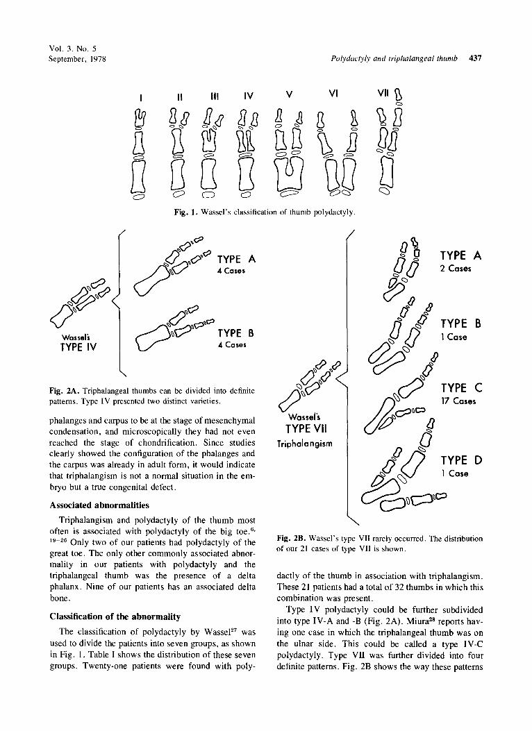
Fig. 1. Wassel's classification of thumb polydactyly.
TYPE A .. Cases
Wassels TYPE IV
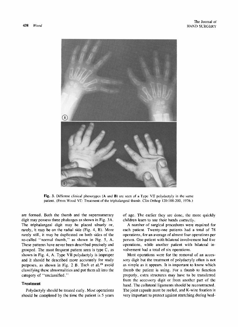
Fig. 2A. Triphalangeal thumbs can be divided into definite patterns . Type IV presented two distinct varieties.
phalanges and carpus to be at the stage of mesenchymal condensation, and microscopically they had not even reached the stage of chondrification. Since studies clearly showed the configuration of the phalanges and the carpus was already in adult form, it would indicate that triphalangism is not a normal situation in the embryo but a true congenital defect.
Associated abnormalities
Triphalangism and polydactyly of the thumb most often is associated with polydactyly of the big toe. 6
,
19-26 Only two of our patients had polydactyly of the great toe. The only other commonly associated abnormality in our patients with polydactyly and the triphalangeal thumb was the presence of a delta phalanx . Nine of our patients has an associated delta bone .
Classification of the abnormality
The classification of polydactyly by WasseJ27 was used to divide the patients into seven groups, as shown in Fig. I. Table I shows the distribution of these seven groups. Twenty-one patients were found with poly-
~<9 ",& (h~()
~v Wassel's TYPE VII
Triphalangism
g~ [J()
tJ II IJ
TYPE A 2 Cases
lJ /J [J(J~ TYPE B .'V 1 Case
o>/:>~ /) ~ {}
~ ~~O~
(t)~ {)
TYPE C 17 Cases
(j (}O /) TYPE 0 a ~ lCase
c:g~c:JC' Fig. 2B. Wassel's type VII rarely occurred . The distribution of our 21 cases of type VII is shown.
dactly of the thumb in association with triphalangism . These 21 patients had a total of 32 thumbs in which this combination was present.
Type IV polydactyly could be further subdivided into type IV -A and -B (Fig. 2A) . Miura28 reports having one case in which the triphalangeal thumb was on the ulnar side. This could be called a type IV-C polydactyly. Type VII was further divided into four definite patterns. Fig. 2B shows the way these patterns
438 Wood The Journal of
HAND SURGERY
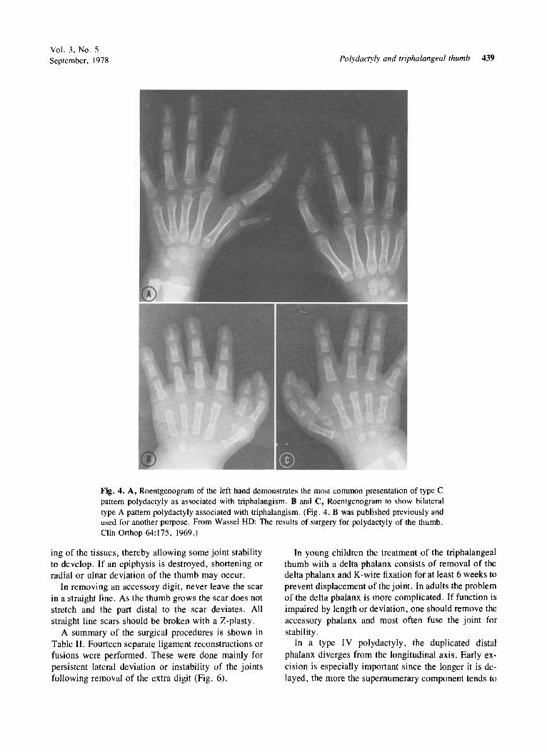
Fig. 3. Different clinical phenotypes (A and B) are seen of a Type VII polydactyly in the same patient. (From Wood VE: Treatment of the triphalangeal thumb. Clin Orthop 120: 188-200, 1976.)
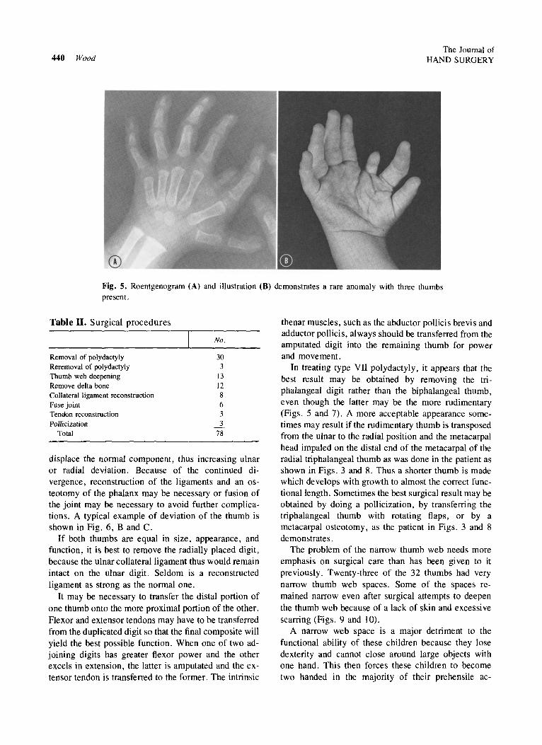
are formed. Both the thumb and the supernumerary digit may possess three phalanges as shown in Fig. 3A. The triphalangeal digit may be placed ulnarly or, rarely, it may be on the radial side (Fig. 4, B). More rarely still, it may be duplicated on both sides of the so-called "normal thumb," as shown in Fig. 5, A. These patterns have never been described precisely and grouped. The most frequent pattern seen is type C, as shown in Fig. 4, A. Type VII polydactyly is improper and it should be described more accurately for study purposes, as shown in Fig. 2 B. Tuch et a1. 29 avoid classifying these abnormalities and put them all into the category of "unclassified."
Treatment
Polydactyly should be treated early. Most operations should be completed by the time the patient is 5 years
of age. The earlier they are done, the more quickly children learn to use their hands correctly.
A number of surgical procedures were required for each patient. Twenty-one patients had a total of 78 operations, for an average of almost four operations per person. One patient with bilateral involvement had five operations, while another patient with bilateral involvement had a total of six operations.
Most operations were for the removal of an accessory digit but the treatment of polydactyly often is not as simple as it appears. It is important to know which thumb the patient is using. For a thumb to function properly, extra structures may have to be transferred from the accessory digit or from another part of the hand. The collateral ligaments should be reconstructed. The joint capsule must be reefed, and K-wire fixation is very important to protect against stretching during heal-
Vol. 3, No.5 September, 1978 Polydactyly and triphalangeal thumb 439
Fig. 4. A, Roentgenogram of the left hand demonstrates the most common presentation of type C pattern polydactyly as associated with triphalangism. B and C, Roentgenogram to show bilateral type A pattern polydactyly associated with triphalangism. (Fig . 4, B was published previously and used for another purpose . From Wassel HD: The results of surgery for polydactyly of the thumb. Clin Orthop 64:175, 1969.)
ing of the tissues, thereby allowing some joint stability to develop. If an epiphysis is destroyed, shortening or radial or ulnar deviation of the thumb may occur.
In removing an accessory digit, never leave the scar in a straight line. As the thumb grows the scar does not stretch and the part distal to the scar deviates. All straight line scars should be broken with a Z-plasty .
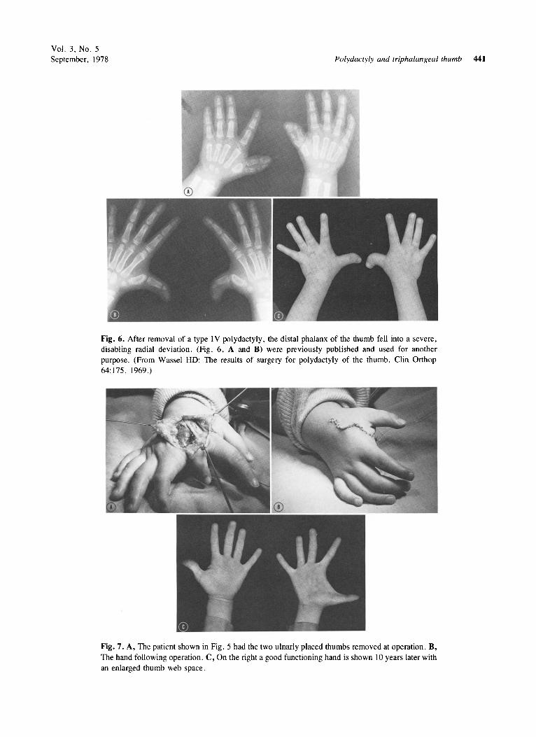
A summary of the surgical procedures is shown in Table II . Fourteen separate ligament reconstructions or fusions were performed. These were done mainly for persistent lateral deviation or instability of the joints following removal of the extra digit (Fig. 6).
In young children the treatment of the triphalangeal thumb with a delta phalanx consists of removal of the delta phalanx and K-wire fixation for at least 6 weeks to prevent displacement of the joint. In adults the problem of the delta phalanx is more complicated. If function is impaired by length or deviation, one should remove the accessory phalanx and most often fuse the joint for stability.
In a type I V polydactyly, the duplicated distal phalanx diverges from the longitudinal axis. Early excision is especially important since the longer it is delayed, the more the supernumerary component tends to
440 Wood The Journal of
HAND SURGERY
Fig. 5. Roentgenogram (A) and illustration (B) demonstrates a rare anomaly with three thumbs present.
Table II. Surgical procedures
Removal of polydactyly Reremoval of polydactyly Thumb web deepening Remove delta bone Collateral ligament reconstruction Fuse joint Tendon reconstruction Pollicization
Total
No.
30 3
13 12 8 6 3 3
78
displace the normal component, thus increasing ulnar or radial deviation. Because of the continued di· vergence, reconstruction of the ligaments and an osteotomy of the phalanx may be necessary or fusion of the joint may be necessary to avoid further complications. A typical example of deviation of the thumb is shown in Fig. 6, Band C.
If both thumbs are equal in size, appearance, and function, it is best to remove the radially placed digit, because the ulnar collateral ligament thus would remain intact on the ulnar digit. Seldom is a reconstructed ligament as strong as the normal one.
It may be necessary to transfer the distal portion of one thumb onto the more proximal portion of the other. Flexor and extensor tendons may have to be transferred from the duplicated digit so that the final composite will yield the best possible function. When one of two adjoining digits has greater flexor power and the other excels in extension, the latter is amputated and the extensor tendon is transferred to the former. The intrinsic
thenar muscles, such as the abductor pollicis brevis and adductor pollicis, always should be transferred from the amputated digit into the remaining thumb for power and movement.
In treating type VII polydactyly, it appears that the best result may be obtained by removing the triphalangeal digit rather than the biphalangeal thumb, even though the latter may be the more rudimentary (Figs. 5 and 7). A more acceptable appearance sometimes may result if the rudimentary thumb is transposed from the ulnar to the radial position and the metacarpal head impaled on the distal end of the metacarpal of the radial triphalangeal thumb as was done in the patient as shown in Figs. 3 and 8. Thus a shorter thumb is made which develops with growth to almost the correct functionallength. Sometimes the best surgical result may be obtained by doing a pollicization, by transferring the triphalangeal thumb with rotating flaps, or by a metacarpal osteotomy, as the patient in Figs. 3 and 8 demonstrates.
The problem of the narrow thumb web needs more emphasis on surgical care than has been given to it previously. Twenty-three of the 32 thumbs had very narrow thumb web spaces. Some of the spaces remained narrow even after surgical attempts to deepen the thumb web because of a lack of skin and excessi ve scarring (Figs. 9 and 10).
A narrow web space is a major detriment to the functional ability of these children because they lose dexterity and cannot close around large objects with one hand. This then forces these children to become two handed in the majority of their prehensile ac-
Vol. 3, No.5 September, 1978 Polydactyly and triphalangeal thumb 441
Fig. 6. After removal of a type IV polydactyly, the distal phalanx of the thumb fell into a severe, disabling radial deviation. (Fig. 6, A and B) were previously published and used for another purpose. (From Wassel HD: The results of surgery for polydactyly of the thumb. Clin Orthop 64:175, 1969.)
Fig. 7. A, The patient shown in Fig. 5 had the two ulnarly placed thumbs removed at operation. B, The hand following operation. C, On the right a good functioning hand is shown \0 years later with an enlarged thumb web space.

442 Wood The Journal of
HAND SURGERY
Fig. 8. A, Pollicization and metacaIpal rotation of a triphalangeal thumb at the time of operation. B, One year later the thumb sits in wide abduction with an adequate web space. (From Wood VE: Treatment of the triphalangeal thumb. Clin Orthop 120:188-200, 1976.)
Fig. 9. When the patient was 3 weeks of age, both extra thumbs were incompletely removed. At the time of these views (5 years of age) the parents were unhappy with the appearance of both hands.
Vol. 3, No.5 September, 1978
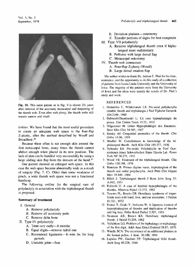
Fig. 10. This same patient as in Fig. 9 is shown 21jz years after removal of the accessory metacarpal and deepening of the thumb web. Even after web plasty, the thumb webs still remain narrow and small.
tivities. We have found that the most useful procedure to create an adequate web space is the four-flap Z-plasty, after the method described by Woolf and Broadbent.30
Because there often is not enough skin around the first metacarpal bone, many times the thumb cannot abduct enough when placed in its new position. The lack of skin can be handled very successfully by using a large sliding skin flap from the dorsum of the hand. 31
One patient showed an enlarged web space. In this case the web space became abnormally wide as a result of surgery (Fig. 7, C). Other than some weakness of pinch, a wide thumb web space was not a functional handicap.
The following outline for the surgical care of polydactyly in association with the triphalangeal thumb is proposed.
Summary of treatment
I. General A. Remove polydactyly B. Remove all accessory parts C. Remove delta bone
II. Type IV polydactyly A. Treat very early-6 months B. Equal digits-remove radial one C. Reconstruct ligaments-K-wire fix for long
periods D. Unstable joint-fuse
Polydactyly and triphalangeal thumb 443
E. Deviation phalanx-osteotomy F. Transfer portions of digits for best composite
III. Type VII polydactyly A. Remove triphalangeal thumb even if bipha
langeal more rudimentary B. Pollicize with large dorsal flap C. Metacarpal osteotomy
IV. Thumb web contracture A. Four-flap Z-plasty (Woolf) B. Large dorsal rotation flap
The author wishes to thank Dr. Adrian E. Flatt for his time, assistance, and the opportunity to do this study of a collection of patients from Lorna Linda University and the University of Iowa. The majority of the patients were from the University of Iowa and the ideas were mainly the results of Dr. Flatt's study and work.
REFERENCES
I. Demarinis F, Wildervanck LS: Pre-axial polydactylia (double thumb and triphalangia.) Ned Tijdschr Geneesk 104:2169, 1960
2. Dubreuil-Chambardel L: Un case hyperphalangie du pouce. Gax Centre Tours 15:25, 1910
3. Hilgereiner H: Ueber Hyperphal3ngie des Daumens. Beitr Klin Chir 54:585, 1907
4. Barsky AJ: Congenital anomalies of the thumb. Clin Ortho 15:96, 1959
5. Mueller W: Contributions to knowledge of the triphalangeal thumb. Arch Klin Chir 185:377, 1936
6. Nylander ES: Pre-axiale Polydaktylie in Funf Generationen Einer Schwedischen Sippe Upsala Lak. Foren Forh NF 36:275, 1931
7. Wood VE: Treatment of the triphalangeal thumb. Clin Ortho 120: 188, 1976
8. Maurizio R: Primus digitus varus, triphalangism of the thumb and radial polydactylia. Arch Putti Chir Organi Mov 19:449, 1964
9. Milch J: Triphalangeal thumb J Bone Joint Surg 33-A:692, 1951
10. Polinelli U: A case of familial hyperphalangia of the thumbs. Minerva Nipiol 12:373, 1962
II. Townes PL, Brocks ER: Hereditary syndrome of imperforate anus with hand, foot, and ear anomalies. J Pediatr 81:321, 1972
12. Komai T, Ozaki Y, Inokuma W: A Japanese kindred of hyperphalangism of thumbs and duplication of thumbs and big toes. Folia Hered Pathol 2:307, 1953
13. Swanson AB, Brown KS: Hereditary triphalangeal thumb. J Hered 53:259, 1962
14. Velichkon LG: Problem of the biphalangy or triphalangy of the first digit. Arkh Anat Gistol Embriol 58:87, 1970
15. Windle BCA: The occurrence of an additional phalanx in the human pollex. J Anat. 26: 100, 1891
16. Lapidus PW, Guidotti FP: Triphalangeal bifid thumb. Arch Surg 49:228, 1944
444 Wood
17. Lapidus PW, Guidotti FP, Coletti CJ: Triphalangeal thumb-report of six cases. Surg Gynecol Obstet 77:178, 1943
18. McMurtry R: Personal communication , Sunnybrook Medical Center, University of Toronto, 1973
19. Bienvenue F: Un Case de Pouces Supplementaire a Trois Phalanges. Rev Orthop Chir Par III :91, 1912
20. Francesconi G: Clinical considerations on a thumb malformation of a familial nature. Minerva Chir 21 :618, 1966
21. Hass SL: Three-phalangeal thumb . Am J Roentgenol Radium Ther Nacl Med 42:677, 1939
22. Jager M, Refior HJ: The congenital triangular deformity of the tubular bones of the hand and foot. Clin Orthop 81 : I 39, 1971
23 . Kirmission E: Pouces a Trois Phalanges . Rev Orthop Par 10:256, 1956
24. Krist jansen A: Supernumerary phalanx in the thumbs . Hyperphalangeal pollicis. Hospitalstid 69: 109, 1926
COPYRIGHT INFORMATION
The Journal of HAND SURGERY
25. Millesi H: Deformations of the fingers following operations for polydactylia. Klin Med (Vienna) 22:266, 1967
26. Wiedemann HR: Hinweis auf eine derzeitig Haufung hypo- and aplasticher Fehlbildungen der Gliedrnassen. Med Welt p . 1863, 1961
27. Wassel HD: The results of surgery for polydactyly of the thumb. Clin Orthop 64: 175, 1969
28. Miura T: Triphalangeal thumb. Plast Reconstr Surg 58:587, 1976
29. Tuch BA, Lipp EB, Larsen IJ , et al : A review of supernumerary thumb and its surgical management. Clin Orthop 125:161-162 , 1977
30. Woolf RM, Broadbent TR: The four-flap Z-plasty. Plast Reconstr Surg 49:48, 1972
31. Tajima T , Watanabe Y, Uchiyama J: Treatment and study of the hypoplastic thumb. Jpn J Plast Reconstr Surg 10:227 , 1967
The appearance of a code at the bottom of the first page of an original article in this JOURNAL indicates the
copyright owner's consent that copies of the article may be made for personal or internal use, or for the
personal or internal use of specific clients . This consent is given on the condition, however, that the copier
pay the stated per copy fee through the Copyright Clearance Center, Inc., P. O. Box 765, Schenectady,
NY 12301,518-374-4430, for copying beyond that permitted by Sections 107 or 108 of the U.S . Copy
right Law. This consent does not extend to other kinds of copying, such as copying for general distribution, for advertising or promotional purposes, for creating new collective works, or for resale .
Recommended