Chinese Journal of Chemistry, 2008, 26, 116—120 Full Paper
* E-mail: [email protected]; Tel./Fax: 0086-025-83792177 Received July 9, 2007; revised August 29, 2007; accepted September 13, 2007. Project supported by the National Natural Science Foundation of China (Nos. 90713023, 20675014, 20535010, 60121101) and the Program (No.
050462) for New Century Excellent Talents in University, the Ministry of Education of China.
© 2008 SIOC, CAS, Shanghai, & WILEY-VCH Verlag GmbH & Co. KGaA, Weinheim
Probing Cellular Binding of Dendrofullerene by in-situ Electrochemical Contact Angle Measurement
ZHOU, Jiana(周謇) ZHANG, Ren-Yuna(张仁云) WU, Chun-Huia(吴春惠) ZHAO, Xue-Yanb(赵学艳) ZHENG, Li-Qiangb(郑利强) FU, De-Ganga(付德刚)
CHEN, Bao-Anc(陈宝安) WANG, Xue-Mei*,a(王雪梅) a State Key Laboratory of Bioelectronics (Chien-Shiung Wu Laboratory), Southeast University, Nanjing,
Jiangsu 210096, China b Ministry Key Laboratory of Colloid and Interface Chemistry, Shandong University, Jinan, Shandong 250100,
China c Zhongda Hospital, School of Clinic Medical Science, Southeast University, Nanjing, Jiangsu 210096, China
Dendrofullerene (C60DF) is a novel fullerene derivative with potential and promising biomedical applications. In this work, electrochemical/contact angle behavior of C60DF in the cellular system has been explored by in-situ electrochemical contact angle measurement. This measuring system is a newly developed technique which can pro-vide electrochemical and contact angle detection simultaneously. The electrochemical results indicate that den-drofullerene may effectively bind and permeate the tumor cell membrane and then distribute into the cancer cells. Our observations of in-situ electrochemical contact angle measurement also illustrate that the permeation and inter-action of C60DF with target cancer cells may lead to some variation of the configurational structure of the relative cell membrane and thus result in the change of hydrophilic/hydrophobic properties of target cellular system. Fur-thermore, through confocus fluorescence microscopy study we found that, upon application of C60DF, the intracel-lular accumulation of anticancer drug daunorubicin in leukemia K562 cells could be remarkably enhanced by C60DF. Therefore fullerene derivatives were demonstrated to be a good candidate that can play an important role in im-proving the intracellular drug uptake in the target cancer cells.
Keywords dendrofullerene, in-situ electrochemical contact angle measurement, confocal fluorescence microscopy, daunorubicin, leukemia K562 cell
Introduction
Since fullerenes (C60) were discovered, characteriza-tion and application of fullerenes and their derivatives have received substantial interest due to the intrinsic interest in their unique structures.1-4 Nevertheless, the relevant fullerenes have few biological applications un-til recent years because of the poor aqueous solubility for the organic functionalization of the highly hydro-phobic C60 moiety. Covalent attachment of some hy-drophilic groups to C60 gives some fullerene amphi-philic adducts and relatively higher solubility in aque-ous solution, enabling them to be introduced into bio-logical applications.5 Among these analogs, den-drofullerene (C60DF) is a new water-soluble C60 deriva-tive, which has a relatively hydrophobic C60 cage and a hydrophilic dendritic tail. This amphiphilic structure could efficiently improve its solubility and make it a sensitive probe for the electrochemical detection and characterization of interfacial property. Additionally, the fact that fullerenes could readily distribute to tissues
suggests that they may eventually be useful to deliver highly polar drugs through membranes to the target tis-sue. Thus, some fullerene derivatives could have the potential to serve as drug carriers to sustain the delivery of drug agents or a synergist for relevant drug accumu-lation since they are biologically stable and have con-venient three-dimensional scaffolds for covalent or noncovalent attachment of multiple drugs. This provides the promise for fullerene drug delivery agents to be made as the versatile vehicles,6 which have great ad-vantages over other drug administration methods such as considerably higher drug concentration in target tis-sues, lower dosage requirements, reduced systemic tox-icity, etc.7
Contact angle measurement has been proved to be an efficient way in detecting the interfacial properties in-cluding the characterization of the hydro-philic/hydrophobic features, wetting properties of monolayer and thin film-functionalized surfaces.8-10 In this study, we have explored a new combination be-tween contact angle and electrochemical detection. This

Dendrofullerene Chin. J. Chem., 2008 Vol. 26 No. 1 117
© 2008 SIOC, CAS, Shanghai, & WILEY-VCH Verlag GmbH & Co. KGaA, Weinheim
in-situ technique was utilized to simultaneously control and detect the variation of interfacial properties as well as electrochemical responding. Recent study has already indicated that some fullerenes could approach and in-teract with cell membrane and have some unique func-tion on certain cell compartments.11 In this study, our observations of the in-situ electrochemical contact angle measurements demonstrate that the interaction between C60DF and cell membrane may lead to some apparent changes of membranous property and result in the hy-drophobic/hydrophilic change of the target system.
The anthracycline antibiotics daunorubicin is a highly effective and broad-spectrum antitumor agent which has been widely used in clinical therapy.12,13 It is already known that daunorubicin is a kind of strong in-tercalators of DNA, which can damage the transcription of DNA of tumor cells and cause the death of target cells. Some reports indicate that anthracycline could gain access to cells by passive diffusion of the neutral form of the drug under the action of a driven force pro-vided by DNA in the nucleus.14,15 But this spontaneous diffusion is not enough for the efficient accumulation of the drug in cancer cells. Besides, anthracycline has some side-effect like cardiotoxicity. Thus, it is desirable to explore the strategy to reduce the drug dosage but increase the intracellular accumulation of daunorubicin in target cancer cells. Considering all of these above, in this work we have explored the synergistic effect of the relevant water-soluble C60DF on the drug accumulation of anticancer drug daunorubicin in target leukaemia K562 cells by using confocal fluorescence microscopy. Our observations indicate that the interaction or per-meation of fullerenes through a lipid bilayer membrane and certain binding or assembly between the C60DF and drug molecules could efficiently facilitate the intracel-lular accumulation of daunorubicin in the target cancer cells.
Experimental
Cell culture
Drug sensitive leukaemia K562 cells were cultured in a flask in RPMI 1640 medium (GIBCO) supple-mented with 10% fetal calf serum (FCS, Sigma), peni-cillin (100 µg•mL-1), and streptomycin (100 µg•mL-1) at 37 ℃ in a humidified atmosphere containing 5% CO2.
The final concentration of cells in the medium is about 2×10⁵/mL. The cells were collected and sepa-rated from 1.0 mL medium by centrifugation 1000 r/min for 10 min. The sediment was blended with saturated C60DF solution (6.5×10⁻⁵ mol/L) in PBS (pH 7.2, 0.1 mol/L).
Atomic force microscopy (AFM) study
AFM characterization was performed using a nano-scope IIIa Multimode AFM (Digital Instruments, Santa Barbara, CA, USA) operating at room temperature (20
±2 ℃). The instrument was mounted on a vibration isolation table to reduce environmental noise. Commer-cially available cantilevers with length 125 µm from Nanosensors were used for Tapping Mode scanning. The resonance frequency of the cantilever was 300—400 kHz. Images were flattened using the appropriate software in order to remove background sloping. The freshly cleaved mica squares were placed in the centre of the metal chip using tape and the relevant sample of C60DF (6.5×10⁻⁵ mol/L) was dropped on the mica and dried before imaging. The metal chip was put on the magnetic holder of the AFM piezo stack. It was critical to assure that the mica was well adhered to the surface to prevent the movement of the sample during tapping mode imaging.
In-situ electrochemical contact angle measurements
In-situ electrochemical contact angle measurements were performed by combining a CAM200 optical con-tact angle analyzer (KSV Instruments, Finland) and a CHI660 electrochemical workstation at room tempera-ture (20±2 ℃). The glassy carbon electrode (φ=5 mm) was utilized as a working electrode while a thin silver wire (φ=0.1 mm) and a platinum wire (φ=0.1 mm) were used as a quasi-reference electrode and a counter electrode, respectively. Such thin wires were chosen to prevent the distortion of the droplets. All potentials were reported here versus the Ag-wire quasi-reference elec-trode. We found that the reference potential of a SCE and the Ag wire in 0.1 mol/L phosphate buffer, pH 7.2, had the relationship of VSCE=VAg wire+0.07 V. The ap-plied potential was changed from 1.6 to -0.8 V and then recurs to 1.6 V. For all systems, the images of the drops were recorded in the respective oxidized and re-duced states of the interfaces. To extract the precise contact angle values, the drop images were fitted using the Young-Laplace equation. The contact angle values were determined with a precision of ±0.5°.
Confocal fluorescence microscopic studies
The freshly prepared cell culture was dropped on a strictly cleaned glass plate immediately before the measurement by using confocal fluorescence micros-copy (Leica TCS SP2) at room temperature (20±2 ℃). The excitation wavelength of fluorescence was 480 nm. Daunorubicin solutions in the absence and presence of C60DF were injected into the relevant cell culture for incubation in which the final concentration of daunoru-bicin and C60DF was 1.0×10⁻⁴ mol/L and 3.3×10⁻⁵ mol/L, respectively. The culture was detected after in-cubating the cells for 0.5 h.
Results and discussion
Initially, C60DF was synthesized by following the procedure as reported in literature.16 The molecular structure of C60DF is illustrated in Figure 1. The unique structure of this fullerene derivative can readily make it self-assembled as a functionalized film, as shown in
118 Chin. J. Chem., 2008, Vol. 26, No. 1 ZHOU et al.
© 2008 SIOC, CAS, Shanghai, & WILEY-VCH Verlag GmbH & Co. KGaA, Weinheim
Figure 2. It is noted that the average height of C60DF is ca. 1 nm. Moreover, the carboxylic acid groups of C60DF make it more hydrophilic than original fullerene moiety, enabling C60DF to bear potential biological ap-plication in aqueous systems. Thus, in the following study we have explored the relevant application and binding behavior of C60DF in the target cellular system by using electrochemical contact angle measurements.
Figure 1 Molecular structure of fullerene derivative C60 den-drofullerene (C60DF).
Figure 2 Typical AFM image of the self-assembled C60DF film. Z scale: 10 nm.
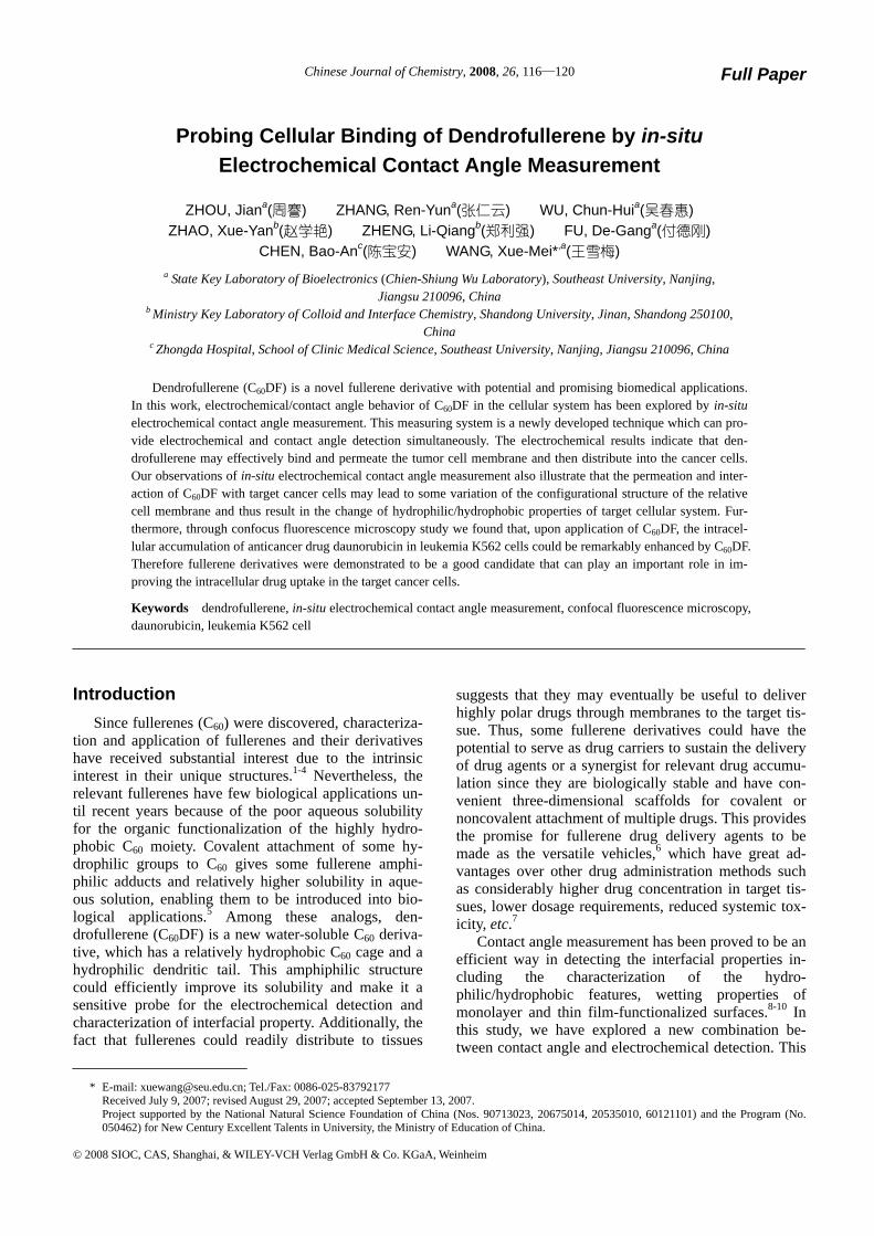
As shown in Figure 3, our electrochemical studies provided the first evidence for the interaction between C60DF and target leukaemia K562 cells. The electro-chemical curves of differential pulse voltammetry (DPV) of C60DF in the absence and presence of leukaemia K562 cells are illustrated in Figure 3. Compared with the original peak current of C60DF (Figure 3a), it ap-pears that after incubation of leukaemia K562 cells with C60DF for about 30 min, the peak current of the C60DF was considerably decreased (Figure 3b). Thus, the rela-tively small amount of C60DF residue outside leukemia K562 cells means a remarkable decrease of the ex-tracellular C60DF concentration after treating the drug-resistant leukemia cells with the C60DF, suggesting the strong interaction or permeability of C60DF to the
respective cancer cell membrane.
Figure 3 Differential pulse voltammetry (DPV) study of (a) C60DF, and (b) C60DF treated leukaemia K562 cells. The final concentration of C60DF is 3.3×10⁻⁵ mol/L and concentration of cell is about 1.0×10⁵/mL. Pulse amplitude: 0.05 V, pulse width: 0.05 s, pulse period: 0.1 s.
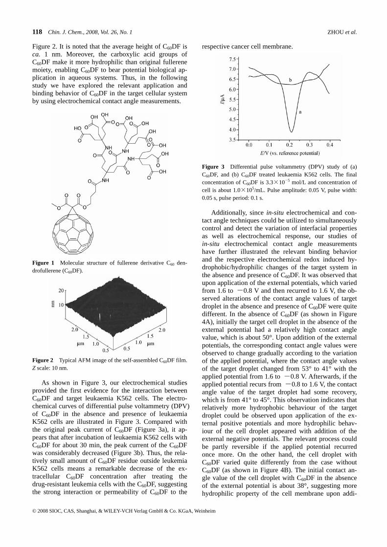
Additionally, since in-situ electrochemical and con-tact angle techniques could be utilized to simultaneously control and detect the variation of interfacial properties as well as electrochemical response, our studies of in-situ electrochemical contact angle measurements have further illustrated the relevant binding behavior and the respective electrochemical redox induced hy-drophobic/hydrophilic changes of the target system in the absence and presence of C60DF. It was observed that upon application of the external potentials, which varied from 1.6 to -0.8 V and then recurred to 1.6 V, the ob-served alterations of the contact angle values of target droplet in the absence and presence of C60DF were quite different. In the absence of C60DF (as shown in Figure 4A), initially the target cell droplet in the absence of the external potential had a relatively high contact angle value, which is about 50°. Upon addition of the external potentials, the corresponding contact angle values were observed to change gradually according to the variation of the applied potential, where the contact angle values of the target droplet changed from 53° to 41° with the applied potential from 1.6 to -0.8 V. Afterwards, if the applied potential recurs from -0.8 to 1.6 V, the contact angle value of the target droplet had some recovery, which is from 41° to 45°. This observation indicates that relatively more hydrophobic behaviour of the target droplet could be observed upon application of the ex-ternal positive potentials and more hydrophilic behav-iour of the cell droplet appeared with addition of the external negative potentials. The relevant process could be partly reversible if the applied potential recurred once more. On the other hand, the cell droplet with C60DF varied quite differently from the case without C60DF (as shown in Figure 4B). The initial contact an-gle value of the cell droplet with C60DF in the absence of the external potential is about 38°, suggesting more hydrophilic property of the cell membrane upon addi-
Dendrofullerene Chin. J. Chem., 2008 Vol. 26 No. 1 119
© 2008 SIOC, CAS, Shanghai, & WILEY-VCH Verlag GmbH & Co. KGaA, Weinheim
tion of C60DF. Besides, it was noted that the contact an-gle value of the target cell droplet in the presence of C60DF changed from 41° to 40° with the applied poten-tial from 1.6 to -0.8 V while the recovery of contact angle at the potential of 1.6 V was also small, varying from 40° to 42°. These observations are consistent with that reported in the literature,11 indicating that the fullerenes could approach and interact with cell mem-brane and thus the interaction of C60DF with the cell membranes might lead to the corresponding changes of the configurational structure of the cell membrane. The microscopic variation of the target system in the ab-sence and presence of C60DF could affect the macro-scopic hydrophilic/hydrophobic properties of the rele-vant cell solution, thus leading to the different contact angle changes of the respective cell droplets. Therefore, it appears that the presence of C60DF could enhance the cellular uptake when distributing into target cancer cells, changing the cell membrane configuration as well as interfacial property, and hence this might further facili-tate the relevant cellular biological process.
Figure 4 The plot of contact angle variation in the absence of C60DF (A) and in the presence of C60DF (B) under the applied potentials from 1.6 to -0.8 V and then recurs to 1.6 V. The con-centration of C60DF is 3.3×10⁻⁵ mol/L and cell concentration is about 1.0×10⁵/mL.
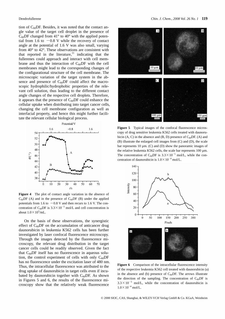
On the basis of these observations, the synergistic effect of C60DF on the accumulation of anticancer drug daunorubicin in leukemia K562 cells has been further investigated by laser confocal fluorescence microscopy. Through the images detected by the fluorescence mi-croscopy, the relevant drug distribution in the target cancer cells could be readily observed. Given the fact that C60DF itself has no fluorescence in aqueous solu-tion, the control experiment of cells with only C60DF has no fluorescence under the excitation laser of 480 nm. Thus, the intracellular fluorescence was attributed to the drug uptake of daunorubicin in target cells even if incu-bated by daunorubicin together with C60DF. As shown in Figures 5 and 6, the results of the fluorescence mi-croscopy show that the relatively weak fluorescence
Figure 5 Typical images of the confocal fluorescence micros-copy of drug sensitive leukemia K562 cells treated with daunoru-bicin (A, C) in the absence and (B, D) presence of C60DF. (A) and (B) illustrate the enlarged cell images from (C) and (D), the scale bar represents 10 µm. (C) and (D) show the panoramic images of the relative leukemia K562 cells, the scale bar represents 100 µm. The concentration of C60DF is 3.3×10⁻⁵ mol/L, while the con-centration of daunorubicin is 1.0×10⁻⁴ mol/L.
Figure 6 Comparison of the intracellular fluorescence intensity of the respective leukemia K562 cell treated with daunorubicin (a) in the absence and (b) presence of C60DF. The arrows illustrate the direction of the sampling. The concentration of C60DF is 3.3×10⁻⁵ mol/L, while the concentration of daunorubicin is 1.0×10⁻⁴ mol/L.
120 Chin. J. Chem., 2008, Vol. 26, No. 1 ZHOU et al.
© 2008 SIOC, CAS, Shanghai, & WILEY-VCH Verlag GmbH & Co. KGaA, Weinheim
was observed when the target cancer cells were treated by daunorubicin in the absence of C60DF. In comparison, when the cells were incubated with daunorubicin and C60DF, much stronger intracellular fluorescence was observed for the target cancer cells. Since higher inten-sity of fluorescence shows higher intracellular drug concentration, the results suggest that the presence of C60DF could efficiently facilitate the respective drug uptake of the target leukemia cells.
Conclusion
In summary, in this study we have demonstrated the novel bio-application of a water-soluble dendrofullerene C60DF. Our observations of the in-situ electrochemical contact angle measurements and laser confocal fluores-cence microscopy illustrate the respective cellular func-tion and synergistic effect of C60DF with anticancer drug daunorubicin. The results indicate that this fullerene derivative could play an important role in cel-lular uptake when distributing into target cancer cells, changing the cell membrane configuration as well as interfacial property and thus enhancing the drug uptake in the cancer cells. Hence, C60DF may have potential valuable application in target drug delivery to sustain the delivery of anticancer drug agents and effectively facilitate the intracellular accumulation of anticancer drugs into the target cancer cells.
References
1 Xie, Q.; Perez-Cordero, E.; Echegoyen, L. J. Am. Chem. Soc. 1992, 114, 3978.
2 Jehoulet, C.; Bard, A. J.; Wudl, F. J. Am. Chem. Soc. 1991, 113, 5456.
3 Fukuzumi, S.; Ohkubo, K.; Imahori, H.; Shao, J.; Ou, Z.; Zheng, G.; Chen, Y.; Pandey, R. K.; Fujitsuka, M.; Ito, O.; Kadish, K. M. J. Am. Chem. Soc. 2001, 123, 10676.
4 Yamago, S.; Tokuyama, H.; Nakamura, E.; Kikuchi, K.; Kananishi, S.; Sueki, K.; Nakahara, H.; Enomoto, S.; Ambe, F. Chem. Biol. 1995, 2, 385.
5 Kam, N. W. S.; Jessop, T. C.; Wender, P. A.; Dai, H. J. Am. Chem. Soc. 2004, 126, 6850.
6 Zakharian, T. Y.; Seryshev, A.; Sitharaman, B.; Gilbert, B. E.; Knight, V.; Wilson, L. J. J. Am. Chem. Soc. 2005, 127, 12508.
7 Nakashima, N.; Nonaka, Y.; Nakanishi, T.; Sagara, T.; Mu-rakami, H. J. Phys. Chem. B 1998, 102, 7328.
8 Wang, X.; Zeevi, S.; Kharitonov, A. B.; Katz, E.; Willner, I. Phys. Chem. Chem. Phys. 2003, 5, 4236.
9 Sigal, G. B.; Mrksich, M.; Whitesides, G. M. J. Am. Chem. Soc. 1998, 120, 3464.
10 Katz, E.; Lioubashevsky, O.; Willner, I. J. Am. Chem. Soc. 2004, 126, 15520.
11 Foley, S.; Crowley, C.; Smaihi, M.; Bonfils, C.; Erlanger, B. F.; Seta, P.; Larroque, C. Biochem. Biophys. Res. Commun. 2002, 294, 116.
12 Rosik, L. O.; Sweet, F. Bioconjugate Chem. 1990, 1, 251. 13 Taatjes, D. J.; Gaudiano, G.; Resing, K.; Koch, T. H. J. Med.
Chem. 1997, 40, 1276. 14 Frezard, F.; Suillerot, A. G. Biochemistry 1991, 30, 5038. 15 Speelmans, G.; Staffhorst, R. W. H. M.; de Kruijff, B.; de
WolP, F. A. Biochemistry 1994, 33, 13761. 16 Braun, M.; Atalick, S.; Guldi, D. M.; Lanig, H.; Brettreich,
M.; Burghardt, S.; Hatzimarinaki, M.; Ravanelli, E.; Prato, M.; van Eldik, R.; Hirsch, A. Chem. Eur. J. 2003, 9, 3867.
(E0707092 CHENG, B.)
Recommended