SCIENTIFIC METHOD APPLIED TO THE SHROUD OF TURINA REVIEW
Raymond N. Rogers* and Anna Arnoldi**
*University of CaliforniaLos Alamos National Laboratory
Los Alamos, NM
**Department of Agrifood Molecular SciencesUniversity of Milan
Milano, Italy
© 2002 All Rights Reserved
ABSRACT
After 25 years of scientific study, I believe that three statements can be supported on the basis ofestablished laws of science and direct observations on the Shroud of Turin.
1. The radiocarbon age determination made in 1988 used an invalid sample, and it gave an erroneousdate for the production of the main part of the cloth.
2. The hypotheses that have appeared since the announcement of an AD 1260-1390 date that invokeradiation of different kinds to explain the image and the date can be categorically discarded.
3. The characteristics of the image can be explained by reference to highly probable, well-knownchemical reactions. No miracles are necessary to explain the image.
INTRODUCTION
The Shroud of Turin first came to public notice and was documented early in the 14th Century in Francewhen its custodian claimed that it was the shroud of Jesus. It has been fiercely controversial ever since.
It has very seldom been displayed to the public, and it has been more an object of veneration that ofstudy. Reports on it have been more subjective than objective; however, its religious concomitant doesnot eliminate the possibility of making truly scientific studies on it. It exists as a material object, andanything that can be observed and measured can be studied by objective methods
The Shroud is a large piece of linen that shows the very faint image of a man on its surface. The imageappears to show a man who has been crucified, with blood flows in the appropriate positions. Thesefeatures have been observed by several different, independent scientific techniques.[1]
The Shroud is now preserved in Turin, Italy. A group of scientists produced a plan for study in 1977.[2]This group incorporated as the Shroud of Turin Research Project (STURP). In 1978, the custodianspermitted limited scientific studies to be made. Results of those observations are reviewed below.
All methods used to establish basic beliefs are subject to the test of general reliability, and they areacceptable only if they yield beliefs which prove to be true. Attempts to develop objective philosophicalmethods for reaching truth from observations have resulted in the logical approach known as ScientificMethod. The STURP plan was based on a rigorous application of this method.
The Bishop of Troyes sent a letter to the pope in AD 1389 claiming that the image had been painted;

2
therefore, that was the primary question to be addressed during the 1978 STURP studies. However, thescientific observations made in Turin in 1978 can and should be used to test any hypotheses involvingthe Shroud.
SCIENTIFIC METHOD
A summary of the major elements of Scientific Method in the context of Shroud studies follows.
1) Identify and clearly state the goal. The goals of the different studies on the Shroud are not alwaysclearly stated in writings on the subject, and ultimate goals were never agreed upon by all of the personsconnected with STURP. There is a huge difference among the following list of possible goals: 1) testwhether the image was a hoax that used known methods for producing an image; 2) estimate theprobability that the Shroud is an "authentic" shroud; 3) prove that the cloth had been the shroud ofJesus; and 4) test whether the Shroud proved the resurrection of Jesus.
2) Assemble all pertinent data. Two of the most damaging things a "scientist" can do during thedevelopment of a "scientific" study is to include speculations on an equal basis with tested facts andexclude observations he does not like. We have seen both problems in Shroud literature. "I think I see,"seems to be accepted by "true believers" on an equal basis with quantitative measurements. Personswho are devoted to "debunking" the Shroud refuse to accept observations that do not further their goals,and persons who want to prove the resurrection refuse to accept observations that seem to conflict withtheir preconceptions.
3) Hypothesize and innovate. An unproved statement that is intended for study and testing is called an"hypothesis." The Method of Multiple Working Hypotheses[3] encourages a scientist to state as manycredible explanations for an observation as possible. This is an enjoyable phase of science; it is perfectlyvalid to let imaginations run wild. Groups often get together to "brainstorm" all of the hypotheses theycan think of. Unfortunately, few attempts have been made to develop multiple, testable hypotheses onimage formation. Persons with fixed goals wish to prove their points, and they refuse to "assemble allpertinent data." Hypotheses for image formation were discussed in detail after the STURPobservations.[4]
4) Test and confirm. The rigorous application of Scientific Method requires that all hypotheses betested equally, and they must be tested against the same comprehensive set of facts and observations.A person who feels comfortable with Scientific Method enjoys "shooting holes" in his hypotheses. Hardfeelings often result when somebody else's favorite hypothesis is picked to pieces, but that is animportant part of the "self-correcting" nature of science. An important part of confirmation is prediction.Prediction enables confirmatory experimentation. Hypotheses that can not be used to formulate testablepredictions are useless. Miracles can not be tested nor confirmed and are not part of science.
5) Occam's Razor. In any contention, both sides can not be correct; however, both sides can be wrong.Competing hypotheses should be tested with Occam's Razor. We usually state it as, "The hypothesisthat includes the smallest number of special assumptions has the highest probability of being closest tothe truth." Scientists normally do not consider it possible to prove a truth. A miracle is a "specialassumption." If one hypothesis demands a miracle and another can be supported by known science andobservations, the miracle should be discarded.
6) The fallacy of the non sequitur. After weak hypotheses have been eliminated according to knownfacts and laws of nature, you hope that at least one remains. After a comprehensive study of the knownfacts, STURP members reported[1] that, "We really do not have a satisfactory, simple explanation forhow the body image got on the cloth."
Many writers who are supporting a religious/miraculous position take that statement to mean somethinglike the following: "If science can not explain the observation, it must have had a miraculous origin." The
3
Greeks recognized the fallacy of such an "argumentative leap" before 300 BC. The fact that science hasnot yet found an explanation proves nothing.
ASSEMBLE ALL PERTINENT DATA
The primary goal of STURP was to test the hypothesis that the Shroud's image was painted. Secondarygoals were to observe the Shroud's technology and composition so that statements could be made on its"authenticity" (whatever that is taken to mean) and possible age. In order to accomplish the goals, alarge number of observations were made before, during, and after the Shroud was made available forstudy in Turin. Facts were also assembled from literature surveys. Some of these facts will be mentionedto illustrate the process of Scientific Method. The observations and data can and should be used to testall hypotheses on the Shroud. No facts can legitimately be ignored.
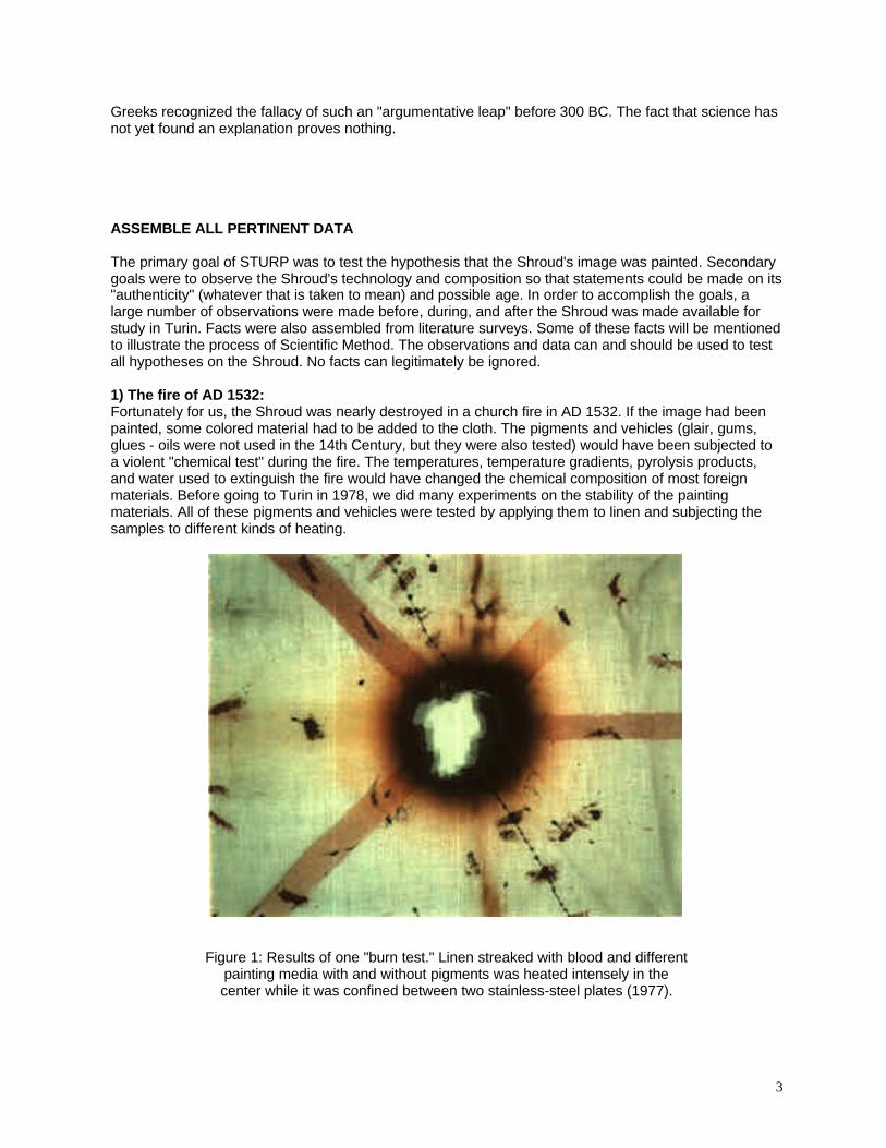
1) The fire of AD 1532:Fortunately for us, the Shroud was nearly destroyed in a church fire in AD 1532. If the image had beenpainted, some colored material had to be added to the cloth. The pigments and vehicles (glair, gums,glues - oils were not used in the 14th Century, but they were also tested) would have been subjected toa violent "chemical test" during the fire. The temperatures, temperature gradients, pyrolysis products,and water used to extinguish the fire would have changed the chemical composition of most foreignmaterials. Before going to Turin in 1978, we did many experiments on the stability of the paintingmaterials. All of these pigments and vehicles were tested by applying them to linen and subjecting thesamples to different kinds of heating.
Figure 1: Results of one "burn test." Linen streaked with blood and differentpainting media with and without pigments was heated intensely in thecenter while it was confined between two stainless-steel plates (1977).
4
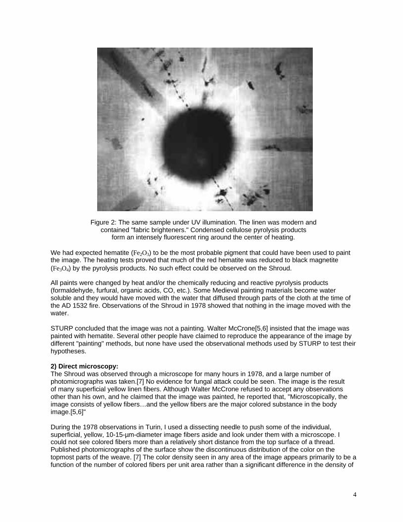
Figure 2: The same sample under UV illumination. The linen was modern andcontained "fabric brighteners." Condensed cellulose pyrolysis products
form an intensely fluorescent ring around the center of heating.
We had expected hematite (Fe2O3) to be the most probable pigment that could have been used to paintthe image. The heating tests proved that much of the red hematite was reduced to black magnetite(Fe3O4) by the pyrolysis products. No such effect could be observed on the Shroud.
All paints were changed by heat and/or the chemically reducing and reactive pyrolysis products(formaldehyde, furfural, organic acids, CO, etc.). Some Medieval painting materials become watersoluble and they would have moved with the water that diffused through parts of the cloth at the time ofthe AD 1532 fire. Observations of the Shroud in 1978 showed that nothing in the image moved with thewater.
STURP concluded that the image was not a painting. Walter McCrone[5,6] insisted that the image waspainted with hematite. Several other people have claimed to reproduce the appearance of the image bydifferent "painting" methods, but none have used the observational methods used by STURP to test theirhypotheses.
2) Direct microscopy:The Shroud was observed through a microscope for many hours in 1978, and a large number ofphotomicrographs was taken.[7] No evidence for fungal attack could be seen. The image is the resultof many superficial yellow linen fibers. Although Walter McCrone refused to accept any observationsother than his own, and he claimed that the image was painted, he reported that, "Microscopically, theimage consists of yellow fibers…and the yellow fibers are the major colored substance in the bodyimage.[5,6]"
During the 1978 observations in Turin, I used a dissecting needle to push some of the individual,superficial, yellow, 10-15-µm-diameter image fibers aside and look under them with a microscope. Icould not see colored fibers more than a relatively short distance from the top surface of a thread.Published photomicrographs of the surface show the discontinuous distribution of the color on thetopmost parts of the weave. [7] The color density seen in any area of the image appears primarily to be afunction of the number of colored fibers per unit area rather than a significant difference in the density of
5
the color of the fibers. This observation was puzzling, and we called it the "half-tone" effect. No fibers ina pure image area were cemented together by any foreign material, and there were no liquid meniscusmarks. These facts seemed to eliminate any image-formation hypothesis that was based solely on theflow of a liquid into the cloth. This also suggests that, if a body was involved in image formation, it wasdry at the time the color formed. Diffusion of gaseous reactants or dyes into the cloth would haveproduced a color gradient (darker on the surface, lighter at depth).
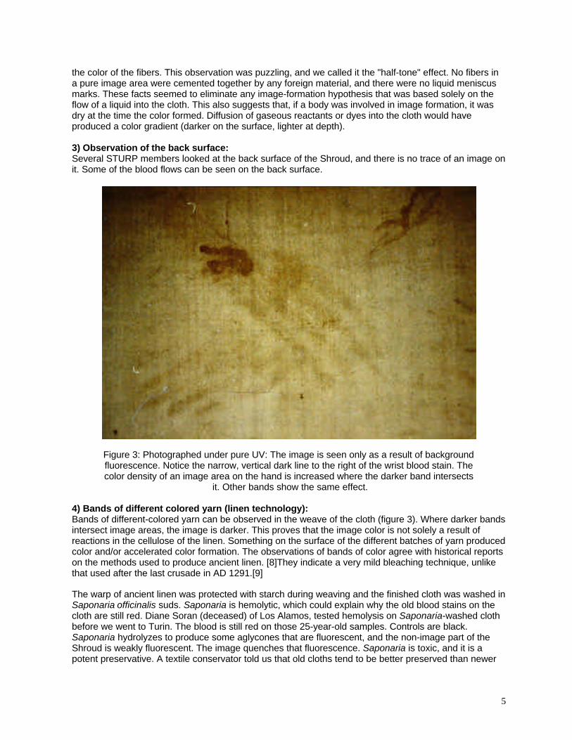
3) Observation of the back surface:Several STURP members looked at the back surface of the Shroud, and there is no trace of an image onit. Some of the blood flows can be seen on the back surface.
Figure 3: Photographed under pure UV: The image is seen only as a result of backgroundfluorescence. Notice the narrow, vertical dark line to the right of the wrist blood stain. Thecolor density of an image area on the hand is increased where the darker band intersects
it. Other bands show the same effect.
4) Bands of different colored yarn (linen technology):Bands of different-colored yarn can be observed in the weave of the cloth (figure 3). Where darker bandsintersect image areas, the image is darker. This proves that the image color is not solely a result ofreactions in the cellulose of the linen. Something on the surface of the different batches of yarn producedcolor and/or accelerated color formation. The observations of bands of color agree with historical reportson the methods used to produce ancient linen. [8]They indicate a very mild bleaching technique, unlikethat used after the last crusade in AD 1291.[9]
The warp of ancient linen was protected with starch during weaving and the finished cloth was washed inSaponaria officinalis suds. Saponaria is hemolytic, which could explain why the old blood stains on thecloth are still red. Diane Soran (deceased) of Los Alamos, tested hemolysis on Saponaria-washed clothbefore we went to Turin. The blood is still red on those 25-year-old samples. Controls are black.Saponaria hydrolyzes to produce some aglycones that are fluorescent, and the non-image part of theShroud is weakly fluorescent. The image quenches that fluorescence. Saponaria is toxic, and it is apotent preservative. A textile conservator told us that old cloths tend to be better preserved than newer
6
ones. Comparison samples loaned to us by the amazing Museum of Egyptology in Turin were stillsupple, and several dated to several thousand years BC. Saponaria produces four glycosidic saponins,all containing gypsogenin. The glycosides hydrolyze to produce sugar chains[10]. The followingcarbohydrates were identified in those chains: galactose, glucose, arabinose, xylose, fucose, rhamnose,and glucuronic acid. Pentose sugars with a furanose structure appear to be the most reactivesugars.[11] The Saponaria sugars should be quite chemically active. Human sebaceous secretions insweat are about 28% free fatty acids. They are a source of "body odor." These fatty acids are chemicallyreactive, and they catalyze many types of reactions. Darker-appearing, pure-image areas did notpenetrate significantly more deeply into the cloth than did lighter areas. The effect was much differentthan that produced by scorching a cloth with a hot statue.
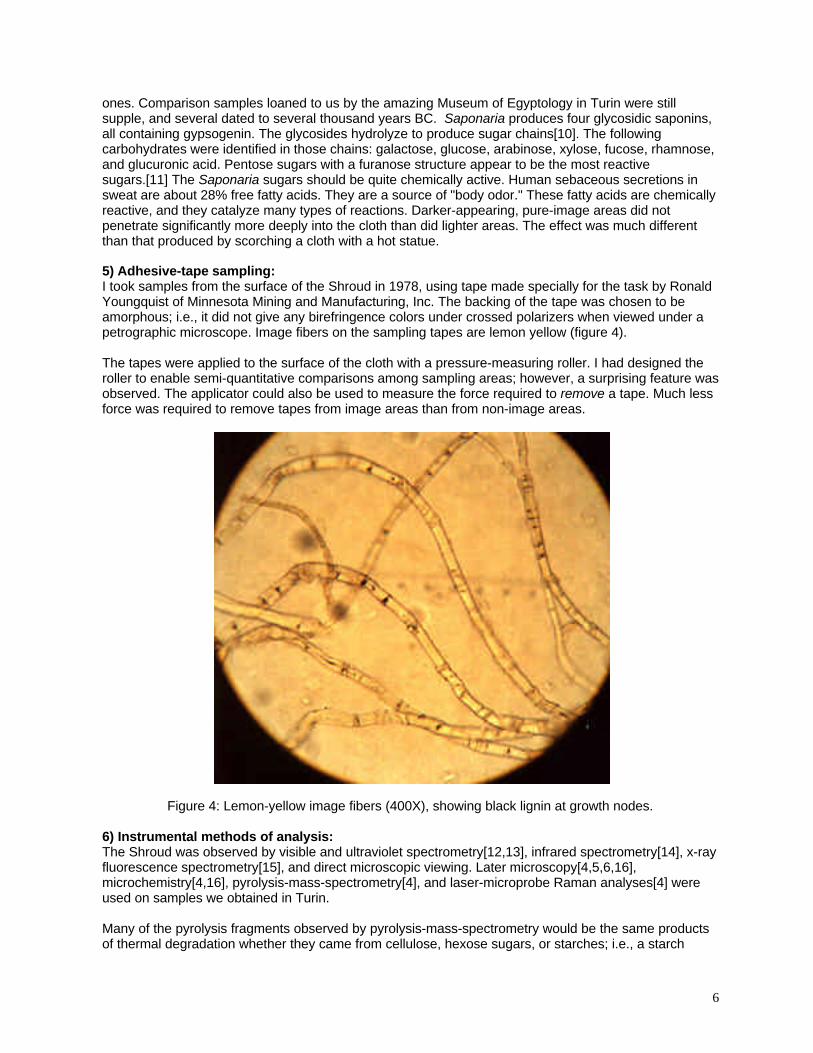
5) Adhesive-tape sampling:I took samples from the surface of the Shroud in 1978, using tape made specially for the task by RonaldYoungquist of Minnesota Mining and Manufacturing, Inc. The backing of the tape was chosen to beamorphous; i.e., it did not give any birefringence colors under crossed polarizers when viewed under apetrographic microscope. Image fibers on the sampling tapes are lemon yellow (figure 4).
The tapes were applied to the surface of the cloth with a pressure-measuring roller. I had designed theroller to enable semi-quantitative comparisons among sampling areas; however, a surprising feature wasobserved. The applicator could also be used to measure the force required to remove a tape. Much lessforce was required to remove tapes from image areas than from non-image areas.
Figure 4: Lemon-yellow image fibers (400X), showing black lignin at growth nodes.
6) Instrumental methods of analysis:The Shroud was observed by visible and ultraviolet spectrometry[12,13], infrared spectrometry[14], x-rayfluorescence spectrometry[15], and direct microscopic viewing. Later microscopy[4,5,6,16],microchemistry[4,16], pyrolysis-mass-spectrometry[4], and laser-microprobe Raman analyses[4] wereused on samples we obtained in Turin.
Many of the pyrolysis fragments observed by pyrolysis-mass-spectrometry would be the same productsof thermal degradation whether they came from cellulose, hexose sugars, or starches; i.e., a starch
7
impurity would not have been detected. UV and visible spectrometry would not see any differencesamong the carbohydrates. The -OH vibrational states of all of the carbohydrates and water are verybroad and intense, and IR spectrometry could not distinguish among them. Laser-microprobe Raman issimilar to IR. We were not looking for trace carbohydrate impurities, we were looking for painting-typeimpurities on the cloth.
The pyrolysis-mass-spectrometry analyses of individual fibers at the NSF Center of Excellence at theUniversity of Nebraska was sufficiently sensitive to detect ppb levels of polyethylene oligomers thatcame from sample bags, but it did not detect any of the possible pigments or painting media. Thepyrolysis-MS analyses did not detect any nitrogen-containing contaminants. This seemed to rule out glair(egg white) as well as any significant microbiological deposits. These results were confirmed bymicrochemical testing.
7) Chemical tests:Image color does not appear under the blood stains when they are removed with a proteolyticenzyme[16]. Whatever process produced the image color must have occurred after the blood flowedonto the cloth, and the image-producing process did not destroy the blood. Microchemical spot tests withaqueous iodine indicated the presence of some starch fractions on Shroud fibers. No proteins could bedetected in either image or non-image areas; however, they were easy to detect in blood stains.[1,4,16]We have recently found that some plant gum, mordants, and dye(s) coat the yarn of the sample whichwas taken by Gilbert Raes in 1973 for textile analysis[17]. These deposits are unique to the Raessample; however, that area was in immediate contact with the radiocarbon sample that was removed fordating in 1988. This fact makes the validity of the radiocarbon sample questionable.
8) Summary of primary observations:All of the observational methods agreed that no pigments, normal painting vehicles, or naturalexudations (other than the blood) had been added to the cloth after its production.
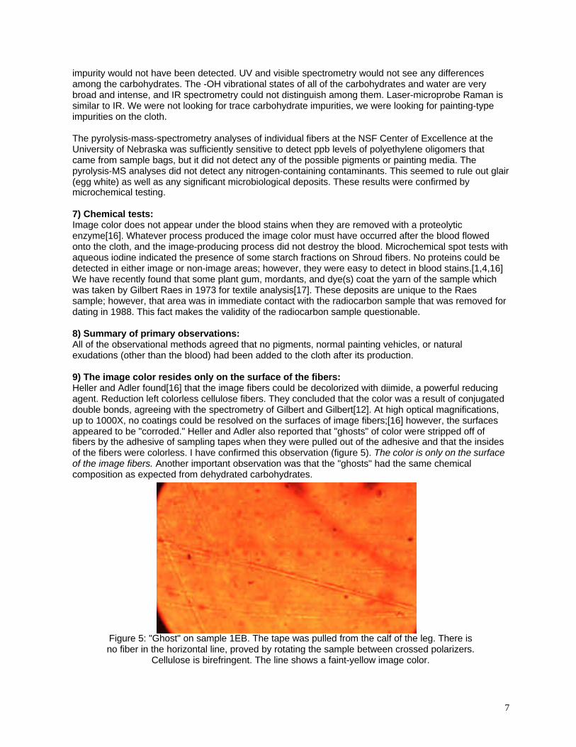
9) The image color resides only on the surface of the fibers:Heller and Adler found[16] that the image fibers could be decolorized with diimide, a powerful reducingagent. Reduction left colorless cellulose fibers. They concluded that the color was a result of conjugateddouble bonds, agreeing with the spectrometry of Gilbert and Gilbert[12]. At high optical magnifications,up to 1000X, no coatings could be resolved on the surfaces of image fibers;[16] however, the surfacesappeared to be "corroded." Heller and Adler also reported that "ghosts" of color were stripped off offibers by the adhesive of sampling tapes when they were pulled out of the adhesive and that the insidesof the fibers were colorless. I have confirmed this observation (figure 5). The color is only on the surfaceof the image fibers. Another important observation was that the "ghosts" had the same chemicalcomposition as expected from dehydrated carbohydrates.
Figure 5: "Ghost" on sample 1EB. The tape was pulled from the calf of the leg. There isno fiber in the horizontal line, proved by rotating the sample between crossed polarizers.
Cellulose is birefringent. The line shows a faint-yellow image color.
8
The STURP observation[16] that the surfaces of image fibers appeared to be "corroded" suggests that avery thin coating of carbohydrate had been significantly dehydrated on the outer surfaces of the fibers.Dehydration causes shrinkage; therefore, any coating of carbohydrate impurities would "craze" duringdehydration. Such a crazed coating would be easy to pull off with adhesive, explaining the easy removalof tapes from image areas. In the context of a discussion on radiation, these observations prove thatonly radiation-induced reactions that color the surfaces of fibers without coloring the cellulose can beconsidered.
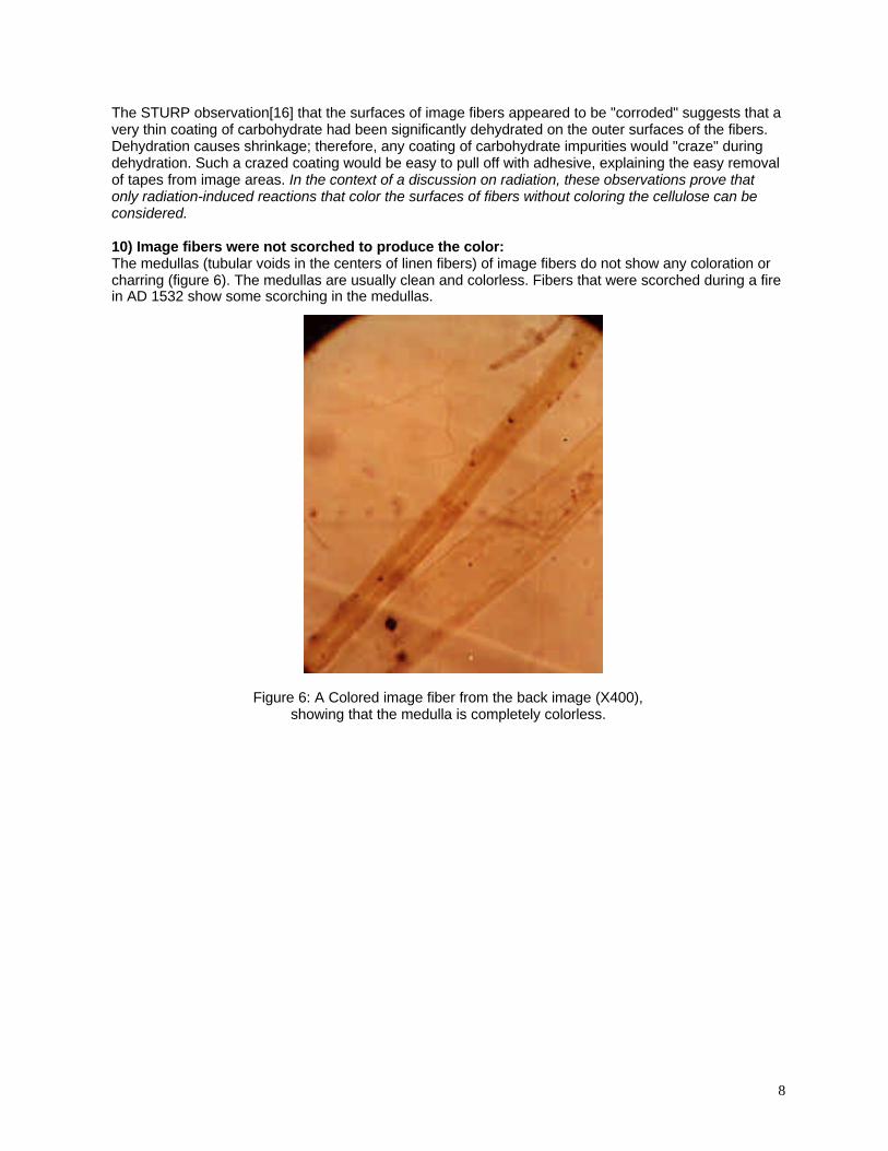
10) Image fibers were not scorched to produce the color:The medullas (tubular voids in the centers of linen fibers) of image fibers do not show any coloration orcharring (figure 6). The medullas are usually clean and colorless. Fibers that were scorched during a firein AD 1532 show some scorching in the medullas.
Figure 6: A Colored image fiber from the back image (X400),showing that the medulla is completely colorless.
9
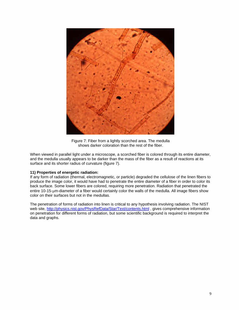
Figure 7: Fiber from a lightly scorched area. The medullashows darker coloration than the rest of the fiber.
When viewed in parallel light under a microscope, a scorched fiber is colored through its entire diameter,and the medulla usually appears to be darker than the mass of the fiber as a result of reactions at itssurface and its shorter radius of curvature (figure 7).
11) Properties of energetic radiation:If any form of radiation (thermal, electromagnetic, or particle) degraded the cellulose of the linen fibers toproduce the image color, it would have had to penetrate the entire diameter of a fiber in order to color itsback surface. Some lower fibers are colored, requiring more penetration. Radiation that penetrated theentire 10-15-µm-diameter of a fiber would certainly color the walls of the medulla. All image fibers showcolor on their surfaces but not in the medullas.
The penetration of forms of radiation into linen is critical to any hypothesis involving radiation. The NISTweb site, http://physics.nist.gov/PhysRefData/Star/Text/contents.html , gives comprehensive informationon penetration for different forms of radiation, but some scientific background is required to interpret thedata and graphs.
10
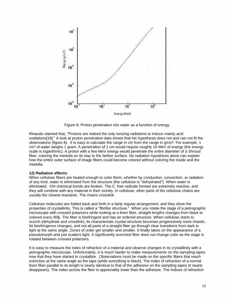
Figure 8: Proton penetration into water as a function of energy.
Rinaudo claimed that, "Protons are indeed the only ionizing radiations to induce mainly acidoxidations[18]." A look at proton penetration data shows that his hypothesis does not and can not fit theobservations (figure 8). It is easy to calculate the range in cm from the range in g/cm². For example, 1cm³ of water weighs 1 gram. A penetration of 1 cm would require roughly 16 MeV of energy (the energyscale is logarithmic). A proton with a few MeV energy would penetrate the entire diameter of a Shroudfiber, coloring the medulla on its way to the farther surface. No radiation hypothesis alone can explainhow the entire outer surface of image fibers could become colored without coloring the inside and themedulla.
12) Radiation effects:When cellulose fibers are heated enough to color them, whether by conduction, convection, or radiationof any kind, water is eliminated from the structure (the cellulose is "dehydrated"). When water iseliminated, -OH chemical bonds are broken. The C· free radicals formed are extremely reactive, andthey will combine with any material in their vicinity. In cellulose, other parts of the cellulose chains areusually the closest reactants. The chains crosslink.
Cellulose molecules are folded back and forth in a fairly regular arrangement, and they show theproperties of crystallinity. This is called a "fibrillar structure." When you rotate the stage of a petrographicmicroscope with crossed polarizers while looking at a linen fiber, straight lengths changes from black tocolored every 90°. The fiber is birefringent and has an ordered structure. When cellulose starts toscorch (dehydrate and crosslink), its characteristic crystal structure becomes progressively more chaotic.Its birefringence changes, and not all parts of a straight fiber go through clear transitions from dark tolight at the same angle. Zones of order get smaller and smaller. It finally takes on the appearance of apseudomorph and just scatters light. A significantly scorched fiber does not change color as the stage isrotated between crossed polarizers.
It is easy to measure the index of refraction of a material and observe changes in its crystallinity with apetrographic microscope. Unfortunately, it is much harder to make measurements on the sampling tapesnow that they have started to crystallize. Observations must be made on the specific fibers that reachextinction at the same angle as the tape (while everything is black). The index of refraction of a normallinen fiber parallel to its length is nearly identical to that of the adhesive on the sampling tapes (it nearlydisappears). The index across the fiber is appreciably lower than the adhesive. The indices of refraction
11
and crystallinity of image fibers are identical to unaffected fibers. Bent, crushed, or otherwise damagedfibers show strain dichroism and will give an erroneous index.
Experiments scorching normal linen fibers agree with observations on scorched fibers from the Shroud.As the scorch color deepens, the two indices of the linen approach the same apparent value. The indexobserved is the average of all of the orientations of the microcrystaline zones in the pseudomorph.Similar fibers have not been observed on image tapes. Other than observing colored medullas,crystallinity and birefringence give good clues for differentiating between scorched and image fibers. Theevidence is strong that the image is not a result of dehydration of the cellulose by any mechanism,thermal or radiation.
The observations of colorless cores in image fibers, ghosts pulled from fibers by the adhesive, thereduction of the color with diimide, lack of fluorescence in an image area, and optical differencesbetween image and scorch fibers seem to eliminate any high-temperature heating event or energeticradiation in image formation. The cellulose of the image had not changed as a result of imageformation. Neutrons produce "recoil protons" when they hit a material that contains hydrogen. The lossof hydrogen also causes crosslinking. Neutrons can not be invoked for a miracle.
13) Low-temperature processes:Although high-temperatures and energetic radiation must be ruled out for image formation, lower-temperature processes are still possible. All that is required is that temperatures never reach the levelwhere cellulose begins to dehydrate at a significant rate. Cellulose starts to dehydrate rapidly between275 and 300°C. Many materials color rapidly at temperatures much lower than that. Some claims havebeen made that thermal radiation (heat/infrared) could not play a part in image formation, becauseintensity of the radiation follows a 1/r2 law. That is not correct. Materials radiate different wavelengths ofelectromagnetic energy at different temperatures. The wavelengths can be calculated from Planck'sRadiation equation. Before a hot surface starts to glow, most of the radiation is produced in the infraredrange.
Surfaces radiate different amounts of heat at different angles depending on the electronic structure ofthe material. G. G. Gubareff, J. E. Janssen, and R. H. Torborg[19] discussed the scientific principles ofthermal radiation in rigorous detail. Polished metals radiate and absorb very little thermal energy, andwhat they do radiate comes off of the surface at a very low angle. Nonmetals radiate much more thermalenergy, and most of it comes out 90° to the surface. It is obvious that thermal radiation does not follow a1/r2 law.
You can observe this effect by buffing the surface of a chrome-plated spatula and putting a fingerprint, aspot of syrup, and a smear of clay on the surface. Heat the piece in the dark. As the temperatureincreases, the first places that you will see glow with radiation will be the print and other non-metallicspots, and they will glow long before the rest of the metal reddens with heat. The glow will be mostvisible from straight above. When the metal starts to glow, its light will be most visible at a low angle. Theemissivity of a human body is like other non-metals or organic materials. Image formation that involvesthermal radiation can not be ruled out; however, it can not explain all of the features of the Shroud.
The thermal conductivity of linen is quite low, about 5 X 10⁻⁴ cal cm-1 s-1 °C-1; therefore, the temperaturegradient extending outward from any heated area will be quite steep: It will be much hotter near contactpoints and cooler away from them. This is important in considering the chemical rates of processes thatcan form a color on a shroud that is in contact with a warm body.
14) Other possible sources of color:We considered iridescence (optical interference in thin layers) and electrons trapped in crystal defects.Those could easily be discarded. All remaining ways must involve chemical changes.
12
15) Chemistry and rate processes:All chemical processes occur at some rate at every temperature above absolute zero, -273°C (-459°F),but the rates follow an exponential function as temperatures change. Some rates are very slow at normaltemperatures. Cellulose does not scorch at a visible rate at room temperature, but it will char quicklyabove about 300°C. Other carbohydrates (e.g., starches and sugars) can color at much lowertemperatures. A piece of cloth heated to several hundred degrees at a point will start to color, but thecolor quickly becomes less intense away from the heated point as a result of the low thermal conductivityof cellulose. Several medical investigators have told me that the postmortem body temperature of aperson who has died of hyperthermia and/or dehydration often reaches 41°C (106°F),[20] and bodytemperatures can actually increase slight ly after death. Some bodies have shown temperatures as highas 43°C (110°F). The bodies are quite dry as a result of the hyperthermia. Many bodies are found at hightemperature in closed automobiles or lying on the desert in the sun. Materials that are stable at normalroom temperatures (about 22°C) can react rapidly at 41-43°C. Many simple chemical processes double(or even triple) their rates for each 10°C (18°F) increase in temperature. A dehydration-type of reactioncould be expected to be about three times faster at normal body temperature than at room temperatureand four to nine times faster at about 41°C.
Chemical rates are modeled with an exponential equation called the Arrhenius expression, k = Ze-E/RT,and rates can be predicted from known, measured chemical kinetics constants (k, the rate constant; Z,the frequency factor; E, the activation energy; R, the gas constant; and T, the absolute temperature).Any chemical process involved in image formation will have properties in accordance with this equation.Heat is also transferred by convection. The circulation of air between a hot surface and a cooler one isdriven by the differences in density between a hot gas and a cooler gas. Convection cells are smallwhere clearances are small and larger where clearances are larger. Convection also transfers vapors,which can include reactive gases, from one surface to another. Fairly thin stagnant zones of gas formnear fixed surfaces. Other gases that approach such zones must diffuse through the stagnant gas toreach the surface. Diffusion of gases through other gases is modeled with Graham's Law of Diffusion.The rates of diffusion are inversely proportional to the square roots of the densities of the gases.Diffusion parallel to the surface of a cloth that covers a body can not be instantaneous, and it will beslower for heavier molecules
In the context of image-formation hypotheses that involve reactive gases, remember that cloth is porous.Gases diffusing to the surface can pass through the pores and be lost. This phenomenon will restrictvapor concentrations as a function of the distance from contact points where a body touches a cloth.Cloth surfaces are active and adsorb gases rapidly, a fact that further limits concentrations as a functionof distance. John Jackson's mathematical analysis of image resolution[2] suggested that no single,simple molecular-diffusion or radiation mechanism could produce the image observed. However, acombination of systems could offer an explanation, e.g., anisotropic heat flow by radiation from the bodyto the cloth, attenuated heat-flow in the cloth, gaseous diffusion, convection, surface properties of cloth,and the dependence of chemical rates on temperature.
16) Summary:Some type of carbohydrate dehydration reaction seems most probable as an explanation for the imagecolor; however, the color appears only on the surface of individual fibers. The color of the image doesnot involve the cellulose. Energetic radiation absolutely can not be used to explain the properties of theimage. That statement does not suggest a miracle.
TEST ALL HYPOTHESES WITH THE SAME FACTS
I. The Radiocarbon Date of 1988
The 1988 radiocarbon age determinations were the best that could have been obtained anywhere in theworld. Effects of sample-preparation methods were studied and careful statistical analyses were made.Damon, et al., reported[21] that "The age of the shroud is obtained as AD 1260-1390, with at least 95%confidence." Unfortunately, that date does not reflect the STURP observations on the linen-productiontechnology and the chemistry of the fibers from the tape samples.
13
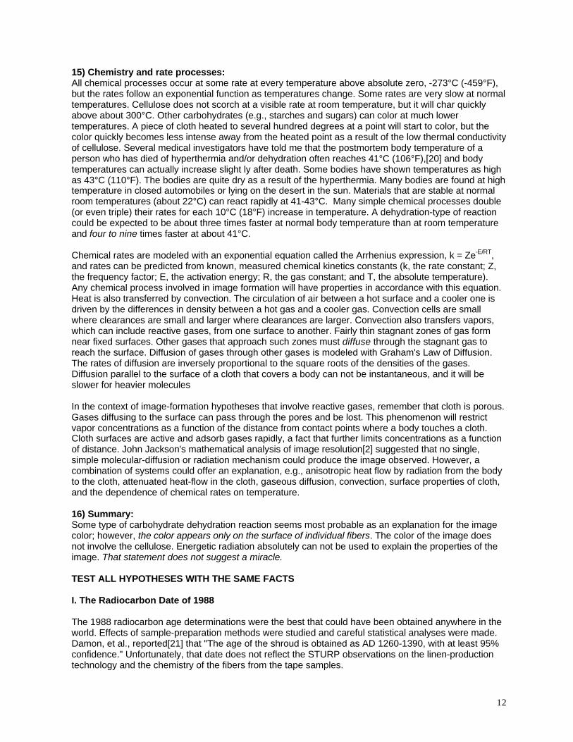
In many cases where questions arise, an appeal is made to "authority." There can be no question aboutthe authority of the radiocarbon investigators; however, true scientists like to see all loose endsquestioned and tested. Persons who object to having their results tested can not be considered to berigorous scientists. Rigorous Scientific Method should be applied in an attempt to resolve the agequestions.
Figure 9: Locations of Raes and radiocarbon samples (bottom margin of clothis to left). There should be compositional similarities between them. Retained
samples should be studied by chemistry and microscopy.
The 1988 sampling operation was described as follows[21]: "The shroud was separated from thebacking cloth along its bottom left-hand edge and a strip (~10 mm x 70 mm) was cut from just above theplace where a sample was previously removed in 1973 for examination. The strip came from a singlesite on the main body of the shroud away from any patches or charred areas." The location of thesample is shown in figure 9.
Unfortunately, the sample was approved at the time of sampling by two textile experts, Franco Testore,professor of Textile Technology at the Turin Polytechnic, and Gabriel Vial, curator of the Ancient TextileMuseum, Lyon, France. No chemical or microscopic investigations were made to characterize thesample. I believe that was a major disaster in the history of Shroud studies
Samples:Professor Gilbert Raes of the Ghent Institute of Textile Technology cut a small sample from the cloth in1973[17]. He found that the samples contained cotton, and he reported that the cotton was an ancientNear Eastern variety, Gossypium herbaceum, on the basis of the distance between reversals in thetape-shaped fibers (about eight per centimeter). I can not confirm the identification of the cotton variety;however, I can confirm the presence of cotton in the Raes sample. The cotton is important. Cotton wasalmost unknown in Europe until about AD 1350[9], when "there was widespread belief that it was thefleece of miniature sheep that lived in trees." Crusaders helped spread knowledge of cotton throughEurope. There were still legal disputes over whether cotton was a kind of linen as late as AD 1631.
14
As part of The Shroud of Turin Research Project (STURP), I took adhesive-tape samples from all areasof the Shroud in 1978[4]. The tape was produced specifically for the project by Ronald Youngquist of theMinnesota Mining and Manufacturing Corporation. He used an amorphous, pure-hydrocarbon adhesivethat would not contaminate the Shroud or the samples, and the adhesive could be removed by washingwith xylene. The tapes were applied to the surface of the Shroud with a pressure-measuring applicator toenable semi-quantitative comparisons among samples. The Shroud was badly damaged in a church firein AD 1532. Nuns patched burn holes and stitched the Shroud to a reinforcing cloth that is now known asthe Holland cloth. I also sampled it in 1978. The Holland cloth provides an authentic, documentedsample of Medieval linen.
In 1980, I received several threads from the 1973 textile sample[17] from Professor Luigi Gonella,Department of Physics, Turin Polytechnic. I now have them numbered and identified as the"Raes threads." I archived remaining tape samples, Holland cloth samples, and Raes threads afterSTURP disbanded. The samples are still available for independent scientific testing of the observationsreported here.
Observations:Visual comparisons among samples were made with a Zeiss petrographic microscope.
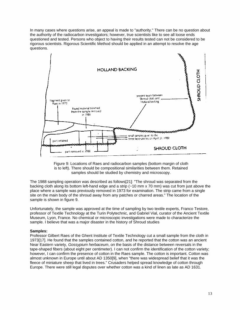
(A) Cotton is not evenly distributed throughout the cloth. Cotton fibers are easy to find mixedintimately with the linen fibers of all of the Raes threads. Figure 10 shows a heavily encrusted cottonfiber on the surface of one of the Raes threads. It can be identified by its flat, tape-like shape, thepresence of one reversal, and the absence of the bamboo-like growth nodes of linen. When the cottonfiber was drawn out of the thread, it showed reversals about 1.2-mm apart. Cotton is not a simplesurface contaminant: It occurs throughout the Raes threads. Fibers retained on the sampling tapes canbe differentiated according to their relative indices of refraction compared with the index of the tape'sadhesive. The two indices of cotton are close to that of the adhesive. Birefringence is first-order white.The index of linen across the fiber is appreciably lower than that of the adhesive.
Figure 10: Heavily encrusted cotton fiber emerging from Raes #14 (400X).

15
I did not attempt to make a quantitative cotton comparison between Raes threads and Shroud tapes,because there was too little cotton of any kind on Shroud samples. We had been puzzled by the Raesreport at the time of the 1978 STURP observations in Turin. We could not find more than traces of cottonon the cloth. The Shroud appeared to be pure linen. We used cotton gloves during the STURP studies of1978 to protect the relic, and they could have been responsible for the traces of modern cotton found ona few Shroud sampling tapes. Samples from the main part of the cloth are significantly different from theRaes samples with regard to cotton content.
(B) Amounts of lignin differ between Raes samples and Shroud fibers. The linen fibers found onShroud tapes average about 13-µm diameter, and they are round in cross-section. They show periodicgrowth nodes, and they look like microscopic lengths of bamboo. Figure 4 shows several linen fibers thatwere pulled from the image at the back of the ankle. It is a completely unpolarized photograph. There isno dichroism or birefringence color. These fibers are characteristic and representative of image fibers.There are dark deposits of lignin on most of the growth nodes. Absolutely no cotton could be foundamong the hundreds of fibers on this tape sample.
Very little lignin is visible at the linen growth nodes of the Raes and Holland cloth samples. Lignin is adark, complex natural structural polymer that is found in all woody plants. Its composition and structureare specific to a given plant, but phenolic units are common to all lignins. It is not a polysaccharide(polymer composed of sugar units) like starch and cellulose. Linen is bleached to remove lignin;however, it is unusual to find a Shroud fiber without some significant deposits of lignin.
(C) Quantitative evaluation of lignin. Simple microscopic viewing is not sufficient to provedifferences among the samples. In order to obtain quantitative data, I counted hundreds of growth nodesin each sample and noted which showed traces of lignin. The table shows that fibers from the Raesthreads, Holland cloth, and modern linen show very little lignin at growth nodes, and the amounts oflignin in those samples are quite consistent.
SAMPLE NODES WITH LIGNIN (%)Modern Commercial 55, very light
Raes Threads 40, lightHolland cloth 60, light
Right Foot, Dorsal Image 54, heavy to moderateFinger, Frontal Image 80, lightAnkle, Dorsal Image 100, heavy to moderate
Scorch control 39, heavy to moderate
Notice that the numbers refer to percentages not numbers of growth nodes observed. No samples of theHolland cloth or Raes threads had heavy deposits of lignin. Unlike the Raes and Holland cloth samples,the fibers on the Shroud tapes vary greatly in amounts of lignin. A large number of observations showsthat lignin ranges from heavy to nil, depending on the location from which the sample was taken. Thereis an explanation for this observation.
(D) Lignin amounts vary among Shroud locations. X-ray-transmission[4,22], contrast-enhanced,ultraviolet[23], and transmitted-light photographs of the Shroud all show specific, discrete bands of yarnwith different x-ray densities and corresponding color densities (figure 3). Both warp and weft yarnsshow this property. Some areas show darker warp yarns and some show darker weft yarns. In someplaces bands of darker color cross. In other places bands of lighter color cross. The effect is somewhatlike a plaid. Many photographs of the Shroud can be viewed on the Shroud web site:http://www.shroud.com.
Linen is bleached to remove the lignin in an attempt to render it pure white. The more quantitative thebleaching process the whiter the product. The bands of different color on the Shroud are the result ofdifferent amounts of lignin left from the bleaching process. The tape samples reflect this variation asobserved differences among quantitative measurements of lignin on the fibers.
16
A conservator at Turin's Museum of Egyptology, Anna Maria Donadoni[24], pointed out locations wherebatches of yarn ended in the weave and new yarn had been inserted in order to continue weaving. Theyarn ends were laid side by side, and the weave was compressed with the comb. The ends are oftenvisible, and the overlaps appear to correspond to zones of different color in the weave.
I believe that the observations of bands of different colors agree with Pliny the Elder's description ofancient linen-production technology[8]. Ancient linen yarn was spun by hand on a spindle whorl. Whenthe spindle was full, the spinner prepared a hank of yarn for bleaching by the fuller. Each hank of yarnwas bleached separately, and each was a little different; indeed, different parts of the same hank showslightly different colors, a little like variegated yarn. The warp yarn was protected with starch during theweaving process, making the cloth stiff. The final cloth was washed with "struthium," Saponariaofficinalis, to make it more supple.
Medieval linen was bleached as the whole cloth. Most commercial bleaching took place in "bleach fields"in the Low Countries, the genesis of the name "Holland cloth" for the Medieval backing on the Shroud.Considerable material was lost during the bleaching process, and the newer linens are less dense thanancient linens, as can be seen by comparing the Holland cloth and patches with the main part of theShroud. The newer linens are also homogeneous. They do not show bands of different-colored yarn inthe weave.
A phloroglucinol-hydrochloric-acid reagent detects vanillin (4-hydroxy-2-methoxybenzaldehyde) withgreat sensitivity. Fresh lignin evolves vanillin in the reagent. You can often smell the vanillin that isevolved from the lignin of warm pine-tree bark. The lignin loses vanillin with time and temperature. Thelignin on older samples of linen gives progressively weaker tests for vanillin as age increases. The ligninon Shroud samples does not give the test. That fact could indicate either significant age for the Shroudor accelerated aging of the lignin as a result of heating during the fire of AD 1532. Differences betweenamounts of lignin on linen fibers in the Raes samples and on Shroud fibers are significant. There isprobably a similar difference between the radiocarbon samples and the main part of the Shroud.
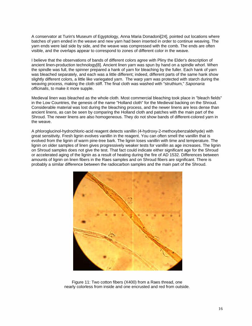
Figure 11: Two cotton fibers (X400) from a Raes thread, onenearly colorless from inside and one encrusted and red from outside.
17
(E) Raes threads show a yellow-brown coating. All Raes threads show colored encrustations on theirsurfaces. Some sections of medulla contain some of the material, showing that it had been able to flowby capillary attraction as a liquid. The encrustation is not removed by nonpolar solvents, but it swells anddissolves in water. There was absolutely no encrustation on either the Holland cloth or fibers from themain part of the Shroud (figure 4). The encrustation is unique to the Raes samples. Any retainedsamples of the material dated in 1988 should be tested for this encrustation.
Figure 11 shows two cotton fibers from Raes thread #5. One of the fibers was taken from inside thethread, and it is nearly colorless. The other fiber was taken from just under the outer surface of thethread, it is deeply colored, and it shows gelatinous material adhering to its surface. A marked differencebetween inside and outside fibers is characteristic of Raes samples.
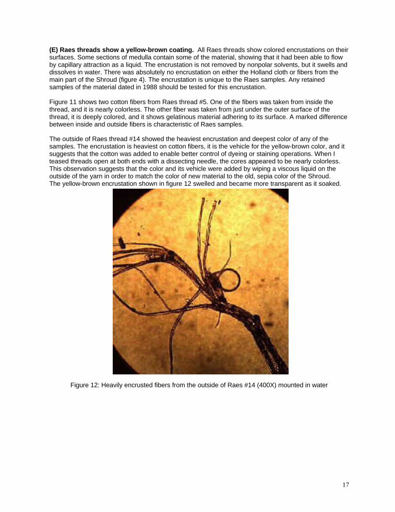
The outside of Raes thread #14 showed the heaviest encrustation and deepest color of any of thesamples. The encrustation is heaviest on cotton fibers, it is the vehicle for the yellow-brown color, and itsuggests that the cotton was added to enable better control of dyeing or staining operations. When Iteased threads open at both ends with a dissecting needle, the cores appeared to be nearly colorless.This observation suggests that the color and its vehicle were added by wiping a viscous liquid on theoutside of the yarn in order to match the color of new material to the old, sepia color of the Shroud.The yellow-brown encrustation shown in figure 12 swelled and became more transparent as it soaked.
Figure 12: Heavily encrusted fibers from the outside of Raes #14 (400X) mounted in water
18
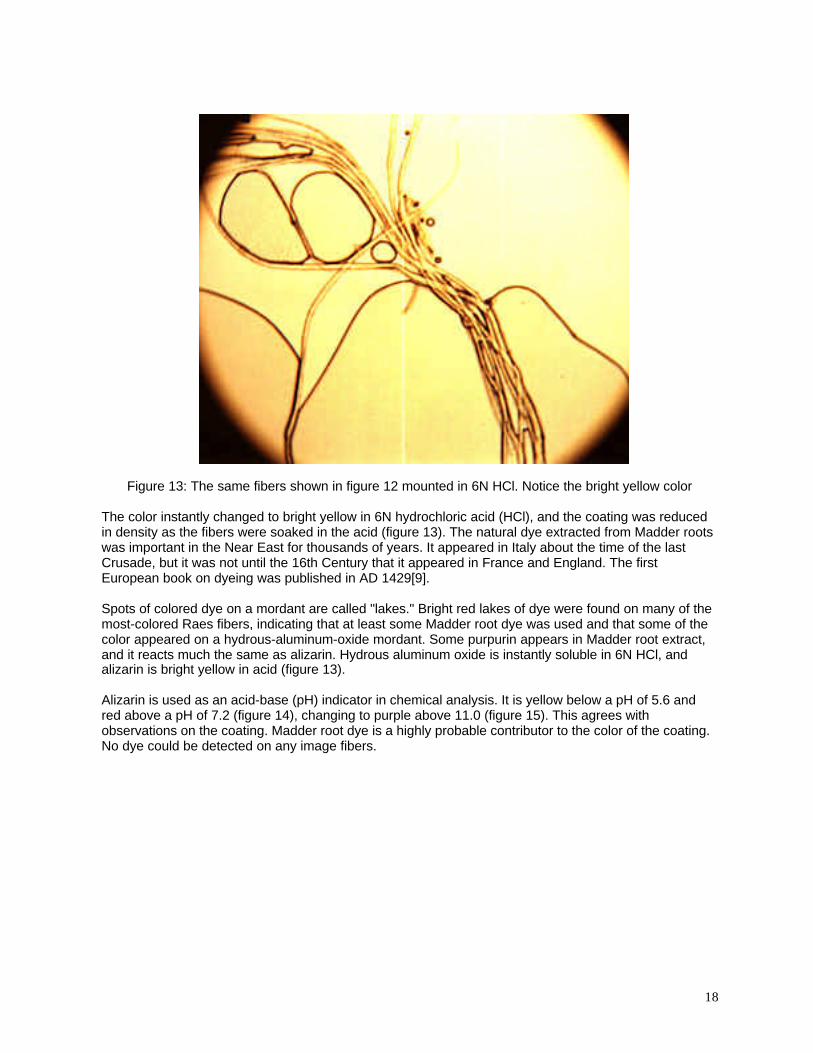
Figure 13: The same fibers shown in figure 12 mounted in 6N HCl. Notice the bright yellow color
The color instantly changed to bright yellow in 6N hydrochloric acid (HCl), and the coating was reducedin density as the fibers were soaked in the acid (figure 13). The natural dye extracted from Madder rootswas important in the Near East for thousands of years. It appeared in Italy about the time of the lastCrusade, but it was not until the 16th Century that it appeared in France and England. The firstEuropean book on dyeing was published in AD 1429[9].
Spots of colored dye on a mordant are called "lakes." Bright red lakes of dye were found on many of themost-colored Raes fibers, indicating that at least some Madder root dye was used and that some of thecolor appeared on a hydrous-aluminum-oxide mordant. Some purpurin appears in Madder root extract,and it reacts much the same as alizarin. Hydrous aluminum oxide is instantly soluble in 6N HCl, andalizarin is bright yellow in acid (figure 13).
Alizarin is used as an acid-base (pH) indicator in chemical analysis. It is yellow below a pH of 5.6 andred above a pH of 7.2 (figure 14), changing to purple above 11.0 (figure 15). This agrees withobservations on the coating. Madder root dye is a highly probable contributor to the color of the coating.No dye could be detected on any image fibers.

19
Figure 14: Surface fibers from Raes #14 reddened in NaHCO3 at a pH of 8.0
Figure 15: Surface fibers from Raes #14 turnedpurple by soaking in a high-pH medium, 2N NaOH
20
The color changes of the dye in the gum coating do not provide a definitive proof that it isalizarin/purpurin. Many dyes show similar color changes with pH, and this observation should beconfirmed with spectrophotometry and additional chemical tests. The barrier to confirmation at present isthe critical lack of samples. The samples that can be sacrificed weigh micrograms, and the dye probablyweighed no more than nanograms. Confirmation may be difficult; however, the important point is that adye similar to alizarin had been added to the gum coating on the Raes samples. They were colored for apurpose using technology that was not used in Italy before the 13th Century or in France before the 16thCentury, about the time the time the Shroud was moved to Turin from France.. The gum coating is notbiogenic.
Other mordants produce different colors with Madder, including blues with calcium compounds. A fewblue lakes can be seen on Raes fibers. The color suggests traces of alizarin on crystals of calcite in thethreads. They are all removed by 6N HCl. A mixture of mordants with alizarin and purpurin can producealmost any desired shade of yellow or brown. In agreement with observations on the individual threads, Icould not detect any significant amount of dye on fibers from the insides of threads.
The gummy coating was totally hydrolyzed by concentrated HCl and 2N NaOH. That fact and itssolubility in water suggest that it is probably a polysaccharide and not a denatured protein. The fact thatsome hydrolyzed in 6N HCl suggests that it is probably a polypentose, composed of five-carbon sugarunits. However, not all of the polysaccharides on the fibers were removed by concentrated HCl. Higher-molecular-weight starch fractions are much more difficult to hydrolyze than are polypentose-containingplant gums. Some starch could be detected on HCl-cleaned Raes fibers with an aqueous iodine reagent.
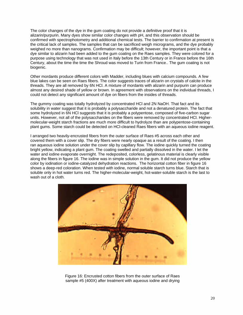
I arranged two heavily-encrusted fibers from the outer surface of Raes #5 across each other andcovered them with a cover slip. The dry fibers were nearly opaque as a result of the coating. I thenran aqueous iodine solution under the cover slip by capillary flow. The iodine quickly turned the coatingbright yellow, indicating a plant gum. The coating swelled and partially dissolved in the water. I let thewater and iodine evaporate overnight. The redeposited, colorless, gelatinous material is clearly visiblealong the fibers in figure 16. The iodine was in simple solution in the gum. It did not produce the yellowcolor by iodination or iodine-catalyzed dehydration reactions. The horizontal cotton fiber in figure 16shows a deep-red coloration. When tested with iodine, normal soluble starch turns blue. Starch that issoluble only in hot water turns red. The higher-molecular-weight, hot-water-soluble starch is the last towash out of a cloth.
Figure 16: Encrusted cotton fibers from the outer surface of Raessample #5 (400X) after treatment with aqueous iodine and drying

21
The encrustation on Raes samples is almost certainly a plant gum. The gum does not appear on any ofthe other linen samples that are associated with the Shroud of Turin. It is highly probable that thealizarin-dyed, gum-coated yarns extend into the adjoining radiocarbon samples.
L. A. Garza-Valdes was allowed to observe fibers from a part of the radiocarbon sample that had beenretained by Giovanni Riggi di Numana after the sampling operation. He reported seeing a coating on thefibers[25]. Although he has misinterpreted the coating, his observations increase the probability that theradiocarbon sample has properties identical to those of the adjoining Raes sample. The gum is probablythe same age as the Raes threads, and it should have had no effect on the age determination. In anycase, it would also have been removed by the cleaning procedures used on the dating sample.However, the presence of a gum coating on retained 1988 radiocarbon-dating samples would prove thatthe samples were not representative of the main part of the relic's cloth. Such a lack of association wouldprove that the radiocarbon date is invalid.
The relatively easy water solubility and hydrolysis of the encrustation suggests gum Arabic. It is obtainedfrom Acacia senegal, and it is mostly composed of pentose-sugar units. It turns bright yellow in aqueousiodine, as observed on the Raes threads. Gum Arabic has been used for thousands of years, and it isstill used in inks, textile printing, and the adhesive on postage stamps.
Figure 17: Raes thread #1 showing an end-to-end splice.The two ends show different colors and amounts of coating.
(F) The Raes samples show a unique splice. Raes thread #1 (figure 17) shows distinct encrustationand color on one end, but the other end is nearly white. The photograph was taken on a 50% gray cardfor color comparison. Fibers have popped out of the central part of the thread, and the fibers from thetwo ends point in opposite directions. This section of yarn is obviously an end-to-end splice of twodifferent batches of yarn. No splices of this type were observed in the main part of the Shroud.
(G) Other observational methods show anomalies in the 14C sample area. The specific area wherethe radiocarbon sample was obtained was photographed in 1978 with low-energy x rays at highresolution[4,22], a pure ultraviolet source[23], and by transmitted 3200°K illumination. All of thesephotographs were available before the Shroud was sampled for radiocarbon analyses. I believe that thesampling area was one of the worst that could have been chosen.
While making the UV photographs (figure 18), the source was heavily filtered to exclude visible light andthe camera was heavily filtered to exclude any effect of the UV on the film. All that appears on the film isthe result of pure fluorescence. The small, triangular,white area in the lower left quadrant is the placewhere the Raes sample was cut in 1973. The normal non-image cloth shows weak fluorescence (upperright). When image appears on the cloth (figure 3), it quenches the fluorescence and gives it a browncolor.
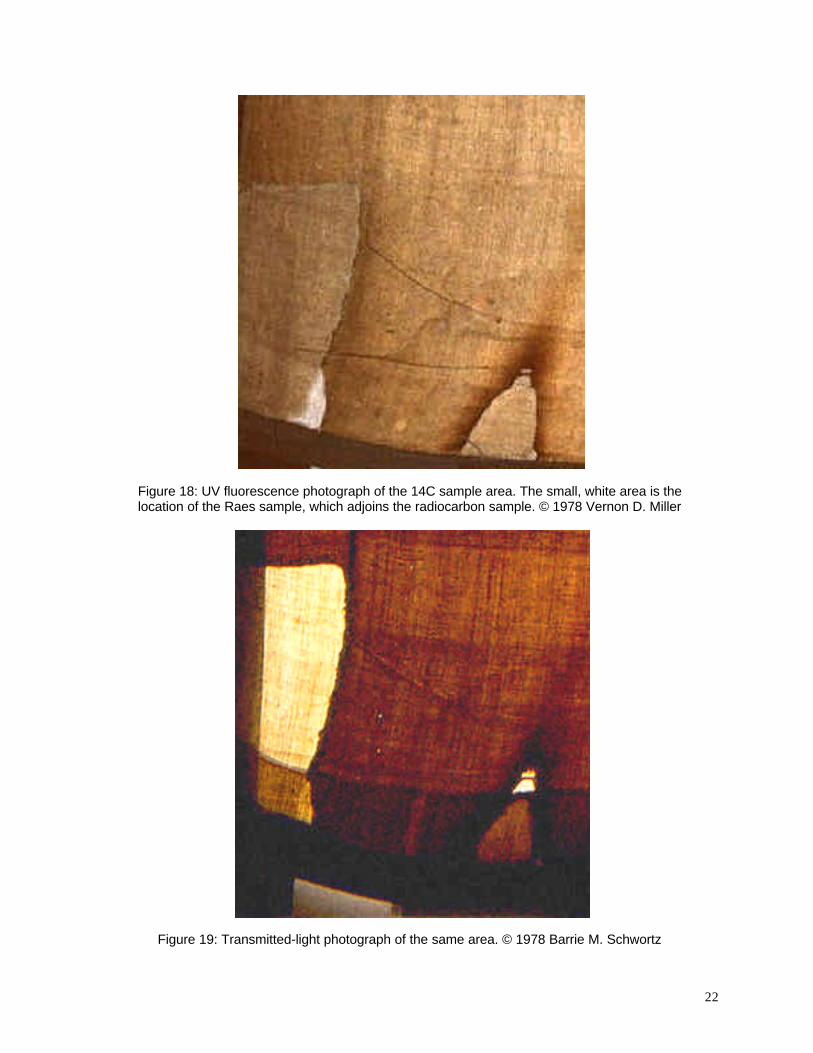
22
Figure 18: UV fluorescence photograph of the 14C sample area. The small, white area is thelocation of the Raes sample, which adjoins the radiocarbon sample. © 1978 Vernon D. Miller
Figure 19: Transmitted-light photograph of the same area. © 1978 Barrie M. Schwortz
23
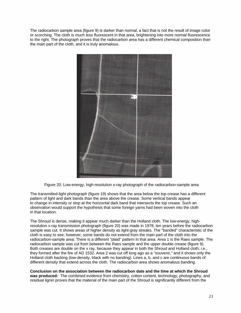
The radiocarbon sample area (figure 9) is darker than normal, a fact that is not the result of image coloror scorching. The cloth is much less fluorescent in that area, brightening into more normal fluorescenceto the right. The photograph proves that the radiocarbon area has a different chemical composition thanthe main part of the cloth, and it is truly anomalous.
Figure 20: Low-energy, high-resolution x-ray photograph of the radiocarbon-sample area
The transmitted-light photograph (figure 19) shows that the area below the top crease has a differentpattern of light and dark bands than the area above the crease. Some vertical bands appearto change in intensity or stop at the horizontal dark band that intersects the top crease. Such anobservation would support the hypothesis that some foreign yarns had been woven into the clothin that location.
The Shroud is dense, making it appear much darker than the Holland cloth. The low-energy, high-resolution x-ray transmission photograph (figure 20) was made in 1978, ten years before the radiocarbonsample was cut. It shows areas of higher density as light-gray streaks. The "banded" characteristic of thecloth is easy to see; however, some bands do not extend from the main part of the cloth into theradiocarbon-sample area: There is a different "plaid" pattern in that area. Area 1 is the Raes sample. Theradiocarbon sample was cut from between the Raes sample and the upper double crease (figure 9).Both creases are double on the x ray, because they appear in both the Shroud and Holland cloth; i.e.,they formed after the fire of AD 1532. Area 2 was cut off long ago as a "souvenir," and it shows only theHolland cloth backing (low-density, black with no banding). Lines a, b, and c are continuous bands ofdifferent density that extend across the cloth. The radiocarbon area shows anomalous banding.
Conclusion on the association between the radiocarbon date and the time at which the Shroudwas produced: The combined evidence from chemistry, cotton content, technology, photography, andresidual lignin proves that the material of the main part of the Shroud is significantly different from the

24
radiocarbon sampling area. The validity of the radiocarbon sample must be questioned with regard todating the production of the main part of the cloth. A rigorous application of Scientific Method woulddemand a confirmation of the date with a better selection of samples.
II. Pseudoscience
The radiocarbon age determination has led to the formulation of many embarrassing "theories" (reallyhypotheses) simultaneously to explain both image formation and the unexpected age. Most of thesehave involved some form of "radiation," and most require a miracle to produce it. Most of the hypothesesabout the date and image have had a theological basis, and most such pseudoscience hypotheses claimthat "this is the only way it could have happened." They then tend to try to work from the postulatedindispensable radiation to a proof of the Biblical resurrection. Goal-directed "theories" andpseudoscience have badly damaged the credibility of rigorous scientific studies on the Shroud of Turin.
Lateral Neural Inhibition
Many observers look at the image for such a long time that they begin to see things that others do not.The phenomenon is responsible for our ability to see figures in clouds. Also, the very faint image isimpossible to see at close range. It was difficult to locate desired sampling locations. It is easy to seeat a distance of four or five meters (something over 10 feet).
Physiologically, the effect is explained in terms of "lateral neural inhibition": the human eye enhancesedge contrasts. The mind plays games with what we think we see. Some devoted observers see imagesof flowers, teeth, bones, etc. on the Shroud. A statement like "I think I see" is totally unacceptable in ascientific discussion.
Unlikely forms of energy
Some phenomena have been postulated without discussion. I have seen invocations of light, ultravioletradiation, soft x rays, protons and other ionizing particles, neutrons, and "other perhaps not discoveredforms of energy." One hypothesis invoked, "axions resonating in the cavity of the tomb" as anexplanation for image formation, and the person had actually purchased an "axion generator" fromRussia.
Scientists at the Livermore National Laboratory, MIT, the Lawrence Berkeley Laboratory, and Fermilabare currently working very hard just to detect axions, a very hard, expensive task. The axion is anhypothetical elementary particle proposed to explain the absence of an electrical dipole moment for theneutron. It has no electric charge, no spin, and would hardly interact with ordinary matter (electrons,photons, quarks, etc.) at all. It would be very unlikely to cause any chemical effects. Even though theaxion -- if it exists -- should have only a tiny mass, axions would theoretically have been producedabundantly in the Big Bang, and relic axions are a possible candidate for the dark matter in the universe.That is the reason they are being studied. They are not even a remote candidate for image production.
Goal-directed pseudoscience
An outstanding example of goal-directed pseudoscience is Antonacci's "Historically ConsistentMethod[26]." He claims that it "was developed by combining research from scientists throughout theworld on all aspects of the body images and blood marks on the Shroud…This theory (sic) states that ifa body instantaneously dematerialized or disappeared, particle radiation would be given off naturally andall the unique features found on the Shroud's body images a nd blood marks would occur" (emphasisadded).
The energy of nuclear weapons is based on the fact that E = mc²; therefore, one bothersome problemwith Antonacci's "theory" is that complete conversion of the mass of a normal human body into energywould have the effect of a huge H bomb, on the order of 200-300 megatons of TNT. That would have
25
vaporized a significant fraction of the Holy Land. Antonacci has done severe damage to the credibility ofstudies on the Shroud.
Failure to test an hypothesis against all data.
A failure to test against all observations can lead to a logically consistent but unsupportablesuperstructure. For example, L. A. Garza-Valdes and S. J. Mattingly have proposed an hypothesis that a"bioplastic" coating on the Shroud contributed to both the "error" in the 14C analysis and imageformation[25,27]. Basing their conclusions on textiles other than the Shroud (e.g., mummy wrappings),they state that[25], "Such coatings have not been previously observed nor confirmed by otherinvestigators," and "Several threads from a putative sample of the linen from the Turin Shroud wereexamined for the presence of these deposits."
Mummy wrappings have experienced a very different environment than has the Shroud. Mummificationtechnology varied with both time and location; however, the wrappings would have been subjected to asignificant amount of protein decomposition products. Color-producing reactions between carbohydratesand proteins and their decomposition products are called Maillard reactions, and they will be discussedin a separate section.
The examination of "the putative" sample from the Shroud was made by Garza-Valdes on the residuefrom the radiocarbon sample that had been retained by Giovanni Riggi in Turin[28]. Garza-Valdes hasnot had access to any other samples. On the basis of his observations, he has jumped to the conclusionthat "…the individual fibers of the cloth are surrounded by a bioplastic coating." However, the first paperreported that[25]: "It should be noted, however, that the amount of organic contamination to producesuch a major change in age is considerable." An addition of about 30% modern carbon would berequired to give the error in the age, and more would be required with older contamination. If the effectwere integrated over the entire history of the Shroud, a huge "bioplastic" coating would be required.
There are four serious problems with the hypothesis: 1) any organic carbon added must be fairlymodern and rich in 14C (it can not be derived from organisms that metabolized original Shroudcarbohydrates as the source of their carbon, e.g., fungi); 2) the amount of modern carbon added must belarge; 3) several different analytical methods have failed to detect any of the elements other than C, H,and O that are necessary for the growth of organisms, and 4) Madder root dye was identified in thecoating on the Raes samples. Some microorganisms do produce large amounts of "extracellularpolymeric substances" (EPS) when they are stressed. The EPS are nearly pure polysaccharides. The"bioplastic hypothesis" is perfectly logical, and it can be tested against the wealth of observations thathave been made on the Shroud. No such tests were made. Mattingly was so certain of his conclusionsthat he actually stated: "...the Turin Shroud is completely coated with both live and deadmicroorganisms. It is not necessary to examine the Shroud linen to make this observation." RigorousScientific Method was not applied.
Although they commented on amounts required to change the Shroud's age, Garza-Valdes andMattingly propose that carbon from times more recent than the 1st Century was added to the Shroud asa product of microbiological action. In order to add more-modern carbon to the cloth, the organisms mustfix carbon from the atmosphere (14C is continually replenished by nuclear reactions in the upperatmosphere). In order to grow, they need water and nutrients, e.g., nitrogen, phosphate, sulfur, and traceelements. The elements other than C, H, and O in biopolymers can be detected with great sensitivity.
The Shroud has always been stored dry, and it has usually been stored out of direct light in some kind ofclosed container, minimizing both the photosynthetic processes that fix carbon dioxide and free accessto CO2.
The STURP tests were planned to test whether anything had been added to the cloth, and observationslooked for organic pigments and painting media as well as inorganic pigments. Nothing other thandehydrated carbohydrate could be found in the image area. In addition to sensitive instrumentalanalytical tests, A. Adler, J. Rogers, and R. Rogers[4,16] spent many hours looking at samples from the

26
image, blood flows, non-image surface, and scorches under microscopes and running microchemicalspot tests. There were no anomalous indices of refraction or impurities. There were no amorphousmaterials cementing fibers together, except for the blood/serum and some starch and gum on threadsfrom the Raes samples.
Without chemical testing, the alizarin-dyed gum on the Raes sample could easily be mistaken for EPS.The misidentification of the coating and failure to analyze it are probably the source for the Garza-Valdes/Mattingly hypothesis.
Several different, sensitive microchemical tests were used in attempts to detect proteins in theimage[16]. Proteins could suggest either protein-containing painting media or bioplastic polymers.Proteins could easily be detected in blood areas. Tests with iodine-azide reagent proved that there wereno sulfur compounds on the surface (except in the blood/serum areas). I am not aware of any "bioplasticpolymers" that are absolutely devoid of amino acids (proteins) and sulfoproteins.
As scientists, Garza-Valdes and Mattingly should have addressed the detailed analyses that were doneduring and after the 1978 observations. They did not do any chemical analyses on samples from themain part of the cloth. Mattingly was sent some fibers from the Raes sample by Alan D. Adler, but whenquestioned about them, he told me that[28]: "I never even looked in the microscope or even handled thethread…I give Garza-Valdes full credit for his hypothesis about the 'bioplastic coating.' It makes perfectsense to microbiologists. Anything with a surface exposure will be coated with microorganisms. I do noteven need to see an object to draw that conclusion." Good science requires observations.
In 2001, Mattingly extended the "bioplastic hypothesis" to the problem of image formation[27].Recognizing that yellow fibers form the image, he said that: "The most readily oxidizable organicmaterials that contribute to a yellowing appearance are lipids (fatty acids) and some pigments (emphasisadded) that are susceptible to oxidation by molecular oxygen."
The atmosphere is the source for "modern," 14C-containing carbon. The addition of modern carbon is theonly way to decrease the apparent age of ancient carbon-containing materials. The most importantorganisms that fix CO2 from the atmosphere are photosynthetic. This is undoubtedly the reason Mattinglymentioned "pigments." Many intensely colored pigments appear in photosynthetic organisms.The final products of photosynthesis are sugars, polysaccharides, nucleic acids, proteins, pigments, etc.Nature builds flax (linen), trees, grass, and little colored microorganisms by photosynthesis. All of the 14Cin our bodies comes originally from photosynthetic processes. The pigments that the photosyntheticorganisms use can be detected with great sensitivity by spectrophotometry, STURP used two differentsystems[12,13], and we did not detect any photosynthetic pigments. If Garza-Valdes and Mattingly hadtested the coating, they could have found the alizarin in it. The Method of Multiple Working Hypotheseswould have helped avoid error. Alizarin is definitely not one of the pigments produced bymicroorganisms. Any gum coating that contains alizarin is definitely not a "bioplastic polymer."
Many of the pigments, e.g., porphyrins and carotenoids, are extremely stable. I have observed several inthe 11,300-year-old sediments at the site of a mammoth kill in Southern Arizona. If they formed on theShroud, they would still be there. If the organisms involved in biopolymer production (like fungi) usedonly the carbohydrates in the Shroud for their metabolic purposes, the biopolymer product would showthe same carbon age as the Shroud. The organisms would use fixed carbon (i.e., the sugar units ofcellulose) and yield carbon dioxide and cell components. Only part of the metabolized carbon could endup in an EPS layer, and the cloth would tend to disappear much faster than the polymer appeared. Clothdoes rot.
All "both live and dead microorganisms" contain proteins, amino acids, and nucleic acids. Algal cellscontain 3.9% nitrogen and 3.3% phosphorus. Fungal cells contain about 0.9% phosphorus and 2.9%nitrogen. Compounds containing these elements can be detected by several of the analytical methodswe applied. All cells of microorganisms give protein microchemical spot tests.
27
If there are no detectable amounts of cell components on the Shroud, there can not be much"biopolymer." The color of the image is indeed a result of a thin coating. "Thin" is the important word.Surface cracking ("corrosion" as Adler called it) of the color can be seen, and flakes can be seen in the"ghosts" on the sampling tapes (figure 5). It takes a thickness on the order of a wavelength of light to getan observable change in index of refraction, and observed indices of an image fiber are identicalto those of a fiber from the Holland cloth or modern linen. The image-color coating seems to beamorphous, but I have been unable to measure its index. I have been able to measure the index of thegum coating on the Raes sample. The thickness of the image color must be less than a sodium-Dwavelength (589 nanometers). Assuming that the image coating could be as thick as 600 nanometers, a20-÷m-diameter fiber (unusually large) would be less than 1% coating by volume. This is certainly notenough material, even if modern, to cause a significant decrease in the apparent age of the Shroud.
The coating on the Raes samples can easily be observed with a normal light microscope with sodium-Dlight; however, it can easily be missed when normal procedures are followed. The usual immersion oilused by microscopists has an index of 1.515, because a normal microscope slide is made of crownglass with an index of 1.517 at 589 nanometers. The index of the coating on the Raes samples varies alittle, but it is very close to 1.515: It can be completely invisible on a normally prepared slide. Water withan index of 1.33 can not be used as an immersion liquid to enhance contrast, because the coating swellsand dissolves. Erroneous measurements will result.
The thickness of the coating on the Raes yarn varies greatly. Cotton fibers tend to have much thickercoatings than linen fibers; however, I would guess that the coating does not average more than about 2µm thick. It would contribute only a few percent to the weight of the Shroud. That would not produce asignificant error in the age determination. In any case, the gum would have been removed by themethods used to clean and prepare the radiocarbon samples for analysis.
I believe that the "bioplastic hypothesis" can easily be disproved. The most probablemisunderstanding/misidentification that generated the hypothesis was the presence of the gum-dyecoating that is unique to the area of the Raes and radiocarbon samples. This coating was probablymistaken for a "bioplastic coating." Unfortunately, the hypothesis was developed without a rigorousapplication of Scientific Method.
Conclusion:I would like to urge persons tempted to call on "science" to prove their point to please use complete,rigorous science. Anything less is scientifically embarrassing and counterproductive to Shroud studies.One recent paper states: "There is a problem with scientists when a non-scientifically explainablephenomenon arises, namely the Resurrection of Jesus Christ." I contend that it is not a problem withscientists. The main problem is the prostitution of science. Science is based on observations of nature:Religions are "revealed."
TEST AND CONFIRM
A large number of attempts have been made to reproduce the image by different methods, and many"theories" (hypotheses) have been proposed. Some of the proposals have actually involved naturalprocesses such as the "vaporographic" theory of Vignon. Some have been far removed from knownreality; e.g., postulating that the image results from "… a burst of energy that can be wide-range-light,UV, soft x-rays or other (perhaps not discovered for now) [emphasis added] that came from within thecorpse." All have failed when compared with observations and measurements on the Shroud.
Attempts to reproduce the image.
(1) Pellicori of STURP studied contact and material-transfer hypotheses[13], and no image-formationhypothesis that is based solely on a vapor-diffusion and/or material-transfer mechanism can beaccepted. Vapors and liquids penetrate the cloth: materials that will color the surface will also diffuse intoand color the inside of the cloth.
28
(2) Image-formation hypotheses that are based solely on any kind of electromagnetic energy must alsobe ruled out. I have already discussed the Lambert/Bouguer law with regard to intensity versuspenetration; however, radiant energy either ablates the surface of a cloth subjected to an intense pulseof energetic photons or; delivered more slowly, it colors the entire facing surface (not just the highestparts). A "flash-of-light" hypothesis had been proposed before the STURP study, but it keepsreappearing[26]. We tested the hypothesis with different-length bursts of laser radiation at differentwavelengths. Long-wave radiation was not sufficiently energetic directly to produce a color (why they putred screens inside clothing-shop windows). Lower-energy photons can not directly induce dehydration.In order to do that, the energy of a photon must be higher than the bond energy of a -OH bond. Indirectdehydration is caused by radiant heating the material above the temperature at which the dehydrationreaction occurs rapidly. That is a relatively slow process. Very intense, 50-ns-long bursts of UV ablatedthe cloth surface, and the samples were reduced to a cloud of very fine particles. We could not get acolor with a "flash of light."
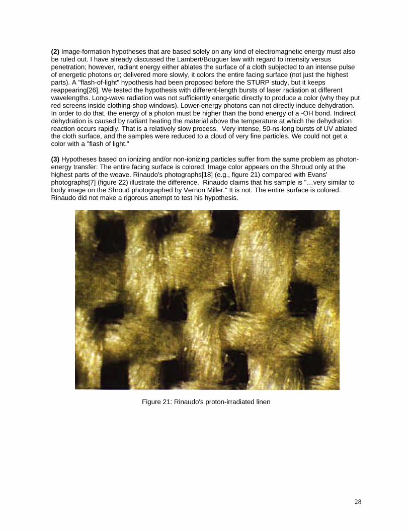
(3) Hypotheses based on ionizing and/or non-ionizing particles suffer from the same problem as photon-energy transfer: The entire facing surface is colored. Image color appears on the Shroud only at thehighest parts of the weave. Rinaudo's photographs[18] (e.g., figure 21) compared with Evans'photographs[7] (figure 22) illustrate the difference. Rinaudo claims that his sample is "…very similar tobody image on the Shroud photographed by Vernon Miller." It is not. The entire surface is colored.Rinaudo did not make a rigorous attempt to test his hypothesis.
Figure 21: Rinaudo's proton-irradiated linen
29
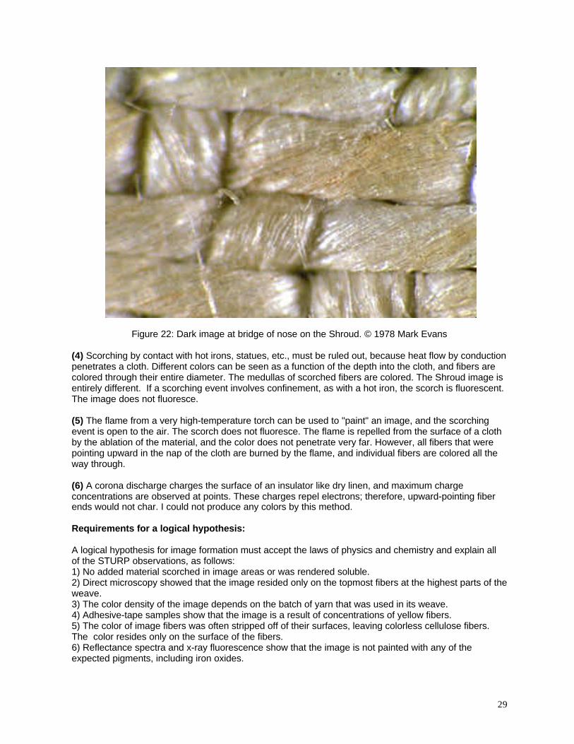
Figure 22: Dark image at bridge of nose on the Shroud. © 1978 Mark Evans
(4) Scorching by contact with hot irons, statues, etc., must be ruled out, because heat flow by conductionpenetrates a cloth. Different colors can be seen as a function of the depth into the cloth, and fibers arecolored through their entire diameter. The medullas of scorched fibers are colored. The Shroud image isentirely different. If a scorching event involves confinement, as with a hot iron, the scorch is fluorescent.The image does not fluoresce.
(5) The flame from a very high-temperature torch can be used to "paint" an image, and the scorchingevent is open to the air. The scorch does not fluoresce. The flame is repelled from the surface of a clothby the ablation of the material, and the color does not penetrate very far. However, all fibers that werepointing upward in the nap of the cloth are burned by the flame, and individual fibers are colored all theway through.
(6) A corona discharge charges the surface of an insulator like dry linen, and maximum chargeconcentrations are observed at points. These charges repel electrons; therefore, upward-pointing fiberends would not char. I could not produce any colors by this method.
Requirements for a logical hypothesis:
A logical hypothesis for image formation must accept the laws of physics and chemistry and explain allof the STURP observations, as follows:1) No added material scorched in image areas or was rendered soluble.2) Direct microscopy showed that the image resided only on the topmost fibers at the highest parts of theweave.3) The color density of the image depends on the batch of yarn that was used in its weave.4) Adhesive-tape samples show that the image is a result of concentrations of yellow fibers.5) The color of image fibers was often stripped off of their surfaces, leaving colorless cellulose fibers.The color resides only on the surface of the fibers.6) Reflectance spectra and x-ray fluorescence show that the image is not painted with any of theexpected pigments, including iron oxides.

30
7) The image spectra were essentially identical to those from aged linen and light scorches. Thestructures of all forms of dehydrated carbohydrates would be very similar, containing complex systemsof conjugated double carbon bonds. Cellulose is not unique.8) Chemical tests showed that there is no protein painting medium or protein-containing coating in imageareas.9) The color can be reduced with diimide, leaving colorless cellulose fibers. The color resides only on thesurface of the fibers, and it is the result of conjugated double bonds.10) The image of the back side of the body shows the same color density and distribution as the front.11) The image does not fluoresce, although scorch margins from the fire of AD 1532 do fluoresce.12) Microchemical tests with iodine indicated the presence of some starch fractions on the cloth.13) The medullas of colored image fibers are not colored: The cellulose was not involved in the color-producing chemistry of the image.
The requirements make it apparent that no single, simple hypothesis will be adequate to explain all ofthe observations made on the Shroud.
Hypothesize: A complex, natural hypothesis for image formation.
The fact that the color resides only on the fiber surfaces leads to the hypothesis that the color formed asa result of chemical reactions involving impurities on the surface. The spectra strongly suggest that theimpurities were carbohydrates that dehydrated as a result of the image-formation process. Thehypothesis on carbohydrate impurities is supported by observations of traces of some starch fractions onimage fibers.
No protein impurities were found in image areas. Although scorch fibers show darkened medullas, thecellulose of image fibers was not colored. This proves that any impurities that produced the color had tohave undergone low-temperature chemical reactions.
Cellulose dehydration follows known chemical rate laws. Cellulose is a very large polymeric chain madefrom glucose (sugar) units that are linked glycosidically. The glucose units exist in the "pyranose" form,rings containing five carbon atoms and one oxygen atom. Pyranose structures appear to be much morestable than are the other common sugar structure, furanose rings. Pyranose systems seem to have anactivation energy for dehydration of about 27 kcal/mole, while furanose systems seem to be much lessstable with an activation energy of about 19 kcal/mole[11]. Because chemical rates are exponential withtemperature, cellulose would react much more slowly than other carbohydrates.
In developing an hypothesis, I had to search for low-temperature chemical processes that produced theobserved type of color. The processes had to involve only impurities and reactants that were eitherdetected on the cloth or could logically be expected from the history of linen technology[8,9] before aboutthe 16th Century.
The bleaching methods used after about AD 1200 made it difficult to identify probable carbohydrateimpurities; however, Pliny the Elder's history[8] suggested that we could expect traces of all of thecarbohydrates that are found in crude starch. In addition, we might expect traces of the glycoside sugarsfrom S. officinalis [10] (e.g., galactose, glucose, arabinose, xylose, fucose, rhamnose, and glucuronicacid). Dehydration-rate calculations made direct dehydration of any carbohydrate impurities, evenpentose sugars, seem very unlikely. Temperatures on the order of 100°C would be required to give acolor in any reasonable amount of time.
A person hoaxing the Shroud image could not have lived long enough to produce a room-temperatureimage. Using a cloth washed in a low-surface-tension solution containing pentose sugars (e.g., aSaponaria solution) and dried in the sun, a non-metallic statue at a temperature above 100°C might haveworked. Such an hypothesis could and should be tested; however, using Occam's Razor, anotherhypothesis seemed more probable.
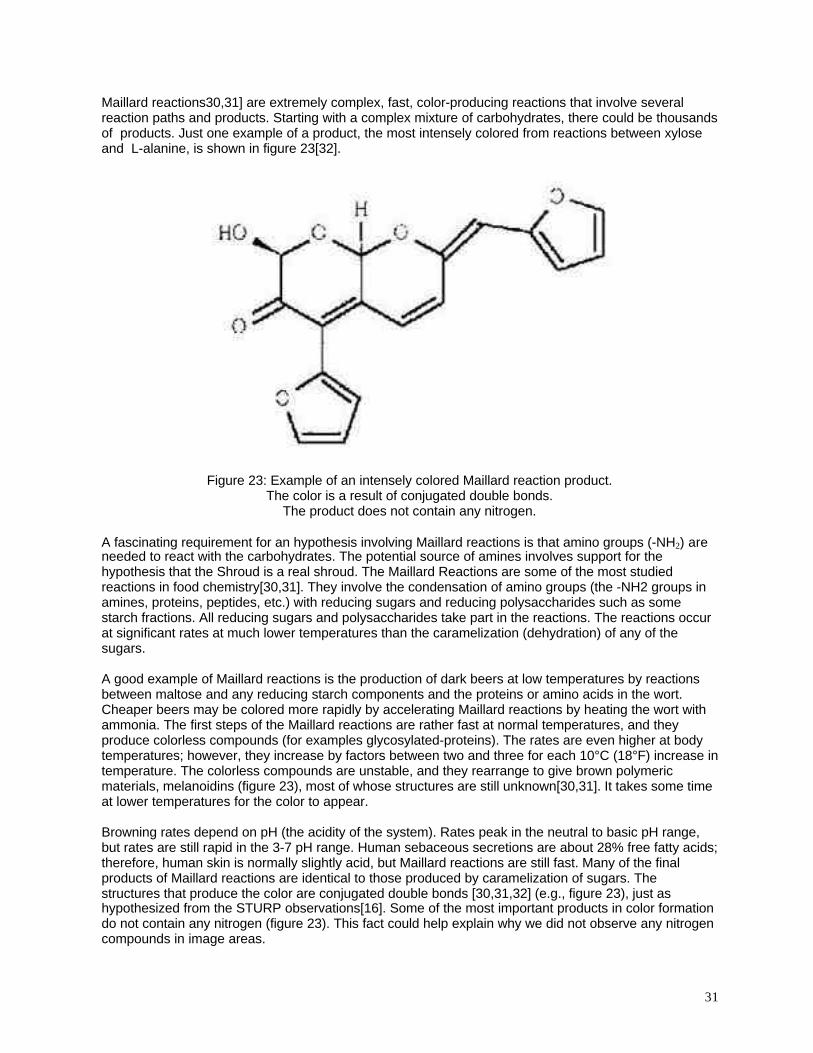
31
Maillard reactions30,31] are extremely complex, fast, color-producing reactions that involve severalreaction paths and products. Starting with a complex mixture of carbohydrates, there could be thousandsof products. Just one example of a product, the most intensely colored from reactions between xyloseand L-alanine, is shown in figure 23[32].
Figure 23: Example of an intensely colored Maillard reaction product.The color is a result of conjugated double bonds.
The product does not contain any nitrogen.
A fascinating requirement for an hypothesis involving Maillard reactions is that amino groups (-NH2) areneeded to react with the carbohydrates. The potential source of amines involves support for thehypothesis that the Shroud is a real shroud. The Maillard Reactions are some of the most studiedreactions in food chemistry[30,31]. They involve the condensation of amino groups (the -NH2 groups inamines, proteins, peptides, etc.) with reducing sugars and reducing polysaccharides such as somestarch fractions. All reducing sugars and polysaccharides take part in the reactions. The reactions occurat significant rates at much lower temperatures than the caramelization (dehydration) of any of thesugars.
A good example of Maillard reactions is the production of dark beers at low temperatures by reactionsbetween maltose and any reducing starch components and the proteins or amino acids in the wort.Cheaper beers may be colored more rapidly by accelerating Maillard reactions by heating the wort withammonia. The first steps of the Maillard reactions are rather fast at normal temperatures, and theyproduce colorless compounds (for examples glycosylated-proteins). The rates are even higher at bodytemperatures; however, they increase by factors between two and three for each 10°C (18°F) increase intemperature. The colorless compounds are unstable, and they rearrange to give brown polymericmaterials, melanoidins (figure 23), most of whose structures are still unknown[30,31]. It takes some timeat lower temperatures for the color to appear.
Browning rates depend on pH (the acidity of the system). Rates peak in the neutral to basic pH range,but rates are still rapid in the 3-7 pH range. Human sebaceous secretions are about 28% free fatty acids;therefore, human skin is normally slightly acid, but Maillard reactions are still fast. Many of the finalproducts of Maillard reactions are identical to those produced by caramelization of sugars. Thestructures that produce the color are conjugated double bonds [30,31,32] (e.g., figure 23), just ashypothesized from the STURP observations[16]. Some of the most important products in color formationdo not contain any nitrogen (figure 23). This fact could help explain why we did not observe any nitrogencompounds in image areas.
32
With regard to the requirement for amine reactants in Maillard processes, decomposing bodies startproducing ammonia and amines, e.g., cadaverine (1,5-diaminopentane) and putrescine (1,4-diaminobutane), fairly quickly, depending on the temperature and humidity. A trained cadaver-searchingdog can find bodies within a few hours after death. Little decomposition is required.
The ammonia and many of the decomposition amines are volatile and basic (they increase the pH into amore favorable range for Maillard reactions), and they rapidly undergo Maillard reactions with anyreducing saccharides they contact. Such sugar-amine reactions offer a natural explanation for the coloron the Shroud, and they suggest that the Shroud of Turin was a real shroud. However, identification of aprobable chemical process does not explain one of the most perplexing observations on the Shroud, thediscontinuous distribution of the color on the topmost parts of the weave.
The fact that color does not penetrate the cloth to any significant depth suggests that any hypothesizedimpurity had to be concentrated at the top of the cloth with little or none throughout the rest of thethickness. Assumed pressure of a body had no effect on the color density or penetration of image colorinto the cloth in the area of the back image. This supports the hypothesis that a limited amount ofsuperficial impurities was involved in image formation. There is a simple explanation.
Saponaria officinalis, also called "soapweed," reduces the surface tension of water making it a goodwetting agent. Both hydrophobic and hydrophilic impurities on cloth are put into solution or suspensionby Saponaria. The process is identical to that involved in every laundry day with every kind of soap ordetergent.
A textile conservator, Anna Maria Donadoni[24], told us that, after ancient cloth was washed in aSaponaria solution, it was "laid out on bushes to dry." Under such conditions, materials that are insolution or suspension in the wash water will concentrate at the drying surface. This is a principle I haveused to transfer traces of soluble materials from irregular surfaces into sheets of filter paper for laterchemical analysis.
Evaporation concentration can explain the superficial nature of the image. An amine vapor that diffusedfrom a body into the cloth could only develop a low-temperature Maillard color on the surface wheresaccharides concentrated. The amines do not react with cellulose. The phenomenon can bedemonstrated with a simple experiment.
Prepare a very dilute solution of food coloring, and divide it into two parts. Add a drop of liquid detergentto one part. Cut some squares of white cloth that are about 10 cm on a side. Place different numbers ofdrops of each solution on clean, smooth nonabsorbent plates, and lay pieces of cloth over the drops. Letthe liquid evaporate. Different types of cloth will show different degrees of concentration of the dye onthe evaporating surfaces.
The Shroud cloth is tightly woven, it is relatively thick, and it does not readily absorb water. With such acloth, any material that can be suspended by Saponaria will primarily migrate to a drying surface and beconcentrated.
The puzzling "half-tone" effect has been mentioned. All of the colored image fibers showedapproximately the same color intensity under a microscope. Assuming that the color formed by reactionswith a very thin deposit of superficial impurities on the fibers, all of the fibers should have shown identicalspectra and roughly the same intensity of color. They did.
Another important observation is the fact that the image-forming process produced slightly different colordensities (but identical spectra) on the different lots of yarn[4] (figure 3). The color-density of the image isnot simply a function of the chemical properties of cellulose: It also depends on the individual propertiesof the batches of yarn. The observed effects must have been caused by different amounts of impuritiesthat originally coated the surfaces of the different hanks of yarn as a result of slightly different productionconditions.
33
Slightly different amounts of impurities on the different batches of linen yarn would cause slightlydifferent surface energies. One major linen impurity is "flax wax," and it produces a hydrophobic surface.Liquids wet the threads as a function of the difference between the surface tension of the washingsolution and the surface energy of the specific linen yarn. This would explain the "banded" appearanceof the Shroud. The original observations and experiments on this phenomenon were done by BenjaminFranklin in 1774[33].
We have now discussed the critical observations on the Shroud, and we can clearly state an alternatehypothesis in accordance with Scientific Method.
Formal statement of the impurity hypothesis for image formation to be tested.
The cloth was produced by technology in use before the advent of large-scale bleaching. Each hank ofyarn used in weaving was bleached individually. The warp yarns were protected and lubricated duringweaving with an unpurified starch paste. The finished cloth was washed in Saponaria officinalis and laidout to dry. Starch fractions, linen impurities, and Saponaria residues concentrated at the evaporatingsurface. The cloth was used to wrap a dead body. Ammonia and other volatile early aminedecomposition products reacted rapidly with reducing saccharides on the cloth in Maillard reactions. Thecloth was removed from the body before liquid decomposition products appeared. The color developedslowly as Maillard compounds decomposed into final colored compounds.
Test and confirm.
Both starch and cellulose are composed of glucose units; however, cellulose is not a reducingpolysaccharide. Crude starch contains a complex mixture of molecular weights as well as some verysoluble free sugars: Its reducing carbohydrates rapidly undergo Maillard reactions. One starchcomponent, dextrin, dissolves in water, and it shows reducing properties with Fehling's solution. It isdissolved in a washing solution, and it concentrates at an evaporating surface. I used it to demonstratethe impurity-hypothesis. The Fehling's test uses a fresh mixture of copper sulfate and alkaline tartratesolutions. Add the sample, and heat gently. A reddish precipitate of cuprous oxide proves a reducingsaccharide.
Age and/or heating in the fire of AD 1532 has changed the lignin in the cloth[34], and it would certainlychange other impurities that are less stable than either lignin or cellulose. Therefore, tests of image-formation hypotheses need fresh material. However, modern linen has been vigorously bleached, and ithas been coated with sizing compounds and fluorescent fabric brighteners. It is useless for experimentalimage-production purposes.
Kate Edgerton (deceased, Norwich, CT) grew flax and made some linen, using both starch andSaponaria. Unfortunately, she ironed the samples with a "warm iron" which colored the cloth andchanged the Saponaria glycosides. Edgerton's samples could be bleached to remove most of the colorby soaking in 3.5% hydrogen peroxide. This treatment did not remove the natural waxes.
Because Edgerton's linen was scarce, many preliminary tests were performed on pure-cellulose filterpaper. These experiments were done by placing drops of test solutions on a dry plastic plate and layinga piece of Whatman's #4 filter paper over them. The liquid migrated through the paper and evaporated atthe surface. No color could be observed in either sunlight or ultraviolet illumination with Saponaria aloneor with the model saccharides (e.g., levulose, dextrose, maltose, lactose, xylose, dextrin, and solublestarch).
The different samples were treated with ammonia vapor for different times. Light colors developed slowlyon the tops of samples that had contained reducing saccharides. A technical grade of dextrin thatreduced Fehling's solution was used to test crude-starch reactions. It acts like crude starch without thefree sugars and highest molecular weight fractions. Paper was laid over different numbers of drops ofdextrin, and the liquid was allowed to migrate into circular spots and evaporate. Samples were then laidover drops of Saponaria solution. The Saponaria caused the polysaccharides to move radially, as in a
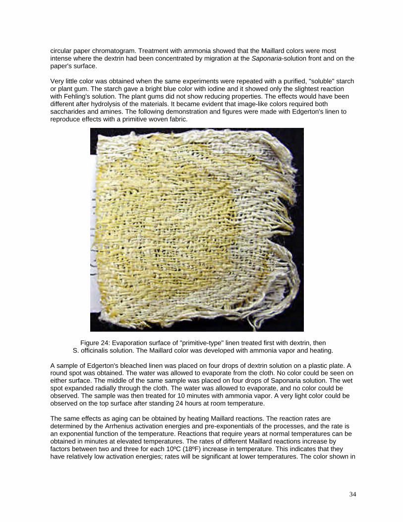
34
circular paper chromatogram. Treatment with ammonia showed that the Maillard colors were mostintense where the dextrin had been concentrated by migration at the Saponaria-solution front and on thepaper's surface.
Very little color was obtained when the same experiments were repeated with a purified, "soluble" starchor plant gum. The starch gave a bright blue color with iodine and it showed only the slightest reactionwith Fehling's solution. The plant gums did not show reducing properties. The effects would have beendifferent after hydrolysis of the materials. It became evident that image-like colors required bothsaccharides and amines. The following demonstration and figures were made with Edgerton's linen toreproduce effects with a primitive woven fabric.
Figure 24: Evaporation surface of "primitive-type" linen treated first with dextrin, thenS. officinalis solution. The Maillard color was developed with ammonia vapor and heating.
A sample of Edgerton's bleached linen was placed on four drops of dextrin solution on a plastic plate. Around spot was obtained. The water was allowed to evaporate from the cloth. No color could be seen oneither surface. The middle of the same sample was placed on four drops of Saponaria solution. The wetspot expanded radially through the cloth. The water was allowed to evaporate, and no color could beobserved. The sample was then treated for 10 minutes with ammonia vapor. A very light color could beobserved on the top surface after standing 24 hours at room temperature.
The same effects as aging can be obtained by heating Maillard reactions. The reaction rates aredetermined by the Arrhenius activation energies and pre-exponentials of the processes, and the rate isan exponential function of the temperature. Reactions that require years at normal temperatures can beobtained in minutes at elevated temperatures. The rates of different Maillard reactions increase byfactors between two and three for each 10ºC (18ºF) increase in temperature. This indicates that theyhave relatively low activation energies; rates will be significant at lower temperatures. The color shown in
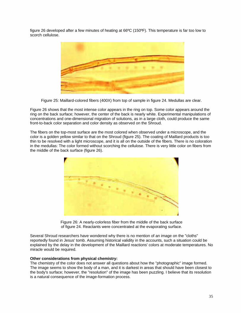
35
figure 26 developed after a few minutes of heating at 66ºC (150ºF). This temperature is far too low toscorch cellulose.
Figure 25: Maillard-colored fibers (400X) from top of sample in figure 24. Medullas are clear.
Figure 26 shows that the most intense color appears in the ring on top. Some color appears around thering on the back surface; however, the center of the back is nearly white. Experimental manipulations ofconcentrations and one-dimensional migration of solutions, as in a large cloth, could produce the samefront-to-back color separation and color density as observed on the Shroud.
The fibers on the top-most surface are the most colored when observed under a microscope, and thecolor is a golden yellow similar to that on the Shroud (figure 25). The coating of Maillard products is toothin to be resolved with a light microscope, and it is all on the outside of the fibers. There is no colorationin the medullas: The color formed without scorching the cellulose. There is very little color on fibers fromthe middle of the back surface (figure 26).
Figure 26: A nearly-colorless fiber from the middle of the back surfaceof figure 24. Reactants were concentrated at the evaporating surface.
Several Shroud researchers have wondered why there is no mention of an image on the "cloths"reportedly found in Jesus' tomb. Assuming historical validity in the accounts, such a situation could beexplained by the delay in the development of the Maillard reactions' colors at moderate temperatures. Nomiracle would be required.
Other considerations from physical chemistry:The chemistry of the color does not answer all questions about how the "photographic" image formed.The image seems to show the body of a man, and it is darkest in areas that should have been closest tothe body's surface; however, the "resolution" of the image has been puzzling. I believe that its resolutionis a natural consequence of the image-formation process.
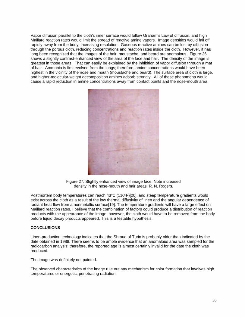
36
Vapor diffusion parallel to the cloth's inner surface would follow Graham's Law of diffusion, and highMaillard reaction rates would limit the spread of reactive amine vapors. Image densities would fall offrapidly away from the body, increasing resolution. Gaseous reactive amines can be lost by diffusionthrough the porous cloth, reducing concentrations and reaction rates inside the cloth. However, it haslong been recognized that the images of the hair, moustache, and beard are anomalous. Figure 26shows a slightly contrast-enhanced view of the area of the face and hair. The density of the image isgreatest in those areas. That can easily be explained by the inhibition of vapor diffusion through a matof hair. Ammonia is first evolved from the lungs; therefore, amine concentrations would have beenhighest in the vicinity of the nose and mouth (moustache and beard). The surface area of cloth is large,and higher-molecular-weight decomposition amines adsorb strongly. All of these phenomena wouldcause a rapid reduction in amine concentrations away from contact points and the nose-mouth area.
Figure 27: Slightly enhanced view of image face. Note increaseddensity in the nose-mouth and hair areas. R. N. Rogers.
Postmortem body temperatures can reach 43ºC (110ºF)[20], and steep temperature gradients wouldexist across the cloth as a result of the low thermal diffusivity of linen and the angular dependence ofradiant heat flow from a nonmetallic surface[19]. The temperature gradients will have a large effect onMaillard reaction rates. I believe that the combination of factors could produce a distribution of reactionproducts with the appearance of the image; however, the cloth would have to be removed from the bodybefore liquid decay products appeared. This is a testable hypothesis.
CONCLUSIONS
Linen-production technology indicates that the Shroud of Turin is probably older than indicated by thedate obtained in 1988. There seems to be ample evidence that an anomalous area was sampled for theradiocarbon analysis; therefore, the reported age is almost certainly invalid for the date the cloth wasproduced.
The image was definitely not painted.
The observed characteristics of the image rule out any mechanism for color formation that involves hightemperatures or energetic, penetrating radiation.
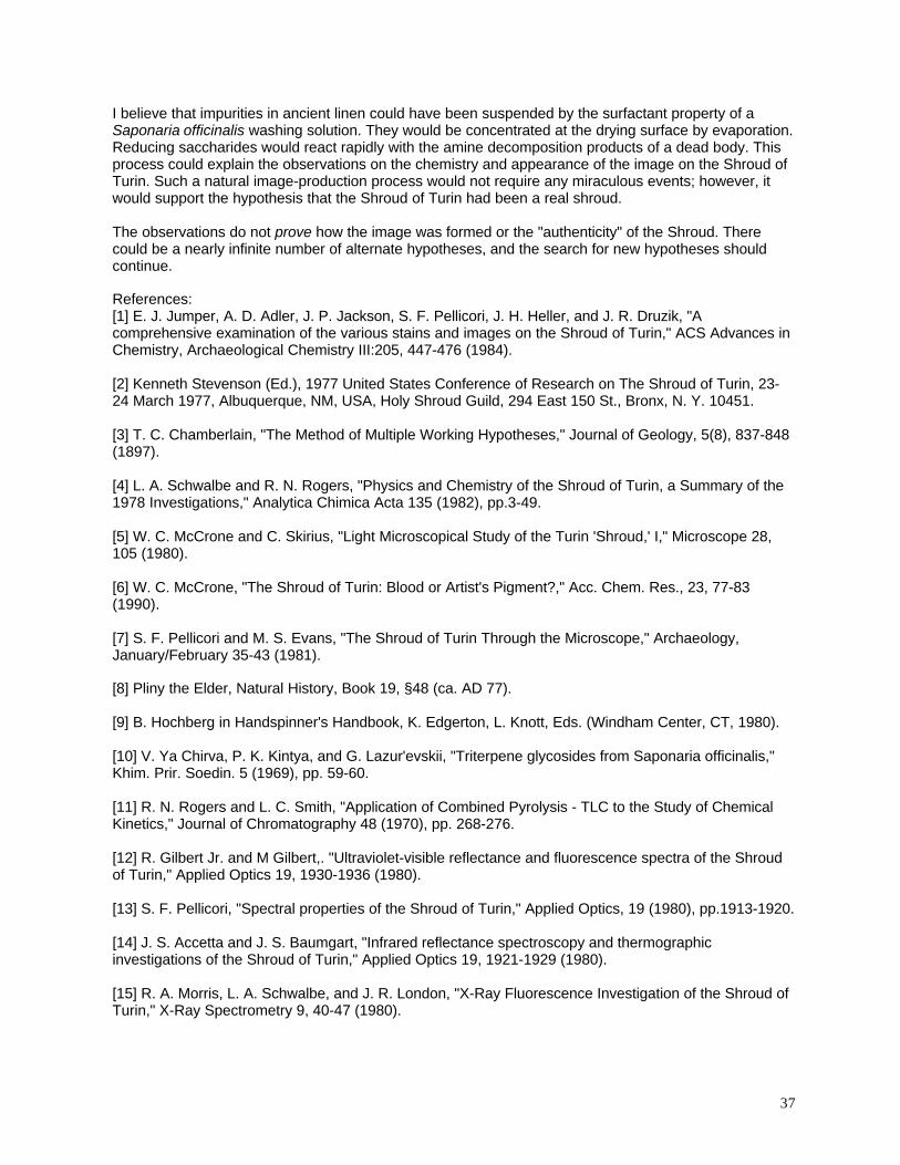
37
I believe that impurities in ancient linen could have been suspended by the surfactant property of aSaponaria officinalis washing solution. They would be concentrated at the drying surface by evaporation.Reducing saccharides would react rapidly with the amine decomposition products of a dead body. Thisprocess could explain the observations on the chemistry and appearance of the image on the Shroud ofTurin. Such a natural image-production process would not require any miraculous events; however, itwould support the hypothesis that the Shroud of Turin had been a real shroud.
The observations do not prove how the image was formed or the "authenticity" of the Shroud. Therecould be a nearly infinite number of alternate hypotheses, and the search for new hypotheses shouldcontinue.
References:[1] E. J. Jumper, A. D. Adler, J. P. Jackson, S. F. Pellicori, J. H. Heller, and J. R. Druzik, "Acomprehensive examination of the various stains and images on the Shroud of Turin," ACS Advances inChemistry, Archaeological Chemistry III:205, 447-476 (1984).
[2] Kenneth Stevenson (Ed.), 1977 United States Conference of Research on The Shroud of Turin, 23-24 March 1977, Albuquerque, NM, USA, Holy Shroud Guild, 294 East 150 St., Bronx, N. Y. 10451.
[3] T. C. Chamberlain, "The Method of Multiple Working Hypotheses," Journal of Geology, 5(8), 837-848(1897).
[4] L. A. Schwalbe and R. N. Rogers, "Physics and Chemistry of the Shroud of Turin, a Summary of the1978 Investigations," Analytica Chimica Acta 135 (1982), pp.3-49.
[5] W. C. McCrone and C. Skirius, "Light Microscopical Study of the Turin 'Shroud,' I," Microscope 28,105 (1980).
[6] W. C. McCrone, "The Shroud of Turin: Blood or Artist's Pigment?," Acc. Chem. Res., 23, 77-83(1990).
[7] S. F. Pellicori and M. S. Evans, "The Shroud of Turin Through the Microscope," Archaeology,January/February 35-43 (1981).
[8] Pliny the Elder, Natural History, Book 19, §48 (ca. AD 77).
[9] B. Hochberg in Handspinner's Handbook, K. Edgerton, L. Knott, Eds. (Windham Center, CT, 1980).
[10] V. Ya Chirva, P. K. Kintya, and G. Lazur'evskii, "Triterpene glycosides from Saponaria officinalis,"Khim. Prir. Soedin. 5 (1969), pp. 59-60.
[11] R. N. Rogers and L. C. Smith, "Application of Combined Pyrolysis - TLC to the Study of ChemicalKinetics," Journal of Chromatography 48 (1970), pp. 268-276.
[12] R. Gilbert Jr. and M Gilbert,. "Ultraviolet-visible reflectance and fluorescence spectra of the Shroudof Turin," Applied Optics 19, 1930-1936 (1980).
[13] S. F. Pellicori, "Spectral properties of the Shroud of Turin," Applied Optics, 19 (1980), pp.1913-1920.
[14] J. S. Accetta and J. S. Baumgart, "Infrared reflectance spectroscopy and thermographicinvestigations of the Shroud of Turin," Applied Optics 19, 1921-1929 (1980).
[15] R. A. Morris, L. A. Schwalbe, and J. R. London, "X-Ray Fluorescence Investigation of the Shroud ofTurin," X-Ray Spectrometry 9, 40-47 (1980).
38
[16] J. H. Heller and A. D. Adler, "A Chemical Investigation of the Shroud of Turin," Canadian Society ofForensic Science Journal 14 (1981), pp.81-103.
[17] G. Raes, Rapport d'Analise. La S. Sindone. Rivista Diocesana Torinese, 79-83 (1976).
[18] J. Rinaudo, "Protonic Model of Image Formation," Third International Congress on the Shroud ofTurin, Turin, Italy, 5-7 June, 1998.
[19] G. G. Gubareff, J. E. Janssen, and R. H. Torborg, Thermal Radiation Properties Survey, HoneywellResearch Center, Minneapolis-Honeywell Regulator Co., Minneapolis, MN, 1960.
[20] R. Irvine MD,. Personal communication, Pathology Section, Office of the Medical Investigator,University of New Mexico, Albuquerque, NM 87131 (2001).
[21] P. E. Damon, D. J. Donahue, B. H. Gore, A. L. Hatheway, A. J. T. Jull, T. W. Linick, P. J. Sercel, L.J. Toolin, C. R. Bronk, E. T. Hall, R. E. M. Hedges, R. Housley, I. A. Law, C. Perry, G. Bonani, S.Trumbore, W. Woefli, J. C. Ambers, S. G. E. Bowman, M. N. Leese, and M. S. Tite, "Radiocarbon datingof the Shroud of Turin," Nature, 337:611-615 (1989).
[22] R. W. Mottern, R. J. London, and R. A. Morris, "Radiographic Examination of the Shroud of Turin - -- a Preliminary Report," Materials Evaluation 38, 39-44 (1979).
[23] V. D. Miller and S. F. Pellicori, "Ultraviolet fluorescence photography of the Shroud of Turin," Journalof Biological Photography 49, 71-85 (1981).
[24] A. M Donadoni,. Personal communication, Turin, Italy, September 1978.
[25] H. E. Gove, S. J. Mattingly, A. R. David, and L. A. Garza-Valdes, "A problematic source of organiccontamination of linen," Nuclear Instruments and Methods in Physics Research B, 123, 504-507 (1997).
[26] M. Antonacci, The Resurrection of the Shroud (M. Evans and Company, Inc., 216 East 49th St.,New York, NY 10017, 2000).
[27] S. J. Mattingly, "The Role of Human Skin Bacteria in the Formation of Photographic-like Images onLinen with Implications for Shroud of Turin Conservation and a Method to Clean Shroud Linen ofMicrobial Contamination for Reliable Radiocarbon dating," British Society for the Turin Shroud, ShroudNewsletter 54, November 2001, pp. 12-17.
[28] S. J. Mattingly, personal communication via e-mail, "Shroud Samples," 18 June 2002.
[29] P. Vignon, The Shroud of Christ (1902), reprinted by University Books (1970).
[30] A. Arnoldi, "Thermal processing and foods quality: analysis and control," in P. Richardson, ThermalTechnologies in Food Processing, 138-159 (Cambridge UK, Woodhead Publishing, 2001).
[31] B. Cämmerer and L. W. Kroh, "Investigation of the influence of reaction conditions on theelementary composition of melanoidins," Food Chem. 53, 55-59 (1995).
[32] T. Hoffman, "Characterization of the most intensely colored compounds from Maillard reactions ofpentoses by application of color dilution analysis," Carbohydr. Res. 313(3-4), 203-213 (1998).
[33] C. Van Doren, Benjamin Franklin, 419, 433-436, (New York: The Viking Press, 1938).
[34] R. N. Rogers, "Supportive comments on the Benford-Marino '16th century repairs' hypothesis,"British Society for the Turin Shroud, Shroud Newsletter 54, 28-33 (2001).
Recommended