-1
Taxonomy and phylogeny of the xerophilic genus Wallemia (Wallemiomycetes
and Wallemiales, cl. et ord. nov.)
Polona Zalar1,*, G. Sybren de Hoog2, Hans-Josef Schroers3, John Michael Frank4 andNina Gunde-Cimerman11Biotechnical Faculty, Biology Department, University of Ljubljana, Vecna pot 111, SI-1000 Ljubljana,Slovenia; 2Centraalbureau voor Schimmelcultures, P.O. Box 85167, NL-3508 AD Utrecht, The Netherlands;3Agricultural Institute of Slovenia, Hacquetova 17, p.p. 2553, 1001 Ljubljana, Slovenia; 433 Tor Rd, Earnham,Surrey, GU9 7BY, UK; *Author for correspondence (e-mail: [email protected]; phone: +386-1-42-333-88; fax: +386-1-257-33-90)
Received 28 May 2004; accepted in revised form 25 November 2004
Key words: Ecology, Food-borne fungi, Internal transcribed spacer ribosomal DNA, Salterns, Smallsubunit ribosomal DNA, Xerotolerance
Abstract
The genus Wallemia comprises xerophilic species. Based on parenthesome ultrastructure it has been linkedto the Filobasidiales (basidiomycetes). Species show a unique type of conidiogenesis, including basauxicdevelopment of fertile hyphae, segregation of conidial units more or less basipetally, and disarticulation ofconidial units into mostly four arthrospore-like conidia. Wallemia is known from air, soil, dried food(causing spoilage), and salt. It can be isolated from hypersaline water of man-made salterns on differentcontinents. Based on analyses of the nuclear small subunit ribosomal DNA (SSU rDNA) Wallemia hasbeen placed into a highly supported clade together with Ustilaginomycetes and Hymenomycetes (Basidi-omycota). Within this clade, it possesses an isolated position distantly related to the Filobasidiales and wascharacterized by numerous nucleotide substitutions not shared by any other fungus. Tests on xerotoleranceindicated that Wallemia presents one of the most xerophilic fungal taxa. Xerotolerance is otherwise rare inthe Basidiomycota. To acknowledge its unique morphology, evolution, and xerotolerance, a new basid-iomycetous class Wallemiomycetes covering an order Wallemiales, is proposed. Based on differences inconidial size, xerotolerance, and sequence data of the rDNA internal transcribed spacer regions (ITSrDNA), at least three Wallemia species are segregated, identified as Wallemia ichthyophaga, Wallemia sebi,and Torula epizoa var. muriae, for which the combination Wallemia muriae is proposed. The three speciesare neotypified. W. ichthyophaga differs from W. sebi and W. muriae in numerous nucleotides of the SSUand ITS rDNA. This high variation within Wallemia indicates existence of at least two cryptic genera notdistinguishable by morphological characters.
Introduction
Wallemia Johan-Olsen is a genus of cosmopolitanxerophilic fungi, frequently involved in food
spoilage. Strains can be isolated from sweet (fruits,jams, cakes, pure sugar), salty (fish, meat, peanuts)and dried foods (Samson et al. 2002), from sea salt(Høye 1902), soil (Domsch et al. 1980), as well as
Antonie van Leeuwenhoek (2005) 87:311–328
DOI 10.1007/s10482-004-6783-x � Springer 2005
from indoor and outdoor air (Takahashi 1997).Initially, Wallemia was described as being halo-philic (Høye 1902; Schoop 1937; Frank and Hess1941). Later it was recognized to be xerophilicbecause growth on artificial media proved to beindependent of the solute used to lower the wateractivity (aw) (Vaisey 1955; Pitt and Hocking 1977).Certain strains of Wallemia can produce the toxinswalleminol and walleminon (Wood et al. 1990;Frank et al. 1999) and cause subcutaneous infec-tions in humans (de Hoog et al. 2000) and prob-ably also allergological problems resulting infarmer’s lung disease (Lappalainen et al. 1998;Roussel et al. 2004).
The genus Wallemia was introduced by Johan-Olsen (1887) for the single species W. ichthyophagaJohan-Olsen, which was described as slow grow-ing, xerophilic, and forming peculiar conidia. Theconidiogenesis was later interpreted or illustratedas phialidic (Barron 1968; von Arx 1970; de Hooget al. 2000) but is now understood as a variation ofthe basauxic mode of development of fertilehyphae, which segregate into larger conidiogenousunits, in a more or less basipetal succession, eachunit disarticulating into four arthrospore-likeconidia that remain connected long time by meansof connectives or disconnect (Hashmi andMorgan-Jones 1973; Madelin and Dorabjee 1974;Cole and Samson 1979).
von Arx (1970) synonymized SporendonemaDesm. with Wallemia and established the combi-nation Wallemia sebi for the species Sporendonemasebi Fr. Wallemia sebi (Fr.) v. Arx is today themost frequently cited Wallemia species andencompasses a large number of synonyms (Ciferriand Redaelli 1934; Ciferri 1958; Cannon 1990).Frank and Hess (1941) distinguished a secondspecies, Sporendonema epizoum Cif. & Red., basedon a study of numerous strains, but this studyremained unacknowledged by subsequent workers.
Terracina (1974) showed dolipore-like septalstructures in W. sebi, similarly to those formed bymany basidiomycetes and some ascomycetousyeasts. He explicitly ascribed the structures sur-rounding the pores to the endoplasmatic reticu-lum. However, they were later interpreted as aspecial kind of parenthesome and used as anargument to describe a new family, the Wallemi-aceae, which was placed into the Filobasidiales(basidiomycetes) (Moore 1986, 1996). While theFilobasidiales contain two other xerophilic taxa,
namely the black yeast genera Moniliella Stolk &Dakin and Trichosporonoides Haskins & Spencer(de Hoog 1979), xerophily is rarely observed inbasidiomycetes but more frequently encounteredin ascomycetes (Samson et al. 2002). Wu et al.(2003) developed SSU rDNA primers for thedetection and identification of airborne fungalspecies, including W. sebi. They phenotypicallycompared Wallemia sequences with those of otherairborne taxa, mainly ascomycetes and zygomy-cetes, among which Wallemia took an isolated,unresolved position.
The present study aims to clarify aspects of thephylogeny, taxonomy, and ecology of the genusWallemia. Recently isolated strains were examinedand compared with various reference strains,including ex-type cultures of taxa considered syn-onymous with W. sebi. A higher-rank phylogenyof the genus was inferred from analyses of thesmall subunit ribosomal DNA gene cluster (SSUrDNA). For the species level, sequence data of therDNA internal transcribed spacer regions 1 and 2(ITS), including the 5.8S rDNA, as well as mor-phological and physiological characteristics werestudied.
Material and methods
Strains and culture conditions
Strains studied are listed in Table 1. They wereisolated from the hypersaline water of salterns inMediterranean (Slovenia, Spain), DominicanRepublic and Namibia using the method des-cribed by Gunde-Cimerman et al. (2000) and fromspoiled prsutto using general microbiologicaltechniques and isolation media for xerophilic fungi(Pitt and Hocking 1997). The strains were depos-ited at the Centraalbureau voor Schimmelcultures(CBS, Utrecht, The Netherlands), the CultureCollection of the National Institute of Chemistry(MZKI, Ljubljana, Slovenia), and the CultureCollection of Extremophilic Fungi (EXF, Ljublj-ana, Slovenia). Reference strains were obtainedfrom the CBS and from the personal collection ofone of us (J.M.F). The strains were maintained on50% glucose medium (MY50G, Pitt and Hocking1997) and on Malt Extract Agar (MEA, Gamset al. 1998) with or without the addition of 5 or10% NaCl, and were preserved either in lyophi-lized condition or under liquid nitrogen.
312
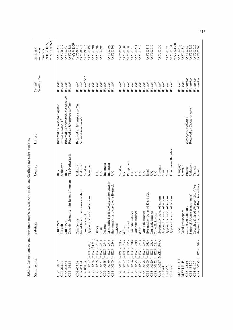
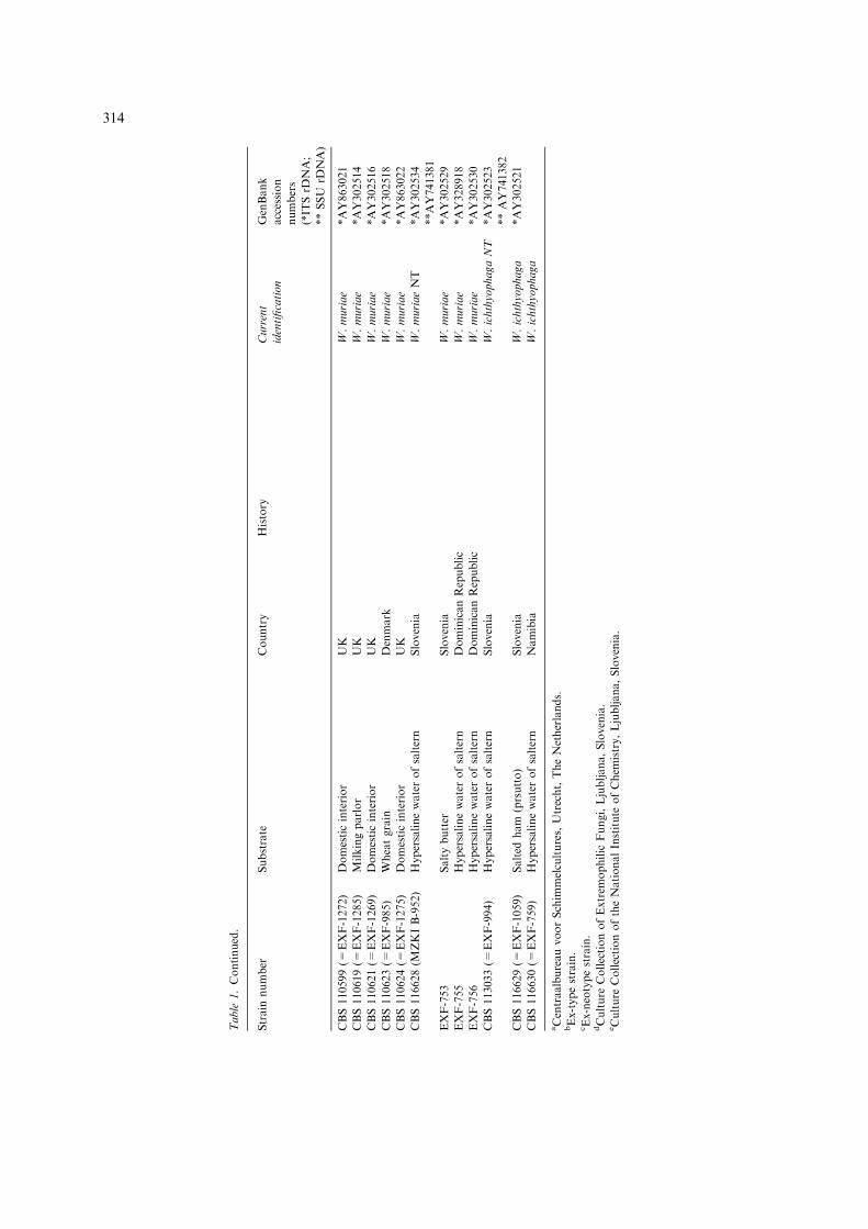
Table
1.Isolatesstudiedandtheirstrain
numbers,substrate,origin,andGenBankaccessionnumbers.
Strain
number
Substrate
Country
History
Current
identification
GenBank
accession
numbers
(*IT
SrD
NA;
**SSU
rDNA)
CBSa200.33
Unknown
Italy
Received
asOospora
d’agatae
W.sebi
*AY302519
CBS202.33
Sea
salt
Unknown
Torula
sacchariTb
W.sebi
*AY328912
CBS213.34
Unknown
Italy
Received
asSporendonem
aepizoum
W.sebi
*AY302520
CBS196.56
Chronic
ulcerativeskin
lesionofhuman
TheNetherlands
Received
asHem
ispora
stellata
W.sebi
*AY302526
**AY741379
CBS633.66
Date
honey
Unknown
Received
asHem
ispora
stellata
W.sebi
*AY328916
CBS453.80
Airofbananacontainer
onship
Unknown
Sporotrichum
navale
TW.sebi
*AY328914
CBS818.96
Sunflower
seed
Sweden
W.sebiNTc
*AY328915
CBS110582(=
EXF-585)
Hypersalinewaterofsaltern
Namibia
W.sebi
*AY302499
CBS110584(=
EXFd-1261)
W.sebi
*AY302501
CBS110585(=
EXF-1263)
Barley
UK
W.sebi
*AY302502
CBS110587(=
EXF-1281)
Cake
UK
W.sebi
*AY302503
CBS110588(=
EXF-1274)
Peanuts
Indonesia
W.sebi
CBS110589(=
EXF-1277)
Dried
salted
fish
Ophiocephalusstriatus
Indonesia
W.sebi
*AY302505
CBS110590(=
EXF-1266)
Haysample
associatedwithlivestock
toxicosis
UK
W.sebi
*AY302506
CBS110591(=
EXF-1268)
Rye
Sweden
W.sebi
*AY302507
CBS110592(=
EXF-1264)
Wheat
UK
W.sebi
*AY302508
CBS110593(=
EXF-1279)
Straw
hat
Philippines
W.sebi
*AY302509
CBS110594(=
EXF-1280)
Domesticinterior
UK
W.sebi
*AY302510
CBS110595(=
EXF-1270)
Domesticinterior
UK
W.sebi
*AY302511
CBS110597(=
EXF-1281)
Wheat
UK
W.sebi
*AY302512
CBS110598(=
EXF-1284)
Domesticinterior
UK
W.sebi
CBS110600(=
EXF-1053)
HypersalinewaterofDeadSea
Israel
W.sebi
*AY302513
CBS110620(=
EXF-1282)
Domesticinterior
UK
W.sebi
*AY302515
CBS110622(=
EXF-1265)
Catw
alk
insilos
UK
W.sebi
CBS116627(M
ZKIe
B-953)
Hypersalinewaterofsaltern
Slovenia
W.sebi
*AY302535
EXF-483
Hypersalinewaterofsaltern
Spain
W.sebi
EXF-622
Hypersalinewaterofsaltern
Spain
W.sebi
*AY302528
EXF-757
Hypersalinewaterofsaltern
DominicanRepublic
W.sebi
*AY302531
**AY741380
MZKIB-384
Seed
Hungary
W.sebi
*AY302532
MZKIB-451
Deadgrasshopper
Slovenia
W.sebi
*AY302533
CBS103.10
Culture
contaminant
France
Hem
ispora
stellata
TW.muriae
*AY302524
CBS184.28
SugarofArenga(sugarpalm
)Unknown
Received
asTorula
sacchari
W.muriae
*AY302525
CBS411.77
DecayingfruitofPhoenix
dactylifera
Tunesia
W.muriae
*AY302527
CBS110583(=
EXF-1054)
HypersalinewaterofRed
Sea
saltern
Israel
W.muriae
*AY302500
313
Table
1.Continued.
Strain
number
Substrate
Country
History
Current
identification
GenBank
accession
numbers
(*IT
SrD
NA;
**SSU
rDNA)
CBS110599(=
EXF-1272)
Domesticinterior
UK
W.muriae
*AY863021
CBS110619(=
EXF-1285)
Milkingparlor
UK
W.muriae
*AY302514
CBS110621(=
EXF-1269)
Domesticinterior
UK
W.muriae
*AY302516
CBS110623(=
EXF-985)
Wheatgrain
Denmark
W.muriae
*AY302518
CBS110624(=
EXF-1275)
Domesticinterior
UK
W.muriae
*AY863022
CBS116628(M
ZKIB-952)
Hypersalinewaterofsaltern
Slovenia
W.muriaeNT
*AY302534
**AY741381
EXF-753
Saltybutter
Slovenia
W.muriae
*AY302529
EXF-755
Hypersalinewaterofsaltern
DominicanRepublic
W.muriae
*AY328918
EXF-756
Hypersalinewaterofsaltern
DominicanRepublic
W.muriae
*AY302530
CBS113033(=
EXF-994)
Hypersalinewaterofsaltern
Slovenia
W.ichthyophagaNT
*AY302523
**AY741382
CBS116629(=
EXF-1059)
Salted
ham
(prsutto)
Slovenia
W.ichthyophaga
*AY302521
CBS116630(=
EXF-759)
Hypersalinewaterofsaltern
Namibia
W.ichthyophaga
aCentraalbureauvoorSchim
melcultures,Utrecht,TheNetherlands.
bEx-typestrain.
cEx-neotypestrain.
dCulture
CollectionofExtrem
ophilic
Fungi,Ljubljana,Slovenia.
eCulture
CollectionoftheNationalInstitute
ofChem
istry,Ljubljana,Slovenia.
314
Morphology and cultural characteristics
Macromorphological characters such as size ofcolonies, spreading tendency, colony structure,exudate production, colony colour, and produc-tion of soluble pigments were described on fivedifferent media, which are as follows: (i) Wallemiamorphology medium I (W-4) with aw = 0.95,containing glycerol and NaCl as controlling sol-utes [yeast extract 2.5 g; KH2PO4 0.64 g;MgSO4 � 7H2O 0.12 g; glycerol 120 g; NaCl40.9 g; H2O 839 ml; agar 15 g; Borrows mi-cronutrients 2 ml (ZnSO4 1 g; FeSO4 1 g; CuSO4
150 mg; MnSO4 100 mg; K2MoO4 100 mg; H2O1 l)], (ii) Wallemia morphology medium II (W-10)with aw = 0.90 [as W-4 but containing 100 gNaCl]; (iii) MEA with aw = 0.998, (iv) MY50Gwith aw = 0.890 and (v) MY10-12 withaw = 0.916 (Pitt and Hocking 1997). Macroscop-ical characters and general growth rates were re-ported from point-inoculated media in plastic Petridishes (9 cm diam) incubated at 24 �C for 14 days.Micromorphological characters were studied fromslide cultures (Gams et al. 1998) using media(i)–(iv) incubated at 24 �C for 7 days and phase-contrast microscopy. The reported characters andmeasurements derived from MY50G. Medium (v)was used for isolation purposes. Water activities ofmedia used for morphological examinations wereverified by the DECAGON CX-1 Water ActivitySystem (Campbell Scientific Ltd). The pH ofmedia (i)–(iii) and (v) was set to 6.5 prior auto-claving according to Pitt and Hocking (1977).
Physiology
Colony growth of strains marked below wasmeasured on media with 11 different wateractivities ranging from aw = 0.99 to 0.77 (mea-sured as described above). For all, glycerol, NaCl,or glucose was used as controlling solute. The basicmedium contained 10 g malt extract, 10 g yeastextract, 1 g K2HPO4 and 20 g agar (Wheeler et al.1988) and had a water activity of aw = 0.999. ThepH of the media was adjusted as described above.The following strains were included in physiologi-cal experiments: CBS 113033, CBS 116629, CBS116630 (all W. ichthyophaga); CBS 202.33, CBS196.56, CBS 633.66, CBS 453.80, CBS 818.96,CBS 110582, CBS 110600, CBS 116627, EXF-483,
EXF-622, EXF-757, MZKIB-384, MZKI B-451(allW. sebi); CBS 103.10, CBS 184.28, CBS 411.77,CBS 110583, CBS 110599, CBS 110619, CBS110621, CBS 110623, CBS 110624, CBS 116628,EXF-753, EXF-755, EXF-756 (all W. muriae). Foreachmedium and each water activity, accomplishedby different NaCl concentrations, mean values ofthree replicates per strain were calculated. Themean of these mean values was calculated forstrains belonging to one species. The standarddeviation was calculated as well.
Molecular methods
DNA was extracted from ca. 1 cm2 of 14 days oldcultures by mechanical lysis (Gerrits van den Endeand de Hoog 1999). For the amplification of SSUrDNA and the internal transcribed spacer regions1 and 2 including the 5.8S rDNA (hereafterreferred to as ‘ITS’), primers NS1 (White et al.1990) and NS24 (Gargas and Taylor 1992) andV9G (de Hoog and Gerrits van den Ende 1998)and LS266 (Masclaux et al. 1995), respectively,were used. PCR conditions were applied as de-scribed by de Hoog et al. (2000). PCR fragmentswere purified using the GFXTM purification kit(Amersham Pharmacia Biotech Inc., Roosendaal,Netherlands). Sequence reactions containingprimers ITS1 or ITS4 (White et al. 1990) for theITS, and Oli1, Oli9, Oli3 (Hendriks et al. 1989),BF83, BF951, BF163, BF1438, and BF1419 (deHoog et al. 2004) for the SSU rDNA as well asaliquots of the BigDye terminator cycle sequencingkit (Applied Biosystems, Foster City, CA) wereanalyzed on an ABI Prism 3700 (Applied Biosys-tems). Sequences were assembled and edited usingSeqMan 3.61 (DNAStar, Inc., Madison, USA).
Sequence data
Newly generated DNA sequences of the SSUrDNA have been deposited in GenBank (http://www.ncbi.nlm.nih.gov) (Table 1). Accession num-bers of published sequences are juxtaposed to theirtaxon names in the phylogenetic tree of Figure 1.References for these sequences are the following:AJ495820, AJ495823, AJ495830 (Bacigalova et al.unpublished), U00969, U00971, U00972 (Berbeeand Taylor 1993), AJ271380, AJ271381 (Doering
315
and Blanz unpublished), AB023413, AB023414(Hamamoto and Nakase unpublished), AJ568017,AJ560318 (Kidd unpublished), AJ496258 (Lopan-dic et al. unpublished), AY083223 (McIlhatton andCurran unpublished), AB038129 (Nagahama et al.unpublished), D85143 (Nishida et al. 1998),AB072226 (Niwata et al. 2002), S83267 (Shah et al.1996), AB000955, AB000959 (Sjamsuridzal et al.1997), AB001728, AB001730 (Sugita and Nakase1998a), AB001749 (Sugita and Nakase 1998b),AB035586, AB035588 (Sugita et al. 2000), D63929(Sugiyama et al. 1995), D31657, D31658, D31659(Suh and Nakase 1995), D12802, D12804 (Suh andSugiyama 1993), D14006 (Suh and Sugiyama1994), D64120 (Suh et al. 1996b), D78330 (Suhet al. 1996a), L22257 (Swann and Taylor 1993),D83189, D83190, D83193 (Takashima and Nakase1996), AB032621 (Takashima and Nakase1999), AB045704 (Takashima and Nakase 2001),
AB075544, AB075545, AB075546 (Takashima andNakase unpublished), AB000645 (Ueda-NishimuraandMikata 2000), X60179 (van de Peer et al. 1992),AF548107, AF548108 (Wu et al. 2003), AJ223490(Xu et al. unpublished).
Phylogenetic analyses
Wallemia sequences were compared with pub-lished and unpublished data available at the Na-tional Center for Biotechnology Information orthe CBS, respectively. SSU rDNA sequences werealigned with sequences of basidiomycetous andascomycetous taxa, partly selected using theBLAST server at http://www.ncbi.nlm.nih.gov/Blast/ (Altschul et al. 1990). Incomplete 3¢ and 5¢parts of sequences were coded as missing charac-ters. Sequences were automatically aligned using
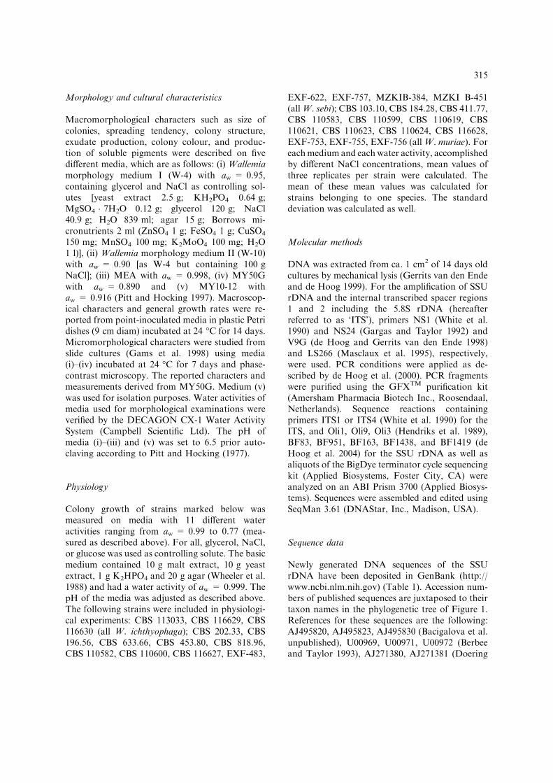
Figure 1. Phylogenetic tree based on SSU rDNA. One of 504 equally parsimonious phylograms inferred from heuristic analyses of the
partial SSU rDNA sequences rooted with Dipodascus agregatus. Bootstrap intervals from 1000 replicates are indicated above branches.
Wallemia forms the supported sistergroup of a clade comprising Ustelaginomycetes and Heterobasidiomycetes (bootstraps = 99%),
which suggests that it is related to the Basidiomycota, within which it comprises a unique phylogenetic position. CI = 0.473;
RI = 0.806.
316
ClustalX 1.81 (Jeannmougin et al. 1998). Thealignments were adjusted manually using BioEdit5.0.9 (Hall 1999). Phylogenetic relationships of thetaxa were estimated from the aligned sequences bythe maximum parsimony criterion as implementedin PAUP 4.0b10 (Swofford 2003). Heuristic sear-ches were performed using parsimony informative,unordered, and equally weighted characters;branch robustness was tested by 1000 searchreplications, each on bootstrapped data sets. Gapswere treated as missing characters. For the SSUrDNA analyses, starting tree(s) were obtained viarandom, 100 times (10 times in bootstrap analyses)repeated sequence addition. For the ITS rDNAsequence addition was simple. A maximumnumber of 1000 trees were allowed.
Results
Molecular phylogeny
Heuristic parsimony analyses of the SSU rDNAaligned sequences yielded 252 equally most-parsi-monious trees 1558 steps long with a consistencyindex (CI) of 0.476 and a retention index (RI) of0.813. The data set contained 2151 bp alignmentpositions including an intron of 339 bp onlyencountered in Protomyces macrosporus Ungerand 446 parsimony informative characters (PIC).The main topology of the 252 trees, one of which isshown in Figure 1, was identical. Wallemia waslocated in a highly supported clade (bootstrapvalue = 98%) of Heterobasidiomycetes (Tri-chosporonales, Tremellales, Filobasidiales, Cys-tofilobasidiales, and Dacrymycetales) togetherwith a moderately supported monophyletic group(bootstrap value = 79%) of Ustilaginomycetes(Microstromatales, Malasseziales, Tilletiales, En-tylomatales, Georgefischeriales, and Exobasidi-ales). Within the former clade, Wallemia clusteredat the base and formed an isolated, 165 steps longbranch. No other genus was found to cluster withWallemia and no fungal SSU rDNA sequence wasavailable sharing any of the numerous nucleotidesubstitutions unique for Wallemia. A highly sup-ported clade (bootstrap value = 100%), com-prising species of the ascomycetous genusTaphrina Fr. and Protomyces macrosporus, wasplaced outside of the basidiomycetous clade, butsistergroup relationship between these clades wasweakly supported (bootstrap value = 56%).
Pneumocystis jirovecii Frenkel and Dipodascopsisuninucleata (Biggs) L.R. Batra & Millner, twoother primitive yeast-like fungi with ascomyceteaffinity, received an unresolved position near theroot of the tree. The number of nucleotide sub-stitutions between the sequences of W. sebi (CBS196.56) and W. ichthyophaga was 81 bp.
Based on the heuristic parsimony analysis of the623 aligned position of the partial large ribosomalsubunit (LSU rDNA) sequences, Wallemia wasplaced among Basidiomycota (bootstrap va-lue = 99%), next to Heterobasidiomycetes (Dac-rymycetales and Tremellales), the later not beingsupported in bootstrap analysis (results notshown). In the LSU rDNA tree as well none of thedeeper branches were supported.
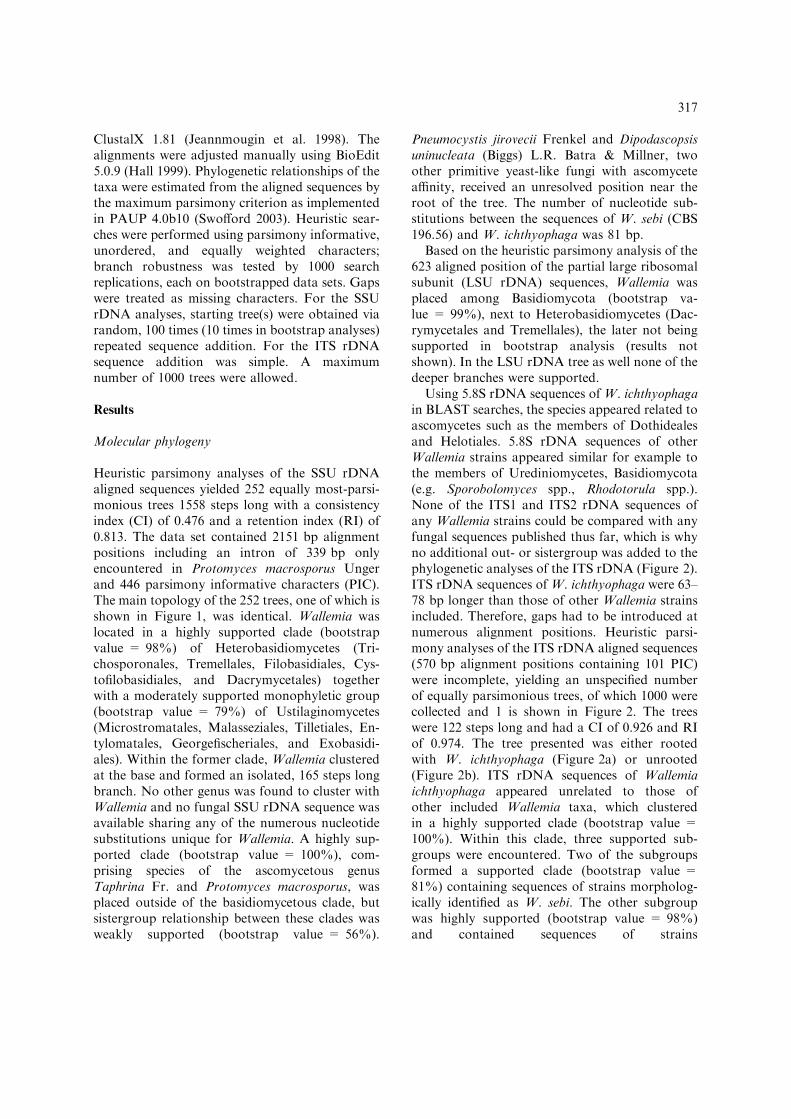
Using 5.8S rDNA sequences of W. ichthyophagain BLAST searches, the species appeared related toascomycetes such as the members of Dothidealesand Helotiales. 5.8S rDNA sequences of otherWallemia strains appeared similar for example tothe members of Urediniomycetes, Basidiomycota(e.g. Sporobolomyces spp., Rhodotorula spp.).None of the ITS1 and ITS2 rDNA sequences ofany Wallemia strains could be compared with anyfungal sequences published thus far, which is whyno additional out- or sistergroup was added to thephylogenetic analyses of the ITS rDNA (Figure 2).ITS rDNA sequences of W. ichthyophaga were 63–78 bp longer than those of other Wallemia strainsincluded. Therefore, gaps had to be introduced atnumerous alignment positions. Heuristic parsi-mony analyses of the ITS rDNA aligned sequences(570 bp alignment positions containing 101 PIC)were incomplete, yielding an unspecified numberof equally parsimonious trees, of which 1000 werecollected and 1 is shown in Figure 2. The treeswere 122 steps long and had a CI of 0.926 and RIof 0.974. The tree presented was either rootedwith W. ichthyophaga (Figure 2a) or unrooted(Figure 2b). ITS rDNA sequences of Wallemiaichthyophaga appeared unrelated to those ofother included Wallemia taxa, which clusteredin a highly supported clade (bootstrap value =100%). Within this clade, three supported sub-groups were encountered. Two of the subgroupsformed a supported clade (bootstrap value =81%) containing sequences of strains morpholog-ically identified as W. sebi. The other subgroupwas highly supported (bootstrap value = 98%)and contained sequences of strains
317
morphologically identified as Torula epizoa Cordavar. muriae Kickx.
Discussion
Analyses of the SSU rDNA (Figure 1) and LSUrDNA (not shown) strongly suggest that the genusWallemia, represented by W. sebi and W. ichthy-
ophaga, is a member of or at least a close relativeof the Basidiomycota. Phylogenetic analyses thusare in support to earlier interpretations based onthe dolipore-like hyphal septum morphology inW.sebi, which was seen similar to those formed gen-erally by basidiomycetes and few ascomycetousyeast (Terracina 1974; Moore 1986). Formation ofarthrospore-like conidia in Wallemia possibly alsoindicates its relationship with the Basidiomycota,
Figure 2. Phylogenetic trees based on ITS rDNA. (a) One of more than 1000 equally and most parsimonious phylograms from an
incomplete heuristic phylogenetic analyses inferred from aligned sequences of the ITS1, 5.8S, and ITS2 rDNA rooted with W.
ichthyophaga. The incomplete analyses yielded an unspecified number of equally parsimonious trees, of which 1000 trees were collected.
Bootstrap intervals from 1000 replicates higher than 70% are indicated near their respective branches. (b) Unrooted radial tree based
on the same analyses as in (a) not showing bootstrap values. The relatedness of W. sebi and W. muriae is highly supported and
numerous molecular steps distinguish W. ichthyophaga from these two species. The moderately supported clade of W. sebi (boot-
straps = 81%) is segregated into two different monophyletic, but phenotypically indistinguishable groups. CI = 0.926; RI = 0.974.
318
in which the arthric development of conidia isfrequently encountered (Stalpers 1978; Waltheret al. 2004). Based on the parenthosome ultra-structure of W. sebi, relationship of Wallemia withthe Filobasidiales has been predicted (Moore1986), but this interpretation is rejected by themolecular phylogeny based on SSU rDNA se-quences (Figure 1).
Based on the SSU rDNA data, Wallemia takesan isolated position as a sister group of a cladecomprising members of the Ustilaginomycetes andHeterobasidiomycetes. In BLAST searches, onlysequences of the Heterobasidiomycetes, Ustilagi-nomycetes, and Taphrinomycetes appeared com-parable to sequences of Wallemia. Both theHeterobasidiomycetes and Ustilaginomycetesencompass isolated phylogenetic positions amongother basidiomycetes (Scorzetti et al. 2002; Stollet al. 2003) and are rich in taxa with yeast-likeanamorphs such as Rhodotorula F.C. Harrison,Cryptococcus Vuill., Malassezia Baill., TilletiopsisDerx, and Tilletiaria Bandoni & B.N. Johri(Boekhout et al. 1998) and biotrophic, plantparasitic life styles such as Tilletiopsis, Tilletiaria,and Exobasidium Woronin (Blanz and Doring1995; Begerow et al. 2000). These features areconsidered to represent phylogenetically old lifeforms. Biotrophic life style and yeast anamorphsare also encountered in the Taphrinomycetes(Eriksson and Winka 1997), which, in respectto certain morphological features, encompassan intermediate between ascomycetes andbasidiomycetes and are regarded as phylogeneticallyold taxa (Nishida et al. 1995). Because of itsrelative relatedness to taxa with yeast-likeanamorphs and biotrophic life-styles, it can beassumed that Wallemia is a phylogeneticallyancient taxon.
Other supposedly primitive or phylogeneticallyold ascomycetes, like Protomyces Unger, Pneu-mocystys P. Delanoe & Delanoe, Schizosacchar-omyces Lindner (Archiascomycetes) did not showany relatedness to Wallemia but were placed atunique positions in the tree on long terminalbranches. Huge molecular distances which resultedin hardly alignable sequences have also beenreported for example for two morphologicallysimilar groups of the Dipodascaceae (Ueda-Ni-shimura and Mikata 2000) and Zygomycota(Voigt et al. 1999), respectively. Also in Wallemia,several subregions of the SSU rDNA as well as the
LSU rDNA were rich in nucleotide substitutions.Within the LSU rDNA, numerous synapomor-phies were encountered, not being shared by anyother fungal taxon. The large amount of nucleo-tide substitutions in these taxa probably accumu-lated over extended time during evolution or is dueto the fact that their close relatives became extinctand may therefore indicate that they are phyloge-netically old.
Large molecular distances, however, were alsoencountered in the LSU rDNA ofW. ichthyophagaandW. sebi and in sequences of the ITS1 and ITS2rDNA of W. ichthyophaga on the one andW. muriae and W. sebi on the other hand (Fig-ure 2). While the sequences of W. muriae and W.sebi were well alignable, sequences of W. ichthy-ophaga were hardly alignable to those of W. mu-riae and W. sebi and multiple gaps had to beincluded at various places. It is therefore possiblethat the genus Wallemia in fact comprises a com-plex of several phylogenetically remote genera andthat taxa in between and linking the extant Wall-emia species have become extinct or have not yetbeen isolated and analyzed. The formation ofsarcina-like structures only in W. ichthyophaga(Figure 3m) may indicate that W. ichthyophaga isalso morphologically distinct from the other twospecies. A potentially close relative of Wallemiamight be the genus Arthrowallemia CastanedaRuiz, a genus of leaf-inhabiting fungi because ofmorphologically similar conidia formed by holo-thallic conidiogenesis (Castaneda Ruiz et al. 1998).No material from living cultures, however, wasavailable to test this hypothesis.
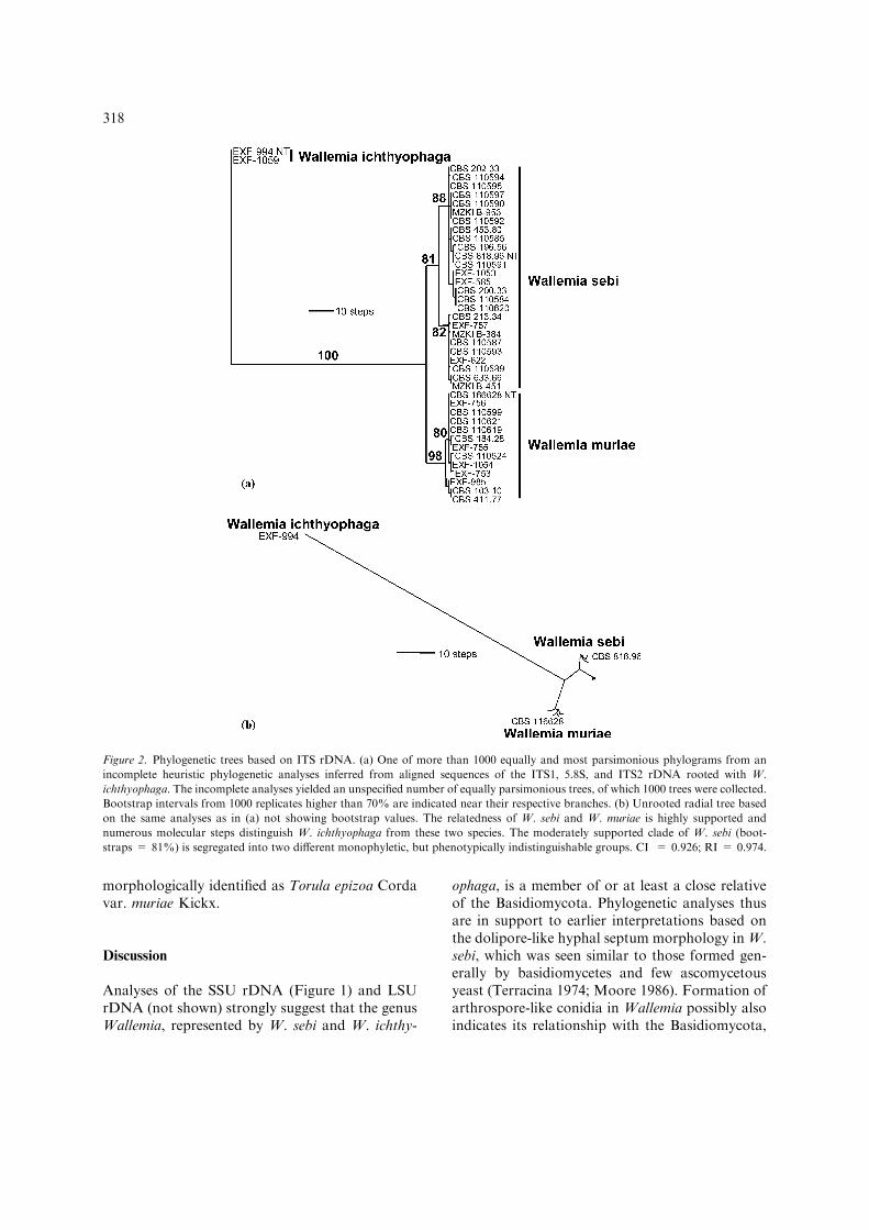
In spite of considerable molecular distancesbetween W. ichthyophaga to W. sebi and W. mu-riae, their close relatedness is particularly wellcorroborated by morphological characters. In allthree species, similar arthrospore-like conidia inunits of mostly four were formed from morpho-logically similar conidiophores (Figure 3c, g, h, l).The morphology of the conidiophores and thebasauxic mode of conidiogenesis were consideredto be unique in the fungal Kingdom and highlytypical for W. sebi (Hashmi and Morgan-Jones1973; Madelin and Dorabjee 1974; Cole andSamson 1979) but present in the other two speciesas well. All three species were also characterized byxerophily, which is relatively rare in basidiomyce-tes, but more often encountered in various, butunrelated ascomycetes such as Aspergillus Link
319
Eurotium Link, Chrysosporium Corda, XeromycesL.R. Fraser, and Hortaea Nishim. & Miyaji(Samson et al. 2002). Of these, Xeromyces and
Wallemia exclusively comprise species that arexerophilic or at least xerotolerant, while xerophilyin other genera typically is observed only in certain
Figure 3. Micromorphology of Wallemia spp. (a–e) Wallemia sebi, MY50G. (a) Hyphae (CBS 196.56); (b) sympodially elongating
conidiophor (CBS 196.56); (c) conidiogenous cell producing conidia in packages of four (MZKI B-953); (d, e) conidia in chains (MZKI
B-384, CBS 196.56, respectively); (f–j) Wallemia muriae, MY50G. (f) Swollen conidium germinating into hypha (EXF-753); (g, h)
conidiogenous cells producing conidia (CBS 110624; EXF-755, respectively); (i) single conidia (CBS 411.77); (j) conidia in chains
(EXF-755). (k–o) Wallemia ichthyophaga, MY50G. (k) Muriform hypha (EXF-759); (l) Conidiogenous cell forming conidia (EXF-
1059). (m) sarcina-like structures of cells (EXF-994); (n) single conidia (EXF-759); (o) conidia in chains (EXF-759). Scale bars on all
the figures indicate 10 lm.
320
specialized taxa but not in all species of therespective genus.
Particularly because of its phylogenetic position(Figure 1), but also because of its unique mor-phology (Hashmi and Morgan-Jones 1973;Madelin and Dorabjee 1974; Cole and Samson1979) and physiology, a new class and order, theWallemiomycetes and Wallemiales, respectively,were introduced in this paper.
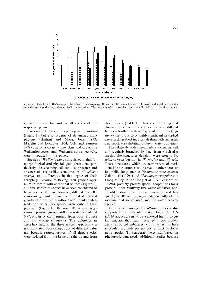
Species of Wallemia are distinguished mainly bymorphological and physiological characters, par-ticularly the size range of conidia, presence andabsence of sarcina-like structures in W. ichthy-ophaga, and differences in the degree of theirxerophily. Because of having their growth opti-mum in media with additional solutes (Figure 4),all three Wallemia species have been considered tobe xerophilic. W. sebi, however, differed from W.ichthyophaga and W. muriae in that it showedgrowth also on media without additional solutes,while the other two species grew only in theirpresence (Figure 4). Because W. ichthyophagashowed positive growth still at a water activity of0.77, it can be distinguished from both, W. sebiand W. muriae (Figure 4). The difference inxerophily among the three species apparently isnot correlated with occupations of different habi-tats because representatives of all three specieswere isolated from the brine of salterns and from
dried foods (Table 1). However, the suggesteddistinction of the three species that also differedfrom each other in their degree of xerophily (Fig-ure 4) may prove to be highly significant in appliedareas such as food industry dealing with materialsand substrata exhibiting different water activities.
The relatively wide, irregularly swollen, as wellas irregularly branched hyphae, from which alsosarcina-like structures develop, were seen in W.ichthyophaga but not in W. muriae and W. sebi.These structures, which are reminiscent of meri-stem-like structures also observed in other xero- orhalophilic fungi such as Trimmatostroma salinumZalar et al. (1999a) and Phaeotheca triangularis deHoog & Beguin (de Hoog et al. 1997; Zalar et al.1999b), possibly present special adaptations for agrowth under relatively low water activities. Sar-cina-like structures, however, were formed fre-quently in W. ichthyophaga independently of themedium and solute used and the water activityapplied.
The adapted concept of Wallemia species is alsosupported by molecular data (Figure 2). ITSrDNA sequences in W. sebi showed high molecu-lar variation that mainly resulted in two moder-ately supported subclades within W. sebi. Thesesubclades probably present two distinct phyloge-netic species. To segregate these taxa based onphenotypic data needs additional studies because
Figure 4. Physiology ofWallemia spp. Growth ofW. ichthyophaga,W. sebi andW. muriae (average values) on media of different water
activities accomplished by different NaCl concentrations. The measures of standard deviation are indicated by bars on the columns.
321
relatively strongly variable size ranges of conidiawere encountered in both subgroups and no dif-ference in their xerophilic behaviour was obvious.
The name Hemispora stellata, of which the ex-type strain CBS 103.10 was identified as W. mu-riae, has been used repeatedly for clinical isolatesidentified as such (Ciferri and Redaelli 1934).Following older concepts, taxa of Wallemia mightbe considered as opportunistic human pathogenswith low virulence, but in recent years only a singleWallemia strain has been isolated from a skinlesion of a human patient (Table 1). Because of itsrare occurrence in skin disorders, it is possible thatpreviously isolated Wallemia strains actually pre-sented culture contaminants of microsporidialdermatophytes that demand specific media andconditions for growth. In contrast, recent reportsattribute a possible role of W. sebi in other clinicalcases such as lung diseases (Lappalainen et al.1998; Roussel et al. 2004).
Taxonomy
Wallemiomycetes Zalar, de Hoog et Schroers, cl.nov.
Classis Basidiomycotis vulgo xerophilis, basi-diosporis ignotis. Septae cum doliporis. Conidi-onenesis basauxis. Conidia arthrosporidia.
Type order. Wallemiales (see below).Wallemiales Zalar, de Hoog et Schroers, ord.
nov.Ordo Basidiomycotis vulgo xerophilis, basi-
diosporis ignotis. Septae cum doliporis. Conidio-genesis basauxis. Conidia arthrosporidia.
Type family. Wallemiaceae R.T. Moore.Wallemiomycetes Zalar, de Hoog and Schroers
cl. nov. et Wallemiales Zalar, de Hoog andSchroers ord. nov.
Conidiophores unbranched or sympodially pro-liferating, continuous with conidiogenous cells,smooth-walled. Conidiogenous cells verruculose,basauxially extending, distally disarticulating intoarthrospore-like conidia. Conidia verruculose,short cylindrical, becoming spherical.Hyphal septawith a single pore, flaring out near the periphery ofthe pore, barrel-shaped, dolipore-like. Ecophysiol-ogy. Typically xerophilic.
Type familiy. Wallemiaceae R.T. Moore, inSneh B, Jabaji-Hare S, Neate S. and Dijst. G.(eds), Rhizoctonia Species: Taxonomy, Molecular
Biology, Ecology, Pathology and Disease Control.Kluwer Acad. Publ., Dordrecht, The Netherlands.p. 20, 1996.
Type genus and type species. Wallemia ichthy-ophaga.
Phylogenetic affinities. Basidiomycota.Habitat. Hypersaline water, food with low water
activity due to salt and other solutes and driedplant products.
Distribution. Cosmopolitan.References. For conidiogenesis: Cole and Sam-
son (1979), Hashmi and Morgan-Jones (1973),Madelin and Dorabjee (1974) and Moore (1986);for ultrastructure of septal pores: Moore (1986,1996) and Terracina (1974); for life style: Frankand Hess (1941).
Commentary. Moore (1996) classified the Wal-lemiaceae into the Filobasidiales based on ultra-structure of the septal pore in Wallemia sebi,particularly on the parenthesomes, which weredescribed as vesiculate.
Wallemia ichthyophaga Johan-Olsen 1887.Christiania Videnkabs-Selskab Forhandl. no. 12,pp. 1–20. – Figure 3k–o.
Colony characteristics. Colonies punctiform,cerebriform, dusty, soft, and slimy typicallyspreading deeply into the agar, on W-4, W-10 andMY50G 3.0–5.0 mm diam, about 3 mm high; onW-4 and W-10 dark reddish brown with darkgreenish brown reverse; on MY50G light brown;on media with glycerol and glucose variously pig-mented, beige, yellow, light green, or brown,sometimes shiny with an irregular surface, con-sisting of one to several, up to 3–5 mm diam largeglobular structures; not growing on MEA ormedia without additional solutes; exudates notobserved.
Microscopy. Hyphae hyaline, smooth, 4.0–10.0 lm wide, with up to 2.0 lm thick walls,forming an irregularly branched, transversely andsometimes also longitudinally septate mycelium(Figure 3k). Conidiophores more or less solitary,erect, subhyaline, unbranched, smooth-walled,slightly constricted below the apex, continuing intofertile conidiogenous cells (Figure 3l); conidioge-nous cells cylindrical, verruculose, basauxicallyextending, at ca. 10–12.5 lm height, basipetallydisarticulating into packages of four arthrospore-like conidia. Conidia one-celled, pale brown,initially short cylindrical, soon becoming spheri-cal, with thick, echinulate walls, 4.0–5.0 lm diam,
322
forming fragile chains (Figure 3 n, o); conidiatypically swelling to 10.0 lm diam, dividingirregularly to form sarcina-like structures up to200 lm diam, from which new conidiophores arise(Figure 3m).
Physiology. Growth requiring additional sol-utes, no growth on ordinary media such as MEA;growth not depending of the solute type; growth ata minimum aw = 0.96, optimum at aroundaw = 0.90, maximum at aw = 0.77 (Figure 4).
Type. Norway, isolated from salted klip-fish(cited by Ciferri 1958). No authentic materialpreserved. NEOTYPE: Slovenia, Secovlje salterns,isolated in Oct 1999 by L. Butinar from hypersa-line water; dried MY10-12 culture of CBS 113033(=EXF-994) preserved in herb. CBS.
Habitat. Occurring in hypersaline water of sal-terns in Slovenia and Namibia; on salted meat andfish; also recorded from stock fish (salted cod),where it produced sarcina-like structures on saltcrystals (J.C. Frisvad, personal communication);until now not recorded from environmental sub-strates with high sugar content, although it cangrow also on sugary culture media.
Diagnostic characters. Growth only onmedia withadditional solutes, best growth at aw = 0.90 (e.g. byusing 10% of NaCl and 12% of glucose); minimalwater activity required is aw = 0.96; conidial sizerange; presence of sarcina-like structures.
References. Frank and Hess (1941) and Ciferri(1958).
Commentary. The identity of W. ichthyophaga issupported by the size range of the conidia, whichwas given in the original description as 3–4 lmdiam and by the presence of sarcina-like cells thatderive from the swelling and irregular dividing ofconidia (Ciferri 1958). Strains identified here as W.ichthyophaga fit remarkably well to the fungusdescribed by Johan-Olsen, however, they formslightly larger conidia. Wallemia ichthyophaga isxerophilic because it grows at water activity below0.85. Wang (1965) reported it several times frompaper, but the identity of these strains could not beconfirmed.
Wallemia sebi (Fr.) von Arx 1970. The Generaof Fungi Sporulating in Pure Culture. CramerVerlag, p. 181 – Figure 3a–e.= Sporendonema sebi Fr., Syst. Mycol. 3: 435.1832.? = Torula epizoa Corda, Deutschl. Fl., ed. J.Sturm, Nurnberg 1: 9. 1829.
” Sporendonema epizoum (Corda) Ciferri etRedaelli, J. Trop. Med. Hyg. 37: 170. 1934.= Sporotrichum navale Joly, Revue Mycol. 26:98. 1961. Supported by ex-type strain CBS 453.80.? = Torula minuta Høye 1902. = Sporendonemaminutum (Høye) Frank & Hess, J. Fish. Res. BoardCan. 5: 291. 1941. Characterized as halotolerant,which is why it could be a synonym of W. sebi.
Colony characteristics. Colonies punctiform,typically spreading deeply into the agar, on MEA,3–6 mm diam, compact, powdery, rust brown topurplish-brown, with brown reverse; on MY50G4.0–12.5 mm diam, with a yellowish brownreverse; on W-4 4.0–8.0 mm diam; colony shapevariable, typically domed without or sometimeswith short marginal spreading area; margin whiteor of same colour as the colony, shaggy to irreg-ular; surface smooth but velvety in the central partor, on MY50G, powdery due to strong sporula-tion; exudates randomly present as yellow drop-lets, mainly formed on W-10.
Microscopy. Hyphae hyaline, smooth- and thin-walled, 1.5–2.5 lm wide, forming a compactmycelium (Figure 3a). Conidiophores erect, den-sely, parallel arranged, subhyaline, unbranched orsometimes sympodially elongating, smooth-wal-led, slightly or not constricted at the apex justbelow a darker integument, which continues intofertile conidiogenous cells (Figure 3b, c); conidi-ogenous cells cylindrical, verruculose, basauxicallyextending, at ca. 7.5–20 lm length basipetallydisarticulating into packages of four arthrospore-like conidia (Figure 3c). Conidia one-celled, palebrown, initially short cylindrical, soon becomingspherical, verrucose, thick-walled, (1.5–)2.0–2.5 lm diam, forming up to 1 mm long, straight orbending chains (Figure 3d, e). Conidial chains ofneighbouring conidiophores densely aggregated,remaining intact in undisturbed colonies or easilyfalling apart leading to randomly distributed sa-tellite colonies.
Physiology. Growth not depending of the solutetype, also on media without additional soluteshaving aw = 0.99; growth optimum betweenaw = 0.976 and 0.957, maximum at aw = 0.83(Figure 4).
NEOTYPE: Sweden, isolated from sunflowerseed, dried MEA culture of CBS 818.96 depositedin herb. CBS, designated herewith.
Habitat. Occurring in salty environments(hypersaline water of salterns or salty fish), air in
323
indoor environments, on seeds (rye, wheat), dryplant material (hay); also isolated from air inindoor environment.
Diagnostic characters. Growth on media withoutadditional solutes such asMEA; conidial size range.
References. Domsch et al. (1980), de Hoog et al.(2000) and Samson et al. (2002).
Commentary. Wallemia sebi is the only speciesof Wallemia capable of growth on media withoutadditional solutes, such as MEA. Because itgrows at water activity below 0.85, W. sebi isxerophilic. Fries (1832) synonymized Torula epi-zoa Corda, which originated from salty meat inBelgium, as Sporendonema sebi Fr., which wasdescribed from tallow (tasteless solid fat extractedfrom animal fat). Both authors did not mentionmeasurements of morphological structures. Incontrast, the concept of W. sebi proposed here, isbased on morphological and physiological char-acteristics. Although originally described fromanimal fat, W. sebi has been isolated mainly fromvarious substrata other than meat or tallow butonce from a salted fish. These substrates share acertain degree of dryness, but are not exception-ally osmotic. In order to stay as close as possibleto the strain reported in the protologue of S. sebi,we select CBS 818.96 from sunflower seed inSweden as NEOTYPE of Wallemia sebi. Ourconcept is in consistency with previous concepts(Ciferri and Redaelli 1934; Ciferri 1958; Cannon1990), which all consider Sporendonema sebi asthe basionym of the taxon, that later von Arxcombined as W. sebi.
Wallemia muriae (Kickx) Zalar & de Hoog,comb. nov. – Figure 3f–j.
” Torula epizoa Corda var. muriae Kickx, Fl.Crypt. Flandres 2: 299. 1847.= Hemispora stellata Vuillemin, Bull. Soc. My-col. Fr. 22: 1–6. 1906. Colonies star-shaped, 0.5–2.5 mm diam. Conidia 2.6–3.5 lm diam,verruculose. The ex-type strain of H. stellata,CBS 103.10, is a representative of W. muriae.Vuillemin (1931) redescribed this strain, which heoriginally described as a culture contaminant, butlisted the species as originating from a humanpatient.? = Oidium morrhuae Farlow, Bull U.S. Fish., p.4. 1886. The species was isolated from salted fish.Its habitat and description indicates that it is likelya synonym of Wallemia muriae.
Colony characteristics. Colonies punctiform,typically spreading deeply into the agar, on W-4and W-10 3.0–5.0 mm diam, walnut- to purplish-brown, with dark greenish-brown reverse; onMY50G 5.5–8.0 mm diam; not growing on MEAor media without additional solutes; colony shapesomewhat domed, typically with a spreadingmarginal area: on MY50G flat, extremely dustydue to strong sporulation, developing in concentricrings; exudates occasionally present on W-10, asyellow droplets.
Microscopy. Hyphae hyaline, smooth- and thin-walled, 2.5–3.5 lm wide, forming a compactmycelium. Conidiophores aggregated, subhyaline,unbranched, rising laterally from hyphae, smooth-walled, slightly constricted below the apex,continuing into fertile conidiogenous cells(Figure 3g, h); conidiogenous cells cylindrical,verruculose, basauxically extending, at ca. 10–12.5 lm height basipetally disarticulating intopackages of four arthrospore-like conidia(Figure 3h). Conidia one-celled, pale brown,initially short cylindrical, soon becoming spheri-cal, verrucose, with up to 1 lm thick walls, 2.5–3.0 lm diam, forming chains (Figure 3i, j).
Physiology. No growth on media withoutadditional solutes such as MEA; growth notdepending of the solute type; growth minimum ataw = 0.985, optimum at around aw = 0.96,maximum at aw = 0.83 (Figure 4).
Type. Belgium, described from fish brine. Noauthentic material preserved. NEOTYPE: Slove-nia, Secovlje salterns, isolated in July 1997 by P.Zalar from hypersaline water; dried MY10-12culture of CBS 116628, deposited in herb. CBS,designated herewith.
Habitat. Occurring on sugary food products(date honey, cake), salty food (peanuts), hypersa-line water of salterns world-wide, dry substrates(straw, seeds), and in indoor environments; onerecord from an insect is available.
Diagnostic characters. Growth only on mediawith additional solutes, best growth at aw = 0.985(e.g. by using 2% NaCl, 10% glucose, or 6%glycerol); conidial size range.
Commentary. The identity of W. muriae is sup-ported by the size range of the conidia, which wasgiven in the original description as 3–3.5 lm diambased on in vivo measurements. Strains identifiedhere as W. muriae have 2.5–3 lm large conidiabased on in vitro measurements and are interme-
324
diate of W. ichthyophaga and W. sebi. Wallemiamuriae is xerophilic because it grows at wateractivity below 0.85.
Dichotomous key to accepted Wallemia species
(1a) Colonies well growing at 24 �C on MEA,reaching 3–6 mm diam in 14 days; conidiashortly cylindrical, 1.5–2.5 lm diam; sarcina-like structures absent. . . . . . . . . . . . . . . . . . . . . . . . . . . . . . . . . . . . W. sebi
(1b) Colonies only growing on MEA with addi-tional solutes (NaCl, glucose); conidia largerthan 2.5 lm diam, sarcina-like structuresabsent or present . . . . . . . . . . . . . . . . . . . . . . . . . 2
(2a) Colonies on MY50G dark brown, with acerebriform surface; conidia 3.5–5.0 lm indiam, sarcina-like structures present . . . . . . . .. . . . . . . . . . . . . . . . . . . . . . . . . . . W. ichthyophaga
(2b) Colonies on MY50G walnut brown, with apowdery surface; conidia 2.5–3.0 lm diam;sarcina-like structures absent. . . . . . . . . . . . . . .. . . . . . . . . . . . . . . . . . . . . . . . . . . . . . . . .W. muriae
List of doubtful names listed as synonyms ofWallemia sebi (Fr.) von Arx.
Bergellinia monospora Borzi, Malpighia 2: 469–476. 1888. Described from an infected external earcanal of human from Sicily, Italy. The descriptioncould not be interpreted.
Catenularia fuliginea Saito, J. Coll. Sci. Imp.Univ. Tokyo 18: 51. 1904. Saccardo synonymisedthis species, isolated from cheese, with Torulasimplex (Lindner) Sacc. The original description ofthe fungus could not be interpreted.
Oidium pulvinatum Cooke, Rav. Am. Fungi n.770. 1883–1884. The species was described from aliving leaf of Carya tomentosa in South Carolina,probably accidentally included by Ciferri (1958) ina list of synonyms of W. sebi.
Oospora d’agatae Saccardo, in: Pasini, AttiRiun. Soc. Ital. Dermat. Sifil. 26: 106. 1930. Thespecies was described on the basis of a clinicalisolate. Its identity is doubtful.
Oospora ochracea Corda, Ic. Fung., vol. I, p.16, 1837. The citation is reproduced from Sac-cardo (1884) but not listed in this publication ofCorda. The name was assigned to an isolate fromcondensed sap of Sambucus nigra berries inBohemia (Czech Republic). The conidia were
reported to be 35 lm diam. The species is ofdoubtful identity.
Penicillium simplex Lindner, Mikrosk. Bet-riebsk. Garungsgew. p. 170. 1895. The name wasintroduced for an isolate from wine must in Ger-many. Raper and Thom (1949) listed it as adoubtful species.
Sporendonema terrestre Oudemans, Meded.Kon. Akad. Wetensch., Afd. Natuurk., 3e Reeks,2: 115. 1885. The species was described from soil,cannot be interpreted and is doubtful.
Torula pulvinata (Bonorden) Saccardo, (= Al-ysidium pulvinatum Bonorden) – Abh. Geb. Myk.2: 86. 1886. The species was described from woodof Abies sp., which is an unusual habitat forWallemia. The morphological description cannotbe interpreted.
Torula rubiginosa Rivolta, Paras. Veget. Introd.Studio Mal. Parass., p. 254. 1873. The species wasisolated from hay in Italy. Its conidial size wasgiven as 9 lm, which excludes Wallemia.
Torula rufescens Fresenius, Beitr. Mykol, p. 36.1850–1863. The species was described for a whitefungus with spherical, reddish conidia measuring5–8 lm in diam. The description was based on anisolate from human eye. Its identity is doubtful.
Torula acchari Corda, Ic. Fung. 4: 23. 1840. Thespecies was described for a white fungus on sugar,probably an Acremonium species forming conidiain chains.
Acknowledgements
We thank Barbara Kastelic Bokal and KasperLuijsterburg for technical assistance. We thankAndre Aptroot for providing Latin description.We acknowledge the support of the ‘EuropeanCommunity – Access to Research Infrastructureaction of the Improving Human PotentialProgramme’.
References
Altschul S.F., Gish W., Miller W., Myers E.W. and Lipman
D.J. 1990. Basic local alignment search tool. J. Mol. Biol.
215: 403–410.
Barron G.L. 1968. The Genera of Hyphomycetes from Soil.
The Williams and Wilkins Co., Baltimore.
Begerow D., Bauer R. and Boekhout T. 2000. Phylogenetic
placements of ustilaginomycetous anamorphs as deduced
from nuclear rDNA sequences. Mycol. Res. 104: 53–60.
325
Berbee M.L. and Taylor J.W. 1993. Dating the evolutionary
radiations of the true fungi. Can. J. Bot. 71: 1114–1127.
Blanz P. and Doring H. 1995. Taxonomic relationships in the
genus Exobasidium (Basidiomycetes) based on ribosomal
DNA analysis. Stud. Mycol. 38: 119–127.
Boekhout T., Bandoni R.J., Fell J.W. and Kwon-Chung K.J.
1998. Discussion of teleomorphic and anamorphic genera of
heterobasidiomycetous yeasts. In: Kurtzman C.F. and Fell
J.W. (eds), The Yeasts, a Taxonomic Study. Elsevier Science,
New York, pp. 609–625.
Cannon P.F. 1990. Name changes in fungi of microbiological,
industrial and medical importance. Part 4. Mycopathologia
111: 75–83.
Castaneda Ruiz R.F., Garcia D. and Guarro J. 1998. Arthro-
wallemia, a new genus of hyphomycetes from tropical litter.
Mycol. Res. 102: 16–18.
Ciferri R. 1958. Mauginiella a synonym of Sporendonema. Atti
Int. Bot. Univ. Pavia Lab. Crittog., Ser. 5(15): 126–133.
Ciferri R. and Redaelli P. 1934. Sporendonema epizoum (Corda)
Cif. et Red., an entity including Hemispora stellata and
Oospora d’Agatae. J. Trop. Med. Hyg. 37: 167–170.
Cole G.T. and Samson R.A. 1979. Patterns of Development in
Conidial Fungi. Pitman Publ. Co., London, San Francisco,
Melbourne.
de Hoog G.S. 1979. The black yeasts, II: Moniliella and allied
genera. Stud. Mycol. 19: 1–90.
de Hoog G.S., Beguin H. and de Vegte W.H. 1997. Phaeotheca
triangularis, a new meristematic black yeast from a humidi-
fier. Anton. Leeuw. 71: 289–295.
de Hoog G.S. and Gerrits van den Ende A.H.G. 1998.
Molecular diagnostics of clinical strains of filamentous Bas-
idiomycetes. Mycoses. 41: 183–189.
de Hoog G.S., Gottlich E., Platas G., Genilloud O., Leotta G.
and van Brummelen J. 2004. Evolution and ecology of the
genus Thelebolus in Antarctica. Stud. Mycol, in press.
de Hoog G.S., Guarro J., Gene J. and Figueras M.J. 2000.
Atlas of Clinical Fungi, 2nd ed. Centraalbureau voor
Schimmelcultures/Universitat Rovira i Virgili, Utrecht/Reus.
Domsch K.H., Gams W. and Anderson T.H. 1980. Compen-
dium of Soil Fungi. Academic Press, London.
Eriksson O.E. and Winka K. 1997. Supraordinal taxa of Asc-
omycota. Myconet 1(1): 1–16.
Frank M. and Hess E. 1941. Halophilic brown molds of the
genus Sporendonema emend. Ciferri et Redaelli. J. Fish. Res.
Board Can. 5: 287–292.
Frank J.M., Kingston E., Jeffery J.C., Moss M.O., Murray M.,
Simpson T.J. and Sutherland A. 1999. Walleminol and wal-
leminone, novel caryophyllenes from the toxigenic fungus
Wallemia sebi. Tetrahedron Lett. 40: 133–136.
Fries E.M. 1832. Systema mycologicum, Greifswald.
Gams W., Hoekstra E.S. and Aptroot A. 1998. CBS Course of
Mycology, 4th ed. Centraalbureau voor Schimmelcultures,
Baarn.
Gargas A. and Taylor J.W. 1992. Polymerase chain reaction
(PCR) for amplifying and sequencing nuclear 18S rDNA
from lychenized fungi. Mycologia 84: 589–592.
Gerrits van den Ende A.H.G. and de Hoog G.S. 1999.
Variability and molecular diagnostics of the neurotropic
species Cladophialophora bantiana. Stud. Mycol. 43: 151–
162.
Gunde-Cimerman N., Zalar P., de Hoog G.S. and Plemenitas A.
2000.Hypersalinewater in salterns – natural ecological niches for
halophilic black yeasts. FEMS Microbiol. Ecol. 32: 235–240.
Hall T.A. 1999. BioEdit: a user-friendly biological sequence
alignment editor and analysis program for Windows 95/98/
NT. Nucl. Acids Symp. Ser. 41: 95–98.
Hashmi M.H. and Morgan-Jones G. 1973. Conidium ontogeny
in hyphomycetes. The meristem arthrospores of Wallemia
sebi. Can. J. Bot. 51: 1669–1671.
Hendriks L., Goris A., Neefs J.-M., van de Peer Y., Hennebert
G. and de Wachter R. 1989. The nucleotide sequence of the
small ribosomal subunit RNA of the yeast Candida albicans
and the evolutionary position of the fungi amongst the
Eukaryotes. Syst. Appl. Microbiol. 12: 223–229.
Høye K. 1902. Undersøgelser op klipfiskesoppen. Bergen Mus.
Aarbog. 7: 1–39.
Jeannmougin F., Thompson J.D., Gouy M., Higgins D.G. and
Gibson T.J. 1998. Multiple sequence alignment with Clustal
X. Trends Biochem. Sci. 23: 403–405.
Johan-Olsen O. 1887. Op sop pa klipfisk den sakaldte mid.
Christiania Videnkabs-Selskab Forhandl. 12: 5.
Lappalainen S., Pasanen A.L., Reiman M. and Kalliokoski P.
1998. Serum IgG antibodies against Wallemia sebi and
Fusarium species in Finnish farmers. Ann. Alergy, Astma
Immunol. 81: 589–592.
Madelin M.F. and Dorabjee S. 1974. Conidium ontogeny in
Wallemia sebi. Trans. Br. Mycol. Soc. 63: 121–130.
Masclaux F., Gueho H., de Hoog G.S. and Christen R. 1995.
Phylogenetic relationships of human-pathogenic Cladospori-
um (Xylohypha) species inferred from partial LS rRNA se-
quences. J. Med. Vet. Mycol. 33: 327–338.
Moore R.T. 1986. A note on Wallemia sebi. Anton. Leeuw. 52:
183–187.
Moore R.T. 1996. The dolipore/parenthesome septum in
modern taxonomy. In: Sneh B., Jabaji-Hare S., Neate S. and
Dijst G. (eds), Rhizoctonia Species: Taxonomy, Molecular
Biology, Ecology, Pathology and Disease Control. Kluwer
Acad. Publ., Dordrecht, The Netherlands, pp. 13–35.
Nishida H., Ando K., Ando Y., Hirata A. and Sugiyama J.
1995. Mixia osmundae: transfer from the Ascomycota to the
Basidiomycota based on evidence from molecules and mor-
phology. Can. J. Bot. 73: S660–S666.
Nishida H., Tajiri Y. and Sugiyama J. 1998. Multiple origins of
fungal group I introns located in the same position of nuclear
SSU rRNA gene. J. Mol. Evol. 46(4): 442–448.
Niwata Y., Takashima M., Tornai-Lehoczki J., Deak T. and
Nakase T. 2002. Udeniomyces pannonicus sp. nov., a ball-
istoconidium-forming yeast isolated from leaves of plants in
Hungary. Int. J. Syst. Evol. Microbiol. 52: 1887–1892.
Pitt J.I. and Hocking A.D. 1977. Influence of solute and
hydrogen ion concentration on the water relations of some
xerophilic fungi. J. Gen. Microbiol. 101: 35–40.
Pitt J.I. and Hocking A.D. 1997. Fungi and Food Spoilage, 2nd
ed. Blackie Academic & Professional, London.
Raper K. and Thom C. 1949. A Manual of Penicillia. The
Williams and Wilkins Co., Baltimore, Maryland.
Roussel S., Reboux G., Dalphin J.-C., Bardonnet K., Millon L.
and Piarroux R. 2004. Microbiological evolution of hay and
relapse in patients with farmer’s lung. Occup. Environ. Med.
61: 3e.
326
Samson R.A., Hoekstra E.S., Frisvad J.C. and Filtenborg O.
2002. Introduction to Food- and Airborne Fungi, 6th ed.
Centraalbureau voor Schimmelcultures, Utrecht.
Schoop G. 1937. Salzpilz (Torula epizoa) auf Lebensmitteln.
Deutsche Tierarztl. Wschr. 45: 621–624.
Scorzetti G., Fell J.W., Fonseca A. and Statzell-Tallman A.
2002. Systematics of basidiomycetous yeasts: a comparison of
large subunit D1/D2 and internal transcribed spacer rDNA
regions. FEMS Yeast Res. 2(4): 495–517.
Shah J.S., Pieciak W., Liu J., Buharin A. and Lane D.J. 1996.
Diversity of host species and strains of Pneumocystis carinii is
based on rRNA sequences. Clin. Diagn. Lab. Immunol. 3(1):
119–127.
Sjamsuridzal W., Tajiri Y., Nishida H., Thuan T., Kawasaki
H., Hirata A., Yokota A. and Sugiyama J. 1997. Evolu-
tionary relationships of members of the genera Taphrina,
Protomyces, Schizosaccharomyces, and related taxa within
the archiascomycetes: integrated analysis of genotypic and
phenotypic characters. Mycoscience 38: 267–280.
Stalpers J.A. 1978. Identification of wood-inhabiting Aphyllo-
phorales in pure culture. Stud. Mycol. 16: 1–248.
Stoll M., Piepenbring M., Begerow D. and Oberwinkler F. 2003.
Molecular phylogeny of Ustilago and Sporisorium species
(Basidiomycota, Ustilaginales) based on internal transcribed
spacer (ITS) sequences. Can. J. Bot. 81(9): 976–984.
Sugita T. and Nakase T. 1998a. Molecular phylogenetic study
of the basidiomycetous anamorphic yeast genus Trichosporon
and related taxa based on small subunit ribosomal DNA
sequences. Mycoscience 39: 7–13.
Sugita T. and Nakase T. 1998b. Trichosporon japonicum sp.
nov. isolated from the air. Int. J. Syst. Bacteriol. 48(Pt 4):
1425–1429.
Sugita T., Takashima M., Ikeda R., Nakase T. and Shinoda T.
2000. Phylogenetic and taxonomic heterogeneity of Crypto-
coccus humicolus by analysis of the sequences of the internal
transcribed spacer regions and 18S rDNA, and the phyloge-
netic relationships of C. humicolus, C. curvatus, and the genus
Trichosporon. Microbiol. Immunol. 44(6): 455–461.
Sugiyama J., Inamura T., OkadaG., SjamsuridzalW., Kawasaki
H. and Hirata A. 1995. Divergence and molecular evolution
among basidiomycetous yeasts with the tropical and sub-
tropical genus Graphiola. In: Progress in Microbial Ecology:
Proceedings of Seventh International Symposium on Micro-
bial Ecology, Santos, Sao Paulo, Brazil, pp. 173–180.
Suh S.O. and Nakase T. 1995. Phylogenetic analysis of the
ballistosporous anamorphic genera Udeniomyces and Bullera,
and related basidiomycetous yeasts, based on 18S rDNA
sequence. Microbiology 141(Pt 4): 901–906.
Suh S.O. and Sugiyama J. 1993. Phylogeny among the basid-
iomycetous yeasts inferred from small subunit ribo-
somal DNA sequence. J. Gen. Microbiol. 139(Pt 7): 1595–
1598.
Suh S.O. and Sugiyama J. 1994. Phylogenetic placement of the
basidiomycetous yeasts Kondoa malvinella and Rhodospori-
dium dacryoidum, and the anamorphic yeast Sympodiomy-
cepsis paphiopedili by means of 18S rRNA gene sequence
analysis. Mycoscience 35: 367–375.
Suh S., Takashima M., Hamamoto M. and Nakase T. 1996a.
Molecular phylogeny of the ballistoconidium-forming ana-
morphic yeast genus Bullera and related taxa based on small
subunit ribosomal DNA sequences. J. Gen. Appl. Microbiol.
42: 501–509.
Suh S., Takematsu A., Takashima M. and Nakase T. 1996b.
Molecular phylogenetic study on stalked conidium-forming
yeasts and related basidiomycetous yeast taxa based on 18S
rDNA sequences. Microbiol. Cult. Coll. 12: 79–86.
SwannE.C. andTaylor J.W. 1993.Higher taxa of basidiomycetes:
an 18S rRNA gene perspective. Mycologia 85(6): 923–936.
Swofford D.L. 2003. PAUP*. Phylogenetic Analysis Using
Parsimony (*and other methods). Version 4.0b.4a. Sinauer
Associates, Massachusetts, Sunderland.
Takahashi T. 1997. Airborne fungal colony-forming units in
outdoor and indoor environments in Yokohama, Japan.
Mycopathologia 139: 23–33.
TakashimaM. and Nakase T. 1996. A phylogenetic study of the
genus Tilletiopsis, Tilletiaria anomala and related taxa based
on the small subunit ribosomal DNA sequences. J. Gen.
Appl. Microbiol. 42: 421–429.
Takashima M. and Nakase T. 1999. Molecular phylogeny of
the genus Cryptococcus and related species based on the
sequences of 18S rDNA and internal transcribed spacer
regions. Microbiol. Cult. Coll. 15: 35–47.
Takashima M. and Nakase T. 2001. Tilletiopsis derxii, Tilleti-
opsis oryzicola and Tilletiopsis penniseti, three new species of
the ustilagionomycetous anamorphic genus Tilletiopsis iso-
lated from leaves in Thailand. Anton. Leeuw. 80: 43–56.
Terracina F.C. 1974. Fine structure of the septum in Wallemia
sebi. Can. J. Bot. 52: 2587–2590.
Ueda-Nishimura K. and Mikata K. 2000. Two distinct 18S
rRNA secondary structures in Dipodascus (Hemiascomyce-
tes). Microbiology 146: 1045–1051.
Vaisey E.B. 1955. Osmophilism of Sporendonema epizoum.
J. Fish. Res. Board Can. 2: 901–903.
van de Peer Y., Hendriks L., Goris A., Neefs A., Vancanneyet
M., Kersters K., Berny J.F., Hennebert G.L. and De Wachter
R. 1992. Evolution of basidiomycetous yeasts as deduced
from small ribosomal subunit RNA sequences. Syst. Appl.
Microbiol. 15: 250–258.
Voigt K., Cigelnik E. and O’Donnell K. 1999. Phylogeny and
PCR identification of clinically important Zygomycetes based
on nuclear ribosomal. DNA sequence data. J. Clin. Micro-
biol. 37: 3957–3964.
von Arx J.A. 1970. The Genera of Fungi Sporulating in Pure
Culture. Cramer Verlag, Lehre.
Vuillemin P. 1931. Hemisporees et thallosporees. In: Les
Champignons parasites et les mycoses de l’homme. Leche-
valier, Paris, pp. 74–81.
Walther S., Garnica M. and Weiss M. 2004. The systematic
relevance of conidiogenesis modes in the gilled Agaricales.
Mycol. Res., in press.
Wang C.J.K. 1965. Fungi of Pulp and Paper in New York.
State Univ. Coll. Forestry Tech. Publ. No.
Wheeler K.A., Hocking A.D. and Pitt J.I. 1988. Effects of T
and aw on germination and growth of W. sebi. Trans. Br.
Mycol. Soc. 90: 365–368.
White T.J., Burns T., Lee S. and Taylor J. 1990. Amplification
and direct sequencing of fungal ribosomal RNA genes for
phylogenies. In: Innis M.A., Gelfand D.H., Sninsky J.J. and
White T.J. (eds), PCR Protocols. A Guide to Methods and
Amplifications. Academic, San Diego, pp. 315–322.
327
Wood G.M., Mann P.J., Lewis D.F., Reid W.J. and Moss M.O.
1990. Studies on a toxic metabolite from the mould Wall-
emia. Food Addit. Contam. 7–1: 69–77.
Wu Z., Tsumura Y., Blomquist G. and Wang X.-R. 2003. 18S
rRNA gene variation among common airborne fungi, and
development of specific oligonucleotide probes for the
detection of fungal isolates. Appl. Environ. Microbiol. 69:
5389–5397.
Zalar P., de Hoog G.S. and Gunde-Cimerman N. 1999a.
Trimmatostroma salinum, a new species from hypersaline
water. Stud. Mycol. 43: 61–67.
Zalar P., de Hoog G.S. and Gunde-Cimerman N. 1999b.
Taxonomy of the endoconidial black yeast genera Phaeotheca
and Hyphospora. Stud. Mycol. 43: 38–48.
328
Recommended