Embed Size (px)
Citation preview
25 Important Cases in EAR, NOSE & THROAT
SurgicoMed.com 25 IMPORTANT CASES IN EAR, NOSE & THROAT |
25 Important Cases in EAR, NOSE & THROAT
Case 1: A 10 year old child was having a right mucopurulent otorrhea for the last 4 years. A
week ago he became dizzy with a whirling sensation, nausea, vomiting and nystagmus to the
opposite side; his deafness became complete and his temperature was normal. Three days later
he became feverish, irritable and continuously crying apparently from severe headache. Also he
had some neck retraction. The child was not managed properly and died by the end of the week.
CASE 1
Diagnosis & Reasons
Right chronic suppurative otitis media (mucopurulent otorhea of 4 years duration) complicated by suppurative labyrinthitis (dizziness, nausea and vomiting with nystagmus to the opposite side and complete loss of hearing) and then complicated by meningitis (fever, severe headache and neck retraction).
Explain the following manifestations
Whirling sensation: vertigo due to inner ear inflammation
Nystagmus to the opposite side: suppurative labyrinthitis leading to fast phase of eye movement to the opposite ear and slow phase to the diseased ear nystagmus direction is called according to the fast phase. In serous labyrinthitis with no inner ear cell destruction the direction of nystagmus is toward the diseased ear.
Severe headache: increased intracranial pressure due to meningitis
Neck retraction: due to meningeal inflammation
Further examination &/or investigations
Otologic examination possible finding of a marginal perforation of atticoantral CSOM (cholesteatoma)
Audiogram to reveal SNHL in the affected ear
Kernig's and Brudzinski's signs
Fundus examination to show papilledema
Lumbar puncture: turbid high pressure CSF with pus rich in proteins
Complete blood picture
Treatment Antibiotics that cross the blood brain barrier
Analgesics
25 Important Cases in EAR, NOSE & THROAT
SurgicoMed.com 25 IMPORTANT CASES IN EAR, NOSE & THROAT |
Repeated lumbar puncture to drain infected CSF and to relieve symptoms and to inject antibiotics
Treaetment of the underlying otitis media appropriately according to its type
Case 2: A 50 year old male patient complained of right earache of 2 days duration. The pain was
especially severe on chewing food and during speech. There was also marked edema of the right
side of the face. On examination, pressure on the tragus was painful; and there was a small red
swelling arising from the anterior external auditory meatal wall. Rinne test was positive in the
right ear. The patient gave a history of 2 previous similar attacks in the same ear during the last
six months but less severe.
CASE 2
Diagnosis &
reasons
Recurrent furunculosis of the right external auditory canal (pain in the ear with
movements of the temporomandibular joint or pressure on the tragus, edema of the
face and a small red swelling in the anterior wall of the external auditory canal)
Explain the
following
manifestations
Severe pain on chewing food: movements of the temporomandibular joint lead to
movements of the cartilaginous external auditory canal that is lined by skin
containing hair follicles from which the furuncle arises.
Edema of the right side of the face: extension of the inflammatory edema to the
face in severe cases
Rinne positive: means normal hearing and NO conductive hearing loss because
when air conduction is better than bone conduction it is called Rinne positive
Previous similar attacks: recurrence the most probable cause is Diabetes mellitus
Further
examination &/or
investigations
Otoscopic examination of the tympanic membrane if possible
Blood glucose analysis to discover diabetes
Treatment Antibiotics
Analgesics
Never incise or excise for fear of perichondritis
Local antibiotic or glycerine icthyol ointment
25 Important Cases in EAR, NOSE & THROAT
SurgicoMed.com 25 IMPORTANT CASES IN EAR, NOSE & THROAT |
Proper control of diabetes if discovered
Case 3: A 10 year old child complained of a right mucopurulent otorhea for the last 2 years. He
suddenly became feverish and this was associated with diminution of the ear discharge. There
was also tenderness on pressure behind the auricle. The retroauricular sulcus was preserved.
There was no retroauricular fluctuation.
CASE 3
Diagnosis &
reasons
Right chronic suppurative otitis media (mucopurulent discharge of 2 years duration)
complicated by mastoiditis (fever with decreased ear discharge, tenderness behind
the auricle with preservation of retroauricular sulcus; it is not an abscess because
there is no retroauricular fluctuation).
Explain the
following
manifestations
Diminution of ear discharge: reservoir sign discharge decreases but is still there and whenever discharge decreases fever and other constitutional symptoms increase in intensity
Tenderness behind the auricle: due to inflammation of the bone of the mastoid process and its overlying periosteum
Retroauricular sulcus preserve: as the inflammatory process is subperioteal
No retroauricular fluctuation: it is mastoiditis and so is not a mastoid abscess yet
Further
examination &/or
investigations
Otoscopic examination of the ear possible finding of a cholesteatoma
Look for the rest of the manifestations of mastoiditis as sagging of the
posterosuperior wall of the bony external auditory canal
CT scan of the ear to show opacity in the mastoid bone
Complete blood picture
Treatment Medical treatment in the form of antibiotics and
Drainage of the ear through myringotomy and
Mastoidectomy is essential to remove all disease from the ear
25 Important Cases in EAR, NOSE & THROAT
SurgicoMed.com 25 IMPORTANT CASES IN EAR, NOSE & THROAT |
Case 4: A 9 year old child has been complaining of right continuous offensive ear discharge for
the last 3 years. A month ago he began to suffer from headache, fever and some vomiting for
which he received symptomatic treatment. The patient’s condition was stable for a while, then
after 2 weeks he started to suffer from severe headache and drowsiness. The patient also
noticed difficulty going up and down the stairs. A week later, he developed weakness in the left
arm and left leg, and became markedly drowsy. He became comatose the next day.
CASE 4
Diagnosis &
reasons
Right atticoantral (cholesteatoma) chronic suppurative otitis media (continuous
offensive ear discharge for 3 years) complicated by right temporal lobe abscess
(manifestations of increased intracranial tension with weakness in the opposite side
of the body on the left arm and leg)
Explain the
following
manifestations
Initial headache fever and vomiting: indicates the initial stage of a brain abscess formation in the stage of encephalitis
Stable condition of 2 weeks: latent phase of brain abscess with decreased symptoms
Severe headache and vomiting after 2 weeks: manifestations of a formed brain abscess leading to increased intracranial tension
Difficulty going up and down the stairs: due to hemipareisis (weakness) in the opposite left leg to the diseased ear
Comatose: final stage of brain abscess
Further
examination &/or
investigations
Otoscopic examination of the ear
CT scan with contrast to locate the brain abscess
Complete blood picture to show leucocytosis very good to know prognosis with treatment
Fundus examination to show papilledema
Treatment Antibiotics that cross the blood brain barrier
Drainage or excision of the brain abscess neurosurgically
Tympanomastoidectomy to remove the cholesteatoma from the ear
Avoid lumbar puncture as it might lead to conization of the brainstem and death
25 Important Cases in EAR, NOSE & THROAT
SurgicoMed.com 25 IMPORTANT CASES IN EAR, NOSE & THROAT |
Case 5: A 6 year old child developed severe pain in both ears together with a rise of temperature
(39 C) following an attack of common cold. The child received medical treatment that lead to
drop of his temperature and subsidence of pain; so the physician stopped the treatment.
However, the mother noticed that her child did not respond to her except when she raised her
voice. This decreased response remained as such for the last 2 weeks after the occurrence of the
primary condition.
CASE 5
Diagnosis & reasons
Common cold leading to bilateral acute suppurative otitis media (fever and earache) complicated by nonresolved acute otitis media or otitis media with effusion (only symptom is a hearing loss)
Explain the following manifestations
Ear condition following common cold: due to extension of infection along eustachian tube
Decreased response to sound: fluid due to non-resolved acute otitis media behind the drum leads to decreased vibration of the tympanic membrane
Further examination &/or investigations
Otoscopic examination will reveal in the primary condition a congested maybe bulging tympanic membrane and in the secondary condition a retracted drum showing a fluid level with loss of luster.
Audiogram will show an air bone gap indicating a conductive hearing loss
Tympanogram will show either a type C (negative peak) or a type B (flat) curves
X-ray of the nasopharynx might reveal an underlying adenoid enlargement specially if the condition is recurrent
Treatment Continue antibiotic treatment until hearing returns to normal
May combine treatment with antihistamines, corticosteroids and mucolytics
Insertion of ventillation tubes (grommet) in the drum if condition persistent or recurrent
Usage of tubes relies on tympanometry findings if the curve is type B flat curve
Adenoidectomy is required if there is an enlarged adenoid obstructing the eustachian tube
Case 6: A 3 year old boy presented to the ENT specialist because of an inability to close the right eye and deviation of the angle of the mouth to the left side upon crying of 2 days duration. His mother reported that he had severe pain in the right ear 5 days prior to his present condition. She also added that his earache improved on antibiotic therapy.
25 Important Cases in EAR, NOSE & THROAT
SurgicoMed.com 25 IMPORTANT CASES IN EAR, NOSE & THROAT |
CASE 6
Diagnosis & reasons
Right acute suppurative otitis media (earache that improved with antibiotics of 2 days duration) complicated by right lower motor neuron facial paralysis (inability to close the right eye and deviation of the angle of the mouth to the left side)
Explain the following manifestations
Inability to close the right eye: paralysis of the orbicularis occuli muscle supplied by the facial
Deviation of the angle of the mouth to the left: muscles of the orbicularis oris of the left non paralysed side pull the mouth to the left side
Onset of paralysis 5 days only after the original condition: due to pressure of the inflammatory exudate in the middle ear on a dehiscent (exposed) facial nerve
Further examination &/or investigations
Otoscopic examination may show a congested bulging tympanic membrane
Examination of the rest of the facial nerve to diagnose the proper level of paralysis
Electroneuronography of the facial nerve to estimate the degree of damage
Audiogram and tympanogram
Treatment Urgent myringotomy to drain the middle ear and allow for facial nerve recovery
Antibiotics for acute suppurative otitis media preferabley according to culture and antibiotic sensitivity
Care of the eye during period of paralysis by eye drops, ointment and covering of the eye
Case 7: A 30 year old female complained of bilateral hearing loss more on the right side following the delivery of her first child; hearing loss was marked in quiet places but hearing improved in a noisy environment. Both tympanic membranes showed a normal appearance. Rinne tuning fork test was negative.
CASE 7
Diagnosis & reasons
Bilateral otosclerosis (hearing loss related to pregnancy, more marked in quiet environment, normal tympanic membranes, Rinne tunning fork test negative that is bone conduction better than air conduction indicating conductive hearing loss)
Explain the following manifestations
Hearing loss marked in quiet places: patient has conductive hearing loss in noisy environment the speaker usually raises his voice and so patient hears better (paracusis Wilsii)
25 Important Cases in EAR, NOSE & THROAT
SurgicoMed.com 25 IMPORTANT CASES IN EAR, NOSE & THROAT |
Normal appearance of both tympanic membranes: this is the common finding in rare cases a reddish tympanic memebrane may be present called Schwartze's sign (flamingo red appearance)
Rinne tunning fork test negative: that is bone conduction better than air conduction indicating conductive hearing loss
Further examination &/or investigations
Other symptoms (tinnitus, sensorineural hearing loss, vertigo)
Audiogram shows either air bone gap indicating conductive hearing loss or low bone curve indicating sensorineural hearing loss or both indicating mixed hearing loss
Tympanogram usually shows type As with stunted type curve
CT scan may show decreased density of the bone around the inner ear (otospongiotic focus) indicating activity of the disease
Treatment Stapedectomy (the best) if hearing loss is conductive or mixed
Hearing aid if patient refuses surgery or has pure sensorineural hearing loss
Medical treatment to stop progression of the disease (fluoride therapy) if disease is extensive
Avoid contraceptive pills and preganacy in order to limit the disease
Case 8: After a car accident a young male complained of inability to close the right eye and deviation of the angle of the mouth to the left side together with dribbling of saliva from the right angle of the mouth. There was also a right hearing loss and a blood clot was found in the right external auditory canal. 3 days later a clear fluid appeared in the right ear that increased in amount on straining. A day later the patient was drowsy and developed fever and neck stiffness.
CASE 8
Diagnosis & reasons
Longitudinal fracture of the right temporal bone (accident, blood in external auditory canal and hearing loss) complicated by right lower motor neuron facial paralysis ( inability to close the right eye and deviation of the angle of the mouth to the left side) and complicated by CSF otorhea (clear fluid in the right external auditory canal that increased with straining) and later complicated by meningitis (drowzy, fever and neck stiffness)
25 Important Cases in EAR, NOSE & THROAT
SurgicoMed.com 25 IMPORTANT CASES IN EAR, NOSE & THROAT |
Explain the following manifestations
Dribbling of saliva from angle of mouth: due to facial nerve paralysis leading to inability to coapte the lips so angle of mouth is open and droops downwards with escape of saliva outwards
Hearing loss: most probably due to longitudinal fracture causing tympanic membrane perforation and auditory ossicular disrruption leading to conductive hearing loss also the blood clot may cause obstruction of the external auditory canal leading to conductive hearing loss
Clear fluid increases with straining: CSF otorhea as CSF pressure increases with straining causing increase in the otorhea
Neck stiffness: due to meningeal irritation and inflammation
Further examination &/or investigations
CT scan to diagnose the fracture and study its extent
Topognostic testa for the facial nerve as (Shirmer's, stapedius reflex,….) to know the level of paralysis
Electroneuronography: to study the electrophysiologic status of the facial nerve
Audiogram: to know the type of hearing loss
Examination of fluid dripping from the ear
Lumbar puncture: increased pressure of turbid pus containing CSF
Treatment Treatment of meningitis: antibiotics, lower CSF pressure by repeated lumbar puncture, diuretics and mannitol 10%
Treatment of CSF otorhea: semisitting position, avoid straining, diuretics and close observation of the patient regarding fever and neck stiffness for the development of meningitis
Treatment of facial nerve paralysis: care of the eye, surgical exploration and repair if electroneuronography reveals 90% degeneration of the affected nerve within one week of the onset of paralysis
Treatment of hearing loss: tympanoplasty if the hearing loss or tympanic membrane perforation persists for more than 6-8 weeks
Case 9: A 28 year old male has been complaining of hearing loss in the left ear for the last 6 years. The hearing loss was progressive in nature and accompanied by tinnitus. During the last 6 months there was swaying during walking to the left side, a change in his voice and an inability to close the left eye with deviation of the angle of the mouth to the right side. Otologic examination showed no abnormality. The corneal reflex was lost in the left eye.
25 Important Cases in EAR, NOSE & THROAT
SurgicoMed.com 25 IMPORTANT CASES IN EAR, NOSE & THROAT |
CASE 9
Diagnosis & reasons
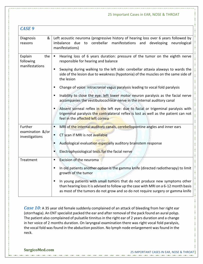
Left acoustic neuroma (progressive history of hearing loss over 6 years followed by imbalance due to cerebellar manifestations and developing neurological manifestations)
Explain the following manifestations
Hearing loss of 6 years duration: pressure of the tumor on the eighth nerve responsible for hearing and balance
Swaying during walking to the left side: cerebellar attaxia alaways to wards the side of the lesion due to weakness (hypotonia) of the muscles on the same side of the lesion
Change of voice: intracranial vagus paralysis leading to vocal fold paralysis
Inability to close the eye: left lower motor neuron paralysis as the facial nerve accompanies the vestibulocochlear nerve in the internal auditory canal
Absent sorneal reflex in the left eye: due to facial or trigeminal paralysis with trigeminal paralysis the contralateral reflex is lost as well as the patient can not feel in the affected left cornea
Further examination &/or investigations
MRI of the internal auditory canals, cerebellopontine angles and inner ears
CT scan if MRI is not available
Audiological evaluation especially auditory brainstem response
Electrophysiological tests for the facial nerve
Treatment Excision of the neuroma
In old patients another option is the gamma knife (directed radiotherapy) to limit growth of the tumor
In young patients with small tumors that do not produce new symptoms other than hearing loss it is advised to follow up the case with MRI on a 6-12 month basis as most of the tumors do not grow and so do not require surgery or gamma knife
Case 10: A 35 year old female suddenly complained of an attack of bleeding from her right ear (otorrhagia). An ENT specialist packed the ear and after removal of the pack found an aural polyp. The patient also complained of pulsatile tinnitus in the right ear of 2 years duration and a change in her voice of 2 months duration. On laryngeal examination there was right vocal fold paralysis, the vocal fold was found in the abduction position. No lymph node enlargement was found in the neck.
25 Important Cases in EAR, NOSE & THROAT
SurgicoMed.com 25 IMPORTANT CASES IN EAR, NOSE & THROAT |
CASE 10
Diagnosis & reasons
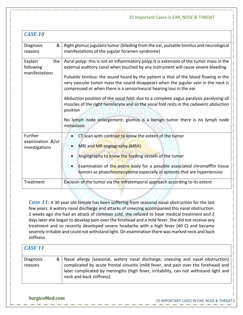
Right glomus jugulaire tumor (blleding from the ear, pulsatile tinnitus and neurological manifestations of the jugular foramen syndrome)
Explain the following manifestations
Aural polyp: this is not an inflammatory polyp it is extension of the tumor mass in the external auditory canal when touched by any instrument will cause severe bleeding
Pulsatile tinnitus: the sound heard by the patient is that of the blood flowing in the very vascular tumor mass the sound disappears when the jugular vein in the neck is compressed or when there is a sensorineural hearing loss in the ear
Abduction position of the vocal fold: due to a complete vagus paralysis paralysing all muscles of the right hemilarynx and so the vocal fold rests in the cadaveric abduction position
No lymph node enlargement: glomus is a benign tumor there is no lymph node metastasis
Further examination &/or investigations
CT scan with contrast to know the extent of the tumor
MRI and MR angiography (MRA)
Angiography to know the feeding vessels of the tumor
Examination of the entire body for a possible associated chromafffin tissue tumors as phaechromocytoma especially in aptients that are hypertensive
Treatment Excision of the tumor via the infratemporal approach according to its extent
Case 11: A 30 year old female has been suffering from seasonal nasal obstruction for the last few years. A watery nasal discharge and attacks of sneezing accompanied this nasal obstruction. 2 weeks ago she had an attack of common cold, she refused to have medical treatment and 2 days later she began to develop pain over the forehead and a mild fever. She did not receive any treatment and so recently developed severe headache with a high fever (40 C) and became severely irritable and could not withstand light. On examination there was marked neck and back stiffness.
CASE 11
Diagnosis & reasons
Nasal allergy (seasonal, watery nasal discharge, sneezing and nasal obstruction) complicated by acute frontal sinusitis (mild fever, and pain over the forehead) and later complicated by meningitis (high fever, irritability, can not withstand light and neck and back stiffness)
25 Important Cases in EAR, NOSE & THROAT
SurgicoMed.com 25 IMPORTANT CASES IN EAR, NOSE & THROAT |
Explain the following manifestations
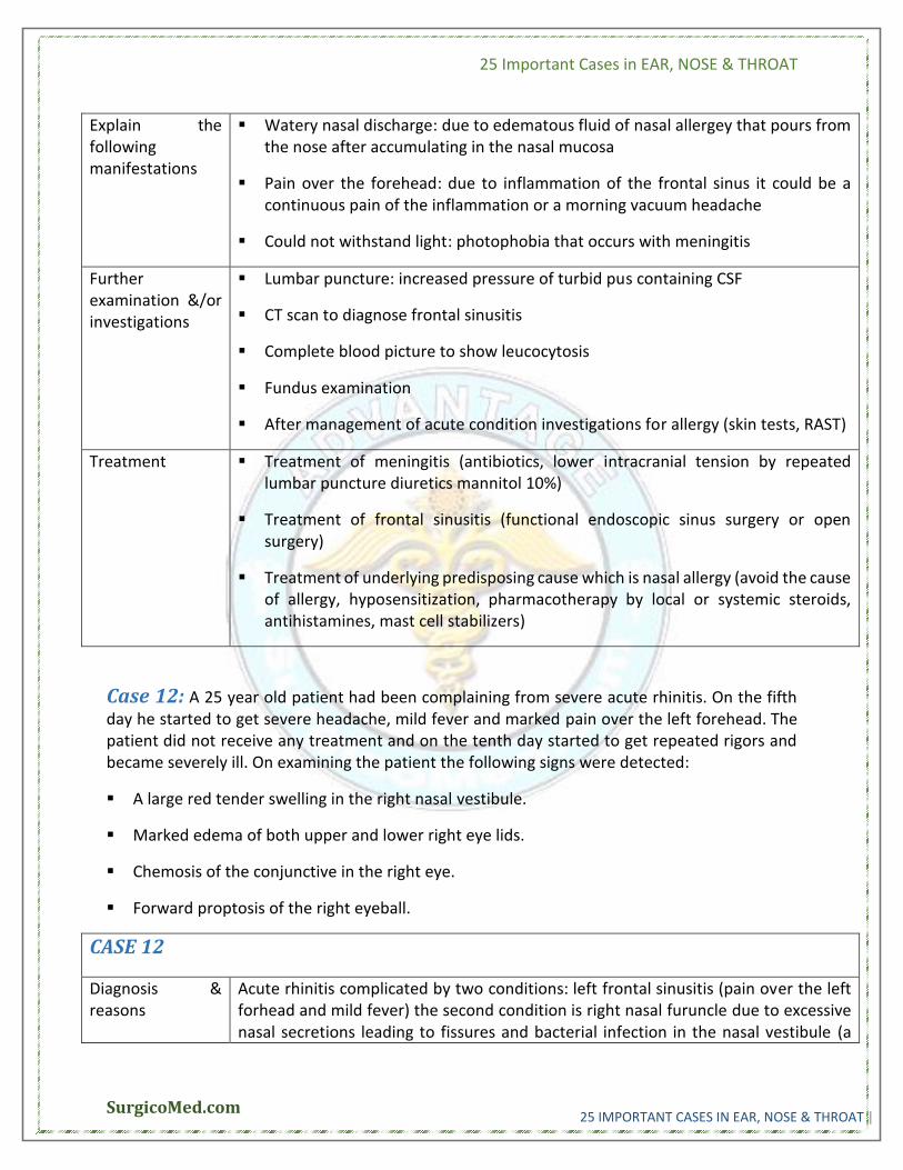
Watery nasal discharge: due to edematous fluid of nasal allergey that pours from the nose after accumulating in the nasal mucosa
Pain over the forehead: due to inflammation of the frontal sinus it could be a continuous pain of the inflammation or a morning vacuum headache
Could not withstand light: photophobia that occurs with meningitis
Further examination &/or investigations
Lumbar puncture: increased pressure of turbid pus containing CSF
CT scan to diagnose frontal sinusitis
Complete blood picture to show leucocytosis
Fundus examination
After management of acute condition investigations for allergy (skin tests, RAST)
Treatment Treatment of meningitis (antibiotics, lower intracranial tension by repeated lumbar puncture diuretics mannitol 10%)
Treatment of frontal sinusitis (functional endoscopic sinus surgery or open surgery)
Treatment of underlying predisposing cause which is nasal allergy (avoid the cause of allergy, hyposensitization, pharmacotherapy by local or systemic steroids, antihistamines, mast cell stabilizers)
Case 12: A 25 year old patient had been complaining from severe acute rhinitis. On the fifth day he started to get severe headache, mild fever and marked pain over the left forehead. The patient did not receive any treatment and on the tenth day started to get repeated rigors and became severely ill. On examining the patient the following signs were detected:
A large red tender swelling in the right nasal vestibule.
Marked edema of both upper and lower right eye lids.
Chemosis of the conjunctive in the right eye.
Forward proptosis of the right eyeball.
CASE 12
Diagnosis & reasons
Acute rhinitis complicated by two conditions: left frontal sinusitis (pain over the left forhead and mild fever) the second condition is right nasal furuncle due to excessive nasal secretions leading to fissures and bacterial infection in the nasal vestibule (a
25 Important Cases in EAR, NOSE & THROAT
SurgicoMed.com 25 IMPORTANT CASES IN EAR, NOSE & THROAT |
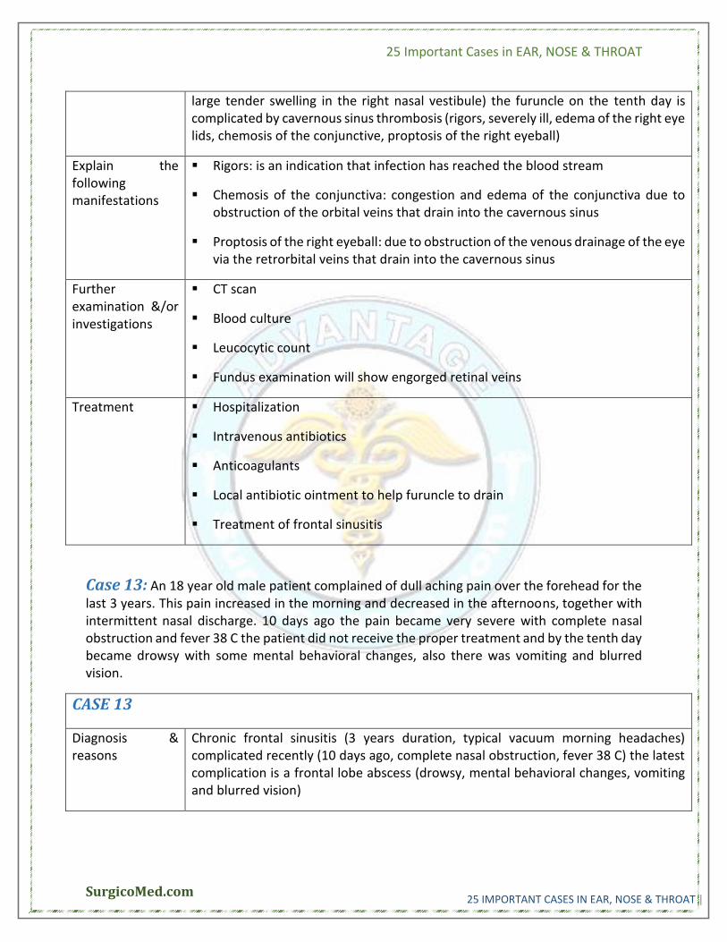
large tender swelling in the right nasal vestibule) the furuncle on the tenth day is complicated by cavernous sinus thrombosis (rigors, severely ill, edema of the right eye lids, chemosis of the conjunctive, proptosis of the right eyeball)
Explain the following manifestations
Rigors: is an indication that infection has reached the blood stream
Chemosis of the conjunctiva: congestion and edema of the conjunctiva due to obstruction of the orbital veins that drain into the cavernous sinus
Proptosis of the right eyeball: due to obstruction of the venous drainage of the eye via the retrorbital veins that drain into the cavernous sinus
Further examination &/or investigations
CT scan
Blood culture
Leucocytic count
Fundus examination will show engorged retinal veins
Treatment Hospitalization
Intravenous antibiotics
Anticoagulants
Local antibiotic ointment to help furuncle to drain
Treatment of frontal sinusitis
Case 13: An 18 year old male patient complained of dull aching pain over the forehead for the last 3 years. This pain increased in the morning and decreased in the afternoons, together with intermittent nasal discharge. 10 days ago the pain became very severe with complete nasal obstruction and fever 38 C the patient did not receive the proper treatment and by the tenth day became drowsy with some mental behavioral changes, also there was vomiting and blurred vision.
CASE 13
Diagnosis & reasons
Chronic frontal sinusitis (3 years duration, typical vacuum morning headaches) complicated recently (10 days ago, complete nasal obstruction, fever 38 C) the latest complication is a frontal lobe abscess (drowsy, mental behavioral changes, vomiting and blurred vision)
25 Important Cases in EAR, NOSE & THROAT
SurgicoMed.com 25 IMPORTANT CASES IN EAR, NOSE & THROAT |
Explain the following manifestations
Morning headache: due to obstruction of the opening of the frontal sinus when the patient sleeps the opening is tightly closed due to edema and the air in the sinus is absorbed creating a negative pressure that causes headache in the morning when the patient stands up the edema is somewhat relieved and air enters the sinus and so the headache disappears or decreases in the afternoon
Mental behavioral changes: the abscess causes pressure on the centers in the frontal lobe of the brain that is responsible for behavior
Blurred vision: increased intracranial tension by the abscess causing vomiting and papilledema
Further examination &/or investigations
Tenderness over the frontal sinus
CT scan with contrast to locate the abscess and diagnose the frontal sinusitis
Leucocytic count important after administering treatment for prognosis
Treatment Neurosurgical excision or drainage of the abscess
Treatment of frontal sinusitis both medically by antibiotics and surgically to drain the frontal sinus
Case 14: A 52 year old male started to develop right sided progressively increasing nasal obstruction 6 months ago. This was followed by blood tinged nasal discharge from the right side as well. Due to looseness of the right second upper premolar tooth, the patient consulted a dentist who advised extraction, this resulted in an oroantral fistula. On examination there was a firm tender swelling in the right upper neck.
CASE 14
Diagnosis & reasons
Cancer of the right maxillary sinus (right blood tinged nasal discharge, looseness of right upper second premolar tooth, swelling in the right upper neck)
Explain the following manifestations
Blood tinged nasal discharge: common early manifestation of cancer of the paranasal sinuses due to the presence of necrotic infected nasal mass
Looseness of the right upper second premolar tooth: due to destruction of the root of the tooth by the malignant tumor as this tooth and the first molar are very close to the floor of the maxillary sinus
Oroantral fistula: due to destruction of the alveolus and the palate by the malignant tumor leading to escape of saliva food and drink from the mouth to the maxillary antrum and then back out of the nose
25 Important Cases in EAR, NOSE & THROAT
SurgicoMed.com 25 IMPORTANT CASES IN EAR, NOSE & THROAT |
Firm tender swelling in the right upper neck: lymph node metastasis from the primary maxillary tumor it could be tender or not tender
Further examination &/or investigations
Other symptoms include: orbital manifestations as diplopia, blindess and pain; headache and trigeminal neuralgic pain; swelling of the cheek; Horner's syndrome due to spread of malignancy from the retropharyngeal lymph node of Rouviere to the upper cervical sympathetic ganglion
CT scan: to diagnose, study the extent of the malignant lesion and its relation to the big blood vessels of the neck and look for other lymph node metastasis
Nasal endoscopy and biopsy to prove malignancy prior to treatment and to know the pathological type of the malignant tumor before deciding on the modality of treatment
General investigations to assess condition of the patient
Treatment Surgical excision by maxillectomy (partial, total or radical according to tumor extent)
Radiotherapy for extensive inoperable lesions
Radical neck dissection for lymph node metastases
Chemotherapy for inoperable tumors that do not respond to radiotherapy
Palliative treatment for inoperable terminal cases
Case 15: A 40 year old female has been complaining of nasal troubles of a long duration in the form of bilateral nasal obstruction, anosmia and nasal crustation. 2 months ago she developed mild stridor that necessitated a tracheostomy later on. She received medical treatment for her condition, but 1 month later developed severe to profound hearing loss that necessitated the use of a hearing aid.
CASE 15
Diagnosis & reasons
Rhinolaryngoscleroma (nasal crustations of long duration, stridor)
Explain the following manifestations
Nasal obstruction: due to the presence of a scleroma mass or crustation or nasal synechia
Stridor: laryngoscleroma causes subglottic stenosis and fibrosis causing biphasic stridor
25 Important Cases in EAR, NOSE & THROAT
SurgicoMed.com 25 IMPORTANT CASES IN EAR, NOSE & THROAT |
Profound hearing loss that necessitated a hearing aid: an old antibiotic used for the treatment of scleroma was streptomycin that was ototoxic causing sensorineural hearing loss now rifampscin is used with no such side effect
Further examination &/or investigations
Examination of the nose shows crusts, nasal mass, offensive discharge
Examination of the larynx will show an area of subglottic stenosis may be in the form of a web
Biopsy: will show a chronic inflammatory process with endarteritis obliterans and two diagnostic structure the Mickulicz cell and the Russel body; the active cell the fibroblast is also seen
Treatment Medical: Rifampscin 300mgm daily twice daily before meals
Surgical: recanalization of the nose to relieve nasal obstruction
Laser excision of the subglottic web to relieve dyspnea and stridor
Follow up the condition until complete cure
Case 16: A 24 year old male patient presented because of severe pain in the throat and the left ear that increased with swallowing of sudden onset and 2 days duration. He gave a history of sore throat and fever a few days prior to the condition. On examination, the patient looked very ill and has a thickened voice. The temperature was 39.5 C and the pulse 110/minute. The patient had fetor of the breath and was unable to open his mouth. There was marked edema of the palate concealing the left tonsil that was found injected. There was a painful hot swelling located below the left angle of the mandible. The left tympanic membrane was normal.
CASE 16
Diagnosis & reasons
Acute tonsillitis (sore throat and fever) complicated by peritonsillar abscess {quinzy} (severe throat pain referred to the left ear, very ill, thickened voice, fever, fetor, unable to open his mouth, edema of the palate, painful hot swelling at the angle of the mandible)
Explain the following manifestations
Pain in the left ear: refeered earache along Jackobsen's tympanic branch (that supplies the middle ear) of the glossopharyngeal nerve (that supplies the palatine tonsil)
Thickened voice: due to palatal edema
Fetor of the breath: severe dysphagia leading to inability to swallow saliva together with the presence of an abscess in the oropharynx
25 Important Cases in EAR, NOSE & THROAT
SurgicoMed.com 25 IMPORTANT CASES IN EAR, NOSE & THROAT |
Unable to open his mouth: trismus due to irritation of the medial pterygoid muscle by the pus under tension in the peritonsillar abscess
Left tonsil injected: markedly congested due to severe inflammatory process
Hot swelling below the left angle of the mandible: jugulodigastric lymph adenitis
Normal tympanic membrane: there is no acute otitis media pain in the ea is referred from the throat
Further examination &/or investigations
Complete blood picture lecocytosis
CT scan
Treatment Medical treatment: antibiotics, analgesics, antipyretics and antiinflammatory drugs
Surgical drainage of the quinzy (pus pointing, palatal edema, throbbing pain, pitting edema)
Tonsillectomy after 2-3 weeks
Case 17: A 5 year old boy was referred to an ENT specialist because of mouth breathing and impairment of hearing of 2 years duration. His mother reported that her child has almost constant mucoid nasal discharge that sometimes changes to a mucopurulent one and he snores during his sleep. On examination, the child has nasal speech and obvious mouth breathing. Examination of the ears showed retracted tympanic membranes. Tympanograms were flat type B.
CASE 17
Diagnosis & reasons
Adenoid enlargement (mouth breathing, nasal discharge, snoring, nasal speech) complicated by bilateral otitis media with effusion (impairement of hearing, retracted tympanic membranes type B tympanograms)
Explain the following manifestations
Mucoid nasal discharge that can change to be mucopurulent: adenoid enlargement may be complicated by ethmoiditis causing the mucopurulent nasal discharge
Snoring: due to bilateral nasal obstruction during his sleep can progress to respiratory obstruction during his sleep (sleep apnea)
25 Important Cases in EAR, NOSE & THROAT
SurgicoMed.com 25 IMPORTANT CASES IN EAR, NOSE & THROAT |
Nasal speech: rhinolalia clausa due to nasal obstruction were the letter m is pronounced as b
Type B tympanograms: due to presence of fluid behind the intact retracted tympanic membrane leading to no vibrations of the drum
Further examination &/or investigations
Other symptoms and signs: adenoid face, stunted growth, poor scholastic achievement, nocturnal enuresis.
X-ray lateral view skull: soft tissue shadow in the nasopharynx causing narrowing of the nasopharyngeal airway
Audiogram: air bone gap indicating conductive hearing loss
Treatment Adenoidectomy
Bilateral ventillation tube (grommet) insertion in the tympanic membranes
Case 18: A male patient 49 year old presented with the complaint of enlargement of the upper deep cervical lymph nodes on both sides of the neck of 6 months duration. The nodes appeared first on the right side later on the other side. The patient gave a history of decreased hearing in the right ear that was intermittent but later became permanent. Recently he developed diminution of hearing in his left ear, nasal regurge, nasal intonation of voice and recurrent mild nosebleeds.
CASE 18
Diagnosis & reasons
Nasopharyngeal carcinoma with lymph node metastasis (early appearance of lymph node metastasis as the nasopharynx is one of the silent areas of the head and neck – occult primary sites; decreased hearing due to eustachian tube affection)
Explain the following manifestations
Bilateral enlargement of upper deep cervical lymph nodes: the nasopharynx may send metastasis to both sides because it is present in the center of the head and neck
Decreased hearing in the right ear: due to eustachian tube destruction by the malignant tumor causing right otitis media with effusion and a retracted tympanic membrane leading to a conductive hearing loss
Nasal regurge: due to palatal paralysis
Nasal intonation of voice: due to nasal obstruction and palatal paralysis it is a combined rhinolalia clausa and aperta
25 Important Cases in EAR, NOSE & THROAT
SurgicoMed.com 25 IMPORTANT CASES IN EAR, NOSE & THROAT |
Further examination &/or investigations
CT scan
Nasopharyngoscopy and biopsy
Audiogram and tympanogram
General investigations
Treatment Radiotherapy for the primary nasopharyngeal carcinoma
Radical neck dissection for residual lymph node metstasis after treatment with radiotherapy
Chemotherapy in certain selected cases according to histopathological finding of biopsy
Palliative treatment for terminal cases
Case 19: A 40 year old female began to experience difficulty in swallowing for the last 3 years. This difficulty in swallowing was to all kinds of food and the condition showed variation in the degree of dysphagia and was associated with a sense of obstruction at the root of the neck. For the last 2 months, she developed rapidly progressive difficulty in swallowing even to fluids together with a change in her voice. Recently she noticed a firm non-tender swelling in the right upper neck.
CASE 19
Diagnosis & reasons
Plummer – Vinson disease (dysphagia of intermittent nature for 3 years to all kinds of food) leading to hypopharyngeal or esophageal malignancy ( progression of dysphagia in the last 2 months, change of voice, appearance of neck swelling indicating lymph node metastasis)
Explain the following manifestations
Sense of obstruction at the root of the neck: the level of obstruction in Plummer Vinson disease is due to the presence of pharyngeal and esophageal webs of fibrous tissue in the lower pharynx and upper esophagus
Change of voice: due to malignant involvement of the recurrent laryngeal nerve leading to vocal fold paralysis
Firm non tender swelling in the right upper neck: lymph node metastasis in the right upper deep cervical lymph node
Further examination
Indirect laryngoscopy: tumor is seen in the hypopharynx with overlying froth
Direct laryngoscopy and biopsy
25 Important Cases in EAR, NOSE & THROAT
SurgicoMed.com 25 IMPORTANT CASES IN EAR, NOSE & THROAT |
&/or investigations
X-ray lateral view neck showing a wide prevertebral space displacing the airway anteriorly
CT scan to show extent of the tumor especially lower extent
Barium swallow
General investigations to assess the general condition of the patient
Treatment Surgical excision by total laryngopharyngectomy and radical neck dissection of metastatic lymph nodes
Radiotherapy
Chemotherapy
Palliative treatmet
Type of treatment depends on general condition of patient, age of patient, extent of tumor and its histopathological type
Case 20: 4 hours following an adenotonsillectomy for a 6 year old the pulse was 110/min, blood pressure 100/70, respiration 20/min and the child vomited 250 cc of a dark fluid. 2 hours later he vomited another 150 cc of the same dark fluid, the pulse became 130/min, the blood pressure became 80/50. The respiration rate remained 20/min.
CASE 20
Diagnosis & reasons
Post-tonsillectomy reactionary hemorrhage (rising pulse, lowering of blood pressure, vomiting of altered blood, 4 hours following an adenotonsillectomy)
Explain the following manifestations
Pulse is 110/min then rises to 130/min: a continuous rising pulse is due to tacchycardia as a compensation for the blood loss
Vomiting of dark fluid: altered blood (acid hematin when blood is changed by stomach HCL)
Further examination &/or investigations
Examination of the throat site of bleeding may be from the tonsil bed or from the adenoid bed
Rapid assessment of hemoglobin
Treatment Antishock measures (fluid and blood transfusion, steroids, coagulants)
Surgical hemostasis under general anesthesia
25 Important Cases in EAR, NOSE & THROAT
SurgicoMed.com 25 IMPORTANT CASES IN EAR, NOSE & THROAT |
Case 21: A 3 year old child was referred to an ENT specialist because of cough, difficulty of respiration and temperature 39.5 C of few hours duration. The child was admitted to hospital for observation and medical treatment. 6 hours later, the physician decided an immediate tracheostomy. After the surgery the child was relieved from the respiratory distress for 24 hours then he became dyspnic again. The physician carried out a minor procedure that was necessary to relieve the child from the dyspnea. Few days later the tracheostomy tube was removed and the child discharged from the hospital.
CASE 21
Diagnosis & reasons
Acute laryngotracheobronchitis – CROUP (dyspnea relieved by tracheostomy placed for a few days only, cough and fever) complicated by an obstruction of the tracheostomy tube by secretions (relieved after cleaning the tube)
Explain the following manifestations
Cough: common with croup due to the presence of tracheal and broncjial imflammation and secretions
Temperature 39.5 C: temperature in croup is varaiable may be mild or severe according to the virus causing the condition
Observation and medical treatment: the main observation is that of the degree of respiratory distress and tacchcyardia to detect early heart failure. Medical treat is mainly steroids and humidification of respired air, mucolytics and expectorants to facilitate getting rid of the secretions in the bronchi and trachea.
Minor procedure: clearnace of the tracheostomy tube from accumulated secretions.
Further examination &/or investigations
Pulse rate
Cyanosis
Chest x-ray to differentiate from foreign body inhalation
Treatment Steroids
Mucolytics
Expectorants
Antibiotics
Humidified oxygen inhalation
Treatment of heart failure
25 Important Cases in EAR, NOSE & THROAT
SurgicoMed.com 25 IMPORTANT CASES IN EAR, NOSE & THROAT |
Case 22: A 45 year old male who is a heavy smoker complained of change in his voice of 3 years duration in the form of hoarseness. During the last 3 months his voice became very hoarse and he developed mild respiratory distress. Later he became severely distressed and required a surgical procedure to relieve the distress. On examination there were bilateral firm non-tender upper neck swellings.
CASE 22
Diagnosis & reasons
Leukoplakia of the vocal folds (hoarseness of 3 years duration) leading to vocal fold carcinoma (glottic carcinoma increased hoarseness, respiratory distress relieved by tracheostomy) with bilateral lymph node metastasis (firm non-tender upper neck swellings)
Explain the following manifestations
Hoarseness: the presence of lesions whether leukoplakia or carcinoma on the vocal fold will limit its vibration capability causing hoarseness
Bilateral firm non-tender swellings in the upper neck: lymph node metastasis not common with vocal fold carcinoma but may occur when the tumor spreads to the neighboring supraglottis or subglottis
Surgical procedure: tracheostomy to bypass the glottic lesion causing respiratory obstruction
Further examination &/or investigations
Other symptoms: cough and hemoptsys
Indirect laryngoscopy: visualize the lesion and vocal fold paralysis
Laryngeal stroboscopy: to examine the vocal fold movement very useful with small vocal fold carcinoma lesions
Direct laryngoscopy and biopsy
CT scan and MRI
Chest X-ray
Treatment Laser excision of the lesion
Laryngofissure and cordectomy
Laryngectomy ( partial or total)
Radiotherapy for small cordal lesions
Chemotherapy and palliative treatment for terminal cases
25 Important Cases in EAR, NOSE & THROAT
SurgicoMed.com 25 IMPORTANT CASES IN EAR, NOSE & THROAT |
Case 23: A 40 year old female had repeated attacks of chest infection not improving by medical treatment. The patient was admitted for investigation of her condition in a hospital. A chest x-ray revealed basal lung infection. During her hospital stay it was noticed that she suffered from chest tightness and choking following meals. The ward nurse noticed that the patient refuses fluid diet and prefers solid bulky food.
CASE 23
Diagnosis & reasons
Cardiac achalasia (basal chest infection due to aspiration, choking following meals and dysphagia more to fluids)
Explain the following manifestations
Chest infection not improving by medical treatment: because of continuous aspiration the original condition of cardiac achalasia must be treated first and the chest infection will improve subsequently
Basal lung infection by X-ray: with aspiration by gravity the basal lung is always affected
Patient refuses fluid diet and prefers solid food: solid food creates a better stimulation by rubbing against the esophageal wall and so the cardiac sphincter opens while fluids need to accumulate in the esophagus before causing a sufficient stimulus
Further examination &/or investigations
X-ray barium swallow esophagus shows a large dilatation of the esophagus and a stenosis at the level of the cardiac sphincter
Esophagoscope
CT scan with barium swallow
Chest X-ray
Treatment Heller's operation
Esophagoscopic dilatation
Case 24: A 4 year old child was referred to an ENT specialist by a pediatrician because of repeated attacks of severe chest infection (three in number) during the last month that usually resolved by antibiotics, expectorants and mucolytics, but the last attack did not resolve. On examination the lower right lobe of the lung showed no air entry and a lot of wheezes all over the chest by auscultation. A chest x-ray revealed an opacified lower right lobe. Temperature 38 C, pulse 120/min and respiration rate 35/min.
CASE 24
25 Important Cases in EAR, NOSE & THROAT
SurgicoMed.com 25 IMPORTANT CASES IN EAR, NOSE & THROAT |
Diagnosis & reasons
Foreign body inhalation in the right lung most probably a vegetable seed as a peanut (attacks of chest infection, no air entry and opacified lower right lobe of the lung, fever tachycardia and dyspnea 35/min normal reting respiratory rate in a child should not exceed 18/min
Explain the following manifestations
Last attack of chest infection did not resolve: the chemical bronchopneumonia caused by the vegetable seed has reached a severity that it could not be controlled by the medical treatment always suspect a foreign body inhalation in a non-responsive chest infection in a child
Wheezes all over the chest: although the foreign body is in the right lung the site of decreased air entry and an opacified lobe by X-ray but the chemical effect of the fatty acids in the vegetable seed is all over the lung causing marked dyspnea and tachypnea as well
Pulse 120/min: respiratory failure is also accompanied by tachycardia which might lead to heart failure
Further examination &/or investigations
Proper history
Tracheobronchoscopy
Treatment Tracheobronchoscopy and removal of the foreign body followed by
Antibiotics
Steroids
Expectorants
Case 25: A 3 year old child suddenly complained of a sore throat and enlarged left upper deep cervical lymph node. Later he suffered from marked body weakness and mild respiratory distress that progressively became severe. Oropharyngeal examination revealed a grayish membrane on the left tonsil, soft palate and posterior pharyngeal wall. 2 days later he developed nasal regurge. His temperature was 38 C and pulse 150/min.
CASE 25
Diagnosis &
reasons
Diphtheria (sore throat, enlarged upper deep cervical lymph node, marked
weakness, respiratory distress, extension of the membrane outside the tonsil,
low grade fever with marked tachycardia)
25 Important Cases in EAR, NOSE & THROAT
SurgicoMed.com 25 IMPORTANT CASES IN EAR, NOSE & THROAT |
Explain the
following
manifestations
Enlarged upper deep cervical lymph node: markedly enlarged (Bull's Neck)
common in diphtheria in the early stages of the disease
Respiratory distress: could be because of heart failure caused by marked
toxemia or due to extension of the diphtheritic membrane to the larynx
Grayish membrane: due to tissue necrosis
Extension of the membrane outside the surface of the tonsil: diphtheria is a
disease of the mucous membrane not only of the tonsil
Pulse 150/min: toxemia causing heart failure leading to a rapid pulse
Further
examination &/or
investigations
Swab from the membrane
Bacteriological diagnosis
Treatment Start treatment immediately do not wait for a definite bacteriological diagnosis
Antitoxin serum 20,000 – 100,000 units daily until the membrane disappears
Bacteriological swabs until the organism disappears from the throat
Antibiotics
Treatment of heart failure if present
Tracheostomy for respiratory distress or even marked heart failure to decrease
the effort of breathing by decreasing the respiratory dead space
Passive and active immunization of the contacts of the patient