Embed Size (px)
Citation preview

8 Localized Periosteal Reaction
CLINICAL IMAGAGINGAN ATLAS OF DIFFERENTIAL DAIGNOSIS
EISENBERG
DR. Muhammad Bin Zulfiqar PGR-FCPS III SIMS/SHL
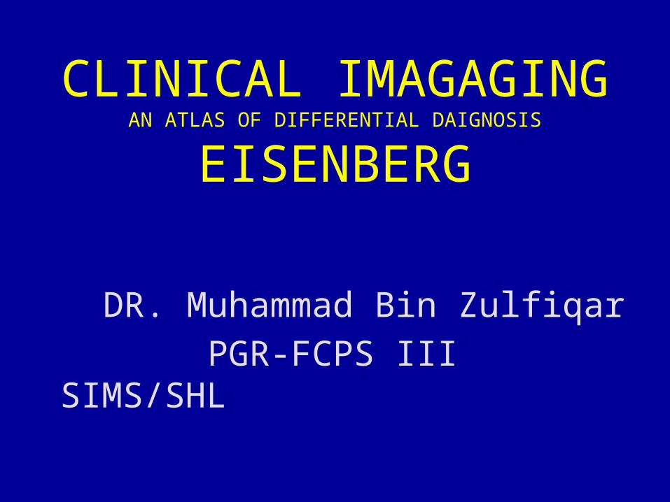
• Fig B 8-1 Osteogenic sarcoma. (A to D) Four examples of osteogenic sarcoma of the femur illustrate the broad spectrum of radiographic changes. There are various amounts of exuberant, irregular periosteal response and ragged bone destruction.
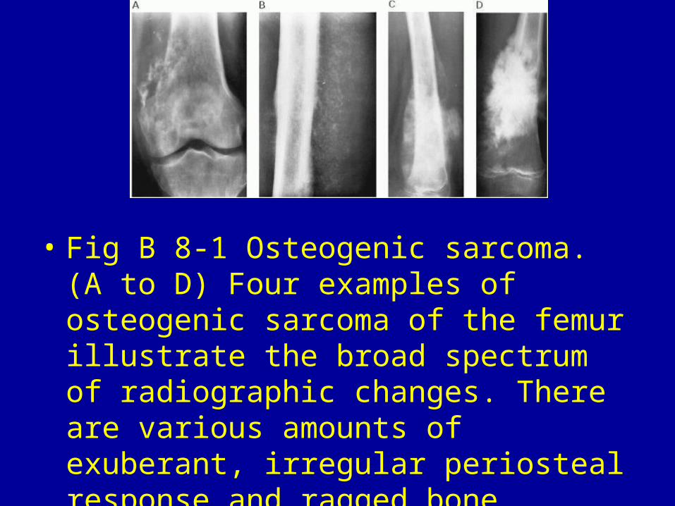
Fig B 8-2 Ewing's sarcoma. Laminated periosteal reaction on one side of the bone and thin periosteal elevation (Codman's triangle) on the other.
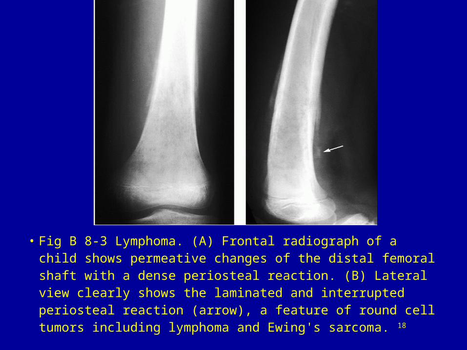
• Fig B 8-3 Lymphoma. (A) Frontal radiograph of a child shows permeative changes of the distal femoral shaft with a dense periosteal reaction. (B) Lateral view clearly shows the laminated and interrupted periosteal reaction (arrow), a feature of round cell tumors including lymphoma and Ewing's sarcoma. 18
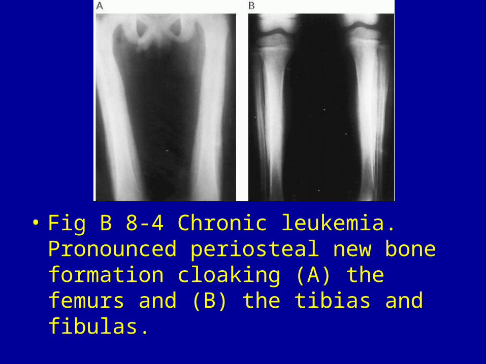
• Fig B 8-4 Chronic leukemia. Pronounced periosteal new bone formation cloaking (A) the femurs and (B) the tibias and fibulas.
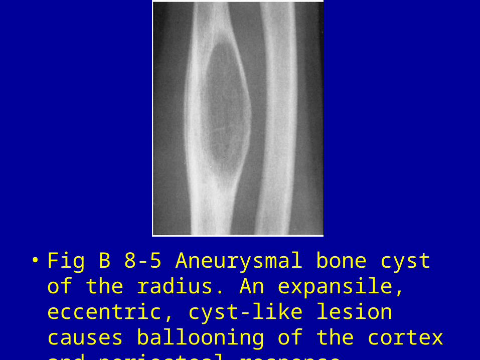
• Fig B 8-5 Aneurysmal bone cyst of the radius. An expansile, eccentric, cyst-like lesion causes ballooning of the cortex and periosteal response.
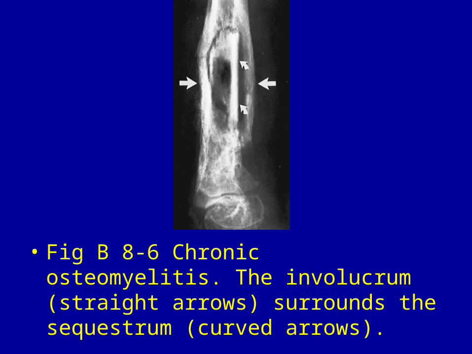
• Fig B 8-6 Chronic osteomyelitis. The involucrum (straight arrows) surrounds the sequestrum (curved arrows).
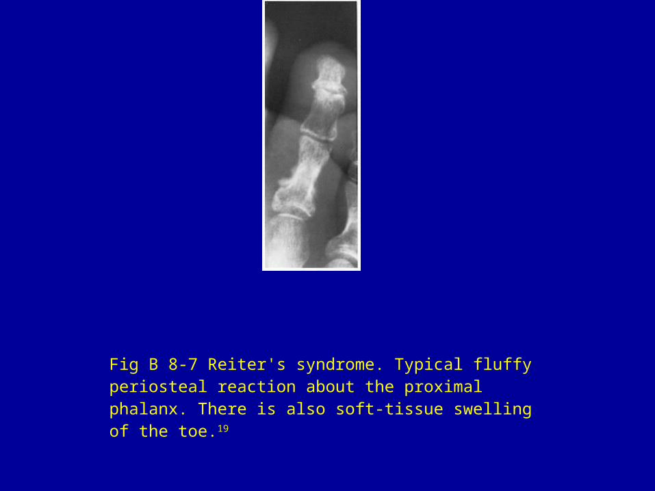
Fig B 8-7 Reiter's syndrome. Typical fluffy periosteal reaction about the proximal phalanx. There is also soft-tissue swelling of the toe.19
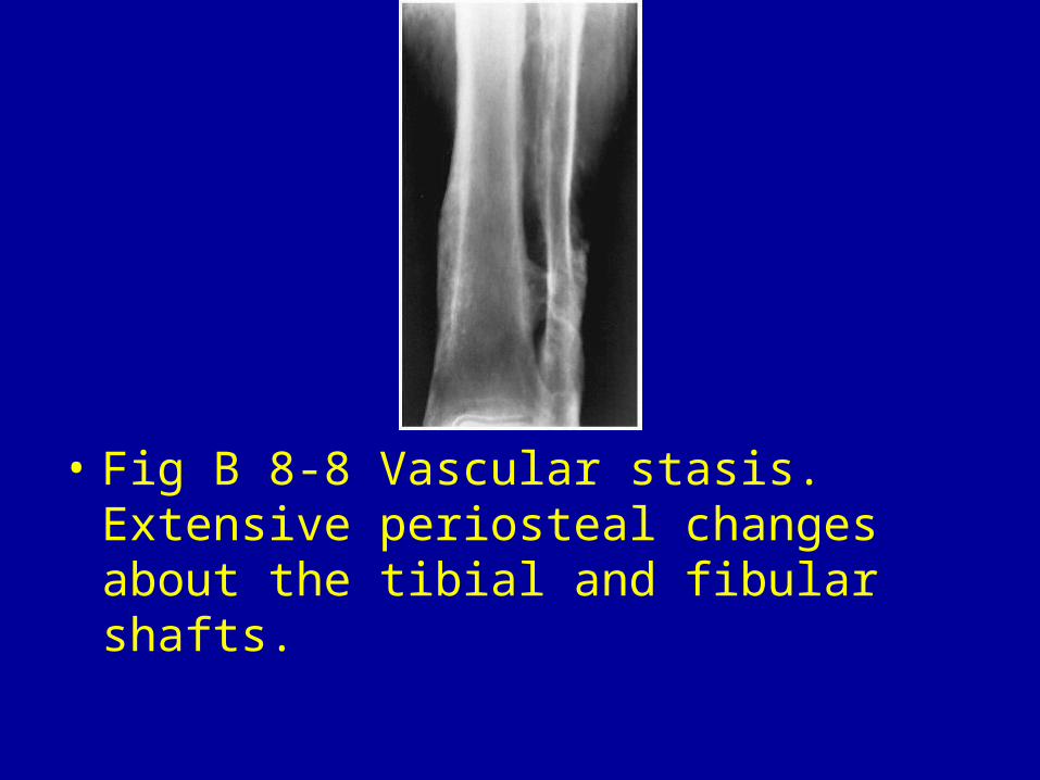
• Fig B 8-8 Vascular stasis. Extensive periosteal changes about the tibial and fibular shafts.
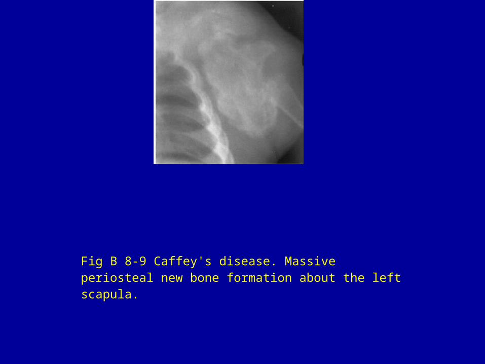
Fig B 8-9 Caffey's disease. Massive periosteal new bone formation about the left scapula.
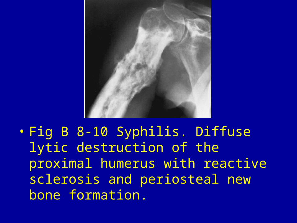
• Fig B 8-10 Syphilis. Diffuse lytic destruction of the proximal humerus with reactive sclerosis and periosteal new bone formation.
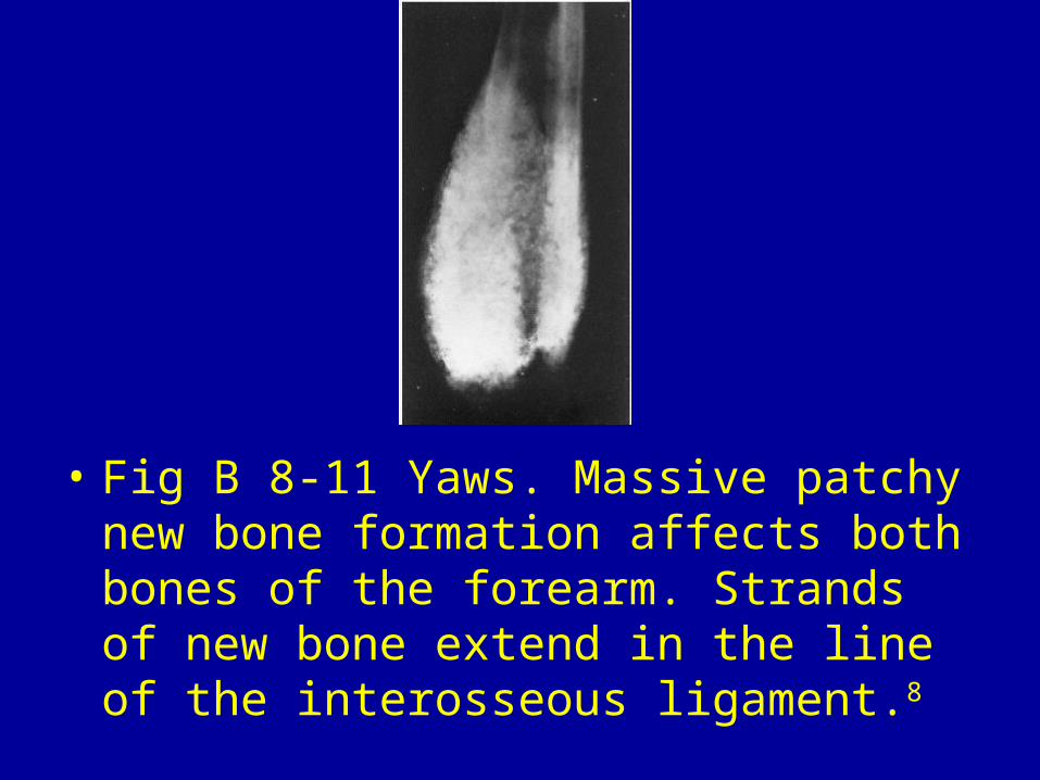
• Fig B 8-11 Yaws. Massive patchy new bone formation affects both bones of the forearm. Strands of new bone extend in the line of the interosseous ligament.8
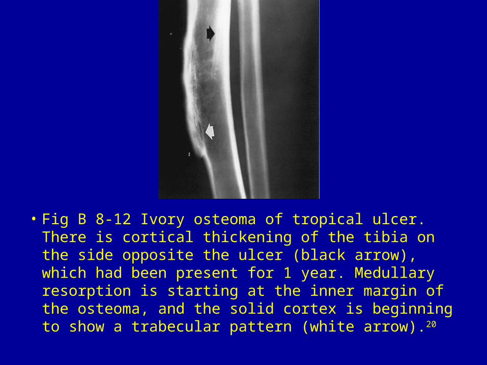
• Fig B 8-12 Ivory osteoma of tropical ulcer. There is cortical thickening of the tibia on the side opposite the ulcer (black arrow), which had been present for 1 year. Medullary resorption is starting at the inner margin of the osteoma, and the solid cortex is beginning to show a trabecular pattern (white arrow).20