Embed Size (px)
Citation preview
The Department of Human anatomy
Peripheral nervous system.The Cranial Nerves Part - 2
The ciliary ganglion The ciliary ganglion is a parasympathetic ganglion
located in the posterior orbit. Preganglionic axons from the Edinger-Westphal nucleus travel along the oculomotor nerve and form synapses in ganglion. The postganglionic axons run in the short ciliary nerves and innervate two eye muscles:
the sphincter pupillae constricts the pupil, a movement known as Miosis. The opposite - dillatator pupillae ,Mydriasis, is the dilation of the pupil.
the ciliaris muscle contracts, releasing tension on the Zonular Fibers, making the lens more convex, also known as accommodation.
Both of these muscles are involuntary – they are controlled by the autonomic nervous system.
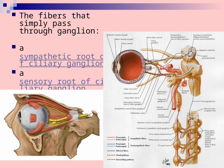
The fibers that simply pass through ganglion:
a sympathetic root of ciliary ganglion
a sensory root of ciliary ganglion
The pterygopalatine ganglion (Synonym: meckel's ganglion) is a parasympathetic ganglion found in the pterygopalatine fossa.
The pterygopalatine ganglion supplies the lacrimal gland, paranasal sinuses, glands of the mucosa of the nasal cavity and pharynx, the gingiva, and the mucous membrane and glands of the hard palate. It communicates anteriorly with the nasopalatine nerve.
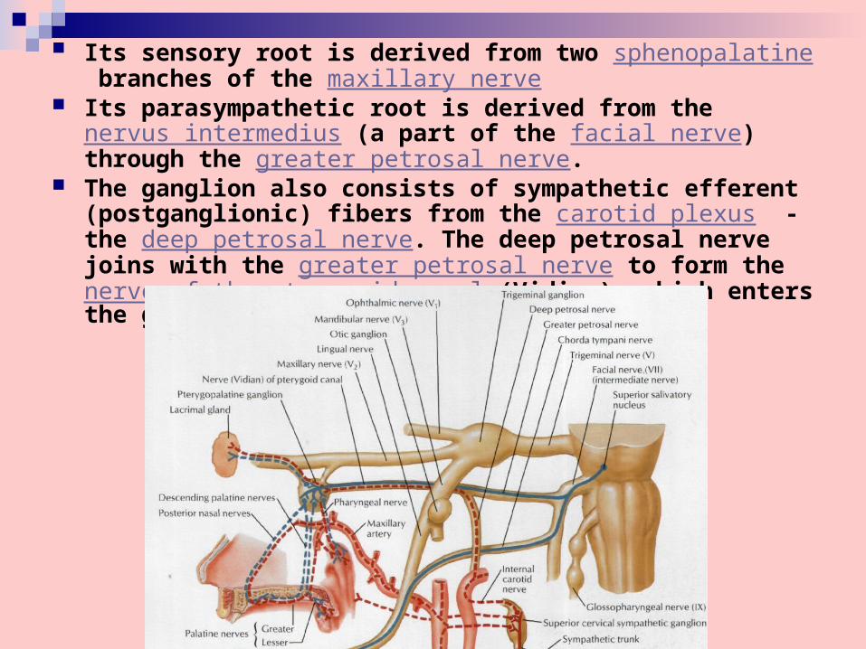
Its sensory root is derived from two sphenopalatine branches of the maxillary nerve
Its parasympathetic root is derived from the nervus intermedius (a part of the facial nerve) through the greater petrosal nerve.
The ganglion also consists of sympathetic efferent (postganglionic) fibers from the carotid plexus - the deep petrosal nerve. The deep petrosal nerve joins with the greater petrosal nerve to form the nerve of the pterygoid canal (Vidian), which enters the ganglion.
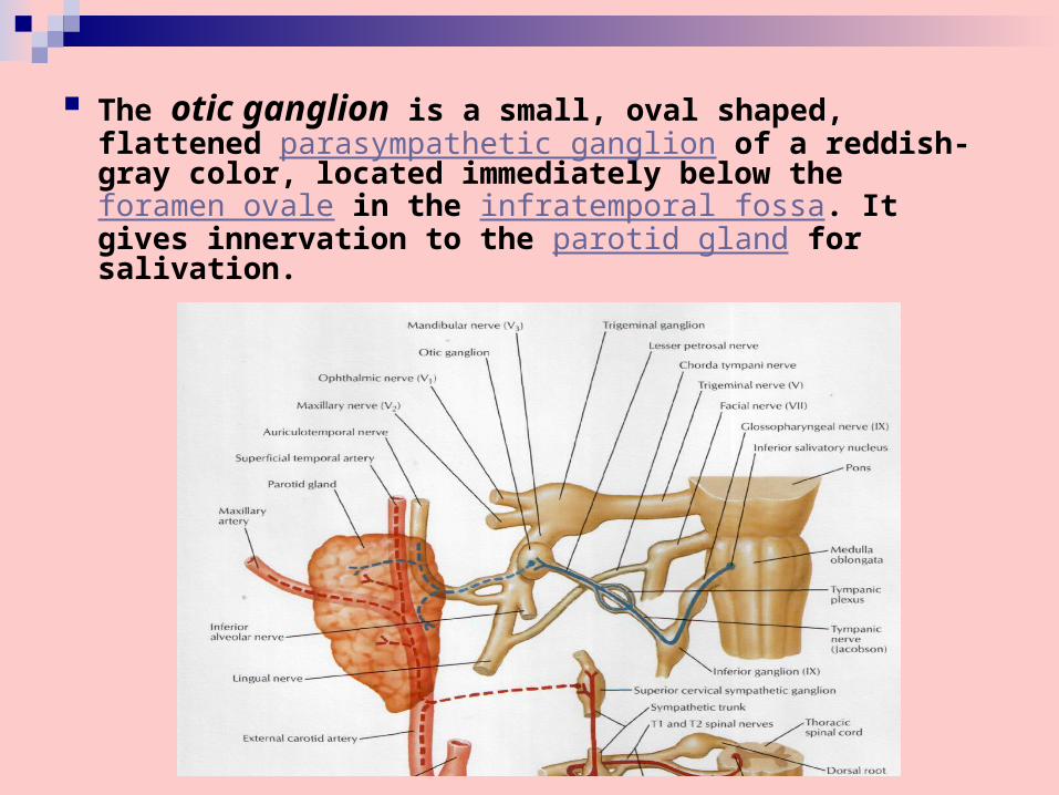
The otic ganglion is a small, oval shaped, flattened parasympathetic ganglion of a reddish-gray color, located immediately below the foramen ovale in the infratemporal fossa. It gives innervation to the parotid gland for salivation.
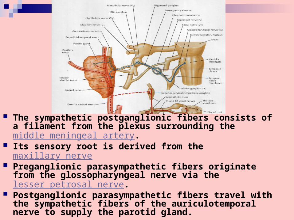
The sympathetic postganglionic fibers consists of a filament from the plexus surrounding the middle meningeal artery.
Its sensory root is derived from the maxillary nerve Preganglionic parasympathetic fibers originate from
the glossopharyngeal nerve via the lesser petrosal nerve.
Postganglionic parasympathetic fibers travel with the sympathetic fibers of the auriculotemporal nerve to supply the parotid gland.
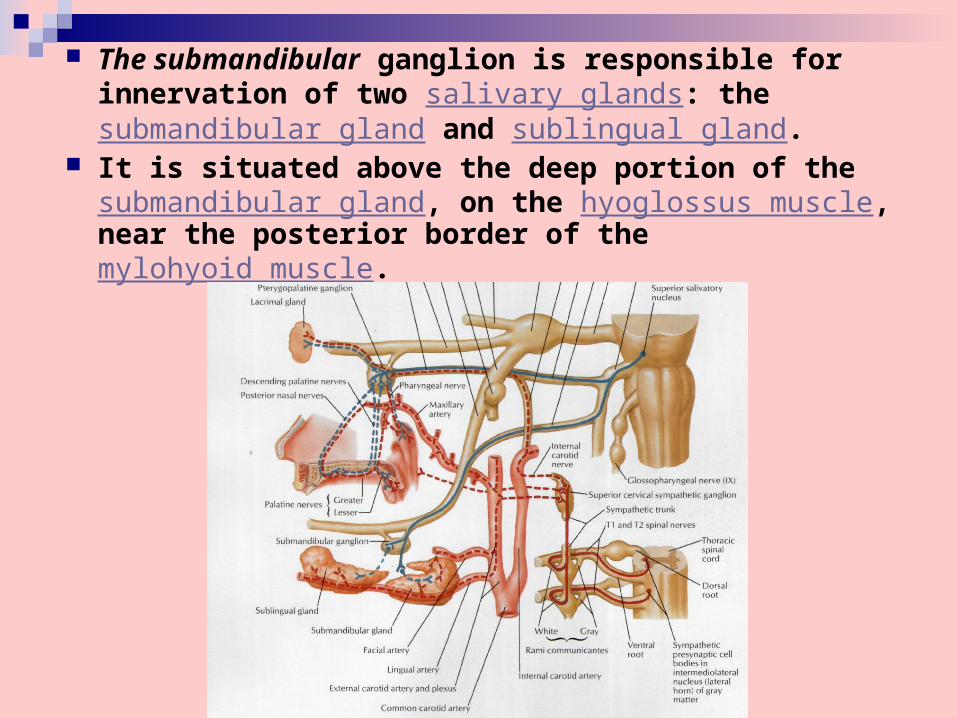
The submandibular ganglion is responsible for innervation of two salivary glands: thesubmandibular gland and sublingual gland.
It is situated above the deep portion of the submandibular gland, on the hyoglossus muscle, near the posterior border of the mylohyoid muscle.
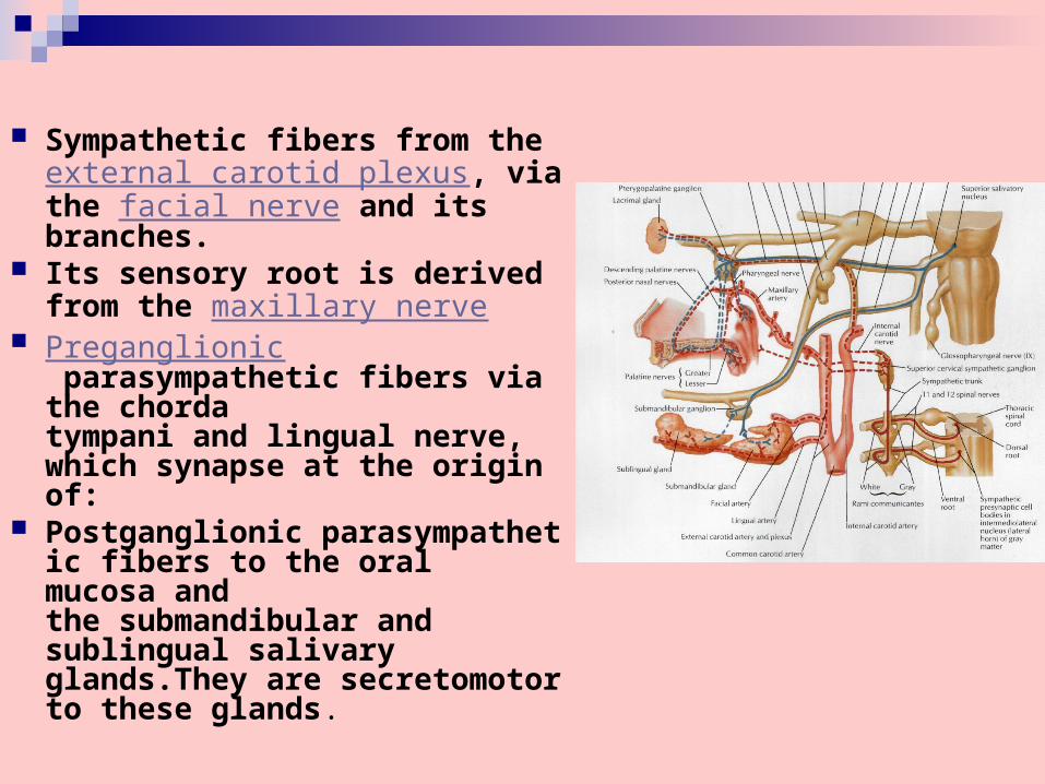
Sympathetic fibers from the external carotid plexus, via the facial nerve and its branches.
Its sensory root is derived from the maxillary nerve
Preganglionic parasympathetic fibers via the chorda tympani and lingual nerve, which synapse at the origin of:
Postganglionic parasympathetic fibers to the oral mucosa and the submandibular and sublingual salivary glands.They are secretomotor to these glands.
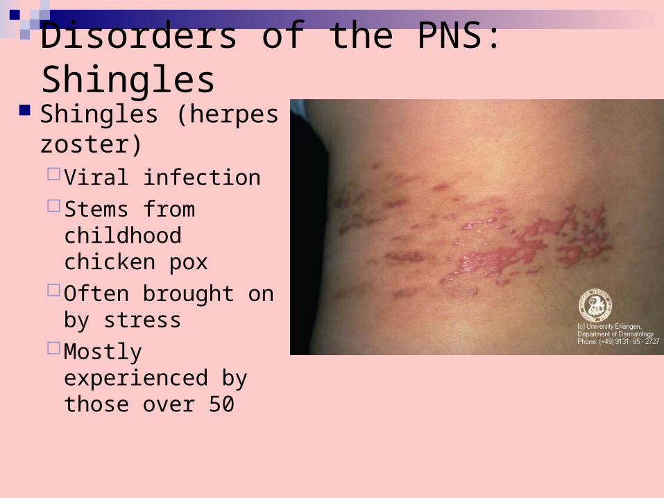
Disorders of the PNS: Shingles Shingles (herpes
zoster) Viral infectionStems from
childhood chicken pox
Often brought on by stress
Mostly experienced by those over 50
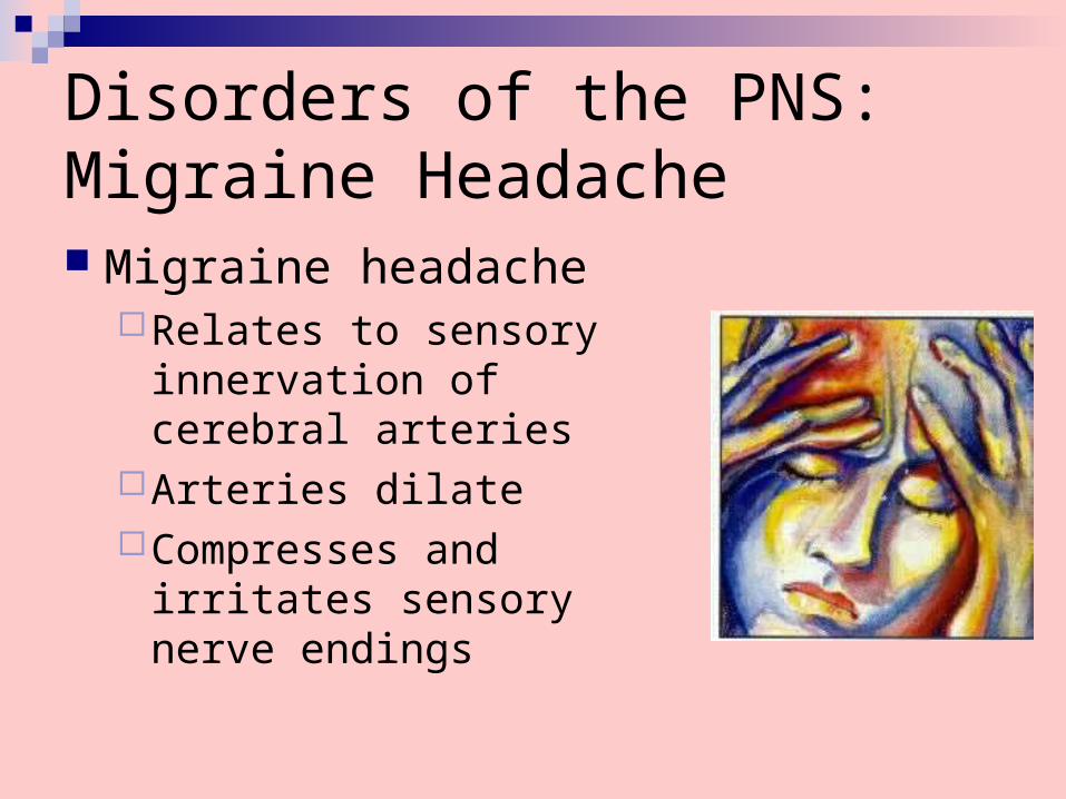
Disorders of the PNS: Migraine Headache Migraine headache
Relates to sensory innervation of cerebral arteries
Arteries dilateCompresses and irritates
sensory nerve endings
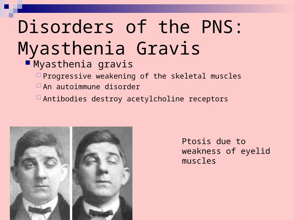
Disorders of the PNS: Myasthenia Gravis
Myasthenia gravis Progressive weakening of the skeletal muscles An autoimmune disorder Antibodies destroy acetylcholine receptors
Ptosis due to weakness of eyelid muscles