Embed Size (px)
Citation preview
ANATOMY OF
EXTERNAL & MIDDLE EAR
Dr. Diptiman Baliarsingh
1st Year PG, Dept. of ENT,
Hi-tech Medical College & Hosp.
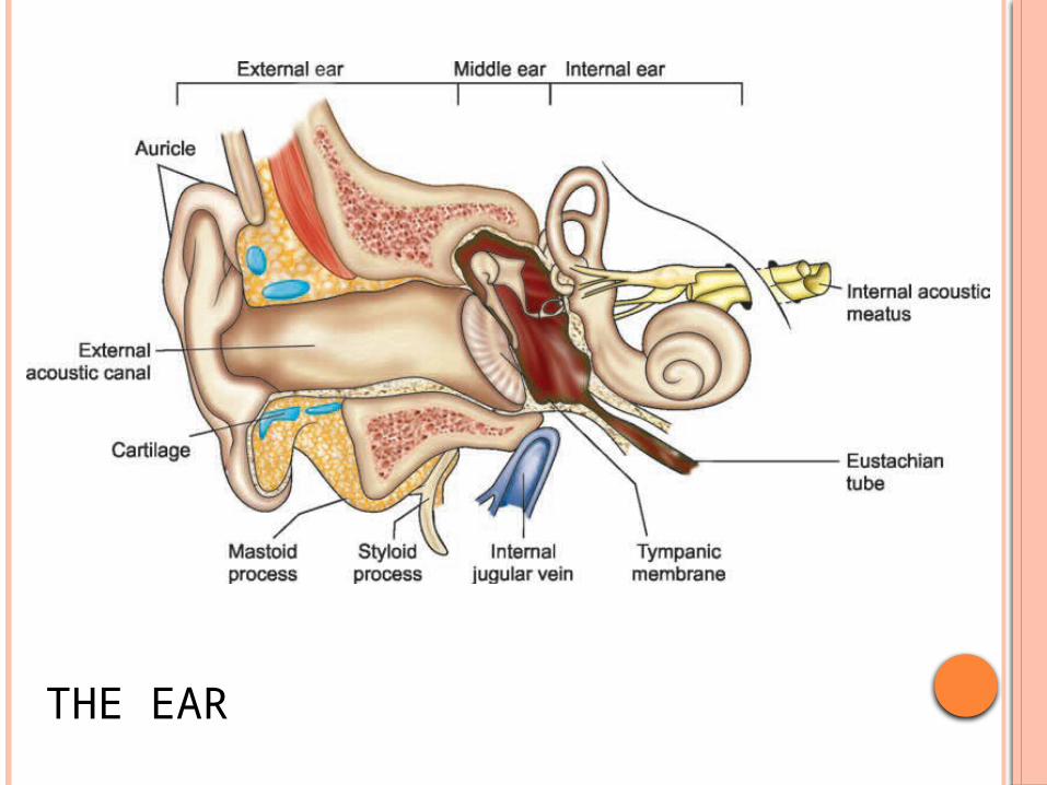
THE EAR
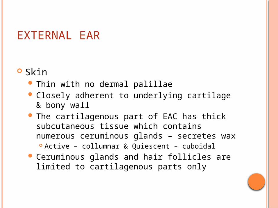
EXTERNAL EAR
Skin Thin with no dermal palillae Closely adherent to underlying cartilage & bony
wall The cartilagenous part of EAC has thick
subcutaneous tissue which contains numerous ceruminous glands – secretes wax Active – collumnar & Quiescent – cuboidal
Ceruminous glands and hair follicles are limited to cartilagenous parts only
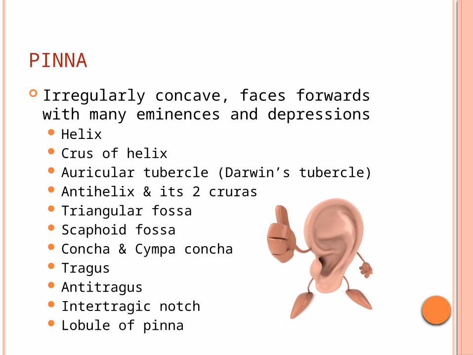
PINNA
Irregularly concave, faces forwards with many eminences and depressions Helix Crus of helix Auricular tubercle (Darwin’s tubercle) Antihelix & its 2 cruras Triangular fossa Scaphoid fossa Concha & Cympa concha Tragus Antitragus Intertragic notch Lobule of pinna
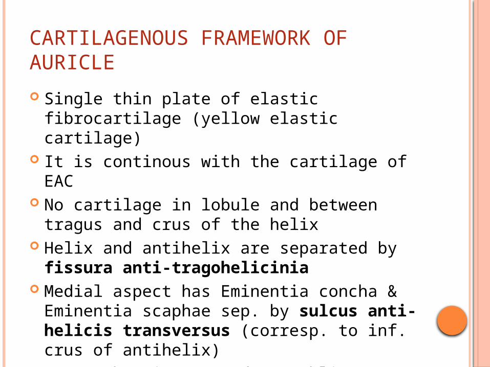
CARTILAGENOUS FRAMEWORK OF AURICLE
Single thin plate of elastic fibrocartilage (yellow elastic cartilage)
It is continous with the cartilage of EAC No cartilage in lobule and between tragus
and crus of the helix Helix and antihelix are separated by fissura
anti-tragohelicinia Medial aspect has Eminentia concha &
Eminentia scaphae sep. by sulcus anti-helicis transversus (corresp. to inf. crus of antihelix)
E. conchae is crossed y a oblique ridge – Ponticulus (atch. of auricularis posterior)
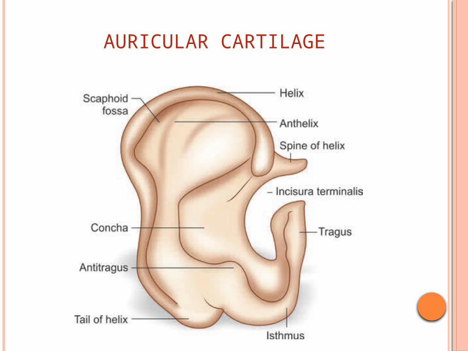
AURICULAR CARTILAGE
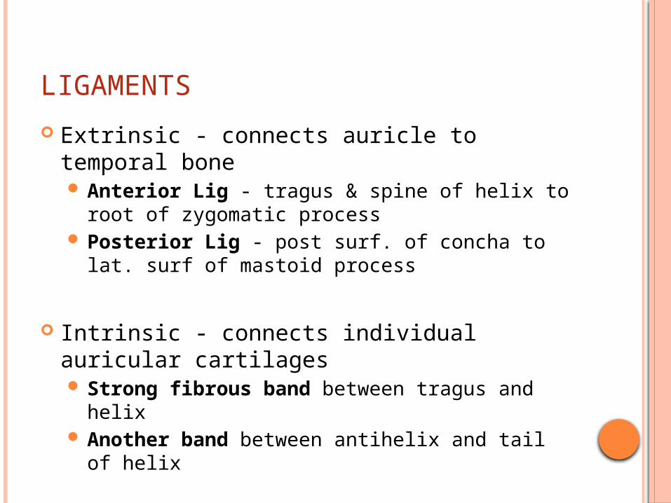
LIGAMENTS
Extrinsic - connects auricle to temporal bone Anterior Lig - tragus & spine of helix to root of
zygomatic process Posterior Lig - post surf. of concha to lat. surf of
mastoid process
Intrinsic - connects individual auricular cartilages Strong fibrous band between tragus and helix Another band between antihelix and tail of
helix
MUSCLES
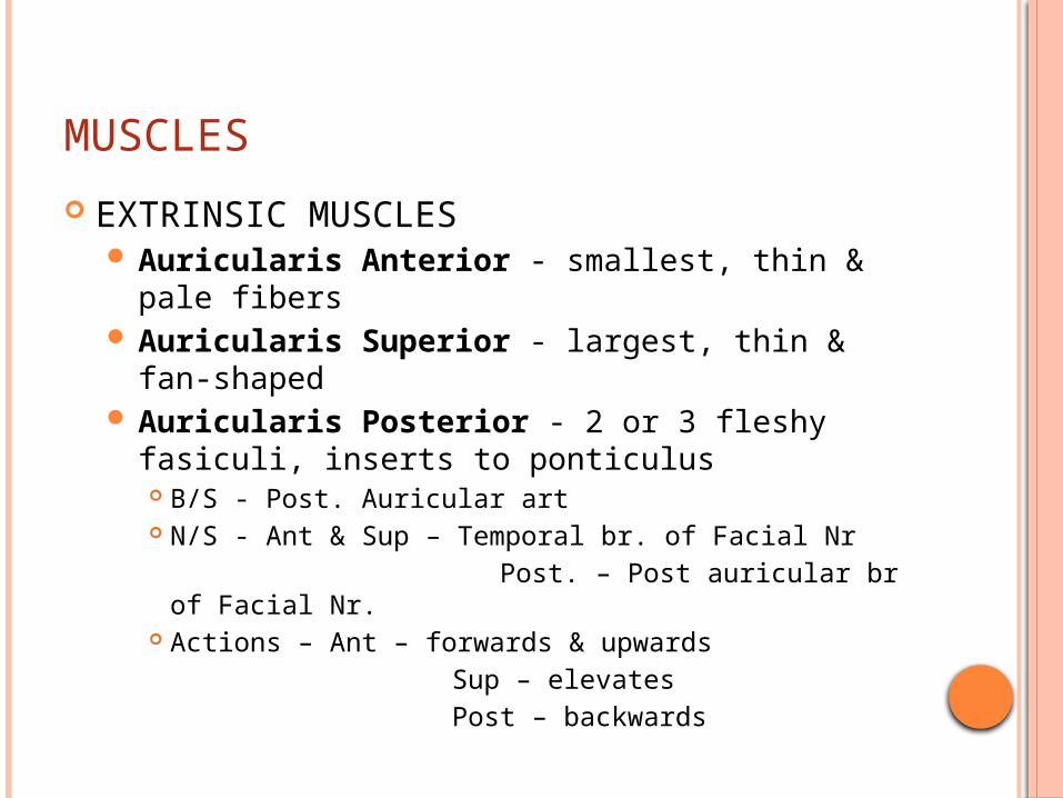
EXTRINSIC MUSCLES Auricularis Anterior - smallest, thin & pale
fibers Auricularis Superior - largest, thin & fan-
shaped Auricularis Posterior - 2 or 3 fleshy fasiculi,
inserts to ponticulus B/S - Post. Auricular art N/S - Ant & Sup – Temporal br. of Facial Nr Post. – Post auricular br of Facial Nr. Actions – Ant – forwards & upwards Sup – elevates Post – backwards
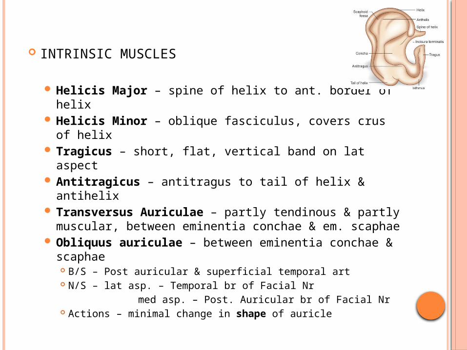
INTRINSIC MUSCLES
Helicis Major – spine of helix to ant. border of helix
Helicis Minor – oblique fasciculus, covers crus of helix
Tragicus – short, flat, vertical band on lat aspect Antitragicus – antitragus to tail of helix & antihelix Transversus Auriculae – partly tendinous & partly
muscular, between eminentia conchae & em. scaphae
Obliquus auriculae – between eminentia conchae & scaphae B/S – Post auricular & superficial temporal art N/S – lat asp. – Temporal br of Facial Nr med asp. – Post. Auricular br of Facial Nr Actions – minimal change in shape of auricle
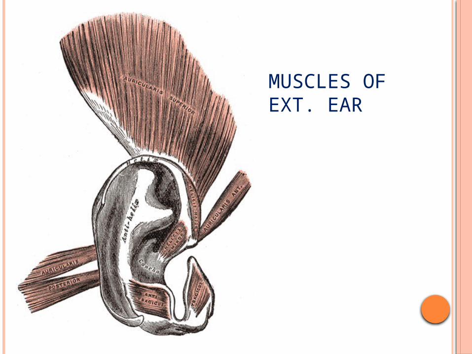
MUSCLES OF EXT. EAR
INNERVATION OF AURICLE
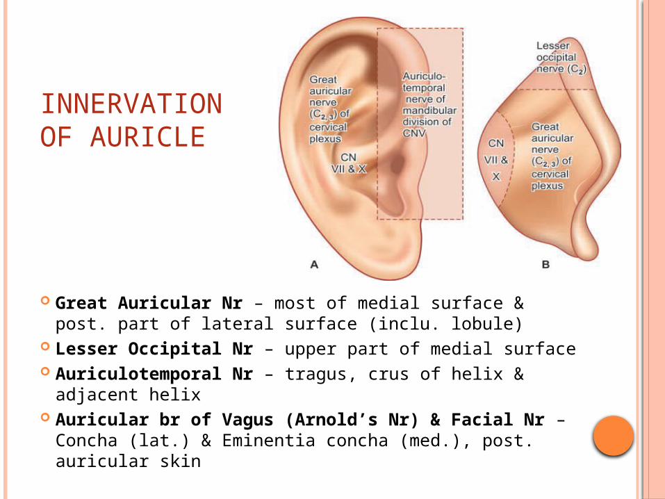
Great Auricular Nr – most of medial surface & post. part of lateral surface (inclu. lobule)
Lesser Occipital Nr – upper part of medial surface Auriculotemporal Nr – tragus, crus of helix &
adjacent helix Auricular br of Vagus (Arnold’s Nr) & Facial Nr –
Concha (lat.) & Eminentia concha (med.), post. auricular skin
EXTERNAL AUDITORY CANAL
Dimensions: EAC measures about 24 mm Extends from the concha to the tympanic membrane Its anterior wall & floor are 6 mm longer than the
posterior wall & roof EAC is usually divided into 2 parts: Its outer one-
third (8 mm) is cartilaginous and its inner two-third (16 mm) is bony.
Direction: EAC is ‘S’ shaped Outer one-third is directed upwards, backwards &
medially Inner two-third is directed downwards, forwards &
medially Anatomically divided into – pars externa, pars media
& pars interna
CARTILAGENOUS EAC
Fissures of Santorini: Transverse slits in the floor of cartilaginous EAC, provide passages for infections and neoplasms to and from the surrounding soft tissue (parotid & mastoid)
Hair follicles are present only in the outer cartilaginous canal and therefore furuncles are seen only here in Cartilagenous EAC
The skin of the cartilaginous canal is thick and contains ceruminous and pilosebaceous glands that secrete wax. The hydrophobic, slightly acidic (pH 6.0–6.5) cerumen is formed in this part of EAC.
BONY EAC
It is mainly formed by the tympanic portion of temporal bone but roof is formed by the squamous part of the temporal bone
In the anterosuperior region, squamous part articulates with tympanic bone (tympanosquamous suture).
Inferiorly and medially squamous part joins with the lateral superior portion of the petrous bone (petrosquamous suture).
Skin of the bony EAC is thin and continuous over the tympanic membrane & skin is devoid of subcutaneous layer, hair follicles and ceruminous glands.
Isthmus: Approximately 6 mm lateral to tympanic membrane, bony EAC has a narrowing called the isthmus. Foreign body impacted medial to bony isthmus of EAC are difficult to remove.
Foramen of Huschke: In children and occasionally in adults, anteroinferior bony EAC may have a deficiency that is called foramen of Huschke. It permits spread of infections to and from EAC and parotid.
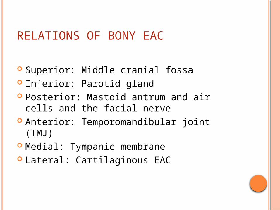
RELATIONS OF BONY EAC
Superior: Middle cranial fossa Inferior: Parotid gland Posterior: Mastoid antrum and air cells and
the facial nerve Anterior: Temporomandibular joint (TMJ) Medial: Tympanic membrane Lateral: Cartilaginous EAC
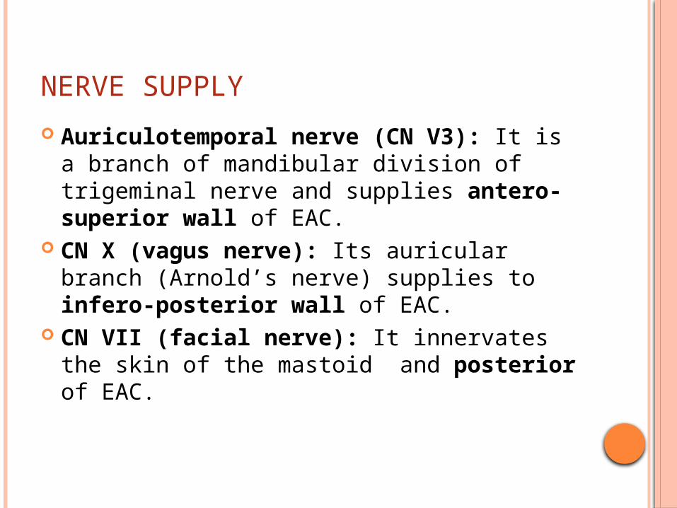
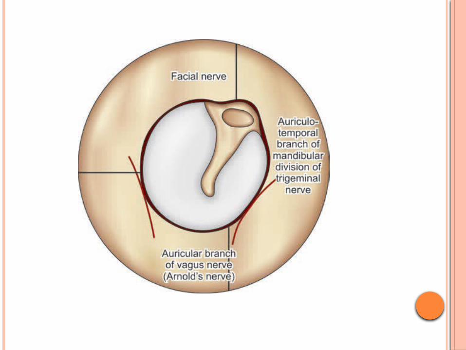
NERVE SUPPLY
Auriculotemporal nerve (CN V3): It is a branch of mandibular division of trigeminal nerve and supplies antero-superior wall of EAC.
CN X (vagus nerve): Its auricular branch (Arnold’s nerve) supplies to infero-posterior wall of EAC.
CN VII (facial nerve): It innervates the skin of the mastoid and posterior of EAC.
CLINICAL IMPORTANCE OF N/S OF EAC
Hitzelberger’s sign: The hypoesthesia of posterior meatal wall occurs due to the pressure on facial nerve (sensory fibers are affected early) in patients with acoustic neuroma.
Vasovagal reflex: While cleaning the EAC, patient may develop coughing, bradycardia, syncope and even cardiac arrest. They can occur because of Arnold’s branch of vagus nerve.
Appetite: Because of vagal innervation, instilling spirit in EAC before meal can stimulate appetite.
Ramsay Hunt syndrome: Vesicles of herpes zoster oticus occur on mastoid and posterior meatal wall which indicate that this part of external ear has facial nerve innervation.
TYMPANIC MEMBRANE
Dimensions: Its dimensions are: 9–10 mm height and 8–9 mm width. It is 0.1 mm thick.
Position: Tympanic membrane (TM) is a �partition wall between the EAC and the middle ear. It is positioned obliquely. It forms angle of 55° with deep EAC. Its posterosuperior part is more lateral than its anteroinferior part.
Structure: Tympanic membrane consists of the following three layers Outer epithelial layer (Cuticular Stratum): It
is continuous with the EAC skin. Keratinised, stratified squamous type. 10 cells thick.* The cells have a propensity for lateral migration
Middle fibrous layer (Fibrous Stratum): It encloses the handle of malleus and consists of three types of fibers: radial, circular and parabolic. In comparison to pars tensa, this layer is very thin in pars flaccida (consists of loose conn. tissue) and not organized into various fibers.
Inner mucosal layer (Mucous Stratum): It is continuous with the middle ear mucosa. Single layer of flat cells. Cilliated collumnar cells are absent over medial aspect of TM.
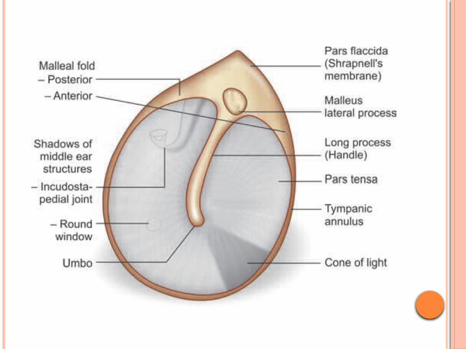
Parts: Tympanic membrane consists of two �parts: Pars tensa: It forms most of tympanic
membrane Annulus tympanicus: TM is thickened in the
periphery and forms a fibrocartilaginous ring called the annulus tympanicus that fits in the tympanic sulcus.
Umbo: The central part of TM near the tip of malleus is tended inwards and is called the umbo.
Cone of light: A bright cone of light radiating from the tip of malleus to the periphery in the anteroinferior quadrant is usually seen during otoscopy.
Pars flaccida (Shrapnell’s membrane): It is situated above the lateral process of malleus between the notch of Rivinus and the anterior and posterior malleal folds. It is not as tense as pars tensa and may appear little pinkish.
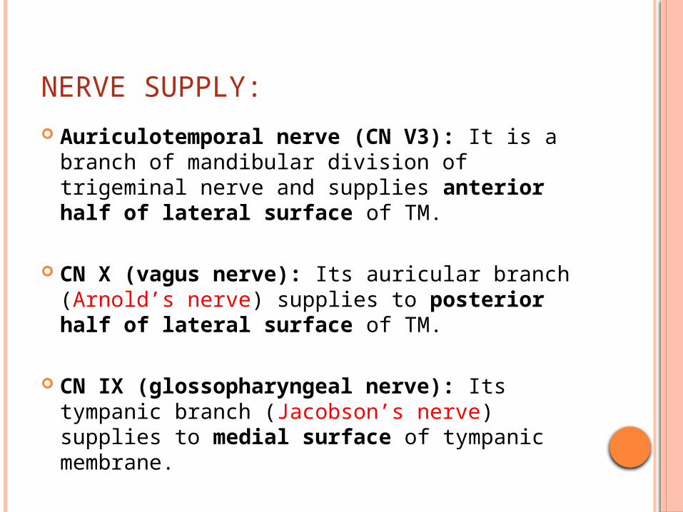
NERVE SUPPLY:
Auriculotemporal nerve (CN V3): It is a branch of mandibular division of trigeminal nerve and supplies anterior half of lateral surface of TM.
CN X (vagus nerve): Its auricular branch (Arnold’s nerve) supplies to posterior half of lateral surface of TM.
CN IX (glossopharyngeal nerve): Its tympanic branch (Jacobson’s nerve) supplies to medial surface of tympanic membrane.
MIDDLE EAR
MIDDLE EAR
The middle ear cleft is lined by mucous membrane and filled with air
Consists of the middle ear, eustachian tube, aditus ad antrum, mastoid antrum and mastoid air cells.
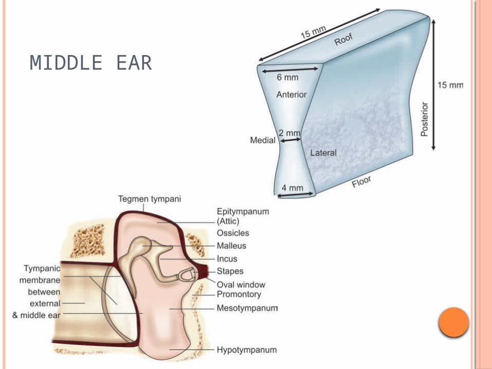
Middle ear is a 1 to 2 cm3 air filled cavity that houses ossicles, stapedius and tensor tympani muscles and chorda tympani nerve and tympanic plexus.
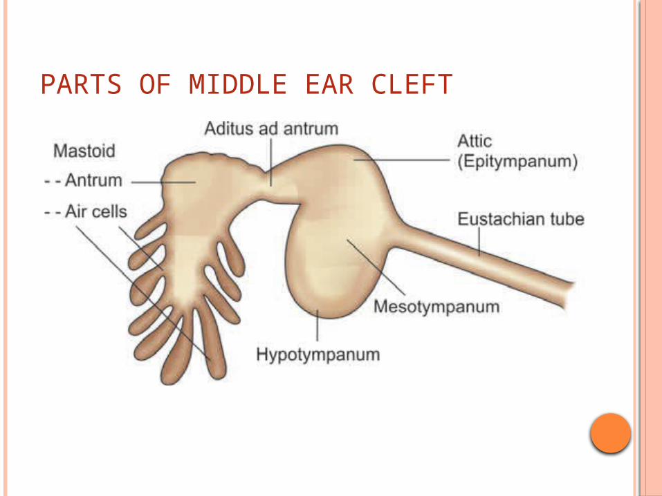
PARTS OF MIDDLE EAR CLEFT
RELATIONS OF MIDDLE EAR CLEFT
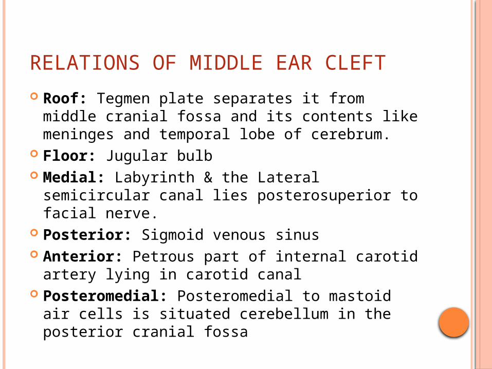
Roof: Tegmen plate separates it from middle cranial fossa and its contents like meninges and temporal lobe of cerebrum.
�Floor: Jugular bulb Medial: Labyrinth & the Lateral semicircular
canal lies posterosuperior to facial nerve. �Posterior: Sigmoid venous sinus Anterior: Petrous part of internal carotid
artery lying in carotid canal Posteromedial: Posteromedial to mastoid
air cells is situated cerebellum in the posterior cranial fossa
PARTS OF MIDDLE EAR
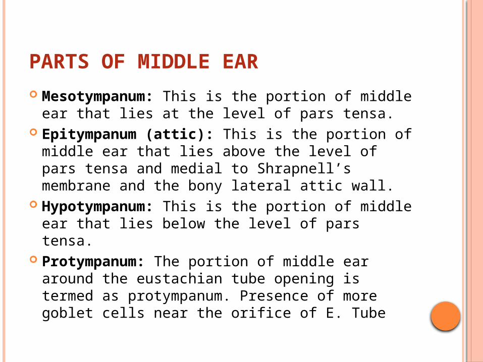
Mesotympanum: This is the portion of middle ear that lies at the level of pars tensa.
Epitympanum (attic): This is the portion of middle ear that lies above the level of pars tensa and medial to Shrapnell’s membrane and the bony lateral attic wall.
Hypotympanum: This is the portion of middle ear that lies below the level of pars tensa.
Protympanum: The portion of middle ear around the eustachian tube opening is termed as protympanum. Presence of more goblet cells near the orifice of E. Tube
MIDDLE EAR
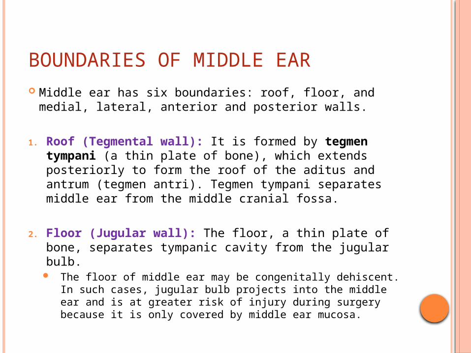
BOUNDARIES OF MIDDLE EAR Middle ear has six boundaries: roof, floor, and medial,
lateral, anterior and posterior walls.
1. Roof (Tegmental wall): It is formed by tegmen tympani (a thin plate of bone), which extends posteriorly to form the roof of the aditus and antrum (tegmen antri). Tegmen tympani separates middle ear from the middle cranial fossa.
2. Floor (Jugular wall): The floor, a thin plate of bone, separates tympanic cavity from the jugular bulb.
The floor of middle ear may be congenitally dehiscent. In such cases, jugular bulb projects into the middle ear and is at greater risk of injury during surgery because it is only covered by middle ear mucosa.
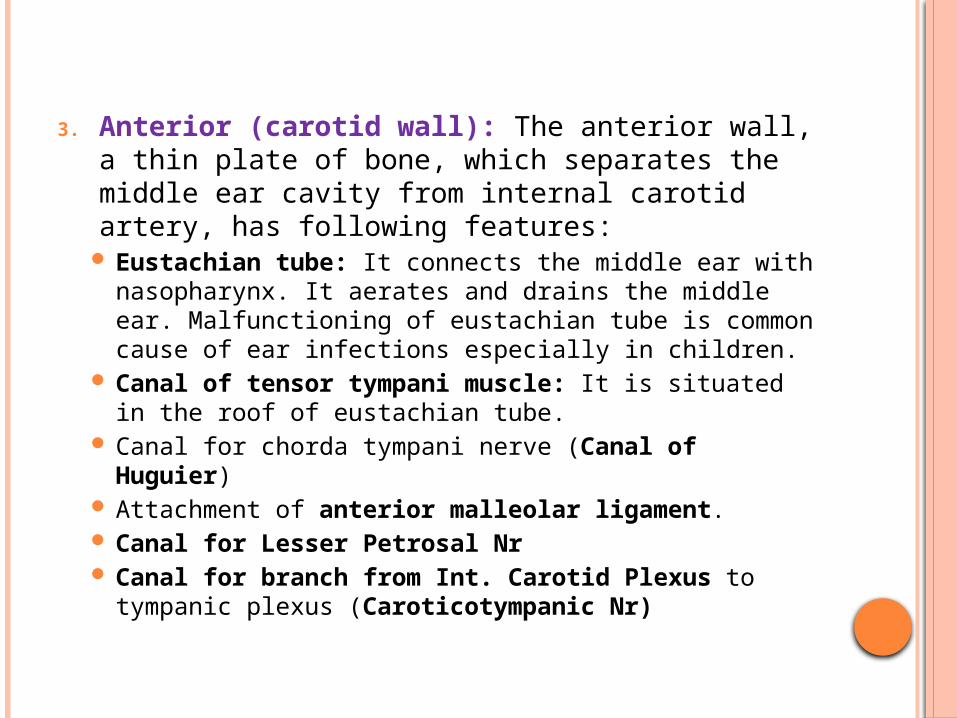
3. Anterior (carotid wall): The anterior wall, a thin plate of bone, which separates the middle ear cavity from internal carotid artery, has following features:
Eustachian tube: It connects the middle ear with nasopharynx. It aerates and drains the middle ear. Malfunctioning of eustachian tube is common cause of ear infections especially in children.
Canal of tensor tympani muscle: It is situated in the roof of eustachian tube.
Canal for chorda tympani nerve (Canal of Huguier)
Attachment of anterior malleolar ligament. Canal for Lesser Petrosal Nr Canal for branch from Int. Carotid Plexus to
tympanic plexus (Caroticotympanic Nr)
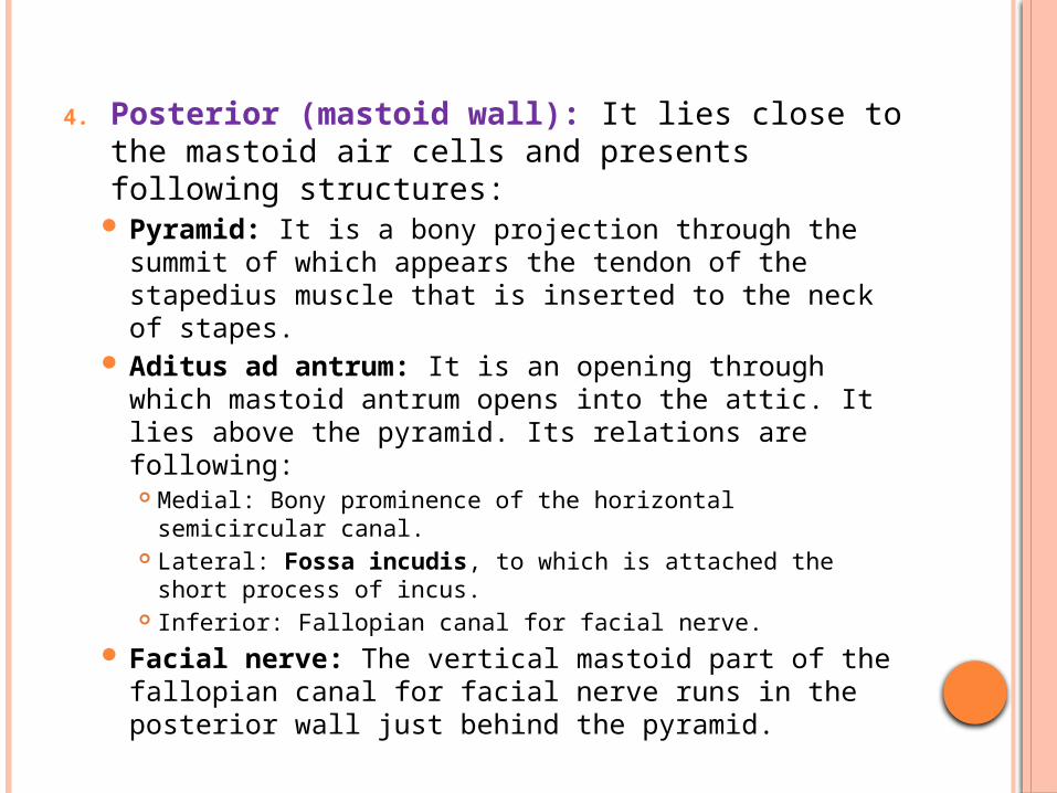
4. Posterior (mastoid wall): It lies close to the mastoid air cells and presents following structures:
Pyramid: It is a bony projection through the summit of which appears the tendon of the stapedius muscle that is inserted to the neck of stapes.
Aditus ad antrum: It is an opening through which mastoid antrum opens into the attic. It lies above the pyramid. Its relations are following: Medial: Bony prominence of the horizontal semicircular
canal. Lateral: Fossa incudis, to which is attached the short
process of incus. Inferior: Fallopian canal for facial nerve.
Facial nerve: The vertical mastoid part of the fallopian canal for facial nerve runs in the posterior wall just behind the pyramid.
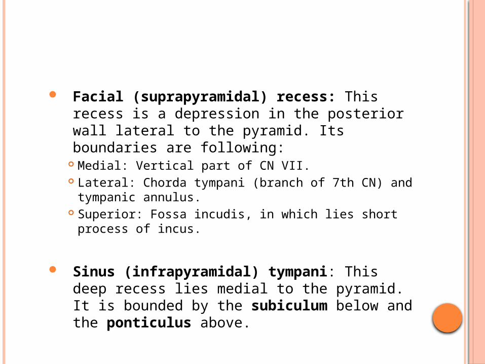
Facial (suprapyramidal) recess: This recess is a depression in the posterior wall lateral to the pyramid. Its boundaries are following:
Medial: Vertical part of CN VII. Lateral: Chorda tympani (branch of 7th CN) and
tympanic annulus. Superior: Fossa incudis, in which lies short process of
incus.
Sinus (infrapyramidal) tympani: This deep recess lies medial to the pyramid. It is bounded by the subiculum below and the ponticulus above.
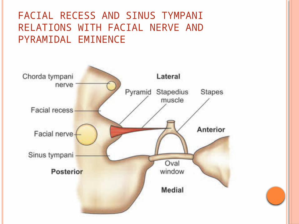
FACIAL RECESS AND SINUS TYMPANI RELATIONS WITH FACIAL NERVE AND PYRAMIDAL EMINENCE
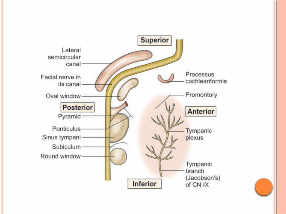
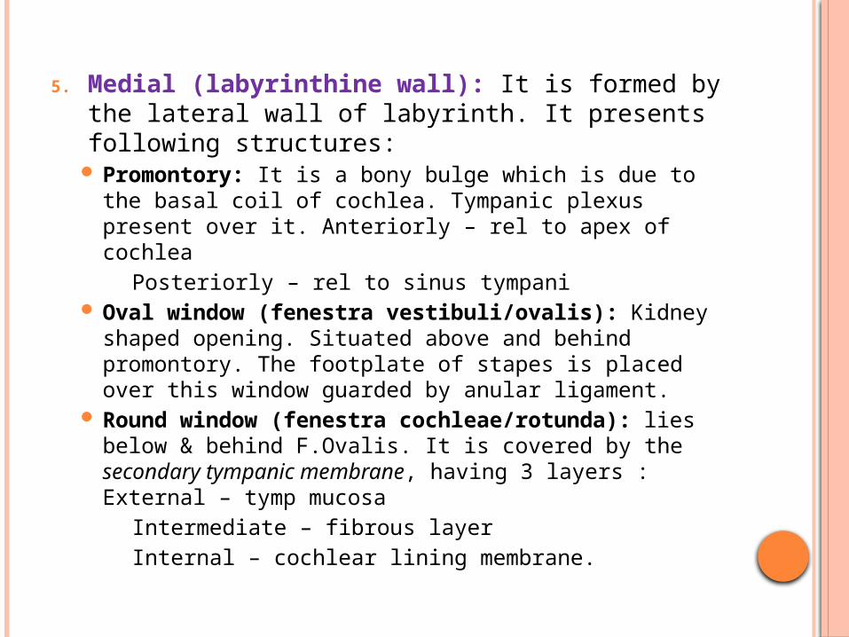
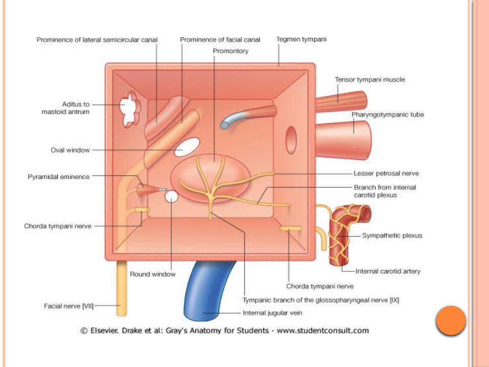
5. Medial (labyrinthine wall): It is formed by the lateral wall of labyrinth. It presents following structures:
Promontory: It is a bony bulge which is due to the basal coil of cochlea. Tympanic plexus present over it. Anteriorly – rel to apex of cochlea
Posteriorly – rel to sinus tympani Oval window (fenestra vestibuli/ovalis): Kidney
shaped opening. Situated above and behind promontory. The footplate of stapes is placed over this window guarded by anular ligament.
Round window (fenestra cochleae/rotunda): lies below & behind F.Ovalis. It is covered by the secondary tympanic membrane, having 3 layers : External – tymp mucosa
Intermediate – fibrous layer Internal – cochlear lining membrane.
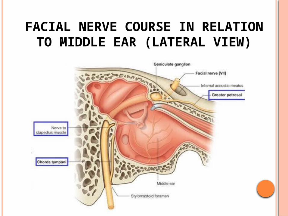
FACIAL NERVE COURSE IN RELATION TO MIDDLE EAR
(LATERAL VIEW)
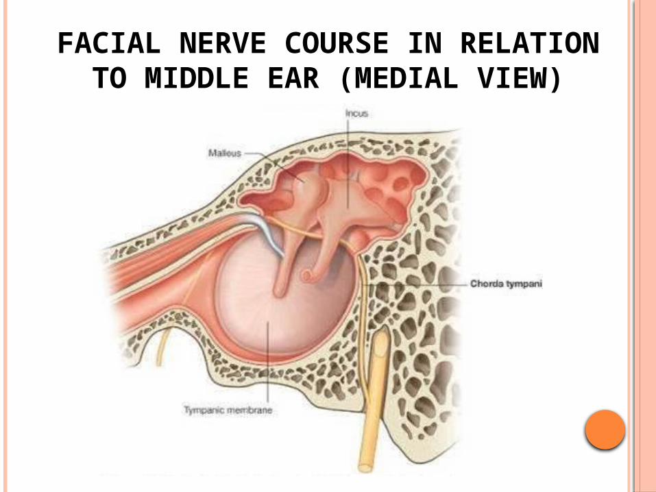
FACIAL NERVE COURSE IN RELATION TO MIDDLE EAR (MEDIAL
VIEW)
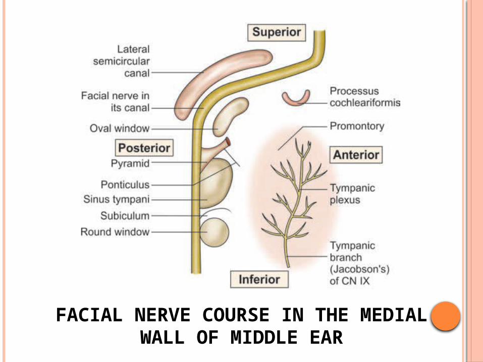
FACIAL NERVE COURSE IN THE MEDIAL WALL OF MIDDLE EAR
Horizontal tympanic part of fallopian canal for facial nerve: It lies above the oval window. The tympanic segment of facial nerve canal may be
congenitally dehiscent and the exposed facial nerve becomes vulnerable to injuries or infection.
Lateral semicircular canal: It lies above the fallopian canal, facial nerve.
Processus cochleariformis: It is a hook-like projection, which lies anterior to the oval window. The tendon of tensor tympani takes a turn on this process and then is inserted on the neck of malleus. Processus cochleariformis is an important surgical
landmark for the level of the genu of the facial nerve.
6. Lateral (membranous wall) Tympanic membrane: Lateral wall is formed
mainly by the tympanic membrane. Some structures of the middle ear (such as long process of incus, incudostapedial joint, round window and eustachian tube) can be seen through the normal semitransparent tympanic membrane.
Scutum: An upper part of epitympanum is formed by outer bony attic wall called scutum.
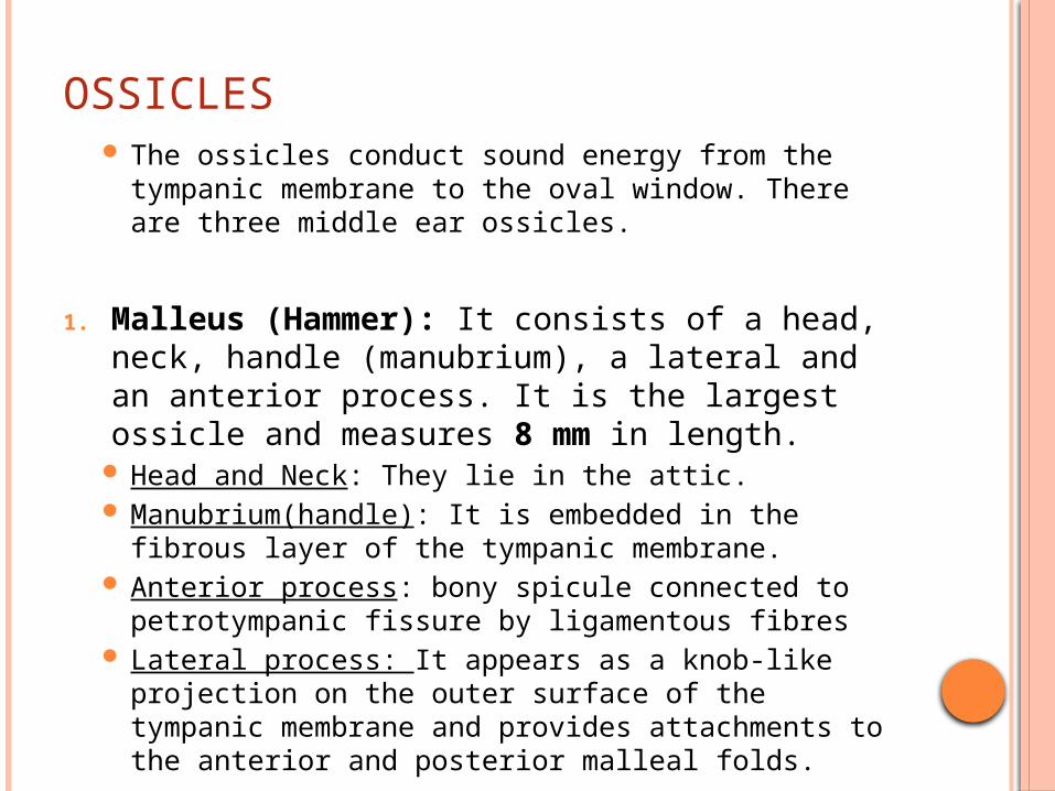
OSSICLES The ossicles conduct sound energy from the
tympanic membrane to the oval window. There are three middle ear ossicles.
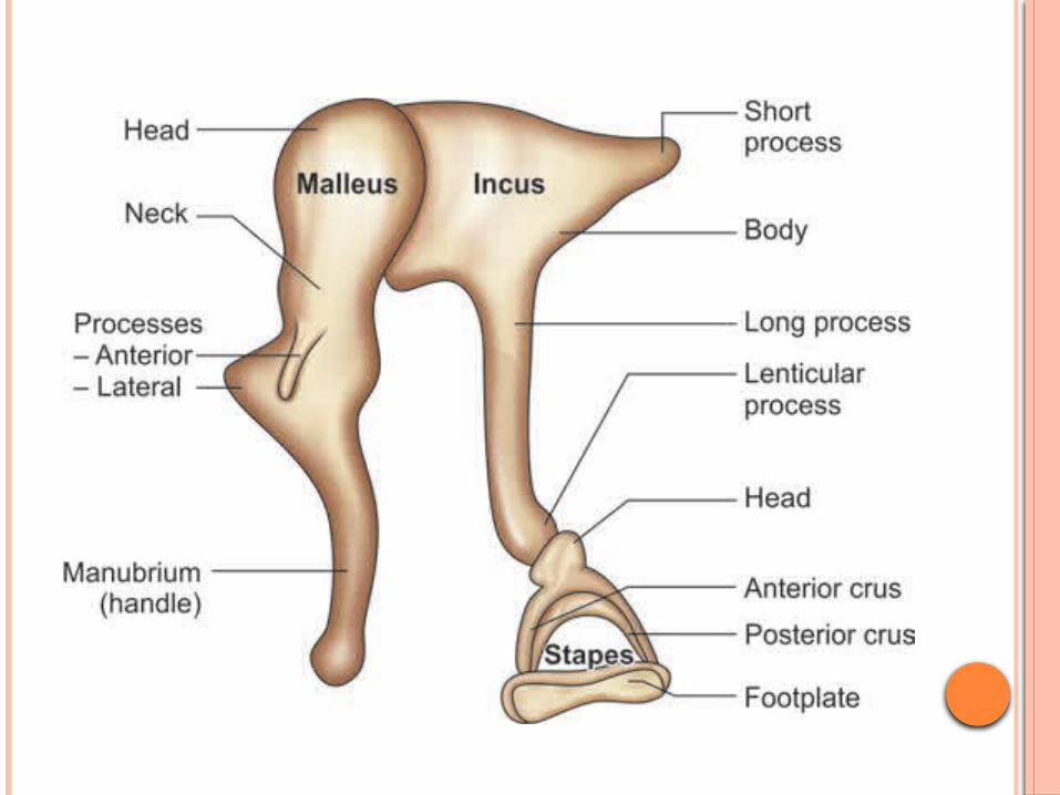
1. Malleus (Hammer): It consists of a head, neck, handle (manubrium), a lateral and an anterior process. It is the largest ossicle and measures 8 mm in length.
Head and Neck: They lie in the attic. Manubrium(handle): It is embedded in the fibrous
layer of the tympanic membrane. Anterior process: bony spicule connected to
petrotympanic fissure by ligamentous fibres Lateral process: It appears as a knob-like
projection on the outer surface of the tympanic membrane and provides attachments to the anterior and posterior malleal folds.
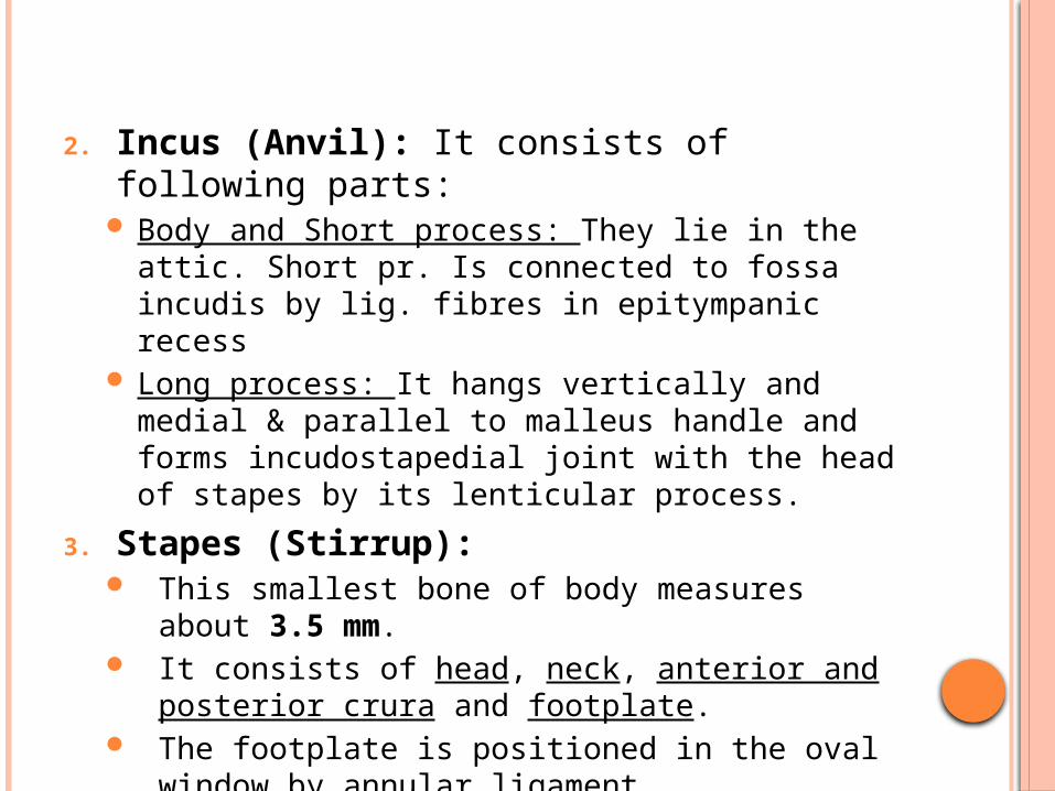
2. Incus (Anvil): It consists of following parts: Body and Short process: They lie in the attic.
Short pr. Is connected to fossa incudis by lig. fibres in epitympanic recess
Long process: It hangs vertically and medial & parallel to malleus handle and forms incudostapedial joint with the head of stapes by its lenticular process.
3. Stapes (Stirrup): This smallest bone of body measures about 3.5
mm. It consists of head, neck, anterior and posterior
crura and footplate. The footplate is positioned in the oval window
by annular ligament
LIGAMENTS OF OSSICLES Malleus
Anterior ligament of Malleus: neck of malleus to ant wall of tympanic cavity Contains muscle fibers called as Laxator tympani/ Musculus externus
mallei Lateral ligament of Malleus: triangular band, from post
border of tympanic inscisure to head of malleus Superior ligament of Malleus: head of malleus to roof of
epitympanic recess Incus
Posterior ligament of Incus: from end of short process to fossa incudis
Superior ligament of Incus: body to roof of epitympanic recess
Stapes Vestibular surf & rim of stapedial base covered with hyaline
cartilage, which is attached to margin of fen. vestibuli by annular ligament
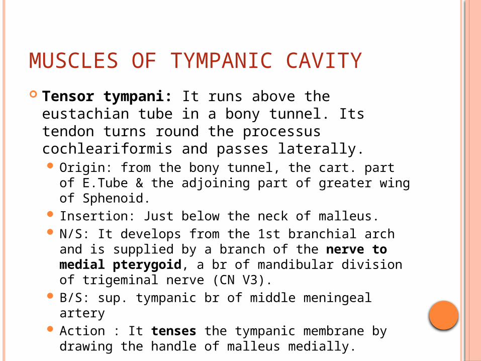
MUSCLES OF TYMPANIC CAVITY Tensor tympani: It runs above the
eustachian tube in a bony tunnel. Its tendon turns round the processus cochleariformis and passes laterally. Origin: from the bony tunnel, the cart. part of
E.Tube & the adjoining part of greater wing of Sphenoid.
Insertion: Just below the neck of malleus. N/S: It develops from the 1st branchial arch and
is supplied by a branch of the nerve to medial pterygoid, a br of mandibular division of trigeminal nerve (CN V3).
B/S: sup. tympanic br of middle meningeal artery Action : It tenses the tympanic membrane by
drawing the handle of malleus medially.
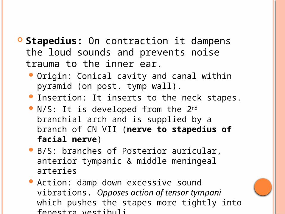
Stapedius: On contraction it dampens the loud sounds and prevents noise trauma to the inner ear. Origin: Conical cavity and canal within pyramid
(on post. tymp wall). Insertion: It inserts to the neck stapes. N/S: It is developed from the 2nd branchial arch
and is supplied by a branch of CN VII (nerve to stapedius of facial nerve)
B/S: branches of Posterior auricular, anterior tympanic & middle meningeal arteries
Action: damp down excessive sound vibrations. Opposes action of tensor tympani which pushes the stapes more tightly into fenestra vestibuli
ACOUSTIC REFLEX
When noises are loud, there occurs reflex contraction of stapedius and tensor tympani which helps to dampen the movement of ossicular chain before vibrations reach the internal ear. Afferent pathway: auditory component of 8th Cr
Nr Efferent Pathway: Facial Nerve – Stapedius &
Mandibular Nerve – Tensor tympani
INTRATYMPANIC NERVES Tympanic plexus (Nerve supply of middle ear):
The tympanic nerve plexus, which lies on the promontory, supplies to the medial surface of the tympanic membrane, tympanic cavity, mastoid air cells and the bony eustachian tube. It is formed by following nerves: Tympanic branch of glossopharyngeal (Jacobson’s
Nerve) : It carries secretomotor fibers to the parotid gland. The pathway of secretomotor fibers to the parotid gland consists of Inferior salivary nucleus CN IX Jacobson’s tympanic branch Tympanic plexus Lesser petrosal nerve Otic ganglion Auriculotemporal nerve Parotid gland. Section of Jacobson’s nerve is carried out in cases of Frey’s
syndrome. Sympathetic fibers: Caroticotympanic nerves come from
the sympathetic plexus, which is present round the internal carotid artery
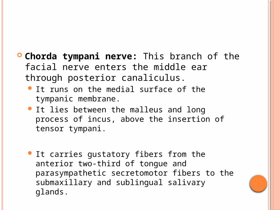
Chorda tympani nerve: This branch of the facial nerve enters the middle ear through posterior canaliculus. It runs on the medial surface of the tympanic
membrane. It lies between the malleus and long process of
incus, above the insertion of tensor tympani.
It carries gustatory fibers from the anterior two-third of tongue and parasympathetic secretomotor fibers to the submaxillary and sublingual salivary glands.
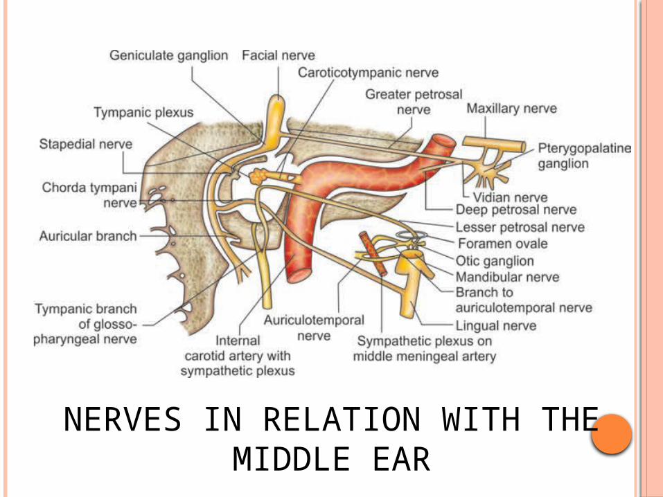
NERVES IN RELATION WITH THE MIDDLE EAR
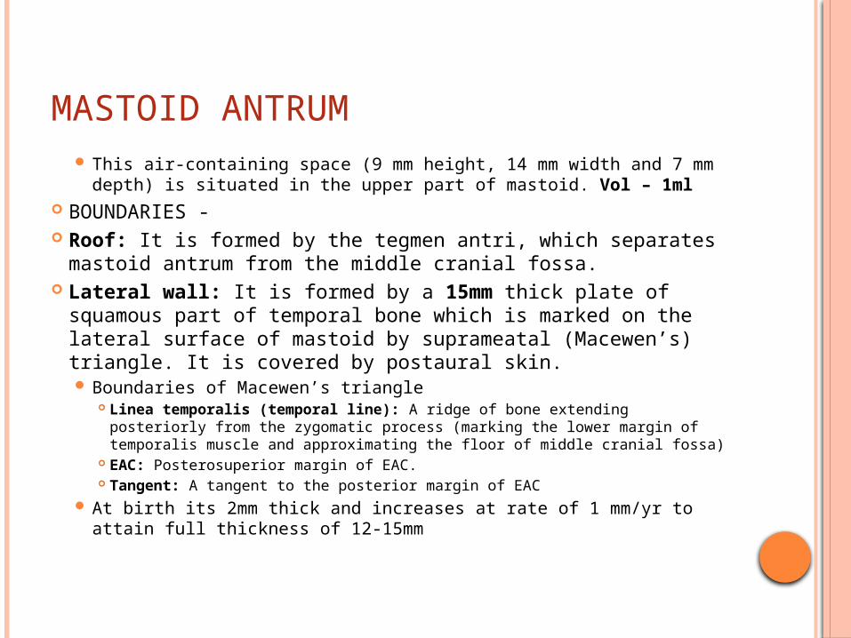
MASTOID ANTRUM This air-containing space (9 mm height, 14 mm width and 7
mm depth) is situated in the upper part of mastoid. Vol – 1ml BOUNDARIES - �Roof: It is formed by the tegmen antri, which separates
mastoid antrum from the middle cranial fossa. �Lateral wall: It is formed by a 15mm thick plate of
squamous part of temporal bone which is marked on the lateral surface of mastoid by suprameatal (Macewen’s) triangle. It is covered by postaural skin. Boundaries of Macewen’s triangle
Linea temporalis (temporal line): A ridge of bone extending posteriorly from the zygomatic process (marking the lower margin of temporalis muscle and approximating the floor of middle cranial fossa)
EAC: Posterosuperior margin of EAC. Tangent: A tangent to the posterior margin of EAC
At birth its 2mm thick and increases at rate of 1 mm/yr to attain full thickness of 12-15mm
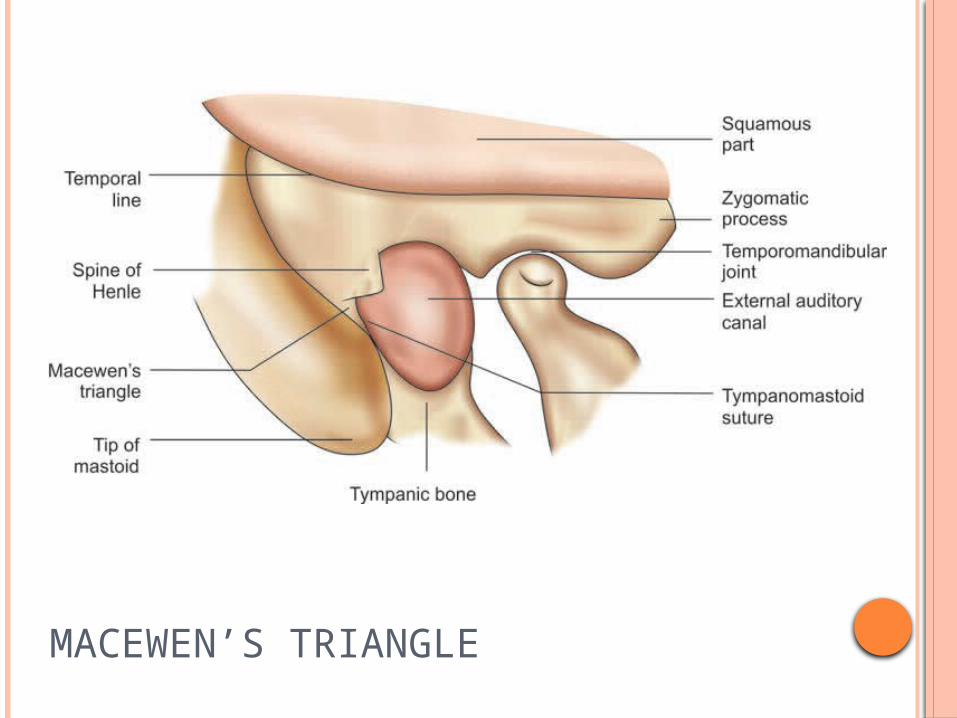
MACEWEN’S TRIANGLE
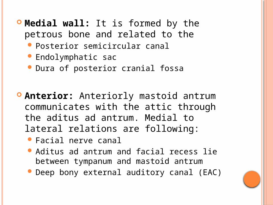
Medial wall: It is formed by the petrous bone and related to the Posterior semicircular canal Endolymphatic sac Dura of posterior cranial fossa
�Anterior: Anteriorly mastoid antrum communicates with the attic through the aditus ad antrum. Medial to lateral relations are following: Facial nerve canal Aditus ad antrum and facial recess lie between
tympanum and mastoid antrum Deep bony external auditory canal (EAC)
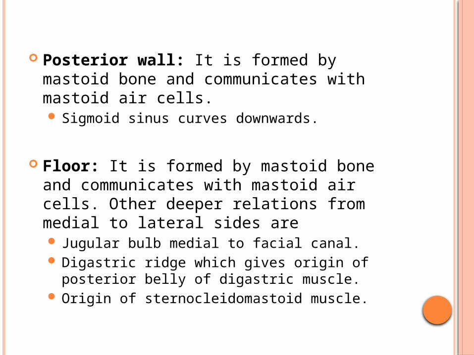
Posterior wall: It is formed by mastoid bone and communicates with mastoid air cells. Sigmoid sinus curves downwards.
�Floor: It is formed by mastoid bone and communicates with mastoid air cells. Other deeper relations from medial to lateral sides are Jugular bulb medial to facial canal. Digastric ridge which gives origin of posterior
belly of digastric muscle. Origin of sternocleidomastoid muscle.
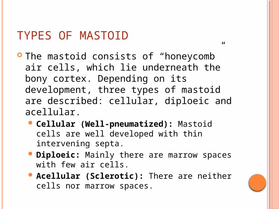
TYPES OF MASTOID
The mastoid consists of “honeycomb” air cells, which lie underneath the bony cortex. Depending on its development, three types of mastoid are described: cellular, diploeic and acellular. Cellular (Well-pneumatized): Mastoid cells
are well developed with thin intervening septa. Diploeic: Mainly there are marrow spaces with
few air cells. Acellular (Sclerotic): There are neither cells
nor marrow spaces.
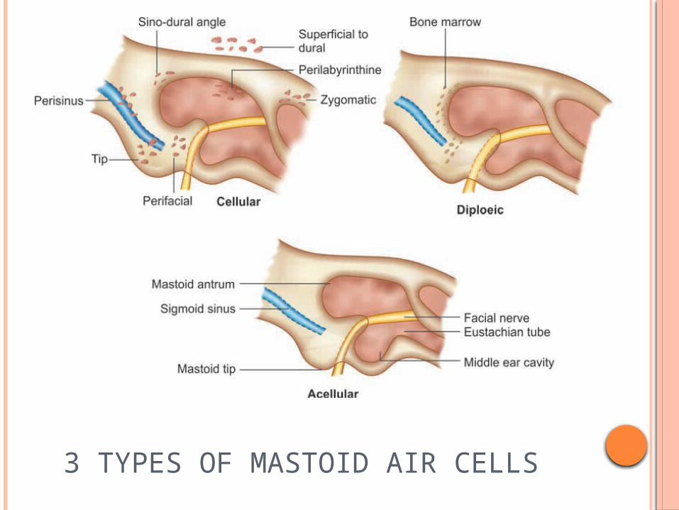
3 TYPES OF MASTOID AIR CELLS
MASTOID AIR CELLS
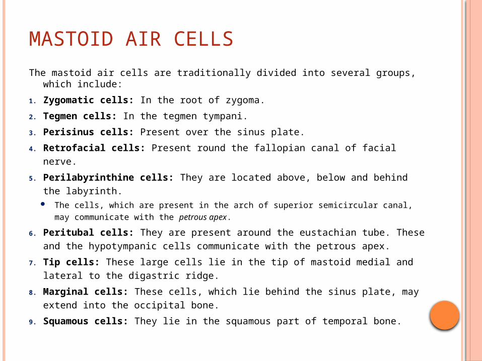
The mastoid air cells are traditionally divided into several groups, which include:
1. Zygomatic cells: In the root of zygoma.
2. Tegmen cells: In the tegmen tympani.
3. Perisinus cells: Present over the sinus plate.
4. Retrofacial cells: Present round the fallopian canal of facial nerve.
5. Perilabyrinthine cells: They are located above, below and behind the labyrinth.
The cells, which are present in the arch of superior semicircular canal, may communicate with the petrous apex.
6. Peritubal cells: They are present around the eustachian tube. These and the hypotympanic cells communicate with the petrous apex.
7. Tip cells: These large cells lie in the tip of mastoid medial and lateral to the digastric ridge.
8. Marginal cells: These cells, which lie behind the sinus plate, may extend into the occipital bone.
9. Squamous cells: They lie in the squamous part of temporal bone.
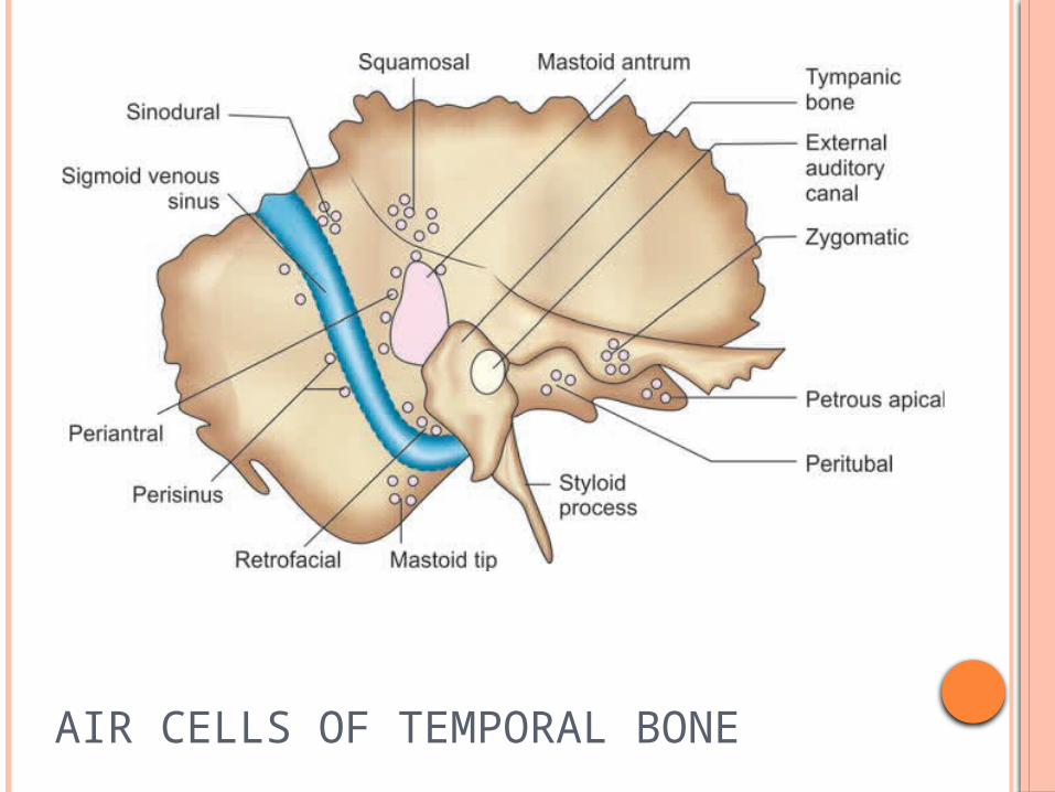
AIR CELLS OF TEMPORAL BONE
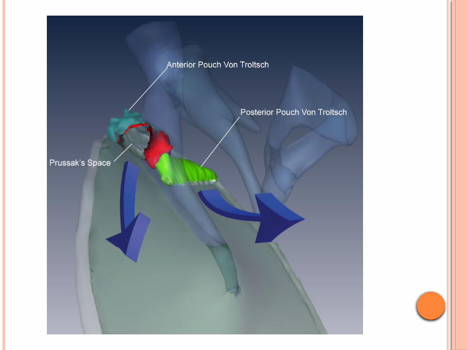
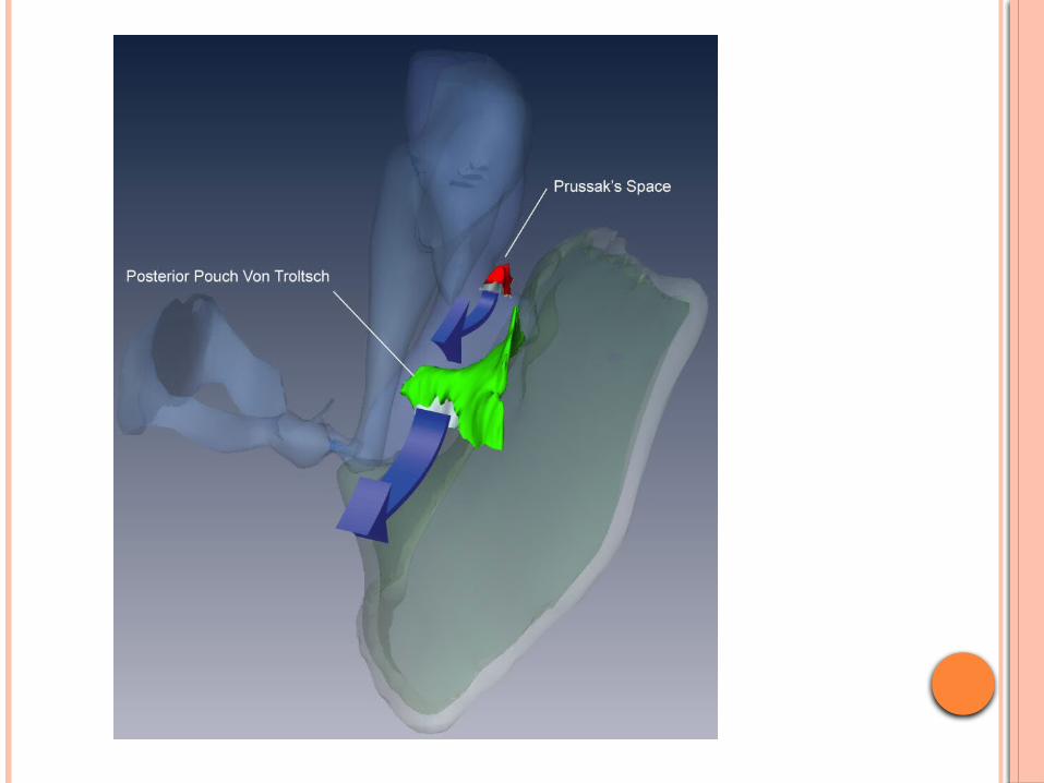
COMPARTMENTS & FOLDS OF MIDDLE EAR
Ossicles and their mucosal folds separate mesotympanum from epitympanum (attic).
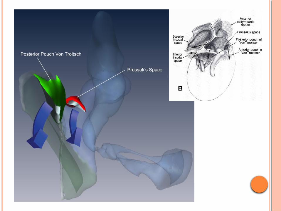
Compartments of Epitympanum1. Prussak’s space: Its boundaries, which
limit spread of infection to other compartments, are following:
Lateral: Membrana flaccida (Shrapnell’s membrane)
Medial: Neck of malleus Floor: Lateral process of malleus Roof: Fibers of lateral malleolar ligament arising
from neck of malleus and inserting along the rim of notch of Rivinus
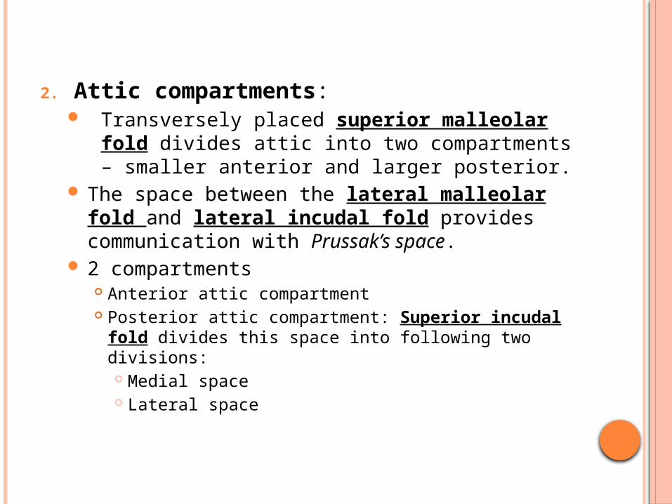
2. Attic compartments: Transversely placed superior malleolar fold
divides attic into two compartments – smaller anterior and larger posterior.
The space between the lateral malleolar fold and lateral incudal fold provides communication with Prussak’s space.
2 compartments Anterior attic compartment Posterior attic compartment: Superior incudal fold
divides this space into following two divisions: Medial space Lateral space

POSTEROSUPERIOR AND LATERAL VIEW OF RIGHT TYMPANIC CAVITY SHOWING COMPARTMENTS AND FOLDS OF MIDDLE EAR
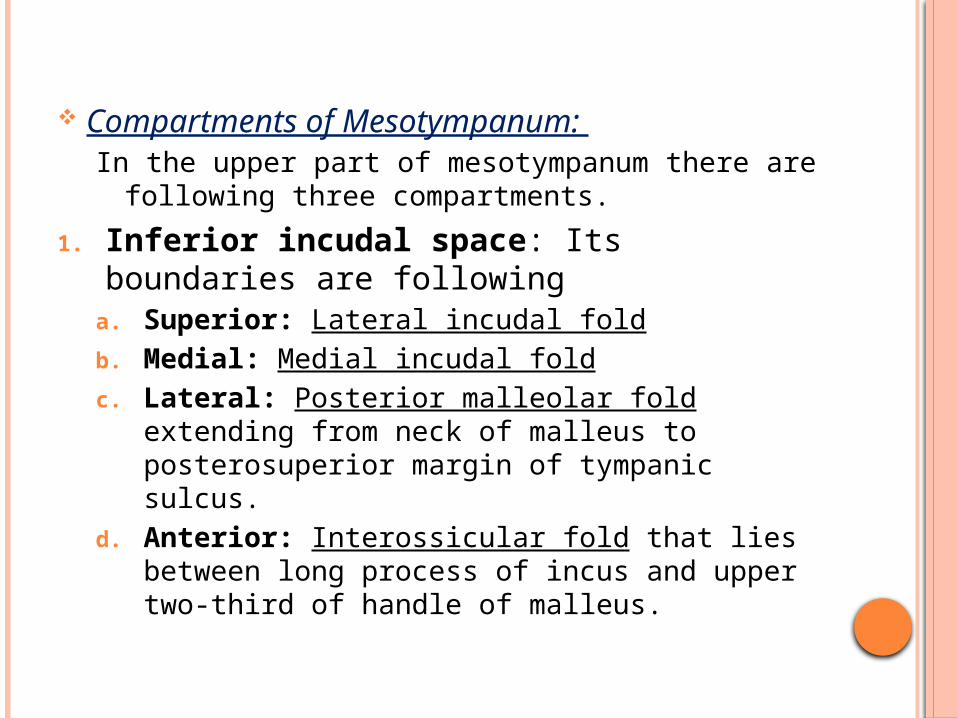
Compartments of Mesotympanum: In the upper part of mesotympanum there are
following three compartments.
1. Inferior incudal space: Its boundaries are following
a. Superior: Lateral incudal foldb. Medial: Medial incudal foldc. Lateral: Posterior malleolar fold extending
from neck of malleus to posterosuperior margin of tympanic sulcus.
d. Anterior: Interossicular fold that lies between long process of incus and upper two-third of handle of malleus.
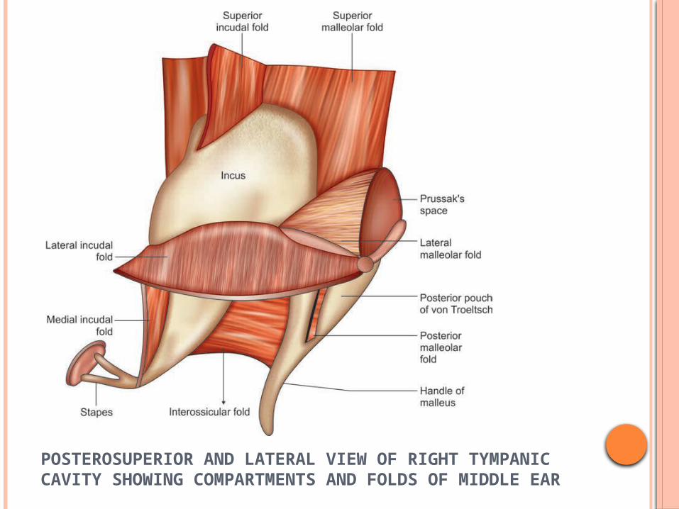
POSTEROSUPERIOR AND LATERAL VIEW OF RIGHT TYMPANIC CAVITY SHOWING COMPARTMENTS AND FOLDS OF MIDDLE EAR
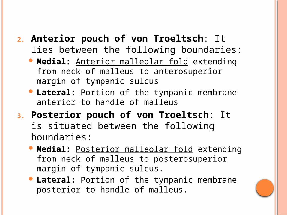
2. Anterior pouch of von Troeltsch: It lies between the following boundaries:
Medial: Anterior malleolar fold extending from neck of malleus to anterosuperior margin of tympanic sulcus
Lateral: Portion of the tympanic membrane anterior to handle of malleus
3. Posterior pouch of von Troeltsch: It is situated between the following boundaries:
Medial: Posterior malleolar fold extending from neck of malleus to posterosuperior margin of tympanic sulcus.
Lateral: Portion of the tympanic membrane posterior to handle of malleus.
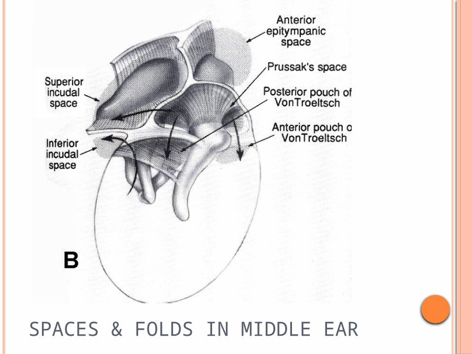
SPACES & FOLDS IN MIDDLE EAR
KORNER’S SEPTUM
Mastoid develops from the squamous and petrous parts of temporal bone.
In some cases petrosquamosal suture persists as a bony plate called Korner’s septum, which separates superficial squamosal cells from the deep petrosal cells.
During the mastoid surgery, Korner’s septum causes difficulty in locating the antrum and the deeper cells.
If not recognized, Korner’s septum leads to incomplete removal of disease during mastoidectomy. Mastoid antrum can be entered into only after the removal of Korner’s septum
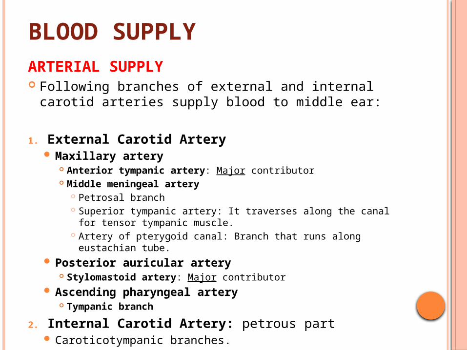
BLOOD SUPPLYARTERIAL SUPPLY Following branches of external and internal carotid
arteries supply blood to middle ear:
1. External Carotid Artery Maxillary artery
Anterior tympanic artery: Major contributor Middle meningeal artery
Petrosal branch Superior tympanic artery: It traverses along the canal for
tensor tympanic muscle. Artery of pterygoid canal: Branch that runs along eustachian
tube. Posterior auricular artery
Stylomastoid artery: Major contributor Ascending pharyngeal artery
Tympanic branch
2. Internal Carotid Artery: petrous part Caroticotympanic branches.
VENOUS DRAINAGE Veins from the middle ear cleft drain into
pterygoid venous plexus, superior petrosal sinus and sigmoid sinus.
LYMPHATIC DRAINAGE The lymphatics of middle ear drain into
retropharyngeal and parotid nodes. Eustachian tube lymphatics drain into retropharyngeal group of lymph nodes . Internal ear does not have any lymphatics

THANK YOU…