Embed Size (px)
Citation preview
CONTENTS INTRODUCTION SKULL INDIVIDUAL BONES
FRONTAL
PAREITAL
OCCIPITAL
ORBITAL
TEMPORAL
SPHENOID
ETHMOID
MAXILLA
ZYGOMATIC
PALATINE
MANDIBLE
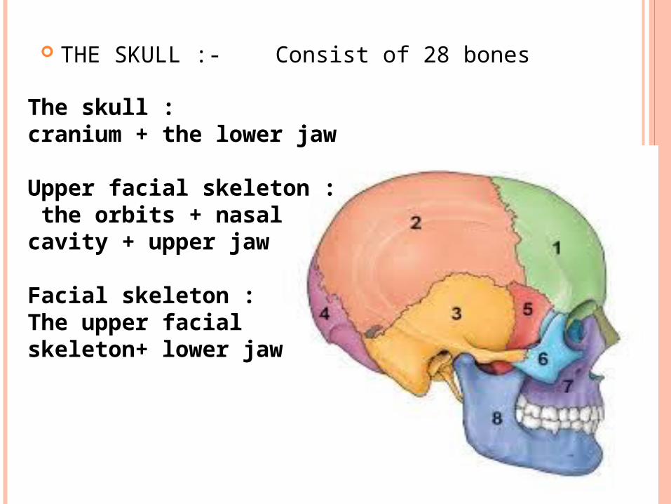
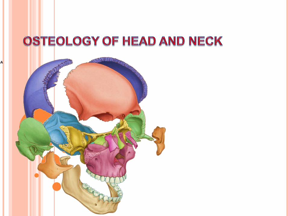
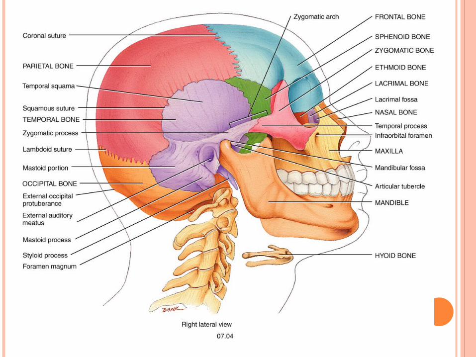
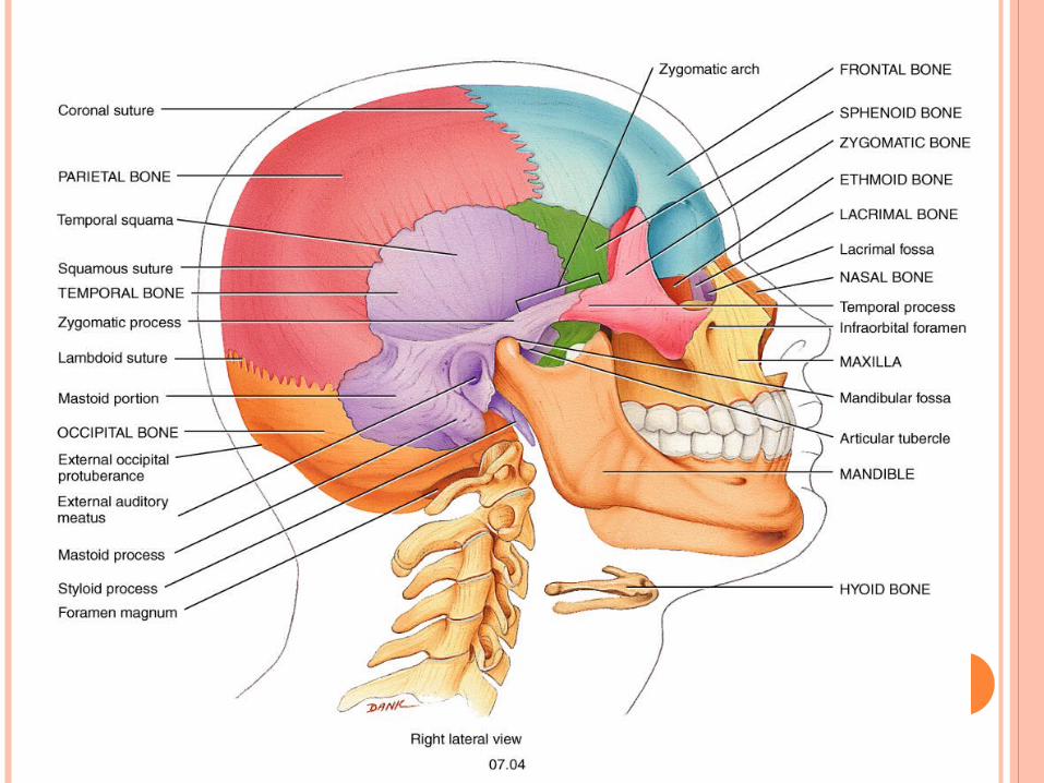
THE SKULL :- Consist of 28 bones
The skull : cranium + the lower jaw
Upper facial skeleton : the orbits + nasal cavity + upper jaw
Facial skeleton :The upper facial skeleton+ lower jaw
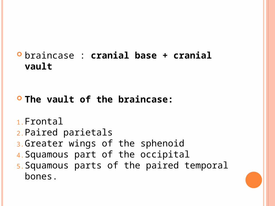
braincase : cranial base + cranial vault
The vault of the braincase:
1. Frontal2. Paired parietals3. Greater wings of the sphenoid4. Squamous part of the occipital5. Squamous parts of the paired temporal
bones.

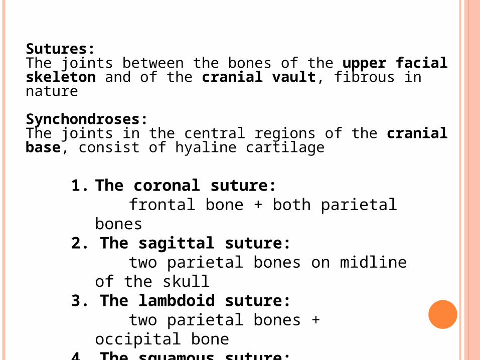
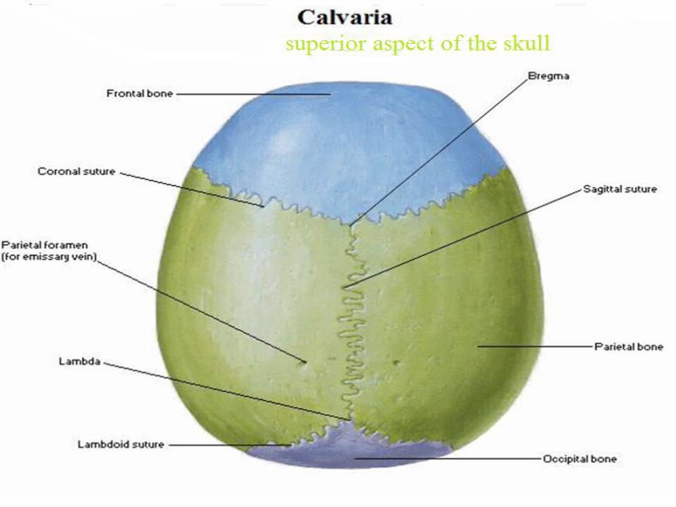
1. The coronal suture: frontal bone + both parietal bones2. The sagittal suture: two parietal bones on midline of the
skull 3. The lambdoid suture: two parietal bones + occipital bone 4. The squamous suture: parietal + temporal bones
Sutures: The joints between the bones of the upper facial skeleton and of the cranial vault, fibrous in nature
Synchondroses: The joints in the central regions of the cranial base, consist of hyaline cartilage
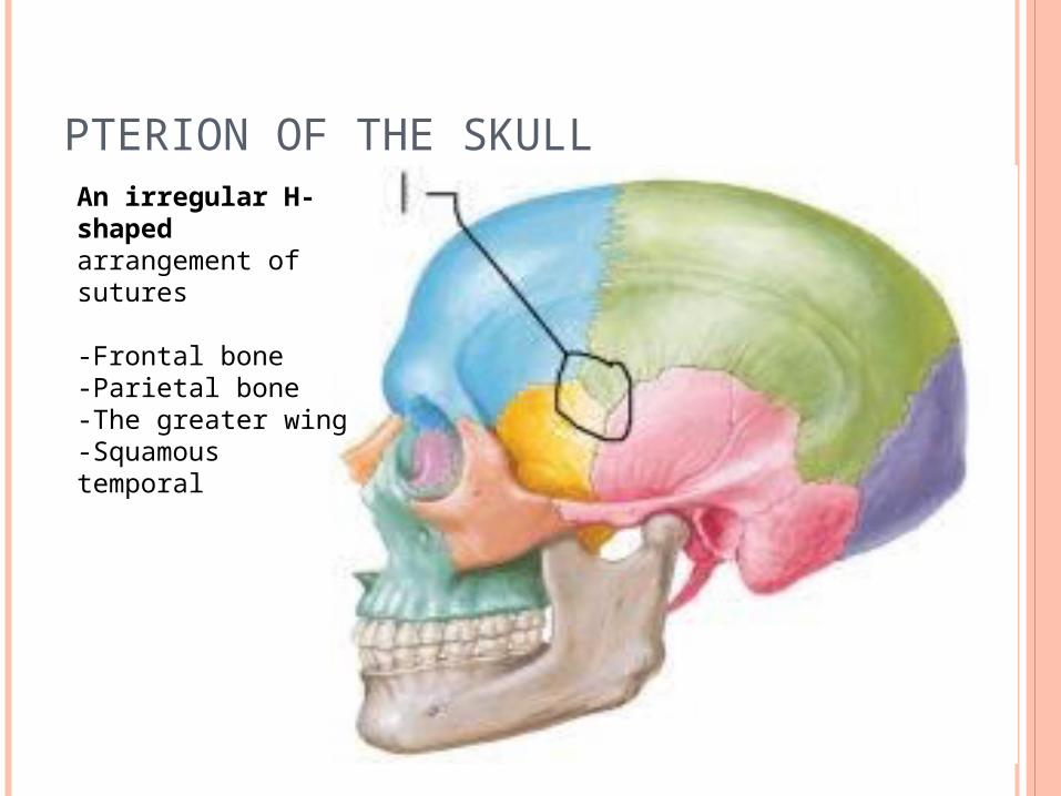
PTERION OF THE SKULLAn irregular H-shaped arrangement of sutures
-Frontal bone -Parietal bone-The greater wing-Squamous temporal
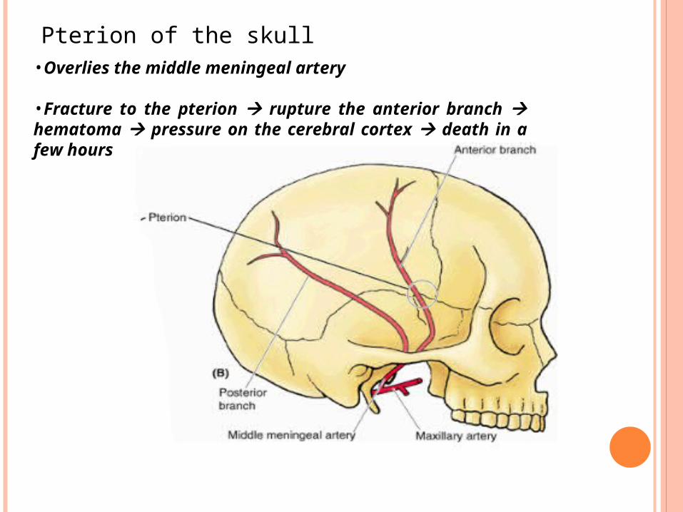
Pterion of the skull
10
•Overlies the middle meningeal artery
•Fracture to the pterion rupture the anterior branch hematoma pressure on the cerebral cortex death in a few hours
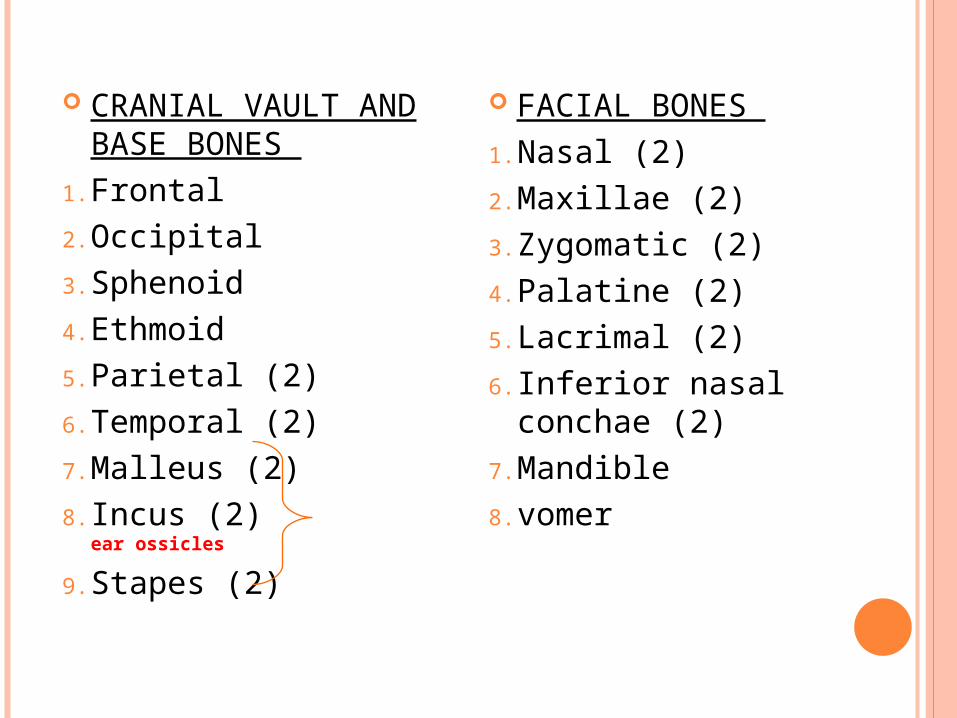
CRANIAL VAULT AND BASE BONES
1. Frontal2. Occipital3. Sphenoid4. Ethmoid5. Parietal (2)6. Temporal (2)7. Malleus (2)8. Incus (2) ear
ossicles
9. Stapes (2)
FACIAL BONES 1. Nasal (2)2. Maxillae (2)3. Zygomatic (2)4. Palatine (2)5. Lacrimal (2)6. Inferior nasal
conchae (2)7. Mandible8. vomer
FRONTAL BONE
FRONTAL BONE
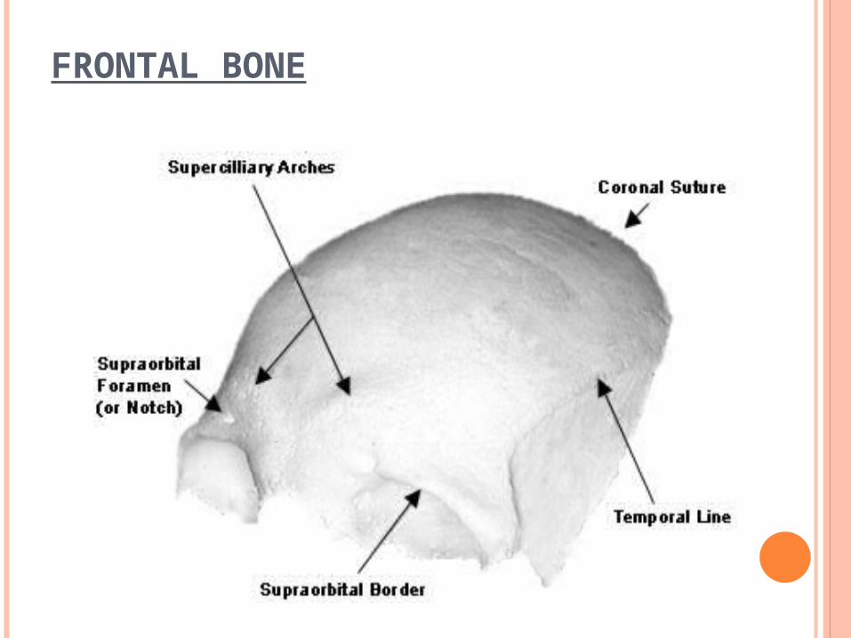
Formes the forehead region curves upwards and backwards from the
supraorbital margins to meet the parietal bones at the coronal suture.
Features :• supraorbital margin• supraorbital notch (foramen)• superciliary arch: a small elevation above the
supraorbital margin, forms the eyebrow ridge in
Articulation :parietal, nasal, ethmoid, lacrimal, maxillary,
and zygomatic bones.
FRONTAL BONE
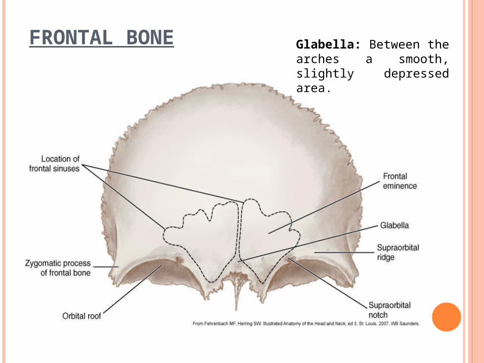
FRONTAL BONE Glabella: Between the arches a smooth, slightly depressed area.
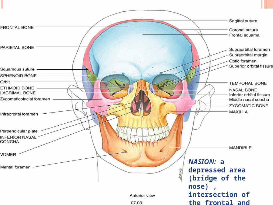
NASION: a depressed area (bridge of the nose) , intersection of the frontal and nasal bones
PARIETAL BONES
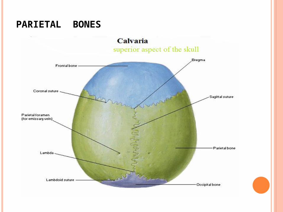
Make up the greater part of the vault
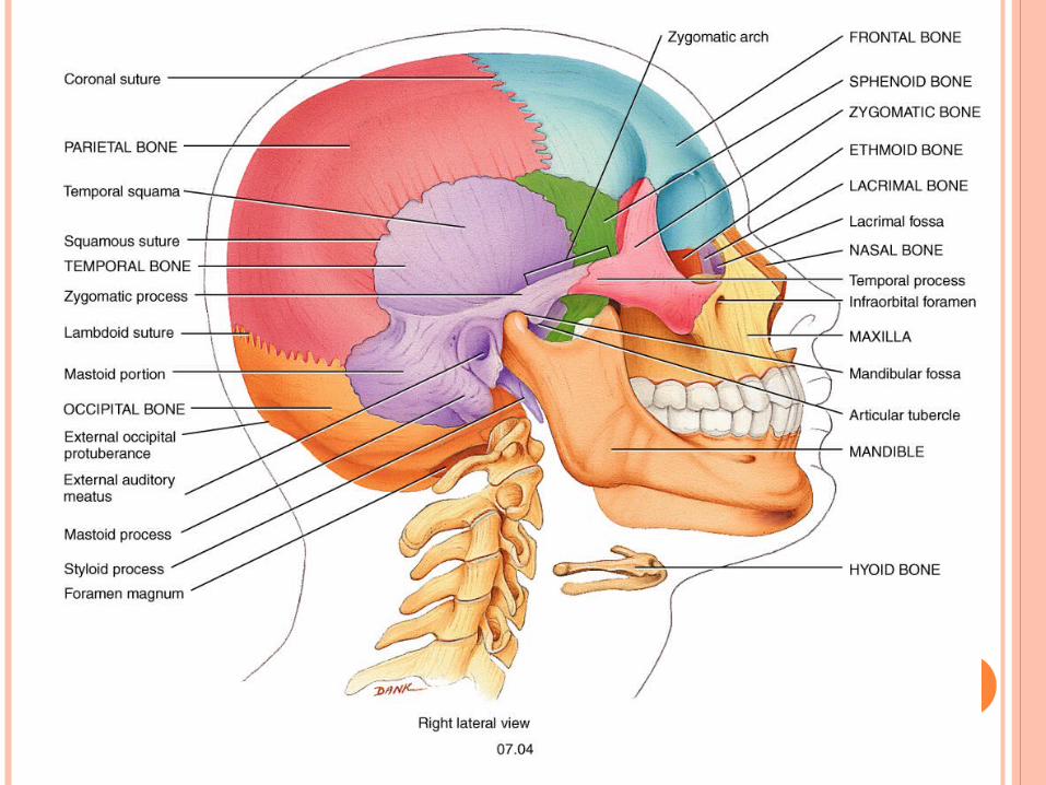
Articulation: • Articulate with each other in the median plane at the
sagittal suture.• Anteriorly with the frontal bone• posteriorly with the occipital bone • Inferiorly with the wing of the sphenoid and the squamous
part of the temporal bone.
Parietal foramen: • situated close to the sagittal suture on the posterior
part of the parietal • transmit emissary veins
PARIETAL BONES

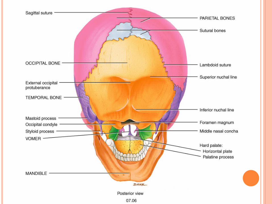
OCCIPITAL BONE
Forms posterior portion of the cranium and cranial base
Forms the posterior cranial fossa Articulates with the temporal bones and parietal bones Articulates with the first cervical vertebra (the atlas)
beneath it
Parts: • Foramen magnum• Basilar part (occiput)• Squamous part
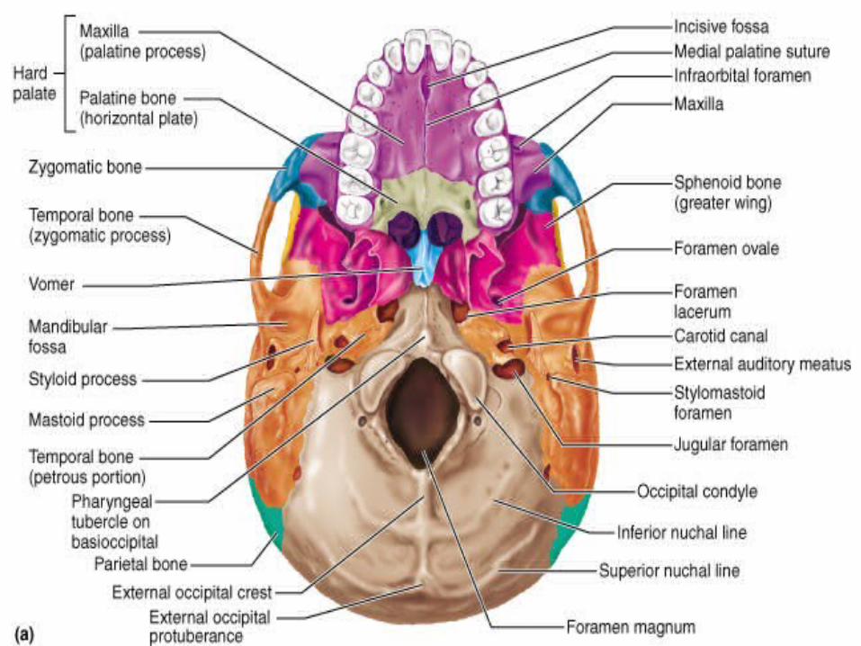
Features: • External occipital crest• External occipital protuberance• Superior nucal line• Inferior nucal line
Features cont.:
Occipital condyles: articulate with the atlas vertebra to form the atlanto-occipital joints.
Pharyngeal tubercle: small elevation, anterior to the foramen magnum, attachment to the uppermost fibres of the superior constrictor muscle.
Hypoglossal foramen: 1)lateral to the condyle.2)internal opening of this canal is situated
anterolateral wall of the foramen magnum.3) transmits: hypoglossal nerve, meningeal
branch of the ascending pharyngeal artery.

ORBITAL BONE

THE ORBIT Roof of the orbit is formed by:• the inferior surface of the orbital part of the
frontal bone. • the inferior surface of the lesser wing of the
sphenoid.
The lateral wall is formed by:• the orbital surfaces of the zygomatic bone • greater wing of the sphenoid
The lateral wall separates the orbital cavity from the temporal fossa anteriorly and from the middle cranial fossa posteriorly.
THE ORBIT The floor of the orbit is formed by: upper surface of the body of the maxilla
At the posteromedial corner of the orbital floor is a small triangular area formed by the orbital plate of the palatine bone
The medial orbital wall is formed by: • the frontal process of the maxilla• the lacrimal and the orbital plate of the
ethmoidal labyrinth • the body of the sphenoid
SUPERIOR ORBITAL FISSURE
The lateral wall and roof are separated posteriorly by the superior orbital fissure
• lying between the greater and lesser wings of the sphenoid
• transmits:1. the nerves to the muscles moving the
eyeballs2. the ophthalmic division of the trigeminal
nerve 3. ophthalmic veins
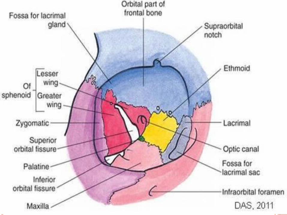
INFERIOR ORBITAL FISSURE
The lateral wall and floor are similarly separated by the inferior orbital fissure
• bordered by the maxilla and greater wing of sphenoid
• posteromedial part the inferior fissure communicates with the pterygopalatine fossa
• its anterolateral part communicates with the infratemporal fossa.
Infraorbital groove :• Starts from the medial part of the inferior
orbital fissure and continues forward • Becomes roofed over to form orbital
canal• then opens on the front of the maxilla at the
infraorbital foramen.
Infraorbital foramen: transmit the infraorbital nerve
OPTIC CANAL
Above the superior orbital fissure lying between the roots of the lesser wing of
the sphenoid. Transmits: The optic nerve and ophthalmic
artery

ETHMOID
ETHMOID
contributes to1. anterior cranial fossa2. the roof and lateral walls of the nasal cavity3. the nasal septum4. the medial walls of the orbits
consists of :1. the perpendicular plate2. the cribriform plate3. the two labyrinth
PERPENDICULAR PLATE
thin lamina of bone which occupies the upper part of the nasal septum.
possesses four margins:1. The superior margin attached to the inferior
surface of the cribriform plate in the midline. 2. The anterosuperior margin articulates with
the frontal and nasal bones. 3. The posterior margin articulates in its upper
part with the crest of the sphenoid and in its lowest part with the superior border of the vomer.
4. The anteroinferior surface is free in the dried skull but in life provides the attachment for the cartilage forming the anterior part of the nasal septum.
CRIBRIFORM PLATE
lies horizontally, occupying the notch between the orbital parts of the frontal.
Forms:1. the floor of the central part of the anterior
cranial fossa 2. the roof of the middle part of the nasal
cavity.
CRIBRIFORM PLATE
Features: perpendicular plate: descends from its
lower surface. crista galli: triangular plate projects
upwards from the midline of the cribriform plate, gives attachment to the falx cerebri.
foramina for the olfactory nerves: cribriform plate is pierced on either side of the crista galli
nasal slit: A narrow gap in the cribriform plate, lies immediately adjacent to the crista galli on each side.
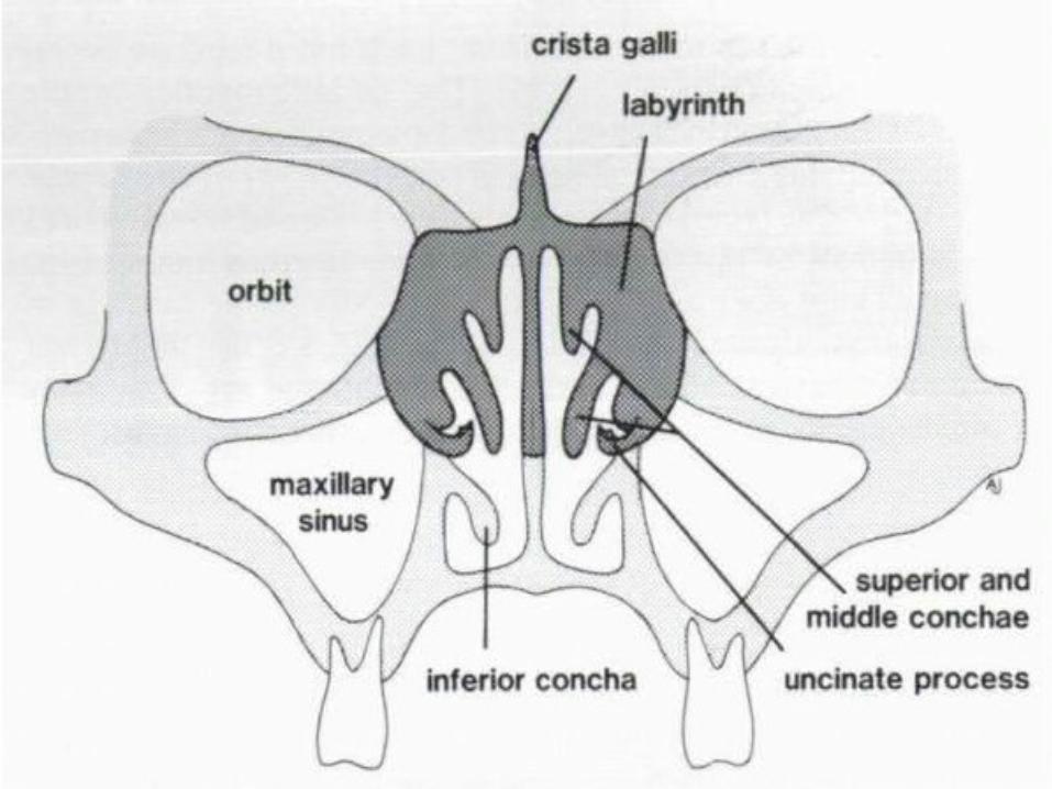
LABYRINTHS
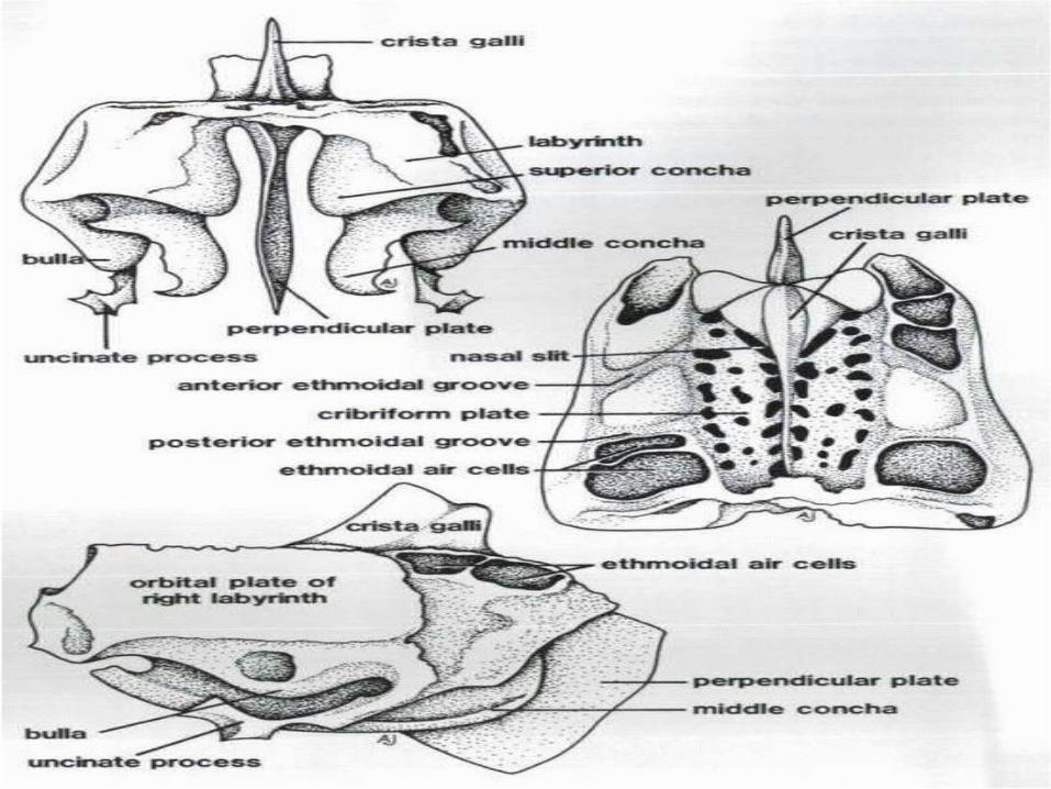
consists two vertical laminae -the medial plate-orbital (lateral) plate separated from each other by a number of
thin-walled ethmoidal air cells. relatively fragile.
LABYRINTHS
The orbital plate• forms part of the medial wall of the orbit
• Articulation : -above with the orbital part of the frontal -below with the orbital surface of the maxilla
and orbital plate of the palatine-in front with the lacrimal-behind with the sphenoid
LABYRINTHS
Projecting downwards and backwards from the anterior part of the orbital plate is the thin curved uncinate process, which helps form the lateral wall of the middle meatus.
LABYRINTHS
The medial plate • forms the upper part of the lateral wall of the
nasal cavity. • It is attached above to the inferior surface of
the cribriform plate.• The lower edge of the plate projects into the
nasal cavity as a horizontal, rolled ridge called the middle nasal concha.
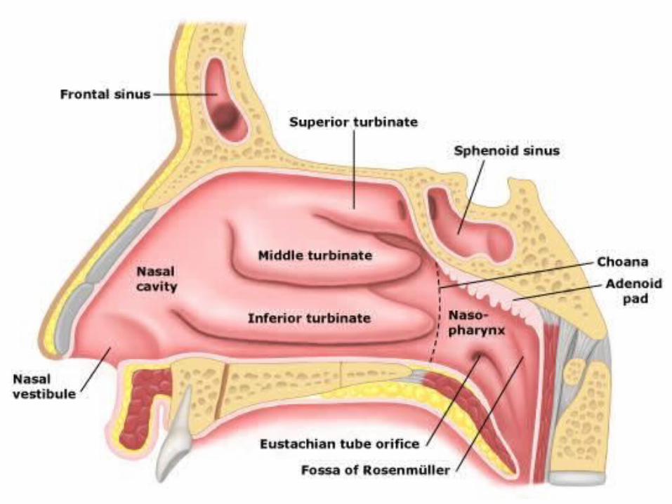
LABYRINTHS NASAL CONCHAE AND MEATUSo Superior nasal concha:• Between its upper and lower borders of the
medial plate • Above the superior concha is the
sphenoethmoidal recess
o Middle nasal concha: from lower edge o Inferior nasal concha
uncinate process :forms the lateral wall of middle meatus
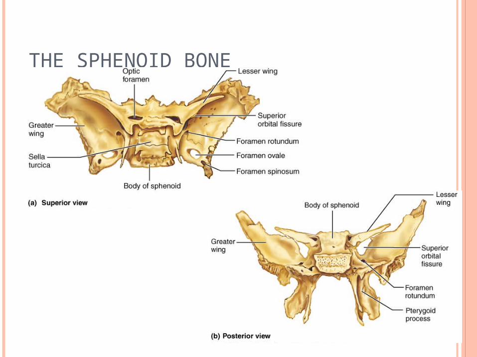
THE SPHENOID BONE
THE SPHENOID BONE Forms part of :1.middle cranial fossa 2.Orbit3.nasal cavity 4. infratemporal fossa
Consists of :1.central body2. two greater wings 3. two lesser wings4. two pterygoid processes.

THE SPHENOID BONE
BODY OF THE SPHENOID BONE
Cuboidal in shape Hollowed out by the two sphenoidal air sinuses. Articulates in front with the cribriform plate of the
ethmoid and behind with the basilar part of the occipital bone
features of the superior surface:1. optic groove2. pituitary fossa3. dorsum sellae (project as the posterior clinoid
processes)
The anterior border of the pituitary fossa frequently bears two small lateral tubercles, the middle clinoid processes.
BODY OF THE SPHENOID BONE
Sphenoidal crest: a median ridge on the anterior surface of the
body, below the articulation with the cribriform plate
this articulates with the posterior edge of the perpendicular plate of the ethmoid.
Either side of the sphenoidal crest are the openings of the sphenoidal sinuses.
provides the posterior part of the roof of the nasal cavity.
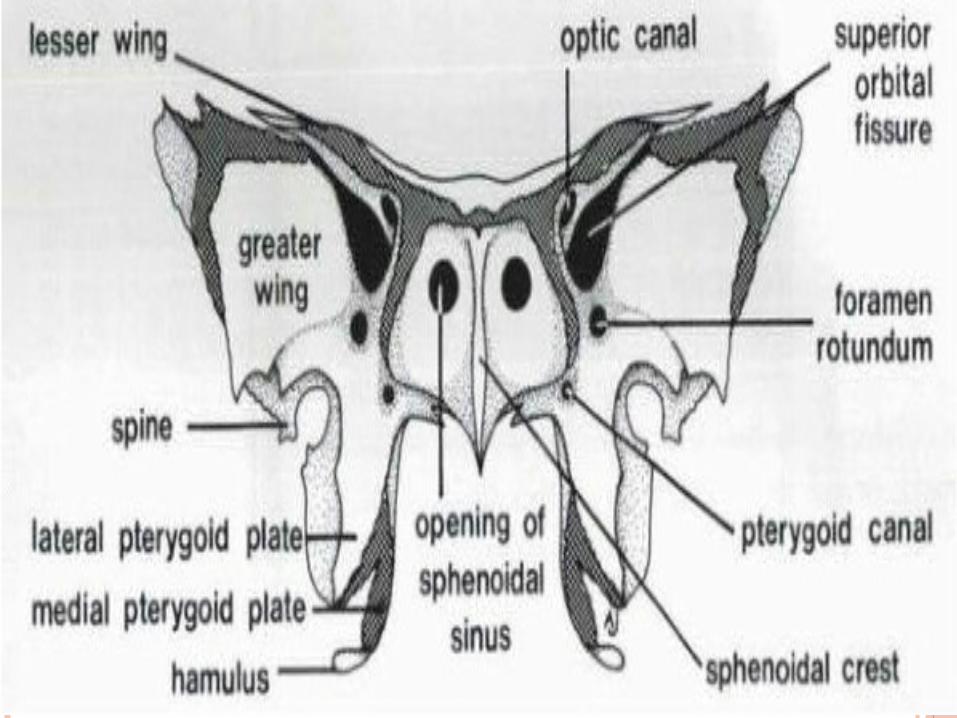
GREATER WINGS
The greater wings project laterally and upwards from the sides of the body.
Each has a superior (cerebral), lateral, and orbital surface.
The superior surface forms the floor of the lateral part of the middle cranial fossa
is pierced by foramina:1. rotundum2. ovale3. spinosum
GREATER WINGS The lateral surface: • convex • divided into upper (temporal) and lower (infratemporal) parts
by the infratemporal crest.
The infratemporal surface:• foramina ovale • foramina spinosum • the spine of the sphenoid
The orbital surface:• faces forwards and somewhat medially• A horizontal ridge divides it into:
A large upper area, forming the posterior part of the lateral orbital wall
A small lower area which provides the upper part of the posterior boundary of the pterygopalatine fossa. Which is pierced medially by the anterior opening of the foramen rotundum
GREATER WINGS
Margins of the greater wing:
• superior and lateral borders makes sutural connections at its tip (i.e. in the region of the pterion) This border ends posteriorly at the spine of the sphenoid.
• posterior border runs medially to the body of the sphenoid.
• medially it forms the anterior boundary of the foramen lacerum.
GREATER WINGS
o Margins of the greater wing cont:• Below the pterion a common border is shared between
the orbital and lateral surfaces. In its upper part this articulates with the zygomatic bone.
• In its lower part the border turns medially to form the lower margin of the orbital surface and the upper boundary of the inferior orbital fissure.
• Medial to the pterion the common border is between the orbital and superior surfaces.
• Laterally this border articulates with the orbital plate of the frontal while medially it provides the inferior border of the superior orbital fissure.
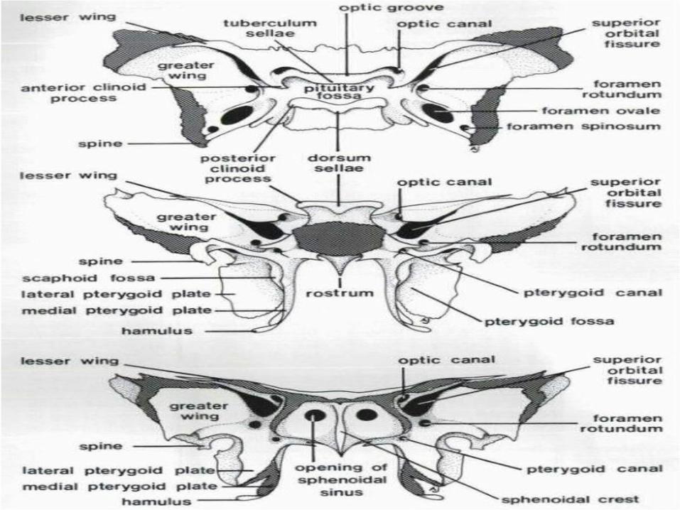
LESSER WINGS
project from the upper part of the body, anterior to the greater wings.
The superior surface forms a small, posterior part of the anterior cranial fossa.
The inferior surface provides :1. superior boundary of the superior orbital fissure2. a small area of the posterior part of the orbital roof.
The posterior surface: ends medially at a projection, the anterior clinoid process.
The lesser wing is attached to the body of the sphenoid by two roots between which lies the optic canal.

66

PTERYGOID PROCESSES
attached to the inferior surface of the sphenoid
bone in the region where the greater wing fuses with the body.
consists of medial and lateral pterygoid plates which are fused anteriorly.
The two plates diverge posteriorly to enclose the pterygoid fossa.
Just below the body of the sphenoid the pterygoid process is pierced by the pterygoid canal.
PTERYGOID PROCESSES upper part of anterior border: provides the posterior boundaries of the
pterygopalatine fossa and pterygomaxillary fissure.
Inferiorly the two plates diverge leaving a fissure into which fits the pyramidal process of the palatine bone.
Adjacent to this fissure the anterior border is roughened for articulation with the perpendicular plate of the palatine bone.
PTERYGOID PROCESSES LATERAL PTERYGOID PLATE
lateral surface:o forms the medial wall of the infratemporal
fossa o origin for the inferior head of the lateral
pterygoid muscle
medial surface o forms the lateral boundary of the pterygoid
fossa o gives attachment to the main part of the
medial pterygoid muscle.
PTERYGOID PROCESSES MEDIAL PTERYGOID PLATE
medial surface forms the lateral boundary of the nasal aperture
lateral surface forms the medial wall of the pterygoid fossa.
PTERYGOID PROCESSES MEDIAL PTERYGOID PLATE
The posterior border of the medial pterygoid plate
lower two-thirds:• Sharp • Gives origin to the superior constrictor muscle
upper one-third:• Blunter• Gives attachment to the pharyngobasilar fascia
(a component of the wall of the nasopharynx) which is pierced by the auditory tube
PTERYGOID PROCESSES
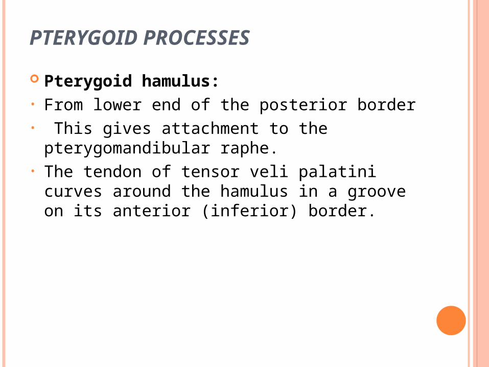
Pterygoid hamulus:• From lower end of the posterior border• This gives attachment to the
pterygomandibular raphe. • The tendon of tensor veli palatini curves
around the hamulus in a groove on its anterior (inferior) border.
THE SPHENOID BONE
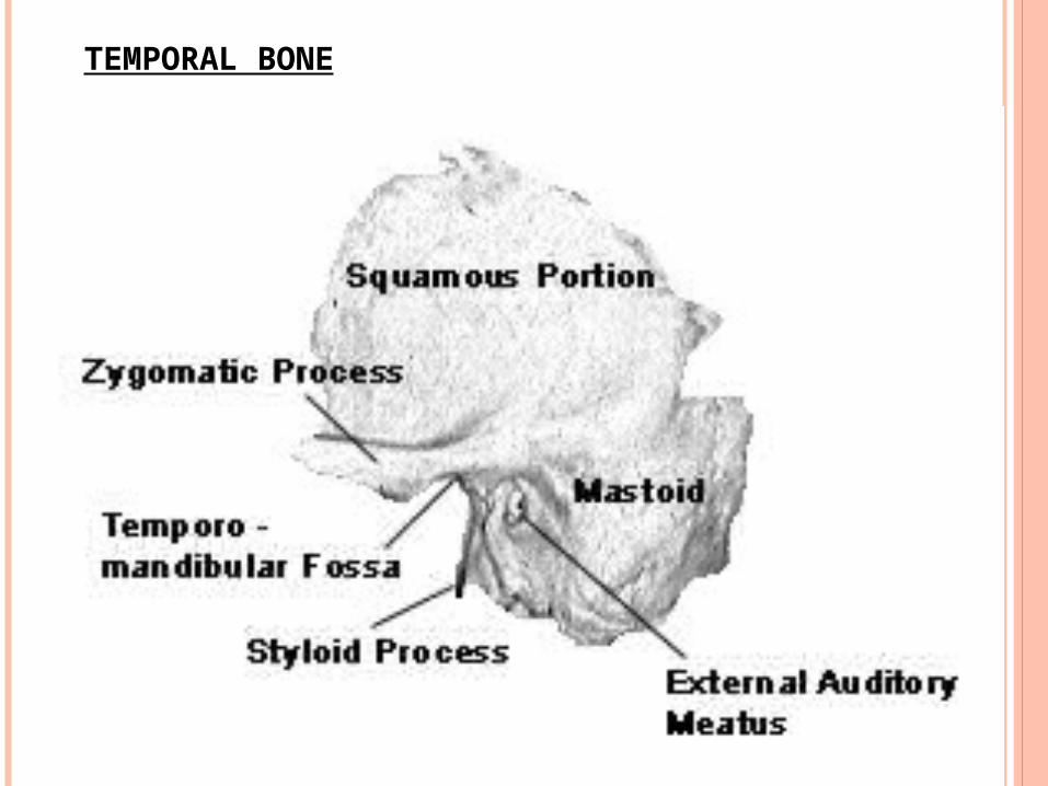
TEMPORAL BONE
TEMPORAL
bone consisting of four fused elements: 1.periotic2.squamous3.tympanic 4.styloid
The periotic ossifies in the cartilage of the auditory capsule.
The squamous is an intramembranously ossifying element which was originally part of the dermal armour shield.
The tympanic is also a dermal bone and is represented in non-mammals by the angular, one of the numerous bones of the lower jaw.
The styloid ossifies in the dorsal part of the cartilage of the hyoid arch.
Contained within the periotic are the middle ear (or tympanic) cavity and the cavity for the internal ear.
Traversing the middle-ear cavity are the three ossicles, malleus, incus, and stapes, which convey vibrations from the ear drum to the internal ear.
PETROUS PART
houses the tympanic and internal ear cavities. it is composed of very hard bone. It lies entirely within the cranial base, wedged between the sphenoid and occipital bones
with its apex directed anteromedially. It has anterior, posterior, and inferior surfaces.
PETROUS PART
The anterior surface forms part of the middle cranial fossa just posterolateral to the apex (and to foramen lacerum in the articulated skull) it is marked by the trigeminal impression which lodges the trigeminal ganglion housed within the trigeminal cave.
Posterolateral to the trigeminal impression much of the anterior surface is formed by a plate of bone called the tegmen tympani.
The tegmen tympani has two small openings from which grooves may be traced leading forwards and medially.
The more posteromedial of the openings is the hiatus for the greater petrosal nerve and the more anterolateral the hiatus for the lesser petrosal nerve. The anterior edge of the tegmen tympani projects downwards between the squamous and tympanic parts of the temporal.
PETROUS PART
On the under surface of the cranial base this downturned edge can be seen as a lip of bone in the medial part of the squamotympanic fissure, dividing it into a posterior petrotympanic and an anterior petrosquamous part
The petrotympanic fissure leads into the tympanic cavity.
Its medial end is widened to form the canaliculus for the chorda tympani through which that branch of the facial nerve escapes from the tympanic cavity to the exterior of the skull.
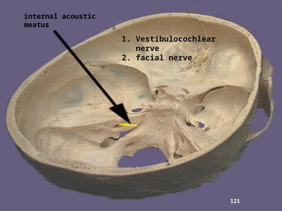
The posterior surface of the petrous bone forms part of the posterior cranial fossa, the superior border of the bone providing the boundary between middle and posterior fossae. approximately in the centre of the posterior surface is the opening of the internal acoustic meatus.
PETROUS PART
The inferior surface makes up part of the under surface of the cranial base.
Very irregular and has a large, circular opening which leads into the carotid canal and behind this the deep jugular fossa which houses the superior bulb of the internal jugular vein.
The ridge separating the carotid opening from the jugular fossa is pierced by the canaliculus for the tympanic branch of the glossopharyngeal nerve.
in front of the opening of the carotid canal, in the angle between the petrous and squamous parts, is the opening of the bony part of the auditory tube. This leads into the anterior part of the tympanic cavity.
PETROUS PART
With the soft tissues in place the tube is continued medially in cartilage to open into the nasopharynx.
The anterior border articulates laterally with the squamous part of the temporal bone at the petrosquamosal suture and medially with the greater wing of the sphenoid at a cartilaginous joint.
PETROUS PART
The posterior border articulates by a cartilaginous joint with the occipital bone.
Immediately adjacent to the jugular fossa the petrous and occipital bones are separated by a wide gap, the jugular foramen.
The apex of the petrous bone forms the posterolateral boundary of the foramen lacerum and is here pierced by the inner opening of the carotid canal.
MASTOID PART
the most posterior part of the temporal bone
bears on its external aspect a conical projection, the mastoid process to which is attached the sternocleidomastoid muscle.
Medial to the process is a deep groove, the mastoid notch, for the attachment of the posterior belly of the digastric muscle.
Further medially still is a shallow groove in which runs the occipital artery.
The endocranial surface is marked by a deep, curved groove for the sigmoid sinus.
The mastoid part articulates by its superior border with the parietal bone and by its posterior border with the occipital bone. Anteriorly it is fused with the squamous and petrous parts.
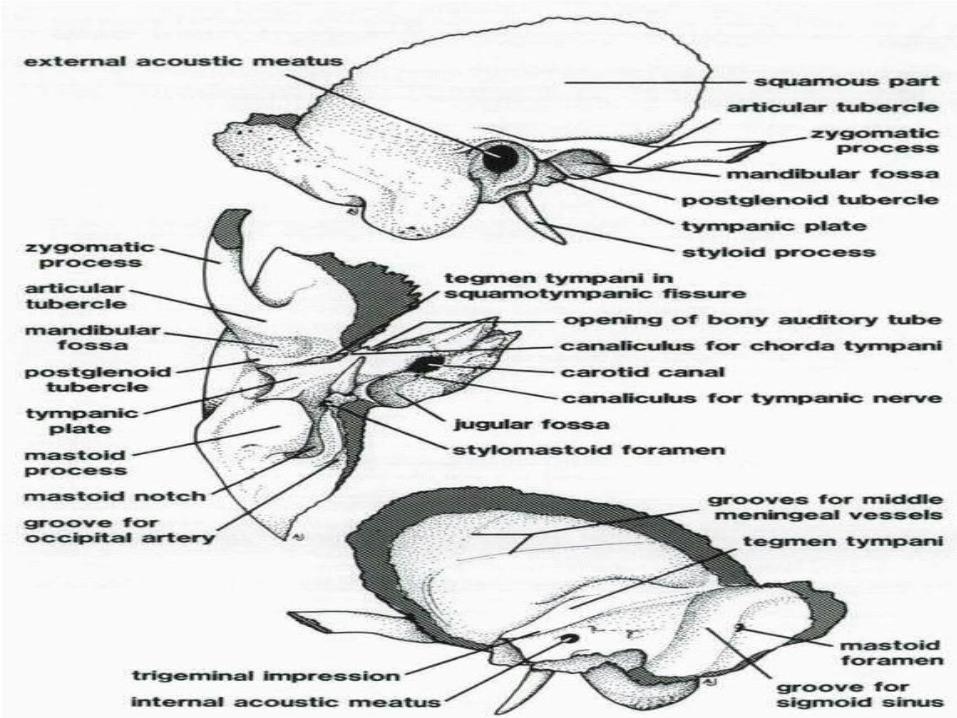
SQUAMOUS PART
is a large plate of bone which forms the lower part of the side wall of the cranial vault and a small area of the lateral part of the cranial base.
The temporal surface is smooth and forms much of the temporal fossa from which the temporalis muscle takes origin.
Projecting forwards from the temporal surface is the zygomatic process which articulates anteriorly with the zygomatic bone to form the zygomatic arch.
The cerebral surface is marked by a number of depressions, corresponding to the convolutions of the temporal lobes of the cerebral hemispheres, and by grooves for the posterior branches of the middle meningeal vessels.
SQUAMOUS PART
The inferior aspect of the squamous part bears the superior articular surface of the temporomandibular joint formed by the articular tubercle and the anterior part of the mandibular fossa.
The fossa is transversely widened and is bounded in front by the articular tubercle and completed behind by the tympanic part of the temporal bone.
Located in the depth of the fossa, between the squamous and tympanic, is the squamotympanic fissure. Only the squamous (i.e. anterior) part of the fossa is articular, the tympanic plate being excluded from the temporomandibular joint by the attachment of the capsule to the anterior lip of the squamotympanic fissure
SQUAMOUS PART
Laterally, the posterior edge of the articular surface is downturned to form a lip of bone called the postglenoid tubercle.
Medially the squamotympanic fissure is divided, into petrotympanic and petrosquamous portions by the lip of the tegmen tympani.
The articular tubercle is sometimes described as the anterior root of the zygomatic process (the posterior root being the ridge of bone which continues from the zygomatic process above the external acoustic meatus).
The posterosuperior border of the squamous part articulates with the parietal bone and the anteroinferior border with the greater wing of the sphenoid bone.
TYMPANIC PLATE
This is a curved plate of bone which is fused with the petrous, mastoid, and squamous parts to complete the external acoustic meatus. It partly ensheaths the base of the styloid process.
STYLOID PROCESS
The styloid process is a slender, curved bony projection of variable length.
It is usually broken in the dried skull. Its upper part is ensheathed by and fused
with the tympanic plate. The process gives attachment to the styloid
muscles and the stylohyoid and stylomandibular ligaments.
Between the styloid and mastoid processes is the stylomastoid foramen through which the facial nerve leaves the skull.
TYMPANIC CAVITY
Is a mediolaterally compressed space within the petrous bone.
In its lateral wall is the ear drum or tympanic membrane which separates the cavity from the external acoustic meatus.
The medial wall is formed by the bony partition separating the tympanic cavity from the internal ear.
This wall has two openings:
1. the fenestra vestibuli
2. fenestra cochleae.
TYMPANIC CAVITY
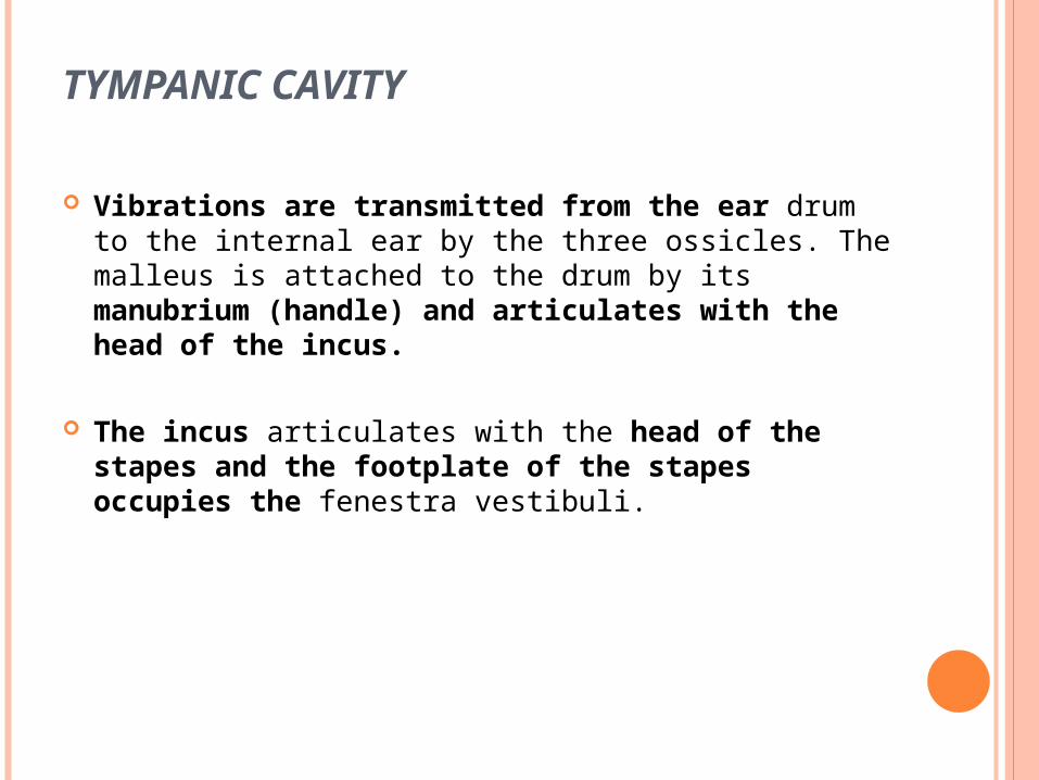
Vibrations are transmitted from the ear drum to the internal ear by the three ossicles. The malleus is attached to the drum by its manubrium (handle) and articulates with the head of the incus.
The incus articulates with the head of the stapes
and the footplate of the stapes occupies the fenestra vestibuli.
TYMPANIC CAVITY
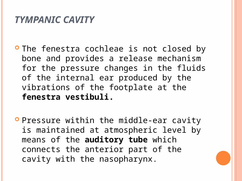
The fenestra cochleae is not closed by bone and provides a release mechanism for the pressure changes in the fluids of the internal ear produced by the vibrations of the footplate at the fenestra vestibuli.
Pressure within the middle-ear cavity is maintained at atmospheric level by means of the auditory tube which connects the anterior part of the cavity with the nasopharynx.
TEMPORAL BONE
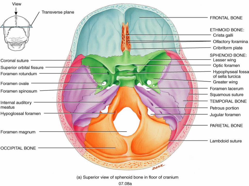
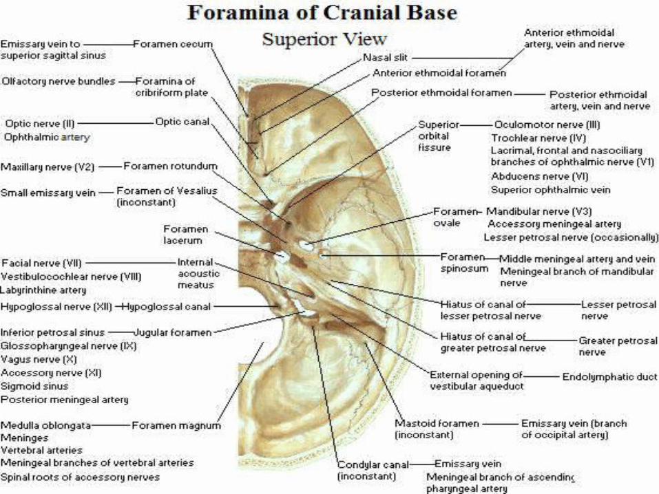
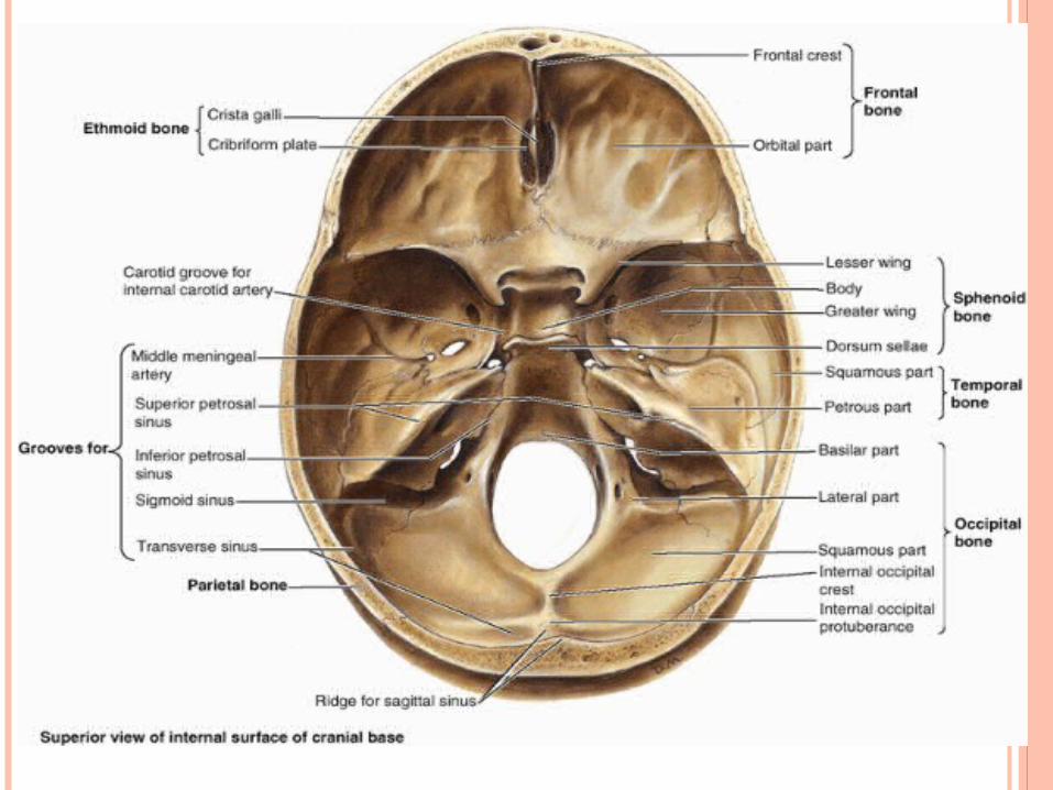
INTERNAL SURFACE OF THE CRANIAL BASE (CRANIAL CAVITY)
subdivided into three regions 1.Anterior cranial fossae2.Middle cranial fossae3.Posterior cranial fossae

99
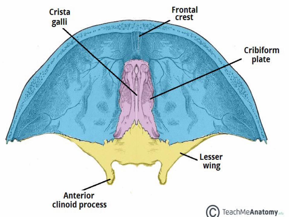
ANTERIOR CRANIAL FOSSA
houses frontal lobes of the cerebral hemispheres.
fossa is made up from:-orbital parts of the frontal bone
-lesser wings of the sphenoid The posterior boundary provided by the
anterior border of the optic groove. Medially each lesser wing bears a prominentprojection termed the anterior clinoid
process.
102
MIDDLE CRANIAL FOSSA
-the temporal lobes of the cerebral hemispheres -the pituitary gland
Boundries Anterior : -posterior borders of the lesser wings of the sphenoid -anterior margin of the optic groove posterior :-superior borders of the petrous parts of the temporal
bones central region: formed by the body of the sphenoid lateral regions: formed by the corresponding greater
wing of the sphenoid, the anterior surface of the petrous temporal and the inferior part of the squamous temporal bone.

104
Sitting on the floor of the lateral portion of the middle cranial fossa is the temporal lobe
Features: • pituitary (hypophysial) fossa: which houses the pituitary gland
(hypophysis cerebri), sella turcica
• optic groove: In front of the pituitary fossa
• optic canal: Follow groove laterally, situated between the roots of the lesser wing transmitting the optic nerve and ophthalmic artery to the orbit.
105
MIDDLE CRANIAL FOSSA
MIDDLE CRANIAL FOSSA
optic chiasma: In the cranial cavity the fibres of the optic nerve undergo a partial decussation (crossing over), lies just above the optic groove.
tuberculum sellae: a rounded swelling between the pituitary fossa and the optic groove
dorsum sellae: Posterior to the fossa, the superolateral angles form the posterior clinoid processes
MIDDLE CRANIAL FOSSA
superior orbital fissure:• Between the greater and lesser wings• transmits: 1. oculomotor nerve2. trochlear nerve3. abducent nerve4. the ophthalmic division of the trigeminal
nerve5. the superior and inferior ophthalmic veins
MIDDLE CRANIAL FOSSA
The medial part of each greater wing is pierced by three foramina.
foramen rotundum: • This runs directly forwards into the
pterygopalatine fossa• transmits the maxillary division of the
trigeminal nerve
MIDDLE CRANIAL FOSSA Foramen ovale• posterior to foramen rotundum • opens downwards into the infratemporal
fossa • Through it passes:1. the mandibular division of the trigeminal
nerve2. lesser petrosal nerve
(which may have its own separate foramen)
3. accessory meningeal artery
MIDDLE CRANIAL FOSSA
Foramen spinosum • postero-lateral to foramen ovale• Transmits:1. the middle meningeal vessels
the main arterial and venous channels to the meninges
2.the nervus spinosus a meningeal branch of the mandibular division of the trigeminal nerve
Occasionally other foramina may be present for small emissary veins.
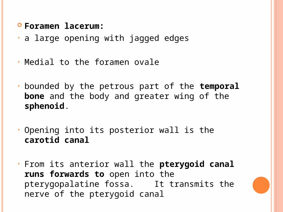
Foramen lacerum:• a large opening with jagged edges
• Medial to the foramen ovale
• bounded by the petrous part of the temporal bone and the body and greater wing of the sphenoid.
• Opening into its posterior wall is the carotid canal
• From its anterior wall the pterygoid canal runs
forwards to open into the pterygopalatine fossa. It transmits the nerve of the pterygoid canal
At a level below these two openings the foramen lacerum is closed by a plug of cartilage pierced by only one or two small emissary veins.

113
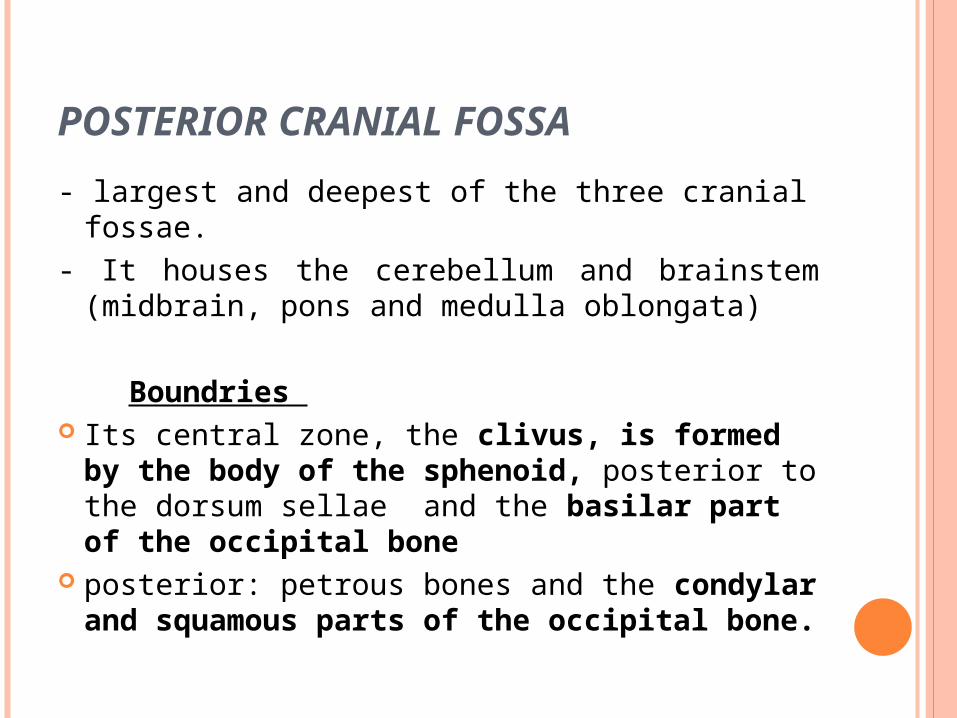
POSTERIOR CRANIAL FOSSA
- largest and deepest of the three cranial fossae.- It houses the cerebellum and brainstem
(midbrain, pons and medulla oblongata)
Boundries Its central zone, the clivus, is formed by the
body of the sphenoid, posterior to the dorsum sellae and the basilar part of the occipital bone
posterior: petrous bones and the condylar and squamous parts of the occipital bone.
115
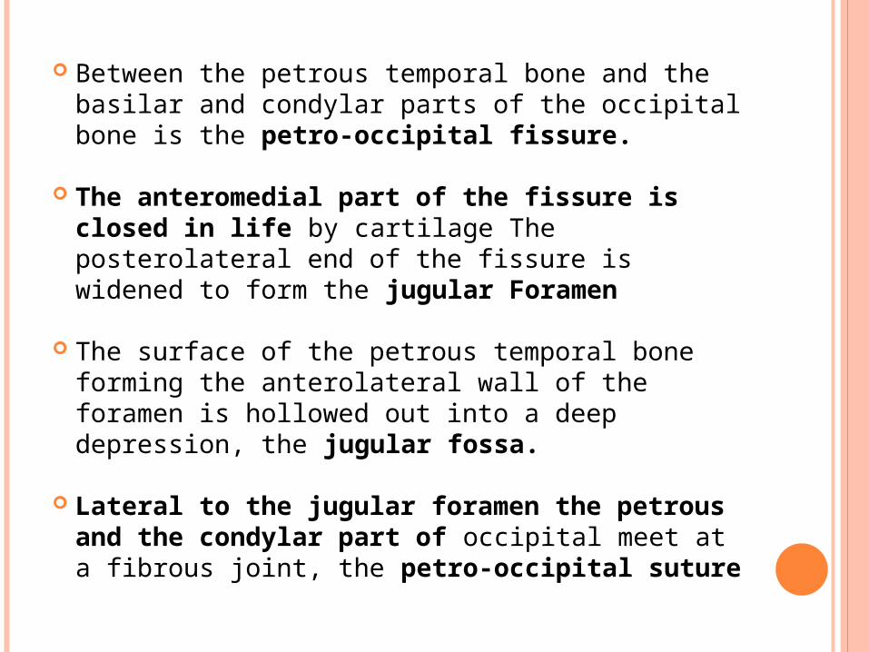
Between the petrous temporal bone and the basilar and condylar parts of the occipital bone is the petro-occipital fissure.
The anteromedial part of the fissure is closed in life by cartilage The posterolateral end of the fissure is widened to form the jugular Foramen
The surface of the petrous temporal bone forming the anterolateral wall of the foramen is hollowed out into a deep depression, the jugular fossa.
Lateral to the jugular foramen the petrous and the condylar part of occipital meet at a fibrous joint, the petro-occipital suture
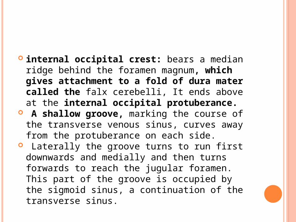
internal occipital crest: bears a median ridge behind the foramen magnum, which gives attachment to a fold of dura mater called the falx cerebelli, It ends above at the internal occipital protuberance.
A shallow groove, marking the course of the transverse venous sinus, curves away from the protuberance on each side.
Laterally the groove turns to run first downwards and medially and then turns forwards to reach the jugular foramen. This part of the groove is occupied by the sigmoid sinus, a continuation of the transverse sinus.
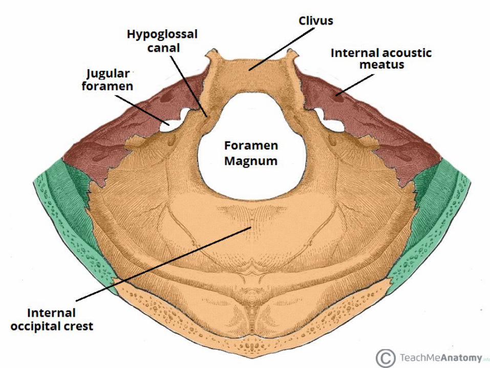
Foramen magnum transmits :
1. medulla oblongata (plus meninges)2. spinal roots of the accessory nerves3. vertebral arteries4. number of other structures
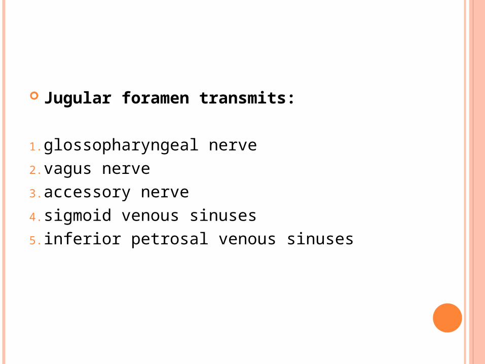
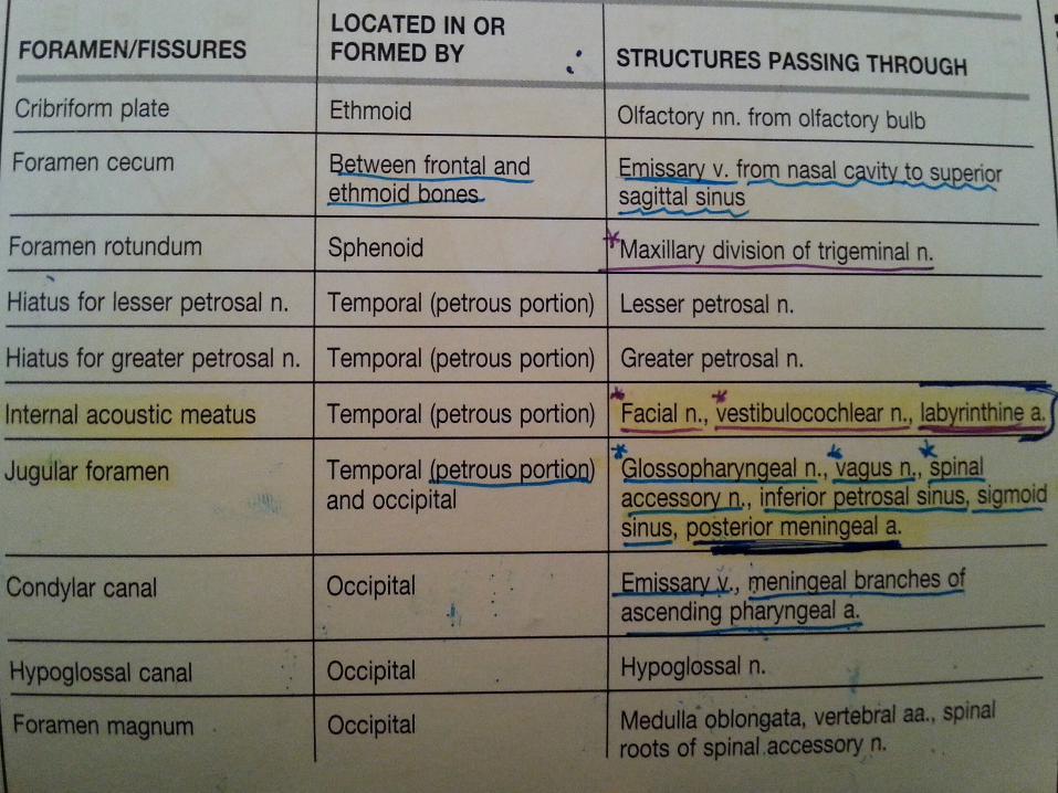
Jugular foramen transmits:
1. glossopharyngeal nerve2. vagus nerve3. accessory nerve4. sigmoid venous sinuses5. inferior petrosal venous sinuses
121
internal acoustic meatus
1. Vestibulocochlear nerve 2. facial nerve
THE UPPER JAW AND BONY PALATE
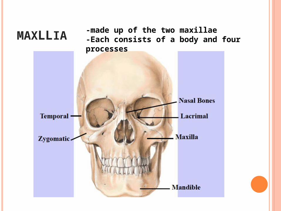
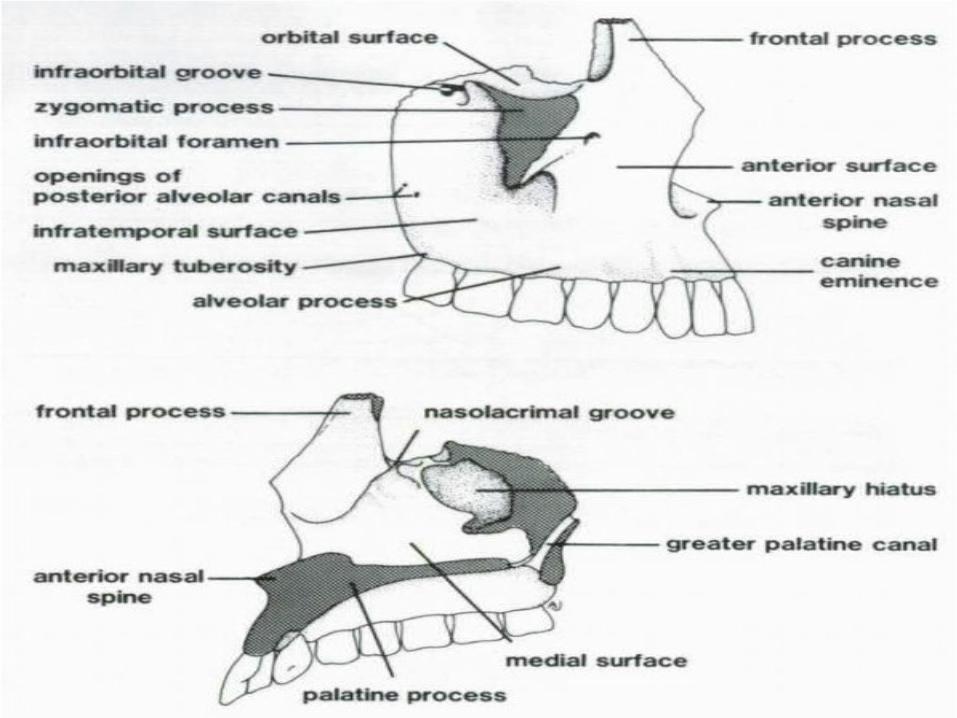
MAXLLIA-made up of the two maxillae-Each consists of a body and four processes
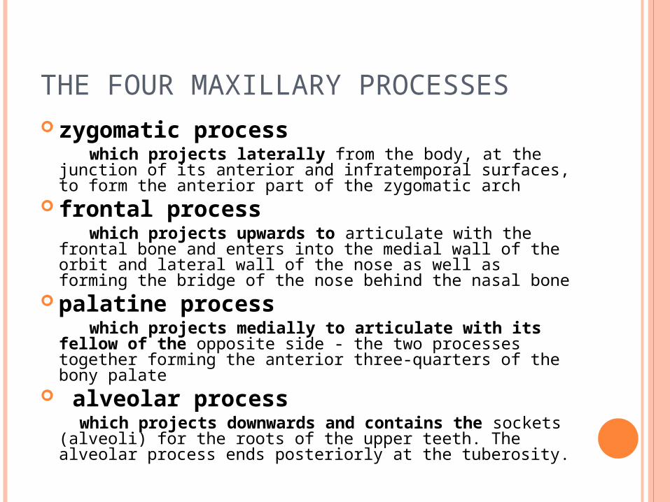
THE FOUR MAXILLARY PROCESSES zygomatic process which projects laterally from the body, at the
junction of its anterior and infratemporal surfaces, to form the anterior part of the zygomatic arch
frontal process which projects upwards to articulate with the frontal
bone and enters into the medial wall of the orbit and lateral wall of the nose as well as forming the bridge of the nose behind the nasal bone
palatine process which projects medially to articulate with its
fellow of the opposite side - the two processes together forming the anterior three-quarters of the bony palate
alveolar process which projects downwards and contains the
sockets (alveoli) for the roots of the upper teeth. The alveolar process ends posteriorly at the tuberosity.
THE MAXILLARY BODY
Roughly pyramidal in shape Hollowed out by the maxillary sinus The upper (orbital) surface of the body
occupies the floor of the orbit The anterior surface forms the curved
external surface of the upper jaw The posterior (infratemporal) surface
provides the anterior wall of the infratemporal fossa
Above the incisor teeth the anterior surface has a shallow depression termed the incisive fossa
laterally is the deeper canine fossa which is separated from the incisive fossa by the canine eminence produced by the root of the canine
Above the canine fossa is the infraorbital foramen.
The anterior surface ends medially at the anterior nasal aperture.
At the inferior margin of this aperture the maxillae of the two sides form a median projection, the anterior nasal spine.
The surface ends below at a prominent rounded eminence, the maxillary tuberosity, located behind the last molar tooth.
More medially the lower part of the infratemporal surface articulates with the pyramidal process of the palatine bone
(the pyramidal process projects posterolaterally from the junction of the perpendicular and horizontal plates of the palatine to intervene between the maxilla and pterygoid process)
while the upper part forms the anterior wall of the pterygopalatine fossa.

posterior alveolar canals: openings through which the posterior alveolar neurovascular bundles reach the upper molar teeth

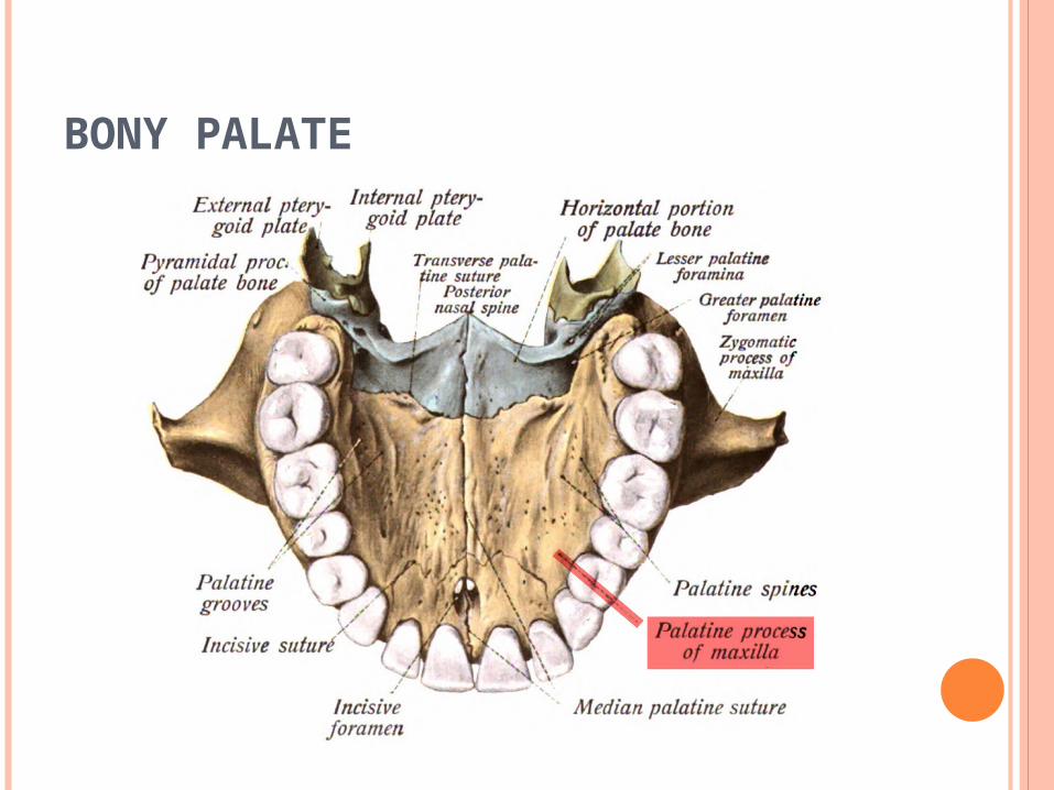
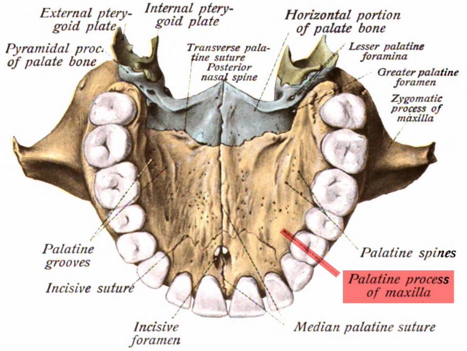
BONY PALATE
BONY PALATE
• provides the floor of the nasal cavity and the roof of the mouth.
• Its anterior three-quarters: palatine processes of the maxillae.
• its posterior one-quarter: the horizontal plates of the palatine bones.
sutures of the palate 1. median suture: between the right and left
sides 2.transverse suture: between the palatine
processes of the maxillae and the horizontal plates of the palatines.

BONY PALATE
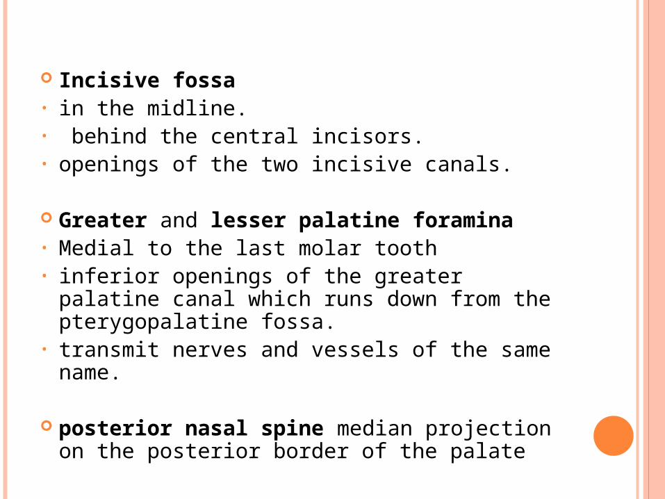
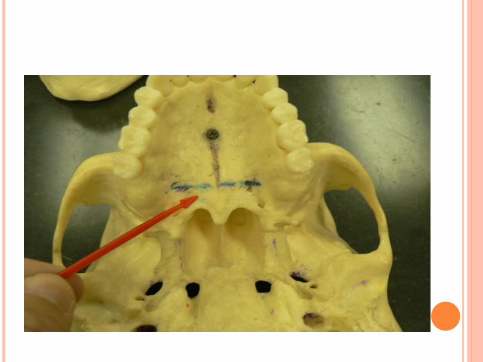
Incisive fossa • in the midline.• behind the central incisors.• openings of the two incisive canals.
Greater and lesser palatine foramina• Medial to the last molar tooth• inferior openings of the greater palatine
canal which runs down from the pterygopalatine fossa.
• transmit nerves and vessels of the same name.
posterior nasal spine median projection on the posterior border of the palate
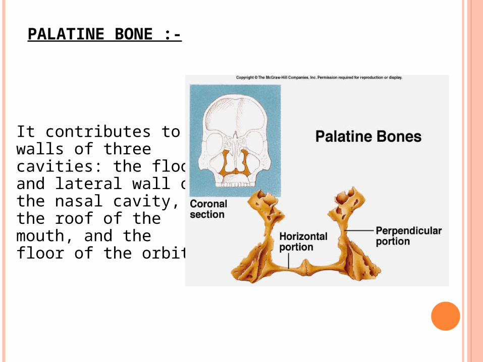
PALATINE BONE :-
It contributes to the walls of three cavities: the floor and lateral wall of the nasal cavity, the roof of the mouth, and the floor of the orbit
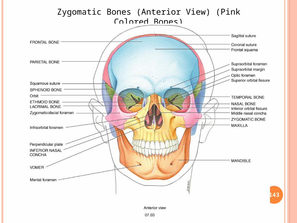
THE ZYGOMATIC BONE
The zygomatic arch1.zygomatic process of the maxilla2.zygomatic bone3.zygomatic process of the squamous part
of the temporal
The temporal fascia is attached to its sharp upper border. The masseter muscle arises from its inferior border and medial surface.
143
Zygomatic Bones (Anterior View) (Pink Colored Bones)
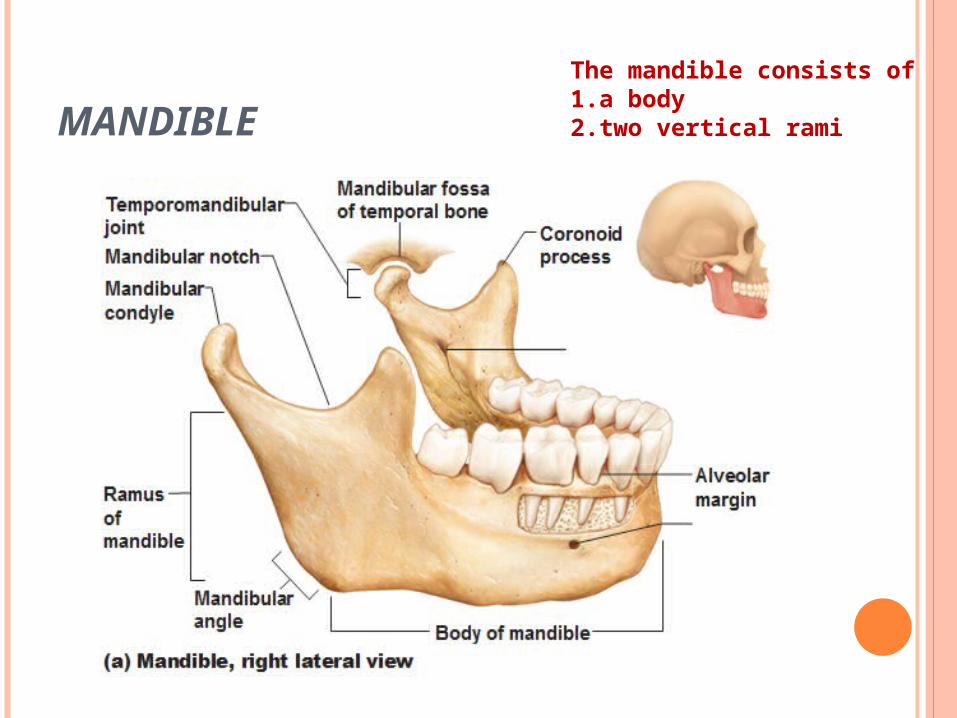
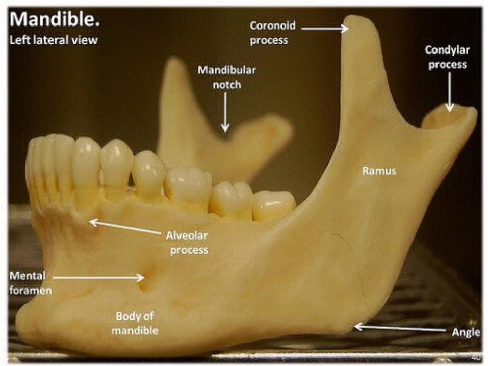
MANDIBLEThe mandible consists of 1.a body 2.two vertical rami
EXTERNAL SURFACE OF THE BODY
vertical ridge: on The external surface below the interproximal space between the central incisor teeth, This indicates the line of fusion of the symphysis menti.
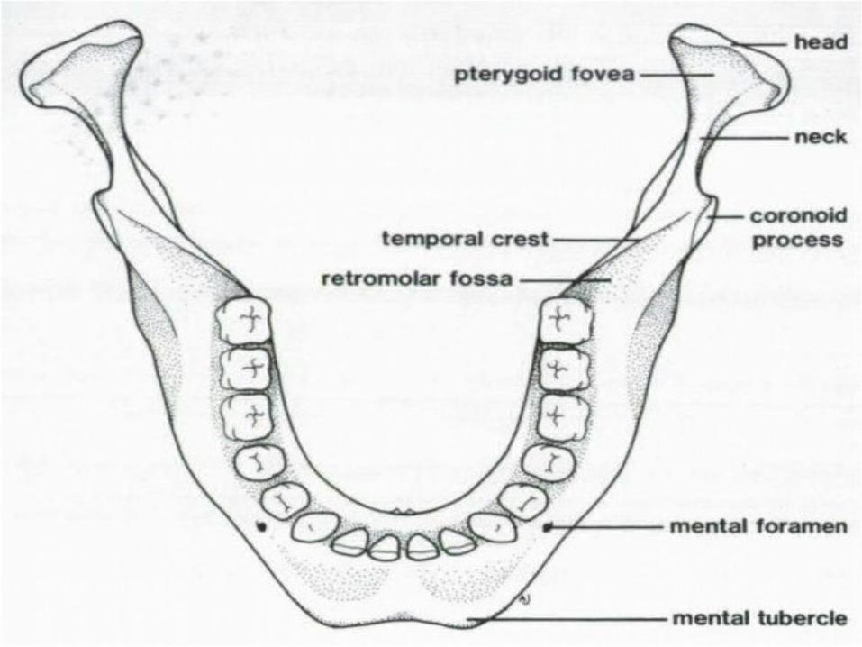
Mental protuberance: a raised area Inferior to vertical ridge, gives the human chin its characteristic shape.
mental tubercle: more prominent area to lateral the protuberance.
oblique line: a faint ridge Running backwards and upwards from the mental tubercle. Below the last molar tooth the ridge becomes more prominent before becoming continuous with the anterior border of the ramus.
mental foramen: above the oblique line, in the region of the premolar teeth, transmits the mental branches of the inferior alveolar nerve and blood vessels
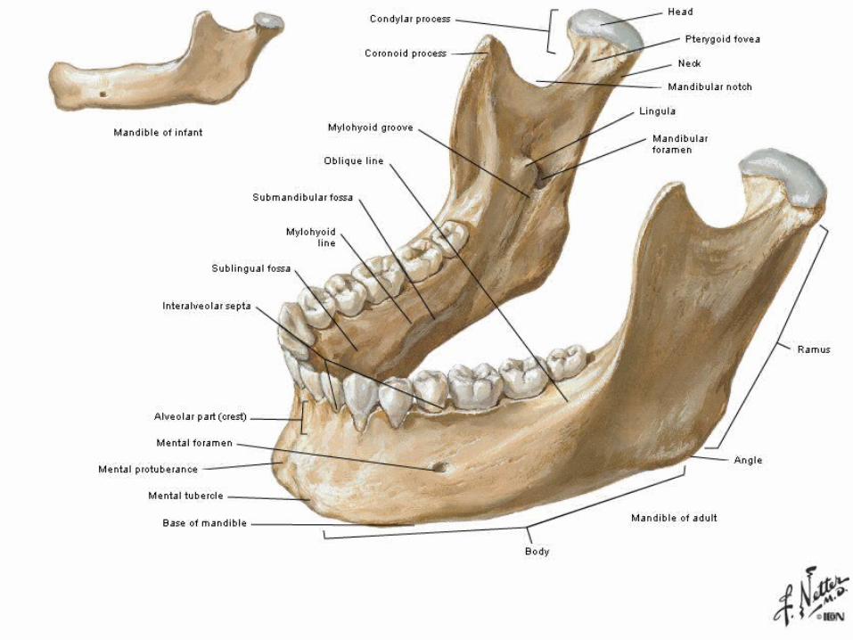

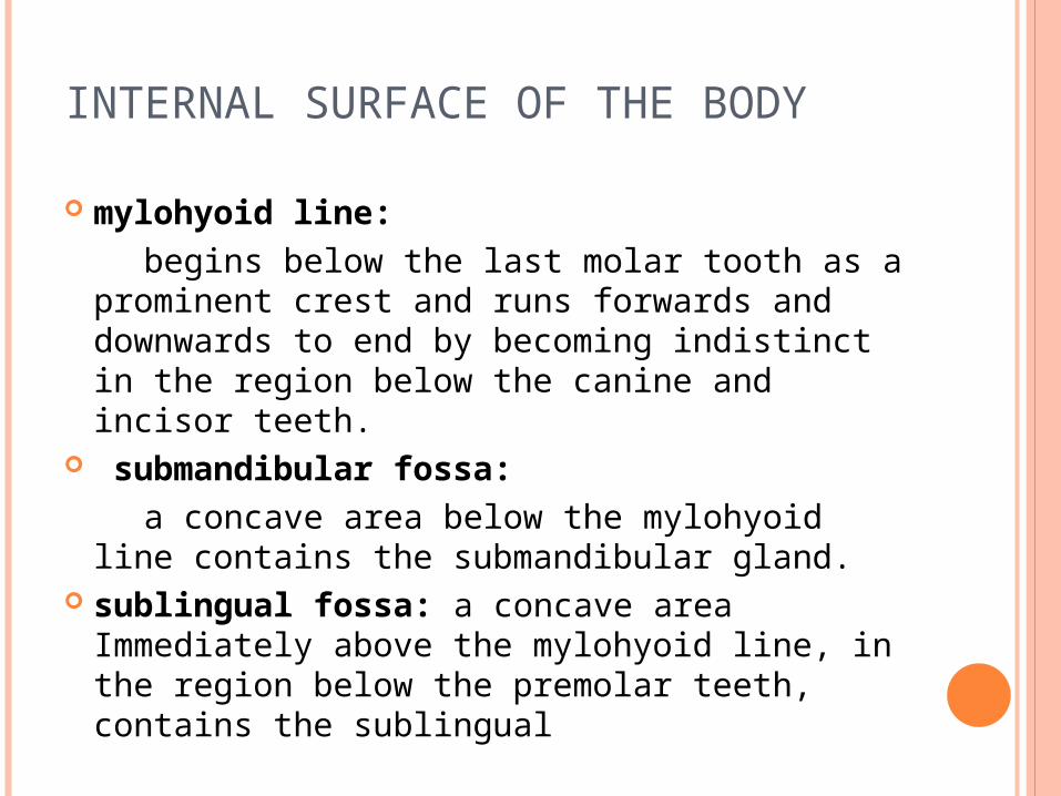
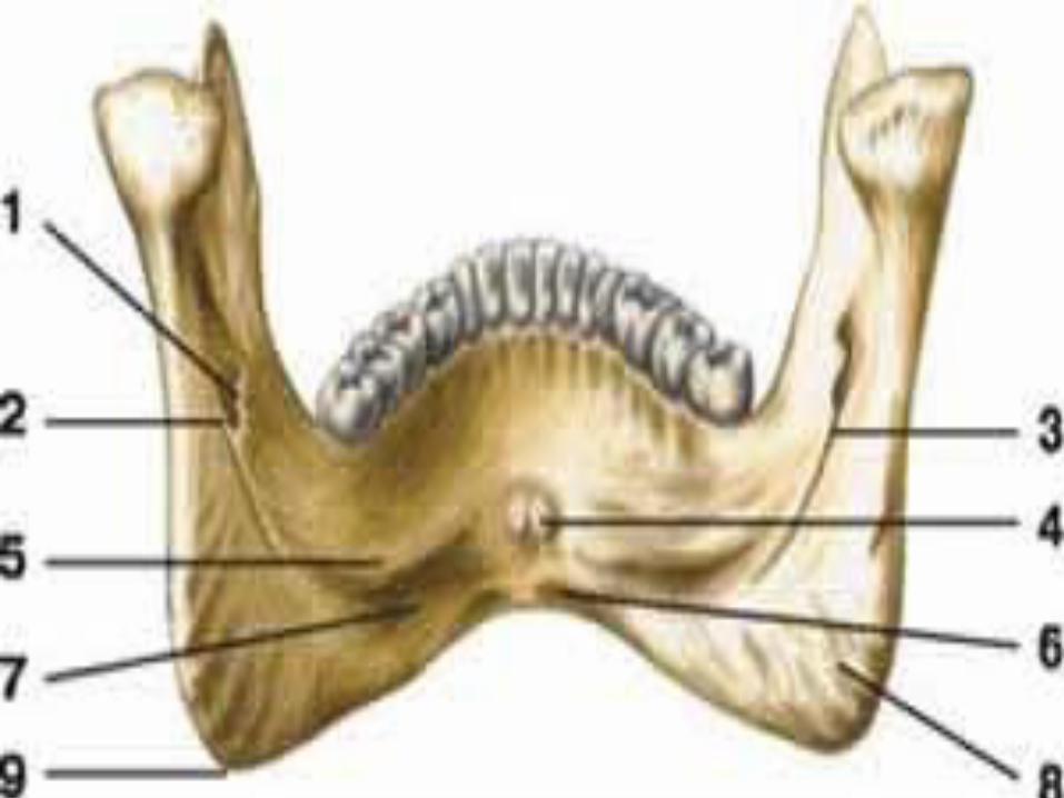
INTERNAL SURFACE OF THE BODY
mylohyoid line: begins below the last molar tooth as a
prominent crest and runs forwards and downwards to end by becoming indistinct in the region below the canine and incisor teeth.
submandibular fossa: a concave area below the mylohyoid line
contains the submandibular gland. sublingual fossa: a concave area
Immediately above the mylohyoid line, in the region below the premolar teeth, contains the sublingual

INTERNAL SURFACE OF THE BODY
superior and inferior mental spines: The inferior part of the internal surface below
the incisor teeth bears two small elevations, for attachment of the genial muscles.
alveolar process: upper part of the body of the mandible, contains sockets for the roots of teeth
digastric fossa: small, roughened area on the inferior border of the body for attachment of the anterior belly of the digastric muscle.
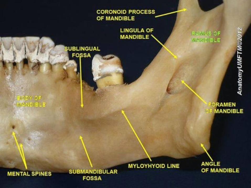
RAMUS OF THE MANDIBLE
a plate of bone. mandibular foramen : Approximately at its centre
transmits the inferior alveolar nerve and blood vessels. And leads into the mandibular canal.
Lingula: a thin plate of bone, on the anterior border of the foramen .
mylohyoid groove: begins at the lower border of the mandibular foramen, just behind the lingula, it runs downwards and forwards onto the body of the mandible.
The area posteroinferior to the mylohyoid groove is roughened for insertion of the medial pterygoid muscle
angle of the mandible: The region where the inferior and posterior borders of the ramus meet.

SUPERIOR BORDER OF THE RAMUS
coronoid and condylar processes are separated from each other by the mandibular notch
The coronoid process: triangular plate, projects upwards and forwards
• temporal crest: is a faint ridge On the medial aspect of the coronoid process, which becomes more prominent as it is traced downwards towards the margin of the alveolar bone medial to the last molar tooth.
• retromolar fossa: Between the temporal crest and the anterior border of the ramus
The superior and anterior margins of the coronoid process and the retromolar fossa provide the area of insertion of the temporalis muscle.
SUPERIOR BORDER OF THE RAMUS condylar process: head of the mandible.
• superior and posterior surfaces of the head are covered with fibrocartilage and articulate with the articular surface of the squamous part of the temporal at the synovial temporomandibular joint.
• condylar Head: wide from side to side but narrow from front to back.
• condylar neck : The constricted part of the condylar process below the head,
• pterygoid fovea: a shallow depression in the anterior aspect of the neck , into which part of the lateral pterygoid muscle is inserted.