Embed Size (px)
Citation preview

Anorectal Malformations: Finding the Pathway out of
the Labyrinth
Dr. Mohit GoelJR II14/11/13
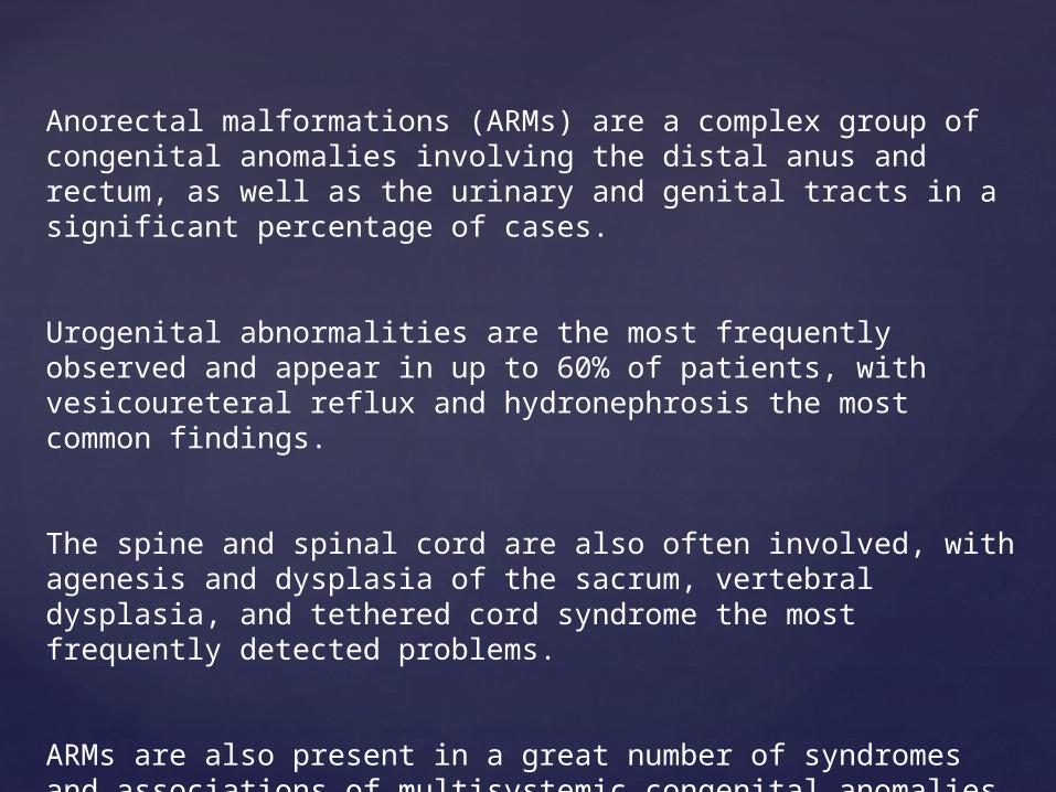
Anorectal malformations (ARMs) are a complex group of congenital anomalies involving the distal anus and rectum, as well as the urinary and genital tracts in a significant percentage of cases.
Urogenital abnormalities are the most frequently observed and appear in up to 60% of patients, with vesicoureteral reflux and hydronephrosis the most common findings.
The spine and spinal cord are also often involved, with agenesis and dysplasia of the sacrum, vertebral dysplasia, and tethered cord syndrome the most frequently detected problems.
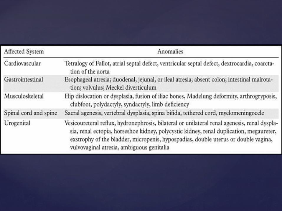
ARMs are also present in a great number of syndromes and associations of multisystemic congenital anomalies

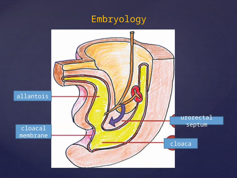
Embryology
allantois
cloacal membran
ecloaca
urorectal septum
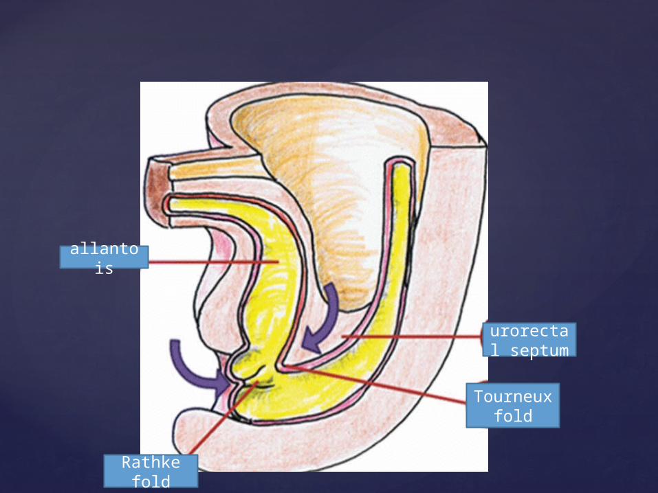
allantois
Rathke fold
Tourneux fold
urorectal septum
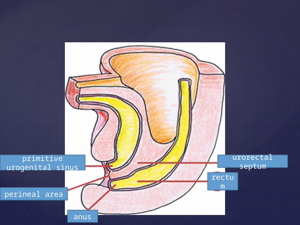
primitive urogenital sinus
perineal area
anus
rectum
urorectal septum
Embryologically, ARMs can thus be subdivided into two main groups according to when the disturbances occur:
Those manifesting as an ectopic anal orifice or fistula are due to early abnormal development of the dorsal part of the cloaca and the cloacal membrane (at weeks 4–7), whereas those manifesting as an abnormal anus in a normal position are due to later defective recanalization of the secondary occluded anal orifice (at weeks 7 and 8).
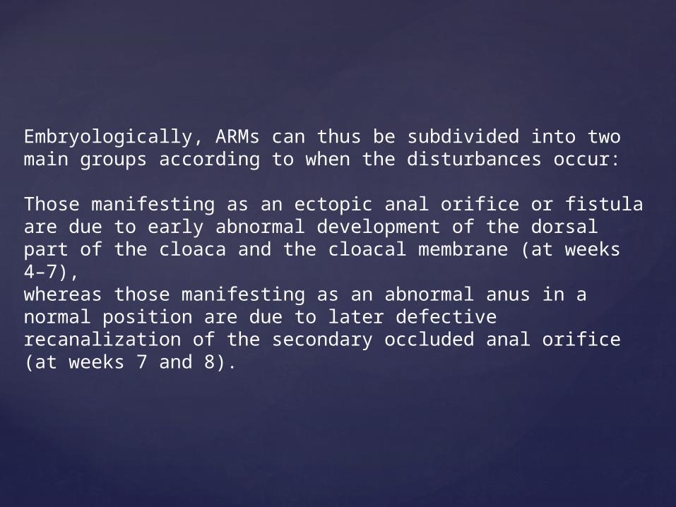
Classification of ARMs
Possible locations of fistulas in males with ARMs according to the Krickenbeck classification. (a) Low-type ARMs have an external anocutaneous opening in the scrotum (1) or perineum (2, 3).
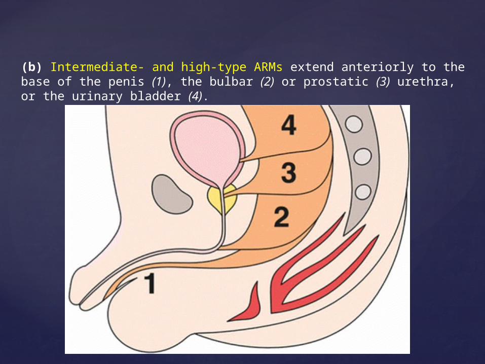
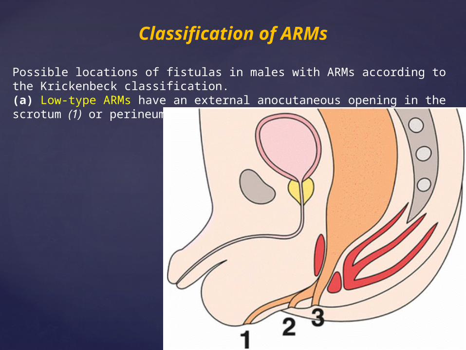
(b) Intermediate- and high-type ARMs extend anteriorly to the base of the penis (1), the bulbar (2) or prostatic (3) urethra, or the urinary bladder (4).
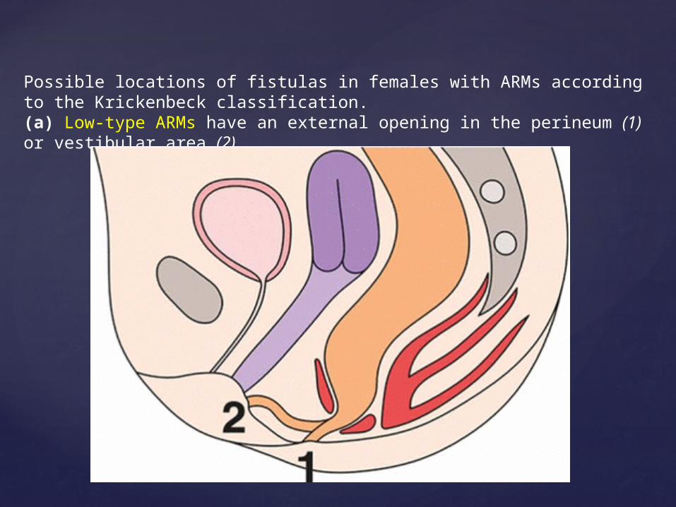
Possible locations of fistulas in females with ARMs according to the Krickenbeck classification. (a) Low-type ARMs have an external opening in the perineum (1) or vestibular area (2).
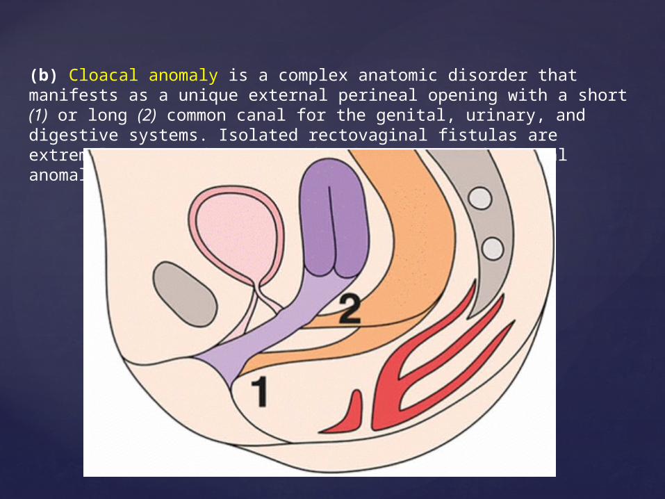
(b) Cloacal anomaly is a complex anatomic disorder that manifests as a unique external perineal opening with a short (1) or long (2) common canal for the genital, urinary, and digestive systems. Isolated rectovaginal fistulas are extremely rare and are considered a variant of cloacal anomaly.
ARMs involving a rectal pouch located below the level of the puborectalis muscle, regardless of whether they are associated with a fistula—perineal or vestibular—are considered low-type ARMs. They may be managed early with a perineal approach involving opening of the rectal pouch and ligature of the fistula, if present.
A rectal pouch lying at or above the level of the puborectal sling is considered an intermediate or high type of ARM; it is treated with colostomy in the first days of life and with posterior sagittal anorectoplasty alone or combined with laparoscopic abdominoperineal rectoplasty in a second intervention
Role of Imaging in Initial Evaluation of ARMs
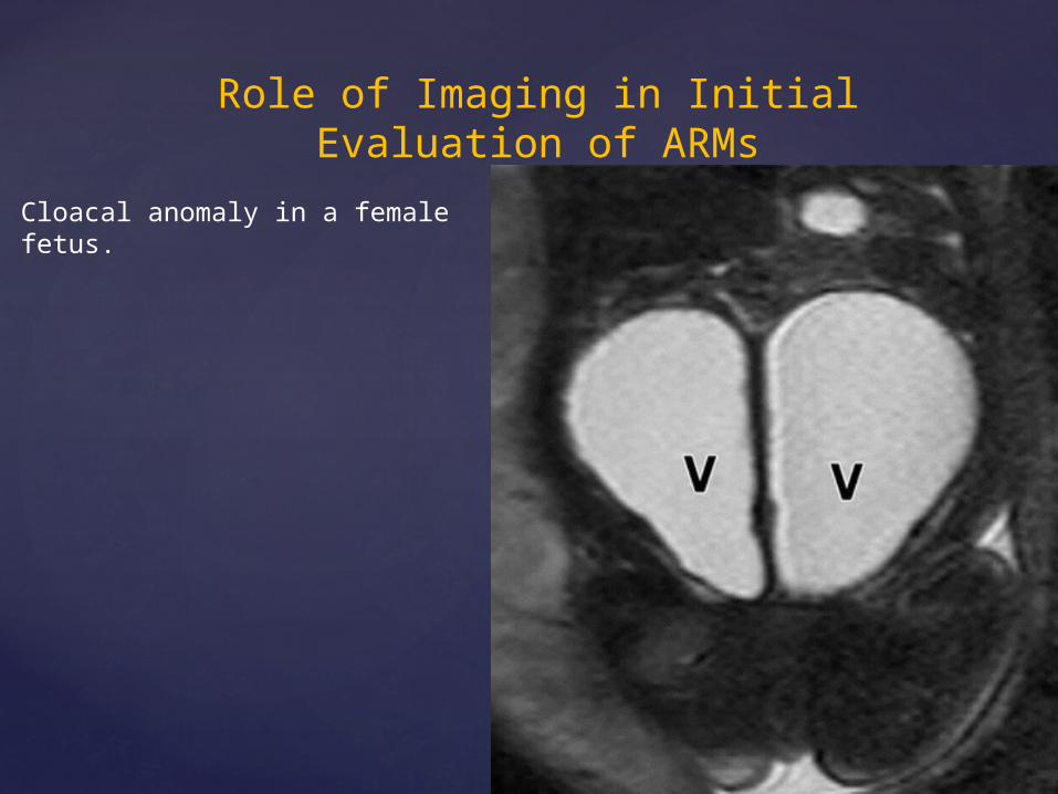
Cloacal anomaly in a female fetus.
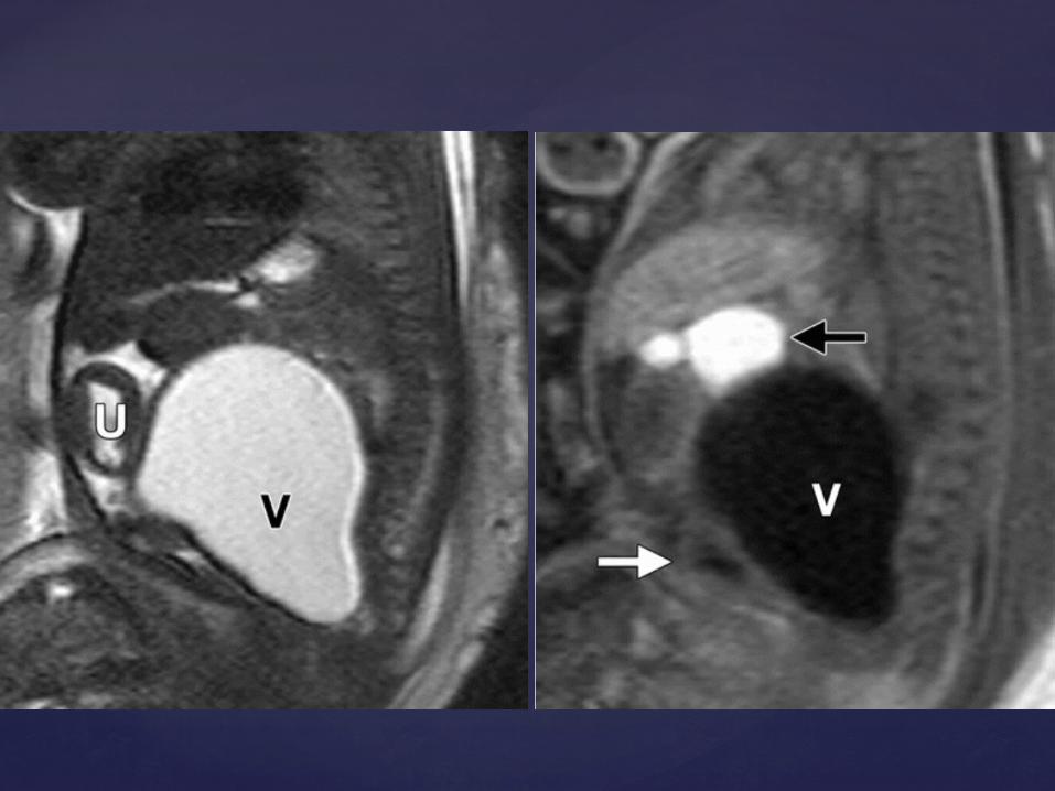
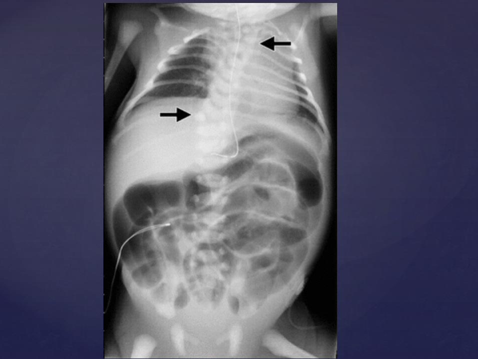
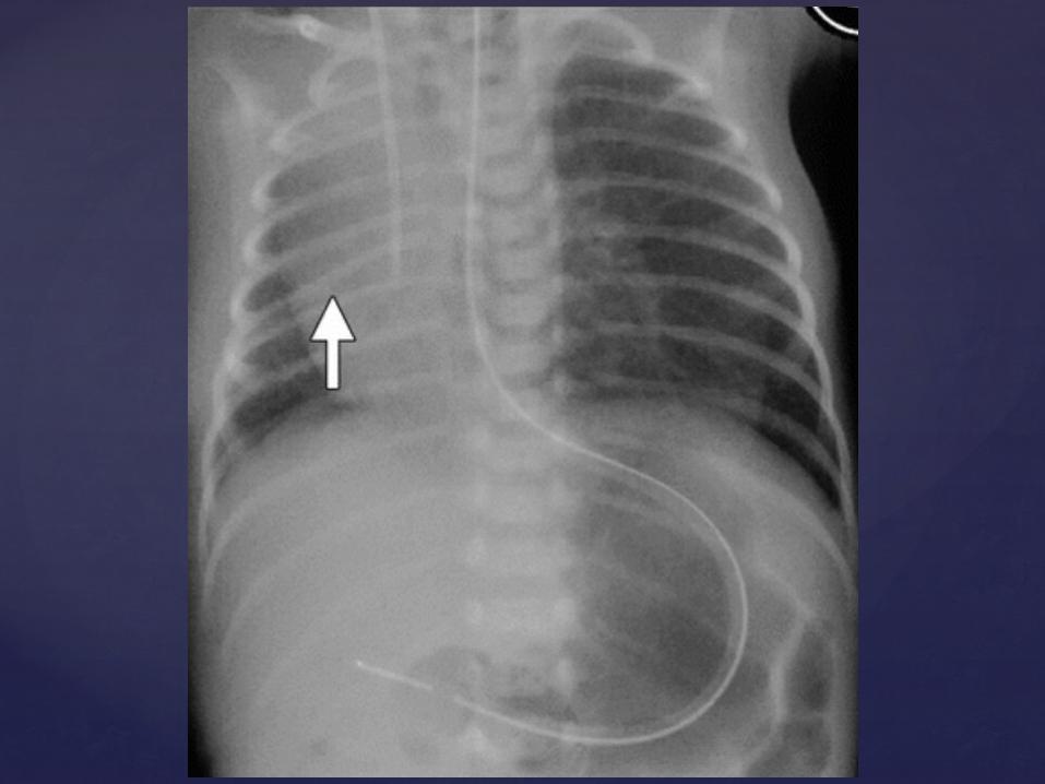
Imaging studies in the first 2 days of life should include radiography of the thorax, spine, and pelvis along with cardiac, perineal, abdominal, pelvic, and spine US to detect possible associated anomalies.
In cases of vertebral or sacral anomalies, spinal MR imaging should be performed.
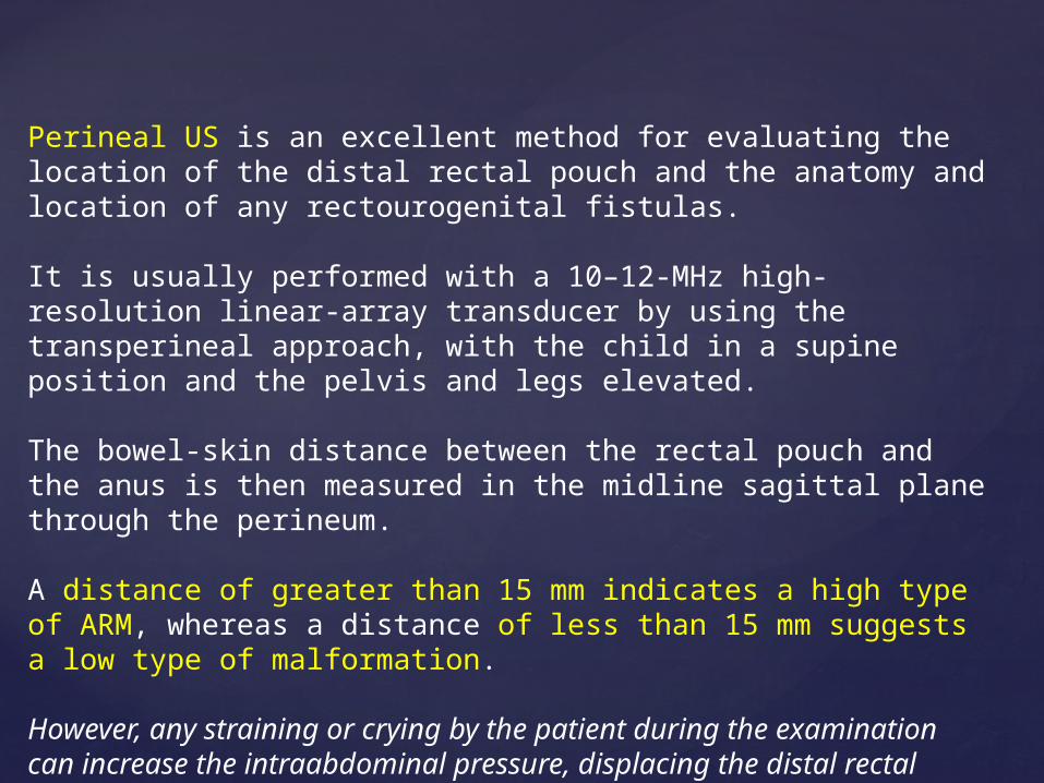
Perineal US is an excellent method for evaluating the location of the distal rectal pouch and the anatomy and location of any rectourogenital fistulas.
It is usually performed with a 10–12-MHz high-resolution linear-array transducer by using the transperineal approach, with the child in a supine position and the pelvis and legs elevated.
The bowel-skin distance between the rectal pouch and the anus is then measured in the midline sagittal plane through the perineum.
A distance of greater than 15 mm indicates a high type of ARM, whereas a distance of less than 15 mm suggests a low type of malformation.
However, any straining or crying by the patient during the examination can increase the intraabdominal pressure, displacing the distal rectal pouch to the perineum and shortening this distance. Fistulas may be identified as linear tracts connecting the rectal pouch to the bladder, urethra, or posterior wall of the vagina.
US evaluation of the urinary tract is limited in the first 24 hours after birth, as upper tract dilatation may be initially absent because of the physiologic dehydration and reduced urinary output of the newborn.
Detection of any genitourinary anomalies will require complementary voiding cystourethrography (VCUG)
Spinal US is currently accepted as a safe and inexpensive screening test for detection of spinal dysraphism. The method provides accurate information about the morphology and integrity of the os sacrum and distal vertebral column and allows identification of the level of the medullary cone and demonstration of a presacral mass.
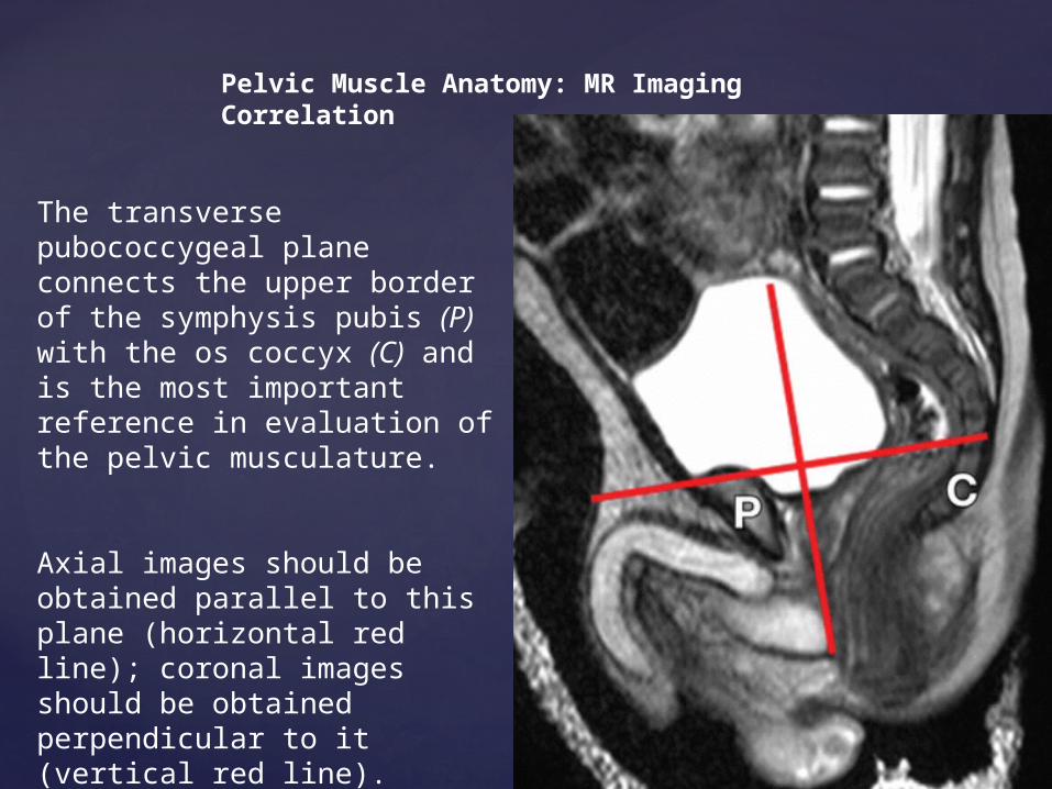
Pelvic Muscle Anatomy: MR Imaging Correlation
The transverse pubococcygeal plane connects the upper border of the symphysis pubis (P) with the os coccyx (C) and is the most important reference in evaluation of the pelvic musculature.
Axial images should be obtained parallel to this plane (horizontal red line); coronal images should be obtained perpendicular to it (vertical red line).
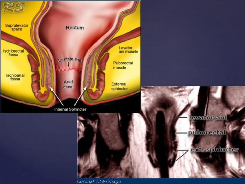
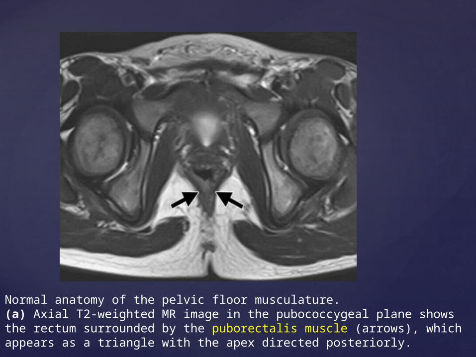
Normal anatomy of the pelvic floor musculature. (a) Axial T2-weighted MR image in the pubococcygeal plane shows the rectum surrounded by the puborectalis muscle (arrows), which appears as a triangle with the apex directed posteriorly.
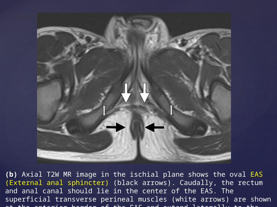
(b) Axial T2W MR image in the ischial plane shows the oval EAS (External anal sphincter) (black arrows). Caudally, the rectum and anal canal should lie in the center of the EAS. The superficial transverse perineal muscles (white arrows) are shown at the anterior border of the EAS and extend laterally to the ischial rami (I).
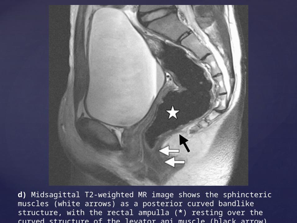
d) Midsagittal T2-weighted MR image shows the sphincteric muscles (white arrows) as a posterior curved bandlike structure, with the rectal ampulla (*) resting over the curved structure of the levator ani muscle (black arrow)
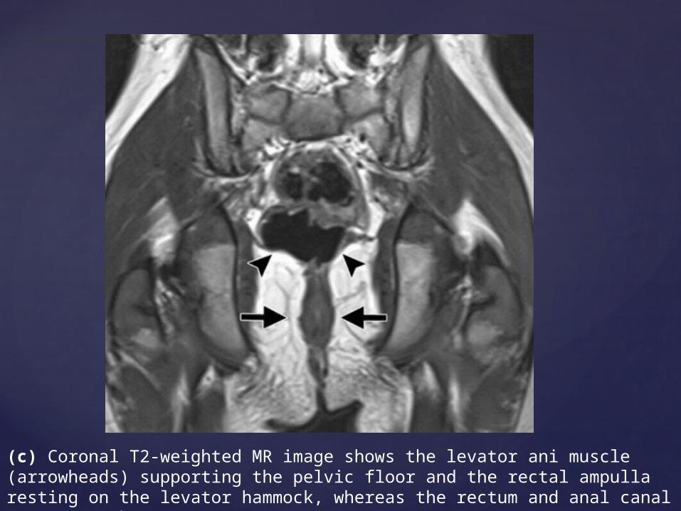
(c) Coronal T2-weighted MR image shows the levator ani muscle (arrowheads) supporting the pelvic floor and the rectal ampulla resting on the levator hammock, whereas the rectum and anal canal penetrate the EAS (arrows).
Pelvic MR Imaging in Boys with ARMs
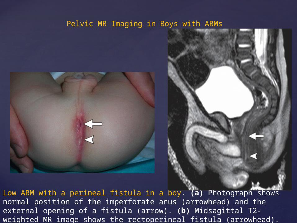
Low ARM with a perineal fistula in a boy. (a) Photograph shows normal position of the imperforate anus (arrowhead) and the external opening of a fistula (arrow). (b) Midsagittal T2-weighted MR image shows the rectoperineal fistula (arrowhead). The anteriorly displaced anorectum (arrow) is seen below the level of the levator ani muscle.
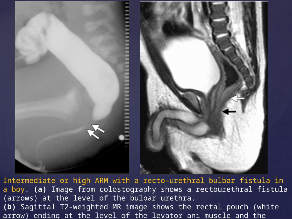
Intermediate or high ARM with a recto–urethral bulbar fistula in a boy. (a) Image from colostography shows a rectourethral fistula (arrows) at the level of the bulbar urethra.(b) Sagittal T2-weighted MR image shows the rectal pouch (white arrow) ending at the level of the levator ani muscle and the fistula (black arrow) extending anteriorly to the bulbar urethra
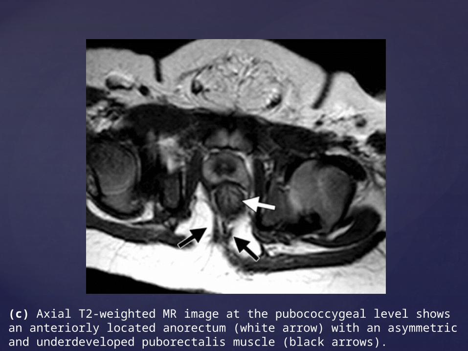
(c) Axial T2-weighted MR image at the pubococcygeal level shows an anteriorly located anorectum (white arrow) with an asymmetric and underdeveloped puborectalis muscle (black arrows).
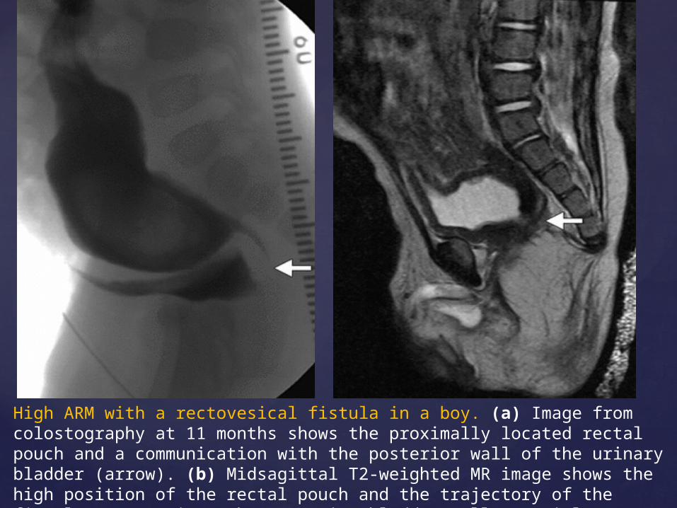
High ARM with a rectovesical fistula in a boy. (a) Image from colostography at 11 months shows the proximally located rectal pouch and a communication with the posterior wall of the urinary bladder (arrow). (b) Midsagittal T2-weighted MR image shows the high position of the rectal pouch and the trajectory of the fistula (arrow) into the posterior bladder wall. Partial agenesis of the sacrum is also seen.
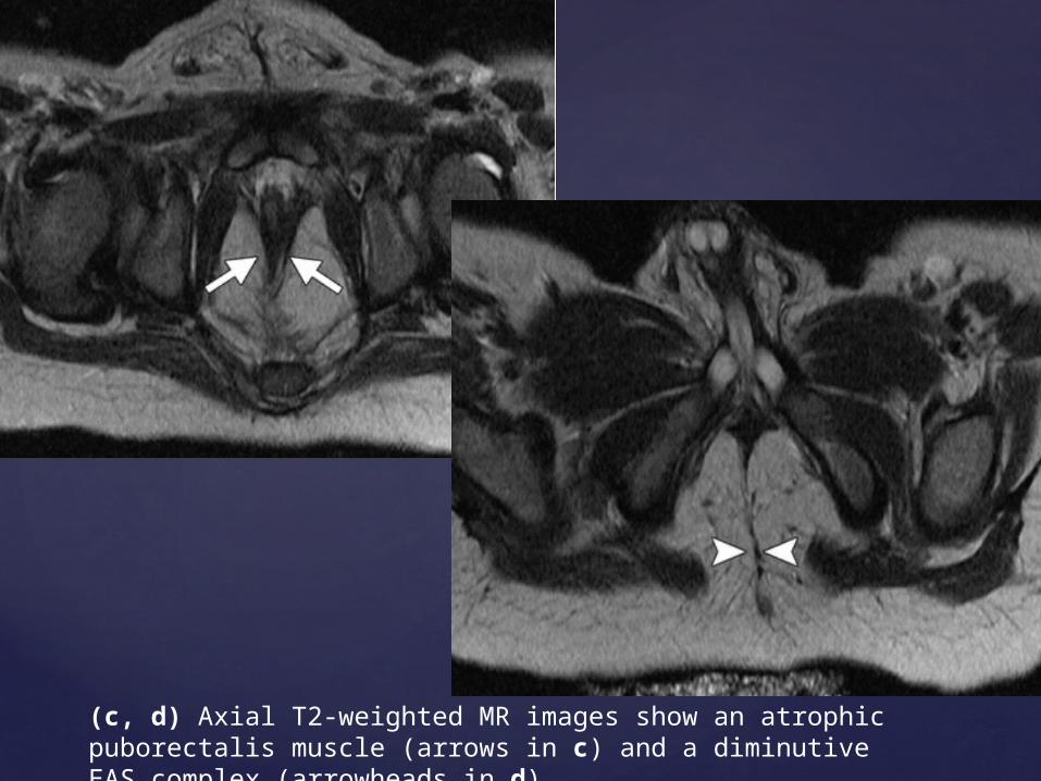
(c, d) Axial T2-weighted MR images show an atrophic puborectalis muscle (arrows in c) and a diminutive EAS complex (arrowheads in d).
Pelvic MR Imaging in Girls with ARMs
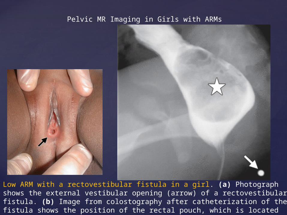
Low ARM with a rectovestibular fistula in a girl. (a) Photograph shows the external vestibular opening (arrow) of a rectovestibular fistula. (b) Image from colostography after catheterization of the fistula shows the position of the rectal pouch, which is located below the pubococcygeal line. * = fecaloma, arrow = normal position of the anus.
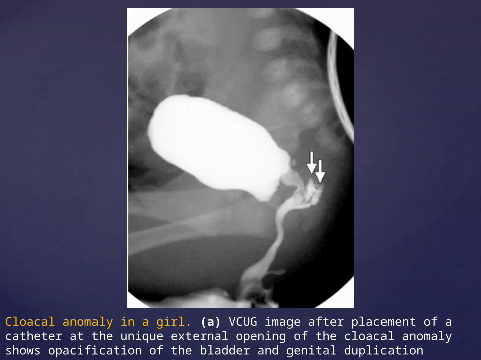
Cloacal anomaly in a girl. (a) VCUG image after placement of a catheter at the unique external opening of the cloacal anomaly shows opacification of the bladder and genital duplication (arrows), but the rectal pouch is not opacified.
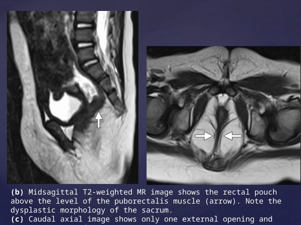
(b) Midsagittal T2-weighted MR image shows the rectal pouch above the level of the puborectalis muscle (arrow). Note the dysplastic morphology of the sacrum. (c) Caudal axial image shows only one external opening and hypotrophic EAS fibers (arrows).
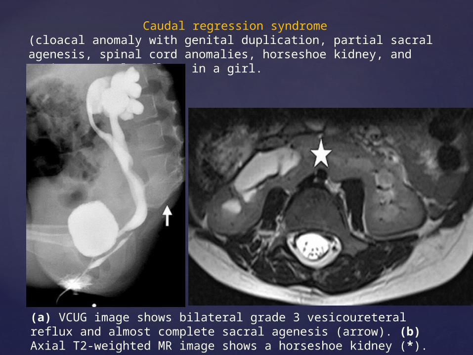
Caudal regression syndrome (cloacal anomaly with genital duplication, partial sacral agenesis, spinal cord anomalies, horseshoe kidney, and vesicoureteral reflux) in a girl.
(a) VCUG image shows bilateral grade 3 vesicoureteral reflux and almost complete sacral agenesis (arrow). (b) Axial T2-weighted MR image shows a horseshoe kidney (*).
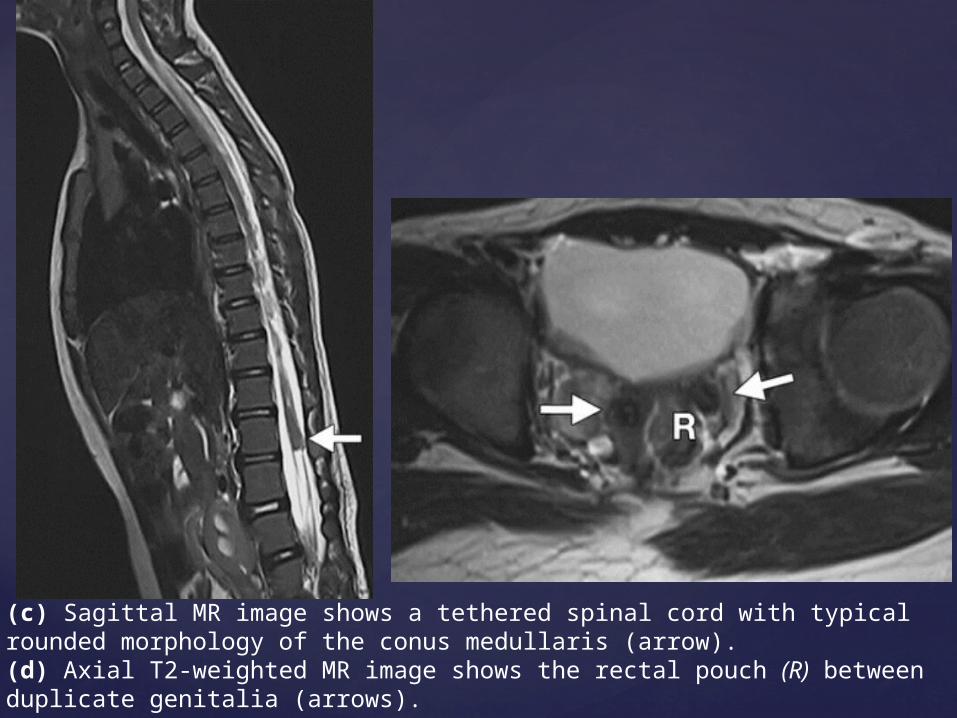
(c) Sagittal MR image shows a tethered spinal cord with typical rounded morphology of the conus medullaris (arrow). (d) Axial T2-weighted MR image shows the rectal pouch (R) between duplicate genitalia (arrows).
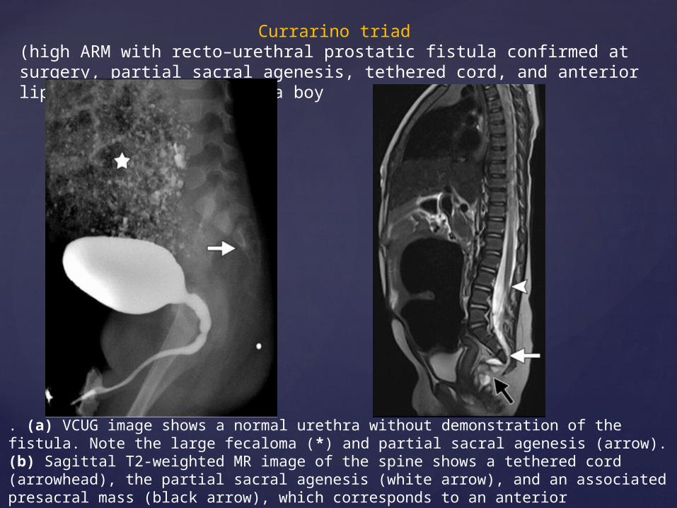
Currarino triad (high ARM with recto–urethral prostatic fistula confirmed at surgery, partial sacral agenesis, tethered cord, and anterior lipomyelomeningocele) in a boy
. (a) VCUG image shows a normal urethra without demonstration of the fistula. Note the large fecaloma (*) and partial sacral agenesis (arrow). (b) Sagittal T2-weighted MR image of the spine shows a tethered cord (arrowhead), the partial sacral agenesis (white arrow), and an associated presacral mass (black arrow), which corresponds to an anterior lipomyelomeningocele.
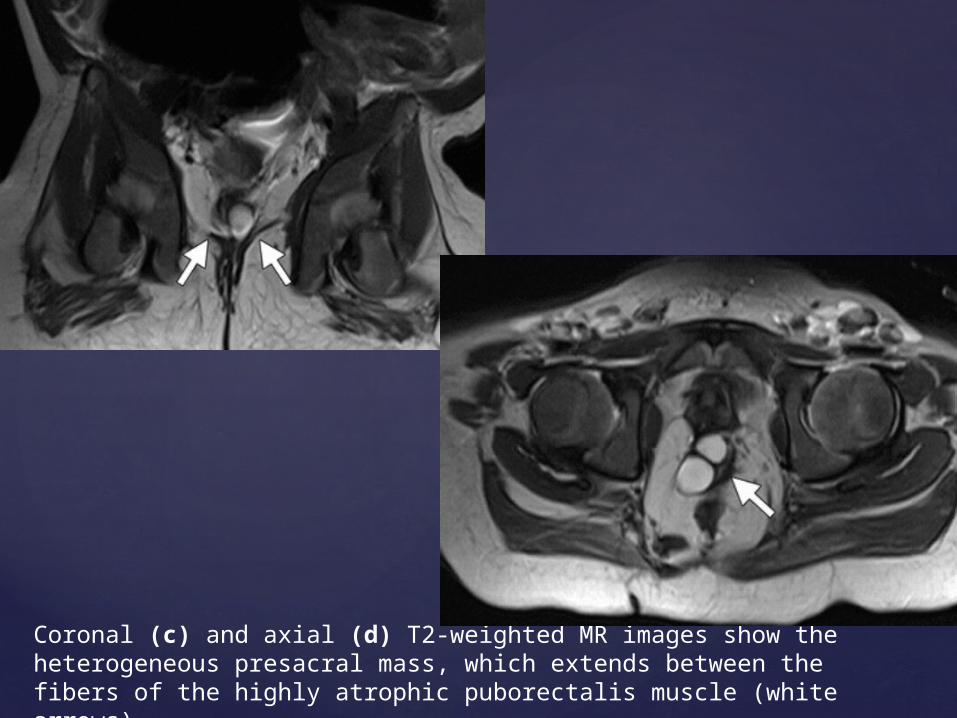
Coronal (c) and axial (d) T2-weighted MR images show the heterogeneous presacral mass, which extends between the fibers of the highly atrophic puborectalis muscle (white arrows).
Conclusion
ARMs are a complex group of congenital anomalies involving the distal anus and rectum.
They result from abnormal development of the urorectal septum in prenatal life. Imaging plays a key role in evaluation of ARMs.
In the first days of life, clinical and imaging findings facilitate early classification of ARMs and allow a decision about whether to perform an immediate colostomy.
In children with intermediate and high types of ARMs, preoperative pelvic MR imaging after the neonatal period allows accurate evaluation of the morphology and grade of development of the sphincteric muscle complex.

THANK YOU