Embed Size (px)
Citation preview
Copyright © John Wiley & Sons, Inc. All rights reserved.
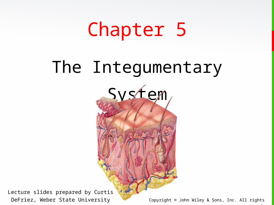
Chapter 5
The Integumentary System
Lecture slides prepared by Curtis DeFriez, Weber State University
Copyright © John Wiley & Sons, Inc. All rights reserved.
Introduction• The organs of the integumentary system include the skin
and its accessory structures including hair, nails, and
glands, as well as blood vessels, muscles and nerves.
Note that all 4 of the basic tissue types are well-
represented in this organ system: Epithelium in the
hair, nails, and the epidermis of the skin; the dermis
contains C.T.; muscle is found attached to the hair
follicles, and in the substance of arteries and veins;
nerves provide an abundance of sensation.
Copyright © John Wiley & Sons, Inc. All rights reserved.
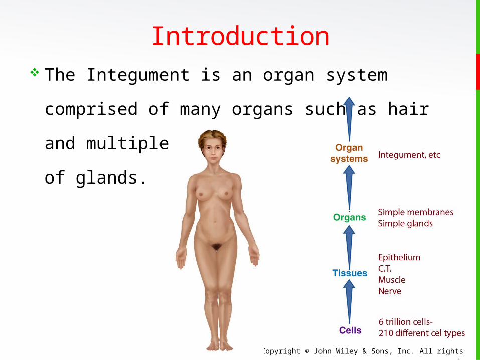
Introduction The Integument is an organ system comprised
of many organs such as hair and multiple types
of glands.

Copyright © John Wiley & Sons, Inc. All rights reserved.
Introduction• The integument can also be thought of as a cutaneous
membrane that covers the outer surface of the body.
It is the largest organ by surface area and weight.
• Its area is about 2 square meters (22 square feet)
and weighs 4.5–5kg (10–11 lb), about 16% of body
weight.
It is 0.5–4 mm thick, thinnest on the eyelids, thickest
on the heels.
We lose almost a kg of skin epithelium a year that
becomes a major part of household “dust”.
Copyright © John Wiley & Sons, Inc. All rights reserved.
Introduction Besides protection, the skin contributes to:
Regulation of body temperature
Sensory perceptions
Synthesis of vitamin D
Emotional expression
It also serves as an important reservoir of
blood.
Copyright © John Wiley & Sons, Inc. All rights reserved.
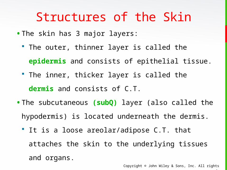
Structures of the Skin• The skin has 3 major layers:
The outer, thinner layer is called the
epidermis and consists of epithelial tissue.
The inner, thicker layer is called the dermis
and consists of C.T.
• The subcutaneous (subQ) layer (also called the
hypodermis) is located underneath the dermis.
It is a loose areolar/adipose C.T. that attaches
the skin to the underlying tissues and organs.
Copyright © John Wiley & Sons, Inc. All rights reserved.
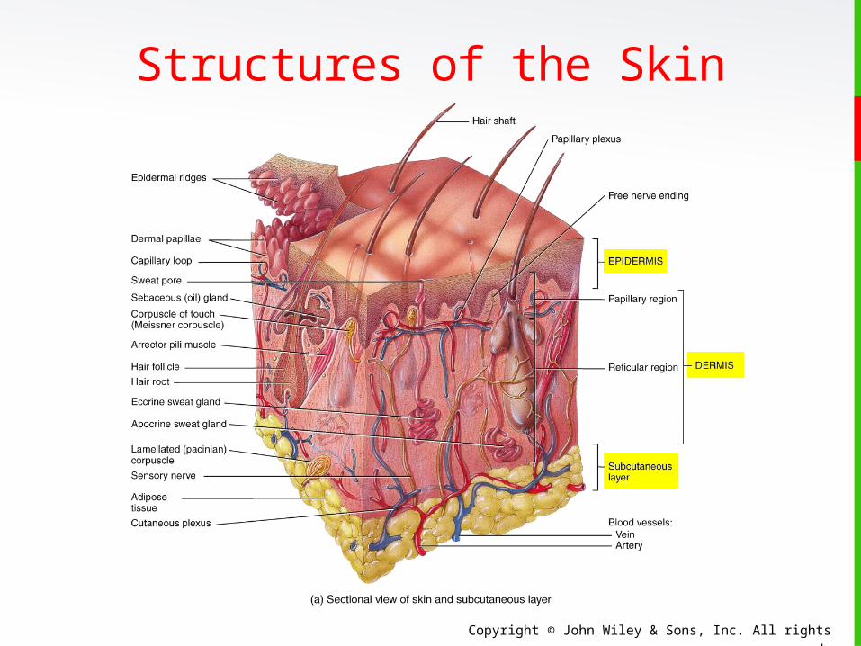
Structures of the Skin
Copyright © John Wiley & Sons, Inc. All rights reserved.
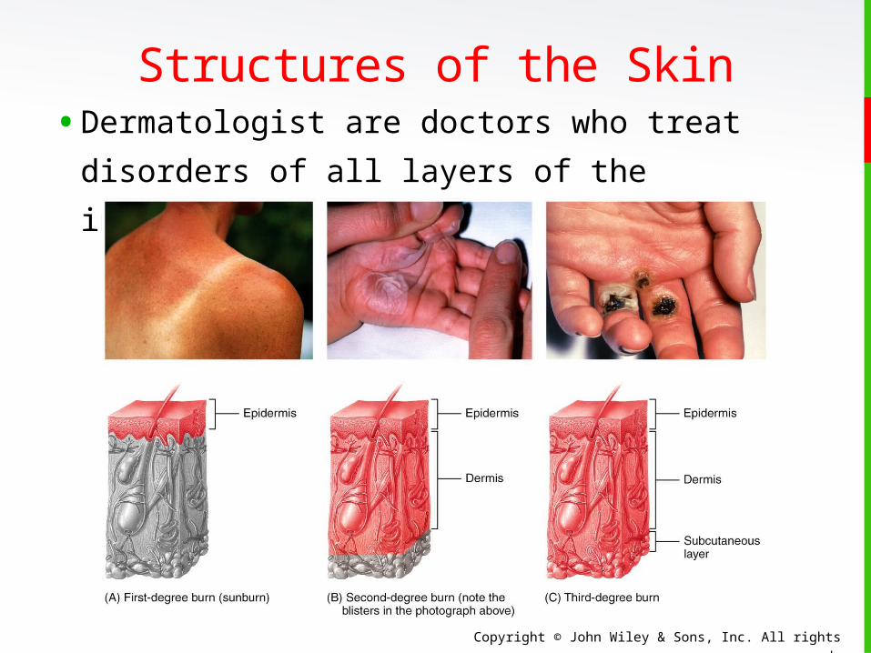
Structures of the Skin• Dermatologist are doctors who treat disorders
of all layers of the integumentary system.
Copyright © John Wiley & Sons, Inc. All rights reserved.
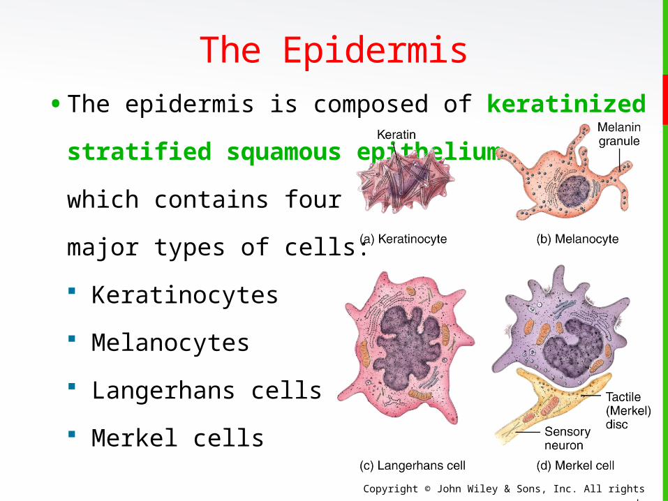
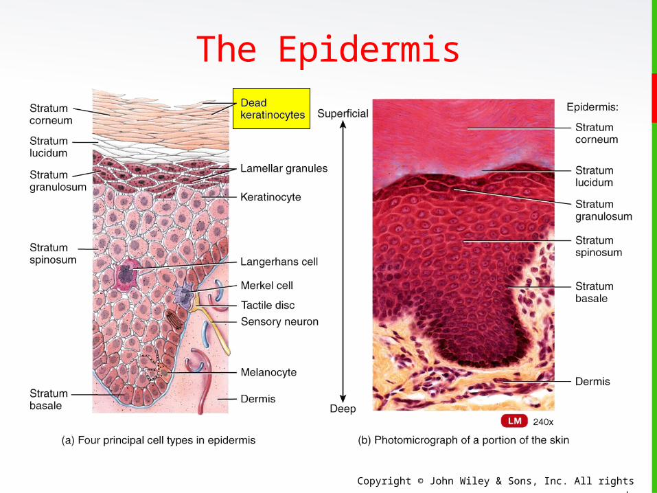
• The epidermis is composed of keratinized
stratified squamous epithelium
which contains four
major types of cells:
Keratinocytes
Melanocytes
Langerhans cells
Merkel cells
The Epidermis
Copyright © John Wiley & Sons, Inc. All rights reserved.
The Epidermis
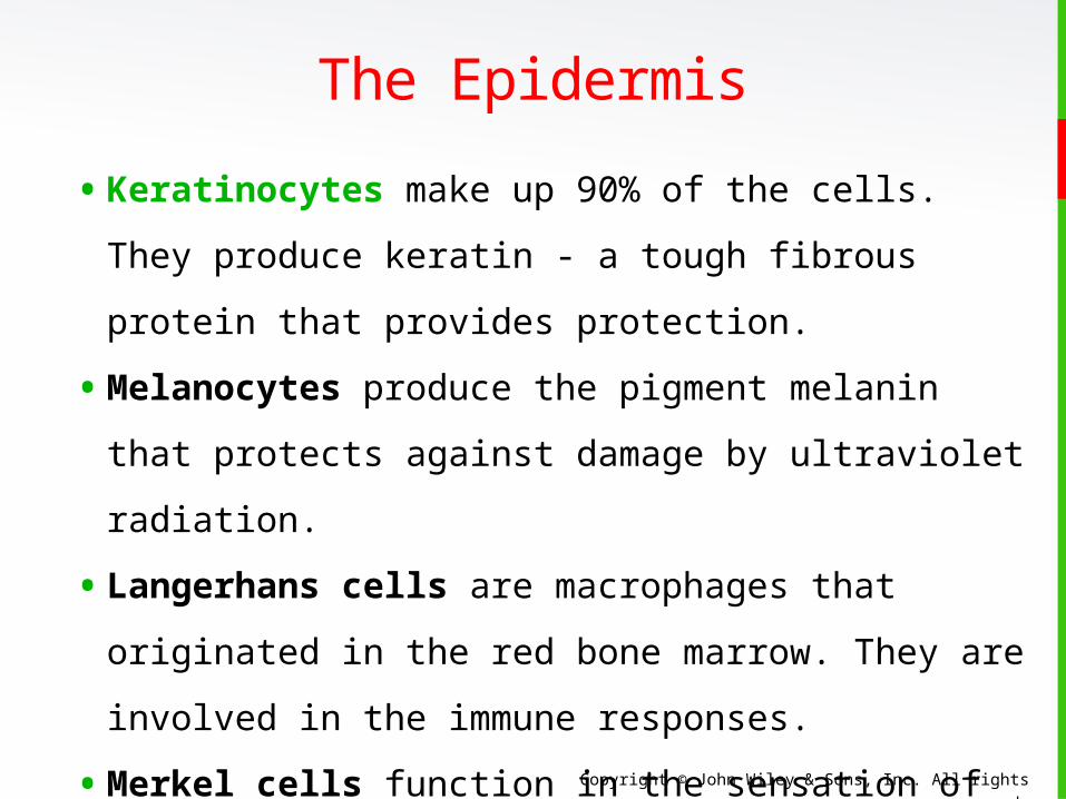
• Keratinocytes make up 90% of the cells. They
produce keratin - a tough fibrous protein that
provides protection.
• Melanocytes produce the pigment melanin that
protects against damage by ultraviolet radiation.
• Langerhans cells are macrophages that
originated in the red bone marrow. They are
involved in the immune responses.
• Merkel cells function in the sensation of touch
along with the other adjacent tactile discs
(receptors).
Copyright © John Wiley & Sons, Inc. All rights reserved.
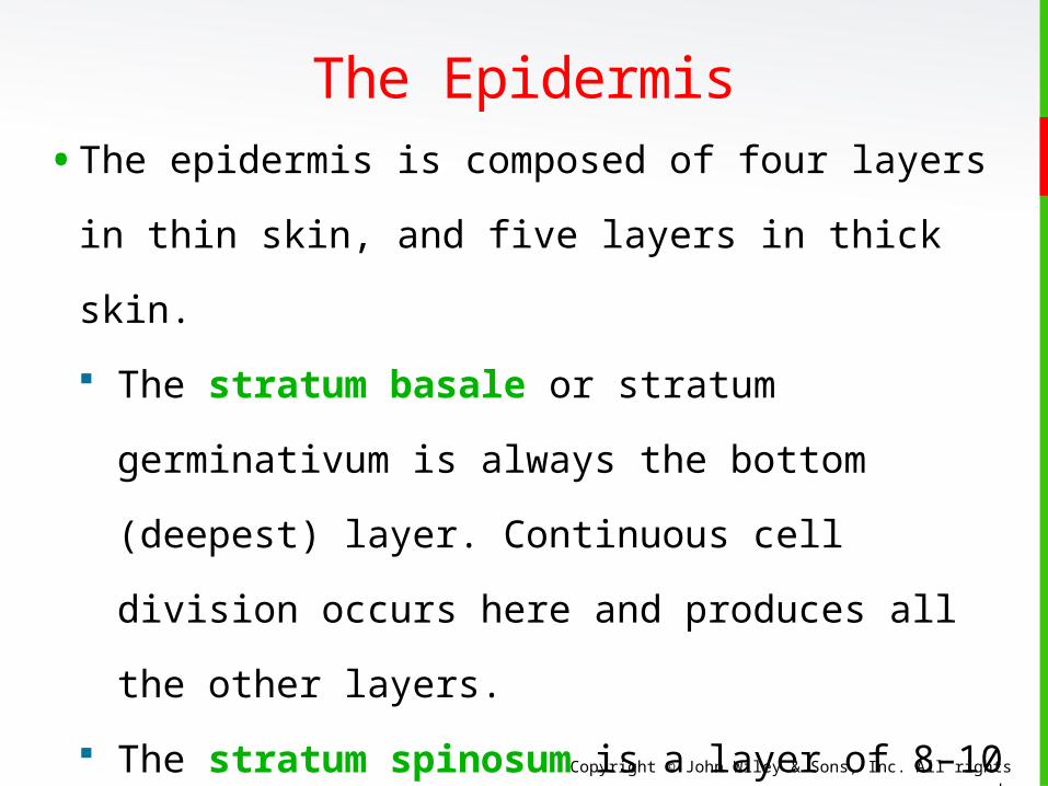
The Epidermis• The epidermis is composed of four layers in thin
skin, and five layers in thick skin.
The stratum basale or stratum
germinativum is always the bottom (deepest)
layer. Continuous cell division occurs here and
produces all the other layers.
The stratum spinosum is a layer of 8–10
keratinocytes
The non-dividing cells of the 3rd layer
(stratum granulosum) are filled with
granules of keratin.
Copyright © John Wiley & Sons, Inc. All rights reserved.
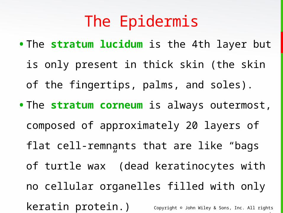
The Epidermis• The stratum lucidum is the 4th layer but is
only present in thick skin (the skin of the
fingertips, palms, and soles).
• The stratum corneum is always outermost,
composed of approximately 20 layers of flat
cell-remnants that are like “bags of turtle wax”
(dead keratinocytes with no cellular organelles
filled with only keratin protein.)
They are continuously shed and replaced by
cells from deeper strata.
Copyright © John Wiley & Sons, Inc. All rights reserved.
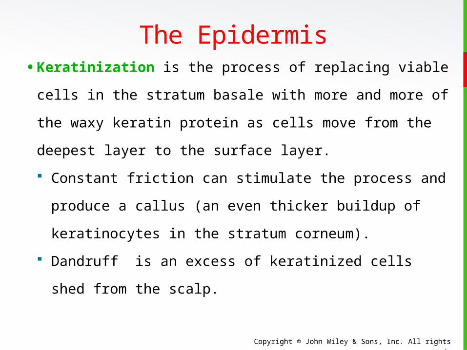
The Epidermis• Keratinization is the process of replacing viable
cells in the stratum basale with more and more
of the waxy keratin protein as cells move from
the deepest layer to the surface layer.
Constant friction can stimulate the process and
produce a callus (an even thicker buildup of
keratinocytes in the stratum corneum).
Dandruff is an excess of keratinized cells shed
from the scalp.
Copyright © John Wiley & Sons, Inc. All rights reserved.
The Epidermis
Copyright © John Wiley & Sons, Inc. All rights reserved.
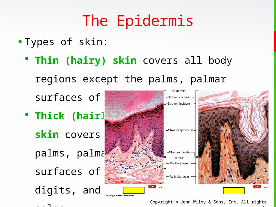
The Epidermis• Types of skin:
Thin (hairy) skin covers all body regions
except the palms, palmar surfaces of digits,
and soles.
Thick (hairless)
skin covers the
palms, palmar
surfaces of
digits, and
soles.
Copyright © John Wiley & Sons, Inc. All rights reserved.
The Epidermis Skin Pigments
Melanin is produced by melanocytes in the
stratum basale
• Eumelanin (brown to black)
• Pheomelanin (yellow to red)
Freckles are clusters of concentrated melanin
triggered
by exposure to sunlight.
Having more freckles is a genetic trait.
Copyright © John Wiley & Sons, Inc. All rights reserved.
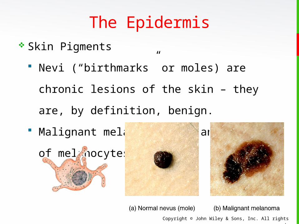
The Epidermis Skin Pigments
Nevi (“birthmarks” or moles) are chronic
lesions of the skin – they are, by definition,
benign.
Malignant melanoma is a cancer
of melanocytes.
Copyright © John Wiley & Sons, Inc. All rights reserved.
The Epidermis Skin Pigments
Vitiligo is a chronic disorder that causes
depigmentation patches in the skin. The
precise pathogenesis, or cause, is not known,
but is most likely a combination of
genetic factors coupled with a disorder of the
immune system (autoimmune disease).
Copyright © John Wiley & Sons, Inc. All rights reserved.
The Epidermis Skin Pigments
Albinism is a congenital disorder
characterized by the complete or partial
absence of pigment in the skin, hair, and
eyes due to a defect of an enzyme involved
in the production of melanin.
Copyright © John Wiley & Sons, Inc. All rights reserved.
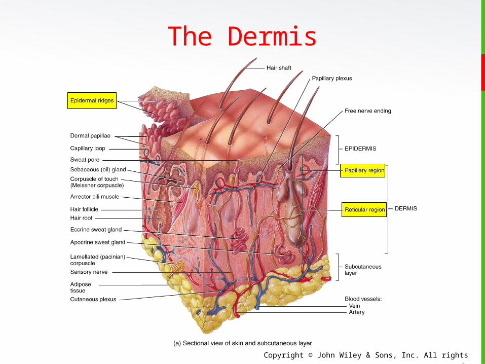
The Dermis• The dermis is composed of connective tissue
containing collagen and elastic fibers.
• It contains two regions:
The papillary region lies just below the
epidermis and consists of areolar
connective tissue containing thin collagen
and elastic fibers, dermal papillae (including
capillary loops), corpuscles of touch and free
nerve endings.
Copyright © John Wiley & Sons, Inc. All rights reserved.
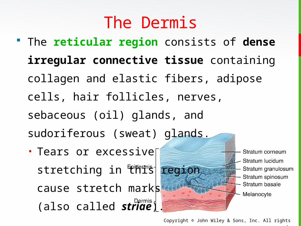
The reticular region consists of dense
irregular connective tissue containing
collagen and elastic fibers, adipose cells, hair
follicles, nerves, sebaceous (oil) glands, and
sudoriferous (sweat) glands.
• Tears or excessive
stretching in this region
cause stretch marks
(also called striae).
The Dermis
Copyright © John Wiley & Sons, Inc. All rights reserved.
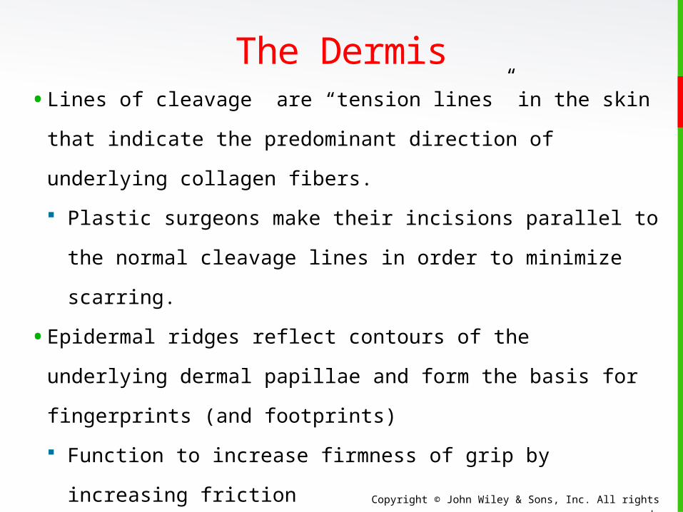
The Dermis• Lines of cleavage are “tension lines” in the skin that
indicate the predominant direction of underlying
collagen fibers.
Plastic surgeons make their incisions parallel to the
normal cleavage lines in order to minimize scarring.
• Epidermal ridges reflect contours of the underlying
dermal papillae and form the basis for fingerprints
(and footprints)
Function to increase firmness of grip by increasing
friction
Copyright © John Wiley & Sons, Inc. All rights reserved.
The Dermis

Copyright © John Wiley & Sons, Inc. All rights reserved.
• The subcutaneous layer is also called the
hypodermis, and it attaches the skin to
underlying tissues and organs.
It contains blood vessels and nerves in transit
to the more superficial layers.
It also contains lamellated
(pacinian) corpuscles
that detect external
pressure applied to the skin.
subQ
The Subcutaneous Layer
Copyright © John Wiley & Sons, Inc. All rights reserved.
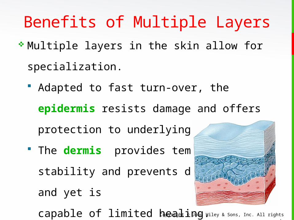
Benefits of Multiple Layers Multiple layers in the skin allow for
specialization.
Adapted to fast turn-over, the epidermis
resists damage and offers protection to
underlying tissues.
The dermis provides temperature stability
and prevents dehydration, and yet is
capable of limited healing.
The subcutaneous tissues insulate,
store fat, and anchor the skin.
Copyright © John Wiley & Sons, Inc. All rights reserved.
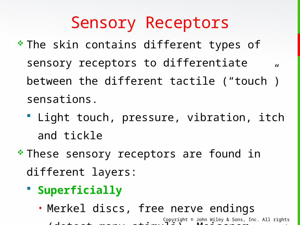
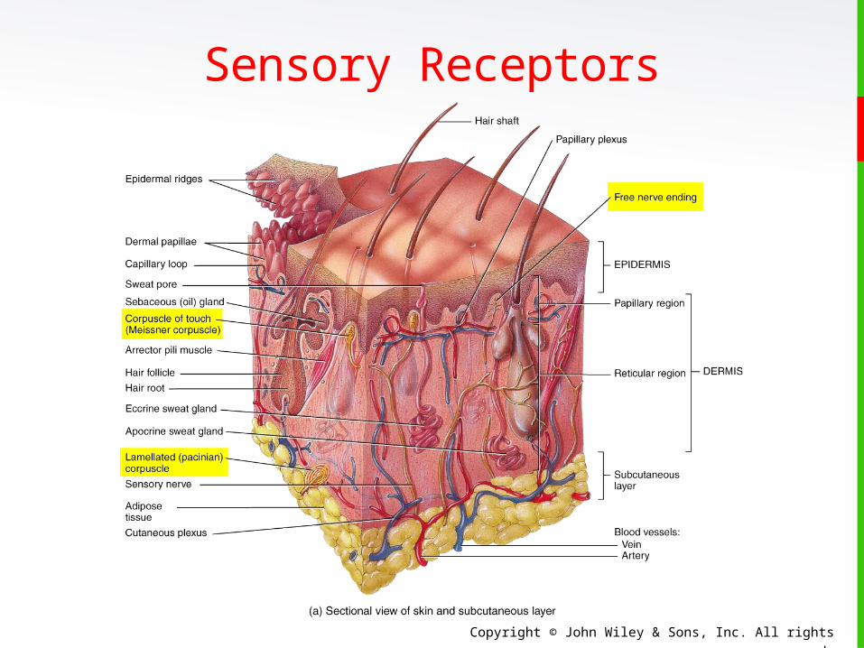
Sensory Receptors The skin contains different types of sensory
receptors to differentiate between the different
tactile (“touch”) sensations.
Light touch, pressure, vibration, itch and
tickle These sensory receptors are found in different
layers:
Superficially
• Merkel discs, free nerve endings (detect
many stimuli), Meissner corpuscles, and
hair root plexuses
Deep
• Pacinian corpuscles
Copyright © John Wiley & Sons, Inc. All rights reserved.
Sensory Receptors
Copyright © John Wiley & Sons, Inc. All rights reserved.
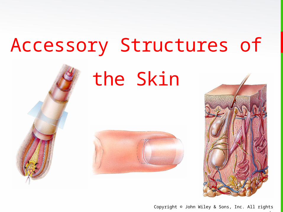
Accessory Structures of
the Skin
Copyright © John Wiley & Sons, Inc. All rights reserved.
Hair Hair is associated with the word “pili”.
It is present on most surfaces except the
palms, anterior surfaces of fingers, and the
soles of the feet.
It is composed of dead, keratinized epidermal
cells.
Genetics determines thickness and
distribution.
Hair helps with touch sensations and protects
the body against the harmful effects of the sun
and against heat loss.
Copyright © John Wiley & Sons, Inc. All rights reserved.
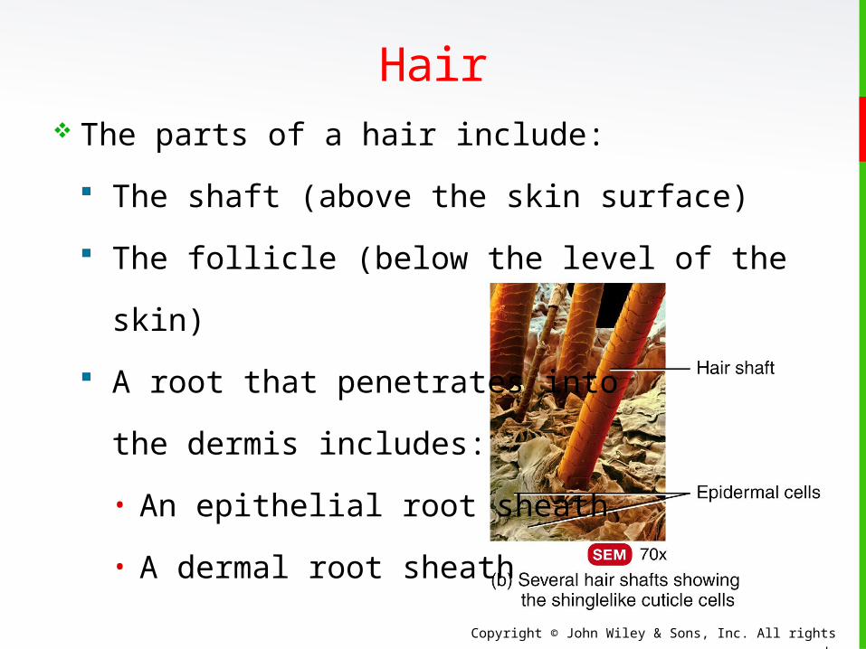
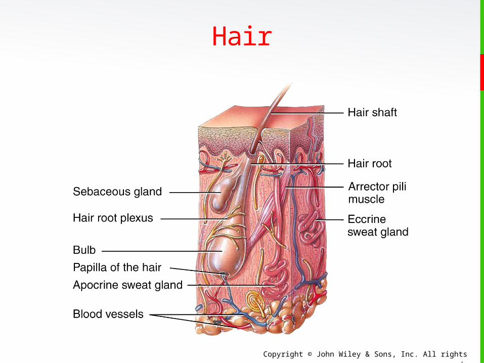
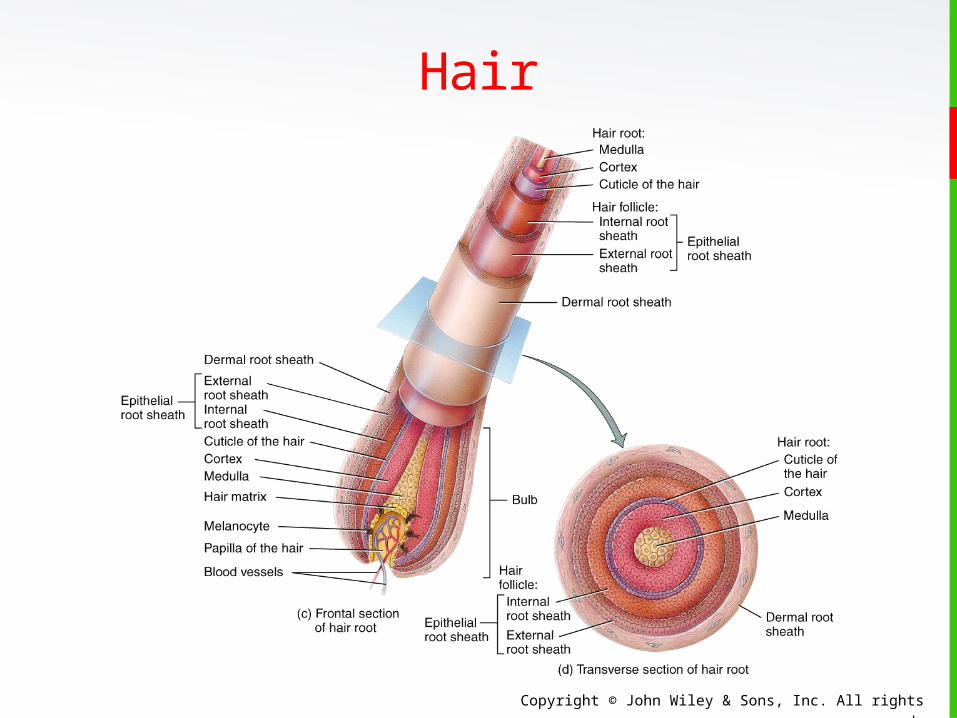
Hair The parts of a hair include:
The shaft (above the skin surface)
The follicle (below the level of the skin)
A root that penetrates into
the dermis includes:
• An epithelial root sheath
• A dermal root sheath
Copyright © John Wiley & Sons, Inc. All rights reserved.
Hair
Copyright © John Wiley & Sons, Inc. All rights reserved.
Hair
Copyright © John Wiley & Sons, Inc. All rights reserved.
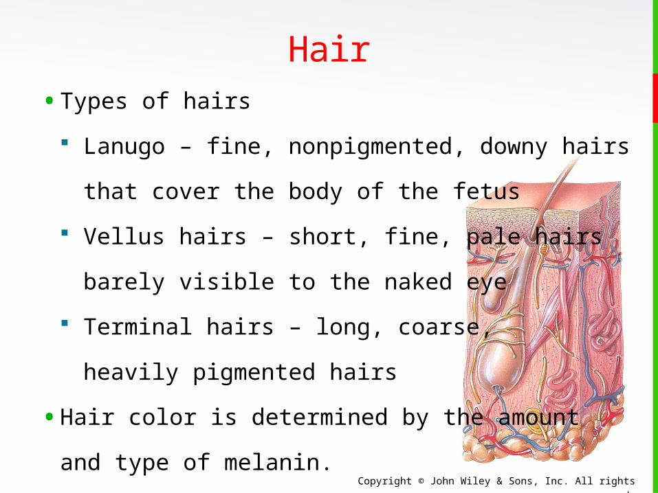
• Types of hairs
Lanugo – fine, nonpigmented, downy hairs
that cover the body of the fetus
Vellus hairs – short, fine, pale hairs
barely visible to the naked eye
Terminal hairs – long, coarse,
heavily pigmented hairs
• Hair color is determined by the amount
and type of melanin.
Hair
Copyright © John Wiley & Sons, Inc. All rights reserved.
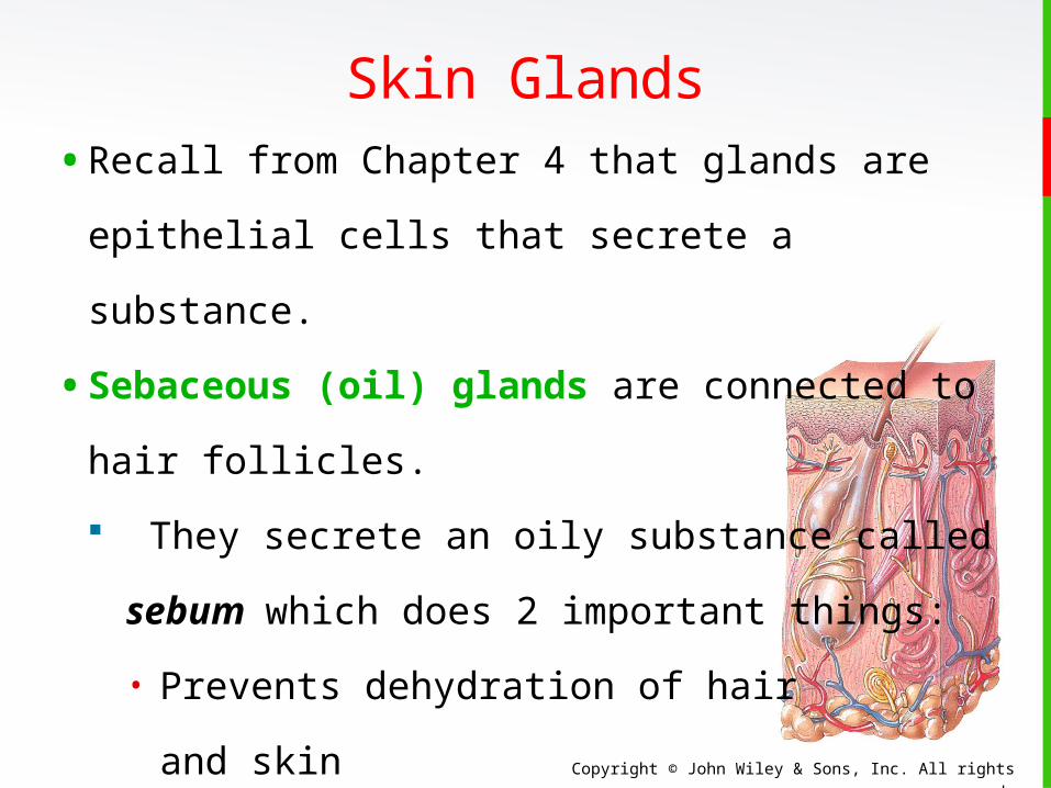
• Recall from Chapter 4 that glands are epithelial
cells that secrete a substance.
• Sebaceous (oil) glands are connected to hair
follicles.
They secrete an oily substance called
sebum which does 2 important things:
• Prevents dehydration of hair
and skin
• Inhibits growth of certain bacteria
Skin Glands
Copyright © John Wiley & Sons, Inc. All rights reserved.
Skin Glands• In addition to oil glands, there are 2 types of
skin sweat glands (also called sudoriferous
glands). Both are simple, coiled tubular glands.
Eccrine sweat glands are the most
numerous. They secrete a watery solution
(600 ml per day) that helps to cool the body
and eliminates small amounts of waste.
Apocrine sweat glands are located mainly
in the skin of the axilla, groin, areolae, and
bearded facial regions of adult males. They
secrete a slightly viscous sweat.
Copyright © John Wiley & Sons, Inc. All rights reserved.
Skin Glands• Eccrine sweat glands release sweat in response to
an emotional stress such as fear or embarrassment.
This type of sweating is referred to as emotional
sweating or a “cold sweat”.
• The secretory portion of apocrine sweat glands is
located mostly in the subcutaneous layer, and the
excretory duct opens into hair follicles, with sweat
secreted during emotional stress and sexual
excitement.
Much of body odor is due to apocrine sweat.
Copyright © John Wiley & Sons, Inc. All rights reserved.
Skin Glands• Ceruminous glands are modified sweat
glands located in the ear canal.
Along with nearby sebaceous glands, they
are involved in producing a waxy secretion
called cerumen (earwax) which provides a
sticky barrier that prevents entry of foreign
bodies into the ear canal.
Copyright © John Wiley & Sons, Inc. All rights reserved.
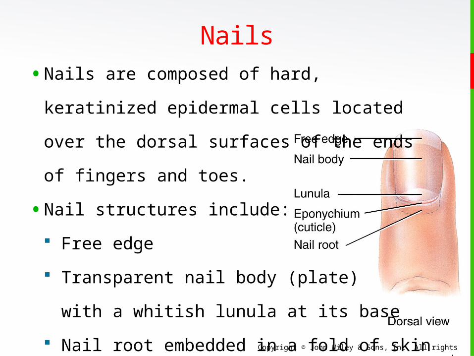
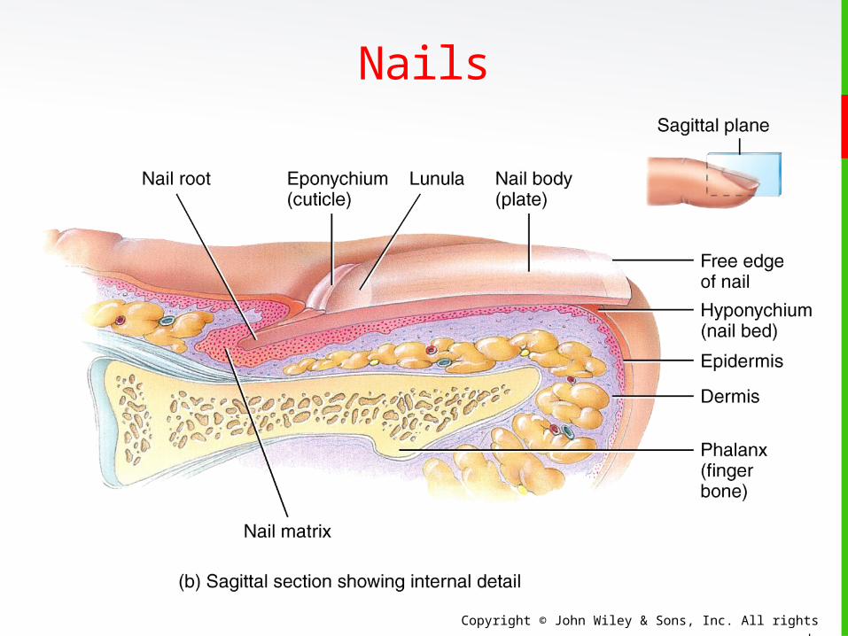
Nails• Nails are composed of hard, keratinized
epidermal cells located over the dorsal
surfaces of the ends of fingers and toes.
• Nail structures include:
Free edge
Transparent nail body (plate)
with a whitish lunula at its base
Nail root embedded in a fold of skin
Copyright © John Wiley & Sons, Inc. All rights reserved.
Nails
Copyright © John Wiley & Sons, Inc. All rights reserved.
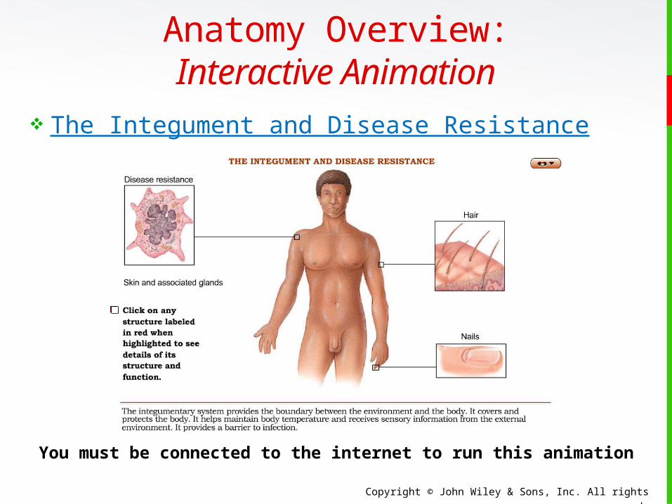
Anatomy Overview:Interactive Animation
The Integument and Disease Resistance
You must be connected to the internet to run this animation
Copyright © John Wiley & Sons, Inc. All rights reserved.
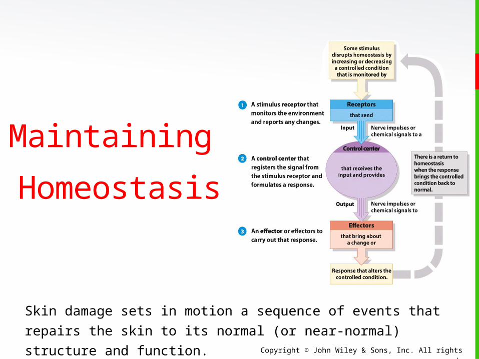
Maintaining
Homeostasis
Skin damage sets in motion a sequence of events that repairs
the skin to its normal (or near-normal) structure and function.
Copyright © John Wiley & Sons, Inc. All rights reserved.
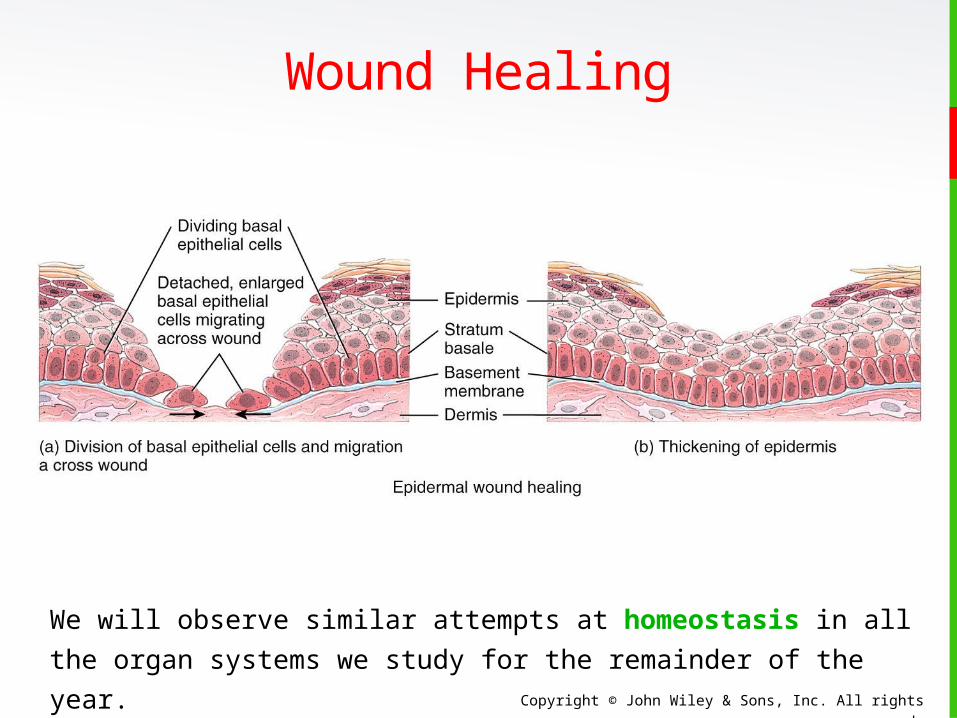
Wound Healing Two kinds of wound-healing processes can occur,
depending on the depth of the injury.
Epidermal wound healing occurs following
superficial wounds that affect only the epidermis.
• Return to normal function is the rule.
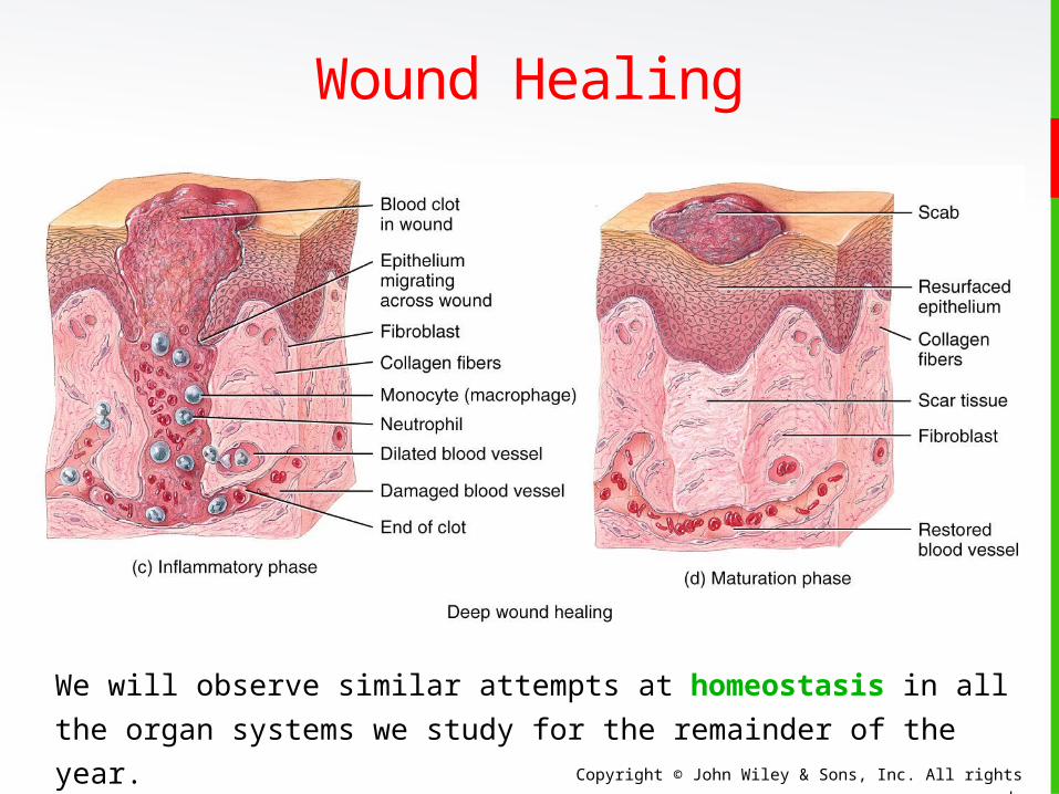
Deep wound healing occurs when an injury
extends to the dermis and subcutaneous layer.
• Loss of some function and development of scar
tissue is the rule.
Copyright © John Wiley & Sons, Inc. All rights reserved.
Wound Healing
We will observe similar attempts at homeostasis in all the
organ systems we study for the remainder of the year.
Copyright © John Wiley & Sons, Inc. All rights reserved.
Wound Healing
We will observe similar attempts at homeostasis in all the
organ systems we study for the remainder of the year.
Copyright © John Wiley & Sons, Inc. All rights reserved.
Burns A burn is tissue damage caused by excessive
heat, electricity, radioactivity, or corrosive
chemicals that denature (break down) the
proteins in the skin cells.
Burns destroy some of the skin's important
contributions to homeostasis—protection
against microbial invasion and desiccation,
and thermoregulation.
Burns are graded according to their severity.
Copyright © John Wiley & Sons, Inc. All rights reserved.
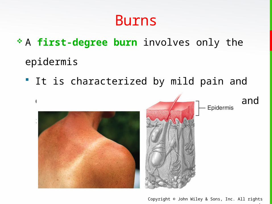
Burns A first-degree burn involves only the
epidermis
It is characterized by mild pain and erythema
(redness) but no blisters and skin functions
remain intact.
Copyright © John Wiley & Sons, Inc. All rights reserved.
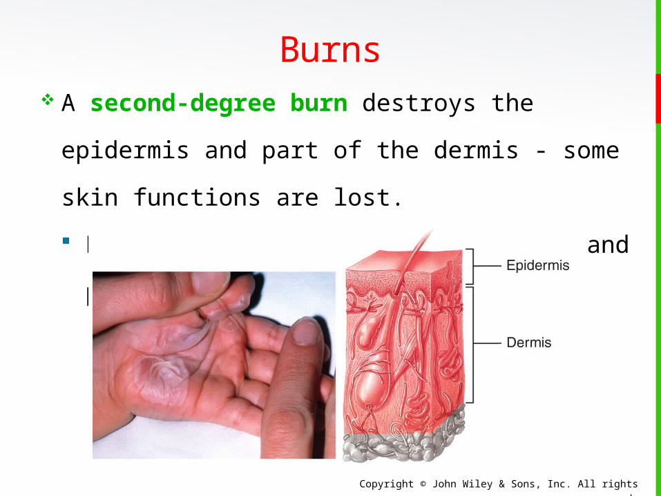
Burns A second-degree burn destroys the
epidermis and part of the dermis - some skin
functions are lost.
Redness, blister formation, edema, and pain
result.
Copyright © John Wiley & Sons, Inc. All rights reserved.
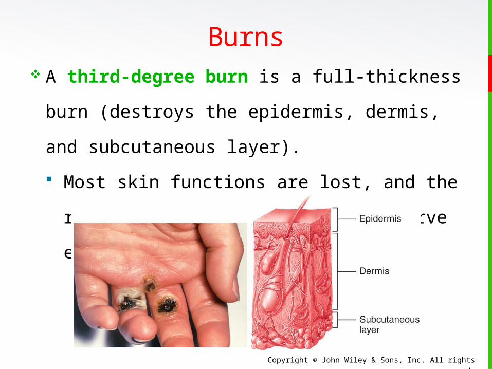
Burns A third-degree burn is a full-thickness burn
(destroys the epidermis, dermis, and
subcutaneous layer).
Most skin functions are lost, and the region is
numb because sensory nerve endings have
been destroyed.
Copyright © John Wiley & Sons, Inc. All rights reserved.
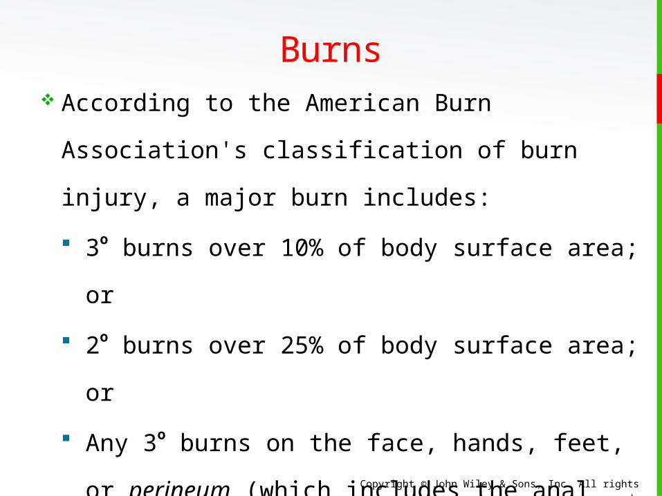
Burns According to the American Burn Association's
classification of burn injury, a major burn
includes:
3o burns over 10% of body surface area; or
2o burns over 25% of body surface area; or
Any 3o burns on the face, hands, feet, or
perineum (which includes the anal and
urogenital regions)
When the burn area exceeds 70%, more than
half the victims die.

Copyright © John Wiley & Sons, Inc. All rights reserved.
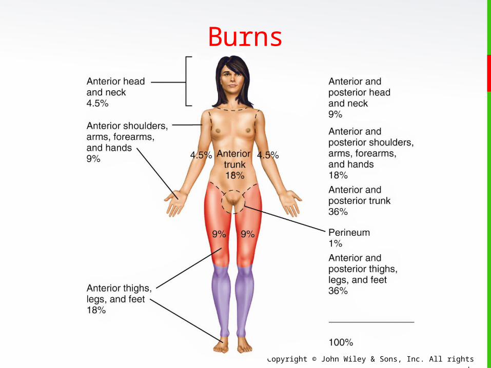
Burns A quick means for estimating the surface area
affected by a burn in an adult is the rule of
nines: Count 9% if both the anterior and posterior surfaces of the
head and neck are affected.
Count 9% for both the anterior and posterior surfaces of
each upper limb (total of 18% for both upper limbs).
Count four times nine or 36% for both the anterior and
posterior surfaces of the trunk, including the buttocks.
Count 9% for the anterior and 9% for the posterior surfaces
of each lower limb as far up as the buttocks (total of 36%
for both lower limbs).
Copyright © John Wiley & Sons, Inc. All rights reserved.
Burns
Copyright © John Wiley & Sons, Inc. All rights reserved.
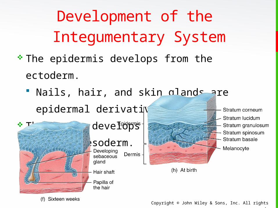
Development of the Integumentary System
The epidermis develops from the ectoderm.
Nails, hair, and skin glands are epidermal
derivatives. The dermis develops
from the mesoderm.
Copyright © John Wiley & Sons, Inc. All rights reserved.
Aging The integumentary system changes with age:
Wrinkles develop.
Dehydration and cracking occurs.
Sweat production increases.
An increase in the numbers of functional
melanocytes results in gray hair and atypical skin
pigmentation.
Subcutaneous fat is lost, and there is a general
decrease in skin thickness.
Nails may also become more brittle.
Copyright © John Wiley & Sons, Inc. All rights reserved.
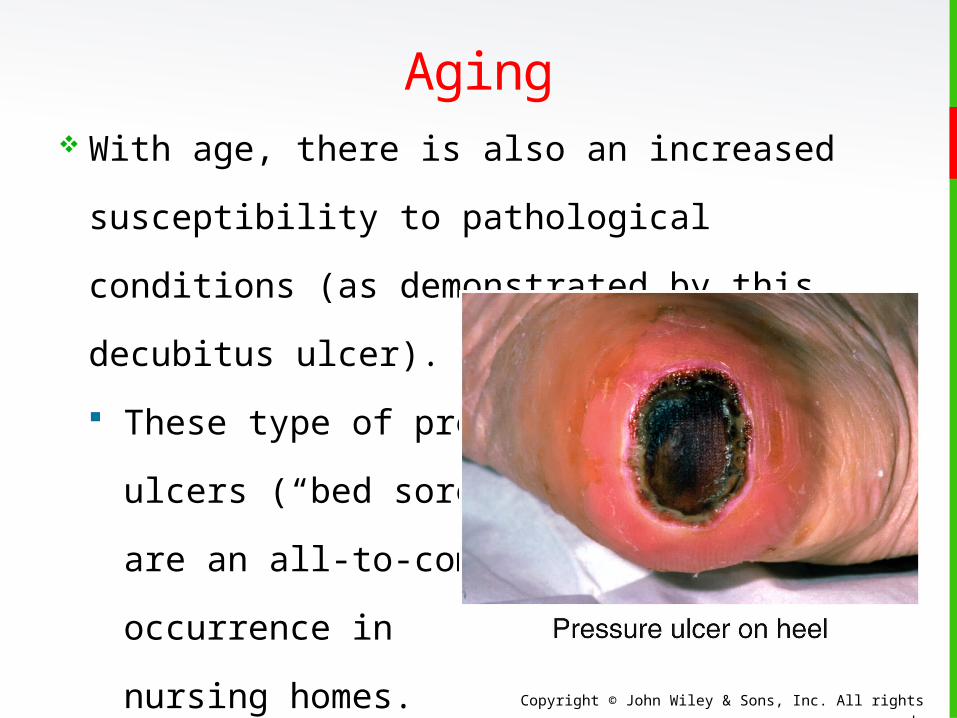
Aging With age, there is also an increased
susceptibility to pathological conditions (as
demonstrated by this decubitus ulcer).
These type of pressure
ulcers (“bed sores”)
are an all-to-common
occurrence in
nursing homes.

Copyright © John Wiley & Sons, Inc. All rights reserved.
End of Chapter 5
Copyright 2012 John Wiley & Sons, Inc. All rights
reserved. Reproduction or translation of this work
beyond that permitted in section 117 of the 1976
United States Copyright Act without express
permission of the copyright owner is unlawful.
Request for further information should be addressed
to the Permission Department, John Wiley & Sons, Inc.
The purchaser may make back-up copies for his/her
own use only and not for distribution or resale. The
Publisher assumes no responsibility for errors,
omissions, or damages caused by the use of these
programs or from the use of the information herein.