Embed Size (px)
Citation preview
By : Gan Quan Fu, PT,
MSc Human Anatomy (Batch 3)
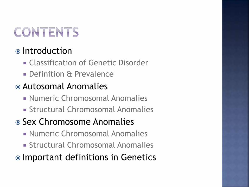
Introduction
Classification of Genetic Disorder
Definition & Prevalence
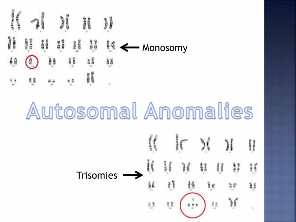
Autosomal Anomalies
Numeric Chromosomal Anomalies
Structural Chromosomal Anomalies
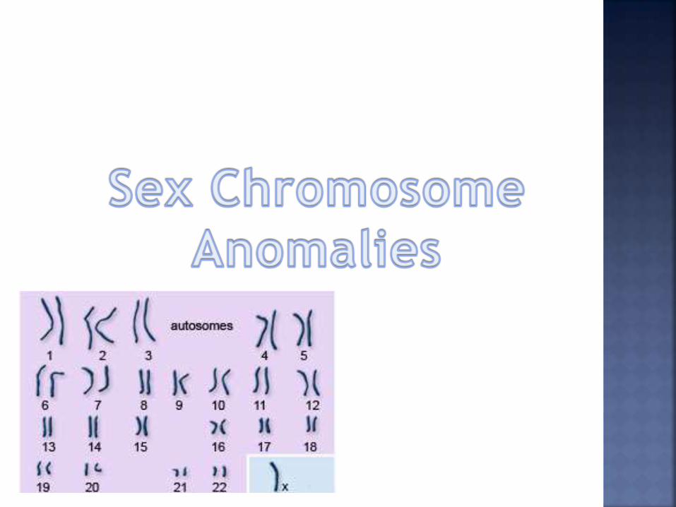
Sex Chromosome Anomalies
Numeric Chromosomal Anomalies
Structural Chromosomal Anomalies
Important definitions in Genetics
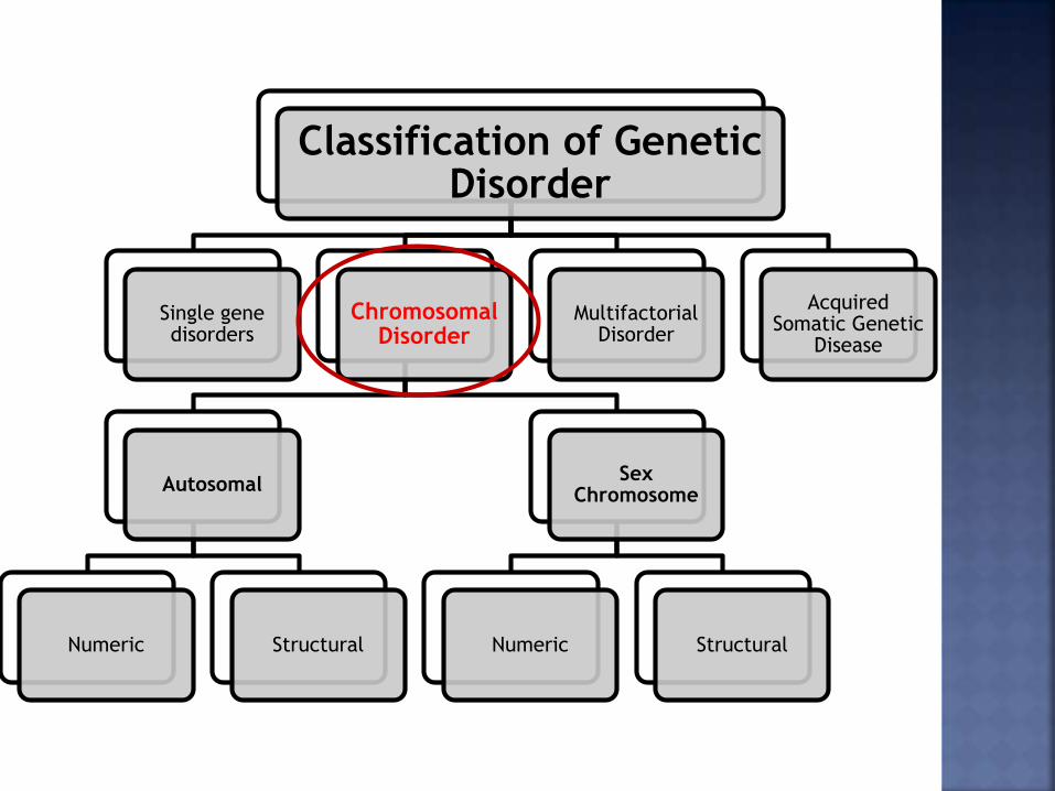
Classification of Genetic Disorder
Single gene disorders
Chromosomal Disorder
Autosomal
Numeric Structural
Sex Chromosome
Numeric Structural
Multifactorial Disorder
Acquired Somatic Genetic
Disease
Chromosomal Anomalies = Missing, extra, or
irregular portion of chromosomal DNA.
Most foetus with some chromosomal
abnormality do not survive.
Affects approximately 1 out of 200 of new-
borns.
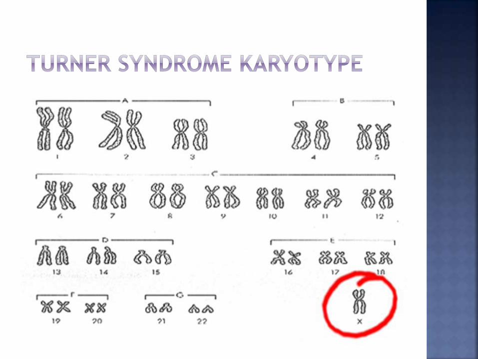
Karyotype = Full set of chromosomes from an
individual. (Chromosomal Anomalies can be
detected via Karyotype Testing.)
Abnormalities depends on type of chromosome
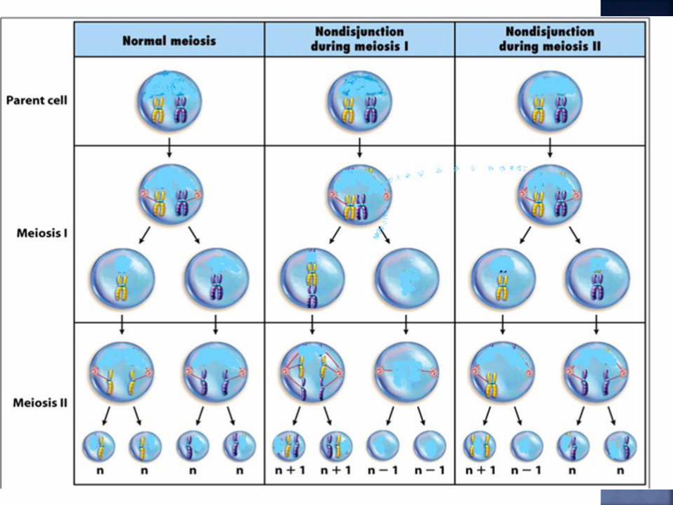
affected due to non-disjunction chromosomes.
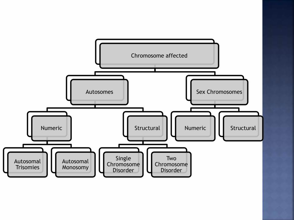
Chromosome affected
Autosomes
Numeric
Autosomal Trisomies
Autosomal Monosomy
Structural
Single Chromosome
Disorder
Two Chromosome
Disorder
Sex Chromosomes
Numeric Structural
Monosomy
Trisomies
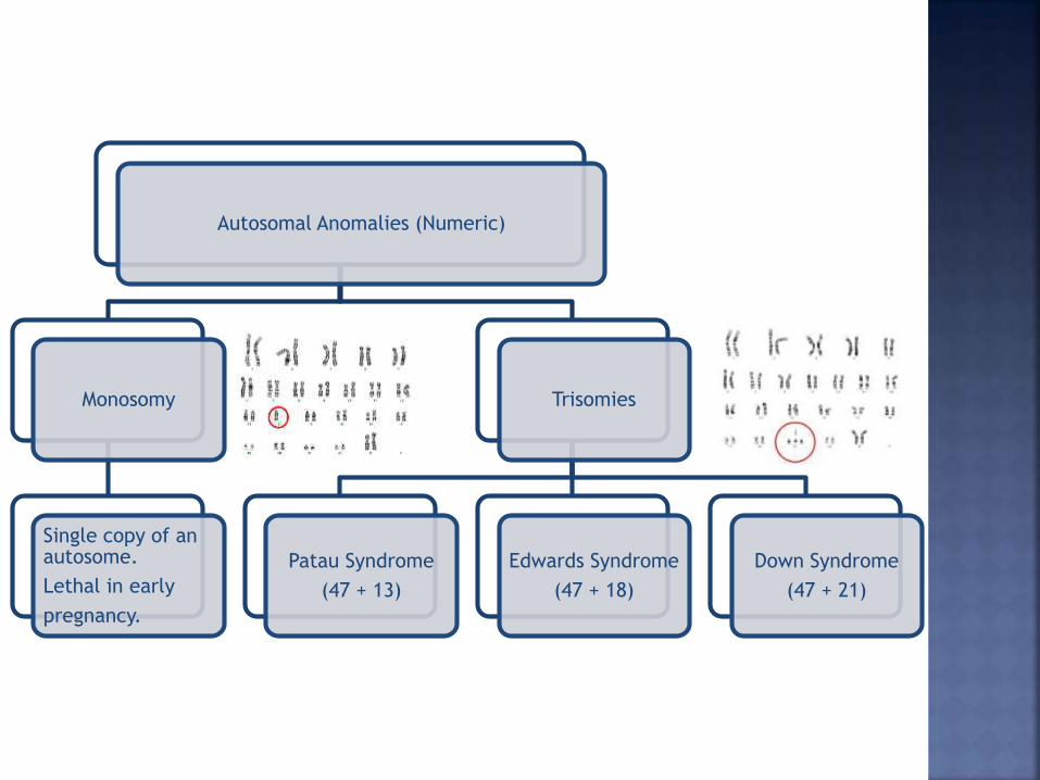
Autosomal Anomalies (Numeric)
Monosomy
Single copy of an autosome.
Lethal in early
pregnancy.
Trisomies
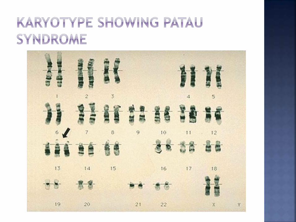
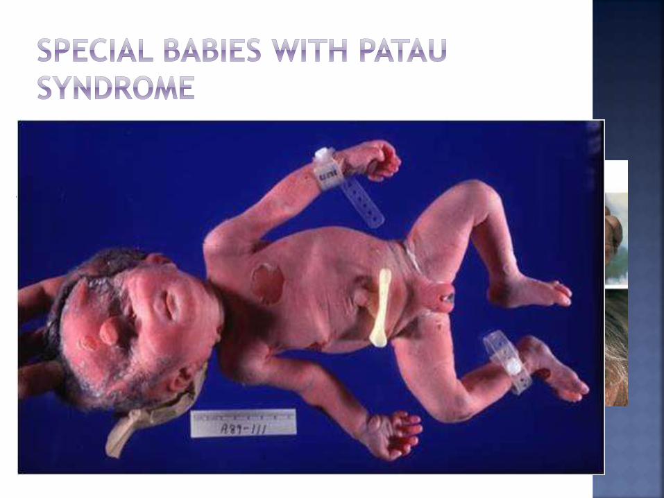
Patau Syndrome
(47 + 13)
Edwards Syndrome
(47 + 18)
Down Syndrome
(47 + 21)
An additional chromosome 13 resulting
from nondisjunction during meiosis.
Incidence = 1 in 10,000 -20,000 live
births.
More than 80% die within the first year of
life.
Anomalies can be seen on:
Nervous & Optic System
Musculoskeletal & Cutaneous
Urogenital & Cardiovascular System
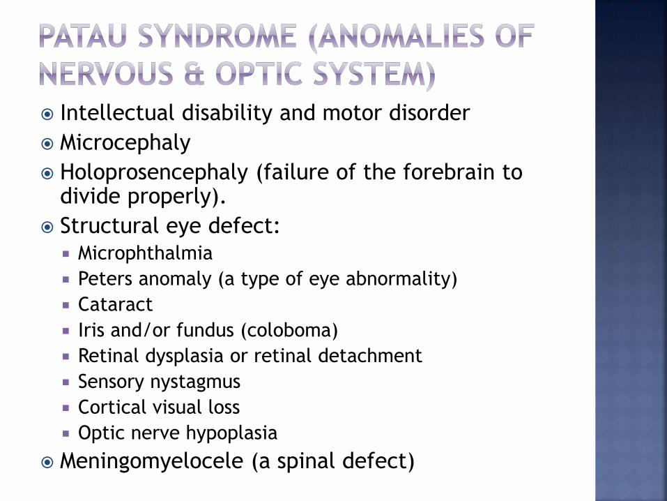
Intellectual disability and motor disorder
Microcephaly
Holoprosencephaly (failure of the forebrain to divide properly).
Structural eye defect:
Microphthalmia
Peters anomaly (a type of eye abnormality)
Cataract
Iris and/or fundus (coloboma)
Retinal dysplasia or retinal detachment
Sensory nystagmus
Cortical visual loss
Optic nerve hypoplasia
Meningomyelocele (a spinal defect)
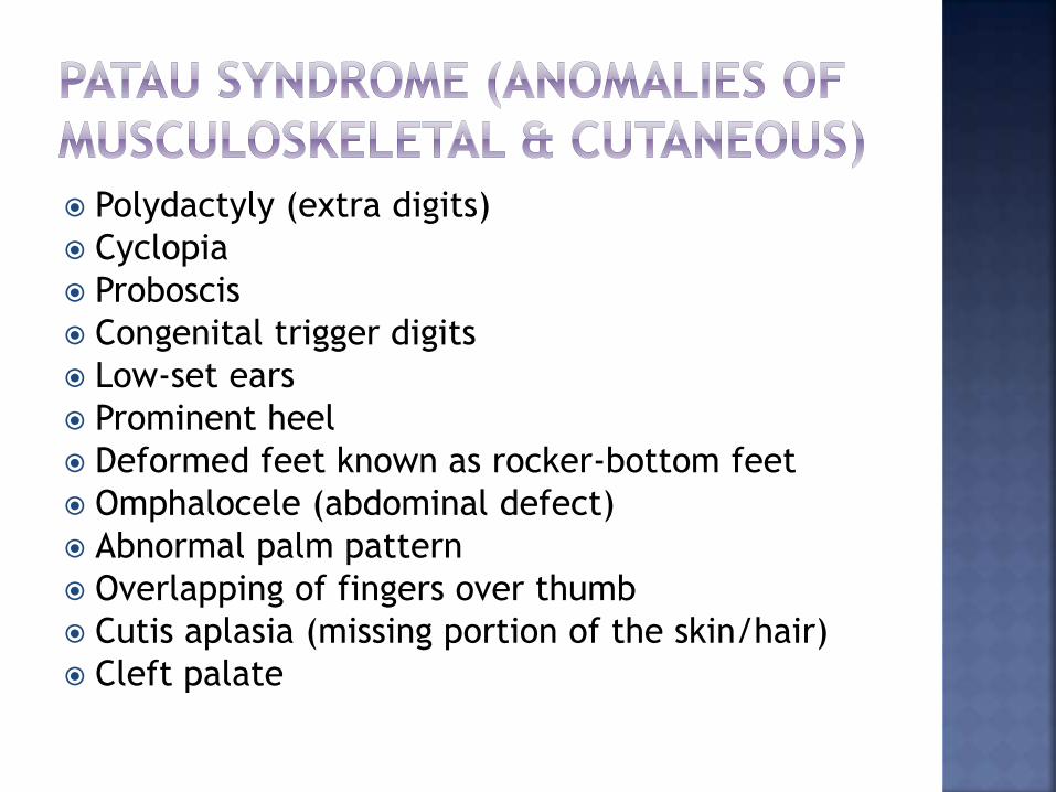
Polydactyly (extra digits)
Cyclopia
Proboscis
Congenital trigger digits
Low-set ears
Prominent heel
Deformed feet known as rocker-bottom feet
Omphalocele (abdominal defect)
Abnormal palm pattern
Overlapping of fingers over thumb
Cutis aplasia (missing portion of the skin/hair)
Cleft palate
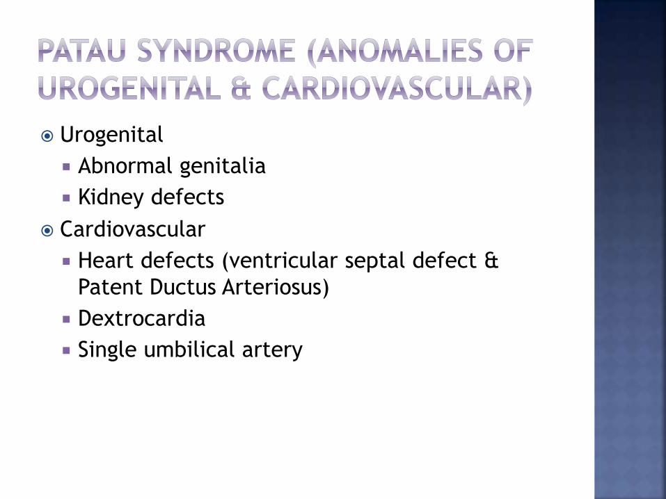
Urogenital
Abnormal genitalia
Kidney defects
Cardiovascular
Heart defects (ventricular septal defect &
Patent Ductus Arteriosus)
Dextrocardia
Single umbilical artery
A. Midline defect with
cleft lip & palate.
B. Clenched hand with
overlapping fingers.
C. Postaxial polydactyly.
D.Equinovarus deformity.
E. Punched out aplasia
cutis scalp lesions.
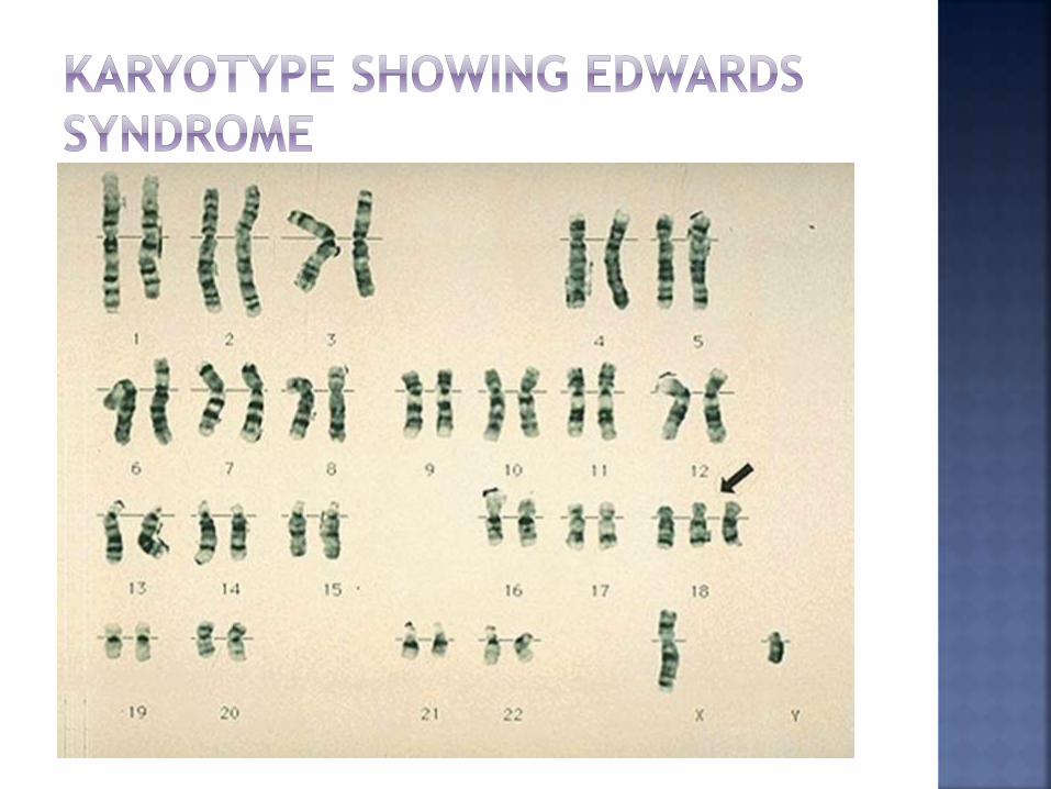
An additional chromosome 18 resulting from
nondisjunction during meiosis.
Incidence =1 in 6000 - 8000 live birth.
Majority of fetuses with this syndrome die
before birth; >90% dead in 1st year.
80% affected were females.
Kidney malformations
Structural heart defects at birth
Ventricular septal defect
Atrial septal defect
Patent ductus arteriosus
Omphalocele (Intestines protruding outside the body)
Esophageal atresia
Intellectual disability
Developmental delays
Growth deficiency
Feeding difficulties
Breathing difficulties
Arthrogryposis (a muscle disorder that causes multiple
joint contractures at birth)
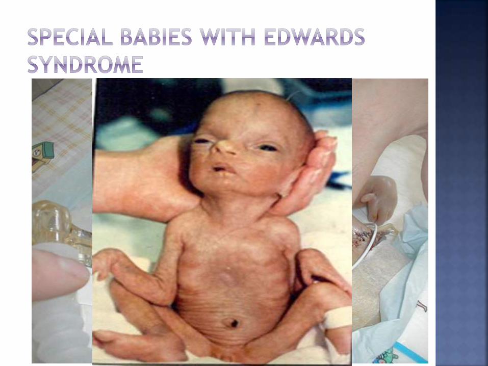
Small head (microcephaly) accompanied by a prominent back portion of the head (occiput),
Low-set, malformed ears,
Abnormally small jaw (micrognathia),
Cleft lip/cleft palate,
Upturned nose,
Narrow eyelid folds (palpebral fissures),
Widely spaced eyes (ocular hypertelorism),
Drooping of the upper eyelids (ptosis),
A short breast bone,
Clenched hands
Choroid plexus cysts
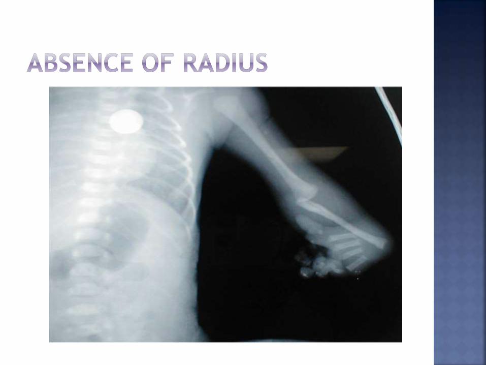
Underdeveloped thumbs and/or nails
Absent radius
Webbing of the second and third toes, clubfoot or rocker bottom feet
Males will have undescended testicles.
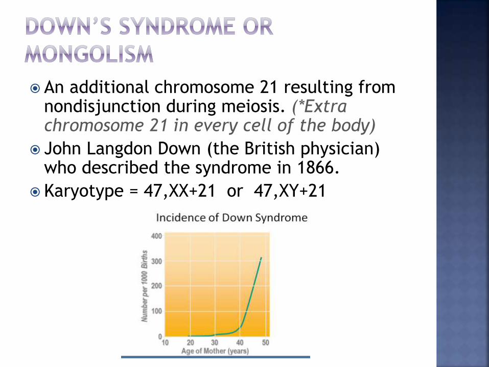
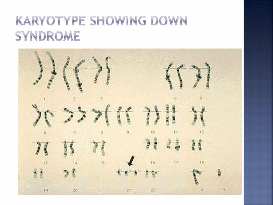
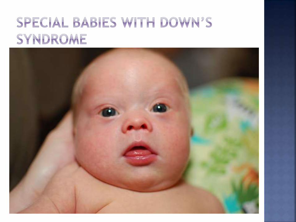
An additional chromosome 21 resulting from nondisjunction during meiosis. (*Extra chromosome 21 in every cell of the body)
John Langdon Down (the British physician) who described the syndrome in 1866.
Karyotype = 47,XX+21 or 47,XY+21
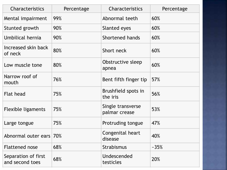
Characteristics Percentage Characteristics Percentage
Mental impairment 99% Abnormal teeth 60%
Stunted growth 90% Slanted eyes 60%
Umbilical hernia 90% Shortened hands 60%
Increased skin back
of neck80% Short neck 60%
Low muscle tone 80%Obstructive sleep
apnea60%
Narrow roof of
mouth76% Bent fifth finger tip 57%
Flat head 75%Brushfield spots in
the iris56%
Flexible ligaments 75%Single transverse
palmar crease53%
Large tongue 75% Protruding tongue 47%
Abnormal outer ears 70%Congenital heart
disease40%
Flattened nose 68% Strabismus ~35%
Separation of first
and second toes68%
Undescended
testicles20%
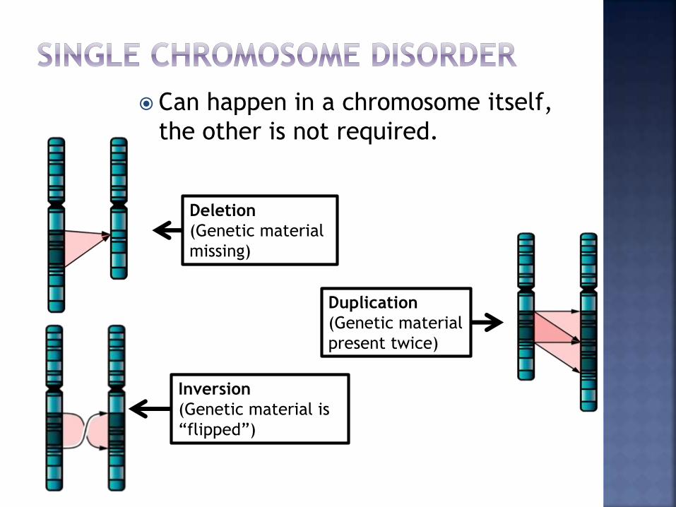
Can happen in a chromosome itself,
the other is not required.
Deletion
(Genetic material
missing)
Duplication
(Genetic material
present twice)
Inversion
(Genetic material is
“flipped”)
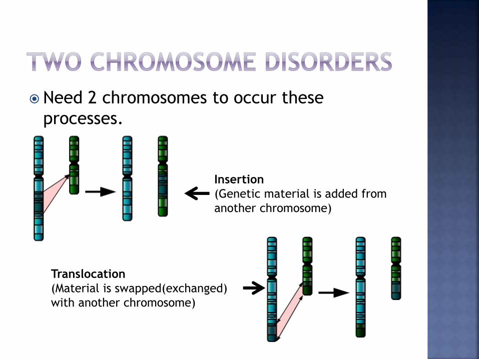
Need 2 chromosomes to occur these
processes.
Insertion
(Genetic material is added from
another chromosome)
Translocation
(Material is swapped(exchanged)
with another chromosome)
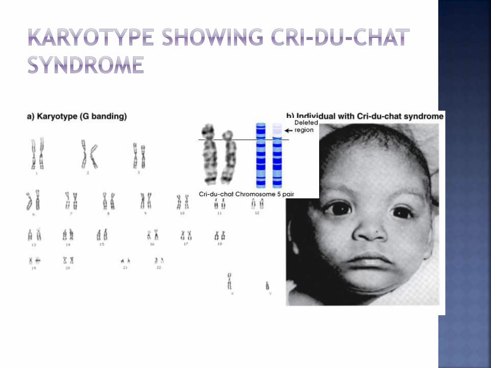
Name is based on the infant’s cry. (high-
pitched and sounds like a cat.)
Incidence =1 in 216,000 birth
Normal 46 chromosomes but, missing a piece
of chromosome number 5.
Most cases are believed to occur during the
development of the egg or sperm, small
number of cases occur when a parent passes
a different, rearranged form of the
chromosome to their child.
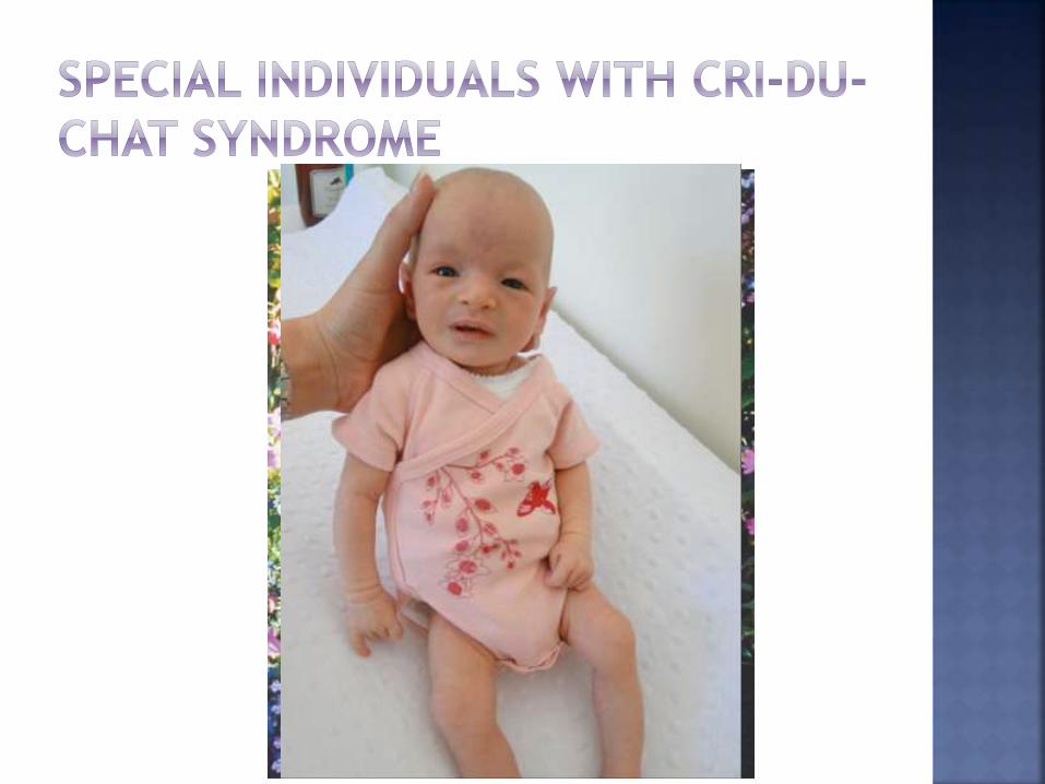
Cry is high pitched and similar to that of a meowing kitten.
Moon-shaped face
Malformed larynx
Difficulty swallowing and sucking (Feeding Problem).
Low birth weight and poor growth.
Severe cognitive, speech, and motor delays, mental retardation
Behavioural problems such as hyperactivity, aggression, and repetitive movements.
Excessive drooling(ptyalism)
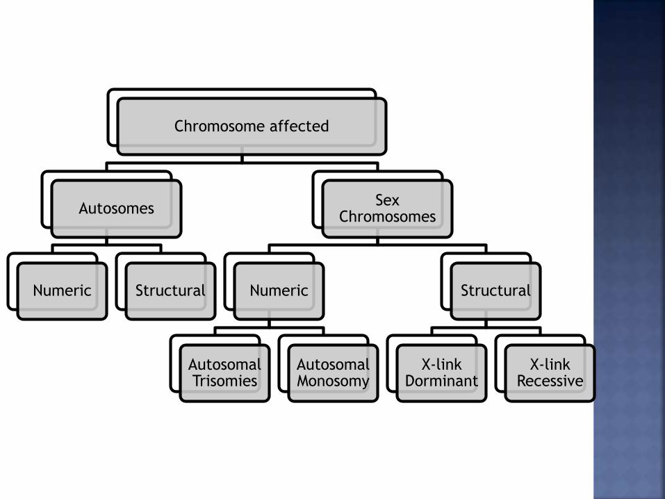
Chromosome affected
Autosomes
Numeric Structural
Sex Chromosomes
Numeric
Autosomal Trisomies
Autosomal Monosomy
Structural
X-link Dorminant
X-link Recessive
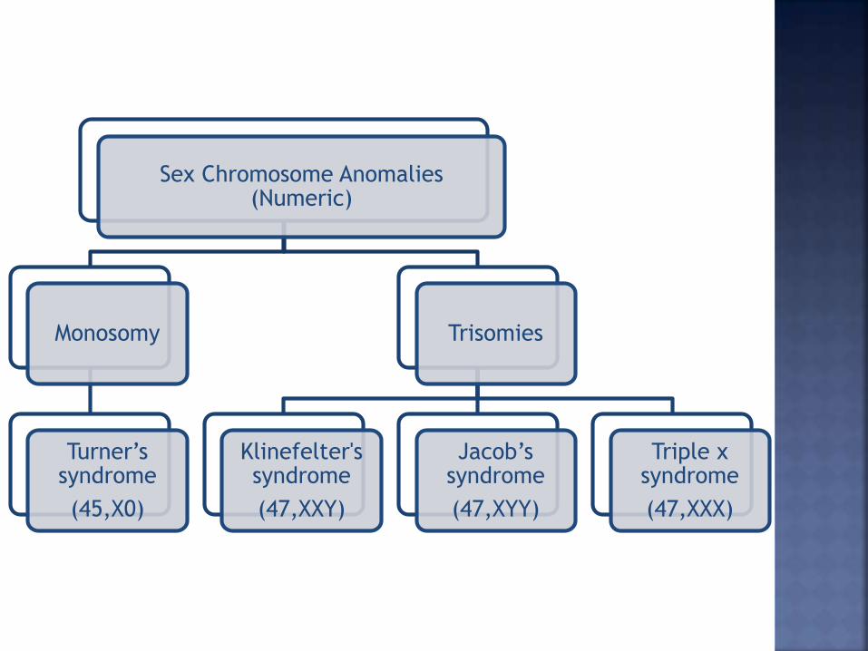
Sex Chromosome Anomalies (Numeric)
Monosomy
Turner’s syndrome
(45,X0)
Trisomies
Klinefelter'ssyndrome
(47,XXY)
Jacob’s syndrome
(47,XYY)
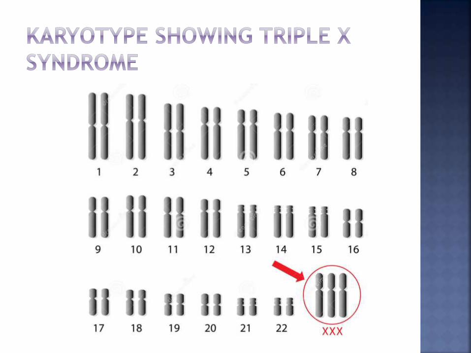
Triple x syndrome
(47,XXX)
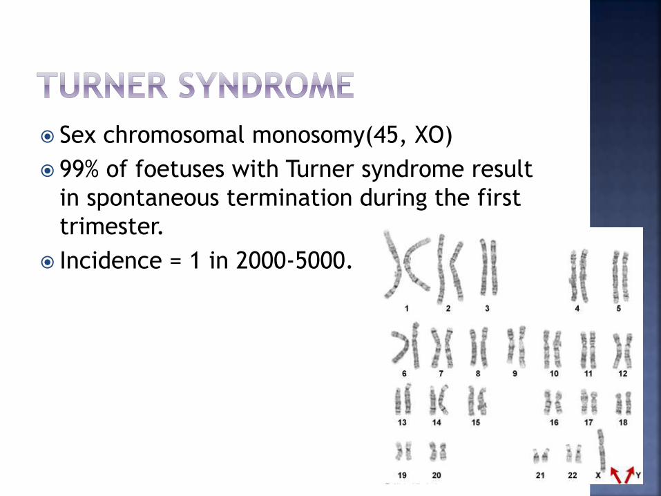
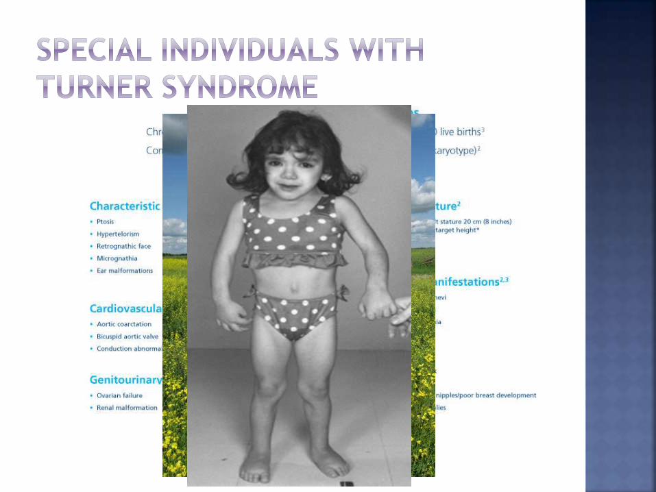
Sex chromosomal monosomy(45, XO)
99% of foetuses with Turner syndrome result
in spontaneous termination during the first
trimester.
Incidence = 1 in 2000-5000.
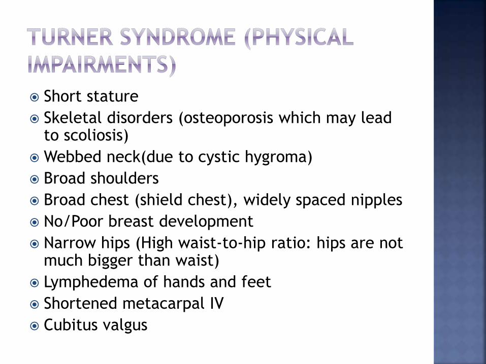
Short stature
Skeletal disorders (osteoporosis which may lead to scoliosis)
Webbed neck(due to cystic hygroma)
Broad shoulders
Broad chest (shield chest), widely spaced nipples
No/Poor breast development
Narrow hips (High waist-to-hip ratio: hips are not much bigger than waist)
Lymphedema of hands and feet
Shortened metacarpal IV
Cubitus valgus
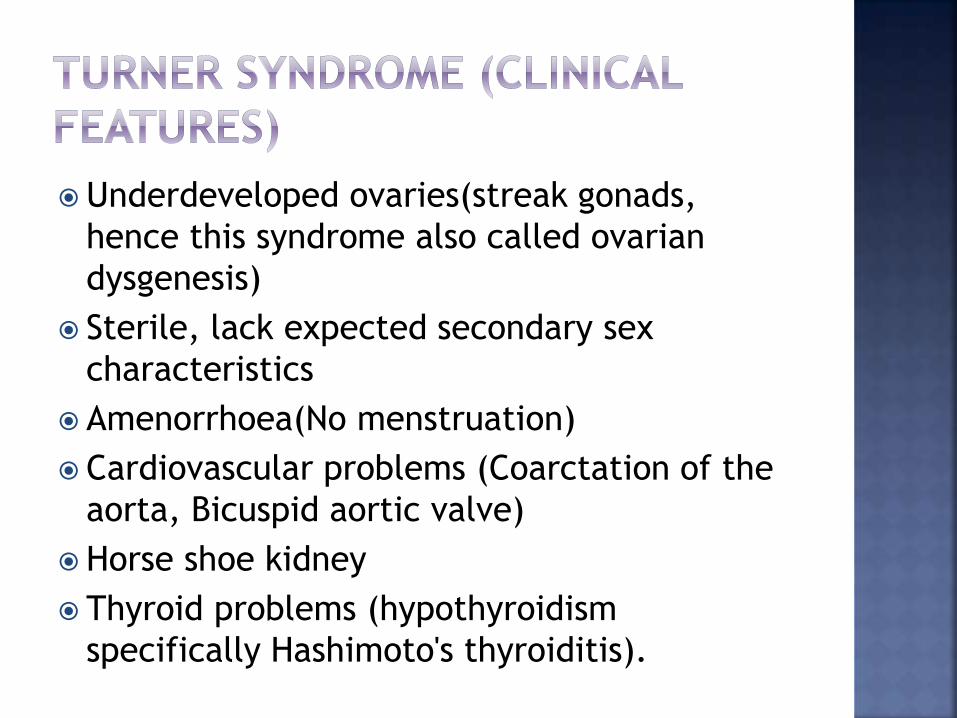
Underdeveloped ovaries(streak gonads,
hence this syndrome also called ovarian
dysgenesis)
Sterile, lack expected secondary sex
characteristics
Amenorrhoea(No menstruation)
Cardiovascular problems (Coarctation of the
aorta, Bicuspid aortic valve)
Horse shoe kidney
Thyroid problems (hypothyroidism
specifically Hashimoto's thyroiditis).
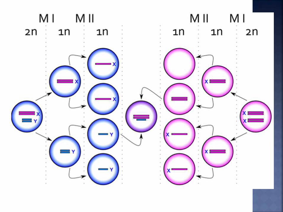
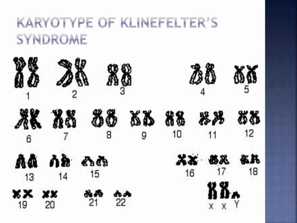
XXY Males
Disorder occurring due to nondisjunction of the X chromosome during Meiosis. Extra X chromosome is nondisjunction during
meiosis II of the germ cell in the female.
Occur when sister chromatids on the X sex chromosome fail to separate.
XX ovum produced and when fertilized with a Y-sperm it yields XXY offspring.
Also occurs X and Y sex chromosomes fail to separate, producing a sperm with an X and Y chromosome & fertilize with normal X ovum produces XXY offspring.
Incidence = 1 in 500 live male births.
48, XXYY (male) syndrome
Incidence = 1 in 18,000–40,000 births
48,XXXY
Incidence = Extremely rare
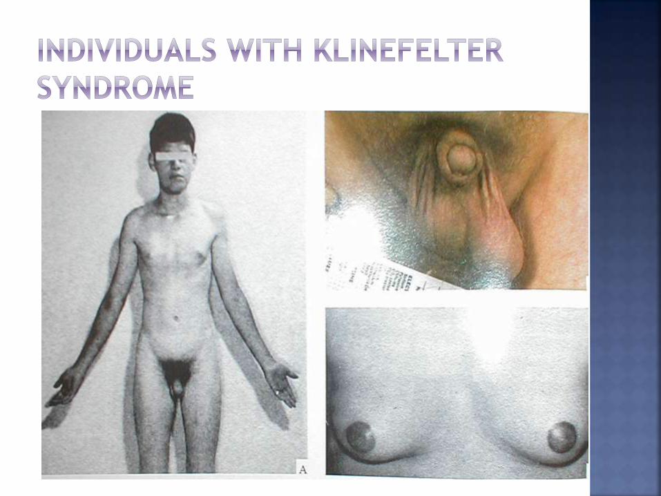
Childhood Weaker muscles and reduced strength.
Puberty (features become more prominent due to
hypogonadism (less amount of testosterone produced): Rounded body type Broader hips Little body hair is present Gynecomastia (increased breast tissue) Microorchidism (i.e. small testicles) Azospermia leading to infertility Micropenis Tall stature IQ is normal
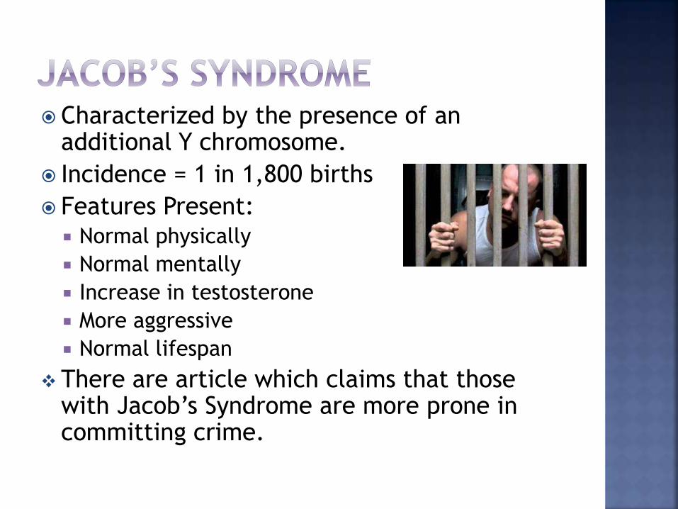
Characterized by the presence of an additional Y chromosome.
Incidence = 1 in 1,800 births
Features Present: Normal physically
Normal mentally
Increase in testosterone
More aggressive
Normal lifespan
There are article which claims that those with Jacob’s Syndrome are more prone in committing crime.

Characterized by the presence of an additional X
chromosome.
When an XX ovum fertilizes with X- sperm.
Incidence = 1 in 1,000 birth
Features Present
Normal physically, Sometimes taller.
Normal mentally, Increase risk of retardation
and learning difficulties.
Fertile.
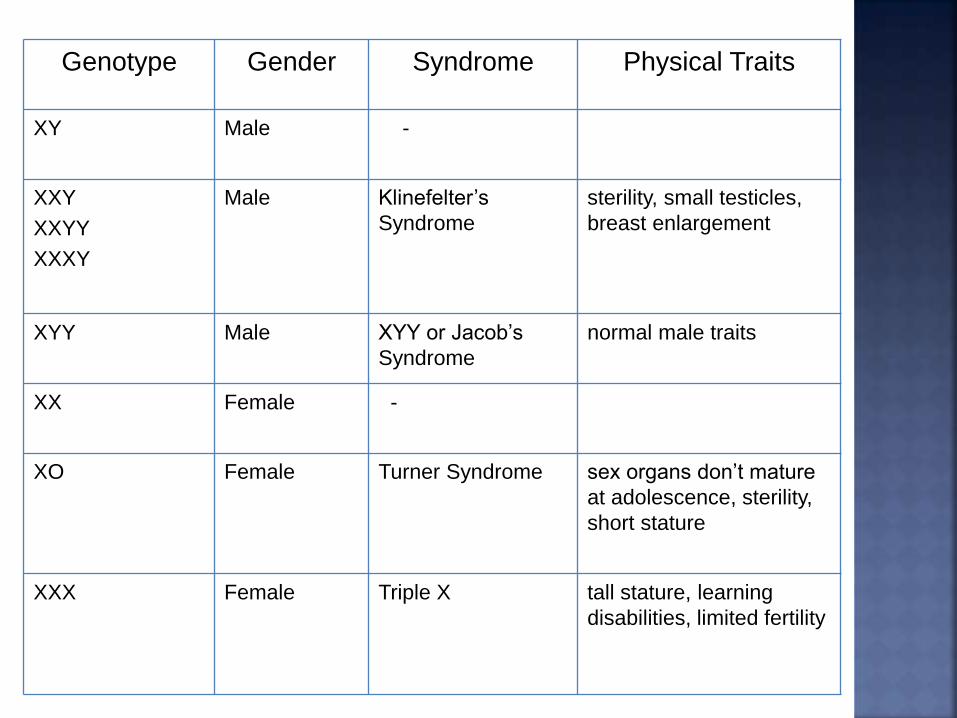
Genotype Gender Syndrome Physical Traits
XY Male -
XXY
XXYY
XXXY
Male Klinefelter’s
Syndrome
sterility, small testicles,
breast enlargement
XYY Male XYY or Jacob’s
Syndrome
normal male traits
XX Female -
XO Female Turner Syndrome sex organs don’t mature
at adolescence, sterility,
short stature
XXX Female Triple X tall stature, learning
disabilities, limited fertility
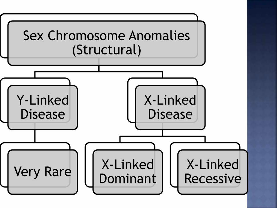
Sex Chromosome Anomalies (Structural)
Y-Linked Disease
Very Rare
X-Linked Disease
X-Linked Dominant
X-Linked Recessive
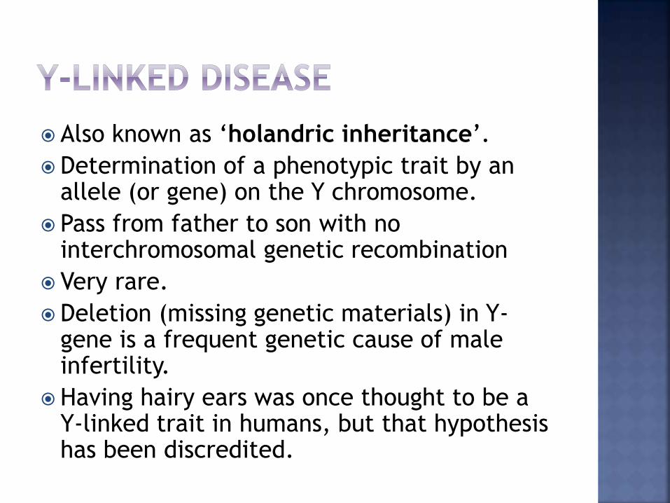
Also known as ‘holandric inheritance’.
Determination of a phenotypic trait by an allele (or gene) on the Y chromosome.
Pass from father to son with no interchromosomal genetic recombination
Very rare.
Deletion (missing genetic materials) in Y-gene is a frequent genetic cause of male infertility.
Having hairy ears was once thought to be a Y-linked trait in humans, but that hypothesis has been discredited.
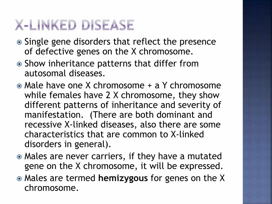
Single gene disorders that reflect the presence of defective genes on the X chromosome.
Show inheritance patterns that differ from autosomal diseases.
Male have one X chromosome + a Y chromosome while females have 2 X chromosome, they show different patterns of inheritance and severity of manifestation. (There are both dominant and recessive X-linked diseases, also there are some characteristics that are common to X-linked disorders in general).
Males are never carriers, if they have a mutated gene on the X chromosome, it will be expressed.
Males are termed hemizygous for genes on the X chromosome.
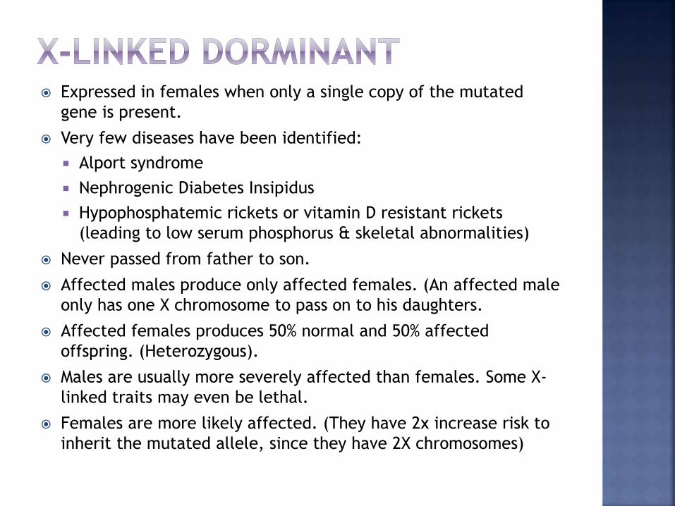
Expressed in females when only a single copy of the mutated
gene is present.
Very few diseases have been identified:
Alport syndrome
Nephrogenic Diabetes Insipidus
Hypophosphatemic rickets or vitamin D resistant rickets
(leading to low serum phosphorus & skeletal abnormalities)
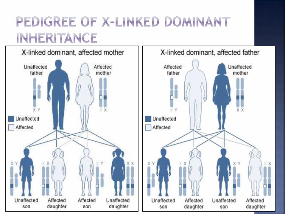
Never passed from father to son.
Affected males produce only affected females. (An affected male
only has one X chromosome to pass on to his daughters.
Affected females produces 50% normal and 50% affected
offspring. (Heterozygous).
Males are usually more severely affected than females. Some X-
linked traits may even be lethal.
Females are more likely affected. (They have 2x increase risk to
inherit the mutated allele, since they have 2X chromosomes)
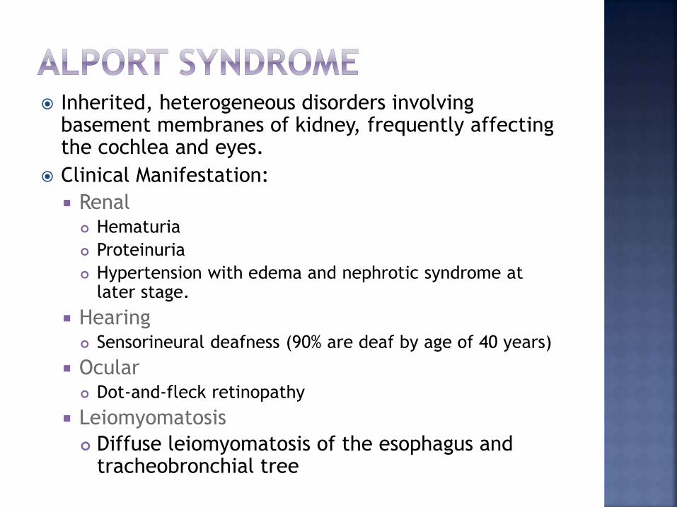
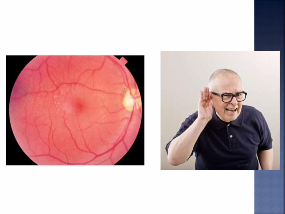
Inherited, heterogeneous disorders involving basement membranes of kidney, frequently affecting the cochlea and eyes.
Clinical Manifestation:
Renal Hematuria
Proteinuria
Hypertension with edema and nephrotic syndrome at later stage.
Hearing Sensorineural deafness (90% are deaf by age of 40 years)
Ocular Dot-and-fleck retinopathy
Leiomyomatosis
Diffuse leiomyomatosis of the esophagus and tracheobronchial tree
Occurs when the kidneys cannot concentrate
urine normally resulting excretion of large
amount of dilute urine. *Kidney tubules do not
respond to Antidiuretic Hormone (ADH).
Very Rare
Symptoms
Intense or uncontrollable thirst, and crave
ice water.
Produce large amounts of urine (usually
more than 3 liters, and up to 15 liters per
day.)
Easily dehydrated
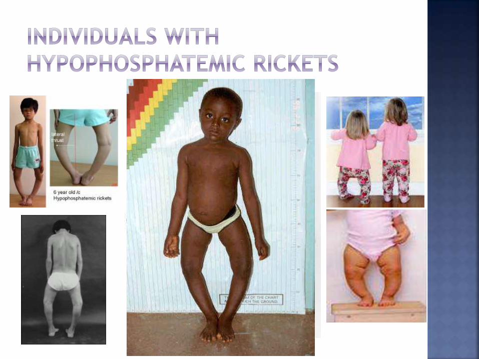
Form of rickets characterized by low serum phosphate levels.
Resistance to treatment with ultraviolet radiation or vitamin D ingestion.
Incidence = 1 in 20,000 newborns
Symptoms: Short stature
Associated with this condition is disproportionate, resulting from deformity and growth retardation of the lower extremities.
Bow Legged and deformities associated with bones
Bone Pain and Joint Pain
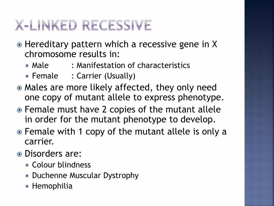
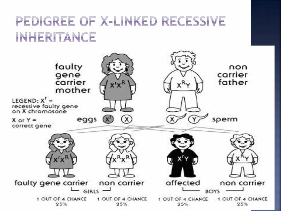
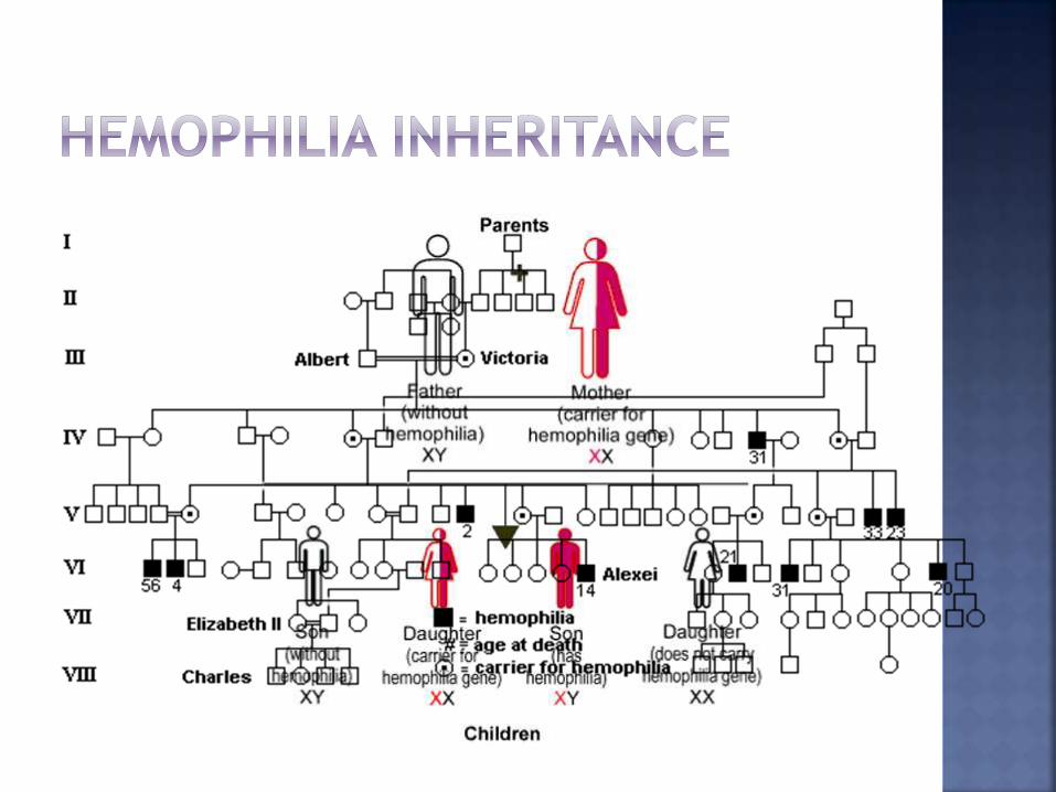
Hereditary pattern which a recessive gene in X chromosome results in:
Male : Manifestation of characteristics
Female : Carrier (Usually)
Males are more likely affected, they only need one copy of mutant allele to express phenotype.
Female must have 2 copies of the mutant allele in order for the mutant phenotype to develop.
Female with 1 copy of the mutant allele is only a carrier.
Disorders are:
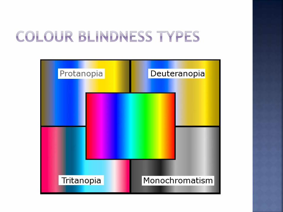
Colour blindness
Duchenne Muscular Dystrophy
Hemophilia
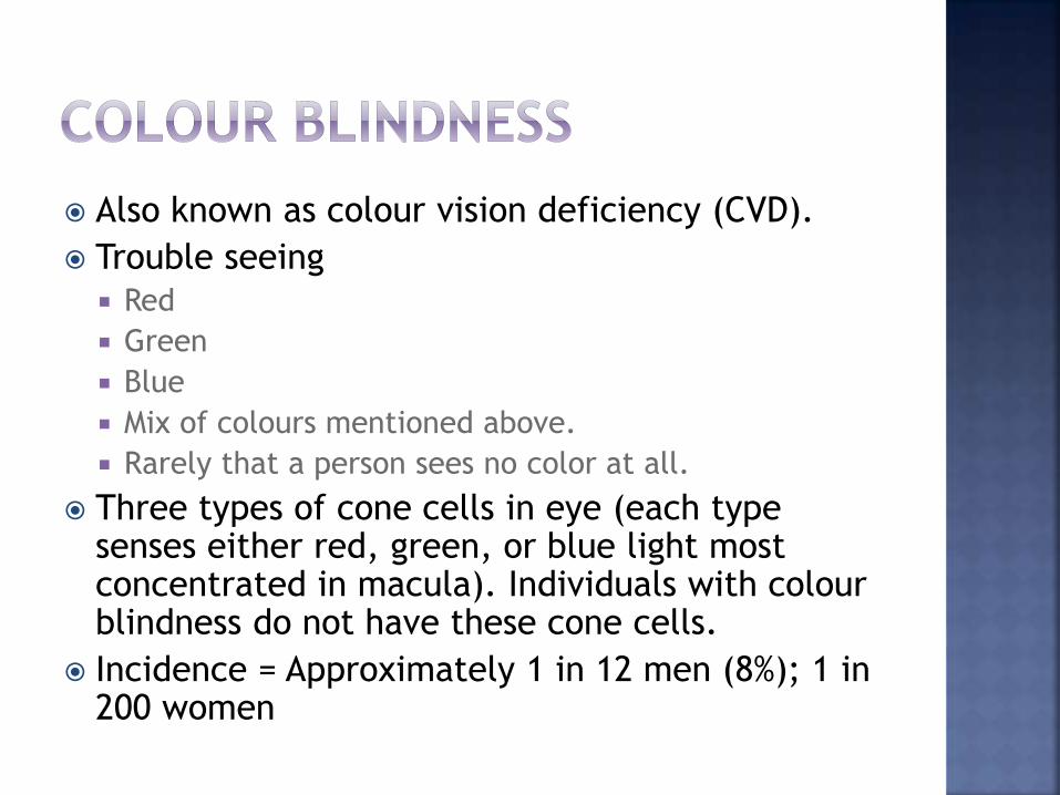
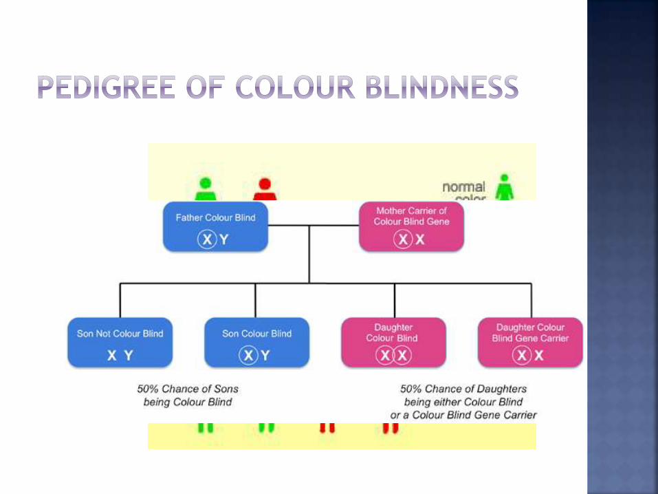
Also known as colour vision deficiency (CVD).
Trouble seeing
Red
Green
Blue
Mix of colours mentioned above.
Rarely that a person sees no color at all.
Three types of cone cells in eye (each type senses either red, green, or blue light most concentrated in macula). Individuals with colour blindness do not have these cone cells.
Incidence = Approximately 1 in 12 men (8%); 1 in 200 women
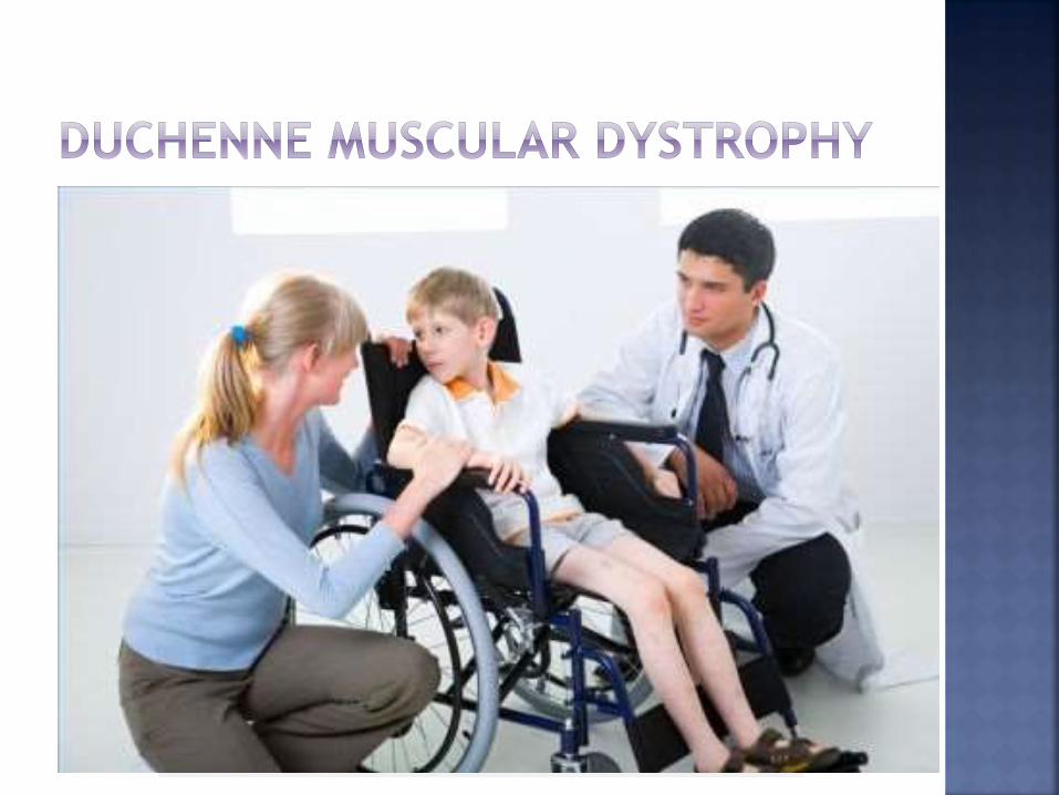
Form of muscular dystrophy (muscle
weakness and loss of muscle tissues) which
worsens quickly.
Caused by a defective gene for dystrophin (A
type of protein in muscles).
Incidence = 1 in 3,600 male infants
Usually appear before age 6 and may appear as early as infancy.
Include:
Fatigue
Learning difficulties (IQ can be below 75)
Intellectual disability
Muscle weakness:
Begins in the legs and pelvis less severe in the arms, neck, and other areas of the body
Problems with motor skills (running, hopping, jumping)
Frequent falls
Trouble getting up from a lying position or climbing stairs
Weakness quickly gets worse
Progressive difficulty walking:
Ability to walk may be lost by age 12
Breathing difficulties and heart disease usually start by age 20
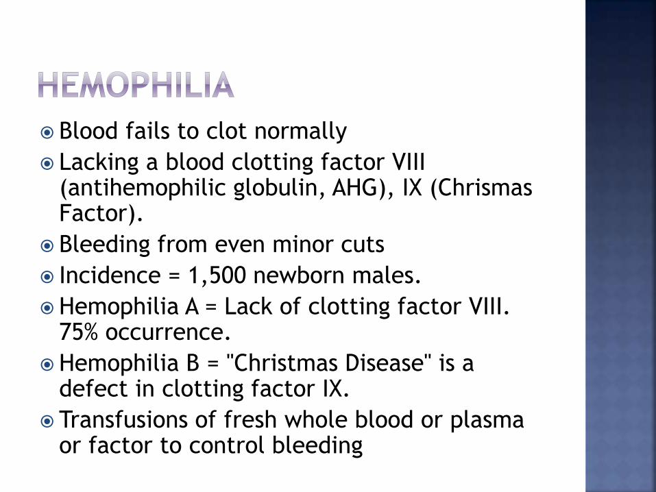
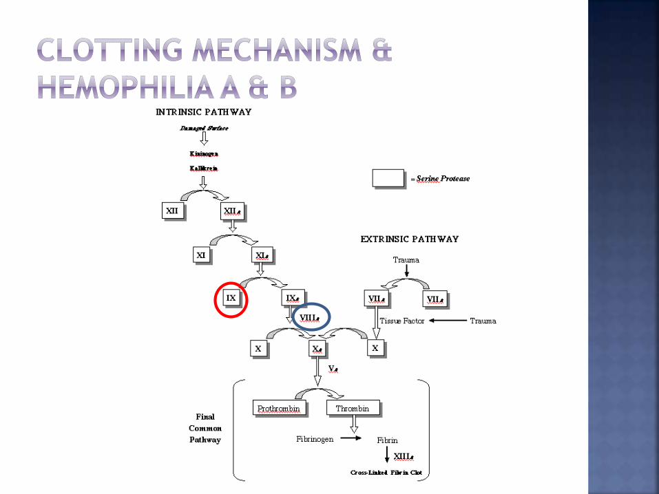
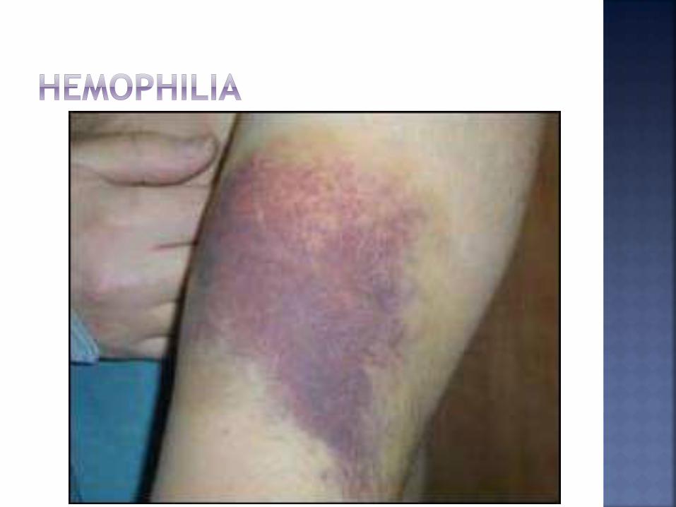
Blood fails to clot normally
Lacking a blood clotting factor VIII (antihemophilic globulin, AHG), IX (ChrismasFactor).
Bleeding from even minor cuts
Incidence = 1,500 newborn males.
Hemophilia A = Lack of clotting factor VIII. 75% occurrence.
Hemophilia B = "Christmas Disease" is a defect in clotting factor IX.
Transfusions of fresh whole blood or plasma or factor to control bleeding
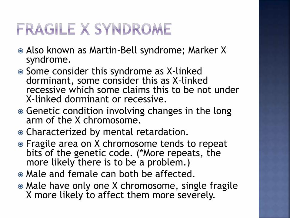
Also known as Martin-Bell syndrome; Marker X syndrome.
Some consider this syndrome as X-linked dorminant, some consider this as X-linked recessive which some claims this to be not under X-linked dorminant or recessive.
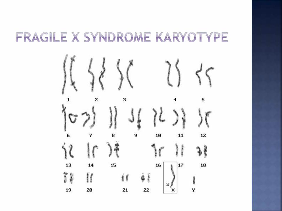
Genetic condition involving changes in the long arm of the X chromosome.
Characterized by mental retardation.
Fragile area on X chromosome tends to repeat bits of the genetic code. (*More repeats, the more likely there is to be a problem.)
Male and female can both be affected.
Male have only one X chromosome, single fragile X more likely to affect them more severely.
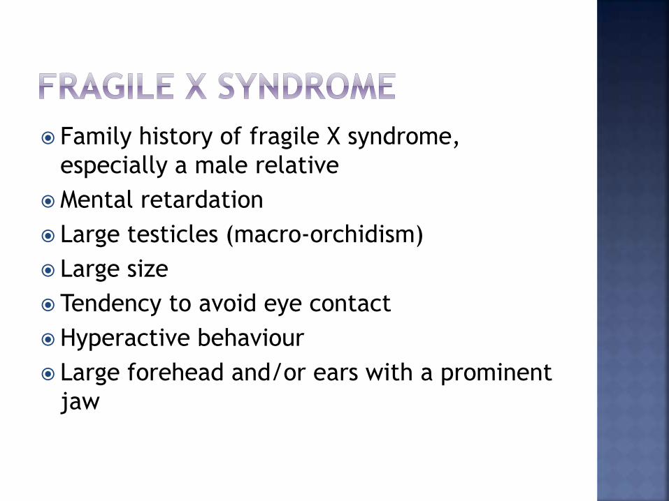
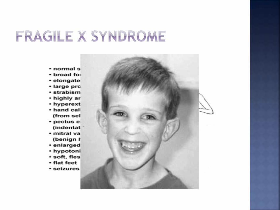
Family history of fragile X syndrome,
especially a male relative
Mental retardation
Large testicles (macro-orchidism)
Large size
Tendency to avoid eye contact
Hyperactive behaviour
Large forehead and/or ears with a prominent
jaw
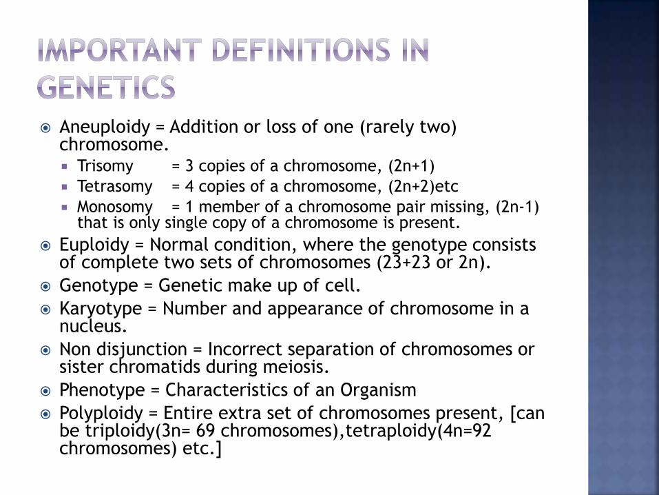
Aneuploidy = Addition or loss of one (rarely two) chromosome. Trisomy = 3 copies of a chromosome, (2n+1)
Tetrasomy = 4 copies of a chromosome, (2n+2)etc
Monosomy = 1 member of a chromosome pair missing, (2n-1) that is only single copy of a chromosome is present.
Euploidy = Normal condition, where the genotype consists of complete two sets of chromosomes (23+23 or 2n).
Genotype = Genetic make up of cell.
Karyotype = Number and appearance of chromosome in a nucleus.
Non disjunction = Incorrect separation of chromosomes or sister chromatids during meiosis.
Phenotype = Characteristics of an Organism
Polyploidy = Entire extra set of chromosomes present, [can be triploidy(3n= 69 chromosomes),tetraploidy(4n=92 chromosomes) etc.]