Embed Size (px)
Citation preview
EAR AFFECTIONS
Dr Amit SinglaSurgery & Radiology,DGCN COVAS, CSKHPKV, PalampurHimachal Pradesh (INDIA)
Otitis Externa
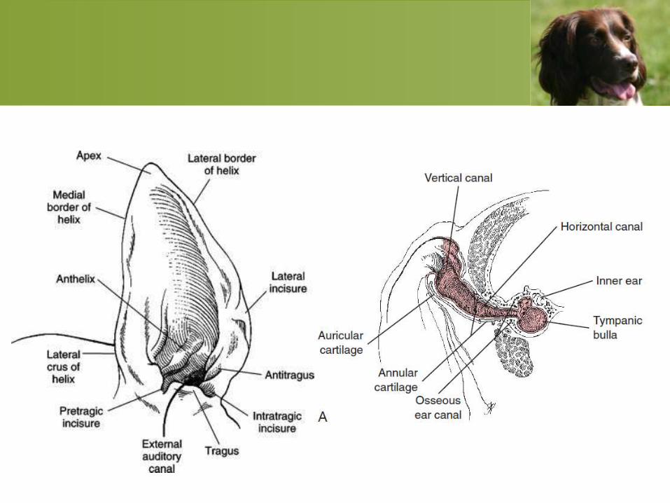
• Otitis externa is an inflammation of the
epithelium of the horizontal and vertical ear
canals and surrounding structures (i.e.,
external auditory meatus and pinna). Swimmer's
ear is a term used to describe otitis externa that
occurs after swimming or bathing.
• Otitis externa is often a clinical manifestation of
a generalized dermatologic condition.
Clinical Signs
• Head shaking
• Scratching and rubbing of the ears
• Discharge from the ears
• Pain around the ears or head
• Malodor
• Behavioral changes:
• Licking of ears by other pets
• The pet’s loss of hearing, although difficult to
document, is a common owner’s complaint.
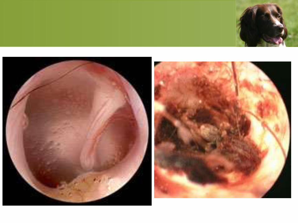
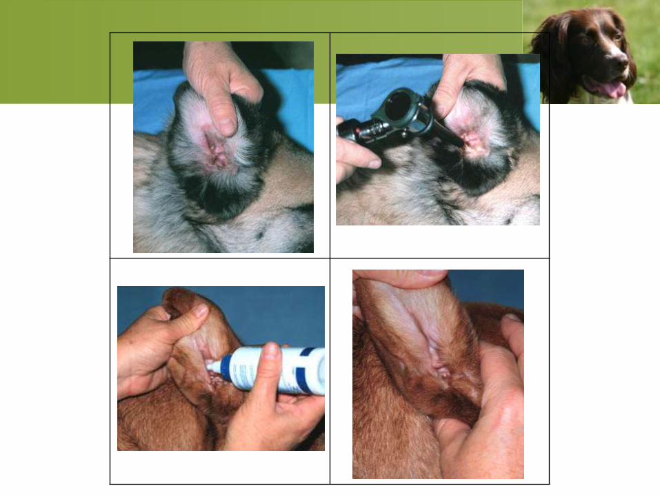
Otoscopic Examination
• For equipment, a standard otoscope with a
diagnostic or operating head is generally
adequate; however, a video otoscope is
strongly recommended for enhanced imaging
and its specialty applications.
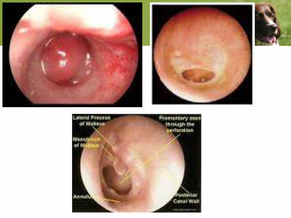
• Evaluate for the size of the ear canals; the
presence of parasites, exudate, hair, or foreign
material; the color of the epithelium; the
presence of ulcers or masses; and the
appearance and integrity of the tympanic
membrane.
• Sedation of the animal may be necessary.
• Topical anesthesia with 1% to 2% lidocaine HCl,
0.5% proparacaine, or other similar agents may be
sufficient.
• General anesthesia is indicated for removal of
most foreign objects; for biopsy, and for
thorough evaluation of the horizontal ear canal.
• Avoid trauma to the ear canal by advancing the
otoscope cone only while directly visualizing the
canal.
Otoscopic Abnormalities
Erythema: reddened epithelium
Exudation:
• Dark, dry, granular exudate is generally found
with ear mite infection;
• Moist, yellow, odoriferous exudate is generally a
sign of bacterial infection
• Brown, waxy exudate is generally consistent
with yeast infection and overgrowth;
• Yellow, waxy-to dry scale may be found with
keratinization disorders.
Cytology of otic exudates is
always indicated to confirm these
clinical clues.
• Hyperplasia (lichenification,
hyperpigmentation) is a sign
associated with chronicity.
• Ulcers suggest more severe disease
and indicate a need for aggressive
treatment—seen especially with
Pseudomonas and yeast
infections.
Medical management
• Purulent yellow or cream-colored exudates
may be associated with gram-negative
infections, particularly Pseudomonas and
Proteus spp.
• Dark brown or black exudates are more
commonly associated with yeast infections
or those caused by Staphylococcus or
Streptococcus spp.
• Do not apply cleansing agents, parasiticides,
ceruminolytic/keratolytic agents, disinfectants,
ototoxic antimicrobials, or oil-based
medications into the ear canal of animals in
which the tympanic membrane is ruptured.
• An average size dog (around 20 kg) would
require 0.5 to 1.0 ml of medication in the ear
canal to penetrate to the level of the tympanic
membrane.
• Gently massage the external canal for 15 to 30
seconds to help to deliver medications deep
into the horizontal canal.
• Lotions and solutions are more easily applied
deep in the external canal.
• Oil-based medications are useful to treat dry,
scaly lesions, such as those of seborrhea sicca.
• Creams, pastes, and powders are difficult to
apply deep in the external ear canal and may
leave a residue. These formulations are rarely
indicated in the treatment of otitis externa in
dogs and cats.
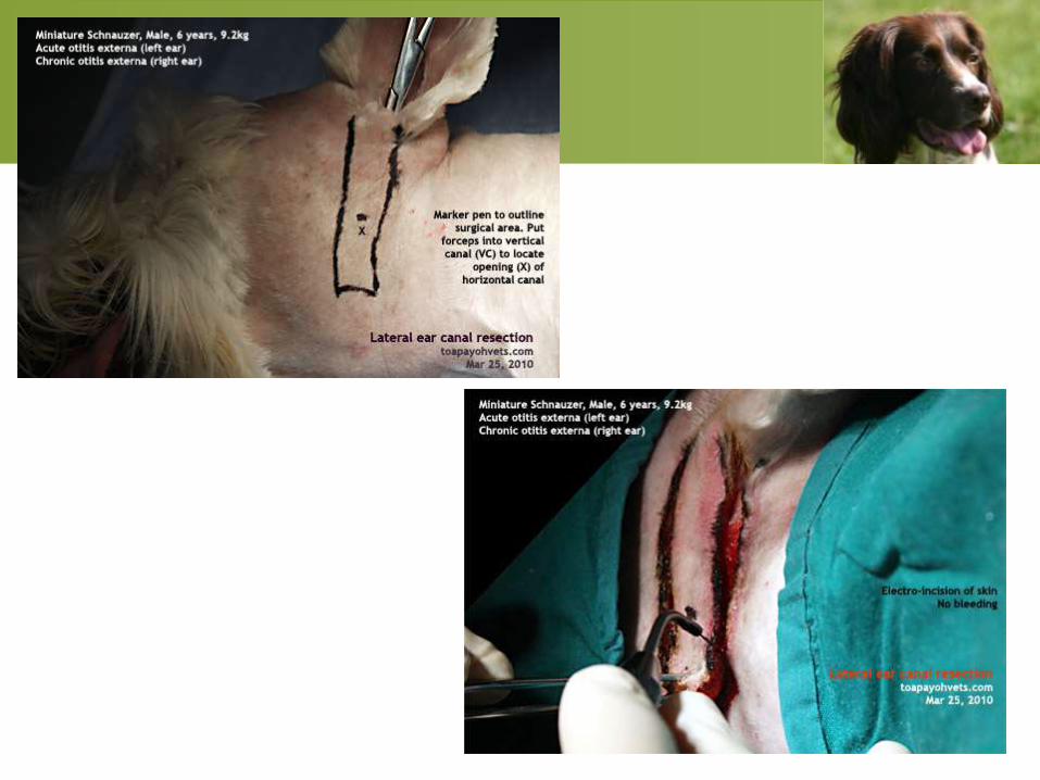
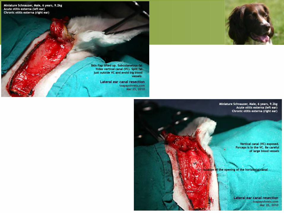
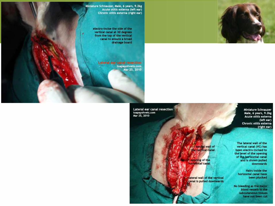
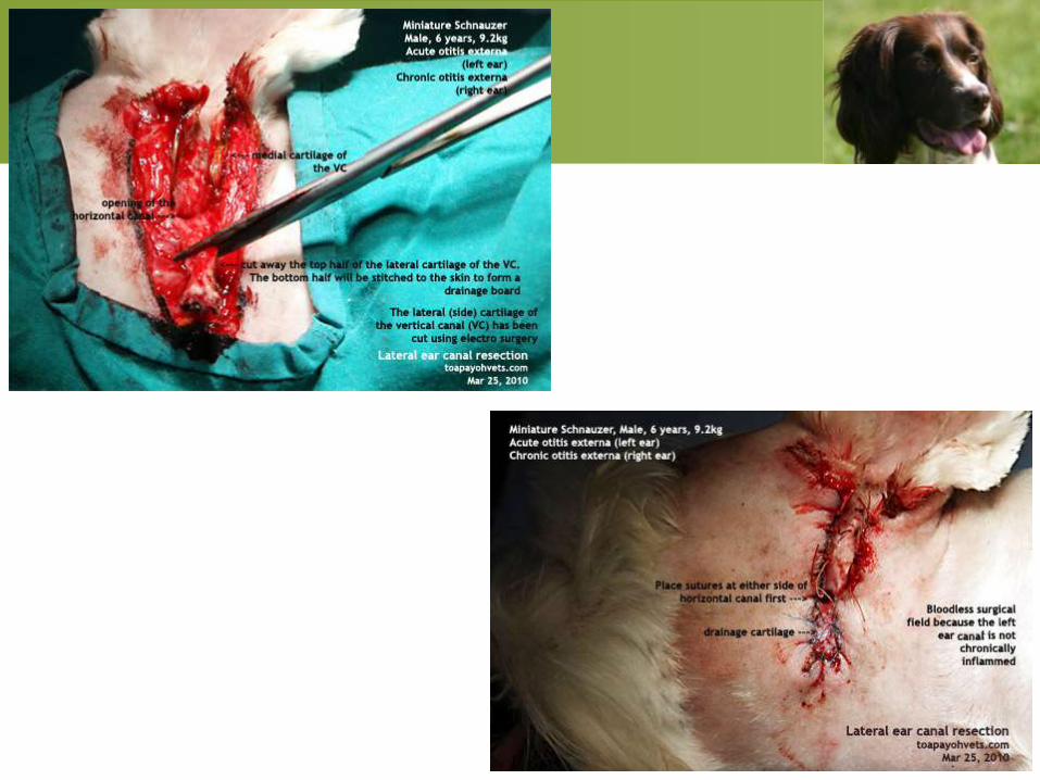
LATERAL EAR CANAL RESECTION
ZEPPS MODIFICATION (LECR)
• Typically, the most common clinical sign of otitis
media is chronic and recurrent otitis externa.
• To expose the medial portion of the vertical
canal and horizontal ear canal.
Prognosis
Prognosis for control of ear disease is good
provided that
• Surgery is performed correctly and for the right
indication.
• No middle ear disease is present.
• Postoperative medical management of otitis
externa is appropriate.
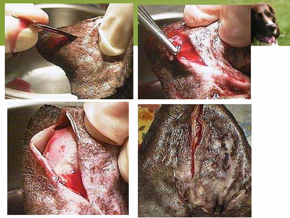
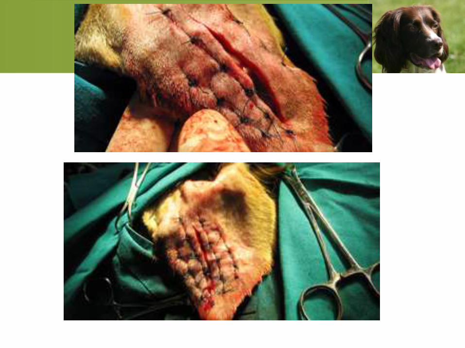
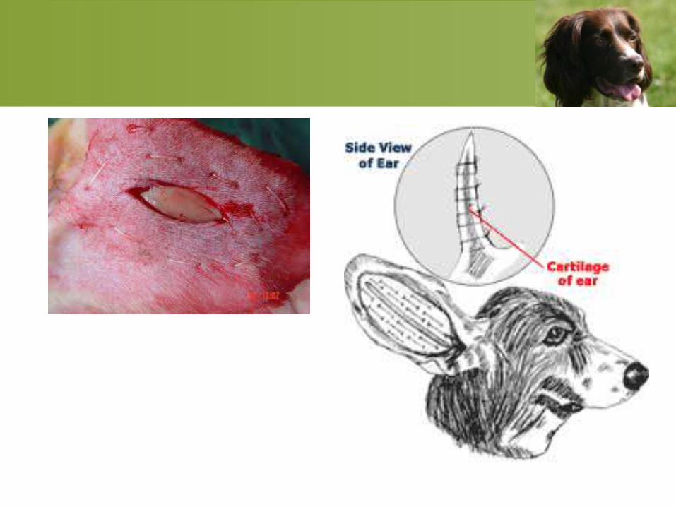
AURICULAR HEMATOMA
• Auricular or aural hematoma is an
accumulation of blood within the cartilage of
the pinna
• Auricular hematomas occur secondary to
inflammatory conditions of the pinna or external
ear canal, such as foreign bodies, food allergy,
bacterial infection, yeast infection, and ear mites
and by violent head shaking or scratching.
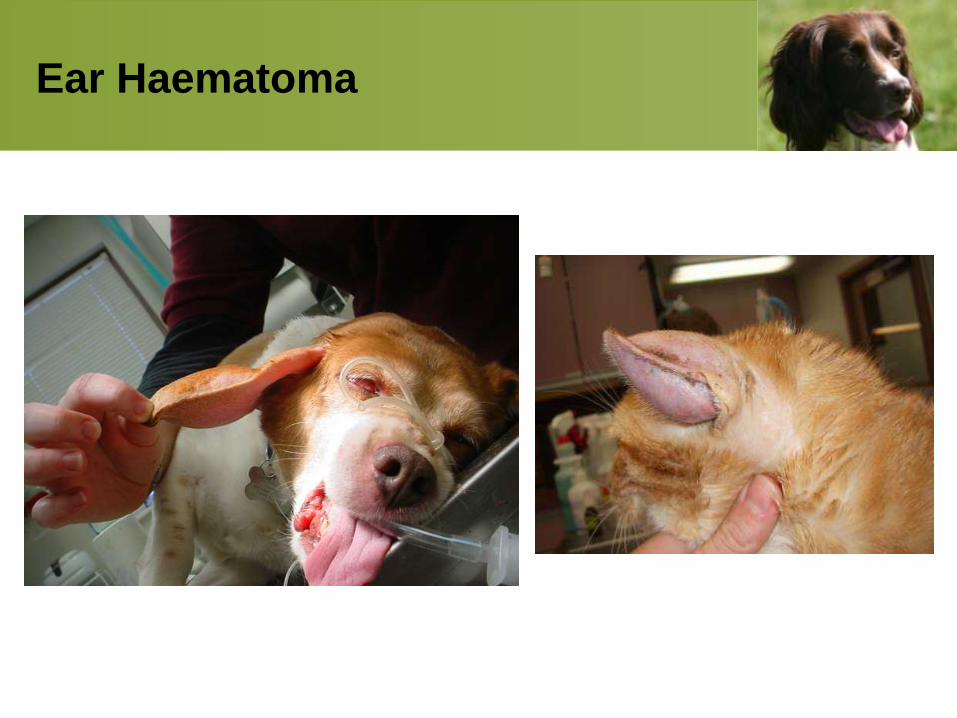
Ear Haematoma
Otitis Media and Otitis Interna
• Otitis media: Otitis media is defined as
inflammation of the middle ear and is an
important perpetuating cause of recurrent otitis
externa.
• Otitis media occurs as a direct extension from an
existing otitis externa through a ruptured
tympanic membrane.
ETIOLOGY
• Bacteria
• Yeast
• Primary secretory otitis media
• Neoplasia and polyps
• Otoliths
• Trauma and foreign bodies.
CLINICAL SIGNS
• Recurrent otitis externa may be the only
clinical sign associated with otitis media.
• Specific clinical signs indicative of otitis media
are facial nerve paralysis and Horner’s
syndrome is due to injury to the sympathetic
nerve fibers, which course near the middle ear,
and is characterized by ptosis, miosis,
enophthalmus, and protrusion of the nictitating
membrane.
Otitis Interna
• Signs of otitis interna are those typically
associated with peripheral vestibular syndrome
and include a head tilt, circling, falling, or
rolling toward the affected side; horizontal or
rotary nystagmus with the fast phase away from
the affected side; and asymmetric ataxia with
strength preserved
Treatment
The goals of treatment are to clean the external
and middle ear; remove infected, inflammatory,
or foreign
• Perform under general anesthesia.
• Soak the ear canal for 10 minutes with a
ceruminolytic ear cleaner, then flush using warm
saline:
• Once the exudate and debris are removed from
the ear canal, evaluate the tympanic membrane
with an otoscope or video otoscope.
• If the tympanic membrane is not intact, perform
cytology and bacterial C/S from the middle ear
cavity
• Once the ear has been cleaned and flushed,
begin systemic and topical
antimicrobial/antifungal treatment based on
cytologic results from the external and middle
ear.
• The most common coccoid bacteria isolated
from the middle ear of dogs with otitis media is
S. intermedius; appropriate antibiotic choices
include
• Cephalexin, 22mg/kg PO q12h
• Amoxicillin and clavulanate, 13.75 to 22mg/kg
PO q12h
• The most common rod bacteria is
Pseudomonas aeruginosa. A fluoroquinolone
such as
• Enrofloxacin, 5 to 20 mg/kg PO q24h
• Marbofloxacin, 2.75 to 5.5mg/kg q24h PO
• Certain systemic antibiotics (primarily
aminoglycosides) are ototoxic and should be
used cautiously.
• Use ketoconazole or itraconazole at 5mg/kg
PO q24h for yeast otitis media.
• Anti-inflammatory doses prednisolone 1mg/kg
PO q24h, then can be used to reduce
hyperplasia and stenosis of the ear canal to
facilitate therapy.

Thanks