Embed Size (px)
DESCRIPTION
This is a lecture by Ruth S. Hwu, MD from the Ghana Emergency Medicine Collaborative. To download the editable version (in PPT), to access additional learning modules, or to learn more about the project, see http://openmi.ch/em-gemc. Unless otherwise noted, this material is made available under the terms of the Creative Commons Attribution Share Alike-3.0 License: http://creativecommons.org/licenses/by-sa/3.0/.
Citation preview
Project: Ghana Emergency Medicine Collaborative
Document Title: Pediatric Trauma: Special Considerations
Author(s): Ruth S. Hwu, MD, (Washington University in St. Louis), 2013
License: Unless otherwise noted, this material is made available under the
terms of the Creative Commons Attribution Share Alike-3.0 License:
http://creativecommons.org/licenses/by-sa/3.0/
We have reviewed this material in accordance with U.S. Copyright Law and have tried to maximize your
ability to use, share, and adapt it. These lectures have been modified in the process of making a publicly
shareable version. The citation key on the following slide provides information about how you may share and
adapt this material.
Copyright holders of content included in this material should contact [email protected] with any
questions, corrections, or clarification regarding the use of content.
For more information about how to cite these materials visit http://open.umich.edu/privacy-and-terms-use.
Any medical information in this material is intended to inform and educate and is not a tool for self-diagnosis
or a replacement for medical evaluation, advice, diagnosis or treatment by a healthcare professional. Please
speak to your physician if you have questions about your medical condition.
Viewer discretion is advised: Some medical content is graphic and may not be suitable for all viewers.
1
Attribution Key
for more information see: http://open.umich.edu/wiki/AttributionPolicy
Use + Share + Adapt
Make Your Own Assessment
Creative Commons – Attribution License
Creative Commons – Attribution Share Alike License
Creative Commons – Attribution Noncommercial License
Creative Commons – Attribution Noncommercial Share Alike License
GNU – Free Documentation License
Creative Commons – Zero Waiver
Public Domain – Ineligible: Works that are ineligible for copyright protection in the U.S. (17 USC § 102(b)) *laws in
your jurisdiction may differ
Public Domain – Expired: Works that are no longer protected due to an expired copyright term.
Public Domain – Government: Works that are produced by the U.S. Government. (17 USC § 105)
Public Domain – Self Dedicated: Works that a copyright holder has dedicated to the public domain.
Fair Use: Use of works that is determined to be Fair consistent with the U.S. Copyright Act. (17 USC § 107) *laws in your
jurisdiction may differ
Our determination DOES NOT mean that all uses of this 3rd-party content are Fair Uses and we DO NOT guarantee that
your use of the content is Fair.
To use this content you should do your own independent analysis to determine whether or not your use will be Fair.
{ Content the copyright holder, author, or law permits you to use, share and adapt. }
{ Content Open.Michigan believes can be used, shared, and adapted because it is ineligible for copyright. }
{ Content Open.Michigan has used under a Fair Use determination. }
2
Pediatric Trauma: Special Considerations
Ruth S. Hwu, MD Pediatric Emergency Medicine Fellow,
PGY-6 Washington University in St. Louis
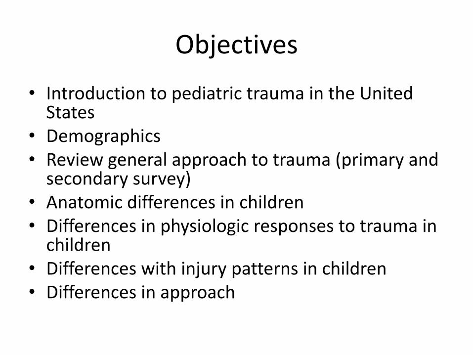
Objectives
• Introduction to pediatric trauma in the United States
• Demographics • Review general approach to trauma (primary and
secondary survey) • Anatomic differences in children • Differences in physiologic responses to trauma in
children • Differences with injury patterns in children • Differences in approach

Pediatric Trauma in the United States
• United States has designated pediatric trauma centers, recognition children receive better care
• Trauma center designation given at state or local level, verified by American College of Surgeons (ACS)
• As of 2010, there were 43 pediatric only trauma centers, 12 were ACS (nationally) verified
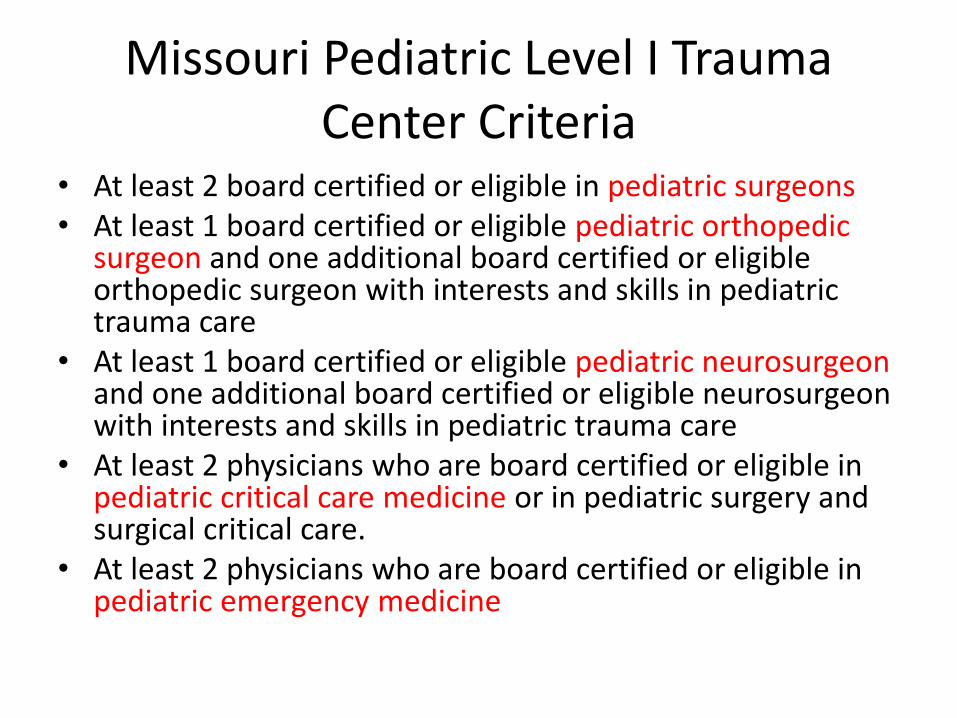
Missouri Pediatric Level I Trauma Center Criteria
• At least 2 board certified or eligible in pediatric surgeons • At least 1 board certified or eligible pediatric orthopedic
surgeon and one additional board certified or eligible orthopedic surgeon with interests and skills in pediatric trauma care
• At least 1 board certified or eligible pediatric neurosurgeon and one additional board certified or eligible neurosurgeon with interests and skills in pediatric trauma care
• At least 2 physicians who are board certified or eligible in pediatric critical care medicine or in pediatric surgery and surgical critical care.
• At least 2 physicians who are board certified or eligible in pediatric emergency medicine
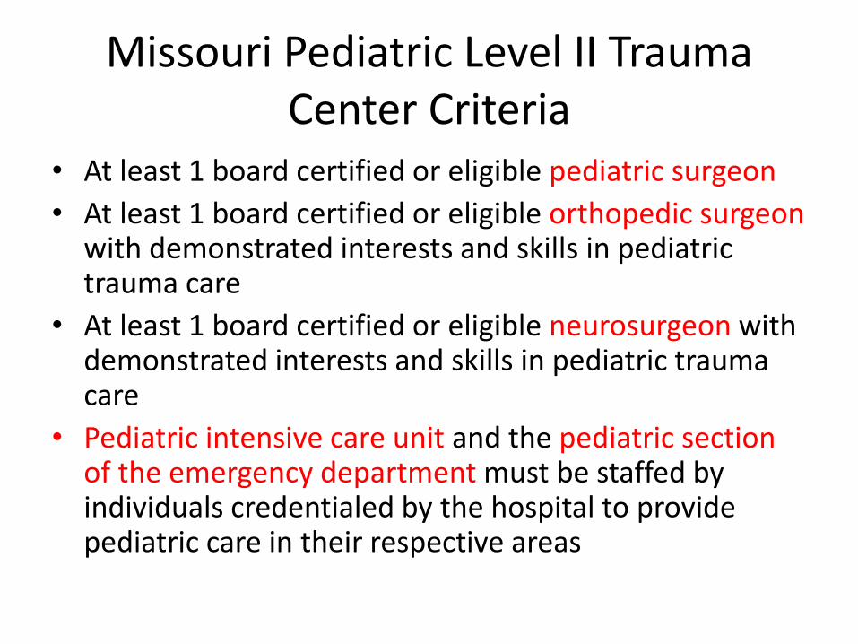
Missouri Pediatric Level II Trauma Center Criteria
• At least 1 board certified or eligible pediatric surgeon
• At least 1 board certified or eligible orthopedic surgeon with demonstrated interests and skills in pediatric trauma care
• At least 1 board certified or eligible neurosurgeon with demonstrated interests and skills in pediatric trauma care
• Pediatric intensive care unit and the pediatric section of the emergency department must be staffed by individuals credentialed by the hospital to provide pediatric care in their respective areas
Demographics
• In United States: – According to the Centers for Disease Control and
Prevention (CDC), unintentional injury is the leading cause of death in children and adults from 1 to 44 years
– The Global Burden of Disease: 2004 update by the World Health Organization (WHO) showed 6900 children less than 14 years of age died of unintentional and intentional injuries
• In Ghana: – In Africa, trauma is second to infectious disease as the
leading killer – According to WHO 2004 data, 4100 children less than 14
years of age died of unintentional and intentional injuries
Demographics
• Of the unintentional injuries, motor vehicle-related injuries were the most common mechanism in both countries
• In 2010, the number of road traffic deaths per 100000 population was 11.4 in the United States and 22.2 in Ghana (Global Health Observatory Data Repository)
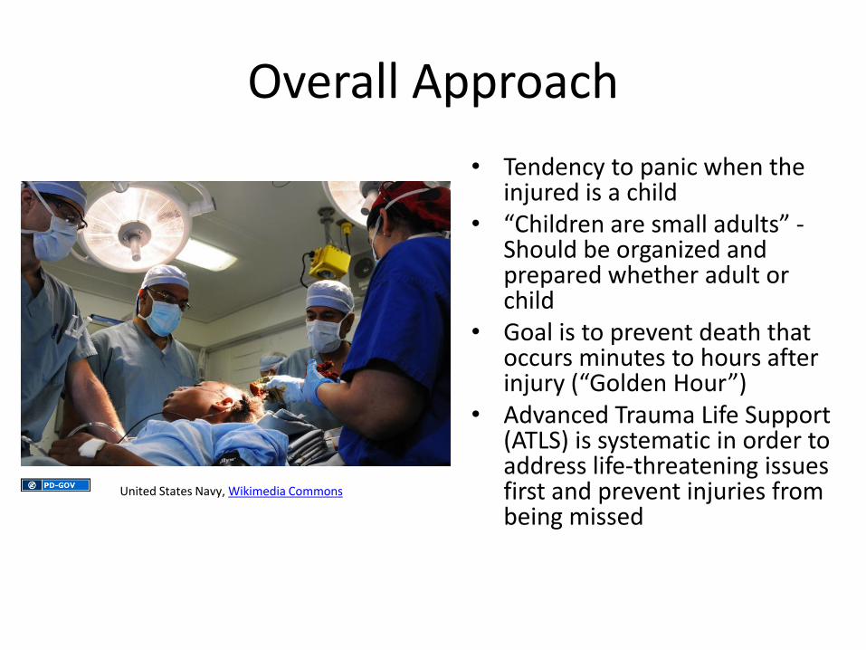
Overall Approach
• Tendency to panic when the injured is a child
• “Children are small adults” - Should be organized and prepared whether adult or child
• Goal is to prevent death that occurs minutes to hours after injury (“Golden Hour”)
• Advanced Trauma Life Support (ATLS) is systematic in order to address life-threatening issues first and prevent injuries from being missed
United States Navy, Wikimedia Commons
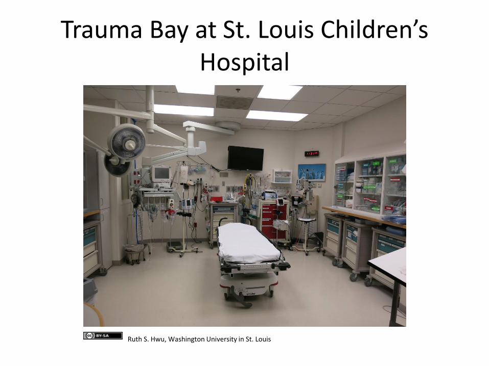
Trauma Bay at St. Louis Children’s Hospital
Ruth S. Hwu, Washington University in St. Louis
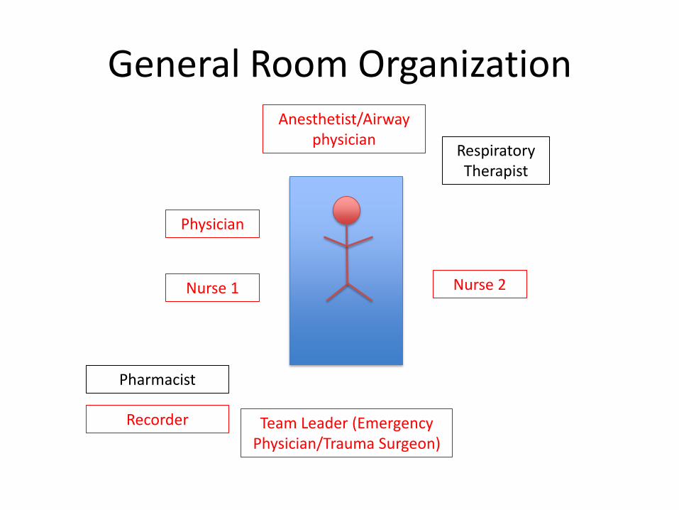
General Room Organization Anesthetist/Airway
physician
Team Leader (Emergency Physician/Trauma Surgeon)
Nurse 1 Nurse 2
Recorder
Pharmacist
Respiratory Therapist
Physician
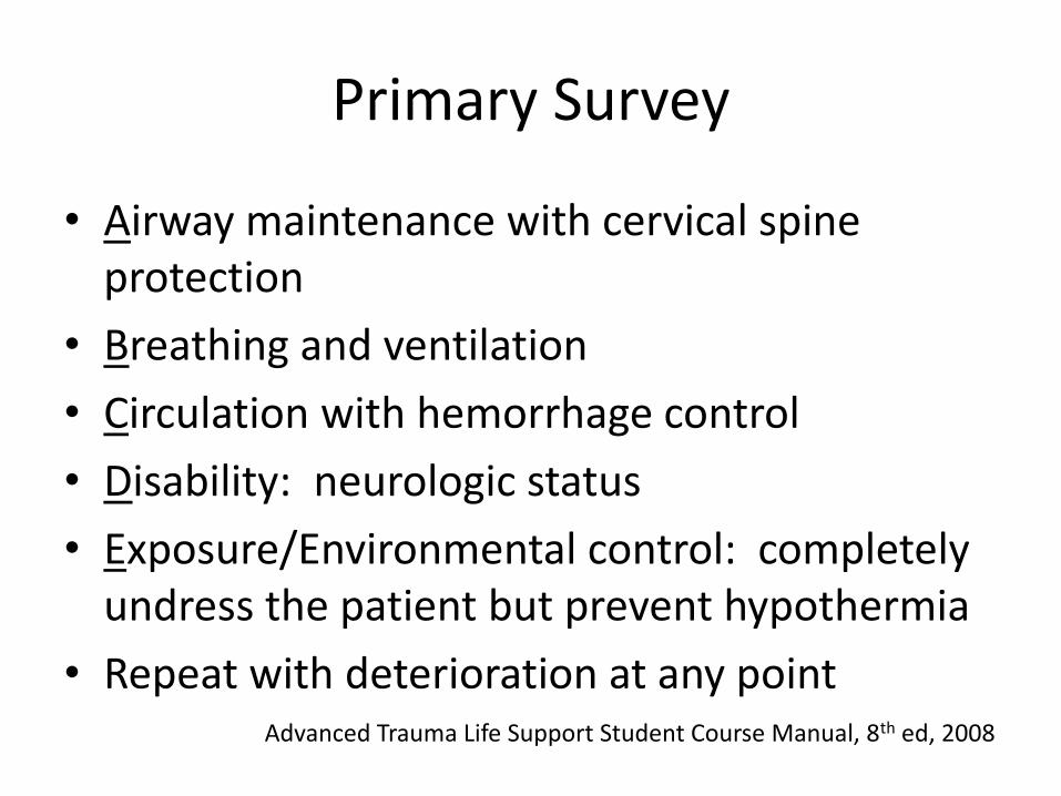
Primary Survey
• Airway maintenance with cervical spine protection
• Breathing and ventilation
• Circulation with hemorrhage control
• Disability: neurologic status
• Exposure/Environmental control: completely undress the patient but prevent hypothermia
• Repeat with deterioration at any point Advanced Trauma Life Support Student Course Manual, 8th ed, 2008
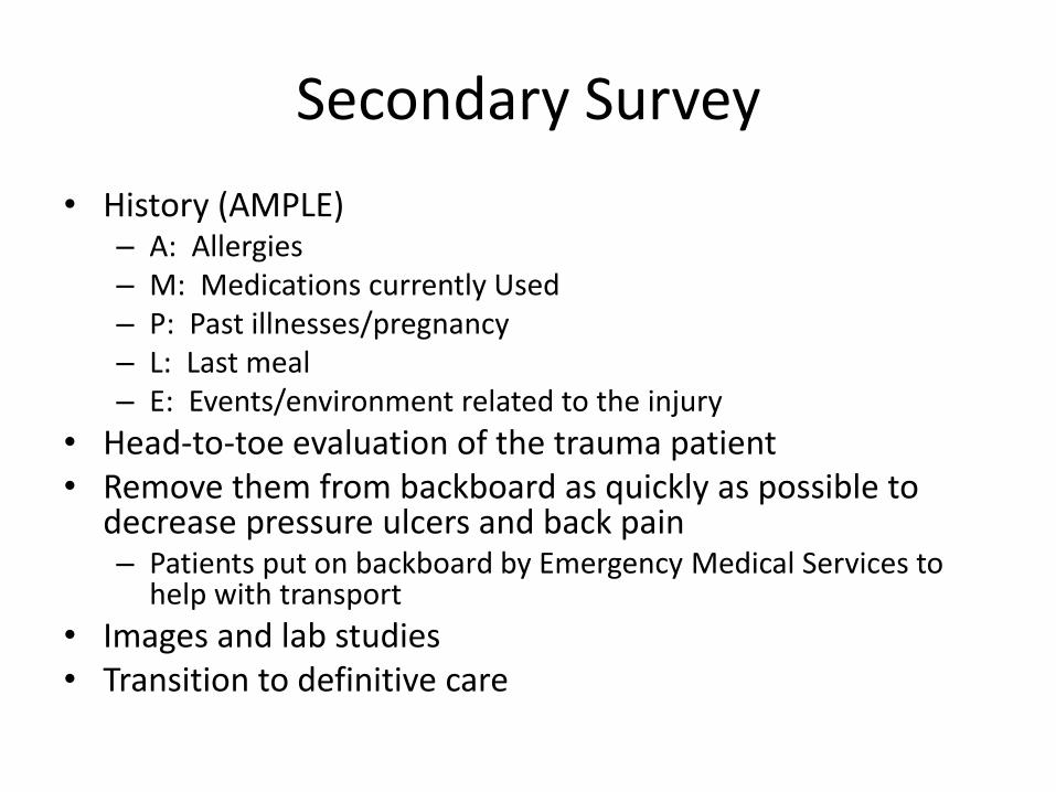
Secondary Survey
• History (AMPLE) – A: Allergies – M: Medications currently Used – P: Past illnesses/pregnancy – L: Last meal – E: Events/environment related to the injury
• Head-to-toe evaluation of the trauma patient • Remove them from backboard as quickly as possible to
decrease pressure ulcers and back pain – Patients put on backboard by Emergency Medical Services to
help with transport
• Images and lab studies • Transition to definitive care
While the general approach is the same, specific issues arise when dealing with children
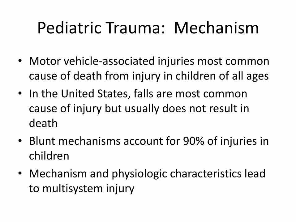
Pediatric Trauma: Mechanism
• Motor vehicle-associated injuries most common cause of death from injury in children of all ages
• In the United States, falls are most common cause of injury but usually does not result in death
• Blunt mechanisms account for 90% of injuries in children
• Mechanism and physiologic characteristics lead to multisystem injury
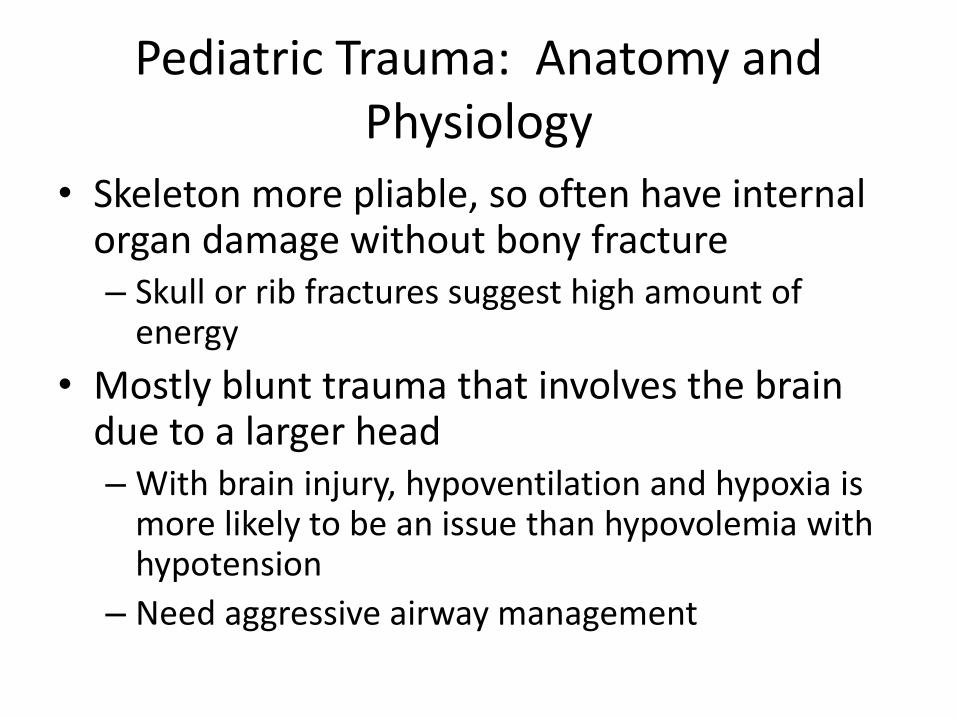
Pediatric Trauma: Anatomy and Physiology
• Skeleton more pliable, so often have internal organ damage without bony fracture – Skull or rib fractures suggest high amount of
energy
• Mostly blunt trauma that involves the brain due to a larger head – With brain injury, hypoventilation and hypoxia is
more likely to be an issue than hypovolemia with hypotension
– Need aggressive airway management
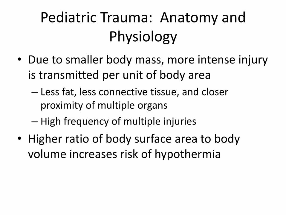
Pediatric Trauma: Anatomy and Physiology
• Due to smaller body mass, more intense injury is transmitted per unit of body area
– Less fat, less connective tissue, and closer proximity of multiple organs
– High frequency of multiple injuries
• Higher ratio of body surface area to body volume increases risk of hypothermia

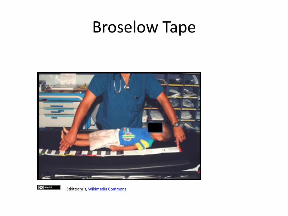
Equipment and Doses Based on Age and Size
• Broselow Pediatric Emergency Tape to help rapid determination of weight based on length
– Provides appropriate fluid volumes, drug doses, and equipment size
• Resuscitation/Code book with estimated weights based on age
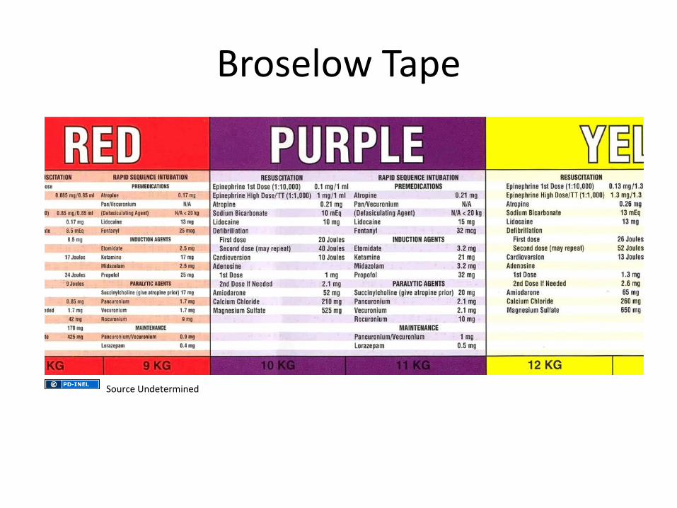
Broselow Tape
Source Undetermined
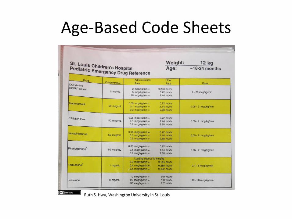
Age-Based Code Sheets
Ruth S. Hwu, Washington University in St. Louis
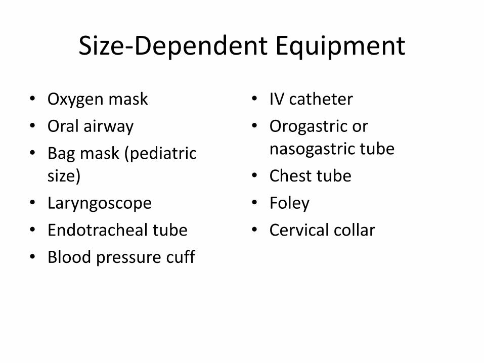
Size-Dependent Equipment
• Oxygen mask
• Oral airway
• Bag mask (pediatric size)
• Laryngoscope
• Endotracheal tube
• Blood pressure cuff
• IV catheter
• Orogastric or nasogastric tube
• Chest tube
• Foley
• Cervical collar
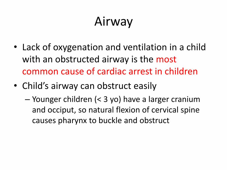
Airway
• Lack of oxygenation and ventilation in a child with an obstructed airway is the most common cause of cardiac arrest in children
• Child’s airway can obstruct easily
– Younger children (< 3 yo) have a larger cranium and occiput, so natural flexion of cervical spine causes pharynx to buckle and obstruct
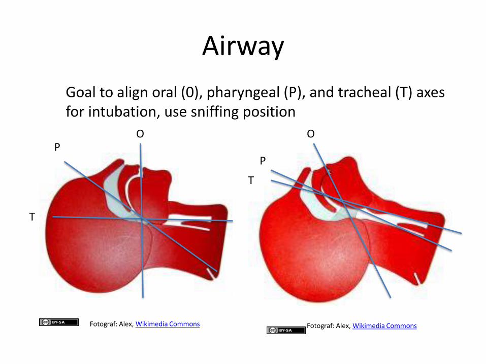
Airway
O P
T
O
P
T
Goal to align oral (0), pharyngeal (P), and tracheal (T) axes for intubation, use sniffing position
Fotograf: Alex, Wikimedia Commons Fotograf: Alex, Wikimedia Commons
Airway
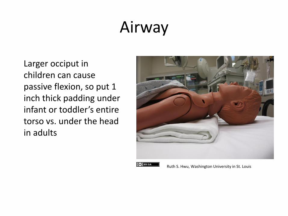
Larger occiput in children can cause passive flexion, so put 1 inch thick padding under infant or toddler’s entire torso vs. under the head in adults
Ruth S. Hwu, Washington University in St. Louis
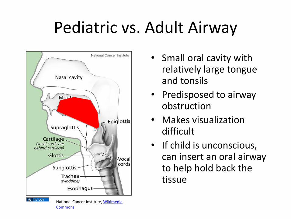
Pediatric vs. Adult Airway
• Small oral cavity with relatively large tongue and tonsils
• Predisposed to airway obstruction
• Makes visualization difficult
• If child is unconscious, can insert an oral airway to help hold back the tissue
National Cancer Institute, Wikimedia Commons
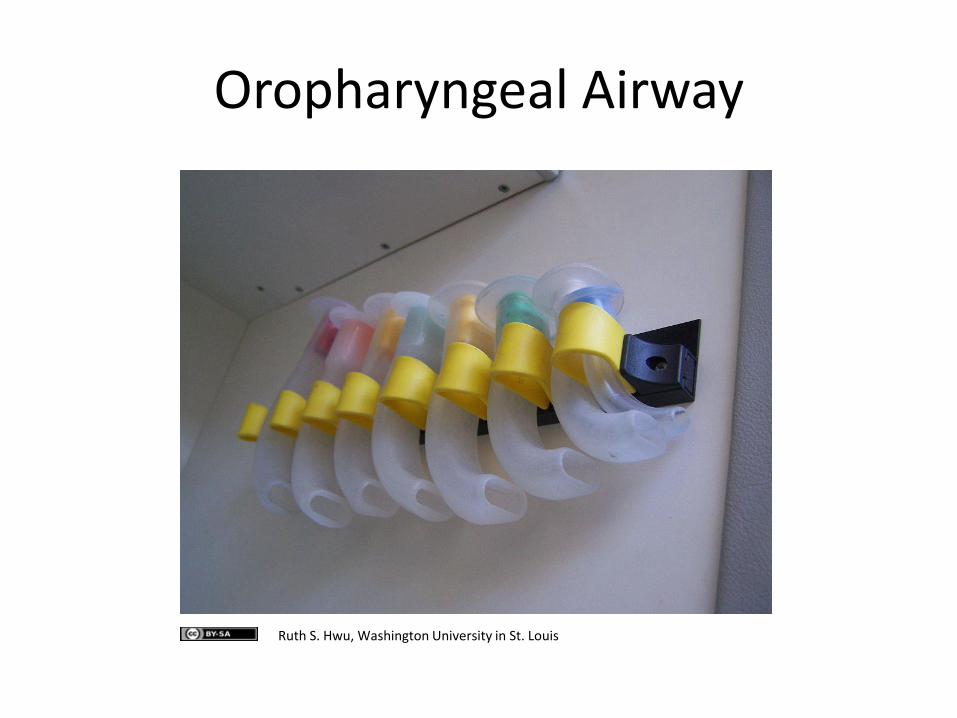
Oropharyngeal Airway
Ruth S. Hwu, Washington University in St. Louis
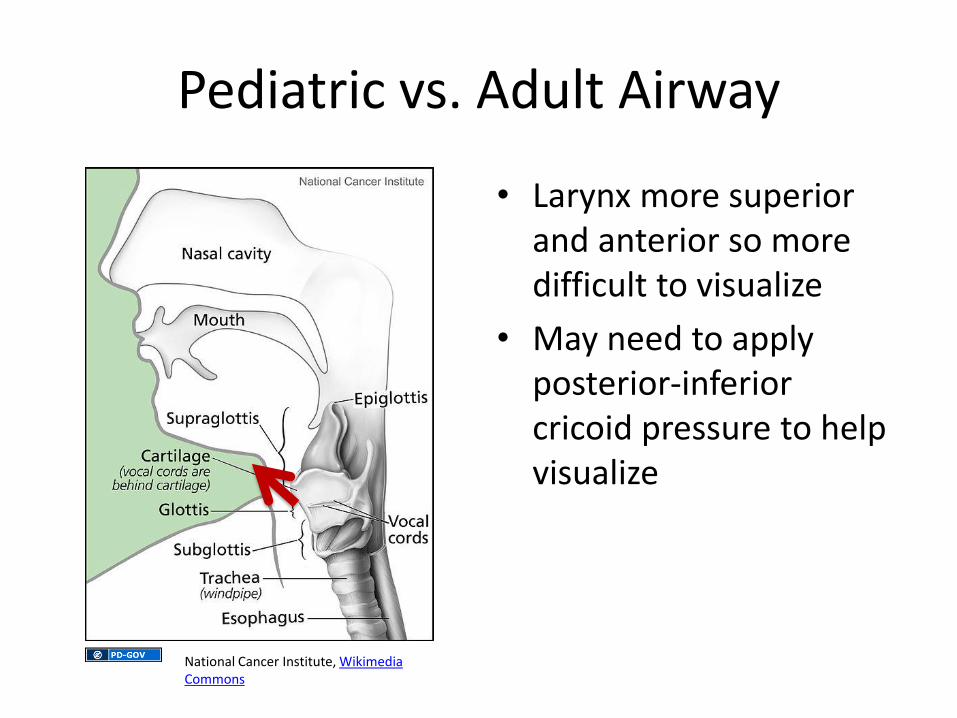
Pediatric vs. Adult Airway
• Larynx more superior and anterior so more difficult to visualize
• May need to apply posterior-inferior cricoid pressure to help visualize
National Cancer Institute, Wikimedia Commons
Pediatric vs. Adult Airway
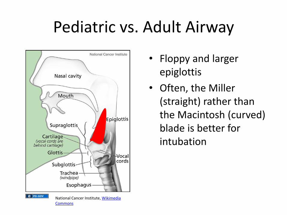
• Floppy and larger epiglottis
• Often, the Miller (straight) rather than the Macintosh (curved) blade is better for intubation
National Cancer Institute, Wikimedia Commons
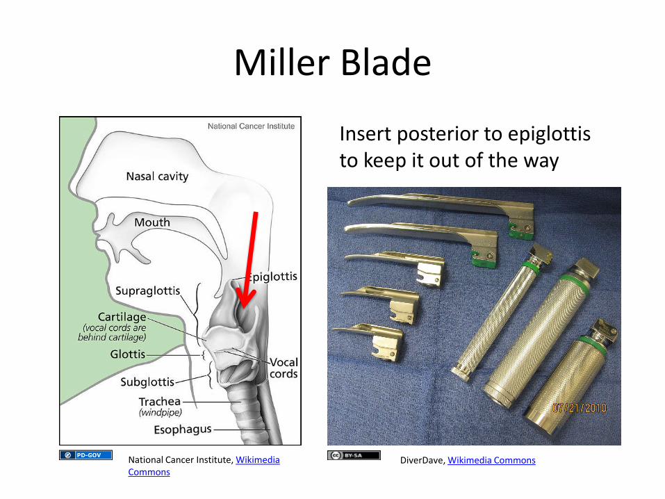
Miller Blade
Insert posterior to epiglottis to keep it out of the way
National Cancer Institute, Wikimedia Commons
DiverDave, Wikimedia Commons
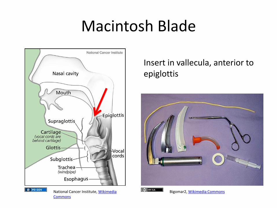
Macintosh Blade
Insert in vallecula, anterior to epiglottis
National Cancer Institute, Wikimedia Commons
Bigomar2, Wikimedia Commons
Pediatric vs. Adult Airway
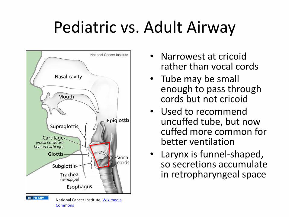
• Narrowest at cricoid rather than vocal cords
• Tube may be small enough to pass through cords but not cricoid
• Used to recommend uncuffed tube, but now cuffed more common for better ventilation
• Larynx is funnel-shaped, so secretions accumulate in retropharyngeal space National Cancer Institute, Wikimedia
Commons
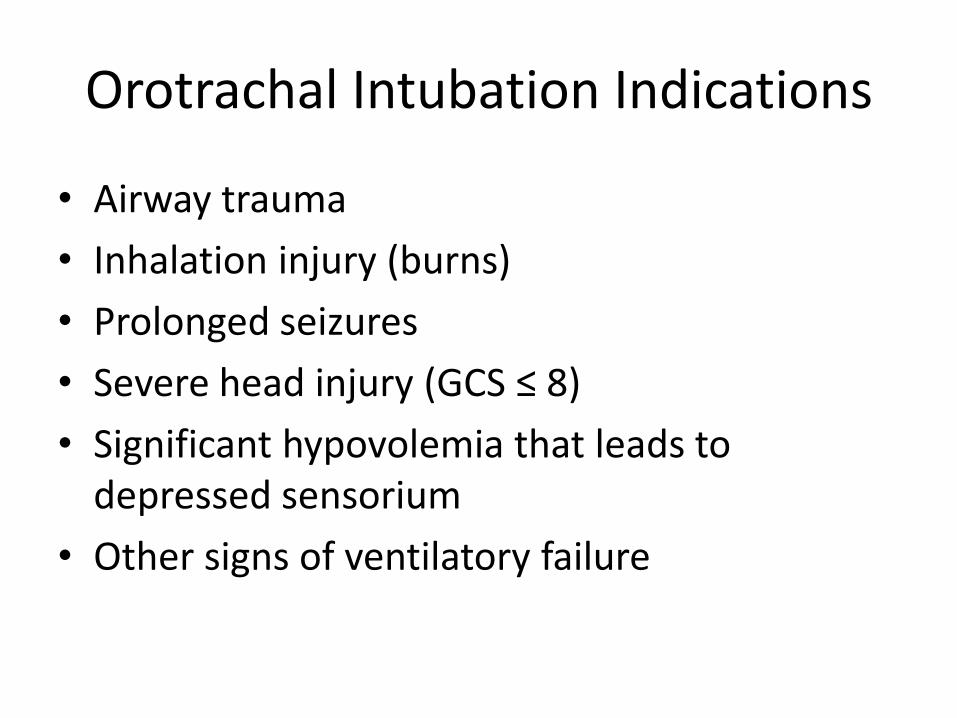
Orotrachal Intubation Indications
• Airway trauma
• Inhalation injury (burns)
• Prolonged seizures
• Severe head injury (GCS ≤ 8)
• Significant hypovolemia that leads to depressed sensorium
• Other signs of ventilatory failure
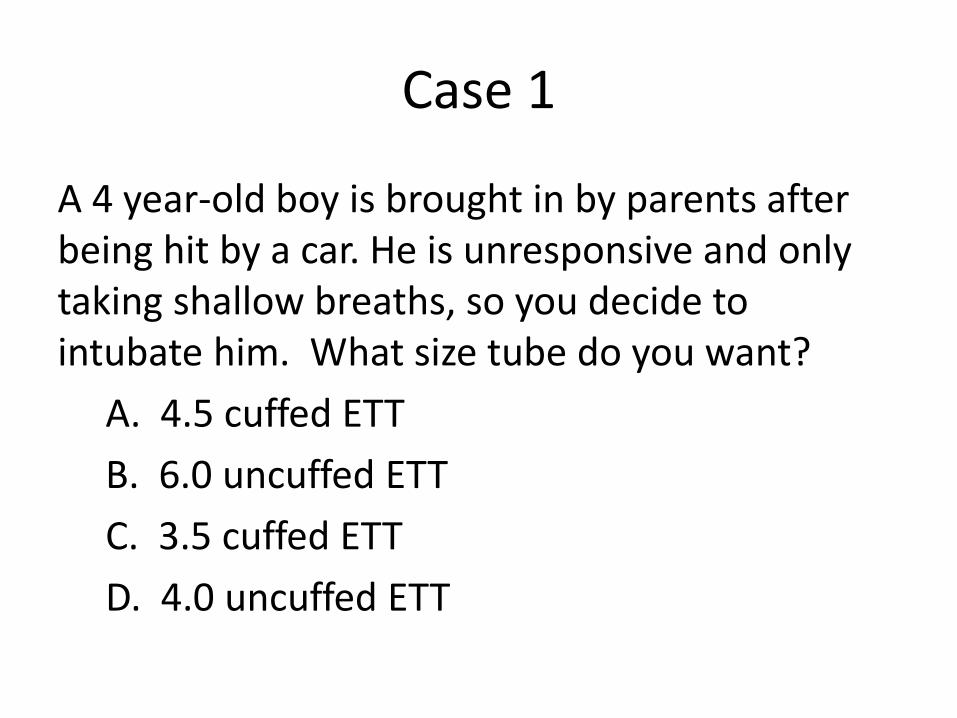
Case 1
A 4 year-old boy is brought in by parents after being hit by a car. He is unresponsive and only taking shallow breaths, so you decide to intubate him. What size tube do you want?
A. 4.5 cuffed ETT
B. 6.0 uncuffed ETT
C. 3.5 cuffed ETT
D. 4.0 uncuffed ETT
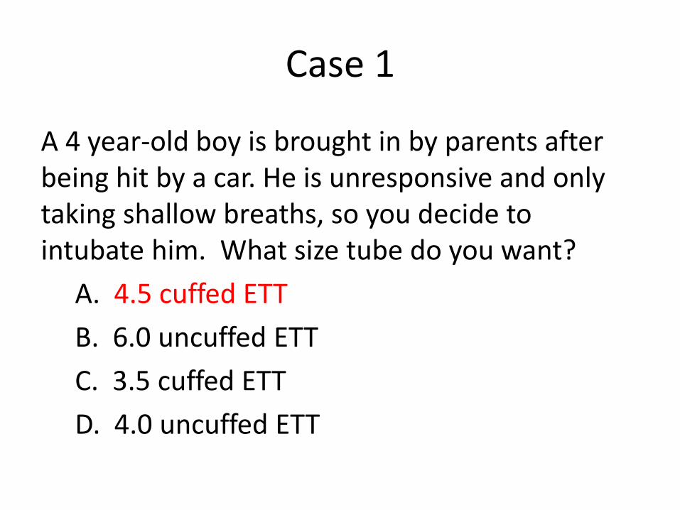
Case 1
A 4 year-old boy is brought in by parents after being hit by a car. He is unresponsive and only taking shallow breaths, so you decide to intubate him. What size tube do you want?
A. 4.5 cuffed ETT
B. 6.0 uncuffed ETT
C. 3.5 cuffed ETT
D. 4.0 uncuffed ETT
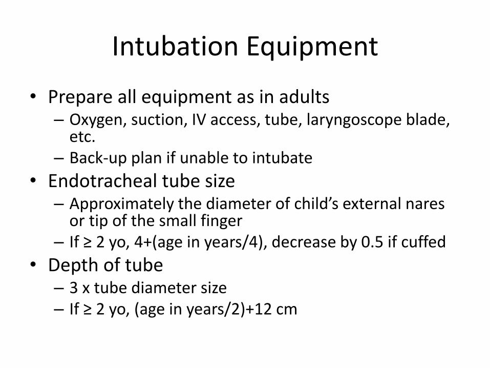
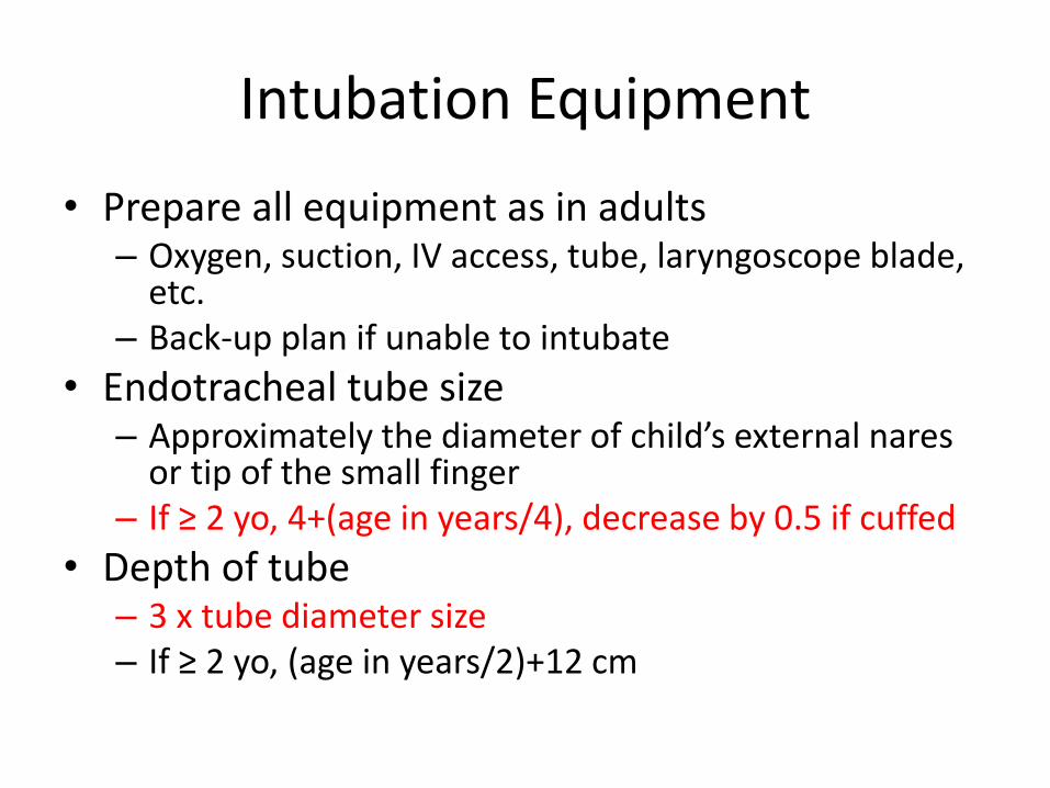
Intubation Equipment
• Prepare all equipment as in adults – Oxygen, suction, IV access, tube, laryngoscope blade,
etc. – Back-up plan if unable to intubate
• Endotracheal tube size – Approximately the diameter of child’s external nares
or tip of the small finger – If ≥ 2 yo, 4+(age in years/4), decrease by 0.5 if cuffed
• Depth of tube – 3 x tube diameter size – If ≥ 2 yo, (age in years/2)+12 cm
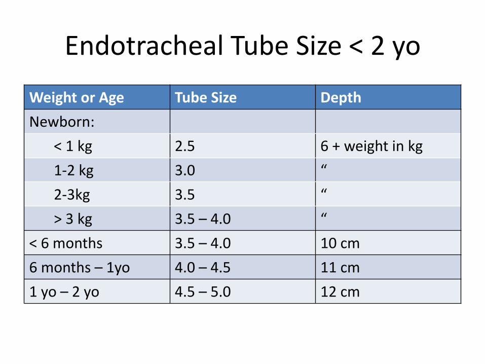
Endotracheal Tube Size < 2 yo
Weight or Age Tube Size Depth
Newborn:
< 1 kg 2.5 6 + weight in kg
1-2 kg 3.0 “
2-3kg 3.5 “
> 3 kg 3.5 – 4.0 “
< 6 months 3.5 – 4.0 10 cm
6 months – 1yo 4.0 – 4.5 11 cm
1 yo – 2 yo 4.5 – 5.0 12 cm
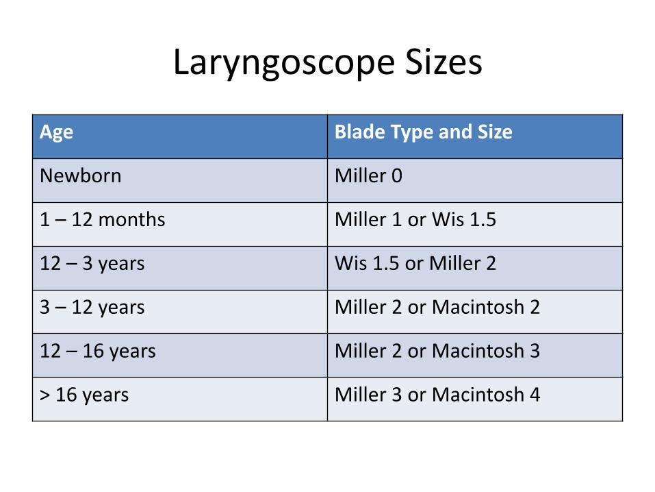
Laryngoscope Sizes
Age Blade Type and Size
Newborn Miller 0
1 – 12 months Miller 1 or Wis 1.5
12 – 3 years Wis 1.5 or Miller 2
3 – 12 years Miller 2 or Macintosh 2
12 – 16 years Miller 2 or Macintosh 3
> 16 years Miller 3 or Macintosh 4
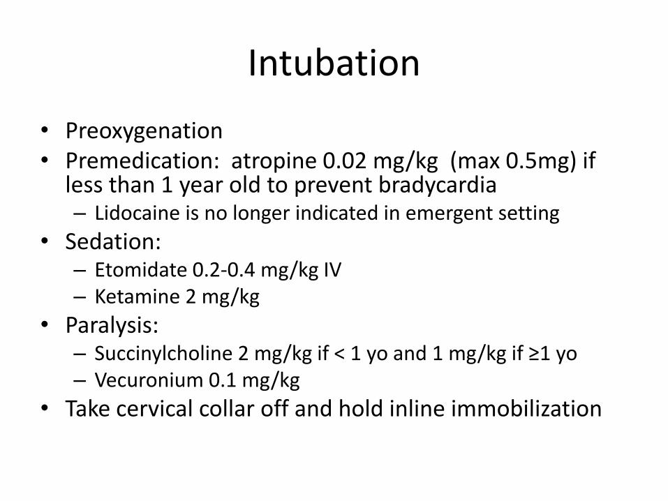
Intubation
• Preoxygenation • Premedication: atropine 0.02 mg/kg (max 0.5mg) if
less than 1 year old to prevent bradycardia – Lidocaine is no longer indicated in emergent setting
• Sedation: – Etomidate 0.2-0.4 mg/kg IV – Ketamine 2 mg/kg
• Paralysis: – Succinylcholine 2 mg/kg if < 1 yo and 1 mg/kg if ≥1 yo – Vecuronium 0.1 mg/kg
• Take cervical collar off and hold inline immobilization
Intubation
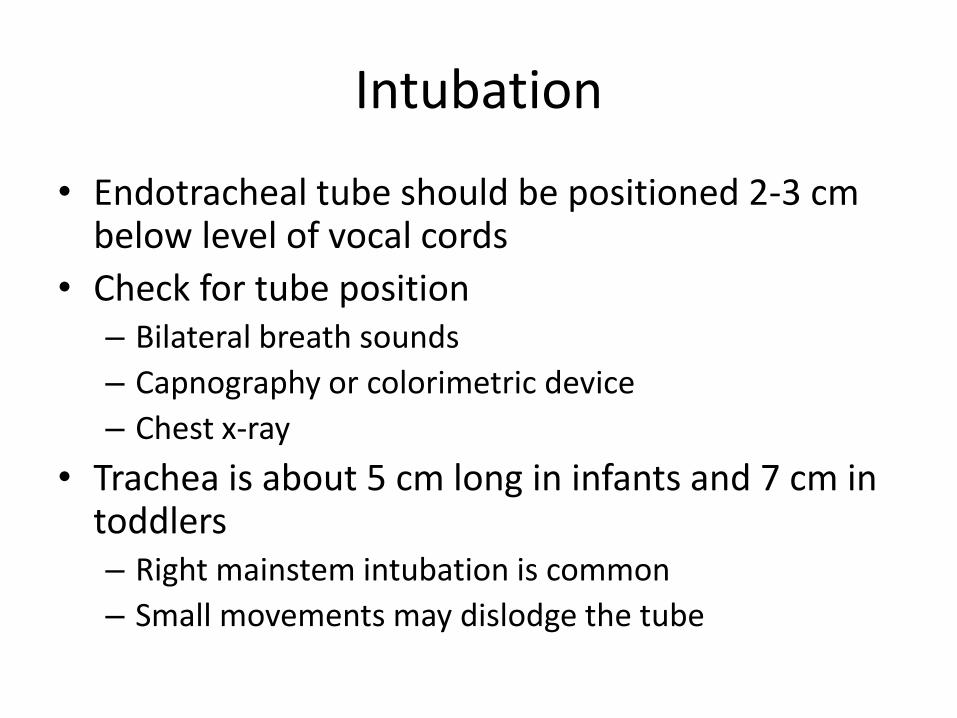
• Endotracheal tube should be positioned 2-3 cm below level of vocal cords
• Check for tube position – Bilateral breath sounds
– Capnography or colorimetric device
– Chest x-ray
• Trachea is about 5 cm long in infants and 7 cm in toddlers – Right mainstem intubation is common
– Small movements may dislodge the tube
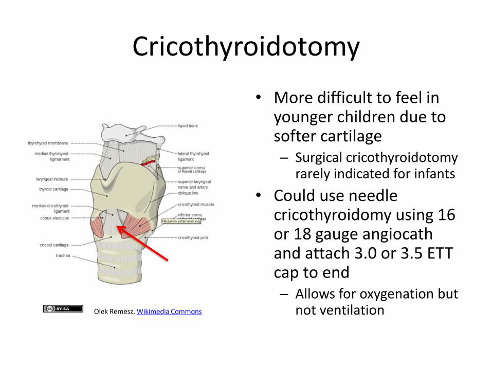
Cricothyroidotomy
• More difficult to feel in younger children due to softer cartilage – Surgical cricothyroidotomy
rarely indicated for infants
• Could use needle cricothyroidomy using 16 or 18 gauge angiocath and attach 3.0 or 3.5 ETT cap to end – Allows for oxygenation but
not ventilation Olek Remesz, Wikimedia Commons
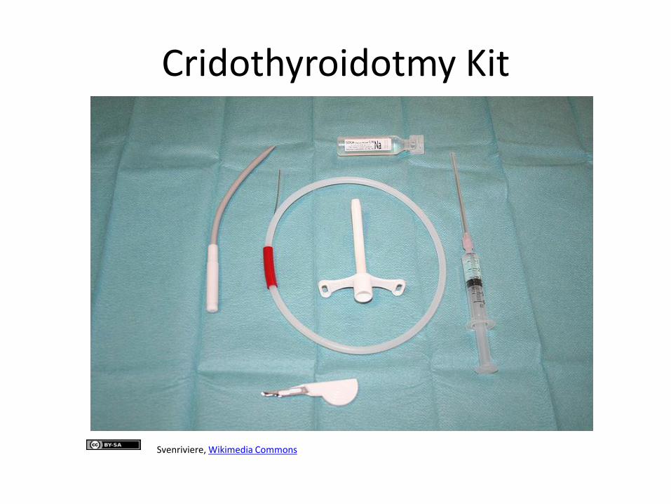
Cridothyroidotmy Kit
Svenriviere, Wikimedia Commons
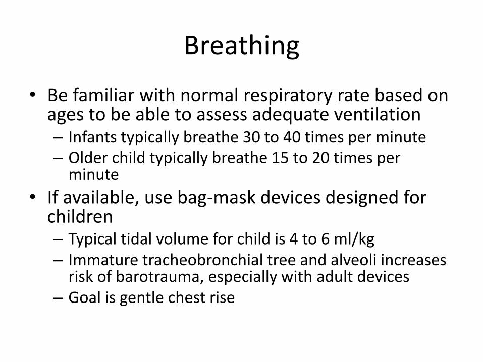
Breathing
• Be familiar with normal respiratory rate based on ages to be able to assess adequate ventilation – Infants typically breathe 30 to 40 times per minute – Older child typically breathe 15 to 20 times per
minute
• If available, use bag-mask devices designed for children – Typical tidal volume for child is 4 to 6 ml/kg – Immature tracheobronchial tree and alveoli increases
risk of barotrauma, especially with adult devices – Goal is gentle chest rise
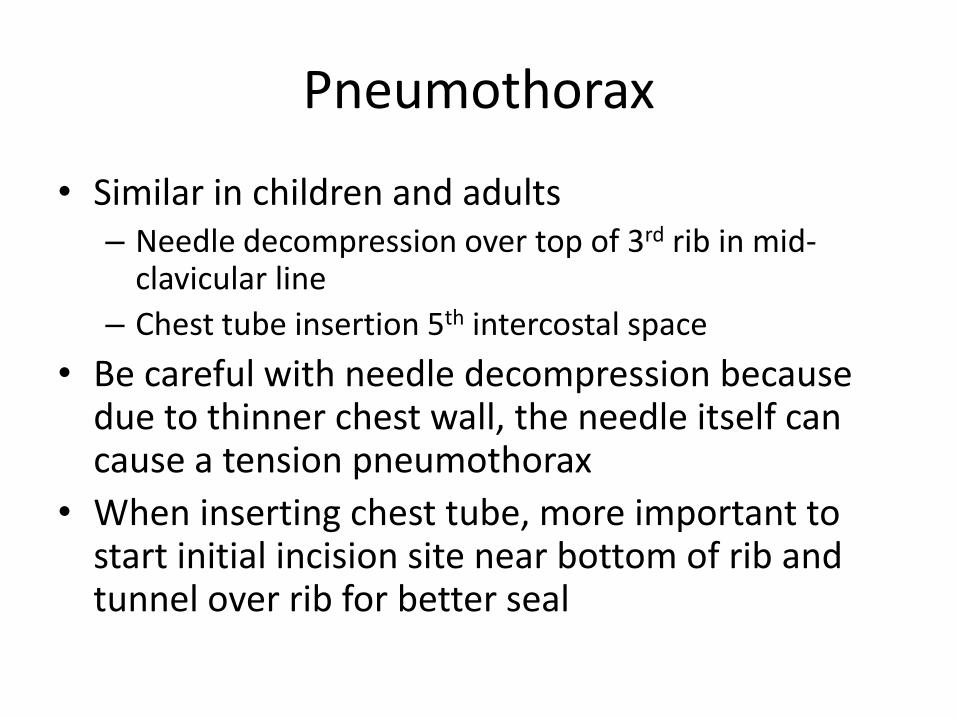
Pneumothorax
• Similar in children and adults – Needle decompression over top of 3rd rib in mid-
clavicular line
– Chest tube insertion 5th intercostal space
• Be careful with needle decompression because due to thinner chest wall, the needle itself can cause a tension pneumothorax
• When inserting chest tube, more important to start initial incision site near bottom of rib and tunnel over rib for better seal
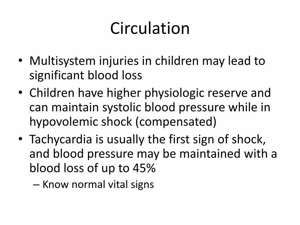
Circulation
• Multisystem injuries in children may lead to significant blood loss
• Children have higher physiologic reserve and can maintain systolic blood pressure while in hypovolemic shock (compensated)
• Tachycardia is usually the first sign of shock, and blood pressure may be maintained with a blood loss of up to 45% – Know normal vital signs
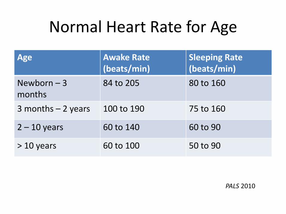
Normal Heart Rate for Age
Age Awake Rate (beats/min)
Sleeping Rate (beats/min)
Newborn – 3 months
84 to 205 80 to 160
3 months – 2 years 100 to 190 75 to 160
2 – 10 years 60 to 140 60 to 90
> 10 years 60 to 100 50 to 90
PALS 2010

Early Signs of Hypovolemic Shock
• Tachycardia
• Poor skin perfusion
– Delayed capillary refill
– Mottling
• Weak peripheral pulse
• Narrowing of pulse pressure to 20 mmHg
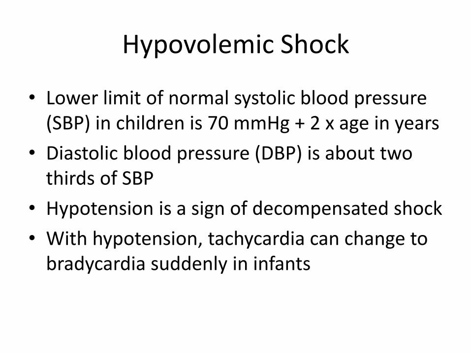
Hypovolemic Shock
• Lower limit of normal systolic blood pressure (SBP) in children is 70 mmHg + 2 x age in years
• Diastolic blood pressure (DBP) is about two thirds of SBP
• Hypotension is a sign of decompensated shock
• With hypotension, tachycardia can change to bradycardia suddenly in infants
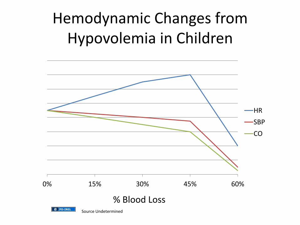
Hemodynamic Changes from Hypovolemia in Children
0% 15% 30% 45% 60%
HR
SBP
CO
% Blood Loss Source Undetermined
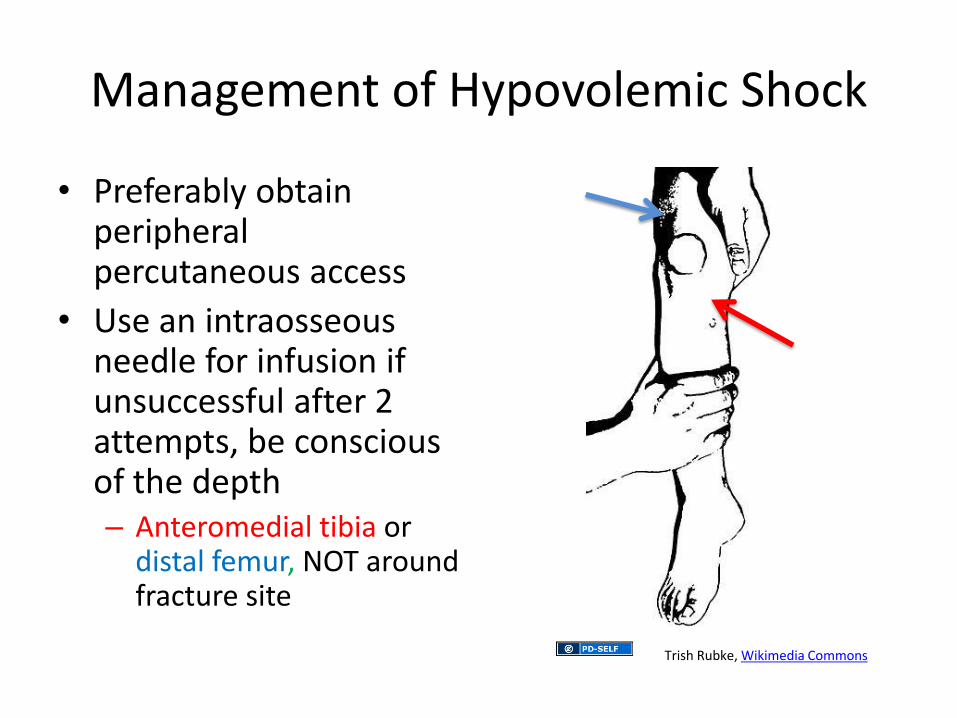
Management of Hypovolemic Shock
• Preferably obtain peripheral percutaneous access
• Use an intraosseous needle for infusion if unsuccessful after 2 attempts, be conscious of the depth – Anteromedial tibia or
distal femur, NOT around fracture site
Trish Rubke, Wikimedia Commons
Fluid and Blood Resuscitation
• Children have blood volumes based on size – Infant blood volume typically 80 ml/kg – Child’s blood volume typically 70 ml/kg
• Give 20 ml/kg normal saline bolus rapidly – Current teaching is to give 3, replaces about 25% of
lost intravascular volume, then start O neg PRBCs – Adult studies have shown sooner use of blood
products may improve survival
• With massive hemorrhage, should give blood products in balanced manner of red blood cells, plasma, and platelets to limit coagulopathy
Response to Resuscitation
• Look for: – Slowing of the heart rate and improved SBP
– Improvement of mental status
– Return of peripheral pulses
– Normal skin color
– Warmth of extremities
– Urine output (1-2 ml/kg/hr)
• As in adults, if only responding transiently or is not responding to crystalloid and blood, consider early operation
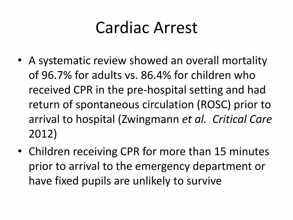
Cardiac Arrest
• A systematic review showed an overall mortality of 96.7% for adults vs. 86.4% for children who received CPR in the pre-hospital setting and had return of spontaneous circulation (ROSC) prior to arrival to hospital (Zwingmann et al. Critical Care 2012)
• Children receiving CPR for more than 15 minutes prior to arrival to the emergency department or have fixed pupils are unlikely to survive
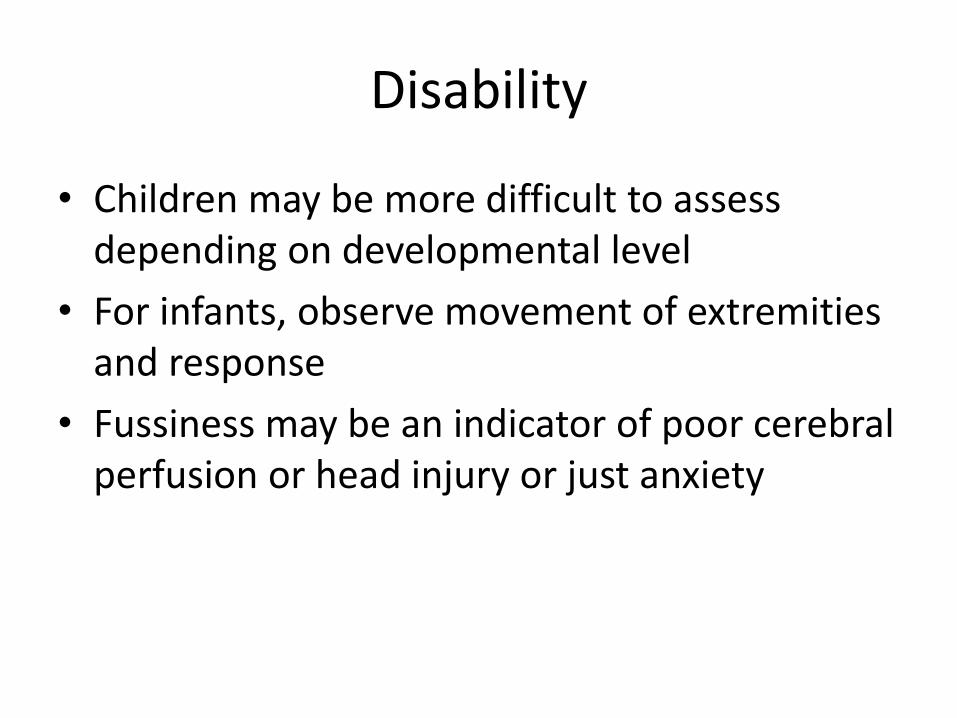
Disability
• Children may be more difficult to assess depending on developmental level
• For infants, observe movement of extremities and response
• Fussiness may be an indicator of poor cerebral perfusion or head injury or just anxiety
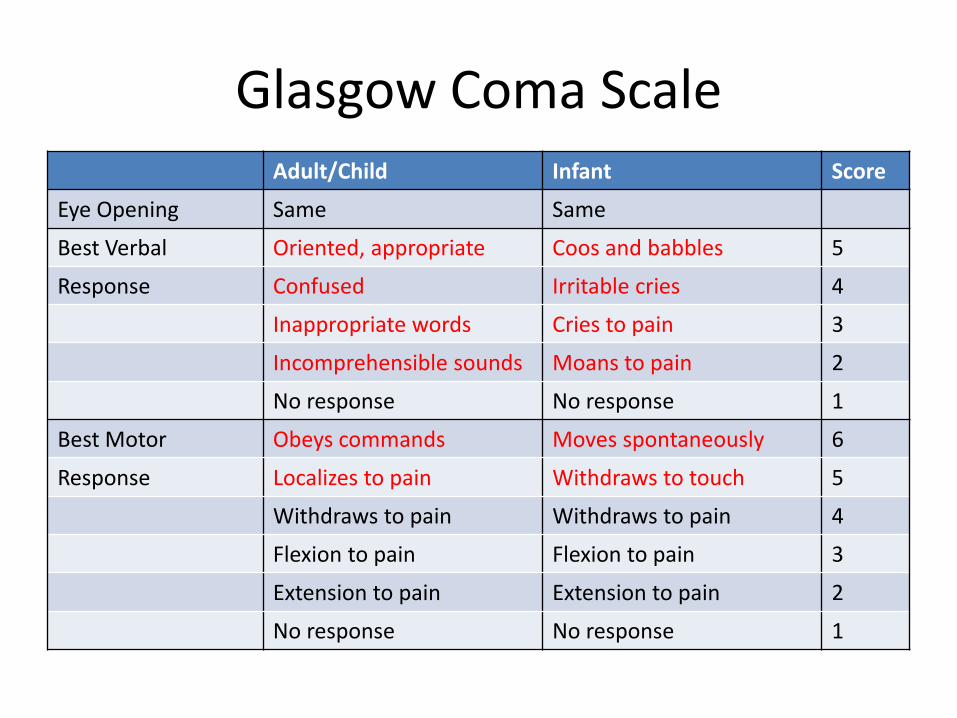
Glasgow Coma Scale Adult/Child Infant Score
Eye Opening Same Same
Best Verbal Oriented, appropriate Coos and babbles 5
Response Confused Irritable cries 4
Inappropriate words Cries to pain 3
Incomprehensible sounds Moans to pain 2
No response No response 1
Best Motor Obeys commands Moves spontaneously 6
Response Localizes to pain Withdraws to touch 5
Withdraws to pain Withdraws to pain 4
Flexion to pain Flexion to pain 3
Extension to pain Extension to pain 2
No response No response 1
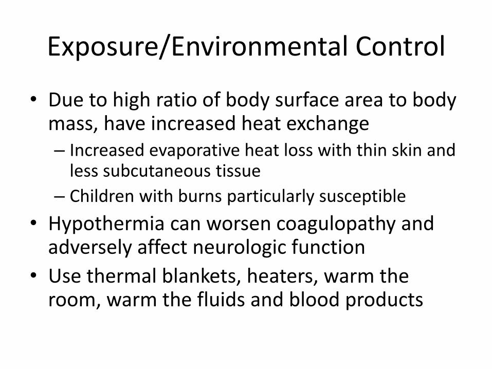
Exposure/Environmental Control
• Due to high ratio of body surface area to body mass, have increased heat exchange – Increased evaporative heat loss with thin skin and
less subcutaneous tissue
– Children with burns particularly susceptible
• Hypothermia can worsen coagulopathy and adversely affect neurologic function
• Use thermal blankets, heaters, warm the room, warm the fluids and blood products
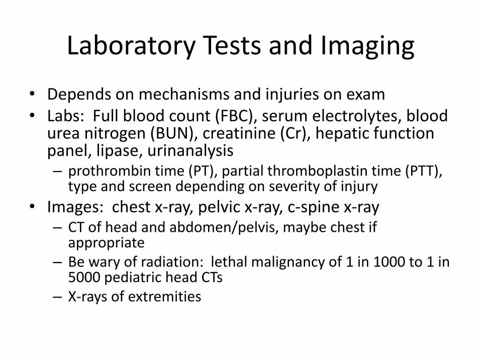
Laboratory Tests and Imaging
• Depends on mechanisms and injuries on exam • Labs: Full blood count (FBC), serum electrolytes, blood
urea nitrogen (BUN), creatinine (Cr), hepatic function panel, lipase, urinanalysis – prothrombin time (PT), partial thromboplastin time (PTT),
type and screen depending on severity of injury
• Images: chest x-ray, pelvic x-ray, c-spine x-ray – CT of head and abdomen/pelvis, maybe chest if
appropriate – Be wary of radiation: lethal malignancy of 1 in 1000 to 1 in
5000 pediatric head CTs – X-rays of extremities
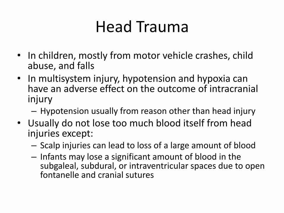
Head Trauma
• In children, mostly from motor vehicle crashes, child abuse, and falls
• In multisystem injury, hypotension and hypoxia can have an adverse effect on the outcome of intracranial injury – Hypotension usually from reason other than head injury
• Usually do not lose too much blood itself from head injuries except: – Scalp injuries can lead to loss of a large amount of blood – Infants may lose a significant amount of blood in the
subgaleal, subdural, or intraventricular spaces due to open fontanelle and cranial sutures
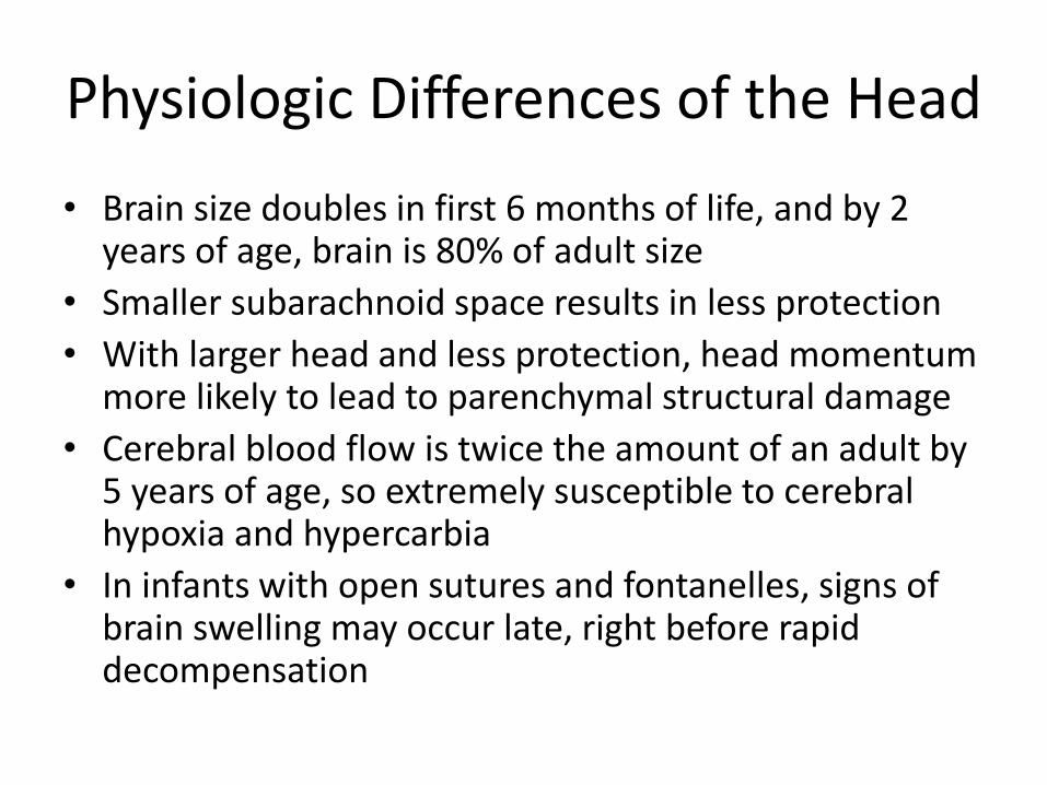
Physiologic Differences of the Head
• Brain size doubles in first 6 months of life, and by 2 years of age, brain is 80% of adult size
• Smaller subarachnoid space results in less protection
• With larger head and less protection, head momentum more likely to lead to parenchymal structural damage
• Cerebral blood flow is twice the amount of an adult by 5 years of age, so extremely susceptible to cerebral hypoxia and hypercarbia
• In infants with open sutures and fontanelles, signs of brain swelling may occur late, right before rapid decompensation
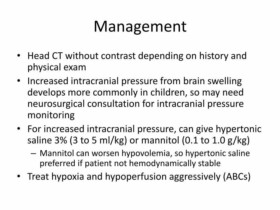
Management
• Head CT without contrast depending on history and physical exam
• Increased intracranial pressure from brain swelling develops more commonly in children, so may need neurosurgical consultation for intracranial pressure monitoring
• For increased intracranial pressure, can give hypertonic saline 3% (3 to 5 ml/kg) or mannitol (0.1 to 1.0 g/kg) – Mannitol can worsen hypovolemia, so hypertonic saline
preferred if patient not hemodynamically stable
• Treat hypoxia and hypoperfusion aggressively (ABCs)
Cervical Spine
• Spinal cord injury uncommon in pediatric group
– Occurs in less than 1% evaluated for trauma
– Accounts for about 5% of all spinal cord injuries
• Anatomic differences:
– Interspinous ligaments and joint capsules more flexible
– Vertebral bodies wedged anteriorly
– Large head of child means fulcrum is higher in the cervical spine and higher injuries
C-spine Management
• Start with plain films of c-spine, unless patient unresponsive and head CT will be done, then can obtain C-spine CT
• Normal x-ray and CT does not necessarily rule out spinal cord injury
– Children may have “spinal cord injury without radiographic abnormalities” (SCIWORA) more commonly than adults
– May need neurosurgical consultation and MRI
Thoracic Trauma
• 8% of injuries in children involve the chest, mostly due to blunt mechanisms
• 66% - 82% of children with chest injuries have multisystem injuries
• 15 - 26% mortality rate in children with thoracic injuries but usually due to other injuries such as blunt head trauma

Thoracic Trauma: Physiologic Differences
• Less ossified bones in child make the chest wall more compliant – More force is transmitted to intrathoracic organs
– Can have serious intrathoracic trauma without much visible damage to chest wall
• Increased mobility of mediastinum increases the risk of tension physiology from pneumothorax or hemothorax
• Commotio cordis more common in children
Types of Thoracic Injuries
• Posterior displacement or dislocation of clavicle (usually at physis) can be associated with injury to esophagus and great vessels
• Pulmonary contusion seen most commonly in children with blunt trauma (49%)
• Rarely see diaphragmatic rupture, aortic transection, tracheobronchial tears, flail chest, sternal fractures
• Life-threatening injuries are uncommon in children due to fewer penetrating mechanisms
Imaging
• Most chest injuries in children can be seen on chest x-ray
• Chest CT: – Not routinely used in children due to lower
incidence of cardiac and great vessel injury
– Obtain if have widened mediastinum or findings on plain film that cannot be explained
• Bedside ultrasound to evaluate for pericardial fluid
Management
• Most chest injuries in children can be managed using supportive care or a chest tube if pneumothorax or hemothorax present
• Indications for emergency department thoracotomy in children similar to adults: – Penetrating thoracic trauma that is hemodynamically
unstable
– Signs of cardiac tamponade
– Thoracic or trauma surgeon available within 45 minutes
Abdominal Trauma
• Abdominal physical exam can be difficult in young children, particularly if crying
• 25% of prepubertal children with multisystem injury have significant abdominal injury
• Smaller anterior-posterior diameter of children and smaller torso gives less area for force to dissipate
• 20% mortality if isolated organ injury – Increases to 20% if gastrointestinal tract involved – Increases to 50% if major vessels involved
Signs of Abdominal Injury
• Ecchymoses, particularly around umbilicus and flank
• Seatbelt sign
• Abdominal distension (but could be from crying)
• Abdominal tenderness or rigidity
• Pain in left shoulder with palpation of left upper quadrant
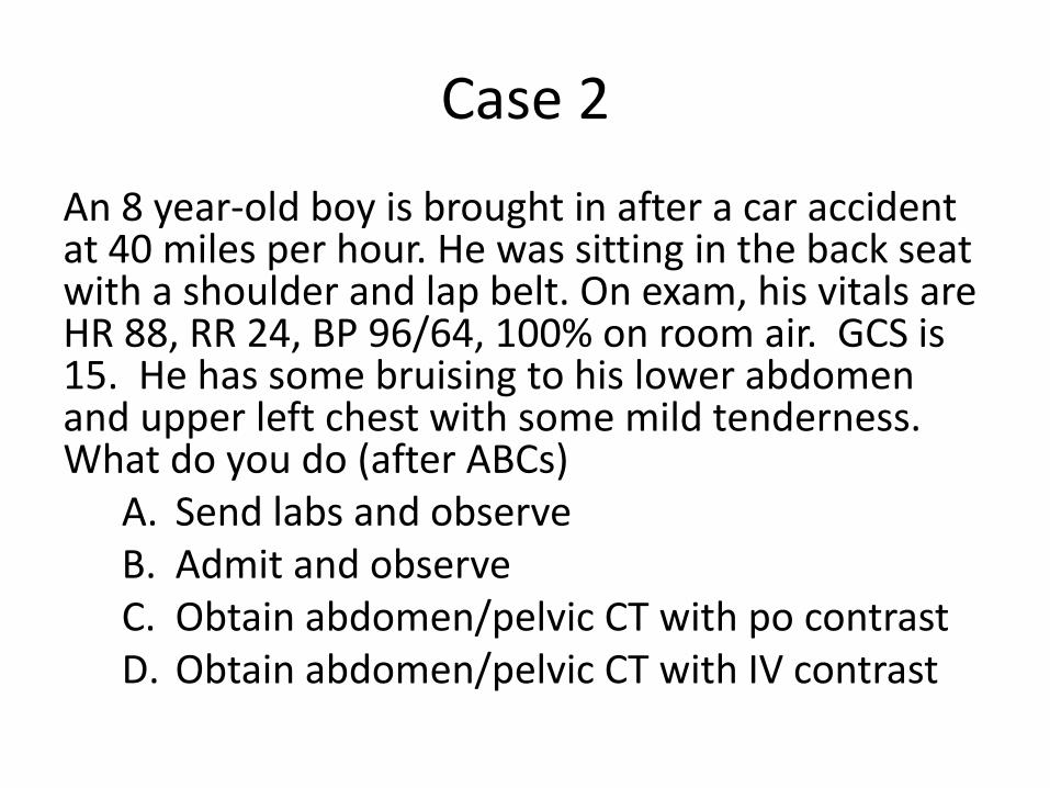
Case 2
An 8 year-old boy is brought in after a car accident at 40 miles per hour. He was sitting in the back seat with a shoulder and lap belt. On exam, his vitals are HR 88, RR 24, BP 96/64, 100% on room air. GCS is 15. He has some bruising to his lower abdomen and upper left chest with some mild tenderness. What do you do (after ABCs)
A. Send labs and observe B. Admit and observe C. Obtain abdomen/pelvic CT with po contrast D. Obtain abdomen/pelvic CT with IV contrast
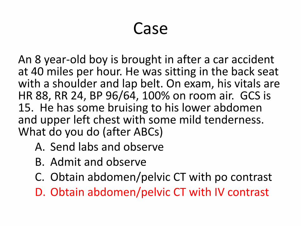
Case
An 8 year-old boy is brought in after a car accident at 40 miles per hour. He was sitting in the back seat with a shoulder and lap belt. On exam, his vitals are HR 88, RR 24, BP 96/64, 100% on room air. GCS is 15. He has some bruising to his lower abdomen and upper left chest with some mild tenderness. What do you do (after ABCs)
A. Send labs and observe B. Admit and observe C. Obtain abdomen/pelvic CT with po contrast D. Obtain abdomen/pelvic CT with IV contrast
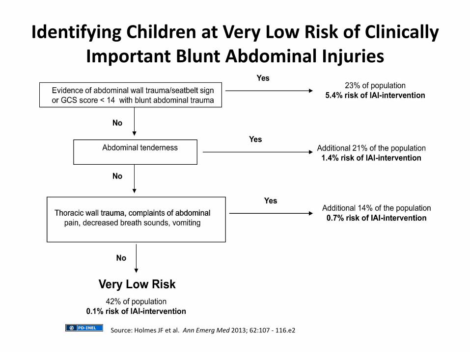
Source: Holmes JF et al. Ann Emerg Med 2013; 62:107 - 116.e2
Identifying Children at Very Low Risk of Clinically Important Blunt Abdominal Injuries

Laboratory Studies
• FBC
• Serum electolytes, BUN, Creatinine
• Hepatic Function Panel
• Lipase
• Urine dipstick (for blood)
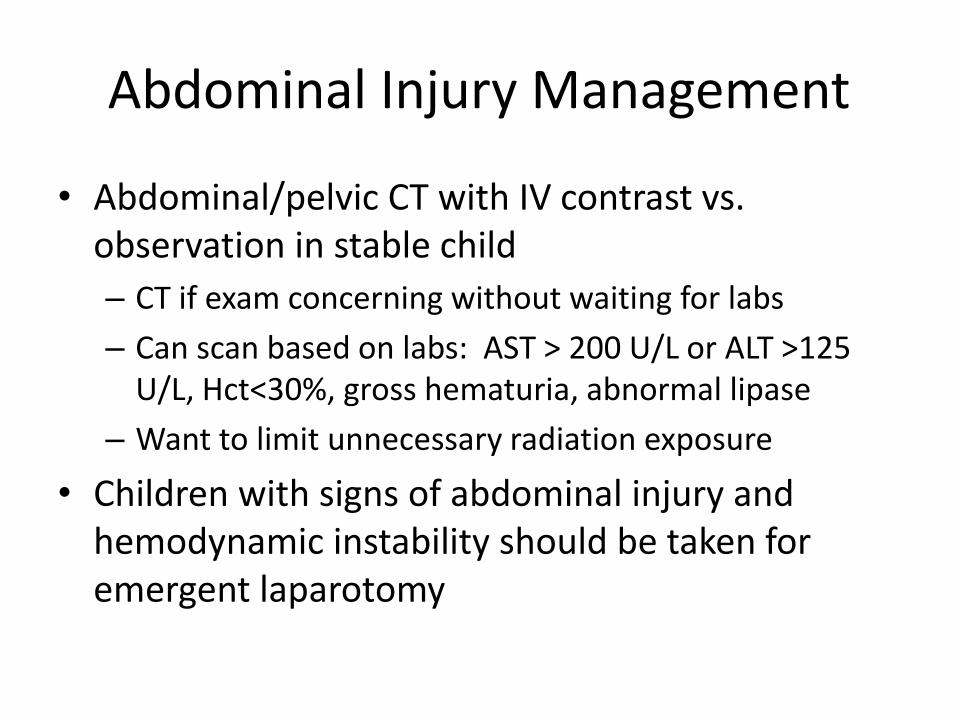
Abdominal Injury Management
• Abdominal/pelvic CT with IV contrast vs. observation in stable child
– CT if exam concerning without waiting for labs
– Can scan based on labs: AST > 200 U/L or ALT >125 U/L, Hct<30%, gross hematuria, abnormal lipase
– Want to limit unnecessary radiation exposure
• Children with signs of abdominal injury and hemodynamic instability should be taken for emergent laparotomy
Focused Assessment Sonography in Trauma (FAST)
• Not as effective in the management of children
• Identifies intra-abdominal fluid, but may not need surgical intervention
• Poor study for identifying intra-parenchymal injuries, which account for up to 1/3 of solid organ injuries in children
Extremity Trauma
• Can be difficult to diagnose in children due to presence of growth plate (physis) or lack of mineralization around the epiphysis – Growth plates may be mistaken for fractures
– Fractures in growth plates may be difficult to see
• Different growth plates close at different times – Typically growth stops 2 years after pubertal
growth spurt completed
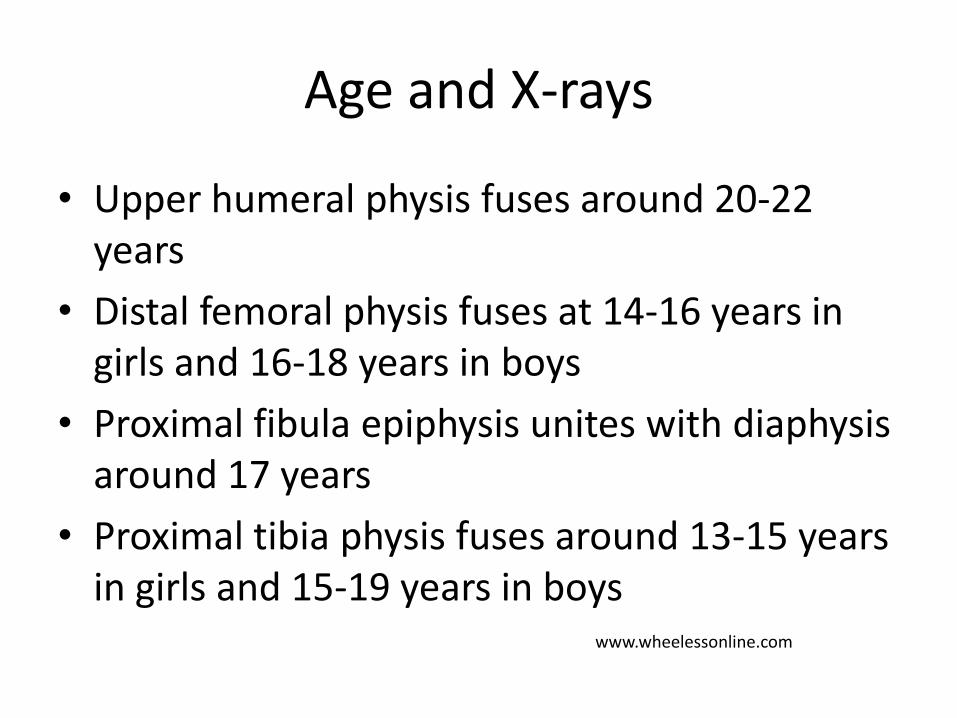
• Upper humeral physis fuses around 20-22 years
• Distal femoral physis fuses at 14-16 years in girls and 16-18 years in boys
• Proximal fibula epiphysis unites with diaphysis around 17 years
• Proximal tibia physis fuses around 13-15 years in girls and 15-19 years in boys
Age and X-rays
www.wheelessonline.com
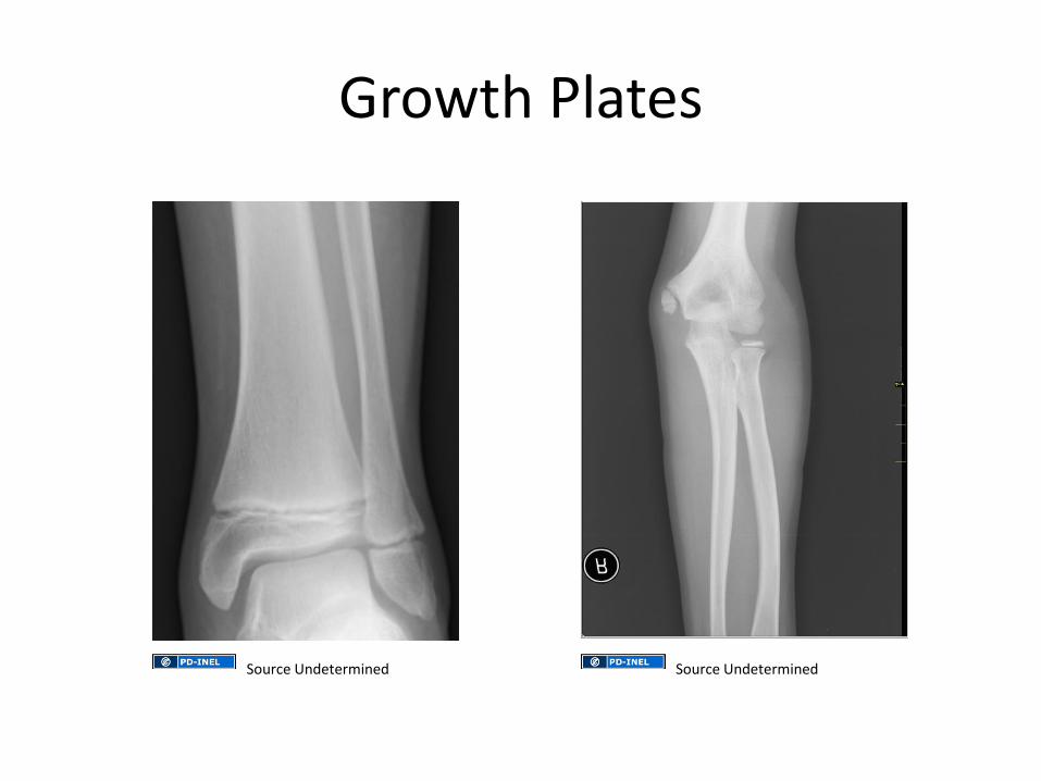
Growth Plates
Source Undetermined Source Undetermined
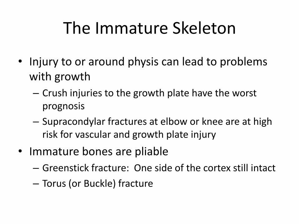
The Immature Skeleton
• Injury to or around physis can lead to problems with growth
– Crush injuries to the growth plate have the worst prognosis
– Supracondylar fractures at elbow or knee are at high risk for vascular and growth plate injury
• Immature bones are pliable
– Greenstick fracture: One side of the cortex still intact
– Torus (or Buckle) fracture
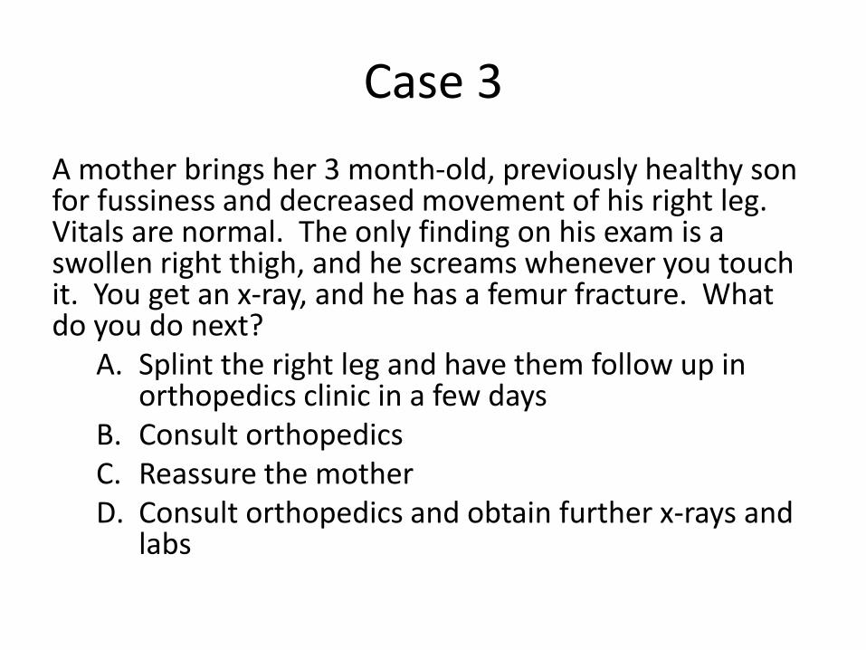
Case 3
A mother brings her 3 month-old, previously healthy son for fussiness and decreased movement of his right leg. Vitals are normal. The only finding on his exam is a swollen right thigh, and he screams whenever you touch it. You get an x-ray, and he has a femur fracture. What do you do next?
A. Splint the right leg and have them follow up in orthopedics clinic in a few days
B. Consult orthopedics C. Reassure the mother D. Consult orthopedics and obtain further x-rays and
labs
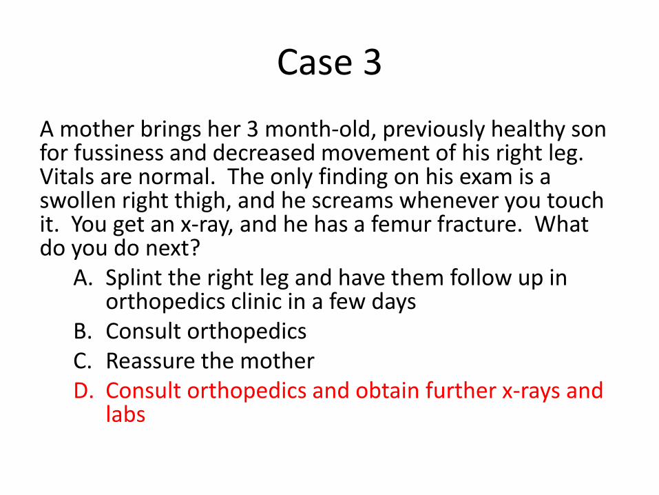
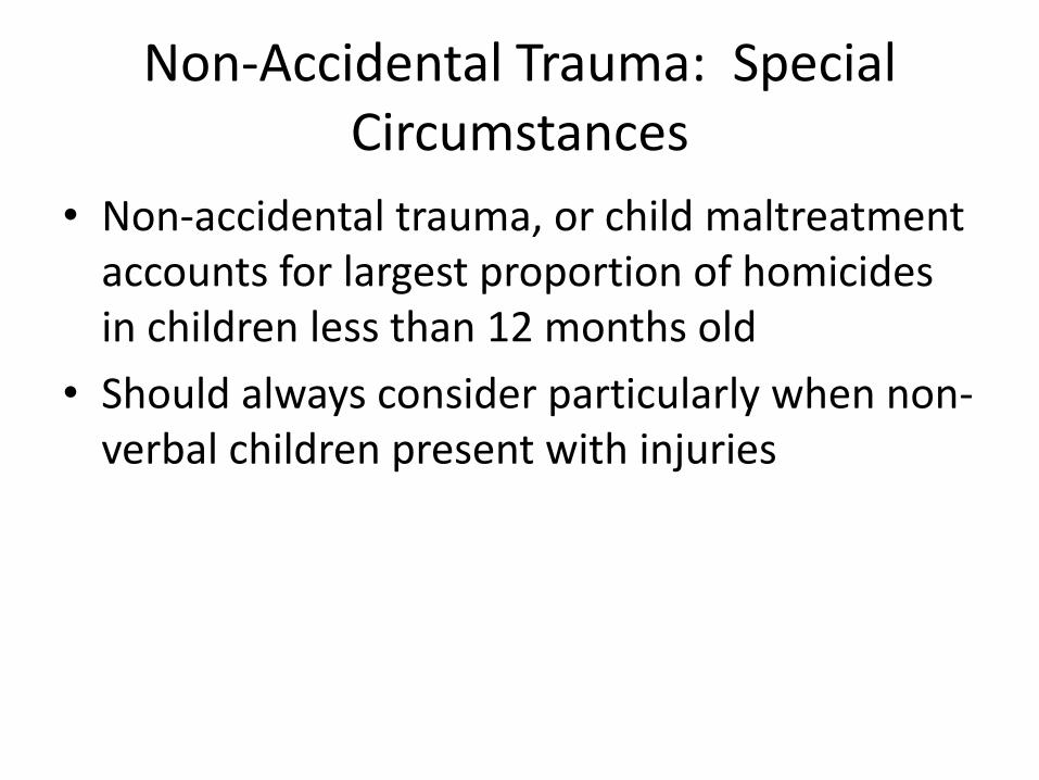
Non-Accidental Trauma: Special Circumstances
• Non-accidental trauma, or child maltreatment accounts for largest proportion of homicides in children less than 12 months old
• Should always consider particularly when non-verbal children present with injuries
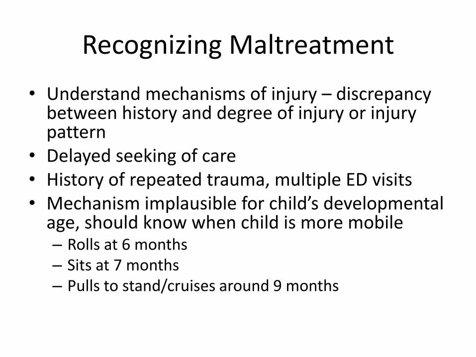
Recognizing Maltreatment
• Understand mechanisms of injury – discrepancy between history and degree of injury or injury pattern
• Delayed seeking of care • History of repeated trauma, multiple ED visits • Mechanism implausible for child’s developmental
age, should know when child is more mobile – Rolls at 6 months – Sits at 7 months – Pulls to stand/cruises around 9 months
Concerning Physical Findings
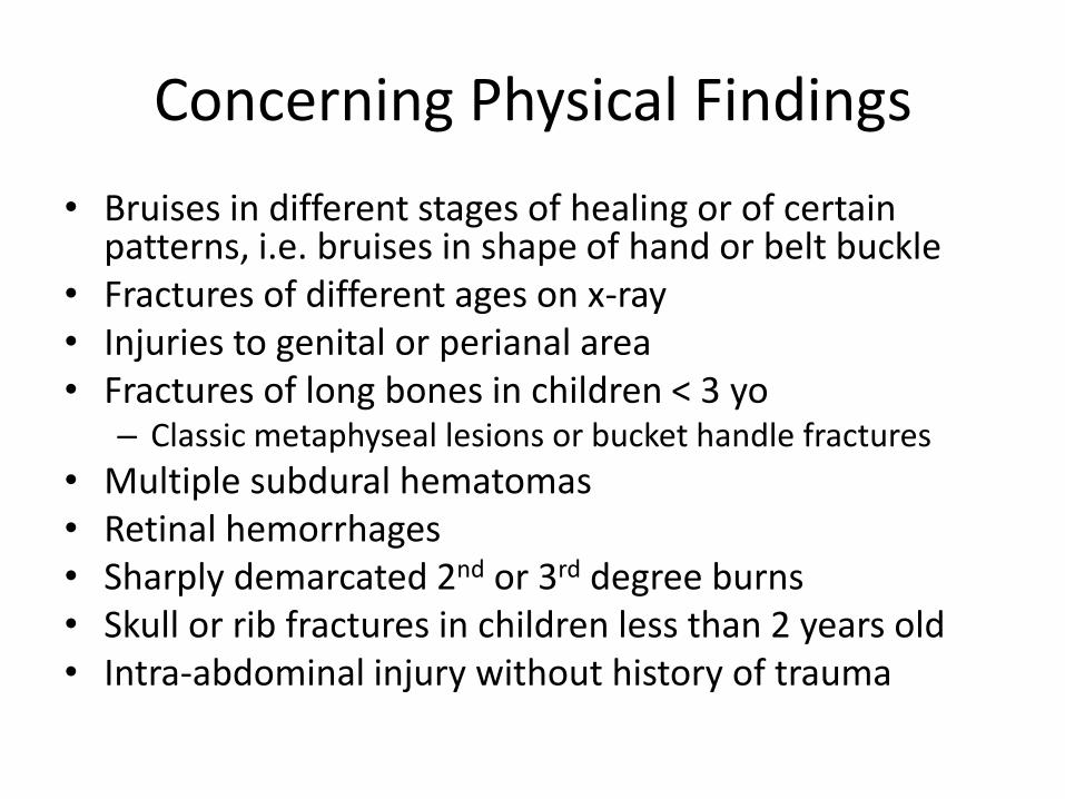
• Bruises in different stages of healing or of certain patterns, i.e. bruises in shape of hand or belt buckle
• Fractures of different ages on x-ray • Injuries to genital or perianal area • Fractures of long bones in children < 3 yo
– Classic metaphyseal lesions or bucket handle fractures
• Multiple subdural hematomas • Retinal hemorrhages • Sharply demarcated 2nd or 3rd degree burns • Skull or rib fractures in children less than 2 years old • Intra-abdominal injury without history of trauma
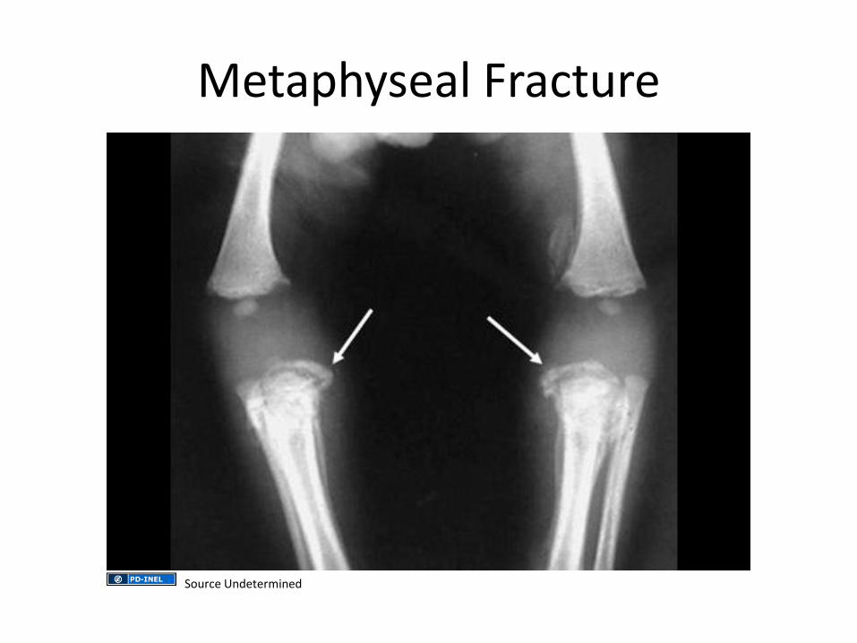
Metaphyseal Fracture
Source Undetermined

Medical Evaluation for Suspected Non-Accidental Trauma
• < 1 years old – Head CT if concerned about head injury,
otherwise brain MRI
– Skeletal survey
– Opthalmology exam (for retinal hemorrhages)
– Labs: FBC, serum electrolytes, BUN, serum creatinine, hepatic function panel, lipase, urinanalysis, PT/PTT (if bruising or had head CT)
– Admit if medical concerns or cannot find safe place to stay
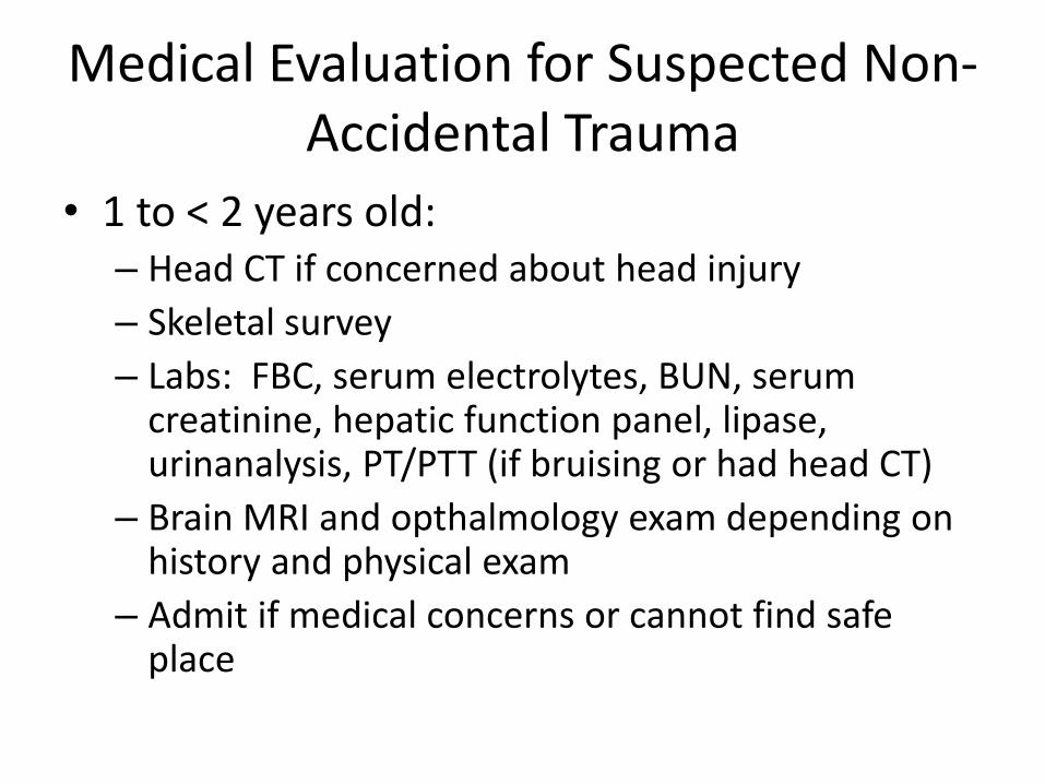
Medical Evaluation for Suspected Non-Accidental Trauma
• 1 to < 2 years old: – Head CT if concerned about head injury
– Skeletal survey
– Labs: FBC, serum electrolytes, BUN, serum creatinine, hepatic function panel, lipase, urinanalysis, PT/PTT (if bruising or had head CT)
– Brain MRI and opthalmology exam depending on history and physical exam
– Admit if medical concerns or cannot find safe place
Medical Evaluation for Suspected Non-Accidental Trauma
• ≥ 2 years old:
– Selective testing depending on injuries and mechanism
– Admit if medical concerns or cannot find safe place to stay
Non-Accidental Trauma
• Mandatory reporting to agencies
– Domestic Violence and Victim Support Unit of the Ghana Police Service
– Social Welfare Department
• 50% of maltreated children who died had previous episodes that went unreported
Summary
• Children are more likely to suffer from blunt mechanisms of injury and thus multisystem injury
• Similar to management of trauma in adults, remember the ABCs
• The airway in children and large occiput make airway positioning a little more difficult
• Children have amazing cardiovascular reserve but can decompensate quickly
• Do not forget to consider non-accidental trauma

References
Chameides L, Samson RA, Schexnayder SM, Hazinski MF, ed (2011). Pediatric Advanced Life Support Provider Manual. American Heart Association: United States of America.
Global Health Observatory. http://www.who.int/gho/countries/gha/en/. Accessed January 25, 2014. Hardcastle TC, Oteng R. Trauma care in Africa: Triumphs and challenges. African J of Em Med. 2011:53-54. Holmes JF, Sokolove PE, Brant WE, et al. Validation of a prediction rule for identification of children with intraabdominal injuries after blung torso trauma.
Ann Emerg Med. 2009; 54:528-533. Holmes JF, Lillis K, Monroe D, et al. Identifying children at very low risk of clinically important blunt abdominal injuries. Ann Emer Med. 2012; 62:107-116. Injury Prevention & Control: Data & Statistics. http://www.cdc.gov/injury/wisqars. Accessed January 25, 2014. Kadish H. Chest wall injuries in children. UpToDate. www.uptodate.com. Accessed January 6, 2014. Kissoon N, Dreyer J, Walia M. Pediatric trauma: differences in pathophysiology, injury patterns and treatment compared with adult trauma. Can Med Assoc
J. 1990; 142(1):27-34. Lee LK, Fleisher G. Trauma management: approach to the unstable child. UpToDate. www.uptodate.com. Accessed January 6, 2014. Lee LK, Fleisher G. Trauma management: unique pediatric considerations. UpToDate. www.uptodate.com. Accessed January 6, 2014. Mendez DR. Initial evaluation and stabilization of children with thoracic trauma. UpToDate. www.uptodate.com. Accessed January 6, 2014. Mendez DR. Overview of blunt abdominal trauma in children. UpToDate. www.uptodate.com. Accessed January 6, 2014. Subcommittee on Advanced Trauma Life Support of the American College of Surgeons Committee on Trauma (2008). Advanced Trauma Life Support for
Doctors. American College of Surgeons: Chicago, IL. Wheeless CR. Wheeless’ Textbook of Orthopaedics. www.wheelessonline.com. Accessed January 27, 2014. Zwingmann J, Mehlhorn AT, Hammer T, Bayer J, Suedkamp NP, Strohm PC. Survival and neurologic outome after traumatic out-of-hospital
cardiopulmonary arrest in a pediatric and adult population: a systematic review. Crit Care. 2012; 16:R117.