Embed Size (px)
Citation preview
IDENTIFICATION OF PATHOGENIC BACTERIA IN CLINICAL MICROBIOLOGY
LABORATORY
G.HARIPRASAD M.Sc.,M.Phil.,Ph.DDepartment of Microbiology
Thoothukudi Govt. Medical College Thoothukudi
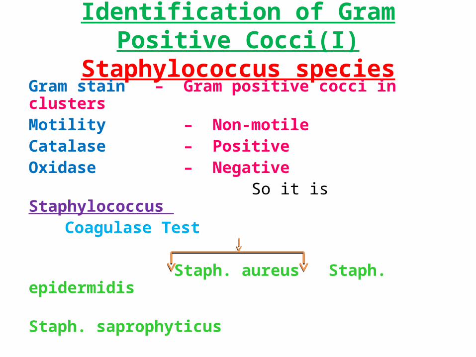
Identification of Gram Positive Cocci(I)Staphylococcus species
Gram stain – Gram positive cocci in clusters Motility – Non-motileCatalase – Positive Oxidase – Negative So it is Staphylococcus
Coagulase Test
Staph. aureus Staph. epidermidis Staph. saprophyticus
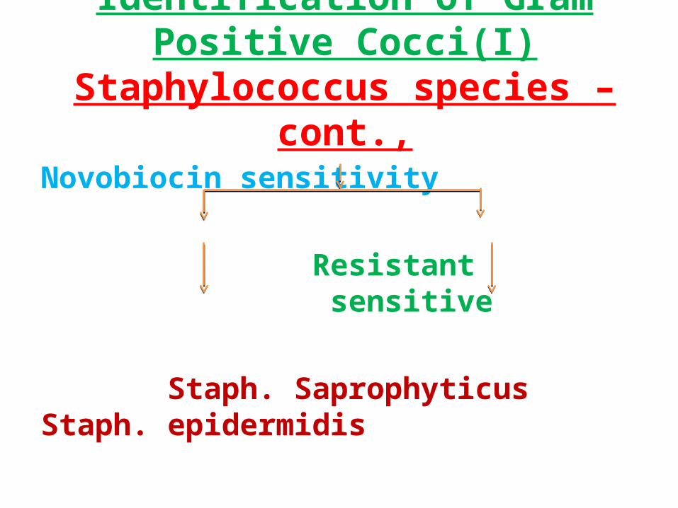
Identification of Gram Positive Cocci(I)Staphylococcus species – cont.,
Novobiocin sensitivity
Resistant sensitive
Staph. Saprophyticus Staph. epidermidis
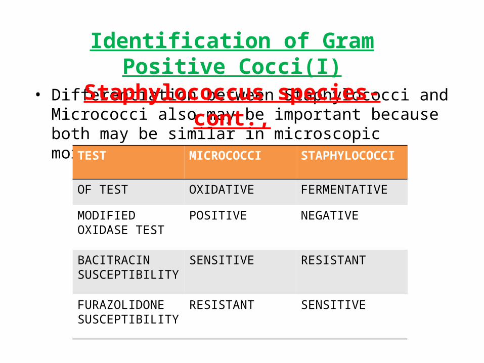
• Differentiation between Staphylococci and Micrococci also may be important because both may be similar in microscopic morphology.
TEST MICROCOCCI STAPHYLOCOCCI
OF TEST OXIDATIVE FERMENTATIVE
MODIFIED OXIDASE TEST
POSITIVE NEGATIVE
BACITRACIN SUSCEPTIBILITY
SENSITIVE RESISTANT
FURAZOLIDONE SUSCEPTIBILITY
RESISTANT SENSITIVE
Identification of Gram Positive Cocci(I)Staphylococcus species- cont.,
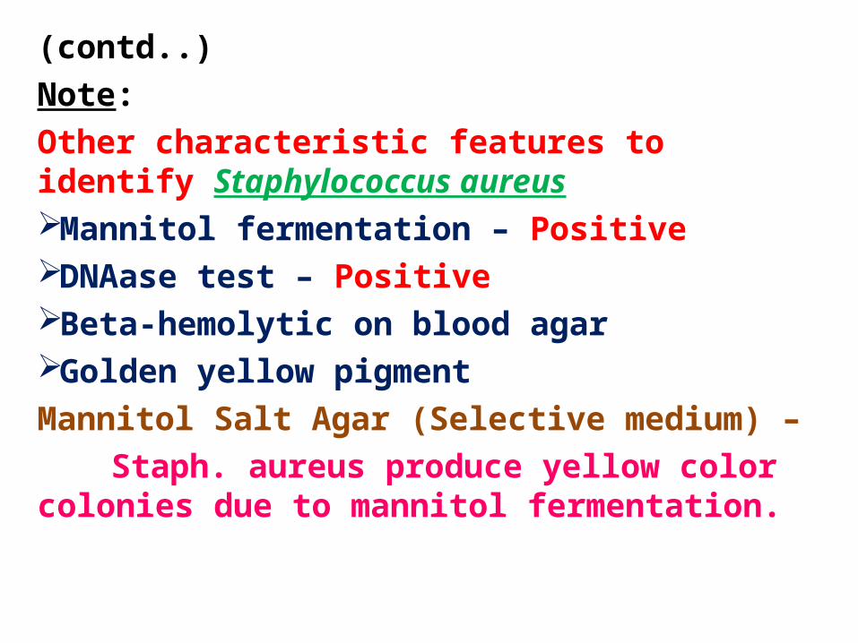
(contd..)Note: Other characteristic features to identify Staphylococcus aureusMannitol fermentation – Positive DNAase test – Positive Beta-hemolytic on blood agar Golden yellow pigment Mannitol Salt Agar (Selective medium) –
Staph. aureus produce yellow color colonies due to mannitol fermentation.
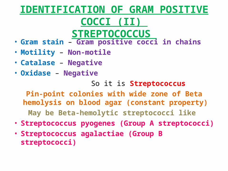
IDENTIFICATION OF GRAM POSITIVE COCCI (II) STREPTOCOCCUS
• Gram stain – Gram positive cocci in chains • Motility – Non-motile • Catalase – Negative • Oxidase – Negative So it is Streptococcus
Pin-point colonies with wide zone of Beta hemolysis on blood agar (constant property)
May be Beta-hemolytic streptococci like • Streptococcus pyogenes (Group A streptococci) • Streptococcus agalactiae (Group B streptococci)
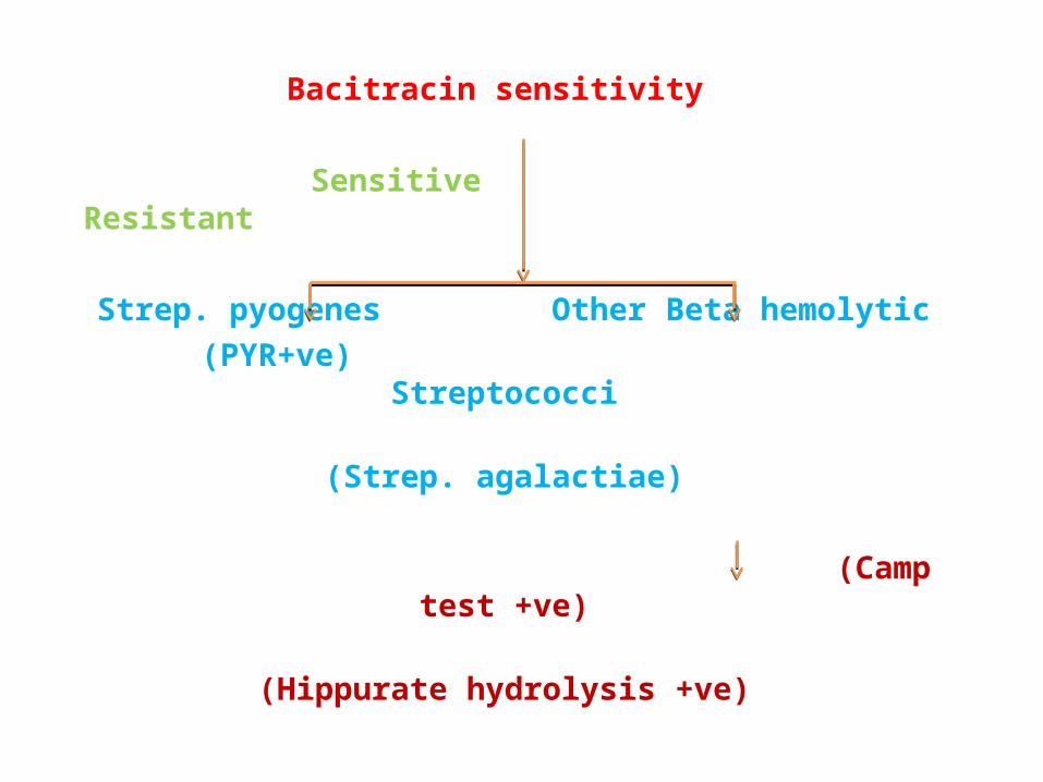
Bacitracin sensitivity
Sensitive Resistant
Strep. pyogenes Other Beta hemolytic (PYR+ve) Streptococci
(Strep. agalactiae)
(Camp test +ve) (Hippurate hydrolysis +ve)
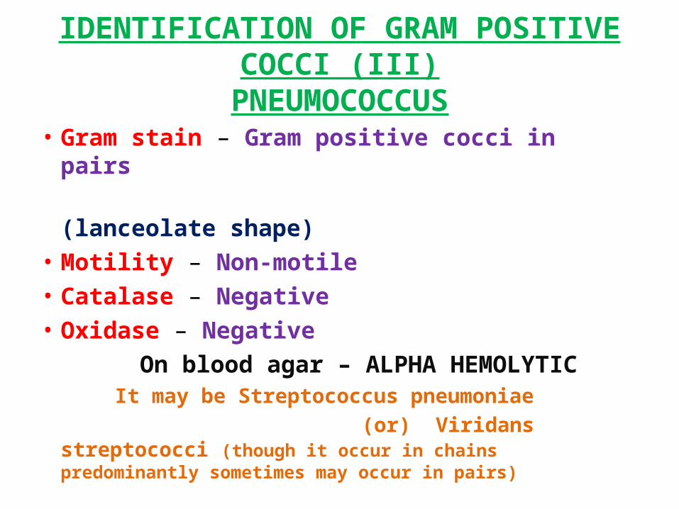
IDENTIFICATION OF GRAM POSITIVE COCCI (III)PNEUMOCOCCUS
• Gram stain – Gram positive cocci in pairs (lanceolate shape)• Motility – Non-motile • Catalase – Negative • Oxidase – Negative On blood agar – ALPHA HEMOLYTIC It may be Streptococcus pneumoniae (or) Viridans streptococci (though it occur
in chains predominantly sometimes may occur in pairs)
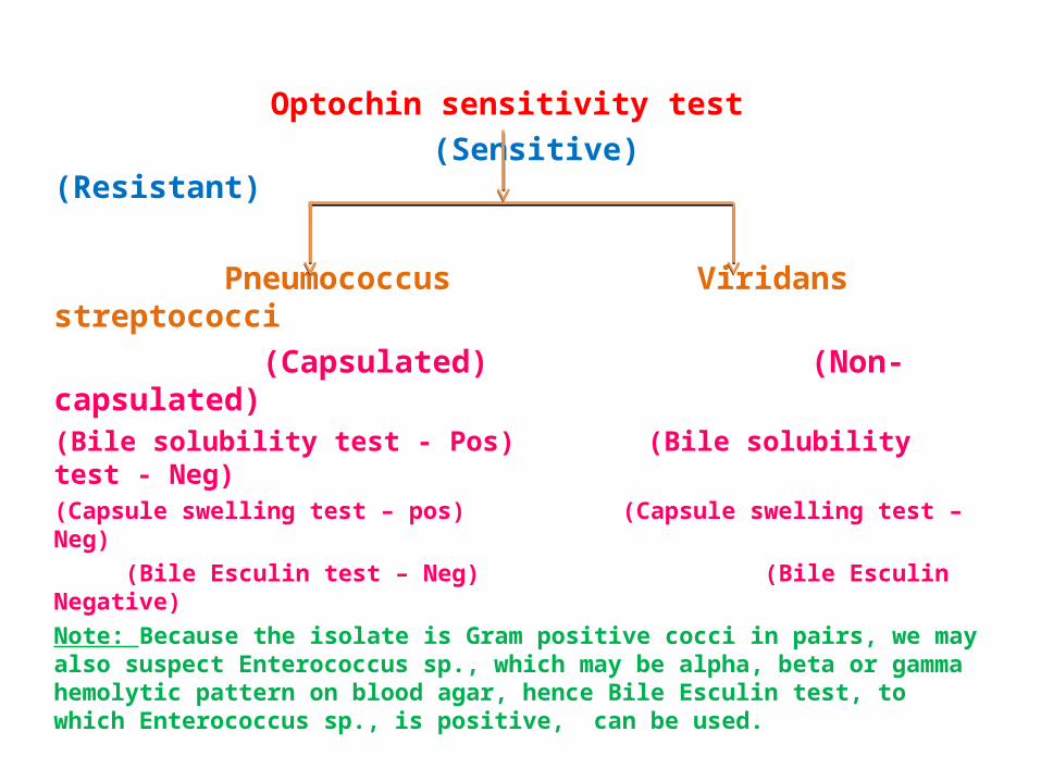
Optochin sensitivity test (Sensitive) (Resistant)
Pneumococcus Viridans streptococci (Capsulated) (Non-capsulated)(Bile solubility test - Pos) (Bile solubility test - Neg)(Capsule swelling test – pos) (Capsule swelling test – Neg) (Bile Esculin test – Neg) (Bile Esculin Negative)Note: Because the isolate is Gram positive cocci in pairs, we may also suspect Enterococcus sp., which may be alpha, beta or gamma hemolytic pattern on blood agar, hence Bile Esculin test, to which Enterococcus sp., is positive, can be used.
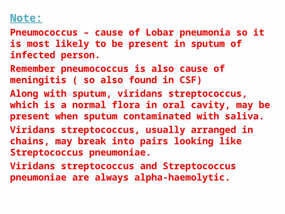
Note:Pneumococcus – cause of Lobar pneumonia so it is most likely to be present in sputum of infected person.Remember pneumococcus is also cause of meningitis ( so also found in CSF)Along with sputum, viridans streptococcus, which is a normal flora in oral cavity, may be present when sputum contaminated with saliva.Viridans streptococcus, usually arranged in chains, may break into pairs looking like Streptococcus pneumoniae. Viridans streptococcus and Streptococcus pneumoniae are always alpha-haemolytic.
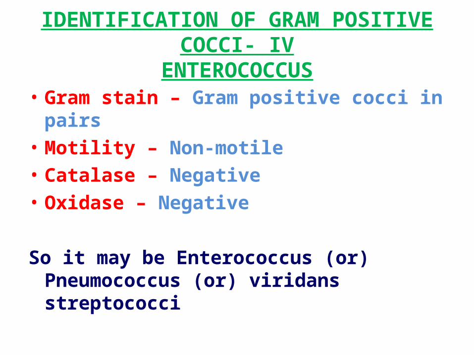
IDENTIFICATION OF GRAM POSITIVE COCCI- IVENTEROCOCCUS
• Gram stain – Gram positive cocci in pairs • Motility – Non-motile • Catalase – Negative • Oxidase – Negative So it may be Enterococcus (or) Pneumococcus
(or) viridans streptococci
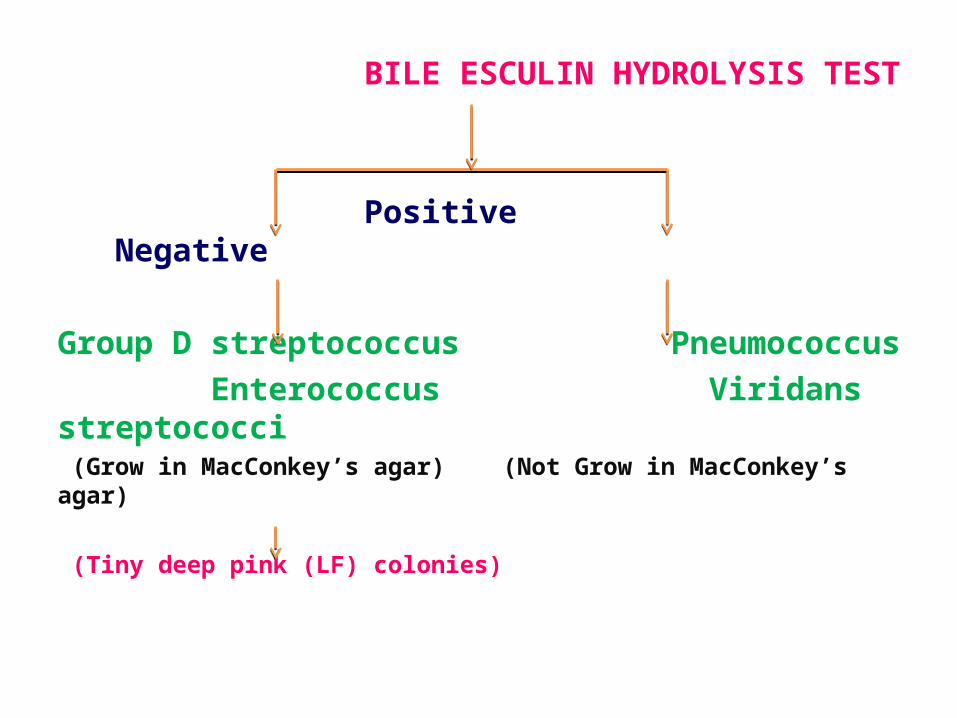
BILE ESCULIN HYDROLYSIS TEST
Positive Negative
Group D streptococcus Pneumococcus Enterococcus Viridans streptococci (Grow in MacConkey’s agar) (Not Grow in MacConkey’s agar)
(Tiny deep pink (LF) colonies)
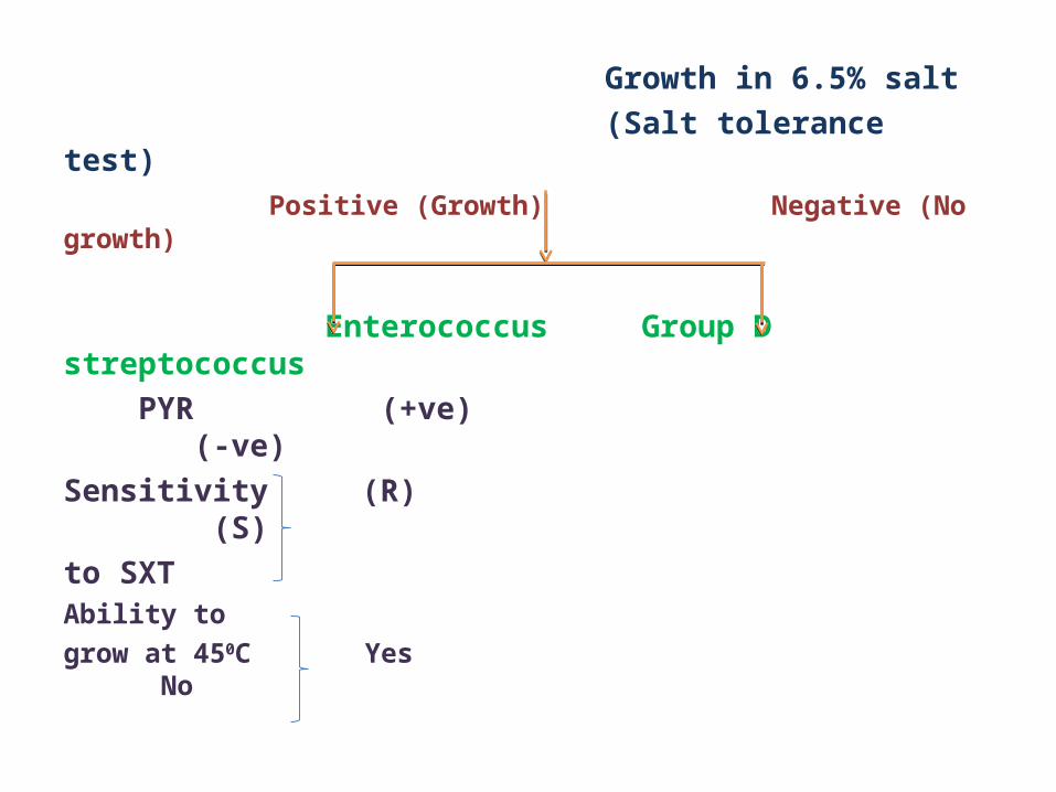
Growth in 6.5% salt (Salt tolerance test) Positive (Growth) Negative (No growth)
Enterococcus Group D streptococcus PYR (+ve) (-ve)Sensitivity (R) (S)to SXTAbility to grow at 450C Yes No
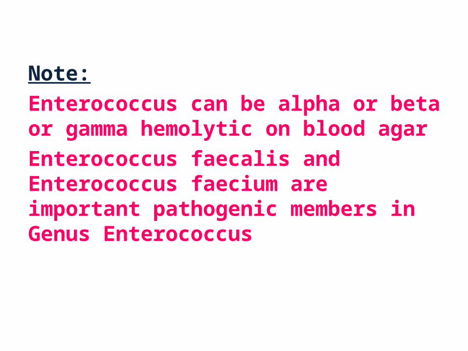
Note:Enterococcus can be alpha or beta or gamma hemolytic on blood agar Enterococcus faecalis and Enterococcus faecium are important pathogenic members in Genus Enterococcus
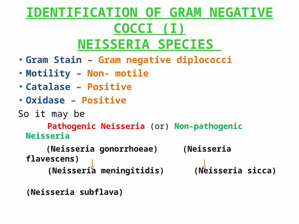
IDENTIFICATION OF GRAM NEGATIVE COCCI (I)NEISSERIA SPECIES
• Gram Stain – Gram negative diplococci • Motility – Non- motile • Catalase – Positive • Oxidase – Positive So it may be Pathogenic Neisseria (or) Non-pathogenic Neisseria (Neisseria gonorrhoeae) (Neisseria flavescens) (Neisseria meningitidis) (Neisseria sicca) (Neisseria subflava)
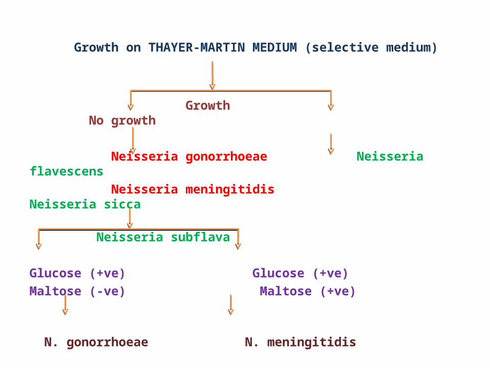
Growth on THAYER-MARTIN MEDIUM (selective medium)
Growth No growth Neisseria gonorrhoeae Neisseria flavescens Neisseria meningitidis Neisseria sicca Neisseria subflava
Glucose (+ve) Glucose (+ve)Maltose (-ve) Maltose (+ve) N. gonorrhoeae N. meningitidis
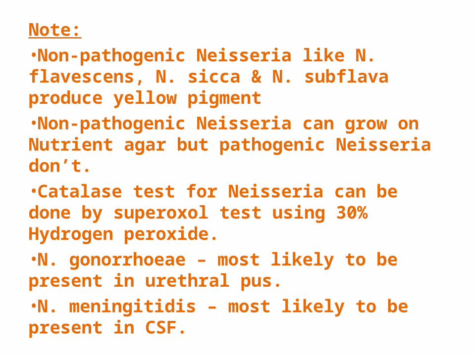
Note:•Non-pathogenic Neisseria like N. flavescens, N. sicca & N. subflava produce yellow pigment•Non-pathogenic Neisseria can grow on Nutrient agar but pathogenic Neisseria don’t.•Catalase test for Neisseria can be done by superoxol test using 30% Hydrogen peroxide.•N. gonorrhoeae – most likely to be present in urethral pus.•N. meningitidis – most likely to be present in CSF.
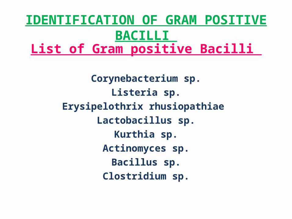
IDENTIFICATION OF GRAM POSITIVE BACILLI
List of Gram positive Bacilli
Corynebacterium sp.Listeria sp.
Erysipelothrix rhusiopathiae Lactobacillus sp.
Kurthia sp.Actinomyces sp.
Bacillus sp.Clostridium sp.
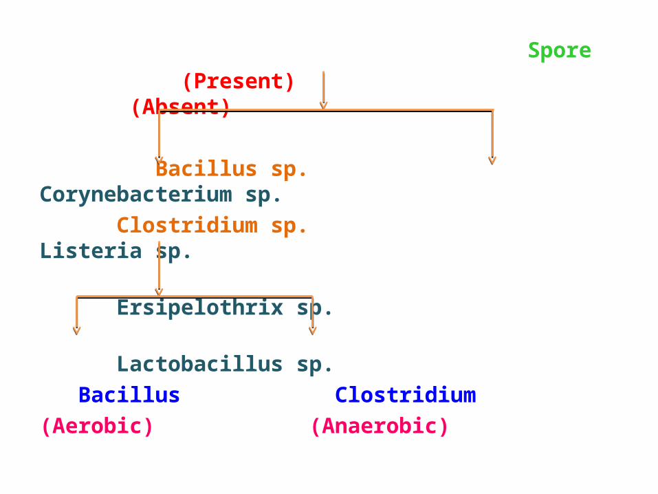
Spore (Present) (Absent)
Bacillus sp. Corynebacterium sp. Clostridium sp. Listeria sp. Ersipelothrix sp. Lactobacillus sp. Bacillus Clostridium (Aerobic) (Anaerobic)
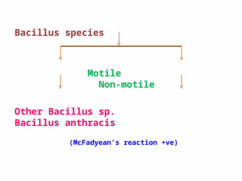
Bacillus species
Motile Non-motile
Other Bacillus sp. Bacillus anthracis (McFadyean’s reaction +ve)
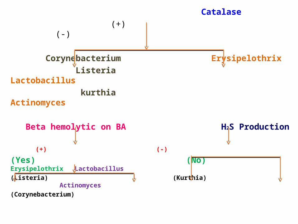
Catalase (+) (-)
Corynebacterium Erysipelothrix Listeria Lactobacillus kurthia Actinomyces
Beta hemolytic on BA H2S Production (+) (-)
(Yes) (No) Erysipelothrix Lactobacillus(Listeria) (Kurthia) Actinomyces(Corynebacterium)
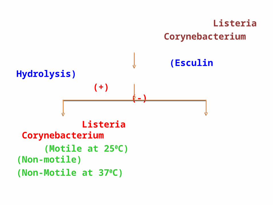
Listeria Corynebacterium
(Esculin Hydrolysis) (+) (-)
Listeria Corynebacterium (Motile at 250C) (Non-motile)(Non-Motile at 370C)
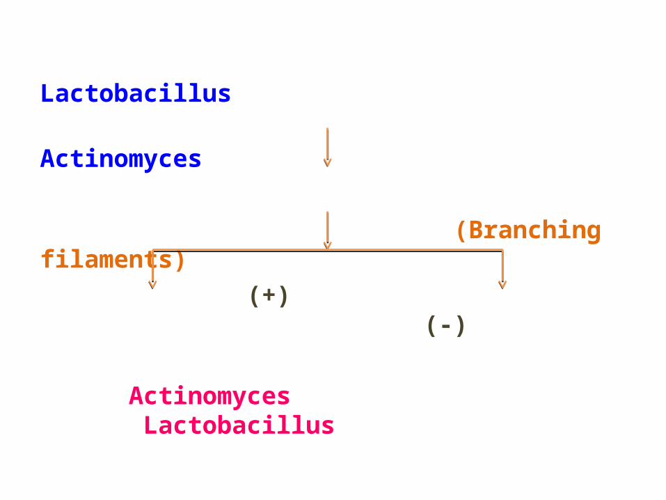
Lactobacillus Actinomyces
(Branching filaments) (+) (-)
Actinomyces Lactobacillus
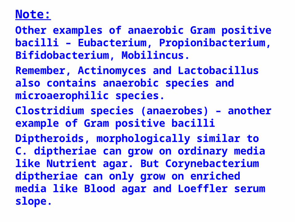
Note:Other examples of anaerobic Gram positive bacilli – Eubacterium, Propionibacterium, Bifidobacterium, Mobilincus.Remember, Actinomyces and Lactobacillus also contains anaerobic species and microaerophilic species. Clostridium species (anaerobes) – another example of Gram positive bacilli Diptheroids, morphologically similar to C. diptheriae can grow on ordinary media like Nutrient agar. But Corynebacterium diptheriae can only grow on enriched media like Blood agar and Loeffler serum slope.
IDENTIFICATION OF GRAM NEGATIVE BACILLI (I)
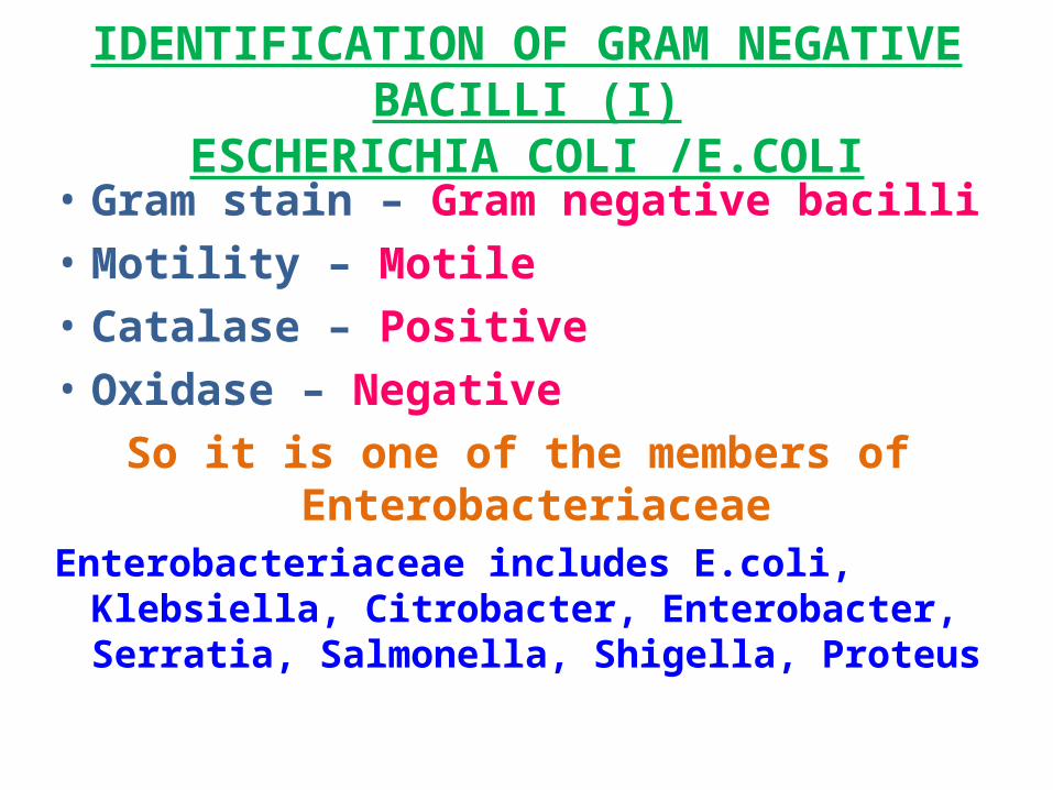
ESCHERICHIA COLI /E.COLI• Gram stain – Gram negative bacilli • Motility – Motile • Catalase – Positive • Oxidase – Negative
So it is one of the members of Enterobacteriaceae
Enterobacteriaceae includes E.coli, Klebsiella, Citrobacter, Enterobacter, Serratia, Salmonella, Shigella, Proteus
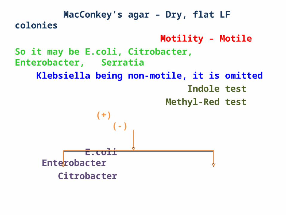
MacConkey’s agar – Dry, flat LF colonies Motility – MotileSo it may be E.coli, Citrobacter, Enterobacter, Serratia Klebsiella being non-motile, it is omitted Indole test Methyl-Red test (+) (-)
E.coli Enterobacter Citrobacter
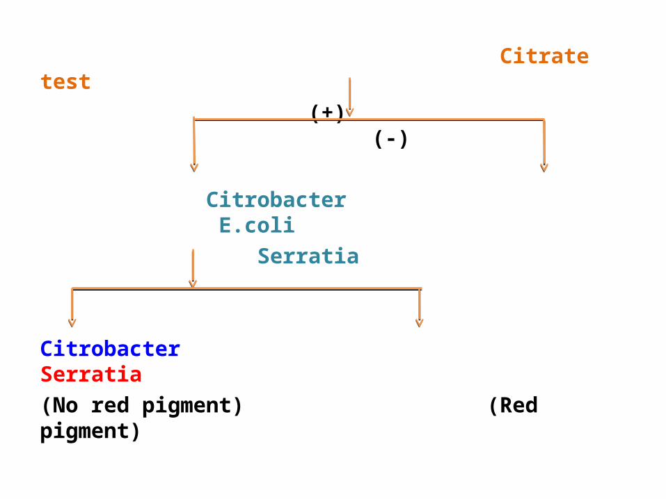
Citrate test (+) (-)
Citrobacter E.coli Serratia
Citrobacter Serratia(No red pigment) (Red pigment)
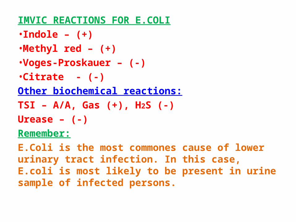
IMVIC REACTIONS FOR E.COLI•Indole – (+)•Methyl red – (+)•Voges-Proskauer – (-)•Citrate - (-)Other biochemical reactions:TSI – A/A, Gas (+), H2S (-)Urease – (-)Remember:E.Coli is the most commones cause of lower urinary tract infection. In this case, E.coli is most likely to be present in urine sample of infected persons.
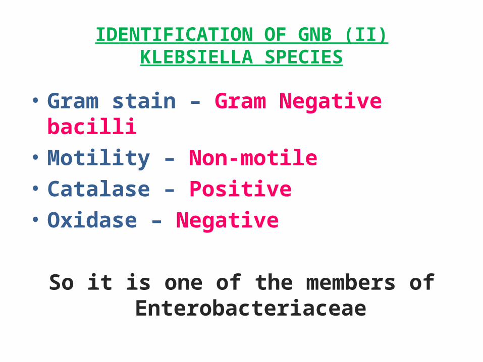
IDENTIFICATION OF GNB (II)KLEBSIELLA SPECIES
• Gram stain – Gram Negative bacilli • Motility – Non-motile • Catalase – Positive • Oxidase – Negative
So it is one of the members of Enterobacteriaceae
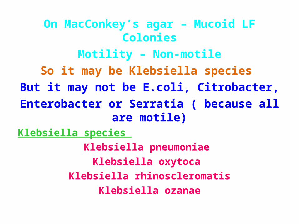
On MacConkey’s agar – Mucoid LF ColoniesMotility – Non-motile
So it may be Klebsiella species But it may not be E.coli, Citrobacter,
Enterobacter or Serratia ( because all are motile)
Klebsiella species Klebsiella pneumoniae
Klebsiella oxytoca Klebsiella rhinoscleromatis
Klebsiella ozanae
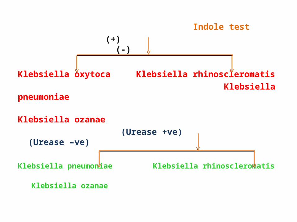
Indole test (+) (-)
Klebsiella oxytoca Klebsiella rhinoscleromatis Klebsiella pneumoniae Klebsiella ozanae (Urease +ve) (Urease –ve)
Klebsiella pneumoniae Klebsiella rhinoscleromatis Klebsiella ozanae
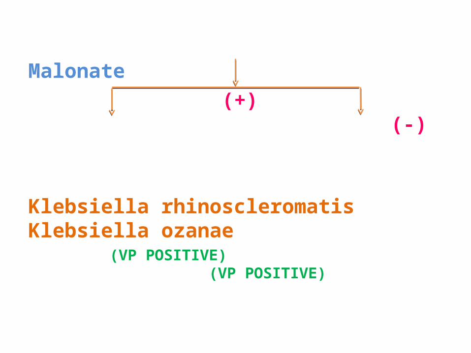
Malonate (+) (-)
Klebsiella rhinoscleromatis Klebsiella ozanae (VP POSITIVE) (VP POSITIVE)
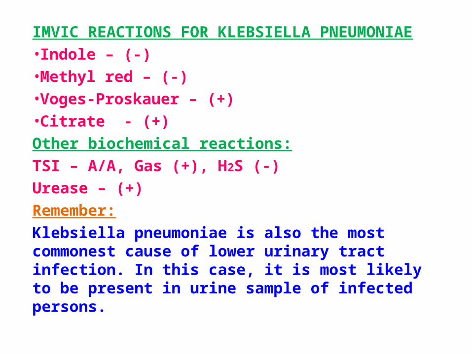
IMVIC REACTIONS FOR KLEBSIELLA PNEUMONIAE•Indole – (-)•Methyl red – (-)•Voges-Proskauer – (+)•Citrate - (+)Other biochemical reactions:TSI – A/A, Gas (+), H2S (-)Urease – (+)Remember:Klebsiella pneumoniae is also the most commonest cause of lower urinary tract infection. In this case, it is most likely to be present in urine sample of infected persons.
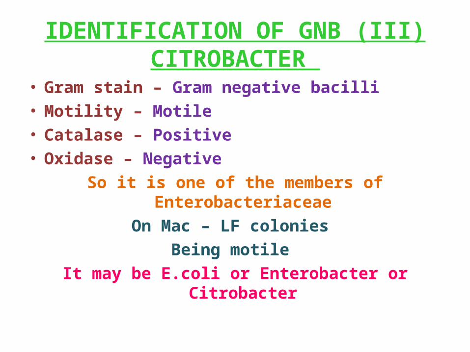
IDENTIFICATION OF GNB (III)CITROBACTER
• Gram stain – Gram negative bacilli• Motility – Motile • Catalase – Positive • Oxidase – Negative
So it is one of the members of EnterobacteriaceaeOn Mac – LF colonies
Being motile It may be E.coli or Enterobacter or Citrobacter
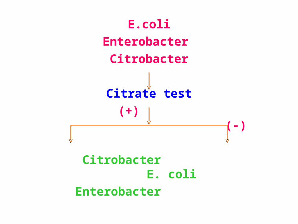
E.coliEnterobacter
Citrobacter
Citrate test (+) (-) Citrobacter E. coli Enterobacter
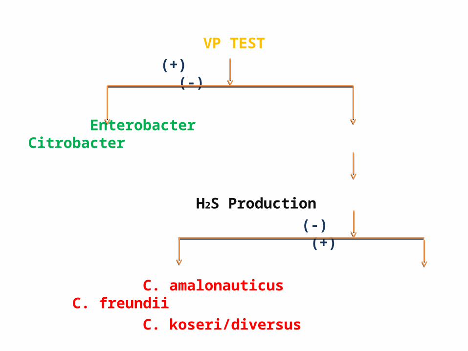
VP TEST (+) (-)
Enterobacter Citrobacter
H2S Production (-) (+)
C. amalonauticus C. freundii C. koseri/diversus
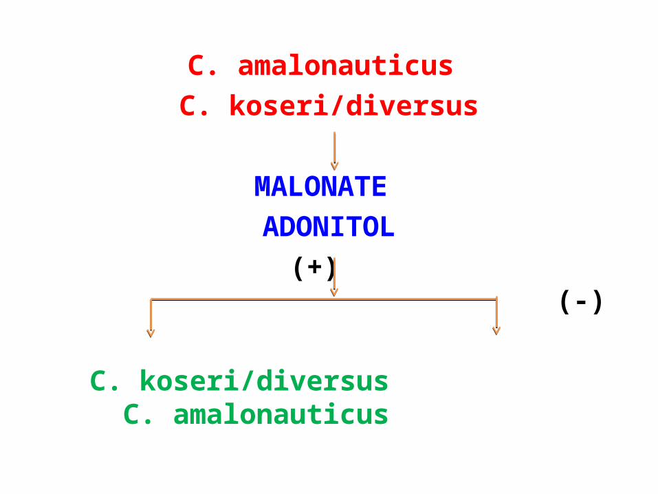
C. amalonauticus C. koseri/diversus
MALONATE ADONITOL
(+) (-)
C. koseri/diversus C. amalonauticus
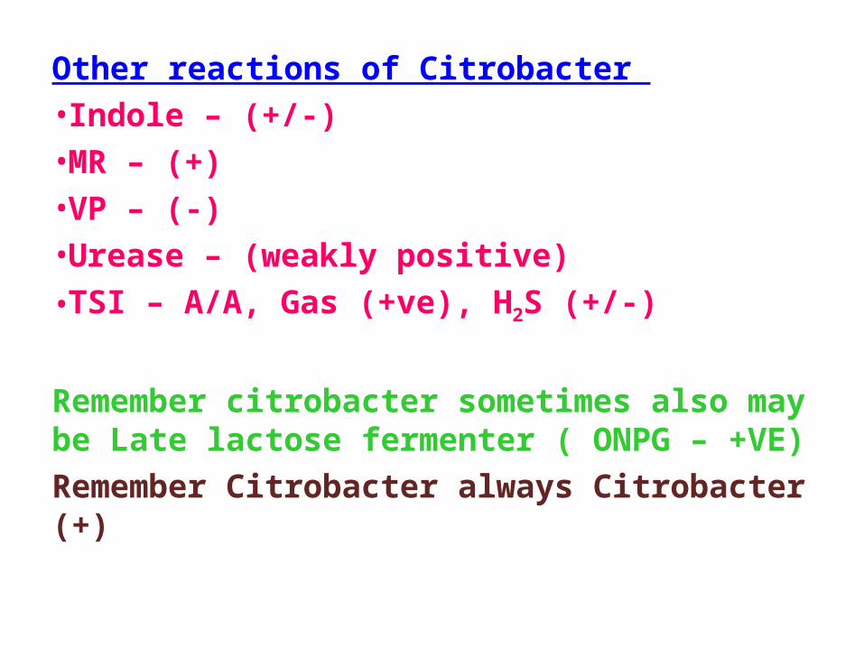
Other reactions of Citrobacter •Indole – (+/-)•MR – (+)•VP – (-)•Urease – (weakly positive)•TSI – A/A, Gas (+ve), H2S (+/-)
Remember citrobacter sometimes also may be Late lactose fermenter ( ONPG – +VE)Remember Citrobacter always Citrobacter (+)
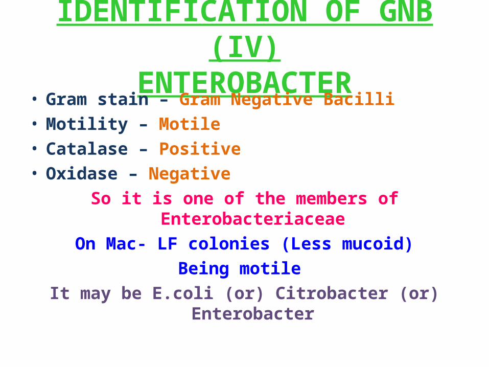
IDENTIFICATION OF GNB (IV)ENTEROBACTER
• Gram stain – Gram Negative Bacilli • Motility – Motile • Catalase – Positive • Oxidase – Negative
So it is one of the members of EnterobacteriaceaeOn Mac- LF colonies (Less mucoid)
Being motile It may be E.coli (or) Citrobacter (or) Enterobacter
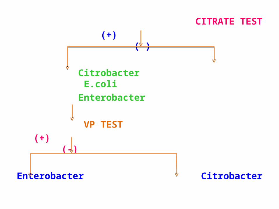
CITRATE TEST (+) (-)
Citrobacter E.coli Enterobacter
VP TEST (+) (-)
Enterobacter Citrobacter
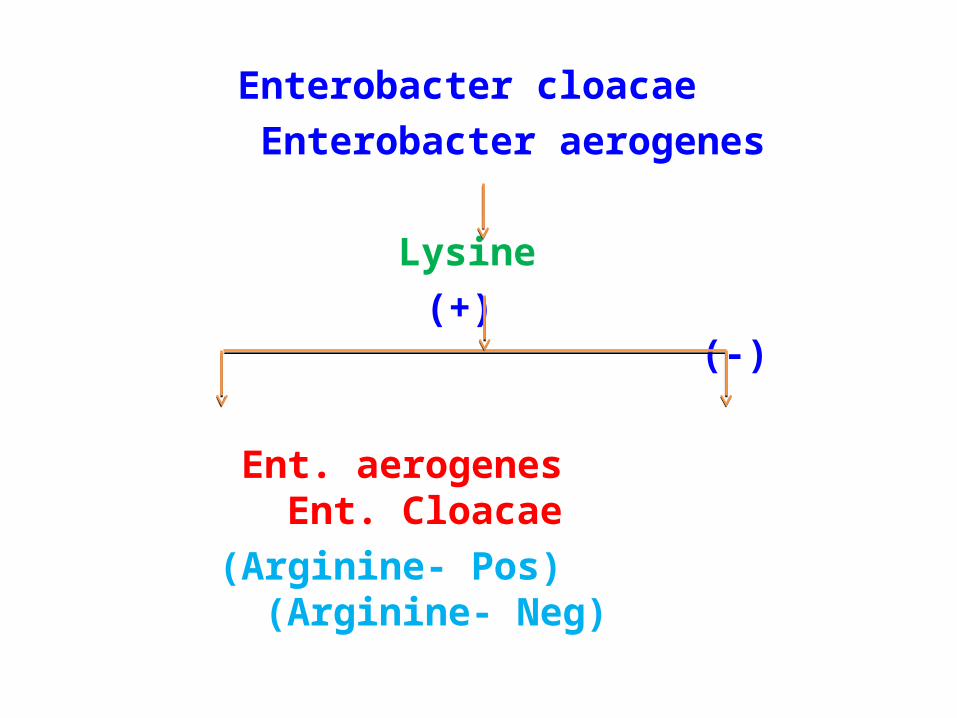
Enterobacter cloacae Enterobacter aerogenes
Lysine (+) (-)
Ent. aerogenes Ent. Cloacae (Arginine- Pos) (Arginine- Neg)
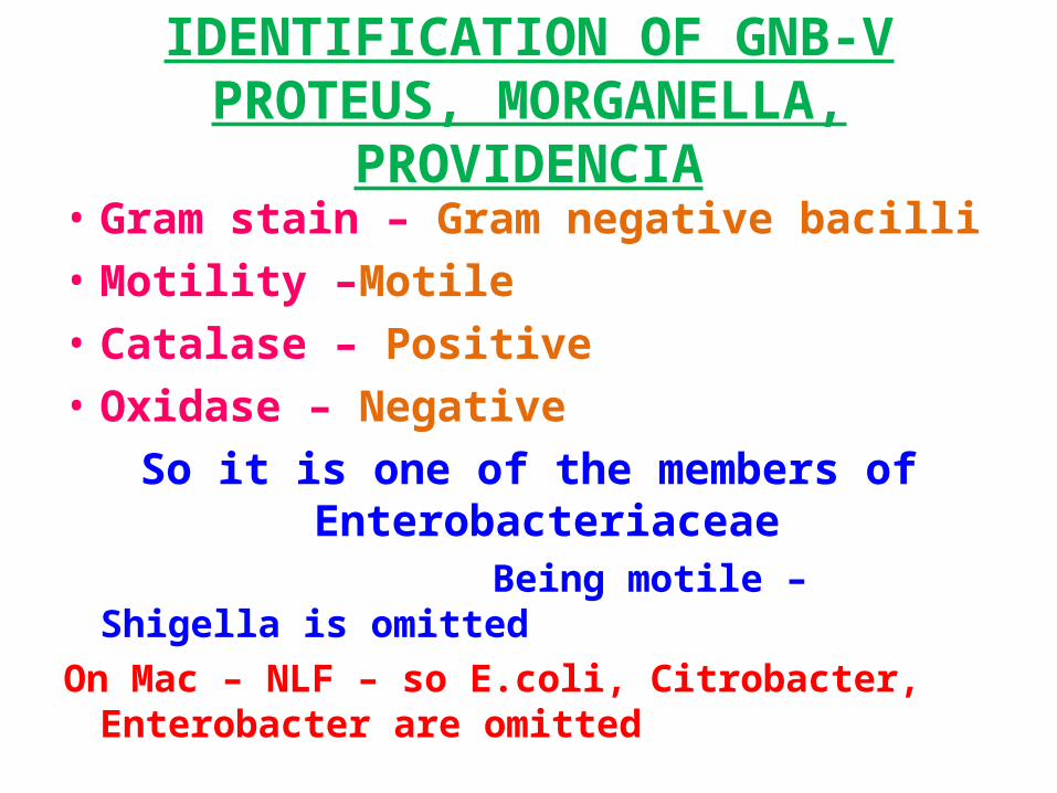
IDENTIFICATION OF GNB-VPROTEUS, MORGANELLA, PROVIDENCIA• Gram stain – Gram negative bacilli • Motility –Motile • Catalase – Positive • Oxidase – Negative
So it is one of the members of Enterobacteriaceae
Being motile – Shigella is omitted On Mac – NLF – so E.coli, Citrobacter, Enterobacter
are omitted
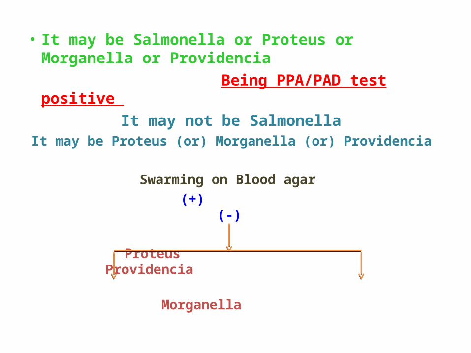
• It may be Salmonella or Proteus or Morganella or Providencia
Being PPA/PAD test positive It may not be Salmonella
It may be Proteus (or) Morganella (or) Providencia
Swarming on Blood agar (+) (-)
Proteus Providencia Morganella
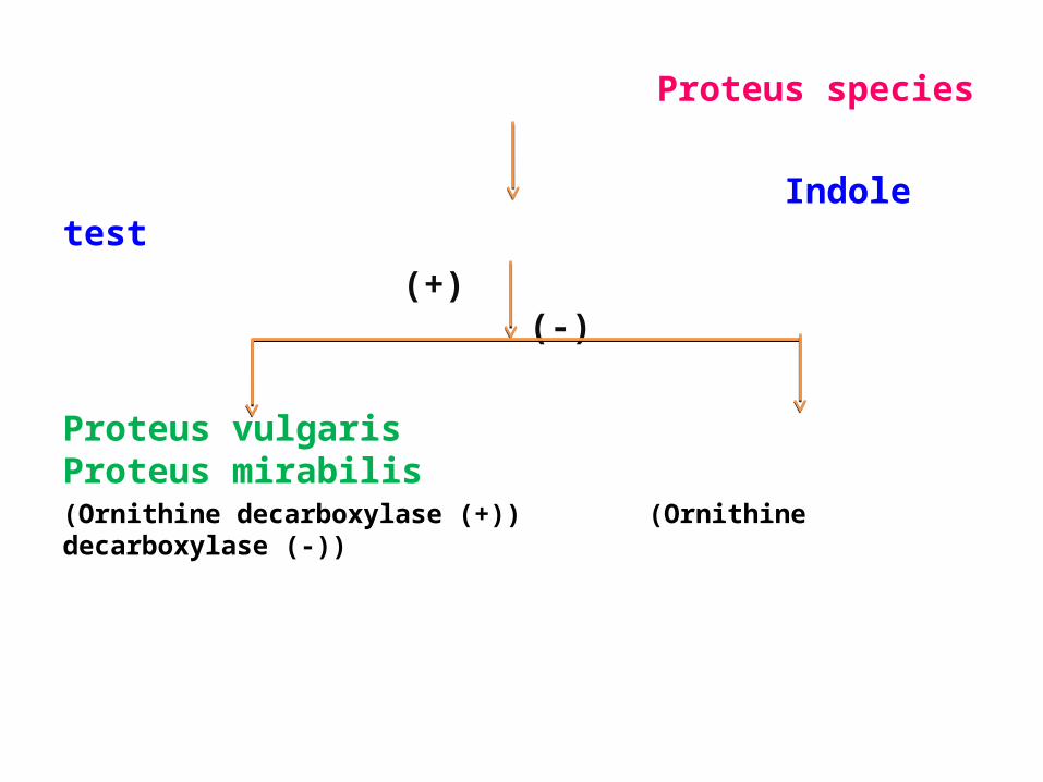
Proteus species
Indole test (+) (-)
Proteus vulgaris Proteus mirabilis(Ornithine decarboxylase (+)) (Ornithine decarboxylase (-))
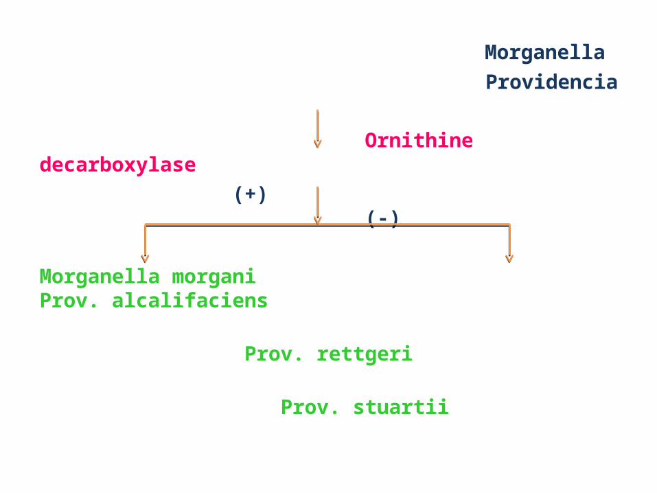
Morganella Providencia
Ornithine decarboxylase (+) (-)
Morganella morgani Prov. alcalifaciens Prov. rettgeri Prov. stuartii
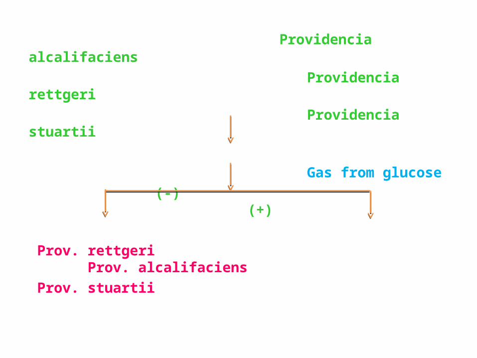
Providencia alcalifaciens Providencia rettgeri Providencia stuartii
Gas from glucose (-) (+)
Prov. rettgeri Prov. alcalifaciens Prov. stuartii
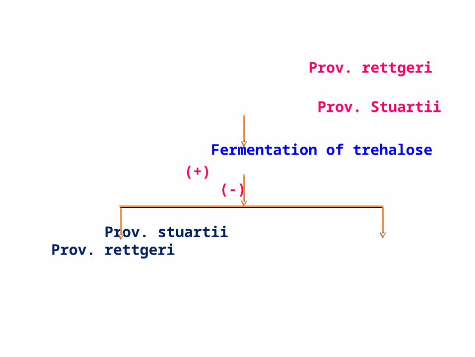
Prov. rettgeri Prov. Stuartii
Fermentation of trehalose (+) (-)
Prov. stuartii Prov. rettgeri
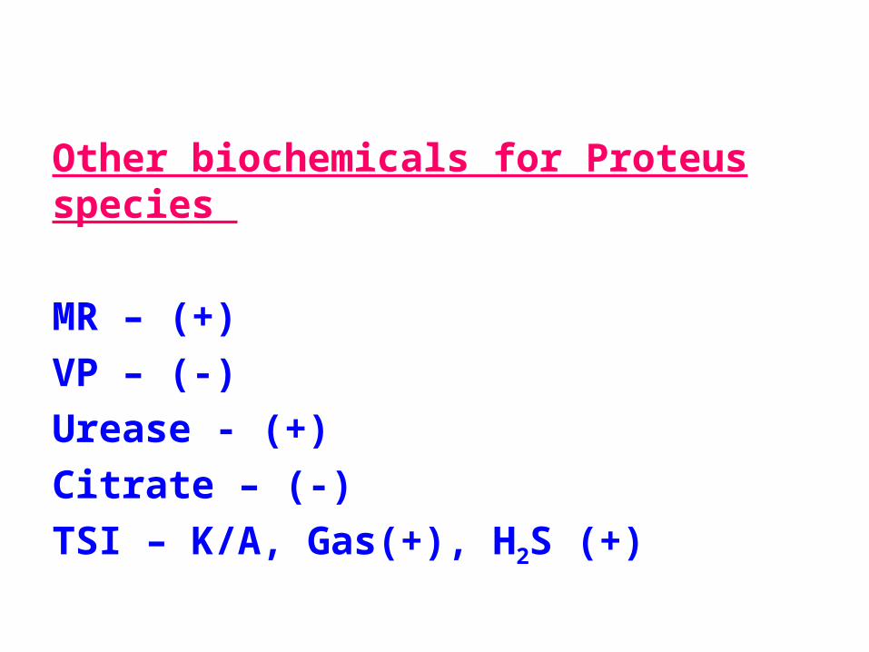
Other biochemicals for Proteus species
MR – (+)VP – (-)Urease - (+)Citrate – (-)TSI – K/A, Gas(+), H2S (+)
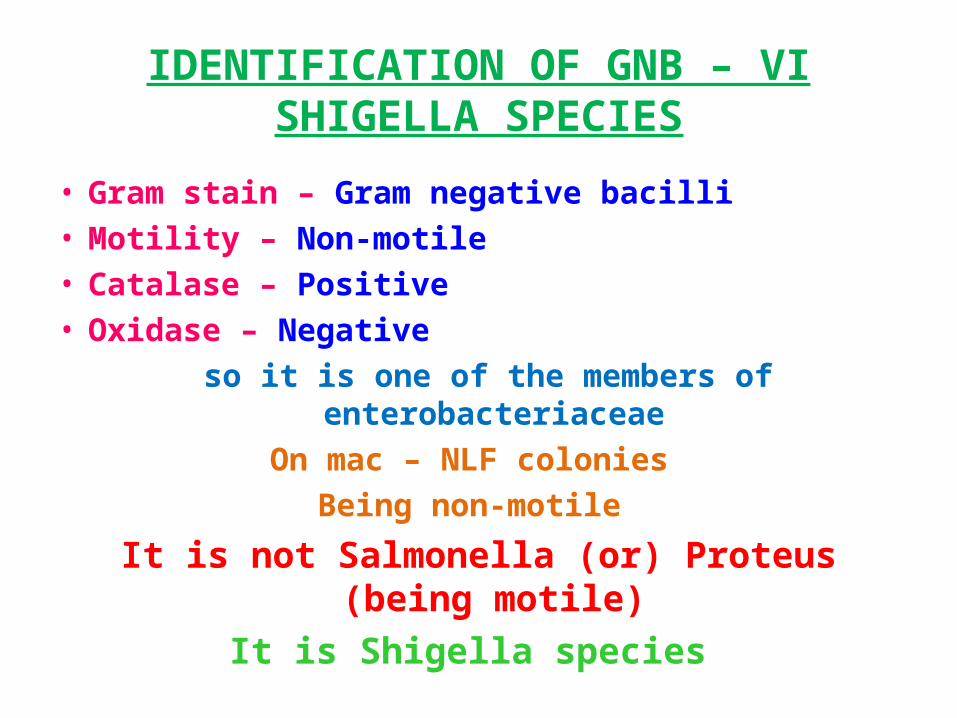
IDENTIFICATION OF GNB – VISHIGELLA SPECIES
• Gram stain – Gram negative bacilli • Motility – Non-motile • Catalase – Positive • Oxidase – Negative
so it is one of the members of enterobacteriaceaeOn mac – NLF colonies
Being non-motile
It is not Salmonella (or) Proteus (being motile)It is Shigella species
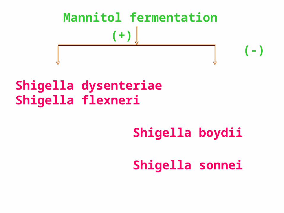
Mannitol fermentation (+) (-)
Shigella dysenteriae Shigella flexneri Shigella boydii Shigella sonnei
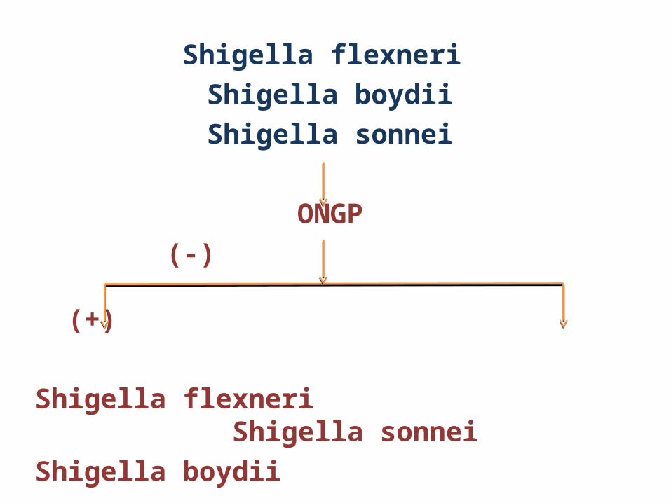
Shigella flexneri Shigella boydiiShigella sonnei
ONGP (-) (+)
Shigella flexneri Shigella sonneiShigella boydii
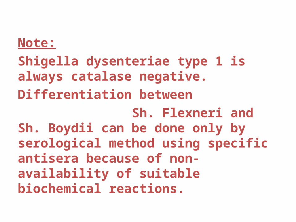
Note:Shigella dysenteriae type 1 is always catalase negative.Differentiation between Sh. Flexneri and Sh. Boydii can be done only by serological method using specific antisera because of non-availability of suitable biochemical reactions.
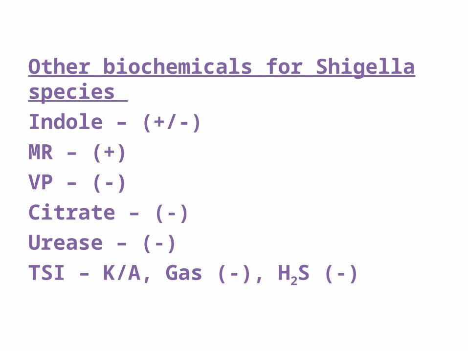
Other biochemicals for Shigella species Indole – (+/-)MR – (+)VP – (-)Citrate – (-)Urease – (-)TSI – K/A, Gas (-), H2S (-)
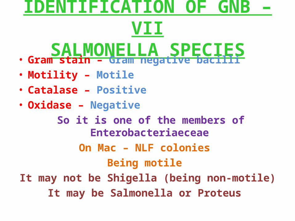
IDENTIFICATION OF GNB – VIISALMONELLA SPECIES
• Gram stain – Gram negative bacilli • Motility – Motile • Catalase – Positive • Oxidase – Negative
So it is one of the members of Enterobacteriaeceae On Mac – NLF colonies
Being motile It may not be Shigella (being non-motile)
It may be Salmonella or Proteus
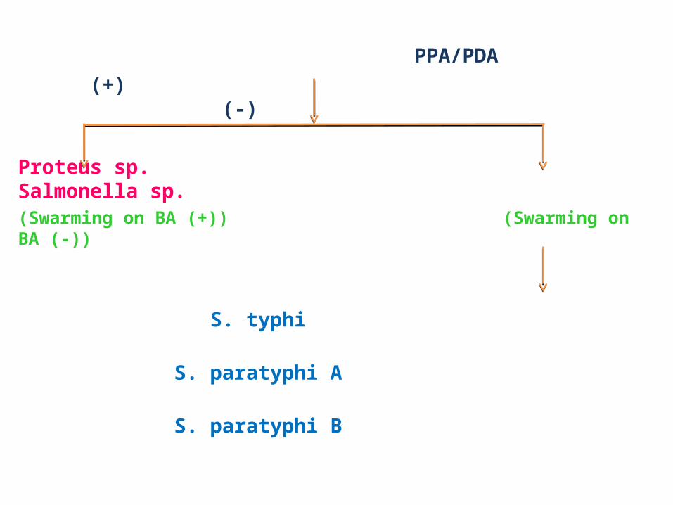
PPA/PDA (+) (-)
Proteus sp. Salmonella sp.(Swarming on BA (+)) (Swarming on BA (-))
S. typhi S. paratyphi A S. paratyphi B
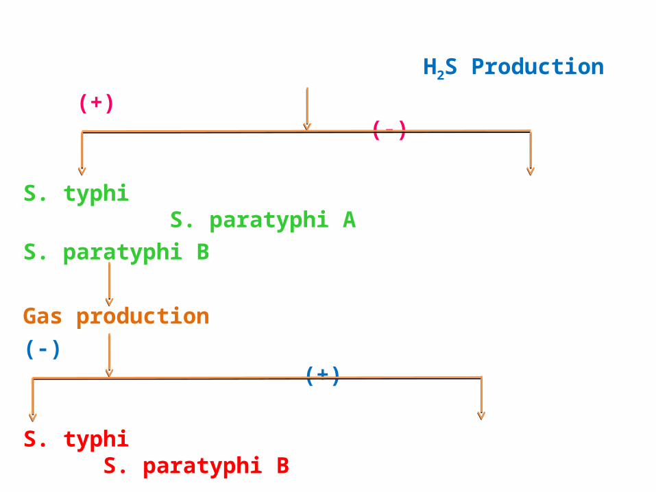
H2S Production
(+) (-)
S. typhi S. paratyphi AS. paratyphi B
Gas production (-) (+)
S. typhi S. paratyphi B
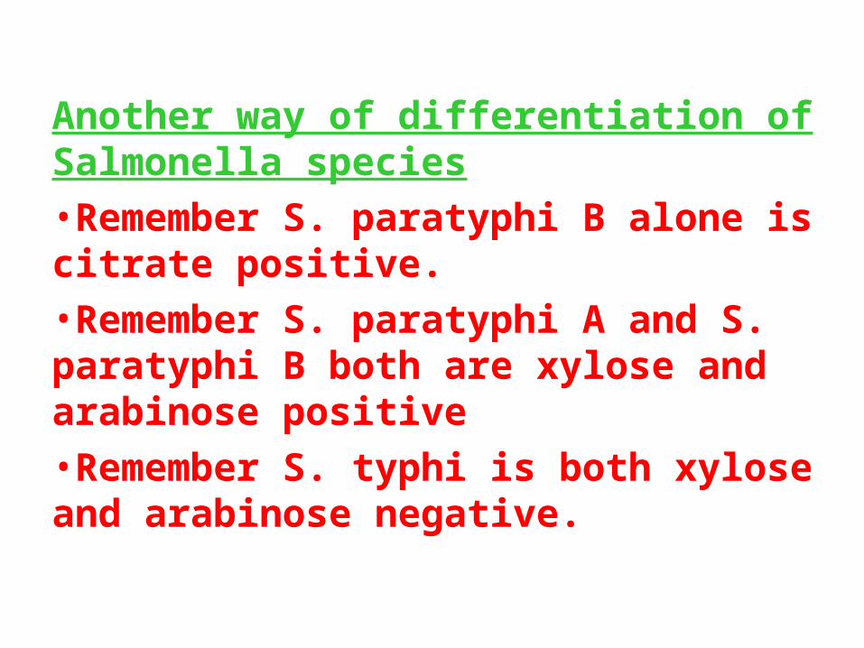
Another way of differentiation of Salmonella species•Remember S. paratyphi B alone is citrate positive.•Remember S. paratyphi A and S. paratyphi B both are xylose and arabinose positive •Remember S. typhi is both xylose and arabinose negative.
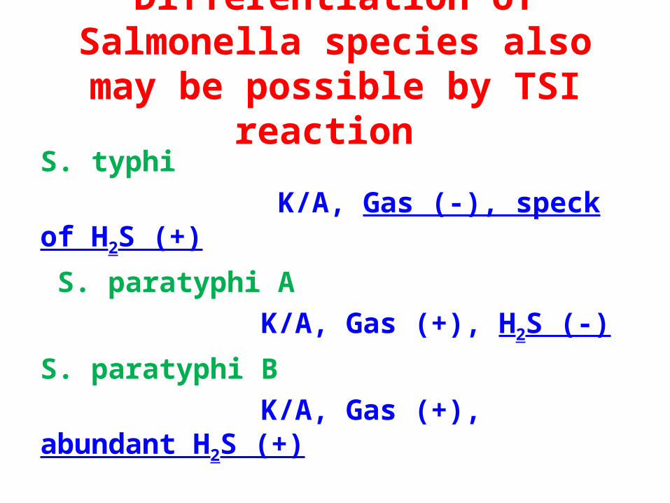
Differentiation of Salmonella species also may be possible by TSI reaction
S. typhi K/A, Gas (-), speck of H2S (+) S. paratyphi A K/A, Gas (+), H2S (-)S. paratyphi B K/A, Gas (+), abundant H2S (+)
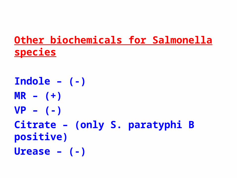
Other biochemicals for Salmonella species
Indole – (-)MR – (+)VP – (-)Citrate – (only S. paratyphi B positive)Urease – (-)
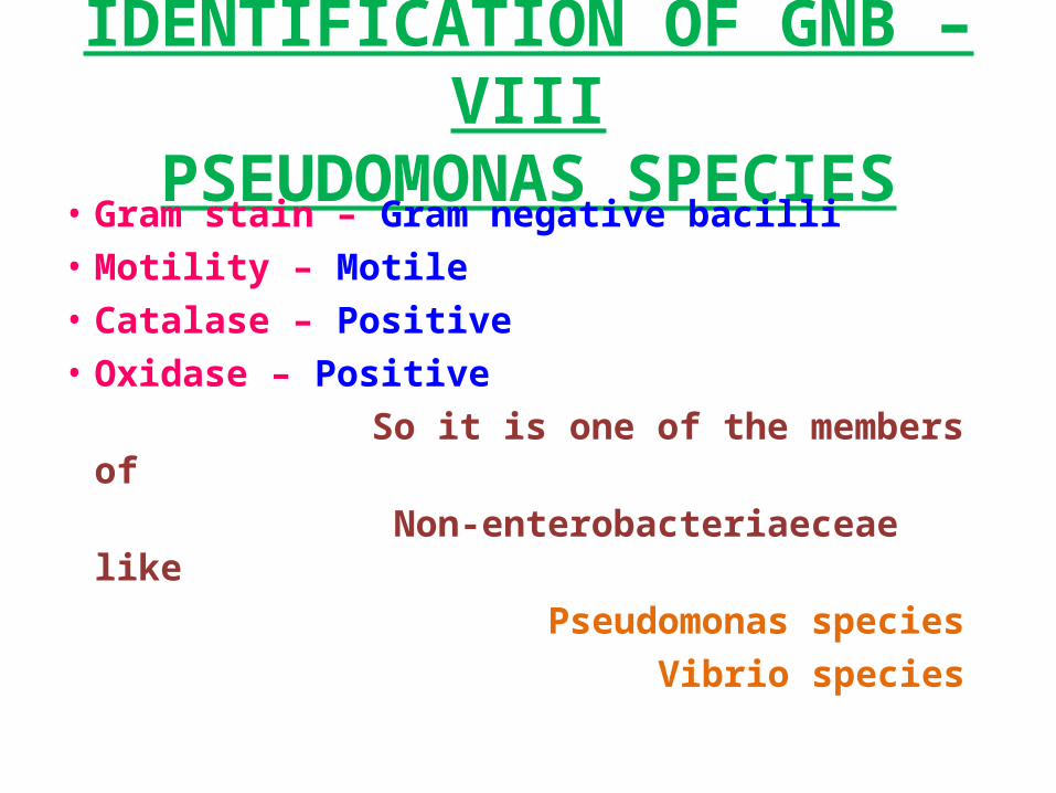
IDENTIFICATION OF GNB – VIIIPSEUDOMONAS SPECIES
• Gram stain – Gram negative bacilli • Motility – Motile • Catalase – Positive • Oxidase – Positive So it is one of the members of Non-enterobacteriaeceae like Pseudomonas species Vibrio species
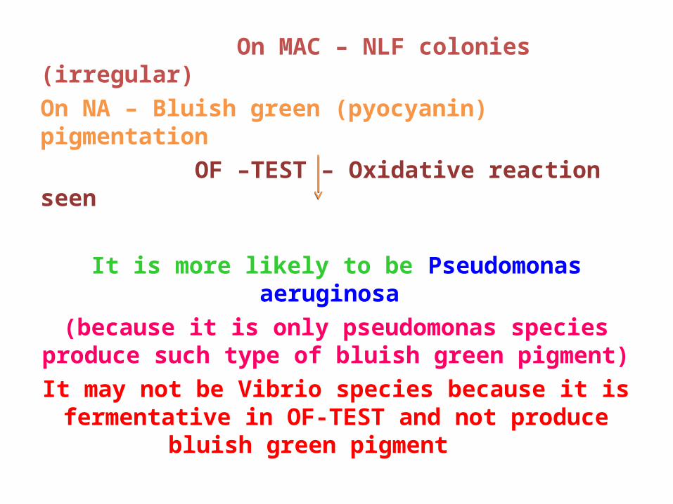
On MAC – NLF colonies (irregular)On NA – Bluish green (pyocyanin) pigmentation OF –TEST – Oxidative reaction seen It is more likely to be Pseudomonas aeruginosa
(because it is only pseudomonas species produce such type of bluish green pigment)
It may not be Vibrio species because it is fermentative in OF-TEST and not produce
bluish green pigment
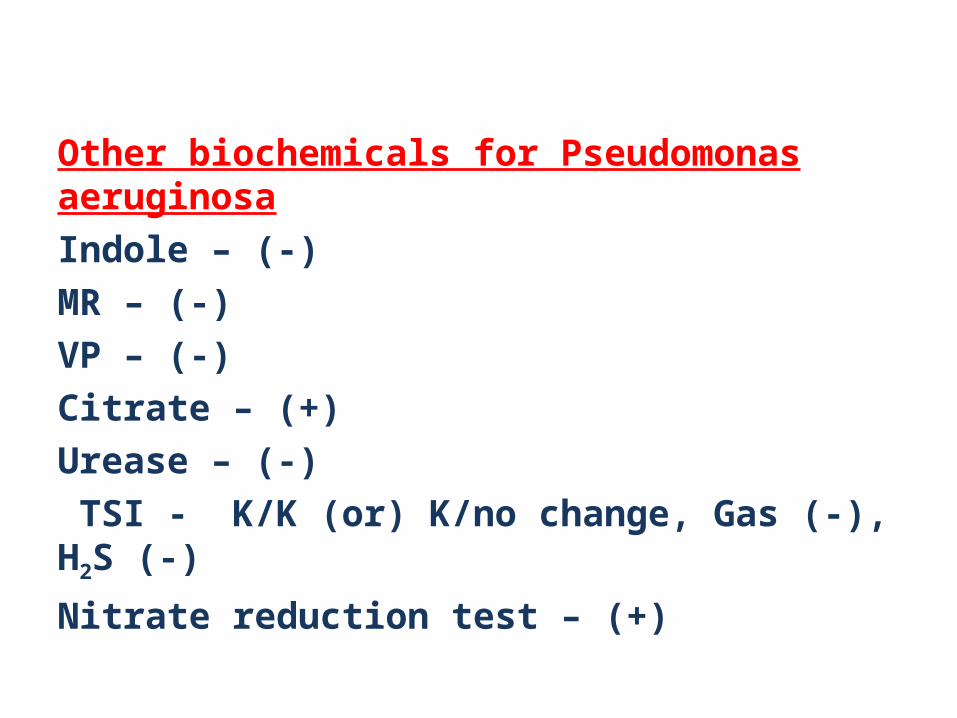
Other biochemicals for Pseudomonas aeruginosaIndole – (-)MR – (-)VP – (-)Citrate – (+)Urease – (-) TSI - K/K (or) K/no change, Gas (-), H2S (-)Nitrate reduction test – (+)
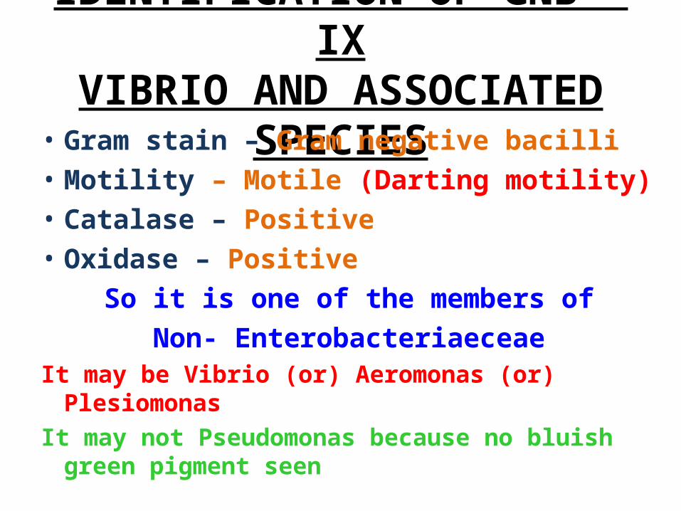
IDENTIFICATION OF GNB – IXVIBRIO AND ASSOCIATED SPECIES• Gram stain – Gram negative bacilli • Motility – Motile (Darting motility)• Catalase – Positive • Oxidase – Positive
So it is one of the members of Non- Enterobacteriaeceae
It may be Vibrio (or) Aeromonas (or) PlesiomonasIt may not Pseudomonas because no bluish green
pigment seen
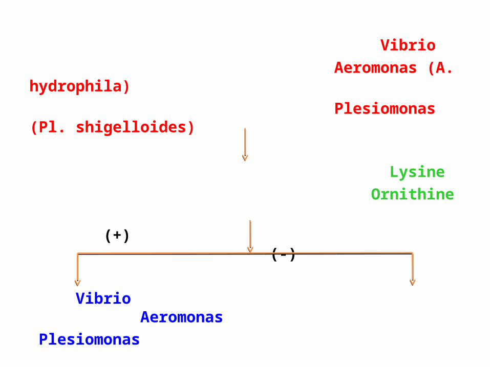
Vibrio Aeromonas (A. hydrophila) Plesiomonas (Pl. shigelloides)
Lysine Ornithine (+) (-)
Vibrio Aeromonas Plesiomonas
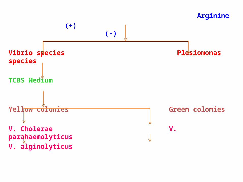
Arginine (+) (-)
Vibrio species Plesiomonas species
TCBS Medium
Yellow colonies Green colonies
V. Cholerae V. parahaemolyticusV. alginolyticus
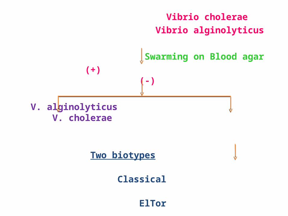
Vibrio cholerae Vibrio alginolyticus
Swarming on Blood agar (+) (-)
V. alginolyticus V. cholerae
Two biotypes Classical ElTor
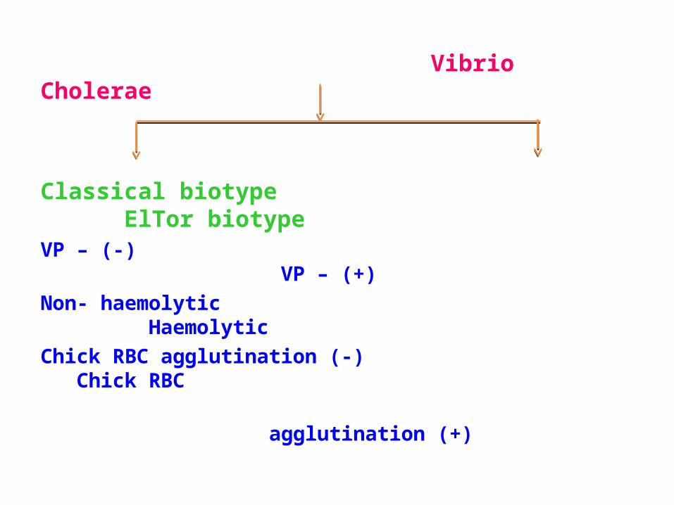
Vibrio Cholerae
Classical biotype ElTor biotypeVP – (-) VP – (+)Non- haemolytic Haemolytic Chick RBC agglutination (-) Chick RBC agglutination (+)
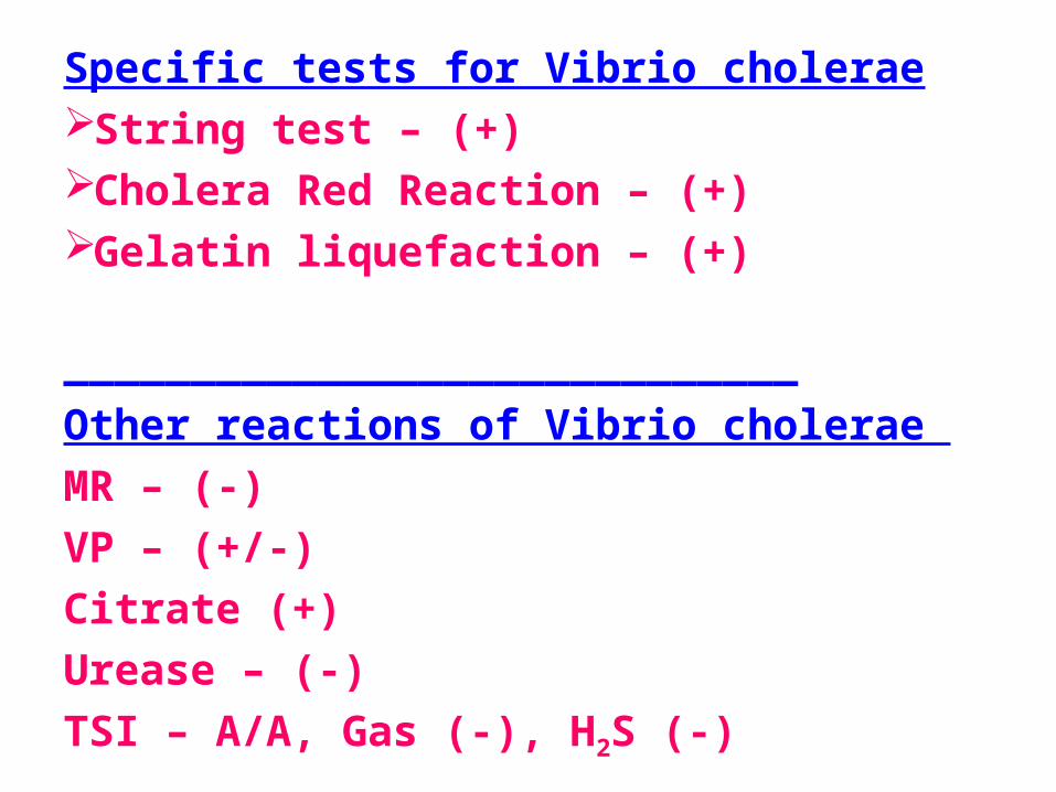
Specific tests for Vibrio choleraeString test – (+)Cholera Red Reaction – (+)Gelatin liquefaction – (+) _____________________________Other reactions of Vibrio cholerae MR – (-)VP – (+/-)Citrate (+)Urease – (-)TSI – A/A, Gas (-), H2S (-)
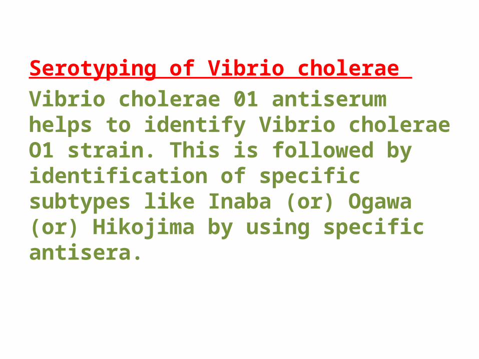
Serotyping of Vibrio cholerae Vibrio cholerae 01 antiserum helps to identify Vibrio cholerae O1 strain. This is followed by identification of specific subtypes like Inaba (or) Ogawa (or) Hikojima by using specific antisera.

• This presentation was created for those who are working as a laboratory technician in clinical microbiology diagnostics.
• Also may find useful for UG, PG, DMLT, PGDMLT in Microbiology.
Mail ID: [email protected]