Embed Size (px)
Citation preview
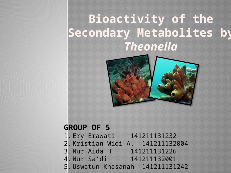
Bioactivity of the Secondary Metabolites by Theonella
GROUP OF 51. Ery Erawati 1412111312322. Kristian Widi A. 1412111320043. Nur Aida H. 1412111312264. Nur Sa’di 1412111320015. Uswatun Khasanah 141211131242
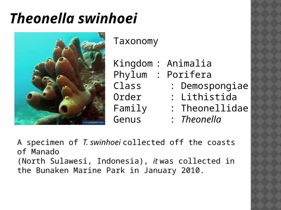
Theonella swinhoei
Taxonomy
Kingdom : AnimaliaPhylum : Porifera Class : DemospongiaeOrder : LithistidaFamily : TheonellidaeGenus : Theonella
A specimen of T. swinhoei collected off the coasts of Manado (North Sulawesi, Indonesia), it was collected in the Bunaken Marine Park in January 2010.
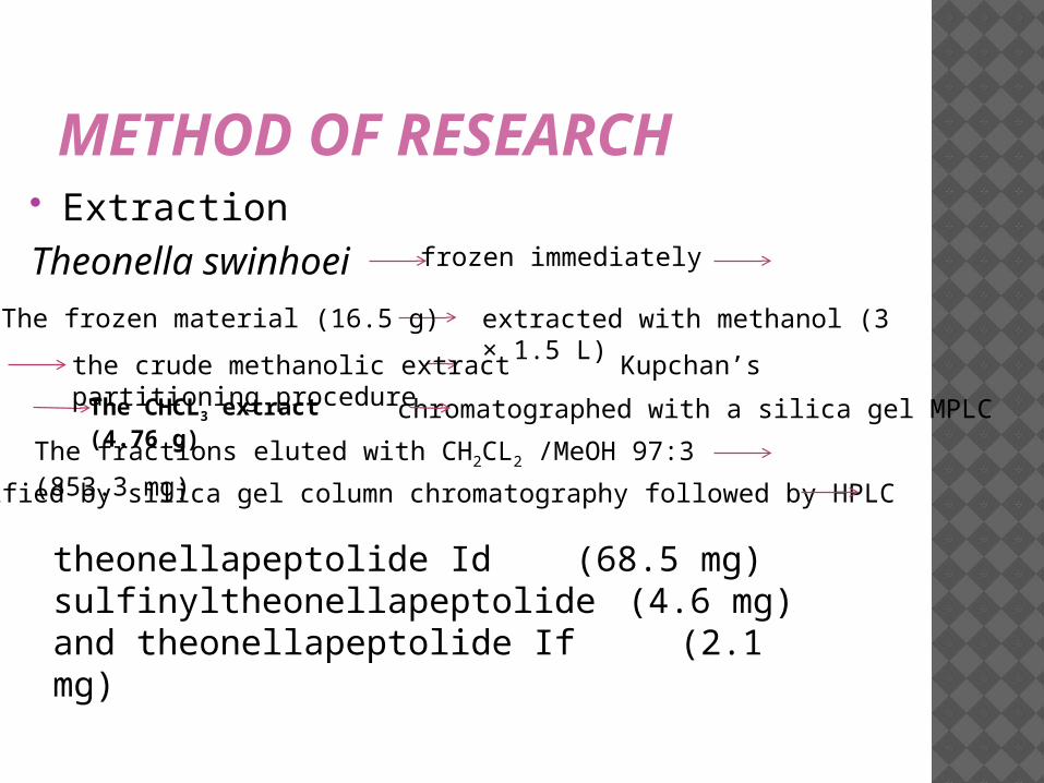
METHOD OF RESEARCH Extraction Theonella swinhoei frozen immediately
The frozen material (16.5 g) extracted with methanol (3 × 1.5 L)
the crude methanolic extract Kupchan’s partitioning procedure
The CHCL3 extract (4.76 g)
chromatographed with a silica gel MPLC
The fractions eluted with CH2CL2 /MeOH 97:3 (853.3 mg)purified by silica gel column chromatography followed by HPLC
theonellapeptolide Id (68.5 mg) sulfinyltheonellapeptolide (4.6 mg)and theonellapeptolide If (2.1 mg)
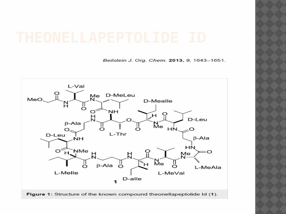
THEONELLAPEPTOLIDE ID
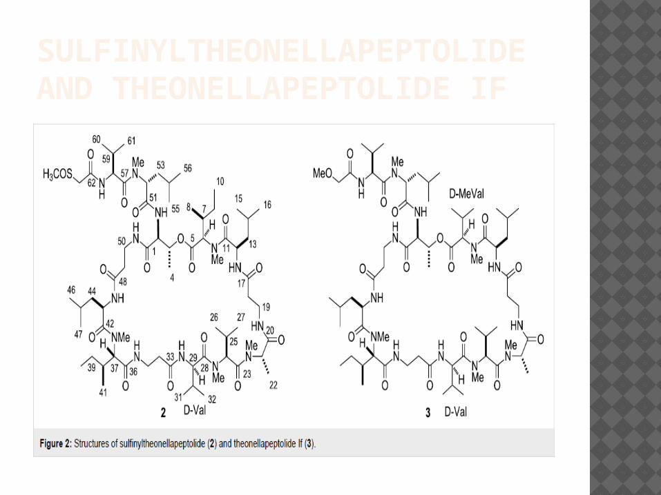
SULFINYLTHEONELLAPEPTOLIDE AND THEONELLAPEPTOLIDE IF
EVALUATION OF ANTIPROLIFERATIVE ACTIVITY
HepG2 (hepatic carcinoma cell line) cells were plated in a 24-wells plate at 3 × 10⁴ cells/well,
Minimum Essential Medium with Earl's 10% fetal bovine serum (FBS) + 1% L-glutamine + 1%+penicillin/streptomycintheonellapeptolide Id (0.1, 1 and 10 μM)Sulfinyltheonellapeptolide (0.1, 1 and 10 μM)theonellapeptolide If (0.1, 1 and 10 μM)
• 100 μL of MTT solution (5mg/mL)
• Added 1 mL of DMSO
• The absorbance was read by using a spectrophotometer at 590 nM.
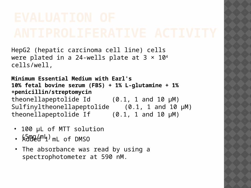
theonellapeptolide Id
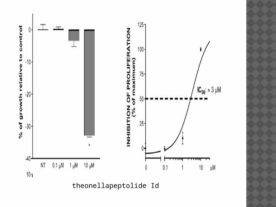
sulfinyltheonellapeptolide
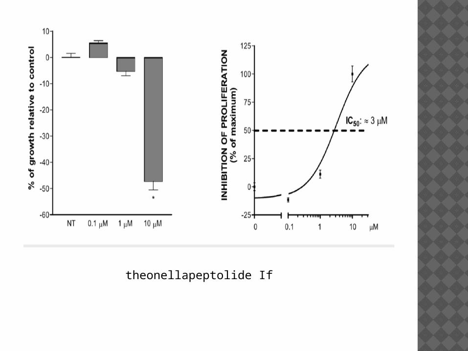
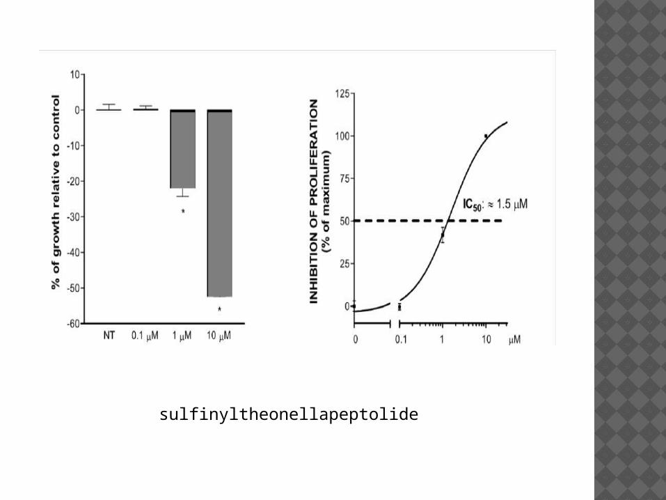
theonellapeptolide If
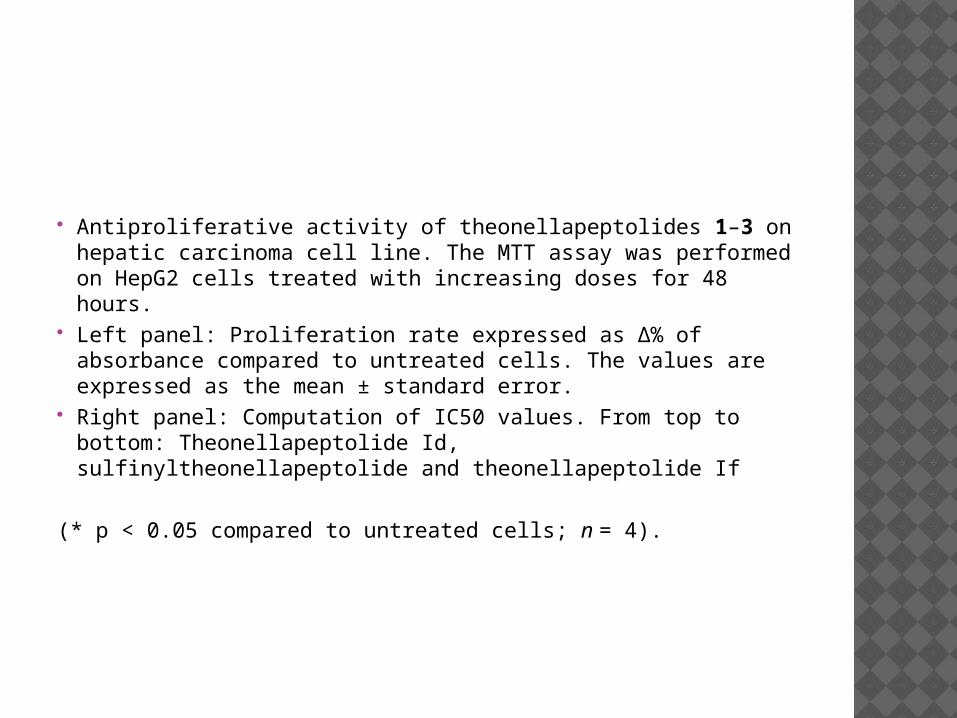
Antiproliferative activity of theonellapeptolides 1–3 on hepatic carcinoma cell line. The MTT assay was performed on HepG2 cells treated with increasing doses for 48 hours.
Left panel: Proliferation rate expressed as Δ% of absorbance compared to untreated cells. The values are expressed as the mean ± standard error.
Right panel: Computation of IC50 values. From top to bottom: Theonellapeptolide Id, sulfinyltheonellapeptolide and theonellapeptolide If
(* p < 0.05 compared to untreated cells; n = 4).
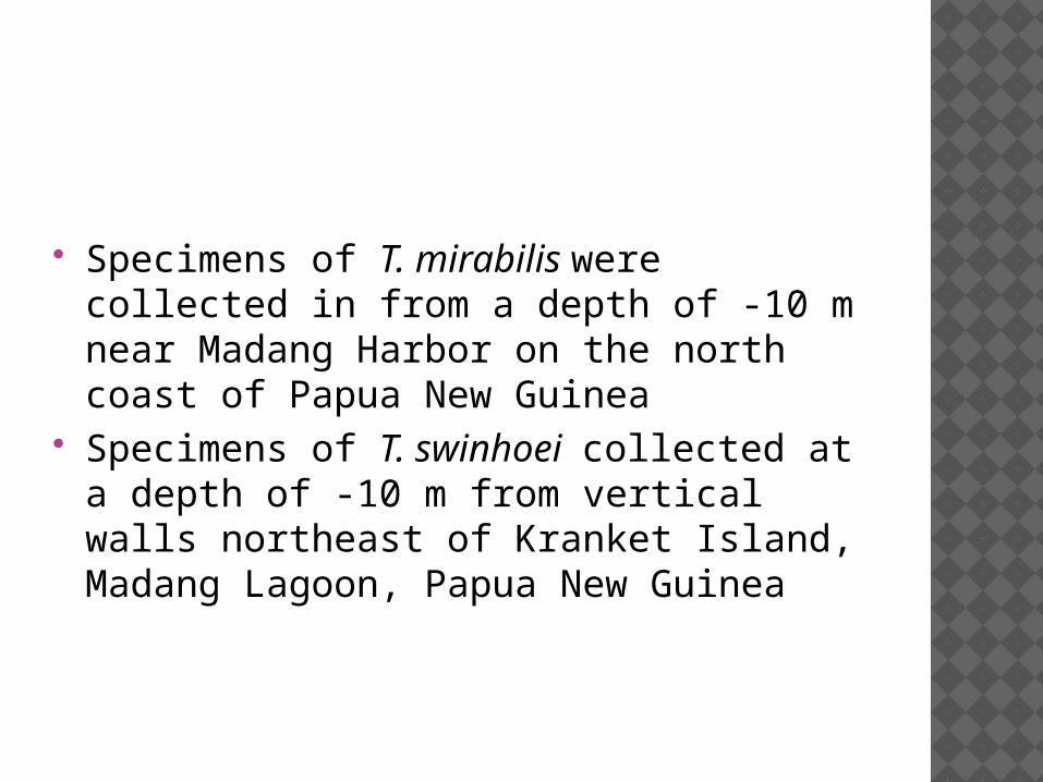
Specimens of T. mirabilis were collected in from a depth of -10 m near Madang Harbor on the north coast of Papua New Guinea
Specimens of T. swinhoei collected at a depth of -10 m from vertical walls northeast of Kranket Island, Madang Lagoon, Papua New Guinea
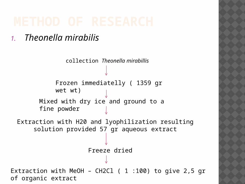
METHOD OF RESEARCH1. Theonella mirabilis
collection Theonella mirabillis
Frozen immediatelly ( 1359 gr wet wt)
Mixed with dry ice and ground to a fine powder
Extraction with H20 and lyophilization resulting solution provided 57 gr aqueous extract
Freeze dried
Extraction with MeOH – CH2Cl ( 1 :100) to give 2,5 gr of organic extract
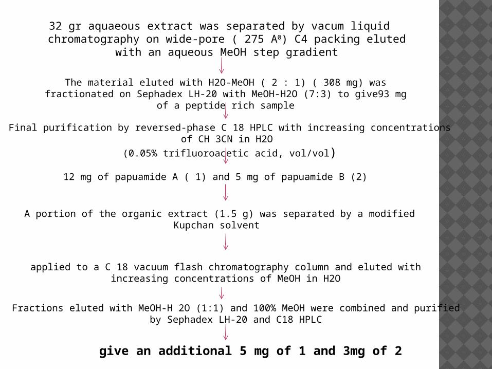
32 gr aquaeous extract was separated by vacum liquid chromatography on wide-pore ( 275 A0) C4 packing eluted with an
aqueous MeOH step gradient
The material eluted with H2O-MeOH ( 2 : 1) ( 308 mg) was fractionated on Sephadex LH-20 with MeOH-H2O (7:3) to give93 mg of a peptide rich
sample
Final purification by reversed-phase C 18 HPLC with increasing concentrations of CH 3CN in H2O
(0.05% trifluoroacetic acid, vol/vol)
12 mg of papuamide A ( 1) and 5 mg of papuamide B (2)
A portion of the organic extract (1.5 g) was separated by a modified Kupchan solvent
applied to a C 18 vacuum flash chromatography column and eluted with increasing concentrations of MeOH in H2O
Fractions eluted with MeOH-H 2O (1:1) and 100% MeOH were combined and purified by Sephadex LH-20 and C18 HPLC
give an additional 5 mg of 1 and 3mg of 2
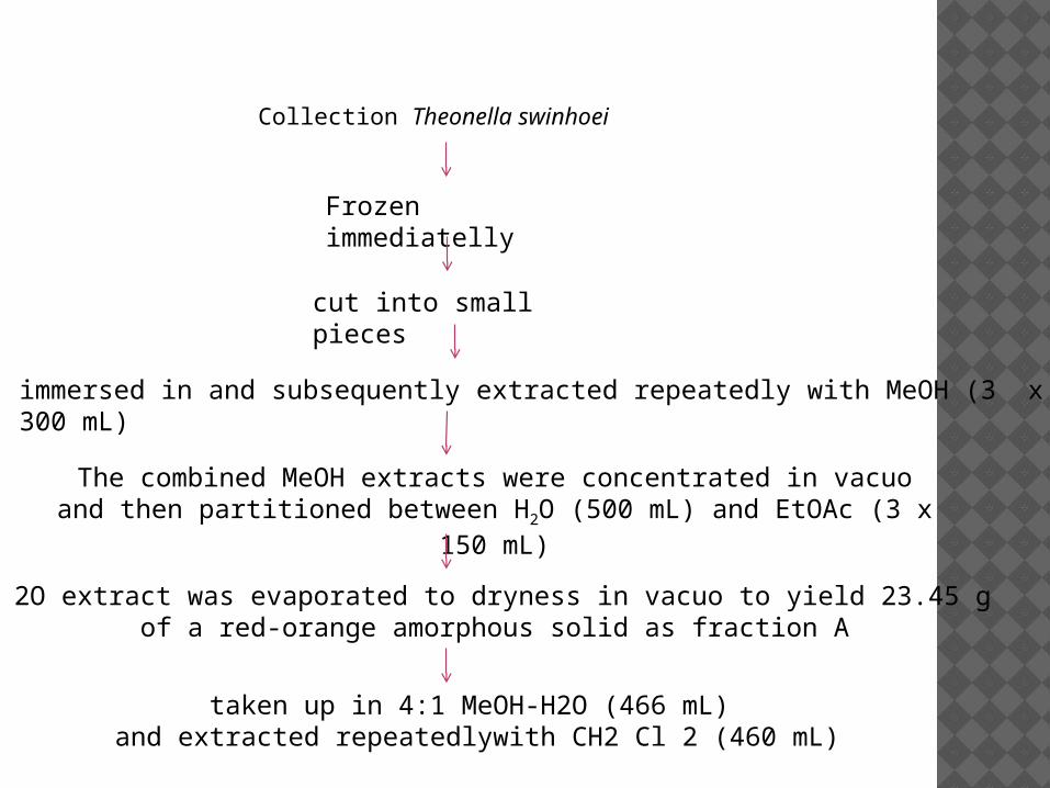
Collection Theonella swinhoei
Frozen immediatelly
cut into small pieces
immersed in and subsequently extracted repeatedly with MeOH (3 x 300 mL)
The combined MeOH extracts were concentrated in vacuo and then partitioned between H2O (500 mL) and EtOAc (3 x 150 mL)
H 2O extract was evaporated to dryness in vacuo to yield 23.45 g of a red-orange amorphous solid as fraction A
taken up in 4:1 MeOH-H2O (466 mL) and extracted repeatedlywith CH2 Cl 2 (460 mL)
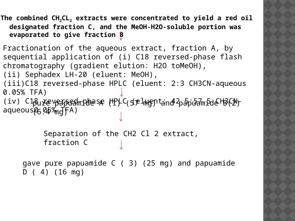
The combined CH2CL2 extracts were concentrated to yield a red oil designated fraction C, and the MeOH-H2O-soluble portion was evaporated to give fraction B
Fractionation of the aqueous extract, fraction A, by sequential application of (i) C18 reversed-phase flash chromatography (gradient elution: H2O toMeOH),(ii) Sephadex LH-20 (eluent: MeOH),(iii)C18 reversed-phase HPLC (eluent: 2:3 CH3CN-aqueous 0.05% TFA)(iv) C18 reversed-phase HPLC (eluent: 42.5:57.5 CH3CN-aqueous0.05% TFA)
pure papuamide A (1) (57 mg) and papuamide B(2) (6.4 mg)
Separation of the CH2 Cl 2 extract, fraction C
gave pure papuamide C ( 3) (25 mg) and papuamide D ( 4) (16 mg)
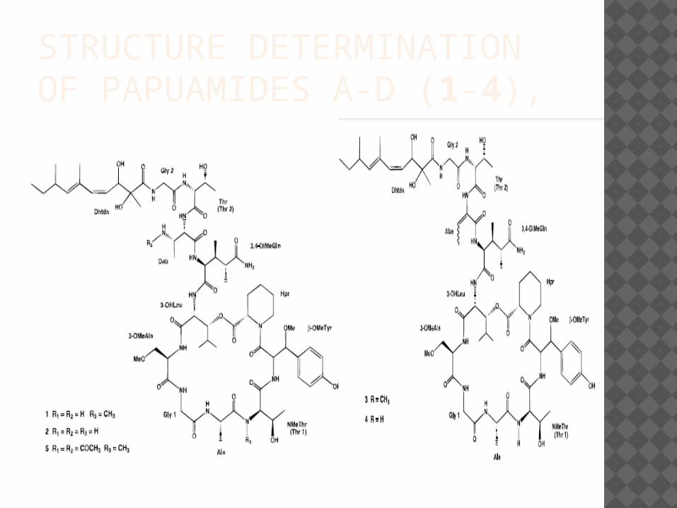
STRUCTURE DETERMINATION OF PAPUAMIDES A-D (1-4),
Papuamides A-D (1-4) are also the first marine-derived peptides reported to contain 3-hydroxyleucine and homoproline residues. These peptides also contain a previously undescribed 2,3-dihydroxy-2,6,8-trimethyldeca-(4 Z,6 E)-dienoic acid moiety N-linked to a terminal glycine residue.
Papuamides A (1) and B(2) inhibited the infection of human T-lymphoblastoid cells by HIV-1 RF in vitro with an EC 50 of approximately 4 ng/mL. Compound 1 was also cytotoxic against a panel of human cancer cell lines with a mean IC50 of 75 ng/mL.

hatur nuhun