Embed Size (px)
Citation preview

Ischaemic Stroke
Jessica Peter Roscoe
Contents...
➢Brief introduction
➢Case study
➢ Imaging
➢Radiological signs
➢Treatment options

Introduction
➢Focal neurological deficit
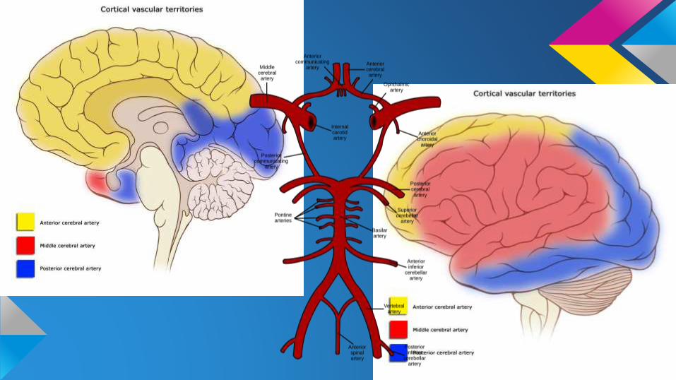
➢ Ischaemic V. Haemorrhagic
➢Modifiable risk factors
➢Symptoms depend on the area of the
brain affected
Case Study
❏ 79yo female
❏ Left sided weakness
❏ Post trauma
❏ Past history MCA territory stroke
❏Rule out: haemorrhagic stroke, ischaemic
stroke, traumatic haemorrhage
❏ 2x CTs performed
Stroke Imaging
➢What imaging is appropriate?
➢How does the imaging contribute to the
diagnosis?
➢How does the imaging contribute to the
management?
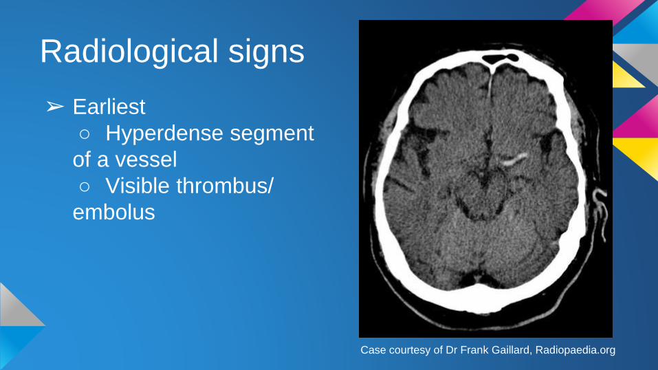
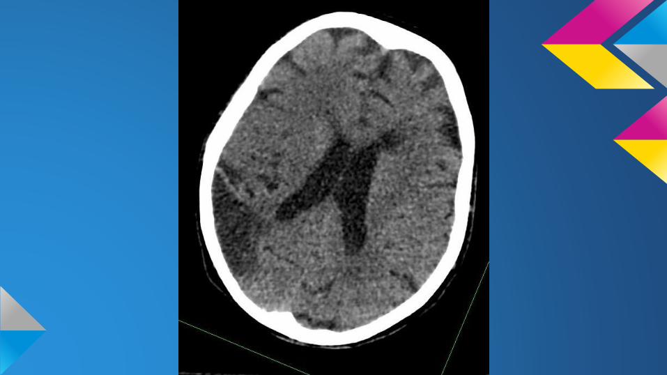
Radiological signs
➢ Earliest
○ Hyperdense segment
of a vessel
○ Visible thrombus/
embolus
Case courtesy of Dr Frank Gaillard, Radiopaedia.org
Radiological signs
➢Hyperacute○ Loss of grey-white matter differentiation
○ Cortical hypodensity
○ Dependent upon the occlusion location and
whether collaterals are present
Radiological signs
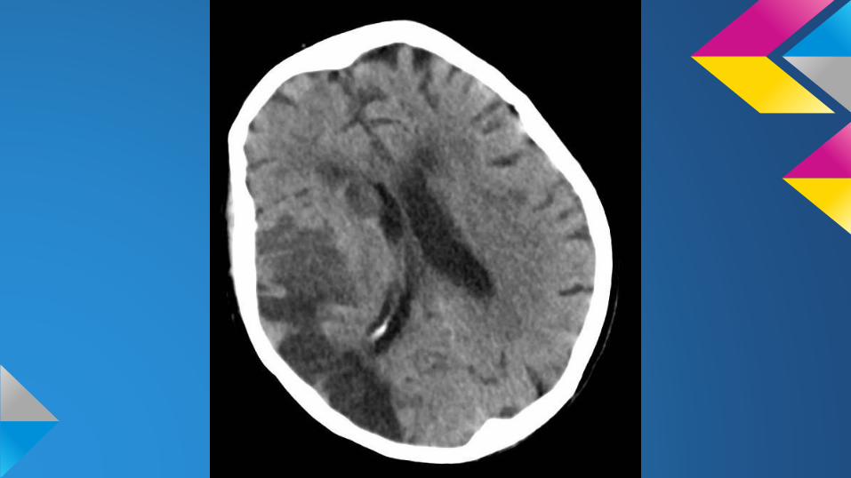
➢Week one○ Marked hypo-attenuation and swelling
Radiological signs
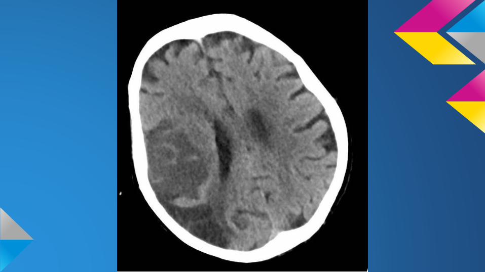
➢Week two/three○ CT fogging - decrease in swelling and
increased attenuation of the cortex
○ Confusing to interpret, can lead to a missed
diagnosis
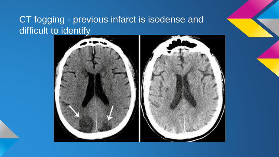
CT fogging - previous infarct is isodense and
difficult to identify
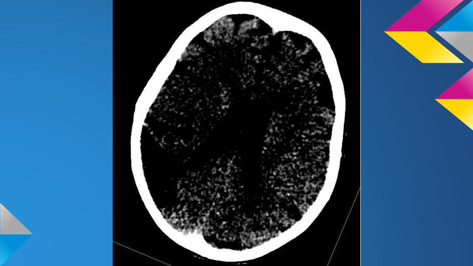
Radiological signs
➢Months later○ Swelling continues to reduce
○ Region of low density
Treating an ischaemic stroke
➢Rapidly restore cerebral blood flow○ Thrombolysis
○ Thrombectomy
➢Rehabilitation

Thank You!
Any Questions?