Embed Size (px)
Citation preview
SPINAL MALFORMATIONS
Dr. Soe Moe AungRadiology PG Student
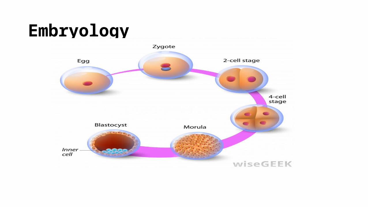
Embryology
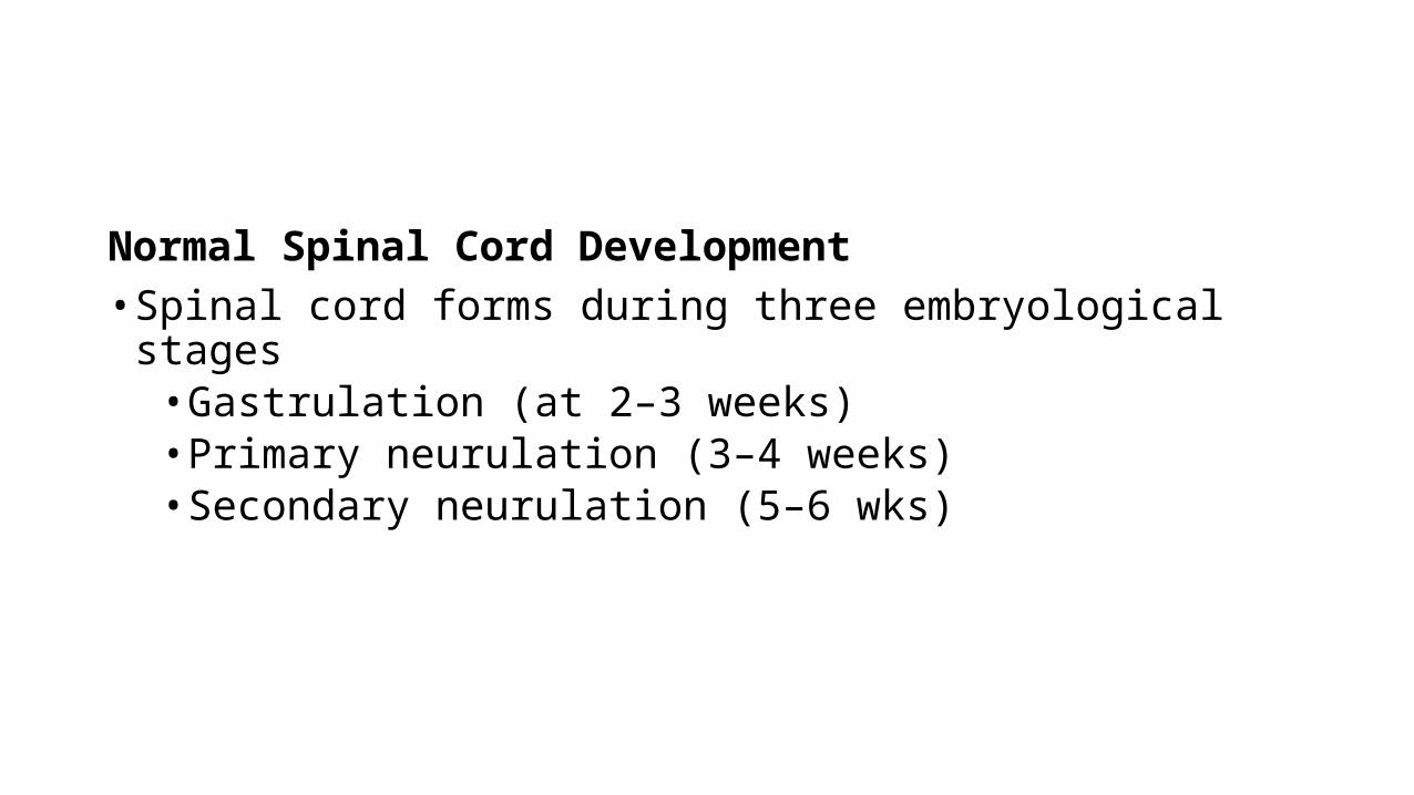
Normal Spinal Cord Development• Spinal cord forms during three embryological stages• Gastrulation (at 2–3 weeks)• Primary neurulation (3–4 weeks) • Secondary neurulation (5–6 wks)
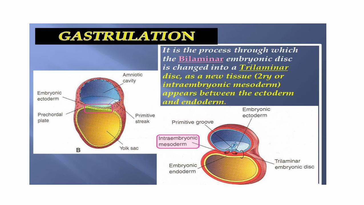

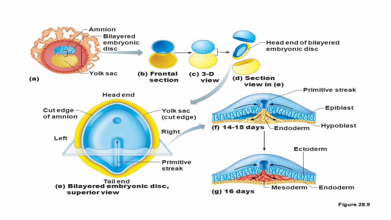
Gastrulation • The embryonic bilaminar disc consists of epiblast (future ectoderm)
and hypoblast (primitive endoderm)• It is converted to a trilaminar disc by migration of cells from the
epiblast through Hensen’s node, a focal region of thickening occurring at the cranial end of the midline ‘primitive streak’ of the disc. • This results in the midline notochord and a layer that will form the
future mesoderm.
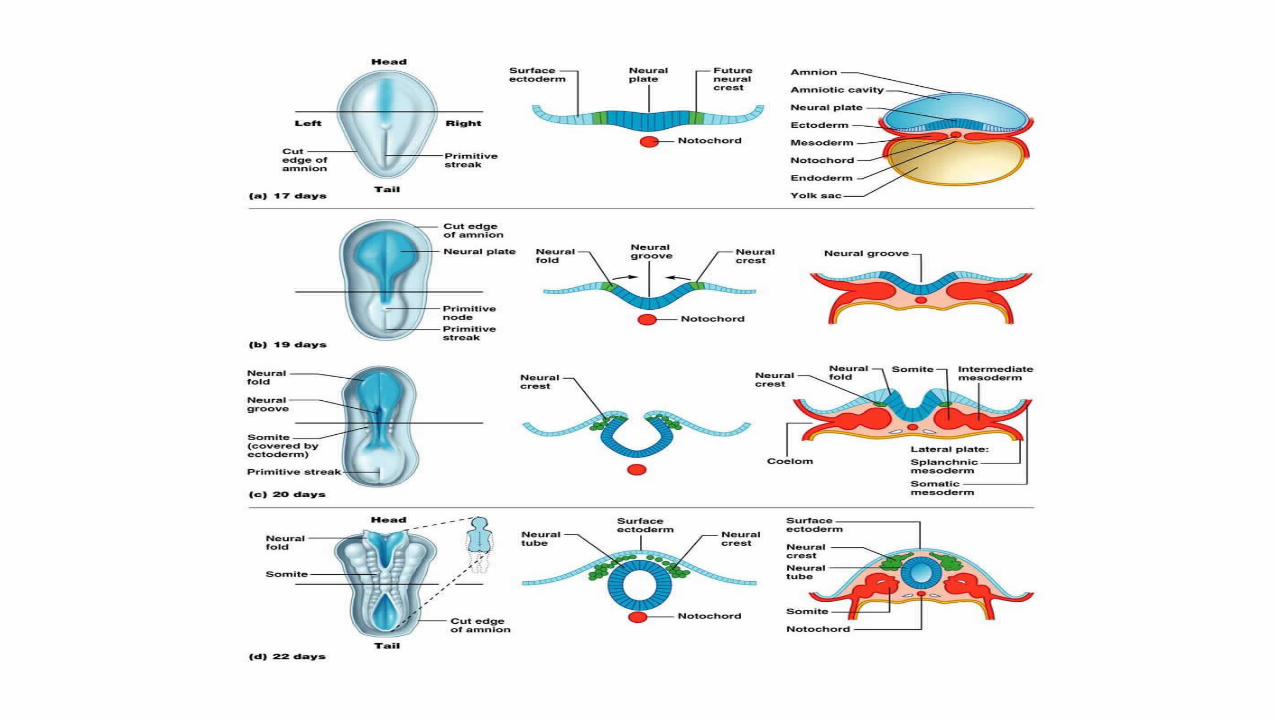
Primary neurulation
• Notochord induces the overlying ectoderm to become neurectoderm and form the neural plate.
• Subsequent folding and bending occurs until the margins unite to form the neural tube.
• Cranial end closes at day 25, while the caudal end closes a couple of days later.
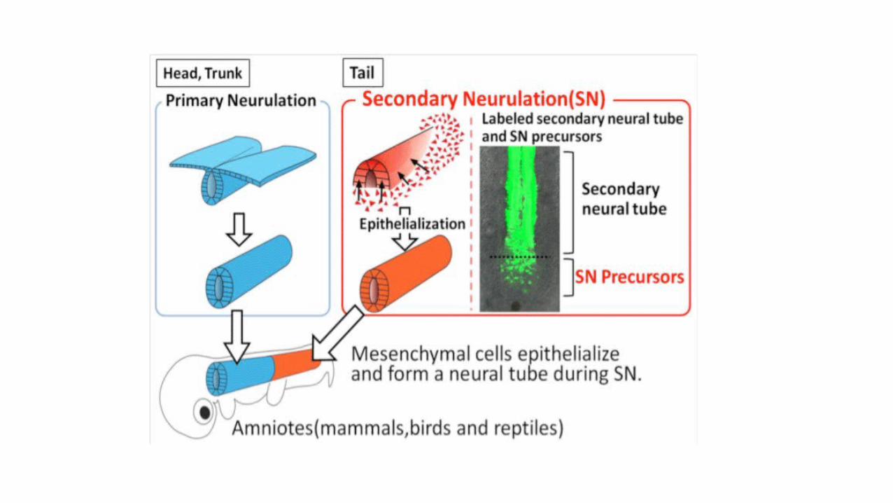
Secondary neurulation
• Finally the caudal cell mass arises from the primitive streak and undergoes retrogressive differentiation with cavitation.
• This is the origin of the foetal neural tissue and vertebrae distal to S2, and will become the conus medullaris.
Terminal ventricle
• focal expansion of the fetal canal as a result of incomplete retrogressive differentiation.
• may be seen as a normal asymptomatic finding in young children and may persist in a small minority into adulthood.
• seen on all post-mortem studies but is bigger in those detectable on MRI.
Spinal dysraphisms • can result from abnormalities occurring during any of above three
periods.
Spinal Dysraphisms • also known as a neural tube defect (NTD)
• comprises a group of congenital spine abnormalities that may cause progressive neurological damage
• may be open, in which case nervous tissue is exposed, or closed, in which case the defect is covered by skin, although a cutaneous lesion such as a dimple, sinus, hairy naevus or haemangioma may be seen as a marker of an underlying defect in 50% of these cases.
Spina bifida • refers to the failure of fusion of the posterior spinal bony elements.
Neural placode • is a flat segment of un-neurulated nervous tissue that may be seen at
the end of the spinal cord or at an intermediate position along its course
Tethered cord syndrome
• low-lying conus medullaris tethered by a short thick filum terminale, seen at or below the level of L3
• majority of normal cord lie between T11/12 and L1/2.
• is a clinical diagnosis of progressive neurological deterioration (usually leg weakness, deformities such as scoliosis or foot abnormalities, loss of bladder and bowel function), presumed to be due to traction damage on the tethered cord.
Open Spinal Dysraphism
• most are myelomeningoceles
• virtually always associated with Chiari II malformation
• neural placode protrudes beyond the level of the skin and there is an expanded CSF-containing sac lined by meninges.
• small proportion of OSDs are myeloceles where the placode is flush with the surface and there is no meningocele component.
• these disorders usually at the lumbosacral level at the spinal cord termination.
• nerve roots (from the everted ventral placode) cross the widely dilated meningocele subarachnoid spaces to enter the neural exit foraminae.
• posterior elements of the vertebral column and the other mesenchymal derivatives (e.g. paravertebral muscles) remain everted.
• Myelomeningoceles are operated on soon after birth; if untreated, the exposed neural tissue is prone to ulceration and infection.
Closed Spinal Dysraphism
• often associated with midline cutaneous stigmata or a mass.
• This may be a subcutaneous lipomatous mass overlying the spinal defect, as in lipomyeloceles and lipomyelomeningoceles.
• In these conditions there is an intraspinal lipoma.
• In a lipomyelocele the junction between the placode and the lipoma lies within the spinal canal, while in lipomyelomeningoceles it lies outside.
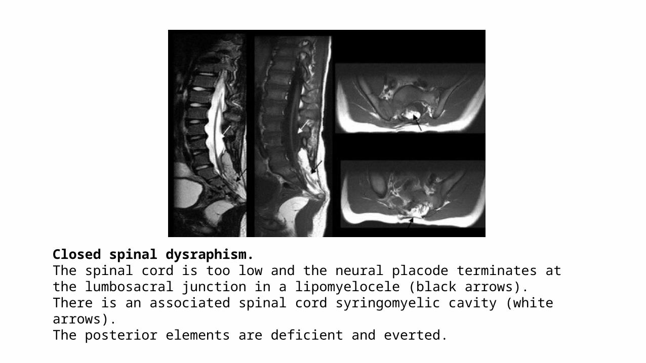
Closed spinal dysraphism. The spinal cord is too low and the neural placode terminates at the lumbosacral junction in a lipomyelocele (black arrows). There is an associated spinal cord syringomyelic cavity (white arrows). The posterior elements are deficient and everted.
• Typically the placode is rotated to one side while the lipoma rotates on the other. • These lipomas may be dorsal, with a normal conus below, or
transitional where the lipoma extends caudally along the conus. • In both these situations the lipoma–placode junction is dorsal and
well-defined.
‘Chaotic’ • 3rd form of closed Spinal Dysraphism• where the lipoma extends ventrally around the neural placode and
the lipoma is much more difficult to resect.• these are less common conditions but are more commonly seen with
sacral agenesis.
Dorsal Dermal Sinus• is an epithelial-lined opening on the skin with variable fistulous
extension to the dural surface.• typically seen in the lumbosacral region• often associated with cutaneous stigmata, such as hairy naevus and
capillary haemangioma.• Dermal openings seen at the sacrococcygeal level are directed
inferiorly below the thecal sac and are known as sacrococcygeal pits.• US may provide useful information about the intradural extension of
the dermal sinus tract and the mobility of the conus.• MRI is more sensitive for additional intraspinal findings such as spinal
cord syrinx and intraspinal dermoid.
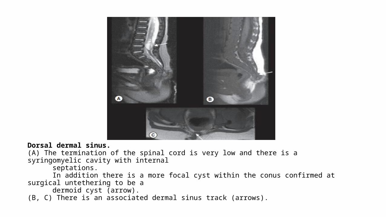
Dorsal dermal sinus. (A) The termination of the spinal cord is very low and there is a syringomyelic cavity with internal septations. In addition there is a more focal cyst within the conus confirmed at surgical untethering to be a dermoid cyst (arrow). (B, C) There is an associated dermal sinus track (arrows).
Diastematomyelia• split notochord syndromes are disorders of notochord midline
integration. • spinal cord is split in two, with each hemicord having one anterior and
one posterior grey matter horn.• There has two types.• Type I• Type II
In type I diastematomyelia • there are two complete dural sacs.• scoliosis or hemivertebrae or bifid/fused vertebrae at the level of the
bony spur. • bony spur is completely extradural and usually midline.
In diastematomyelia type II • There is a single dural sac in which the two hemicords lie, and a
fibrous septum may be seen passing intradurally between the two hemicords. • There may be no septum or there may only be partial clefting of the
cord. • Conus is often low and there may be a tight or fatty filum.
• Both forms of diastematomyelia may be associated with hydromyelia within the spinal cord.
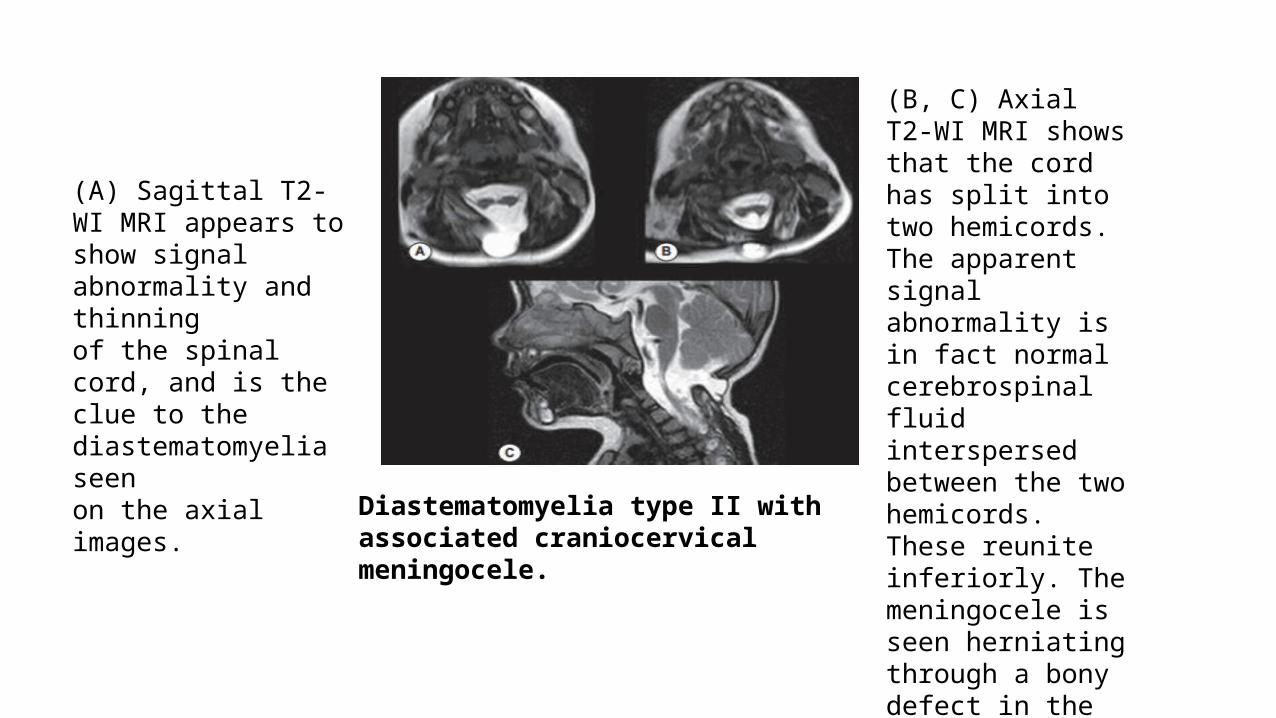
(A) Sagittal T2-WI MRI appears to show signal abnormality and thinningof the spinal cord, and is the clue to the diastematomyelia seenon the axial images.
(B, C) Axial T2-WI MRI shows that the cord has split into two hemicords. The apparent signalabnormality is in fact normal cerebrospinal fluid interspersedbetween the two hemicords. These reunite inferiorly. Themeningocele is seen herniating through a bony defect in thevertebral posterior elements.
Diastematomyelia type II with associated craniocervical meningocele.
Neurenteric Cysts• severest and rarest form of notochordal midline integration anomaly
occurs with dorsal enteric fistulas and neurenteric cysts.• cysts are usually seen intradurally anterior to the spinal cord and • are derived from endodermal remnants trapped between a split
notochord.• They have signal characteristics of CSF or of proteinaceous fluid with T
shortening.
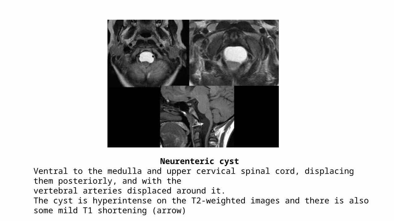
Neurenteric cyst Ventral to the medulla and upper cervical spinal cord, displacing them posteriorly, and with thevertebral arteries displaced around it. The cyst is hyperintense on the T2-weighted images and there is also some mild T1 shortening (arrow)
Disorders of the Caudal Cell Mass/ Caudal Regression Syndrome
• affects development of the caudal cell mass.
• caudal agenesis and the rarer condition of segmental spinal dysgenesis are considered to occur as a result of apoptosis of notochordal cells which have not formed in their correct craniocaudal position.
In caudal agenesis • there is a severe abnormality which results in absence of the vertebral
column at the affected level, as well as a truncated spinal cord, imperforate anus and genital anomalies. • may be seen with • OEIS (omphalocele, exstrophy, imperforate anus, spinal defects), • VACTERL and • Currarino triad• Anorectal malformation• Presacral mass which may be a teratoma or anterior
meningocele
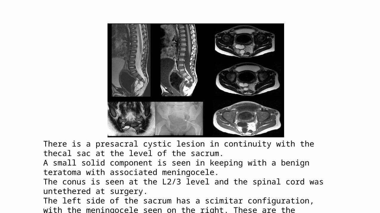
There is a presacral cystic lesion in continuity with the thecal sac at the level of the sacrum. A small solid component is seen in keeping with a benign teratoma with associated meningocele. The conus is seen at the L2/3 level and the spinal cord was untethered at surgery. The left side of the sacrum has a scimitar configuration, with the meningocele seen on the right. These are the features of the Currarino triad.
Segmental Spinal Dysgenesis
• Segmental abnormality affecting the spinal cord, segmental nerve roots and vertebrae
• Associated with a congenital paraparesis and lower limb deformities.
• Short segment of the spinal column is deficient and the spinal canal may be obliterated.
• On imaging, there may be an acute angle kyphus.
• These anomalies may be a result of a postneurulation insult.

THANK YOU!


![Imaging of Hereditary Hemorrhagic Telangiectasia · Spinal and cerebral vascular malformations are mani-festations of underlying vascular dysplasia [12]. These lesions represent abnormal](https://img.pdfslide.net/doc/110x75/5ed59c731b7fdd786a1b540e/imaging-of-hereditary-hemorrhagic-telangiectasia-spinal-and-cerebral-vascular-malformations.jpg)