Embed Size (px)
DESCRIPTION
A detailed description on anatomy of respiratory system.
Citation preview
Anatomy of Respiratory System
Dr. Anand Kumar BansalJunior Resident
Department Of Pulmonary Medicine
1
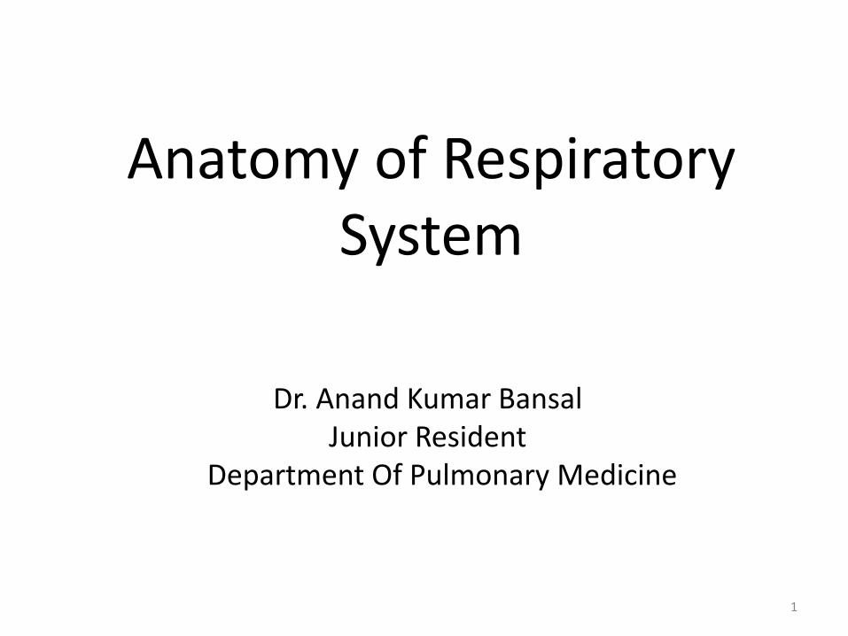
Functional Anatomy of the Respiratory System
Upper respiratory tract
– Nose, nasal cavity, and paranasal sinuses
– Pharynx and larynx
Lower respiratory tract
– Trachea
– Bronchi and smaller bronchioles
– Lungs and alveoli
2
NoseFUNTIONS --• Provides an airway for respiration
• Moistens and warms air
• Filters inhaled air
• Resonating chamber for speech
• Houses olfactory receptors
3
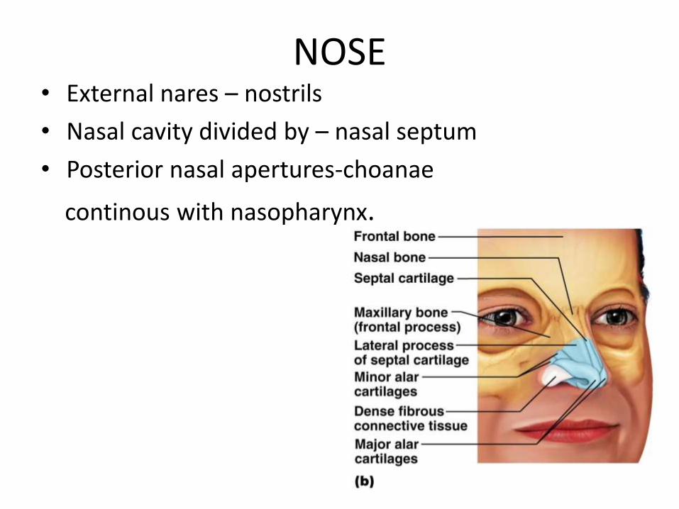
NOSE• External nares – nostrils
• Nasal cavity divided by – nasal septum
• Posterior nasal apertures-choanae
continous with nasopharynx.
4
Nasal Conchae
• Superior and middle nasal conchae– Part of the ethmoid bone
• Inferior nasal conchae– Separate bone
• Project medially from the lateral wall of the nasal cavity
• Create streamline flow of air and provide larger surface area for humidification of air
5
• Two types of mucous membrane
– Olfactory mucosa
Near roof of nasal cavity
Houses olfactory (smell) receptors
– Respiratory mucosa
Lines nasal cavity
Epithelium is pseudostratified ciliated columnar
6
Blood supply of Nose
• Lateral nasal wall is supplied by the sphenopalatine artery posteroinferiorly and by the anterior and posterior ethmoid arteries superiorly.
• The nasal septum also derives its blood supply from the sphenopalatine and the anterior and posterior ethmoid arteries with the added contribution of the superior labial artery (anteriorly) and the greater palatine artery (posteriorly)
7
The Paranasal Sinuses• Air containing cavities in certain bones of skull. • Lined with mucous membrane continuous with that of
nasal cavity.• Communicate with nasal fossa through their various ostia.
Functions -• May have a role in air-conditioning of the inspired air.• May reduce weight of the skull or simply act as protector
to eyes in trauma.• May give resonance to voice.• May thermally insulate skull base and orbit.
8
The nasal sinuses are eight in number, four on each side of the nose.
• Right and Left Frontal sinuses
• Right and Left Ethmoidal sinuses
• Right and Left Maxillary sinuses
• Right and Left Sphenoidal sinuses
9
Clinically , paranasal sinuses are divided into 2 groups-
The Anterior Group draining below the middle turbinate or near the infundibulum consists of:
• Frontal
• Maxillary
• Anterior and Middle Group of Ethmoidal cells
The Posterior Group draining at several locations above the middle turbinate is made up of:
• Posterior Ethmoidal Cells
• Sphenoidal Sinuses
10
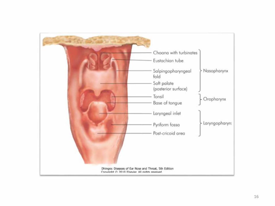
The Pharynx• Pharynx is a conical fibromuscular tube forming
the upper part of air and food passages.
• 12-14cm long
• Divided into three sections by location
– Nasopharynx
– Oropharynx
– Laryngopharynx
11
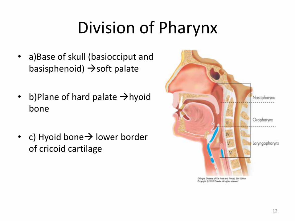
Division of Pharynx
• a)Base of skull (basiocciput and basisphenoid) soft palate
• b)Plane of hard palate hyoid bone
• c) Hyoid bone lower border of cricoid cartilage
12
Nasopharynx
• Only an air passageway• Closed off during swallowing• Contains the opening to the pharyngotympanic tube
(Eustachian or auditory tube)• Nasopharyngeal tonsil (adenoids)
– Located on posterior wall– Destroys entering pathogens
• Tubal tonsil• Provides some protection from infection
• Epithelium– Pseudostratified Ciliated columnar epithelium
13
Oropharynx
• Lies opposite to the oral cavity with which it communicates through oropharyngeal isthmus.
OROPHARYNGEAL ISTHMUS-boundaries.• Soft palate above.• Upper surface of tongue below.• Palatoglossal arch on either sides.Two types of tonsils in the oropharynx
– Palatine tonsils – in the lateral walls of the fauces– Lingual tonsils – covers the posterior surface of the tongue
• Epithelium– Stratified squamous epithelium
14
Laryngopharynx(Hypopharynx)
• Passageway for both food and air• Lowest part of pharynx
Boundaries• Above oropharynx
• Below oesophagus
• Ant laryngeal cavity or inlet
• Epithelium– Lined by Stratified squamous epithelium
15
16
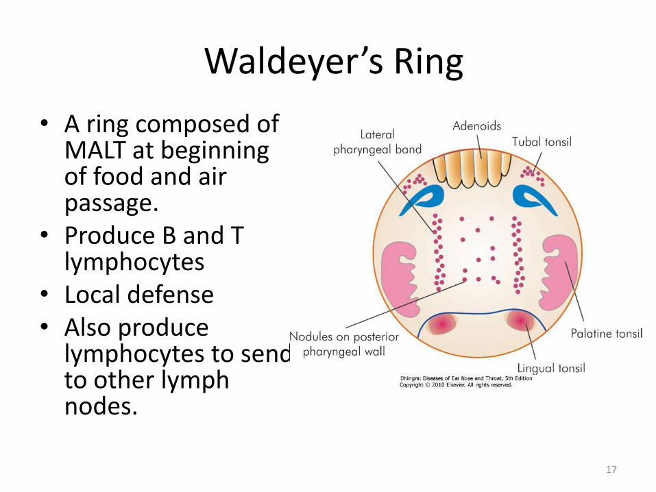
Waldeyer’s Ring
• A ring composed of MALT at beginning of food and air passage.
• Produce B and T lymphocytes
• Local defense• Also produce
lymphocytes to send to other lymph nodes.
17
The Larynx
It lies anterior to the hypopharynxOpposite to C3- C6 vertebrae
Three functions– Provides an open airway and Protection of the lower
airways
– Voice production
– Respiration
18
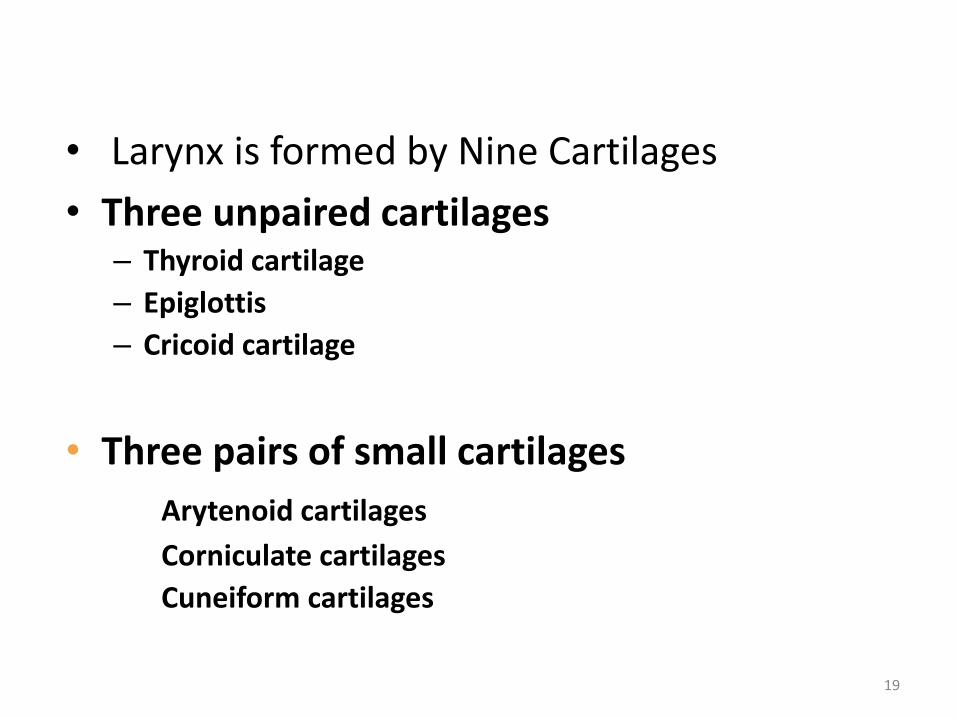
• Larynx is formed by Nine Cartilages
• Three unpaired cartilages– Thyroid cartilage
– Epiglottis
– Cricoid cartilage
• Three pairs of small cartilages
Arytenoid cartilages
Corniculate cartilages
Cuneiform cartilages
19
Vocal ligaments of the larynx
– Vocal folds (true vocal cords) Act in sound production
– Vestibular folds (false vocal cords) No role in sound production
20
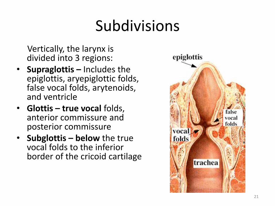
SubdivisionsVertically, the larynx is divided into 3 regions:
• Supraglottis – Includes the epiglottis, aryepiglottic folds, false vocal folds, arytenoids, and ventricle
• Glottis – true vocal folds, anterior commissure and posterior commissure
• Subglottis – below the true vocal folds to the inferior border of the cricoid cartilage
21
Mucosa
• Mucosa of glottic and Supragottic regions is stratified squamous epithelium.
• Mucosa of ventricles and sub-glottic regions is pseudo-stratified ciliated epithelium
• Supra and sub glottic regions particularly ventricles are rich in submucosal mucous or minor salivary glands while glottis is not.
22
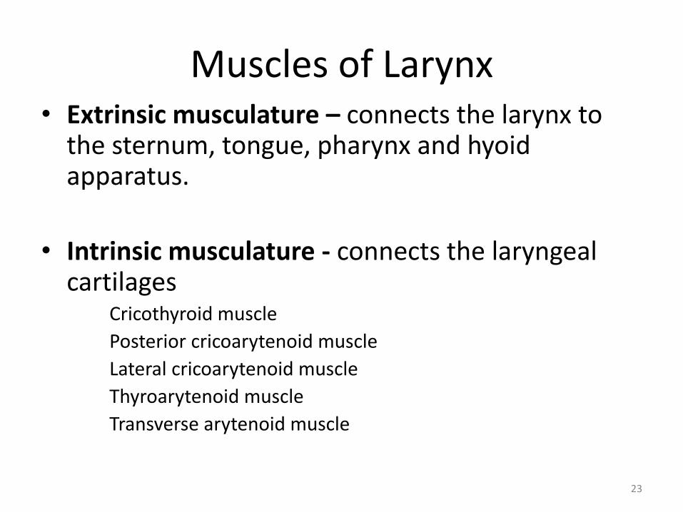
Muscles of Larynx• Extrinsic musculature – connects the larynx to
the sternum, tongue, pharynx and hyoid apparatus.
• Intrinsic musculature - connects the laryngeal cartilages
Cricothyroid muscle
Posterior cricoarytenoid muscle
Lateral cricoarytenoid muscle
Thyroarytenoid muscle
Transverse arytenoid muscle
23
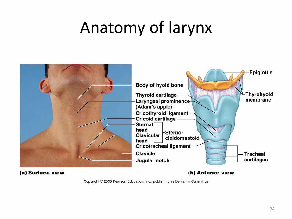
Anatomy of larynx
24
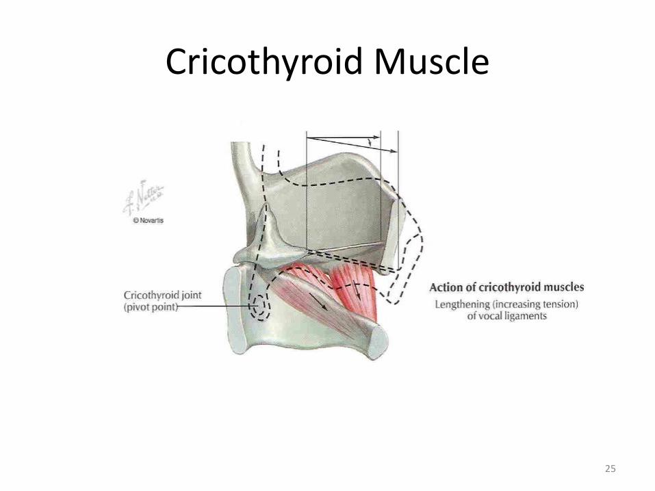
Cricothyroid Muscle
25
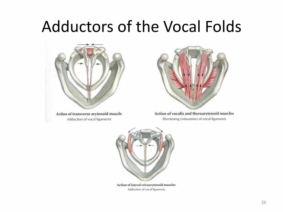
Adductors of the Vocal Folds
26
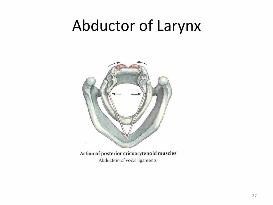
Abductor of Larynx
27
Nerve supply : Derived from the Vagus
Superior laryngeal nerve• Supplies cricothyroid muscle
• Sensory supply to larynx above vocal cords
Recurrent laryngeal nerve• Supplies all intrinsic muscles except cricothyroid
• Sensory supply to larynx below vocal cords
28
ARTERIAL SUPPLY
• Sup. Laryngeal artery which originates from Sup. Thyroid artery branch of External carotid artery
• Inf. Laryngeal artery which originates from Inf. Thyroid artery branch of Thyrocervical trunk
29
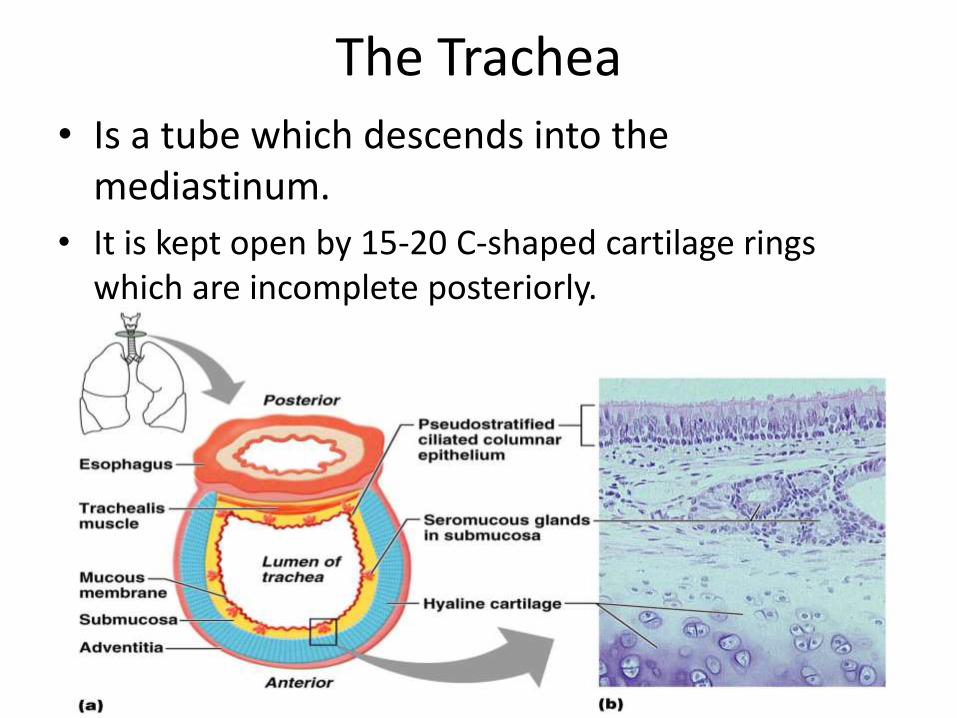
The Trachea• Is a tube which descends into the
mediastinum.
• It is kept open by 15-20 C-shaped cartilage rings which are incomplete posteriorly.
30
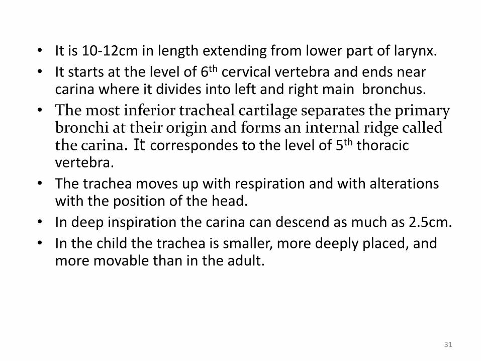
• It is 10-12cm in length extending from lower part of larynx.
• It starts at the level of 6th cervical vertebra and ends near carina where it divides into left and right main bronchus.
• The most inferior tracheal cartilage separates the primary bronchi at their origin and forms an internal ridge called the carina. It correspondes to the level of 5th thoracic vertebra.
• The trachea moves up with respiration and with alterations with the position of the head.
• In deep inspiration the carina can descend as much as 2.5cm.
• In the child the trachea is smaller, more deeply placed, and more movable than in the adult.
31
32
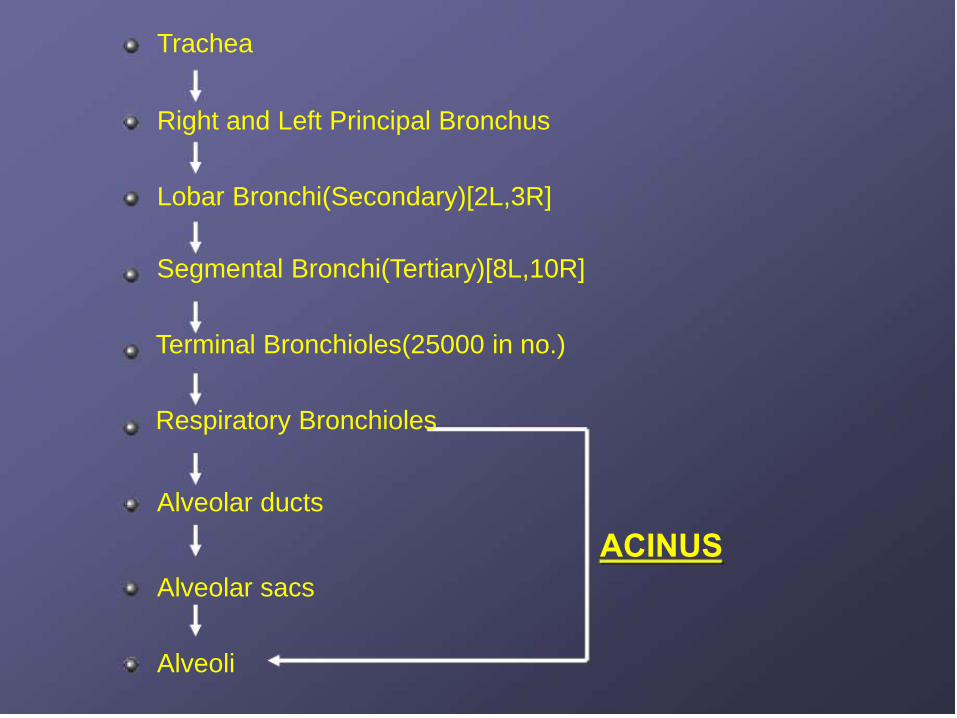
Trachea
Right and Left Principal Bronchus
Lobar Bronchi(Secondary)[2L,3R]
Segmental Bronchi(Tertiary)[8L,10R]
Terminal Bronchioles(25000 in no.)
Respiratory Bronchioles
Alveolar ducts
ACINUS
Alveolar sacs
Alveoli
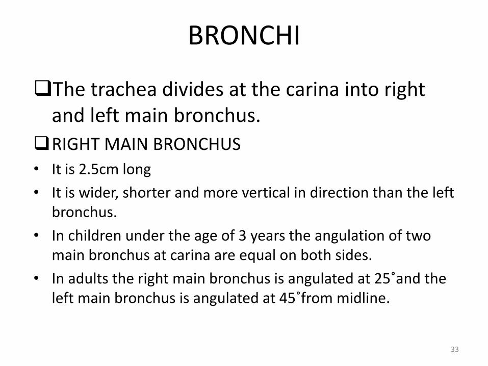
BRONCHI
The trachea divides at the carina into right and left main bronchus.
RIGHT MAIN BRONCHUS
• It is 2.5cm long
• It is wider, shorter and more vertical in direction than the left bronchus.
• In children under the age of 3 years the angulation of two main bronchus at carina are equal on both sides.
• In adults the right main bronchus is angulated at 25˚and the left main bronchus is angulated at 45˚from midline.
33
• The azygos vein arches over it from behind and the right pulmonary artery lies at first below and then in front of it.
• The right main bronchus divides into three lobar bronchi to supply respective lobes.
• Right main bronchus is the commonest site for aspiration .
• If a patient in right lateral position aspirates, the material gravitates into lateral portion of posterior segment of upper lobe.
• If patient aspirates in supine position, the material gravitates to apical segment of lower lobe.
34
LEFT MAIN BRONCHUS
• It is narrower than the right bronchus and is nearly 5cm long.
• The left main bronchus divides into two lobar bronchi for upper and middle lobes.
• It passes beneath the aortic arch, crosses in front of the esophagus, the thoracic duct, and the descending aorta, and has the left pulmonary artery lying at first above, and then in front of it
35
Bronchioles• The bronchi divide dichotomously into several
million terminal bronchioles to terminate in one or more respiratory bronchioles.
• Bronchioles are less than 1 mm in diameter.
• The cartilagenous rings that are seen in bronchioles are replaced by cartilagenousplates as the size of bronchioles decrease.
• The cartilage completely disappear when their size reaches to 0.6mm.
36
• The small terminal bronchioles are supported by smooth muscle cells.
• Distal to each terminal bronchiole is an acinus, which consists of three to four orders of respiratory bronchioles.
• Respiratory bronchioles lead to alveolar ducts. The walls of these ducts consist of alveolar sacs or the mouths of alveoli.
• Smooth muscles are found in the walls of the airways upto the level of alveolar ducts.
37
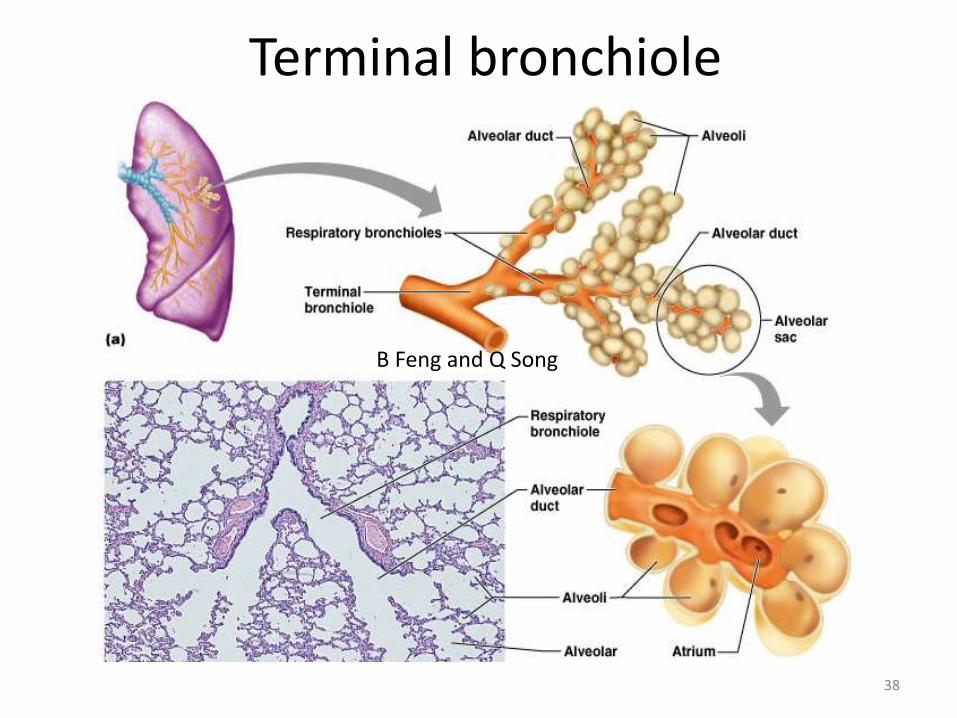
Terminal bronchiole
38
B Feng and Q Song
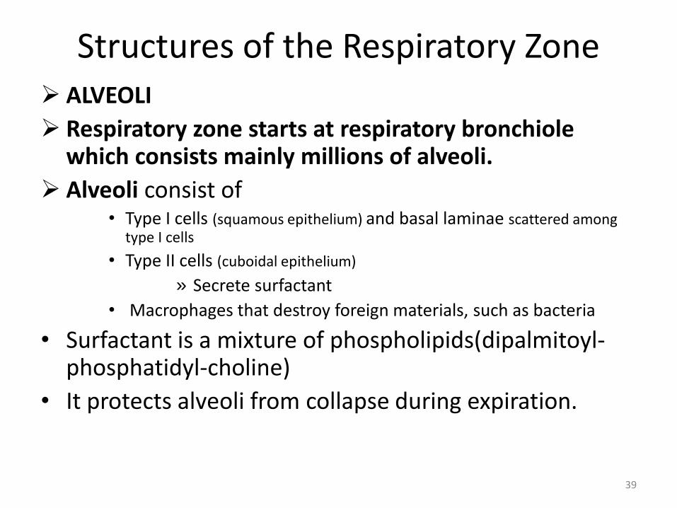
Structures of the Respiratory Zone ALVEOLI
Respiratory zone starts at respiratory bronchiole which consists mainly millions of alveoli.
Alveoli consist of• Type I cells (squamous epithelium) and basal laminae scattered among
type I cells
• Type II cells (cuboidal epithelium)
» Secrete surfactant
• Macrophages that destroy foreign materials, such as bacteria
• Surfactant is a mixture of phospholipids(dipalmitoyl-phosphatidyl-choline)
• It protects alveoli from collapse during expiration.
39
Alveoli
Features of alveoli
– Surrounded by elastic fibers.
– Interconnect by way of alveolar pores
– Internal surfaces
A site for free movement of alveolar macrophages.
40
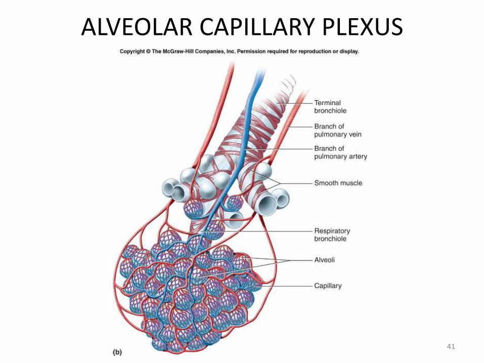
ALVEOLAR CAPILLARY PLEXUS
41
Pleura and Pleural Cavities
• The outer surface of each lung and the adjacent internal thoracic wall are lined by a serous membrane called pleura.
• The outer surface of each lung is tightly covered by the visceral pleura.
• while the internal thoracic walls, the lateral surfaces of the mediastinum, and the superior surface of the diaphragm are lined by the parietal pleura.
• The parietal and visceral pleural layers are continuous at the hilus of each lung.
42
Pleural Cavities
• The potential space between the serous membrane layers is a pleural cavity.
• The pleural membranes produce a thin, serous pleural fluidthat circulates in the pleural cavity and acts as a lubricant, ensuring minimal friction during breathing.
43
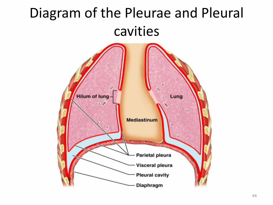
Diagram of the Pleurae and Pleural cavities
44
Lungs
• A pair of respiratory organs situated in the thoracic cavity.
• Each is cone-shaped with anterior, lateral and posterior surfaces contacting ribs
• Superior tip is apex, just deep to clavicle
• Concave inferior surface resting on diaphragm is the base.
• The lungs are heavier in the male than in the female, their proportion to the body being, in the former, as 1:37, in the latter as 1:43.
45
Gross Anatomy of the Lungs
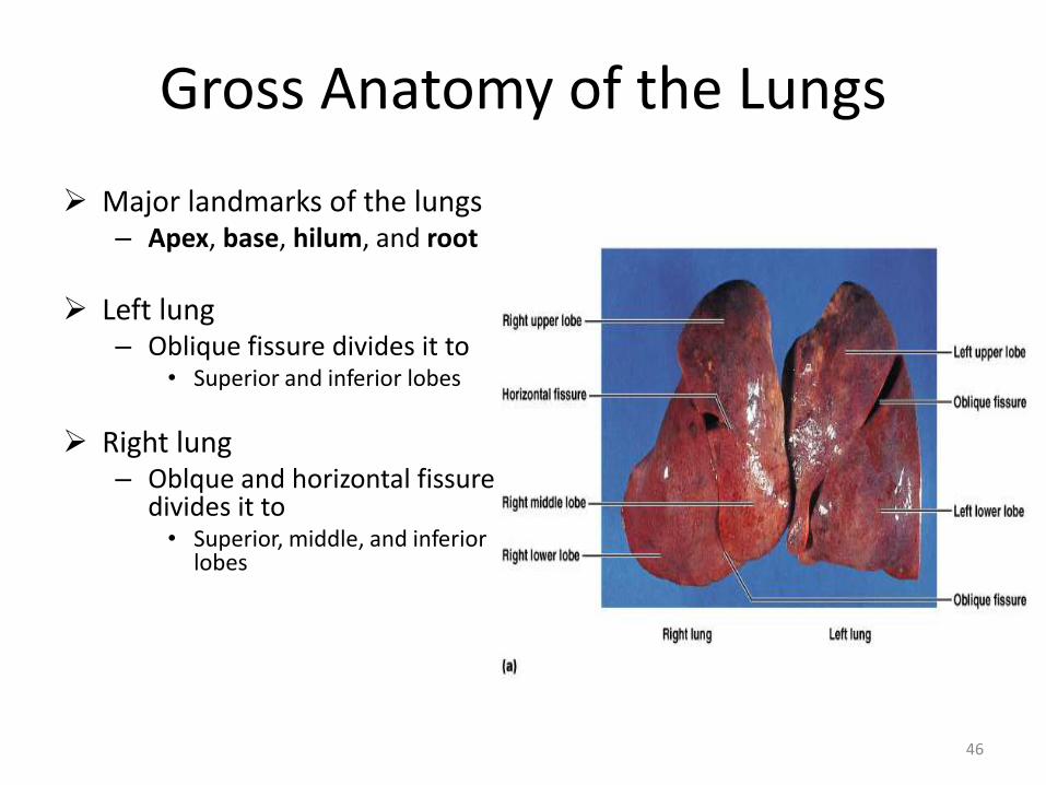
Major landmarks of the lungs– Apex, base, hilum, and root
Left lung– Oblique fissure divides it to
• Superior and inferior lobes
Right lung– Oblque and horizontal fissure
divides it to• Superior, middle, and inferior
lobes
46
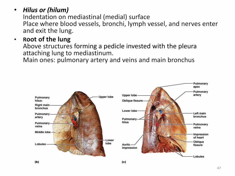
• Hilus or (hilum)Indentation on mediastinal (medial) surfacePlace where blood vessels, bronchi, lymph vessel, and nerves enter and exit the lung.
• Root of the lungAbove structures forming a pedicle invested with the pleura attaching lung to mediastinum.Main ones: pulmonary artery and veins and main bronchus
47
Bronchopulmonary Segments
• Each lobe is made up of Bronchopulmonary segments separated by dense connective tissue– These are well defined sectors of the lung– These are independent respiratory units– Limit spread of infection
• Each Bronchopulmonary segment is aerated by a tertiary or segmental bronchus
• The branch of pulmonary artery accompany the bronchus and lies dorsolateral to it. Thus each segment has its own separate artery
• Pulmonary veins run in the inter-segmental planes, serving adjacent segments thus each segment has more than one vein and each vein drains more than one segment
• Thus a bronchopulmonary segment is not a bronchovascularsegment as it does not have its own vein
48
Bronchopulmonary Segments
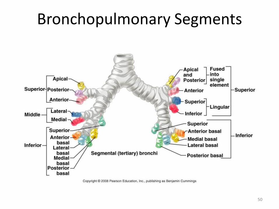
The right lung is subdividedinto three lobes with tensegments:• right upper lobe
– apical – posterior – anterior
• right middle lobe– lateral – medial
• right lower lobe– superior – anterior basal– medial basal– lateral basal– posterior basal
The left lung is subdivided intotwo lobes and thereby, into eightsegments:• left upper lobe
– apicoposterior– anterior – superior lingular– inferior lingular
• left lower lobe– superior– anteromedial basal– lateral basal– posterior basal
49
Bronchopulmonary Segments
50
Blood Supply• The lungs have two functionally distinct
circulatory pathways.
• These are the pulmonary vessels, which convey deoxygenated blood to the alveolar walls and drain oxygenated blood back to the left side of the heart.
• The much smaller bronchial vessels, which are derived from the systemic circulation and provide oxygenated blood to lung tissues which do not have close access to atmospheric oxygen.
51
Innervation of the Lungs
• The lungs are innervated by parasympathetic (vagal) and sympathetic fibres.
• The parasympathetic fibres are derived from vagus. They supply the bronchial muscles and glands and are bronchoconstrictor and secretomotor.
• The sympathetic fibres are derived from 2nd to 5th spinal segments and are inhibitory to the bronchial muscles and glands. They relax the bronchial smooth muscle and also have vasoconstrictor effects.
52
Lymphatic drainage of Lungs
Lymphatic vessels originate in the subpleural(superficial) and deep lymphatic plexuses
• The subpleural lymphatic plexus is superficial, lying deep to the visceral pleura, and drains lymph from the surface of the lung to the bronchopulmonary (hilar) nodes
• The deep lymphatic plexus is in the lung and follows the bronchi and pulmonary vessels to the pulmonary nodes and then bronchopulmonary nodes located at the root of the lung
• Lymph from the parietal pleura drains into lymph nodes of the thoracic wall
53

Thank You!
54


![Anatomy and Physiology Respiratory System [Tab 2] Respiratory System](https://img.pdfslide.net/doc/110x75/56649ebd5503460f94bc631f/anatomy-and-physiology-respiratory-system-tab-2-respiratory-system.jpg)