Embed Size (px)
Citation preview
1. Name the organs forming the respiratory passageway from the nasal cavity to the alveoli of the lungs.
2. Explain how the respiratory muscles cause volume changes that lead to breathing.
3. Sally has a vital lung capacity of 3900 ml. Her tidal volume is 400 ml. Her expiratory reserve volume is 1000 ml. What is her inspiratory reserve volume?
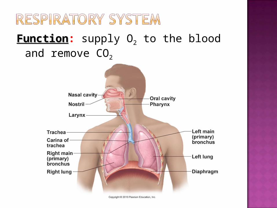
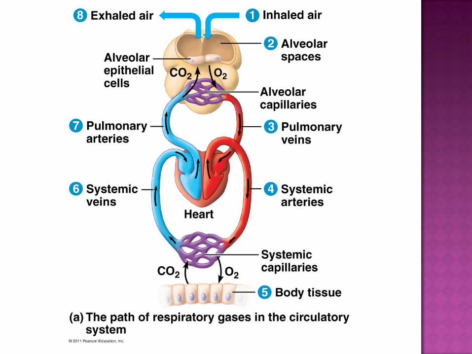
FunctionFunction: supply O2 to the blood and remove CO2
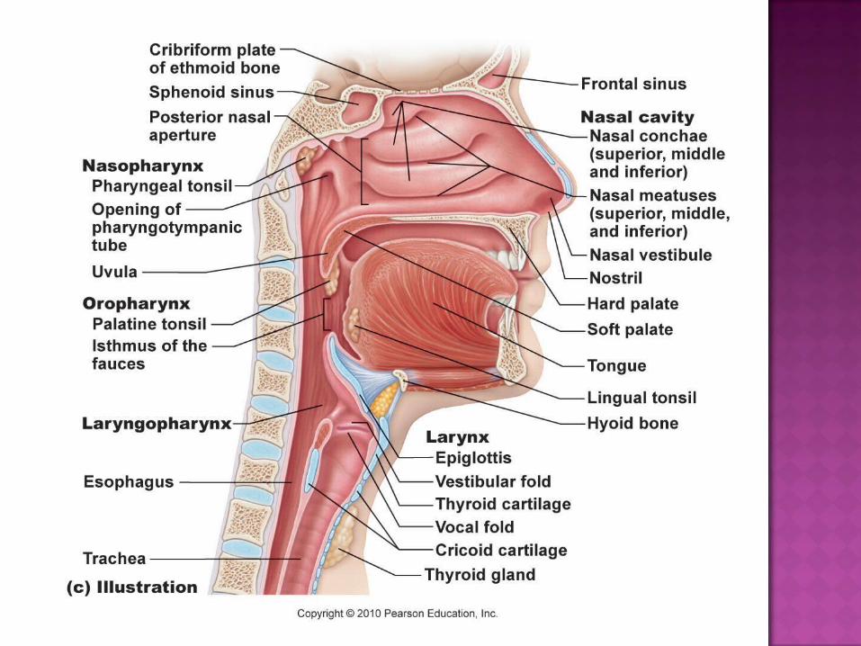
Nose/mouth: filtered, warmed, humidifiedMucus traps bacteria & foreign debrisCilia sweep mucus toward throat digested by
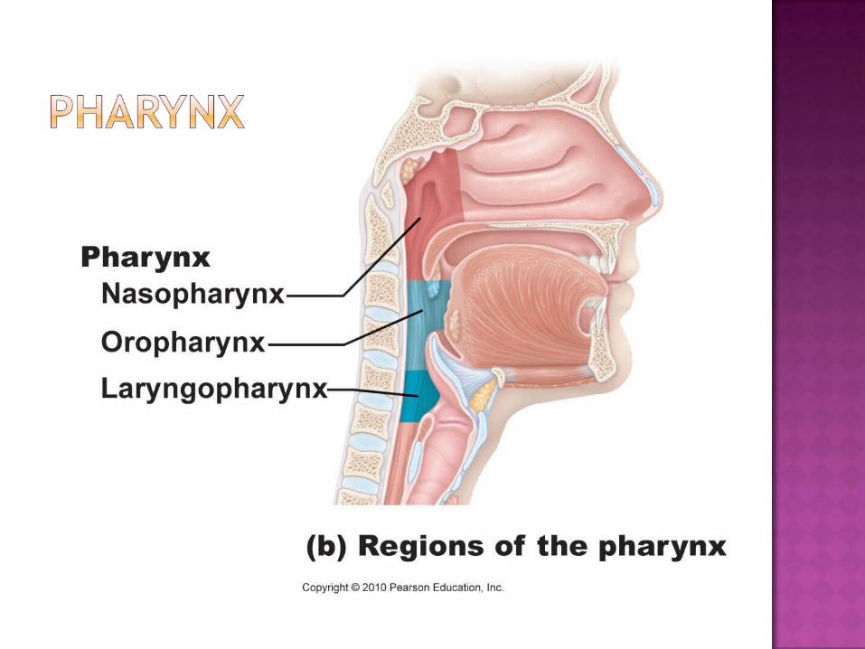
stomach Pharynx: throat (passage for food/air)
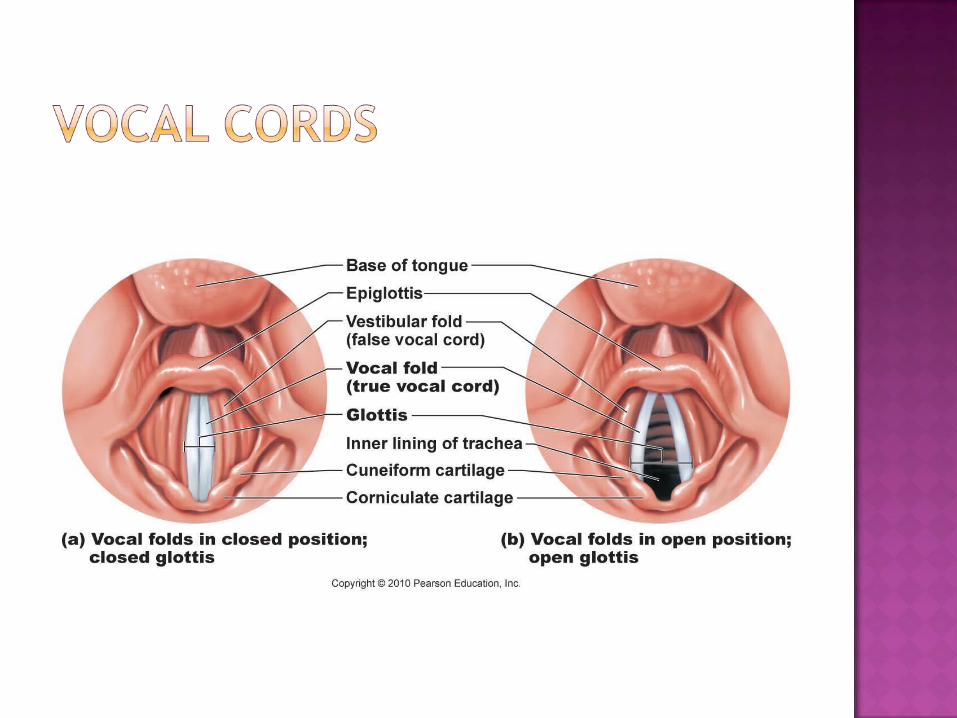
Tonsils: clusters of lymphatic tissue Larynx: contains vocal cords
Epiglottis: covers larynx when liquids/food swallowed
Trachea: windpipe; lined with cartilage (C-shaped)
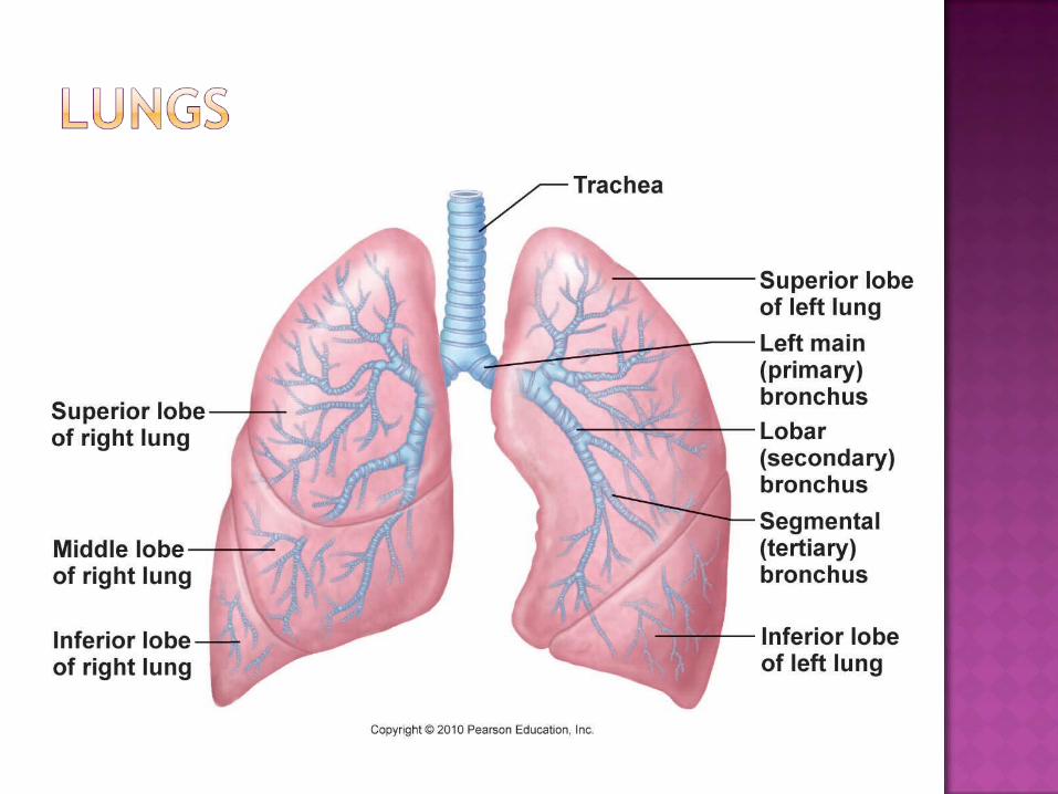
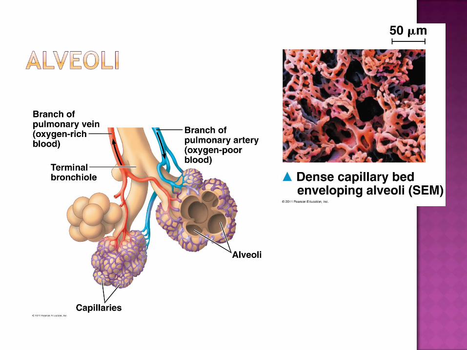
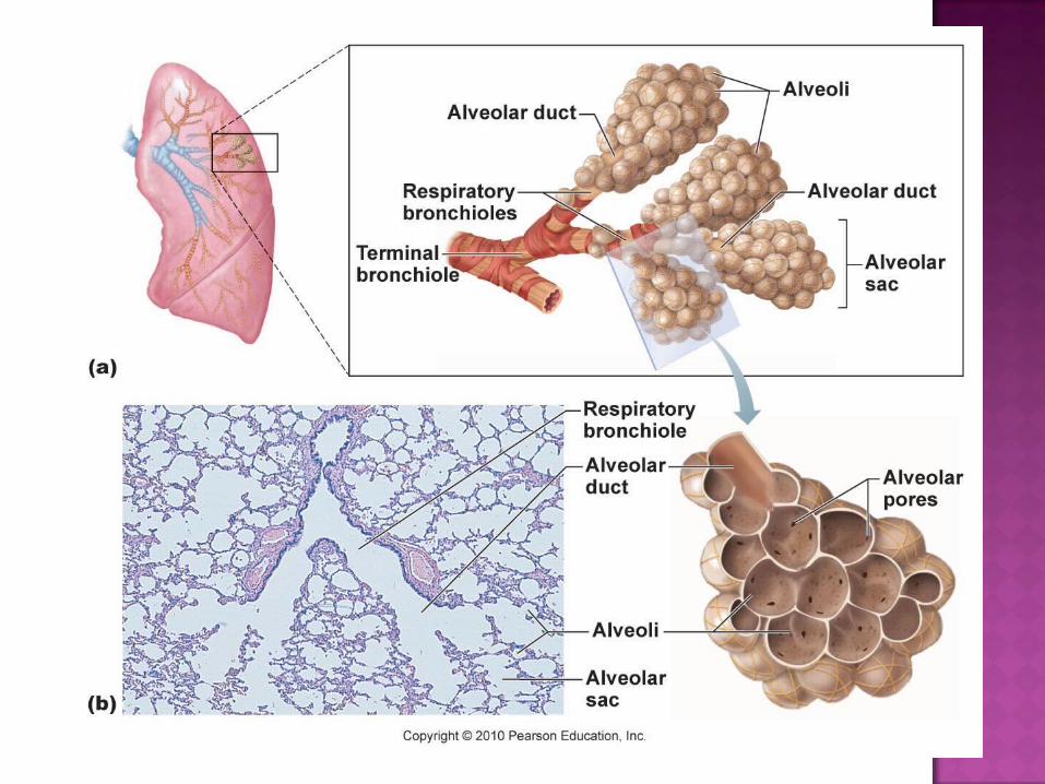
Bronchi: branches to lungs Bronchioles: smaller branches Lungs Alveoli: air sacs for gas exchange
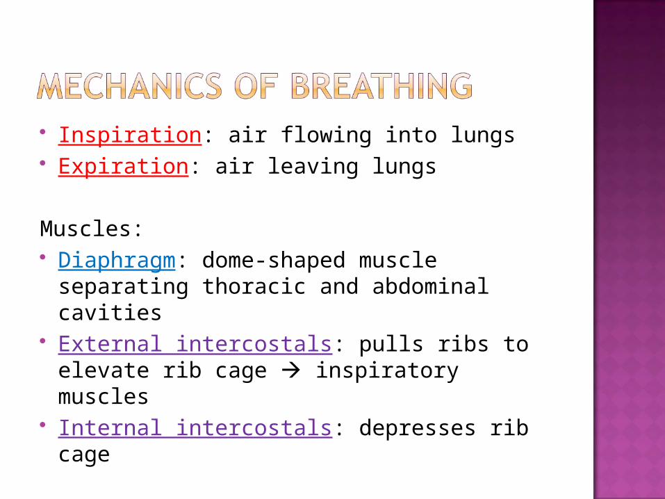
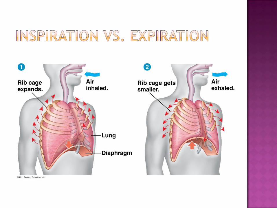
Inspiration: air flowing into lungs Expiration: air leaving lungs
Muscles: Diaphragm: dome-shaped muscle
separating thoracic and abdominal cavities
External intercostals: pulls ribs to elevate rib cage inspiratory muscles
Internal intercostals: depresses rib cage
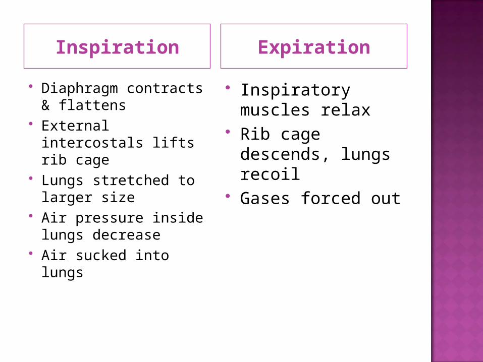
Inspiration Expiration
Diaphragm contracts & flattens
External intercostals lifts rib cage
Lungs stretched to larger size
Air pressure inside lungs decrease
Air sucked into lungs
Inspiratory muscles relax
Rib cage descends, lungs recoil
Gases forced out
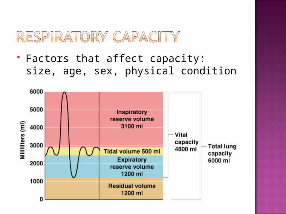
Factors that affect capacity: size, age, sex, physical condition
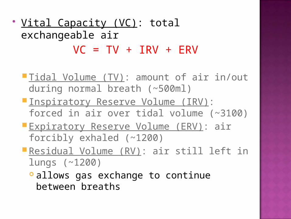
Vital Capacity (VC): total exchangeable air
VC = TV + IRV + ERV
Tidal Volume (TV): amount of air in/out during normal breath (~500ml)
Inspiratory Reserve Volume (IRV): forced in air over tidal volume (~3100)
Expiratory Reserve Volume (ERV): air forcibly exhaled (~1200)
Residual Volume (RV): air still left in lungs (~1200) allows gas exchange to continue between
breaths
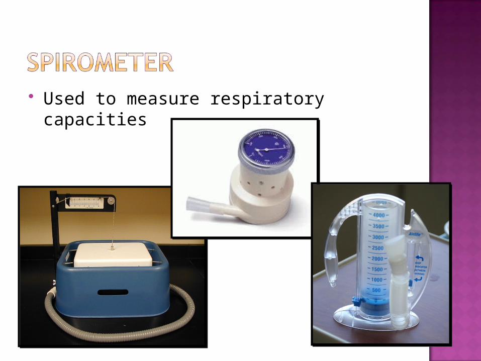
Used to measure respiratory capacities
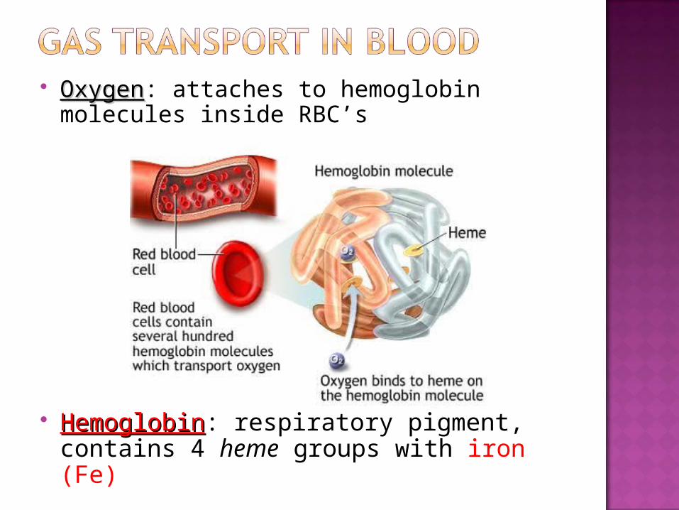
OxygenOxygen: attaches to hemoglobin molecules inside RBC’s
HemoglobinHemoglobin: respiratory pigment, contains 4 heme groups with iron (Fe)
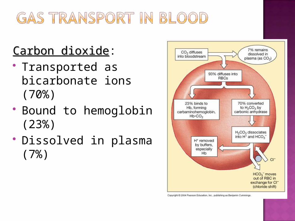
Carbon dioxideCarbon dioxide: Transported as
bicarbonate ions (70%) Bound to hemoglobin
(23%) Dissolved in plasma
(7%)
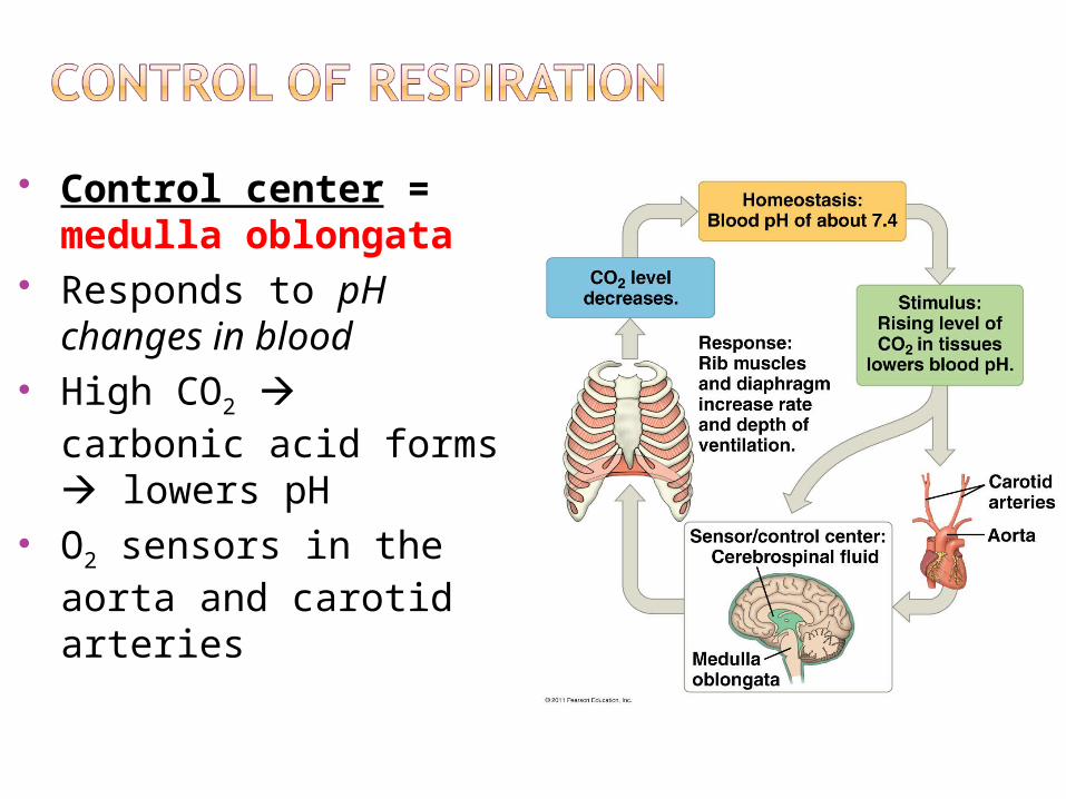
Control center = medulla oblongata
Responds to pH changes in blood
High CO2 carbonic acid forms lowers pH
O2 sensors in the aorta and carotid arteries
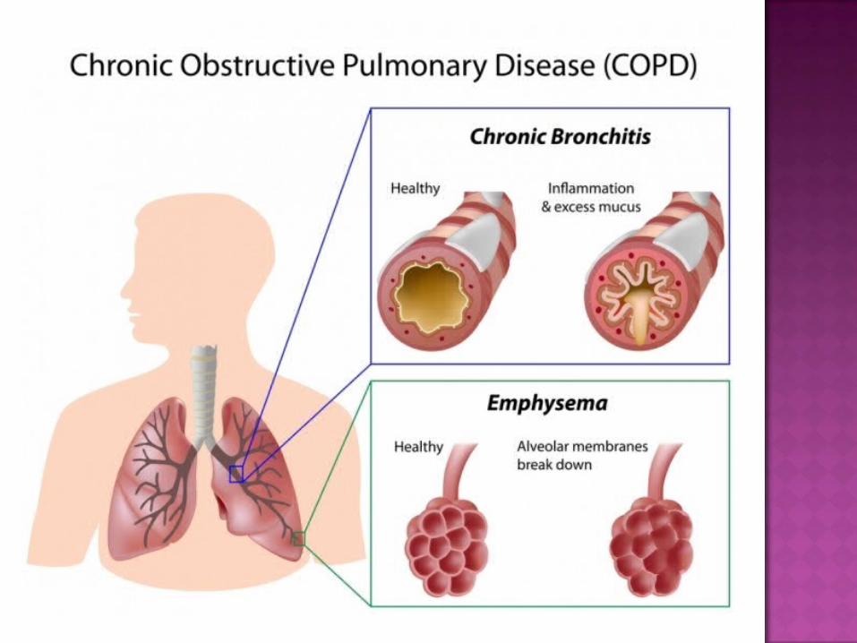
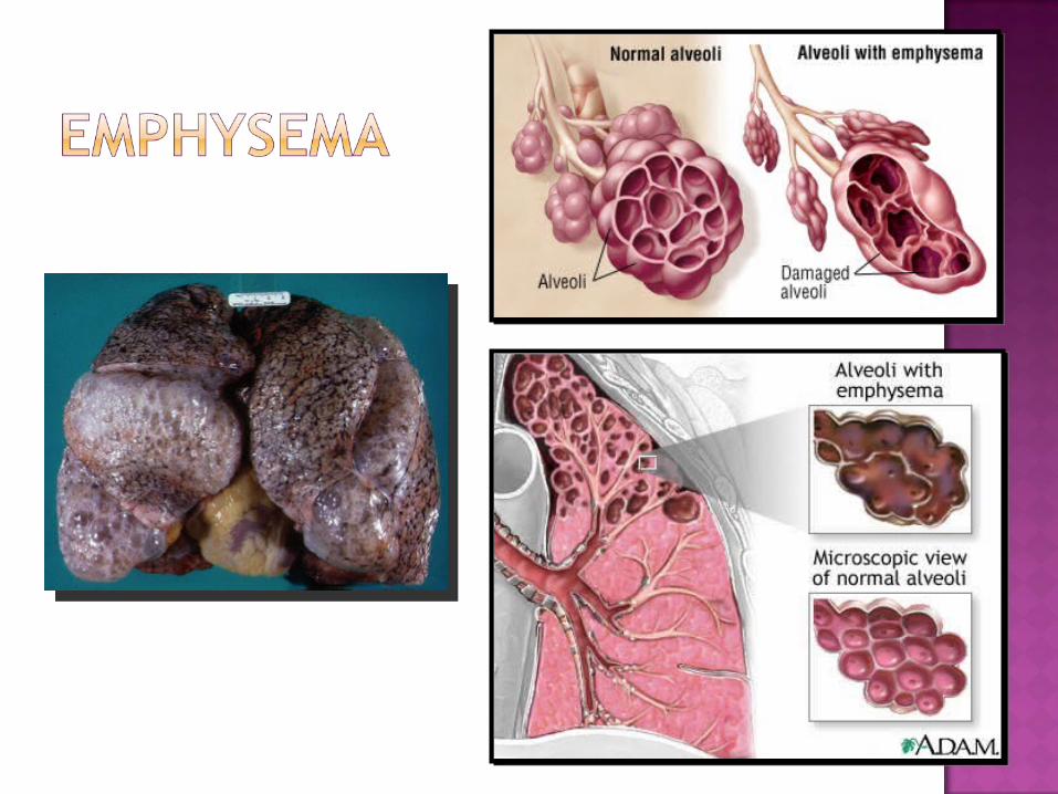
Group of lung diseases blocks airflow and makes breathing difficult
Emphysema (lose elasticity of lung tissue) & chronic bronchitis (excess mucus)
Features:1. History of smoking2. Labored breathing (wheezing, shortness of
breath)3. Coughing & frequent pulmonary infections4. Hypoxic (inadequate O2 delivery – bluish
skin)
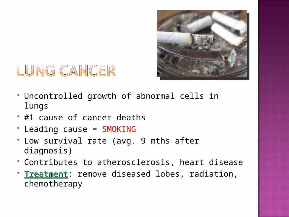
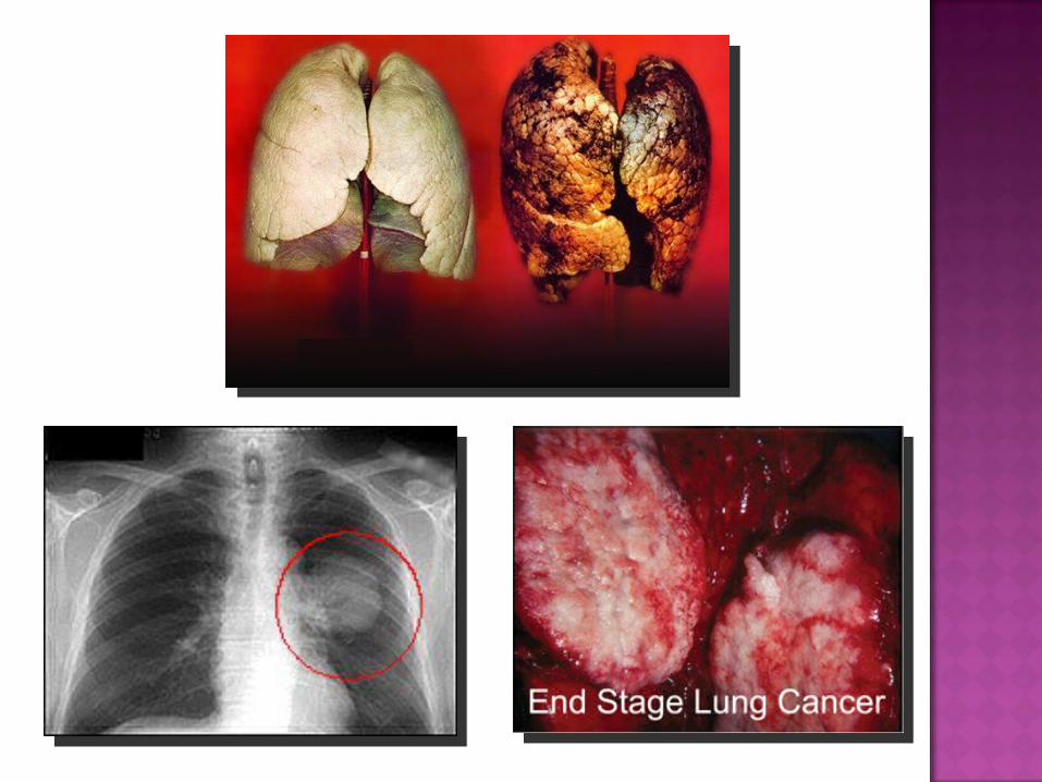
Uncontrolled growth of abnormal cells in lungs #1 cause of cancer deaths Leading cause = SMOKING Low survival rate (avg. 9 mths after diagnosis) Contributes to atherosclerosis, heart disease TreatmentTreatment: remove diseased lobes, radiation,
chemotherapy
Asthma: inflamed, hypersensitive bronchial passages that respond to irritants
Bronchitis: bronchi swollen and clogged Pneumonia: inflammation of lung caused
by infection Tuberculosis (TB): infectious disease
caused by M. tuberculosis bacterium