Embed Size (px)
Citation preview
In-vivo assessment of the relationship In-vivo assessment of the relationship between shear stress and plaque between shear stress and plaque
vulnerability in human coronary arteriesvulnerability in human coronary arteriesFrank Gijsen, Jolanda Wentzel, Attila Thury, Frits Mastik, Johannes
Schaar, Johan Schuurbiers, Pim de Feyter, Ton van der Steen, Patrick Serruys, and Cornelis Slager
Hemodynamics Lab, Department of Biomedical Engineering
Erasmus Medical Center, Rotterdam, The Netherlands
Shear stress and atherosclerosis
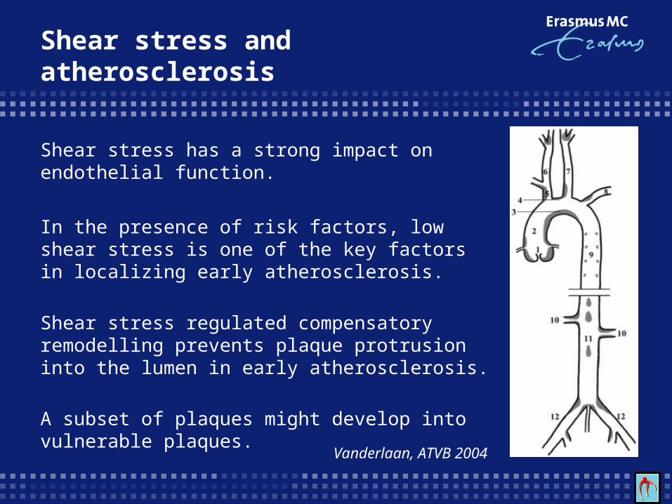
Shear stress has a strong impact on endothelial function.
In the presence of risk factors, low shear stress is one of the key factors in localizing early atherosclerosis.
Shear stress regulated compensatory remodelling prevents plaque protrusion into the lumen in early atherosclerosis.
A subset of plaques might develop into vulnerable plaques.
Vanderlaan, ATVB 2004
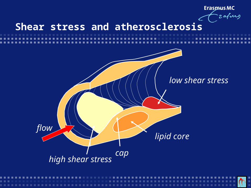
Shear stress and atherosclerosis
cap
lipid core
high shear stress
low shear stress
flow
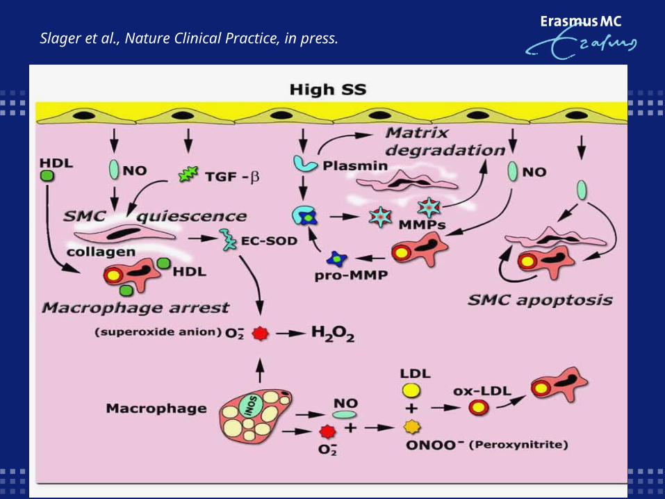
Slager et al., Nature Clinical Practice, in press.
Shear stress and atherosclerosisShear stress and atherosclerosis
Aim:
Investigate, in coronary arteries of patients, the relationship between shear stress and a marker of plaque vulnerability.
Working hypothesis:
Early atherosclerosis: low shear stress is one of the localizing factors of the disease and high shear stress acts protective.
Advanced atherosclerosis: high shear stress, through its anti-inflammatory impact on the endothelium, might enhance plaque vulnerability.
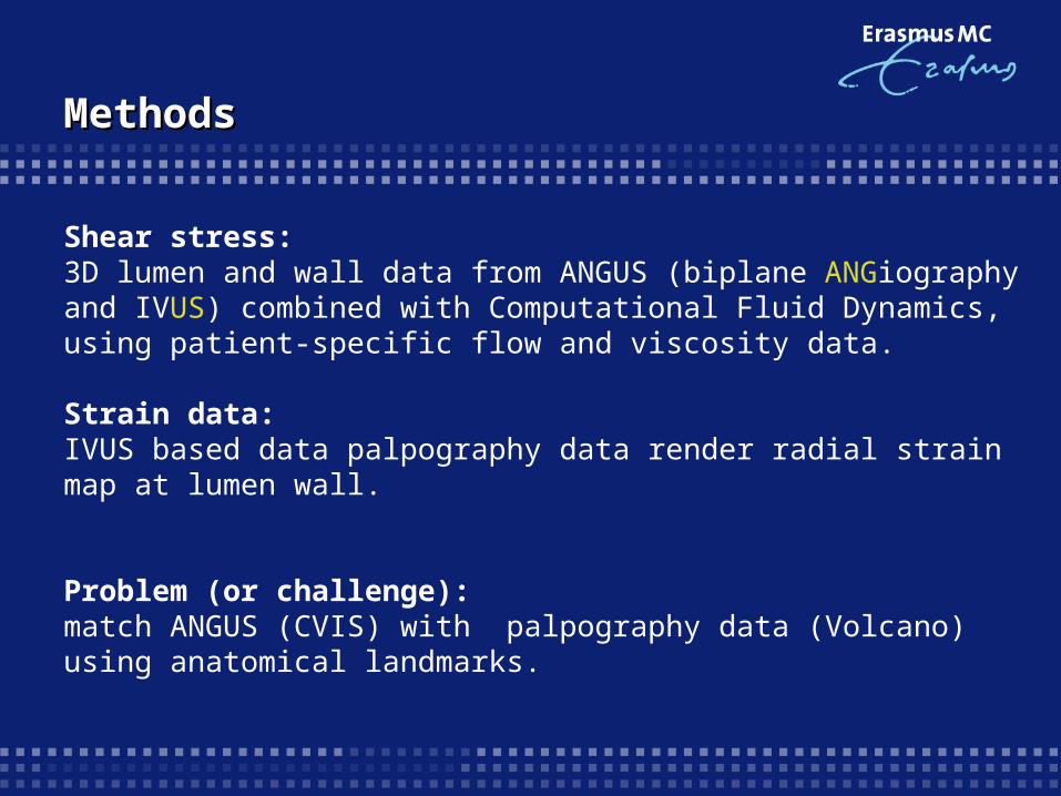
MethodsMethods
Shear stress:3D lumen and wall data from ANGUS (biplane ANGiography and IVUS) combined with Computational Fluid Dynamics, using patient-specific flow and viscosity data.
Strain data:IVUS based data palpography data render radial strain map at lumen wall.
Problem (or challenge):match ANGUS (CVIS) with palpography data (Volcano) using anatomical landmarks.
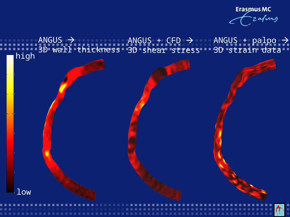
ANGUS 3D wall thickness
ANGUS + CFD 3D shear stress
ANGUS + palpo 3D strain data
high
low
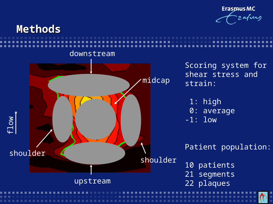
midcap
upstream
shouldershoulder
downstream
flow
Scoring system for shear stress and strain: 1: high 0: average-1: low
Patient population:
10 patients21 segments22 plaques
MethodsMethods
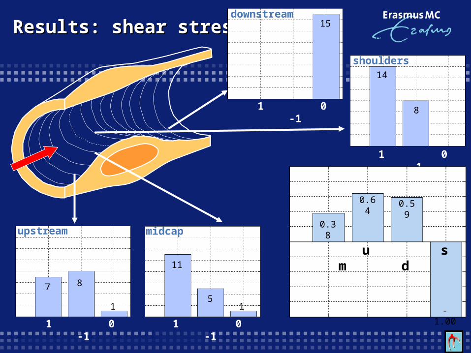
Results: shear stressResults: shear stress
1 0 -1
downstream15
1 0 -1
shoulders14
8
1 0 -1
midcap
11
51
1 0 -1
upstream
1
87
u s m d
0.38
0.64 0.59
-1.00

Results: shear stressResults: shear stress
0.31
0.550.12
-0.47 u s m d
1 0 -1
downstream
9
42
1 0 -1
shoulders
1210
1 0 -1
upstream
8
53
1 0 -1
midcap
4
11
2
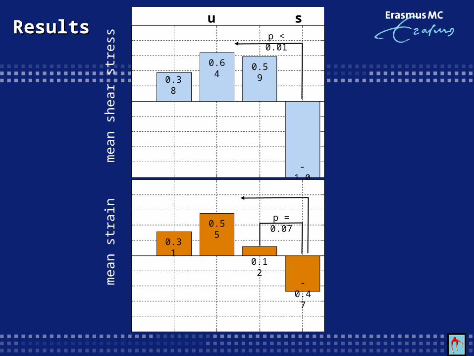
ResultsResults u s m d
mea
n st
rain
p < 0.01
p < 0.01
mea
n sh
ear s
tress
0.38
0.64 0.59
-1.00
p = 0.07
0.31
0.55
0.12
-0.47
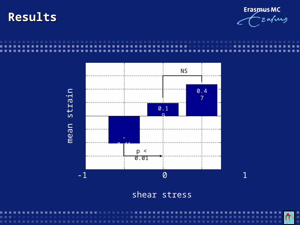
ResultsResults
NS
p < 0.01
mea
n st
rain
shear stress
-1 0 1
0.47
0.19
-0.41
Discussion and conclusions
Low shear stress downstream of a plaque relates to low strain.
Location alone cannot predict where we can find high shear stress and high strain.
High shear stress predicts the location of high strain spots, confirming the hypothesis that high shear stress is related to plaque vulnerability.
Follow-up data have to reveal if this relationship is confirmed.

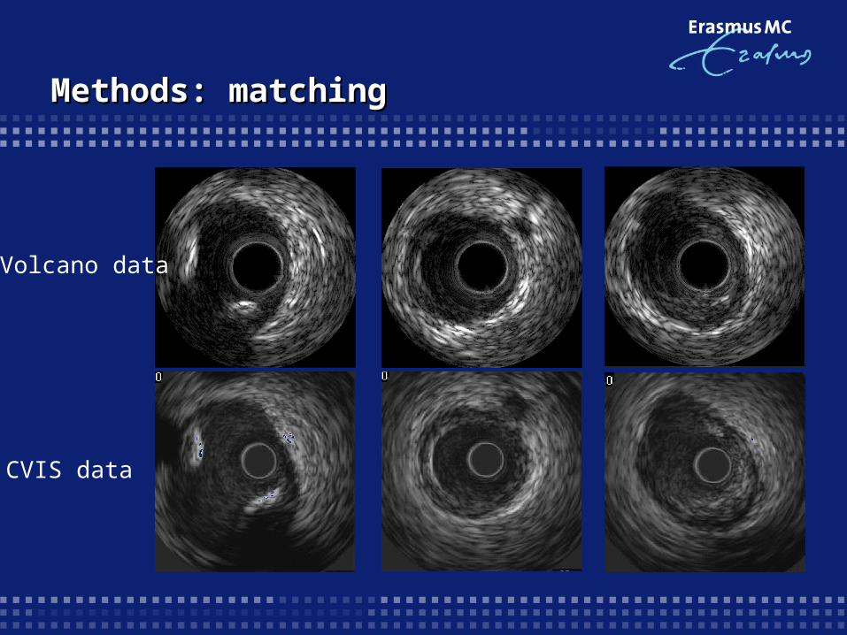
Methods: matchingMethods: matching
Volcano data
CVIS data
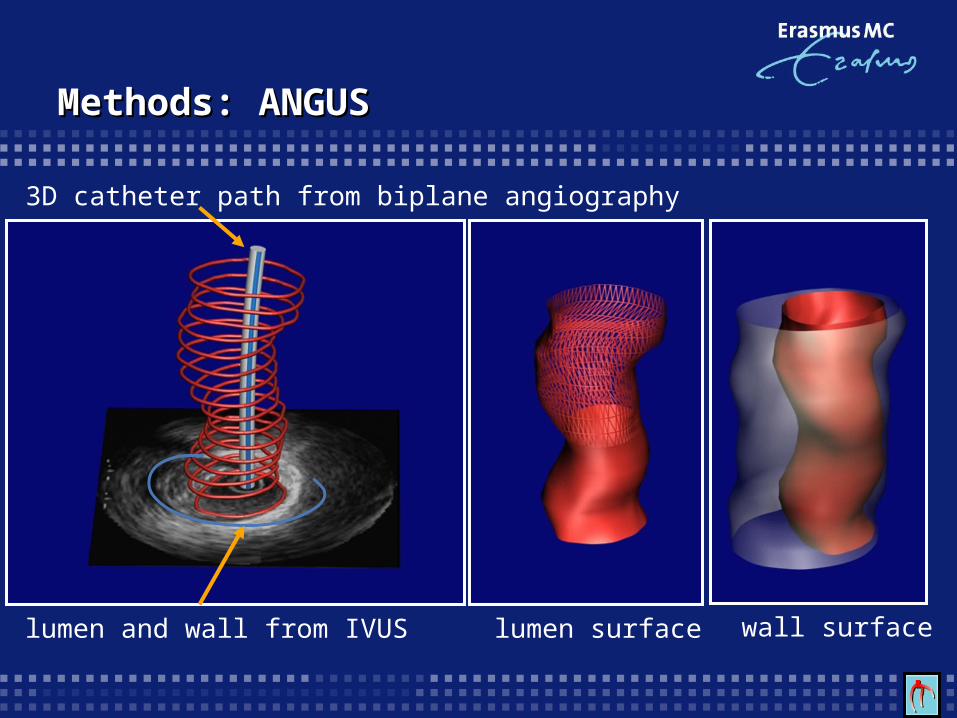
Methods: ANGUSMethods: ANGUS
lumen surface wall surface
3D catheter path from biplane angiography
lumen and wall from IVUS
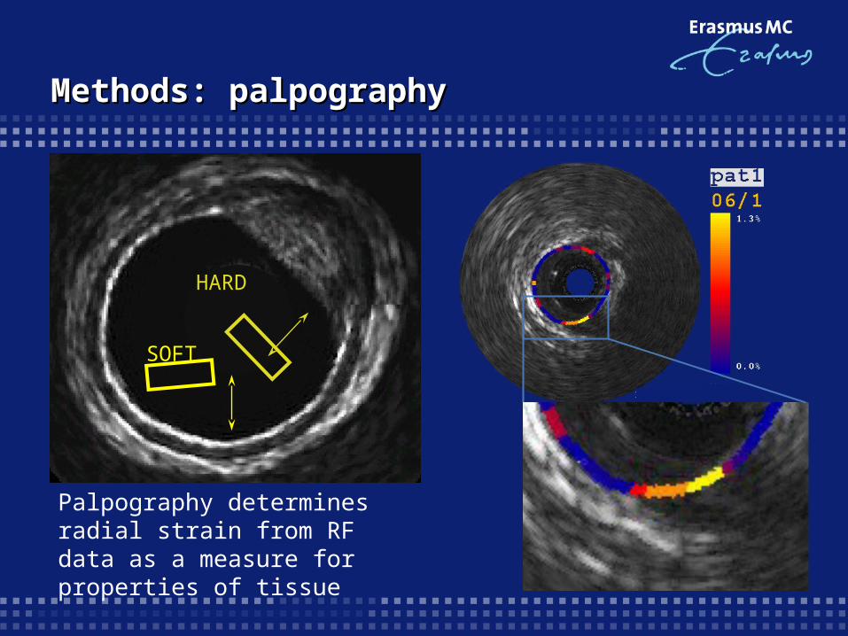
Methods: palpographyMethods: palpography
SOFTHARD
Palpography determines radial strain from RF data as a measure for properties of tissue