Embed Size (px)
Citation preview
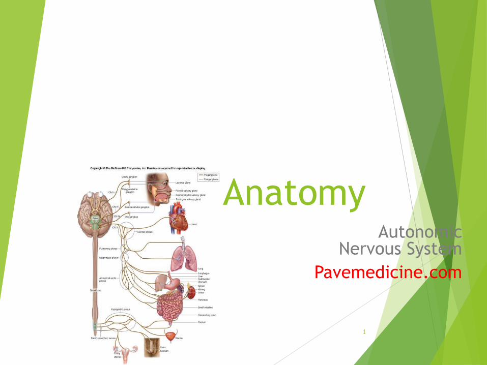
Human AnatomyAutonomic
Nervous System
Pavemedicine.com
1
Autonomic Nervous System
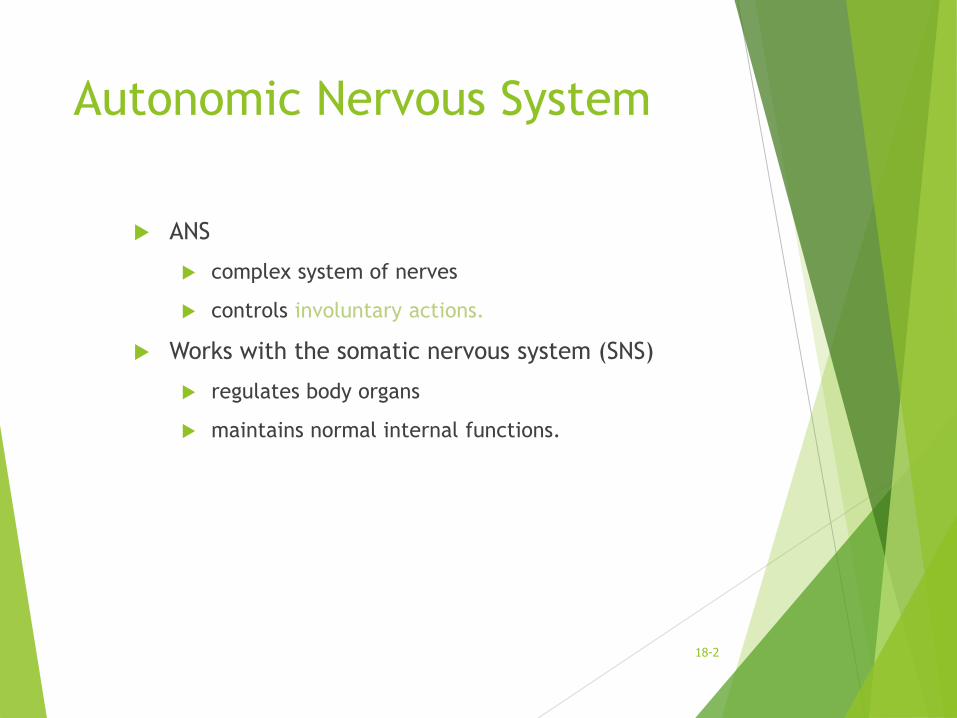
ANS
complex system of nerves
controls involuntary actions.
Works with the somatic nervous system (SNS)
regulates body organs
maintains normal internal functions.
18-2
SNS, PNS, and ANS
SNS and ANS are both part of the peripheral nervous system (PNS).
SNS operates under our conscious control.
ANS functions are involuntary.
18-3

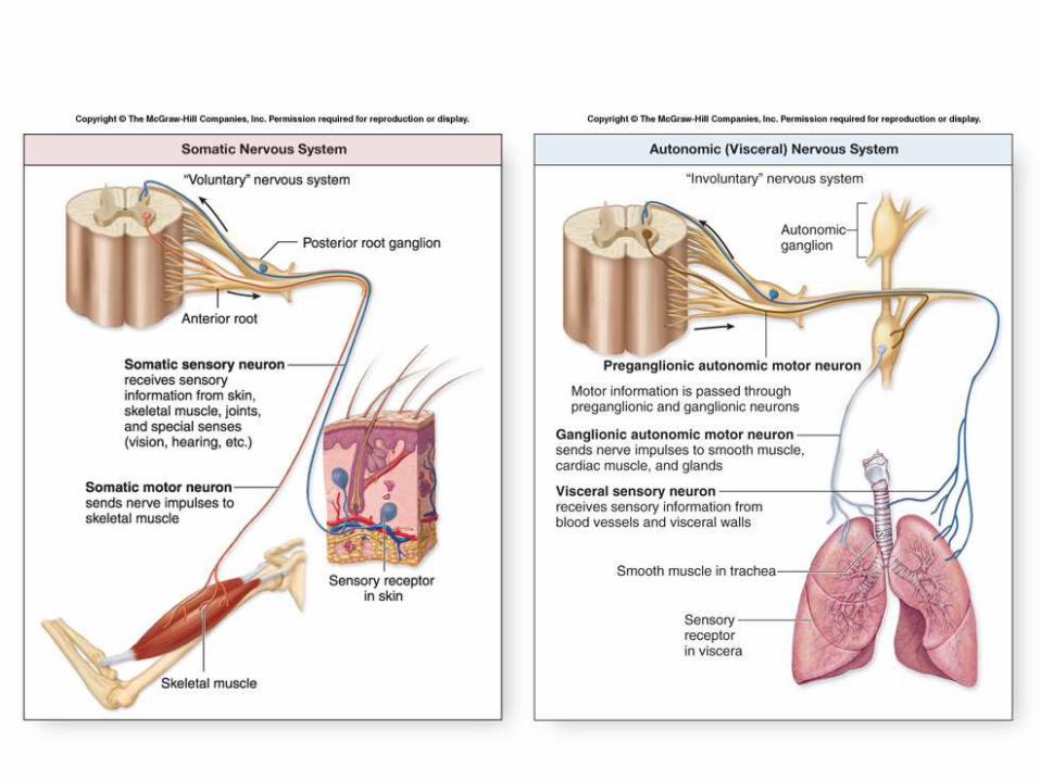
Comparison of SNS and ANS
SNS uses both somatic sensory and somatic motor neurons
Somatic sensory neurons conduct stimulus information from a sensory receptor
Somatic motor neurons innervate skeletal muscle fibers.
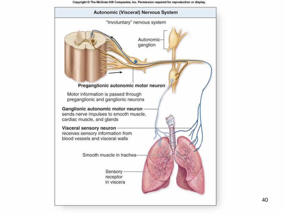
ANS also utilizes sensory and motor neurons.
Visceral sensory neurons provide input to activate the ANS
Visceral motor neurons innervate smooth muscle, cardiac muscle, and glands
18-4
5
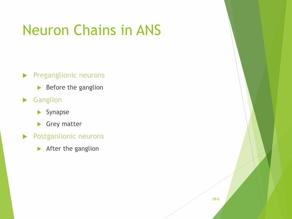
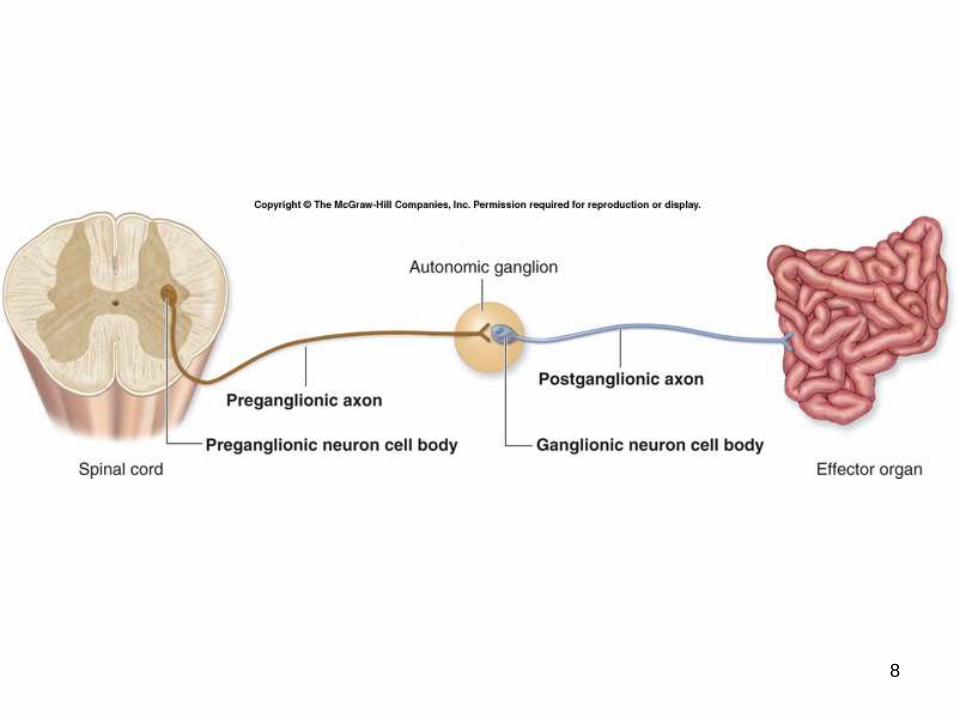
Neuron Chains in ANS
Preganglionic neurons
Before the ganglion
Ganglion
Synapse
Grey matter
Postganlionic neurons
After the ganglion
18-6
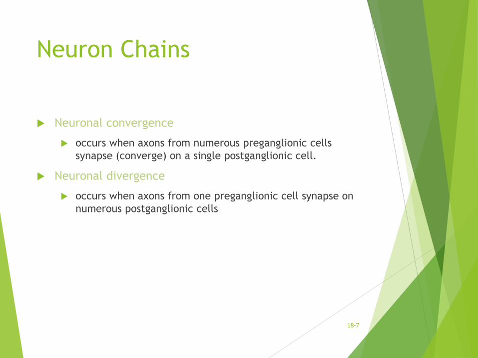
Neuron Chains
Neuronal convergence
occurs when axons from numerous preganglionic cells
synapse (converge) on a single postganglionic cell.
Neuronal divergence
occurs when axons from one preganglionic cell synapse on
numerous postganglionic cells
18-7
8
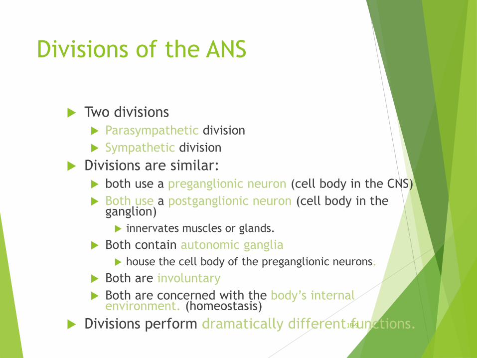
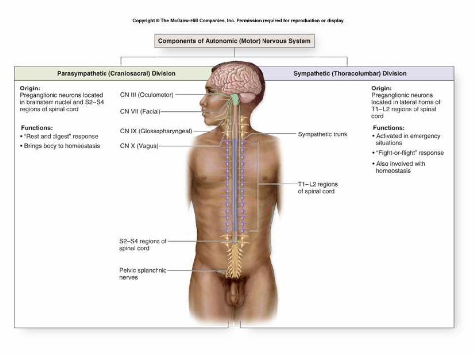
Divisions of the ANS
Two divisions
Parasympathetic division
Sympathetic division
Divisions are similar:
both use a preganglionic neuron (cell body in the CNS)
Both use a postganglionic neuron (cell body in the ganglion)
innervates muscles or glands.
Both contain autonomic ganglia
house the cell body of the preganglionic neurons.
Both are involuntary
Both are concerned with the body’s internal environment. (homeostasis)
Divisions perform dramatically different functions.18-9
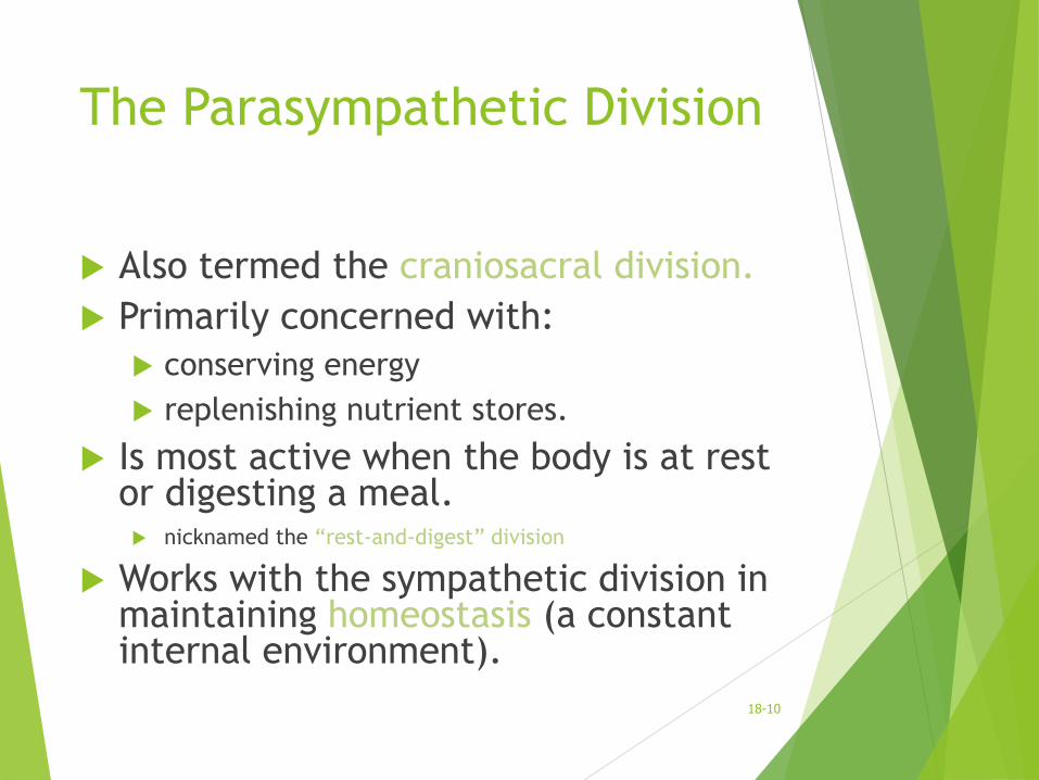
The Parasympathetic Division
Also termed the craniosacral division.
Primarily concerned with:
conserving energy
replenishing nutrient stores.
Is most active when the body is at rest or digesting a meal. nicknamed the “rest-and-digest” division
Works with the sympathetic division in maintaining homeostasis (a constant internal environment).
18-10
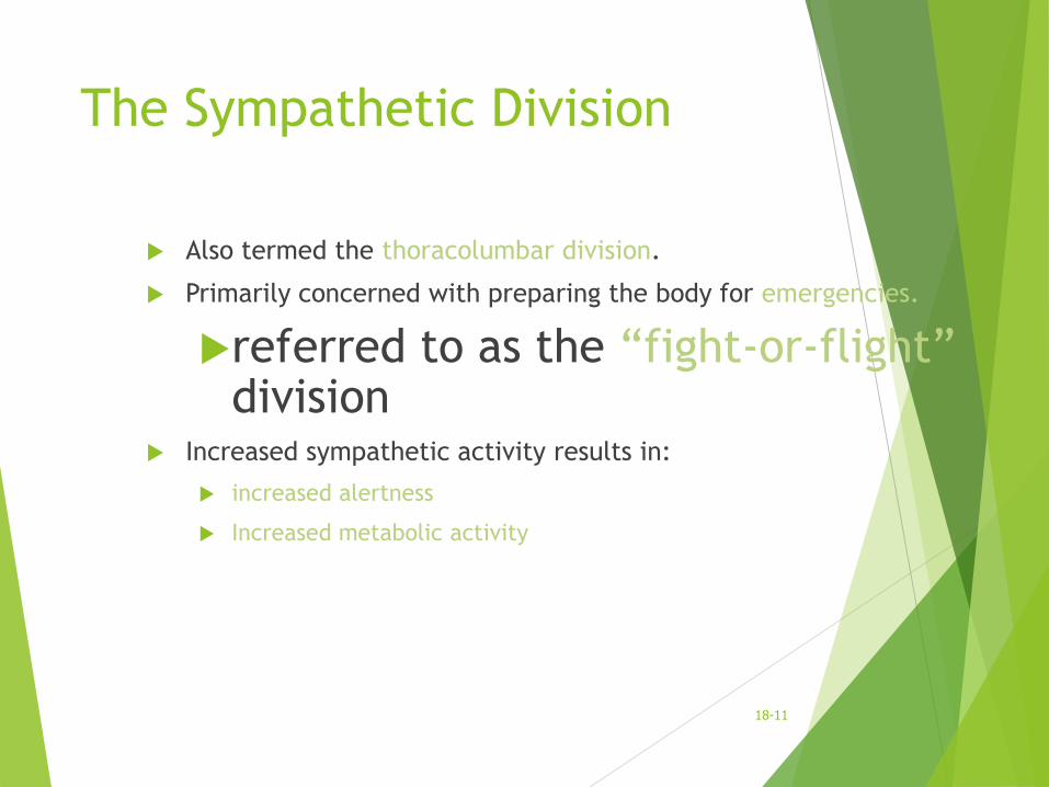
The Sympathetic Division
Also termed the thoracolumbar division.
Primarily concerned with preparing the body for emergencies.
referred to as the “fight-or-flight”division
Increased sympathetic activity results in:
increased alertness
Increased metabolic activity
18-11
12
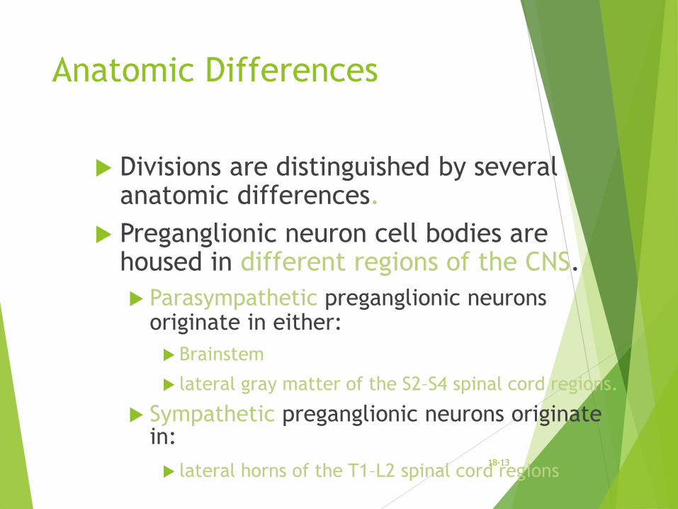
Anatomic Differences
Divisions are distinguished by several anatomic differences.
Preganglionic neuron cell bodies are housed in different regions of the CNS.
Parasympathetic preganglionic neurons originate in either:
Brainstem
lateral gray matter of the S2–S4 spinal cord regions.
Sympathetic preganglionic neurons originate in:
lateral horns of the T1–L2 spinal cord regions18-13
14
Anatomic Differences
Parasympathetic division is structurally simple.
Parasympathetic division is also termed the craniosacral division because its preganglionic neurons are: housed within nuclei in the brainstem
within the lateral gray regions of the S2–S4 spinal cord segments.
Postganglionic neurons in the parasympathetic division are found in
terminal ganglia: are located close to the target organ
intramural ganglia: located within the wall of the target organ
18-15
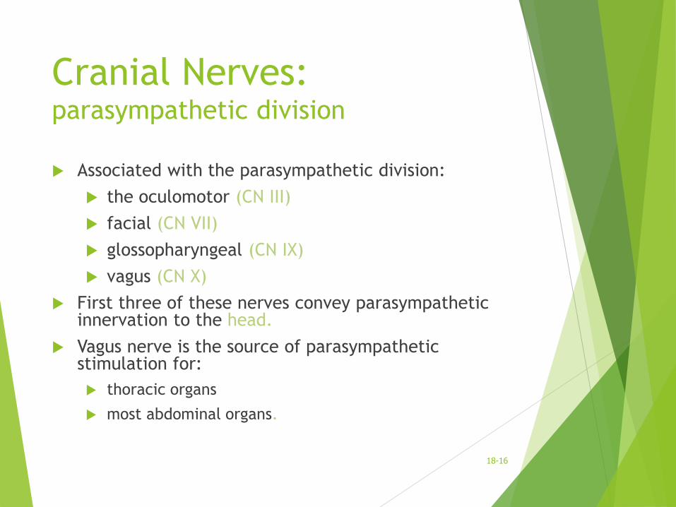
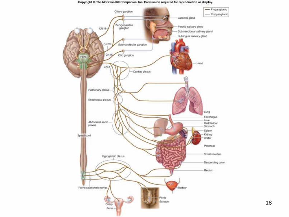
Cranial Nerves: parasympathetic division
Associated with the parasympathetic division:
the oculomotor (CN III)
facial (CN VII)
glossopharyngeal (CN IX)
vagus (CN X)
First three of these nerves convey parasympathetic innervation to the head.
Vagus nerve is the source of parasympathetic stimulation for:
thoracic organs
most abdominal organs.
18-16
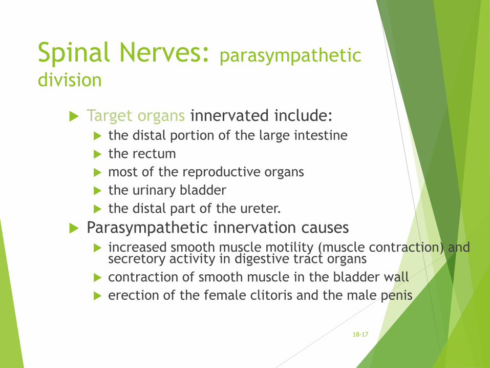
Spinal Nerves: parasympathetic
division
Target organs innervated include: the distal portion of the large intestine
the rectum
most of the reproductive organs
the urinary bladder
the distal part of the ureter.
Parasympathetic innervation causes increased smooth muscle motility (muscle contraction) and
secretory activity in digestive tract organs
contraction of smooth muscle in the bladder wall
erection of the female clitoris and the male penis
18-17
18
Effects and General Functions
of the Parasympathetic
Division Parasympathetic division is most active during
times when the body must process nutrients and conserve energy.
Lack of extensive divergence in preganglionic axons
prevents the mass activation seen in the sympathetic division.
Effects of the parasympathetic nervous system tend to be discrete and localized.
Parasympathetic activity can affect one group of organs without necessarily having to “turn on” all other organs
18-19
Organization and Anatomy of
the Sympathetic Division
Much more complex than the parasympathetic division.
Sympathetic preganglionic neuron cell bodies
housed in the lateral horn of the T1–L2
Preganglionic sympathetic axons:
travel with somatic motor neuron axons
exit the spinal cord
enter first the anterior roots
then the T1–L2 spinal nerves.
Preganglionic sympathetic axons remain with the spinal nerve for a short distance
they branch off and leave the spinal nerve
18-20
Left and Right Sympathetic
Trunks
Immediately anterior to the paired spinal nerves are the left
and right sympathetic trunks.
Each is located immediately lateral to the vertebral column.
A sympathetic trunk is like a pearl necklace:
the “string” of the “necklace” is composed of bundles of
axons
the “pearls” are the sympathetic trunk (or paravertebral)
ganglia
house sympathetic ganglionic neuron cell bodies
18-21
Left and Right Sympathetic
Trunks
One sympathetic trunk ganglion is
approximately associated with each spinal
nerve.
Cervical portions
three sympathetic trunk ganglia
superior, middle, and inferior cervical ganglia
opposed to the eight cervical spinal nerves.
18-22
23
24
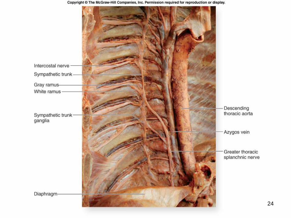
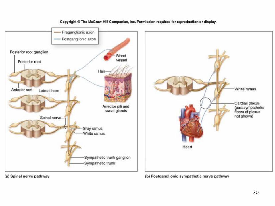
White Rami
Connecting the spinal nerves to each sympathetic
trunk are rami communicantes.
Carry preganglionic sympathetic axons from the
T1–L2 spinal nerves to the sympathetic trunk.
Associated only with the T1–L2 spinal nerves.
Preganglionic axons are myelinated.
the white ramus has a whitish appearance
Similar to “entrance ramps” on a highway.
18-25
Gray Rami
Carry postganglionic sympathetic axons
from the sympathetic trunk to the spinal nerve.
Axons are unmyelinated.
gray rami have a grayish appearance
Similar to “exit ramps” on a highway.
Connect to all spinal nerves.
Sympathetic information that starts in the
thoracolumbar region can be dispersed to all parts of the body.
18-26
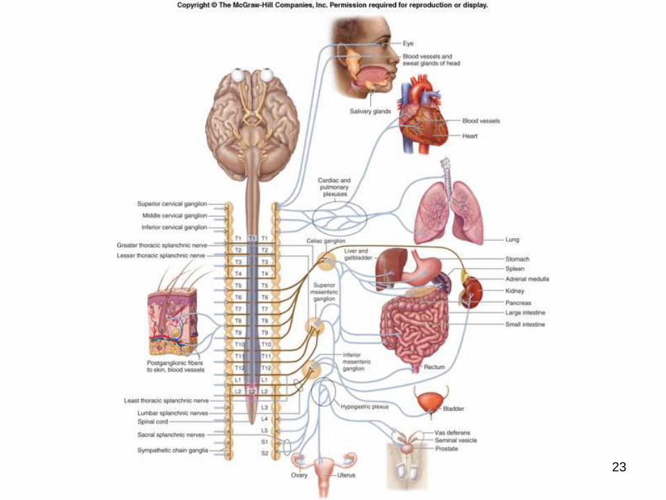
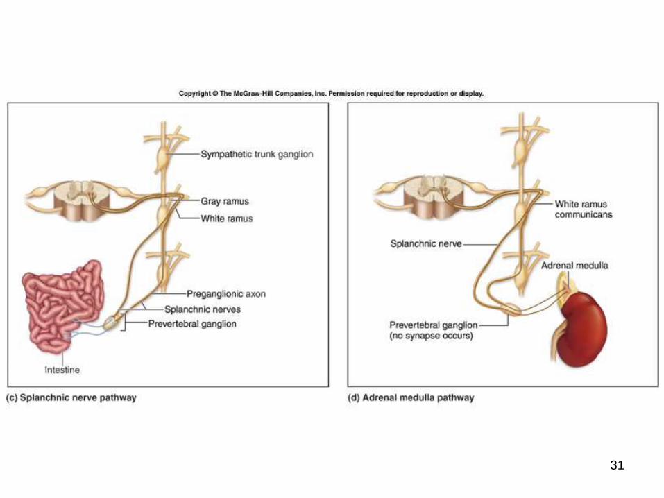
Splanchnic Nerves
Composed of preganglionic sympathetic axons.
Run anteriorly from the sympathetic trunk to most of the viscera.
Should not be confused with the pelvic splanchnic nerves associated with the parasympathetic division.
Larger splanchnic nerves have specific names:
greater thoracic splanchnic nerves
lesser thoracic splanchnic nerves
least thoracic splanchnic nerves
lumbar splanchnic nerves
sacral splanchnic nerves
18-27
Splanchnic Nerves
Terminate in prevertebral (or collateral) ganglia.
Called “prevertebral” because they are immediately anterior to the vertebral column.
Prevertebral ganglia typically cluster around the major abdominal arteries and are named for these arteries.
Example: celiac ganglia cluster around the celiac trunk
Sympathetic postganglionic axons extend away from the ganglia and innervate many of the abdominal organs.
18-28
Types of Prevertebral Ganglia
Differ from the sympathetic trunk ganglia.
Are single structures, rather than paired.
Are anterior to the vertebral column on the anterior surface of
the aorta.
Located only in the abdominopelvic cavity.
Prevertebral ganglia include:
the celiac ganglion
superior mesenteric ganglion
interior mesenteric ganglion.
18-29
30
31
Sympathetic Pathways
Spinal nerve pathway
Postganglionic sympathetic nerve
pathway
The Splanchnic Nerve Pathway
The Adrenal Medulla Pathway
18-32
Fight-or-Flight Function of
the ANS
May involve a single effector or many
effectors.
In mass activation, a large number of
ganglionic neurons activate many effector
organs.
causes a heightened sense of alertness due to
stimulation of the reticular activation system
18-33
Dual Innervation by the
Parasympathetic and Sympathetic
Divisions of the ANS
Innervate organs through specific axon bundles called autonomic plexuses.
Communication by chemical messengers, called neurotransmitters.
specific in each division of the autonomic
nervous system
Usually all organs are innervated by both divisions
of the autonomic nervous system.
Maintains homeostasis through autonomic reflexes that occur in the innervated organs.
18-34
Autonomic Plexuses
Collections of sympathetic postganglionic
axons and parasympathetic preganglionic
axons, as well as some visceral sensory
axons.
Close to one another, but they do not
interact or synapse with one another.
Provide a complex innervation pattern to
their target organs.
18-35
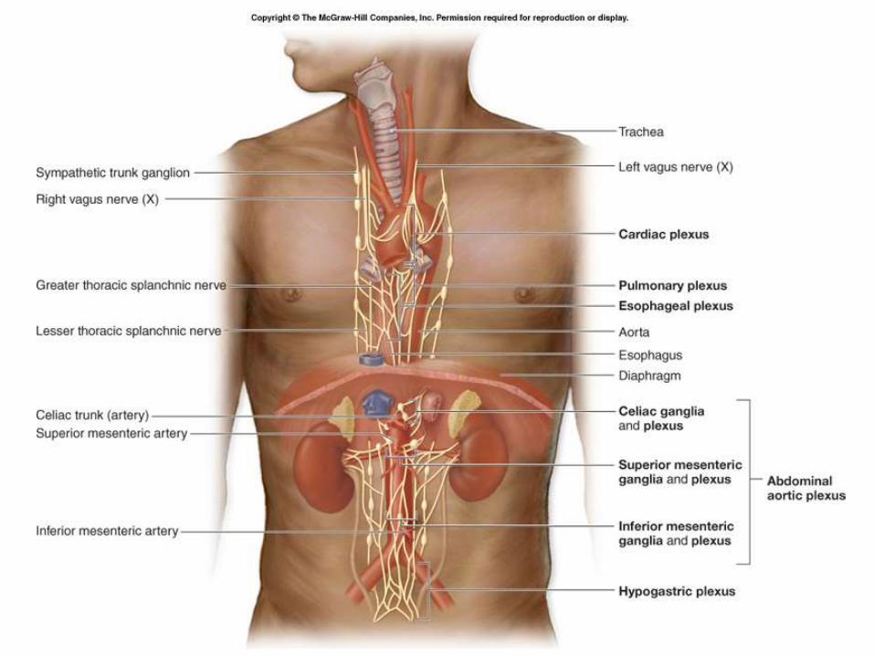
36
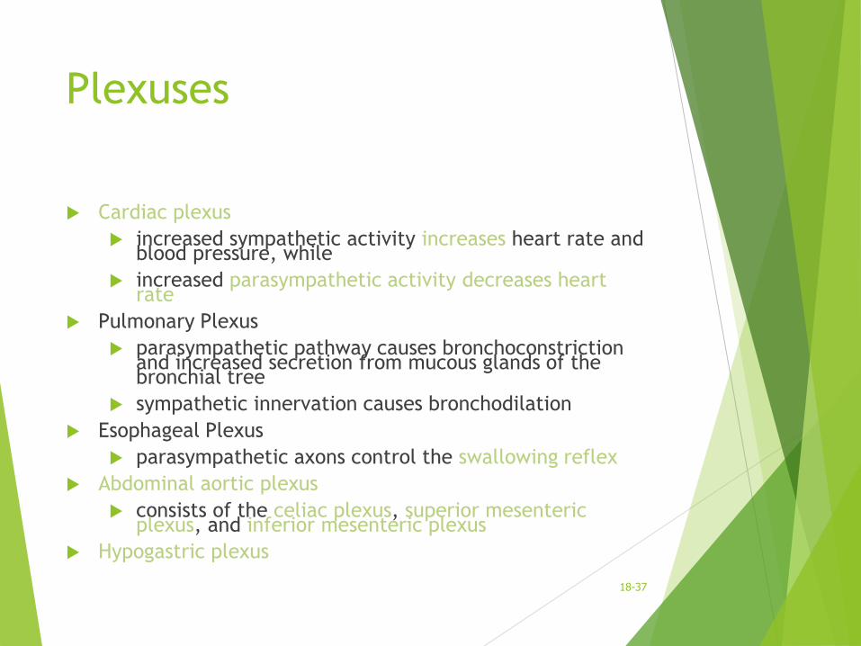
Plexuses
Cardiac plexus
increased sympathetic activity increases heart rate and blood pressure, while
increased parasympathetic activity decreases heart rate
Pulmonary Plexus
parasympathetic pathway causes bronchoconstriction and increased secretion from mucous glands of the bronchial tree
sympathetic innervation causes bronchodilation
Esophageal Plexus
parasympathetic axons control the swallowing reflex
Abdominal aortic plexus
consists of the celiac plexus, superior mesenteric plexus, and inferior mesenteric plexus
Hypogastric plexus
18-37
Neurotransmitters and
Receptors
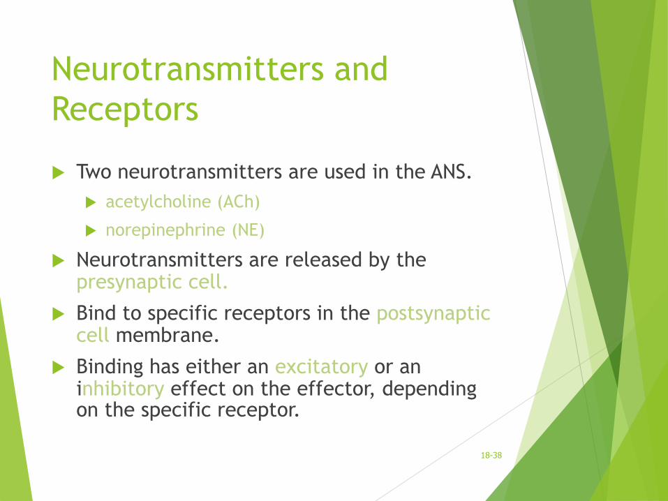
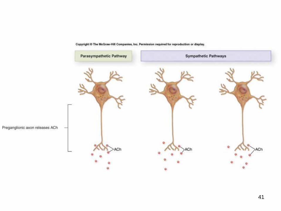
Two neurotransmitters are used in the ANS.
acetylcholine (ACh)
norepinephrine (NE)
Neurotransmitters are released by the presynaptic cell.
Bind to specific receptors in the postsynaptic cell membrane.
Binding has either an excitatory or an inhibitory effect on the effector, depending on the specific receptor.
18-38
Neurotransmitters
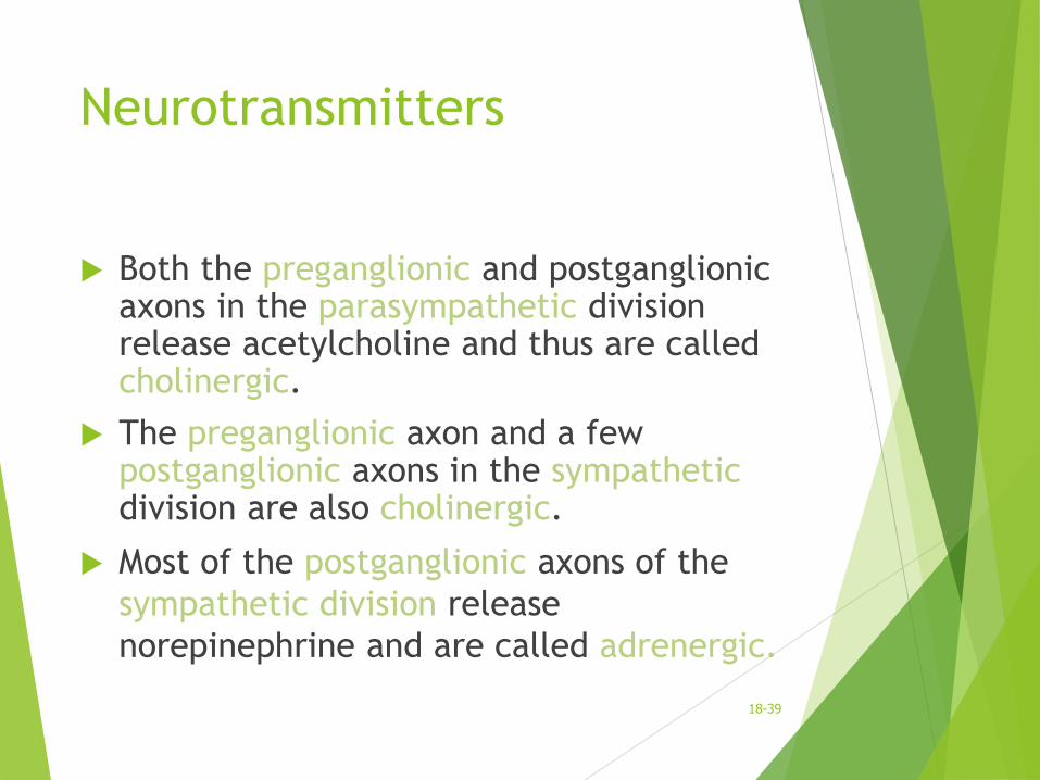
Both the preganglionic and postganglionic axons in the parasympathetic division release acetylcholine and thus are called cholinergic.
The preganglionic axon and a few postganglionic axons in the sympatheticdivision are also cholinergic.
Most of the postganglionic axons of the
sympathetic division release
norepinephrine and are called adrenergic.
18-39
40
41
Dual Innervation
Many visceral effectors are innervated by
postganglionic axons from both ANS divisions.
Actions of the divisions usually oppose
each other.
exert antagonistic effects on the same organ
Opposing effects are also achieved by
increasing or decreasing activity in one
division.
18-42
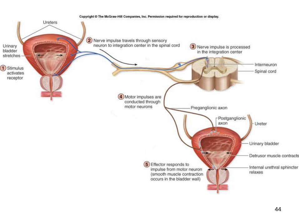
Autonomic Reflexes
ANS helps maintain homeostasis through the involuntary activity of autonomic reflexes or visceral reflexes.
Consist of smooth muscle contractions, cardiac muscle contractions, or secretion by glands that are mediated by autonomic reflex arcs in response to a specific stimulus.
Example: micturition reflex, which partly controls the release of urine
Other reflexes include alteration of heart rate, changes in respiratory rate and depth, regulation of digestive system activities, and alteration of pupil diameter.
Comparable to spinal reflexes.
Classic autonomic reflex involves the reduction of blood pressure.
18-43
44
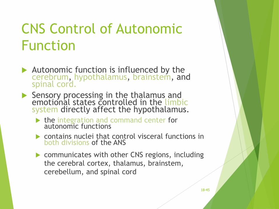
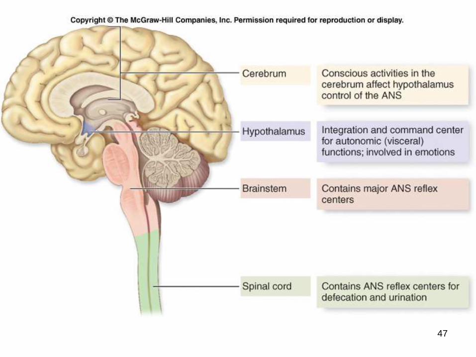
CNS Control of Autonomic
Function
Autonomic function is influenced by the cerebrum, hypothalamus, brainstem, and spinal cord.
Sensory processing in the thalamus and emotional states controlled in the limbic system directly affect the hypothalamus.
the integration and command center for autonomic functions
contains nuclei that control visceral functions in both divisions of the ANS
communicates with other CNS regions, including the cerebral cortex, thalamus, brainstem, cerebellum, and spinal cord
18-45
CNS Control of Autonomic
Function
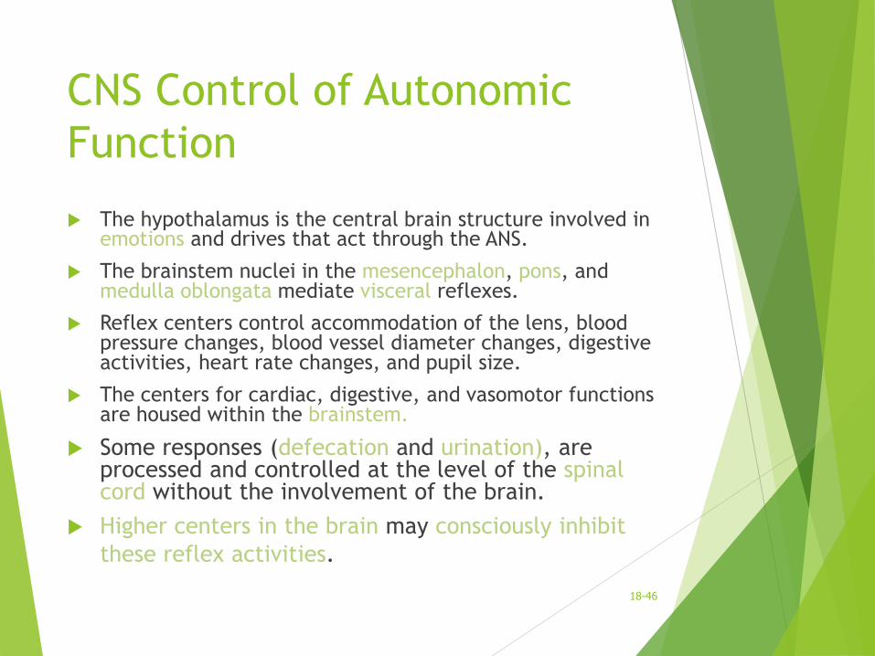
The hypothalamus is the central brain structure involved in emotions and drives that act through the ANS.
The brainstem nuclei in the mesencephalon, pons, and medulla oblongata mediate visceral reflexes.
Reflex centers control accommodation of the lens, blood pressure changes, blood vessel diameter changes, digestive activities, heart rate changes, and pupil size.
The centers for cardiac, digestive, and vasomotor functions are housed within the brainstem.
Some responses (defecation and urination), are processed and controlled at the level of the spinal cord without the involvement of the brain.
Higher centers in the brain may consciously inhibit
these reflex activities.
18-46
47