Embed Size (px)
Citation preview
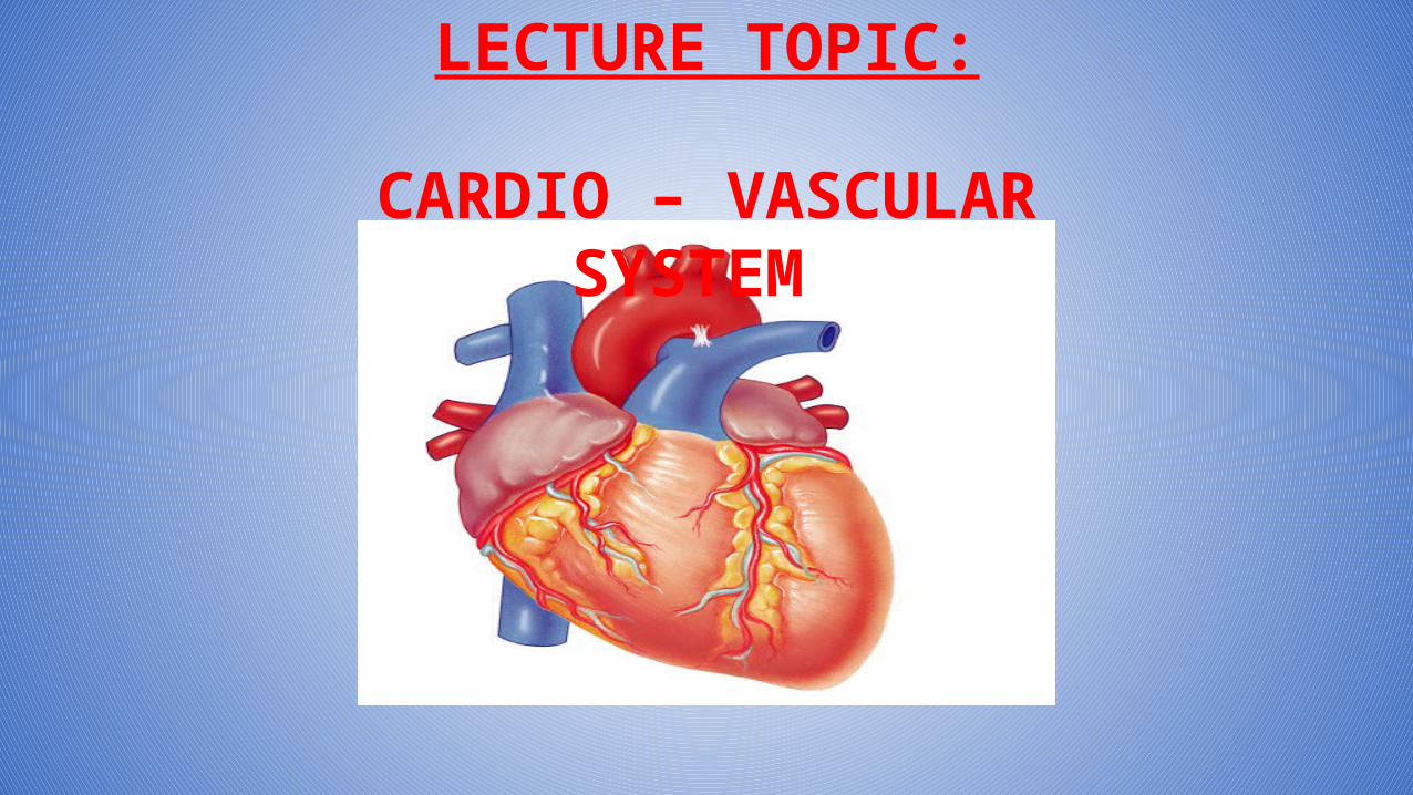
LECTURE TOPIC:
CARDIO – VASCULAR SYSTEM
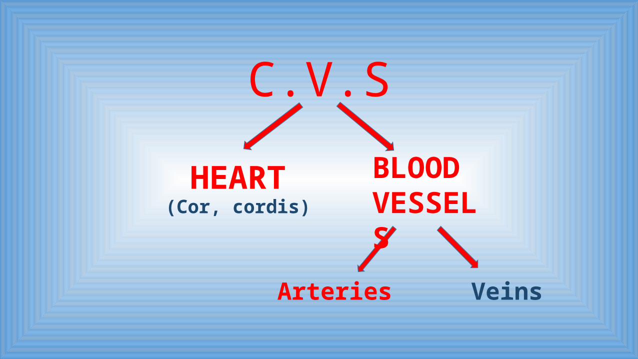
C.V.S
HEART BLOOD VESSELS
Arteries Veins
(Cor, cordis)
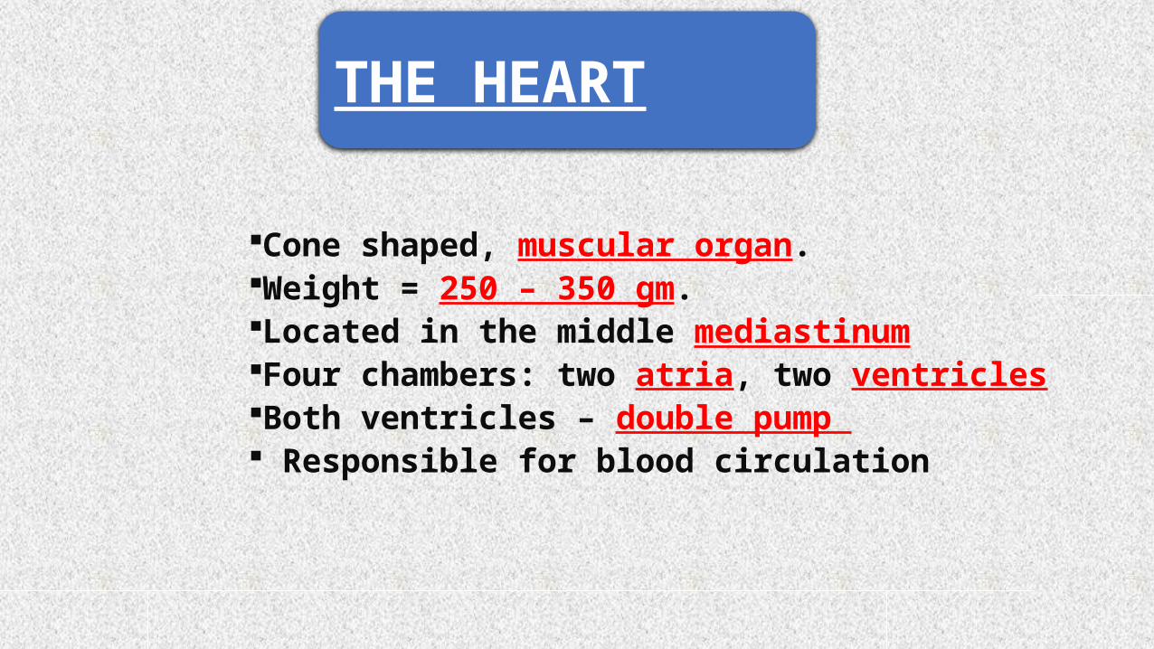
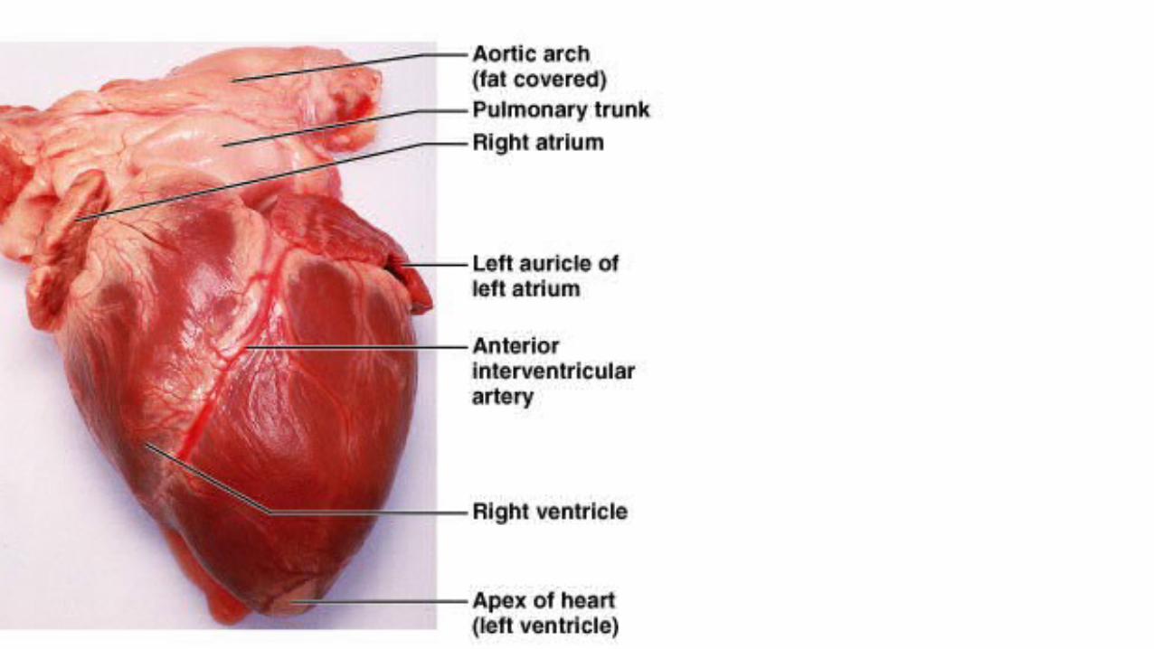
Cone shaped, muscular organ.Weight = 250 – 350 gm.Located in the middle mediastinumFour chambers: two atria, two ventriclesBoth ventricles – double pump Responsible for blood circulation
THE HEART
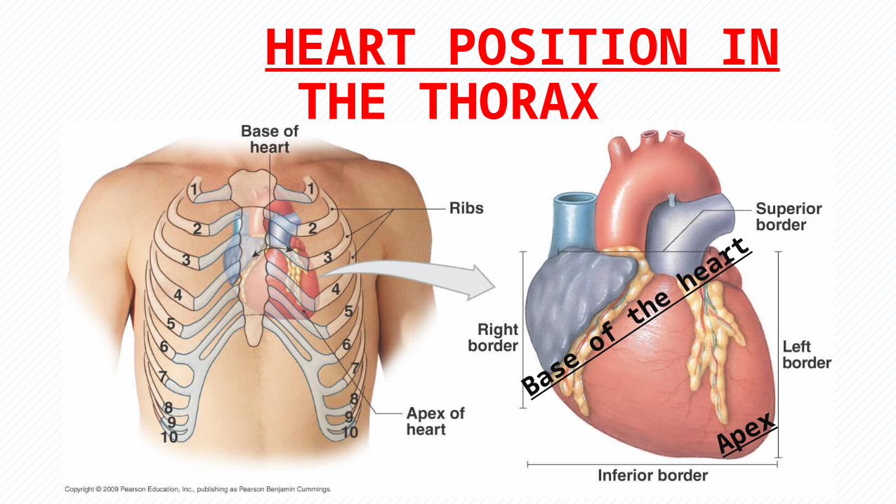
HEART POSITION IN THE THORAX
Apex
Base of the heart
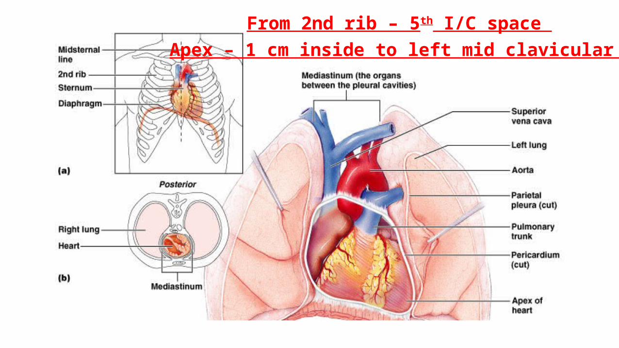
From 2nd rib – 5th I/C space Apex – 1 cm inside to left mid clavicular line
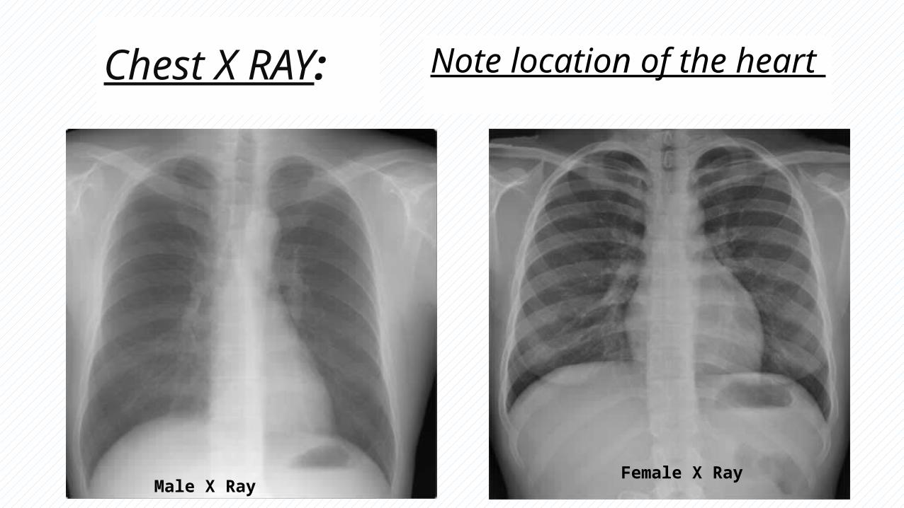
Chest X RAY:
Male X RayFemale X Ray
Note location of the heart
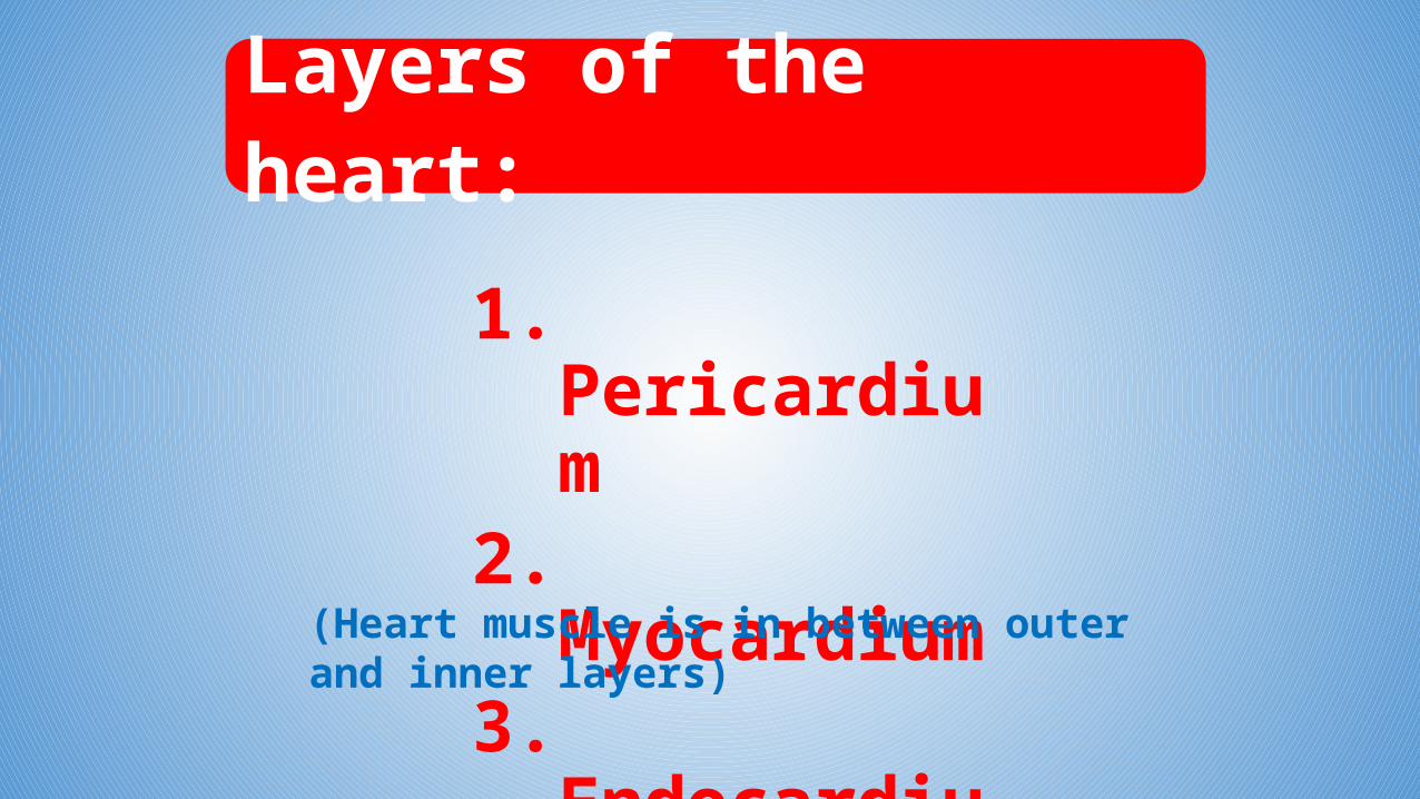
Layers of the heart:
1. Pericardium2. Myocardium3. Endocardium
(Heart muscle is in between outer and inner layers)
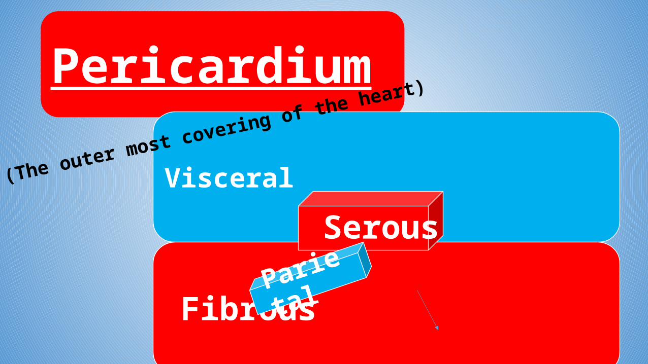
Pericardium
Visceral
Fibrous
(The outer most covering of the heart)
Serous
Parietal
(Visceral = Epicardium)
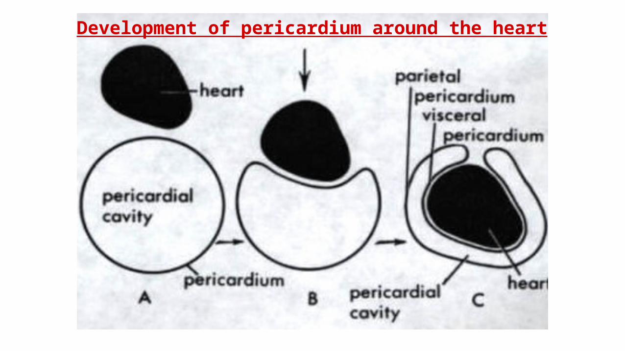
Development of pericardium around the heart
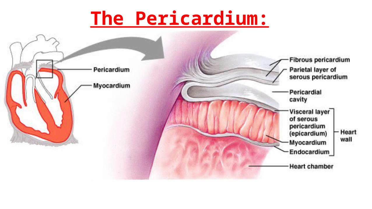
The Pericardium:
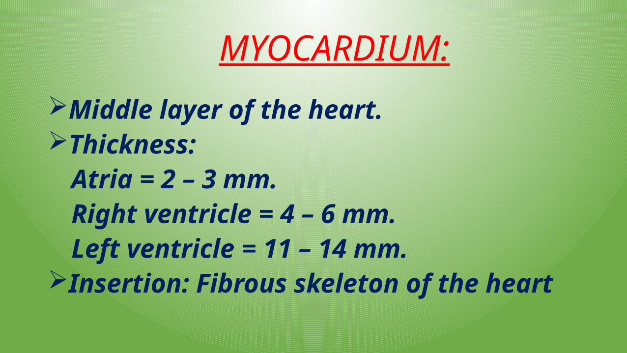
MYOCARDIUM:
Middle layer of the heart.Thickness:
Atria = 2 – 3 mm.Right ventricle = 4 – 6 mm.Left ventricle = 11 – 14 mm.
Insertion: Fibrous skeleton of the heart
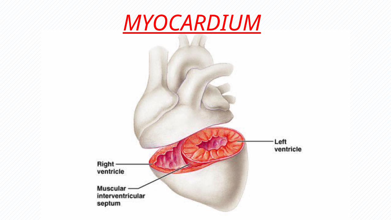
MYOCARDIUM
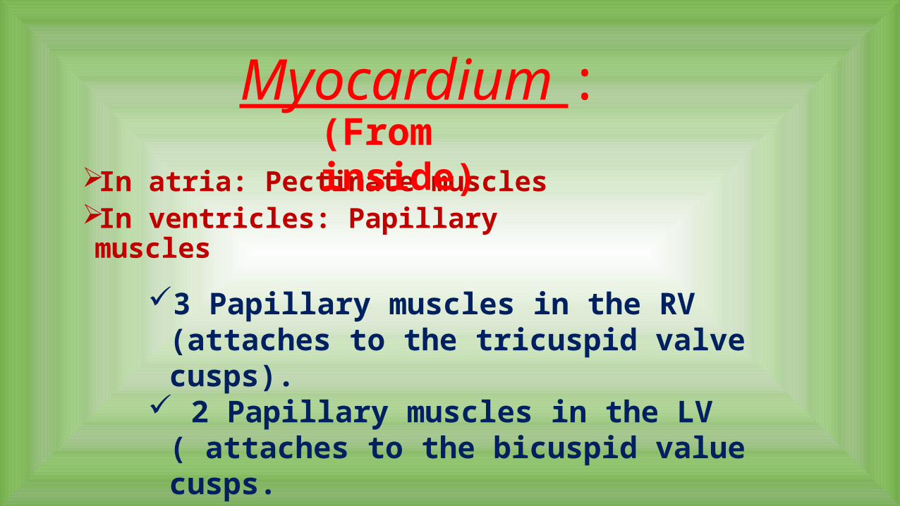
Myocardium :
In atria: Pectinate musclesIn ventricles: Papillary muscles
3 Papillary muscles in the RV (attaches to the tricuspid valve cusps).
2 Papillary muscles in the LV ( attaches to the bicuspid value cusps.
(From inside)
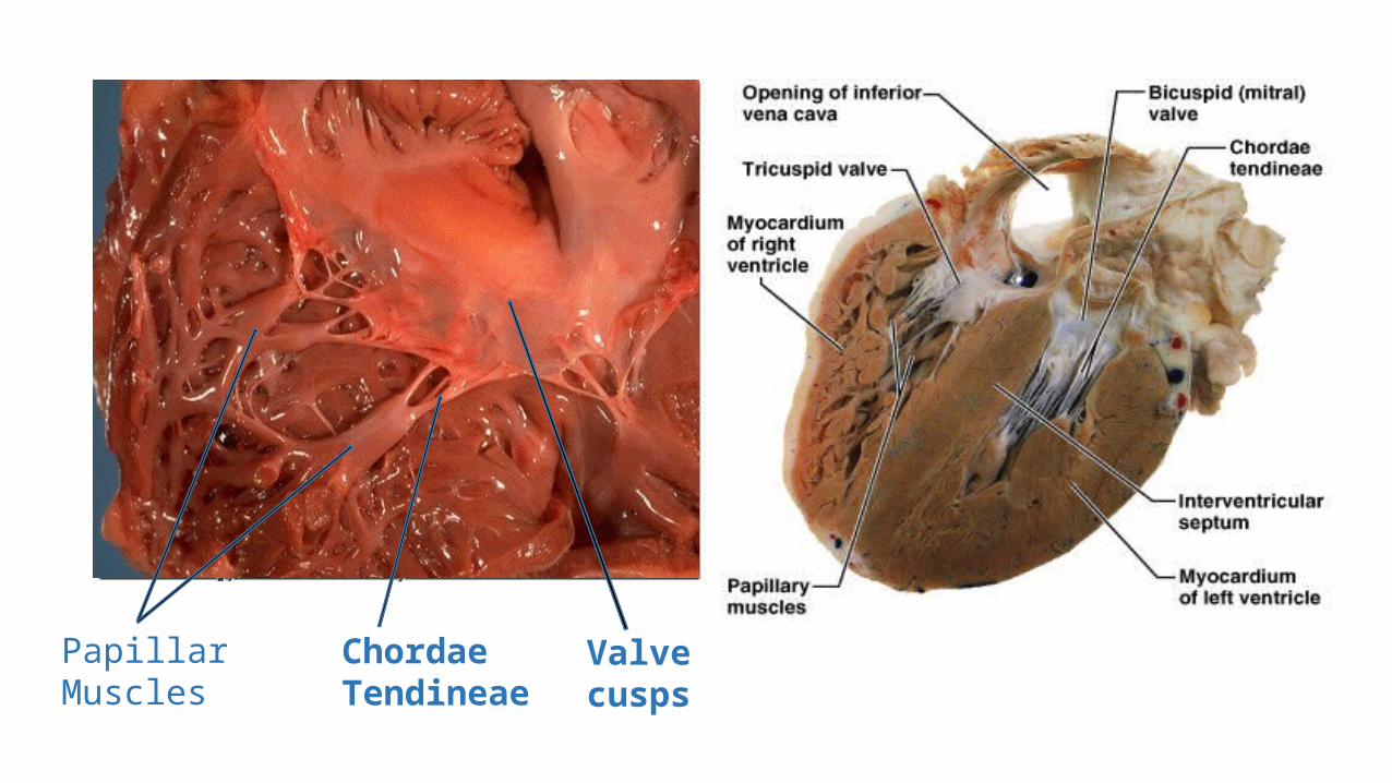
MiddlePapillary Muscles
Chordae Tendineae
Valve cusps
ENDOCARDIUM:
• Inner layer of the heart.
•Covers hart chambers from inside.
• Formed by endothelia.
•Participates in the formation of heart valves.
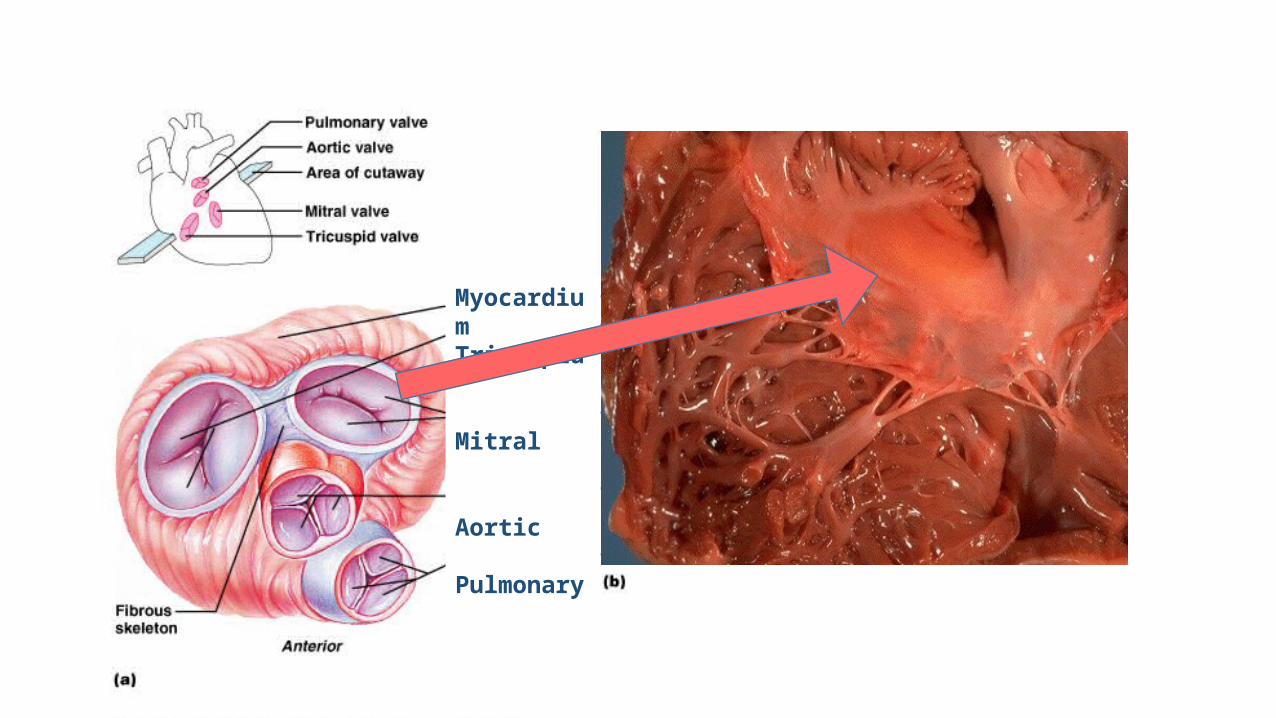
Myocardium Tricuspid
Mitral
Aortic
Pulmonary
Right Atrium
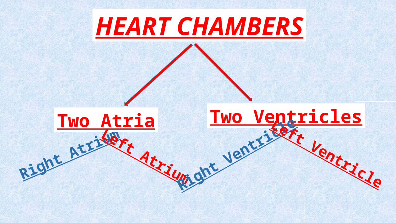
HEART CHAMBERS
Two Atria Two VentriclesLeft Atrium Right Ventric
le Left Ventricle
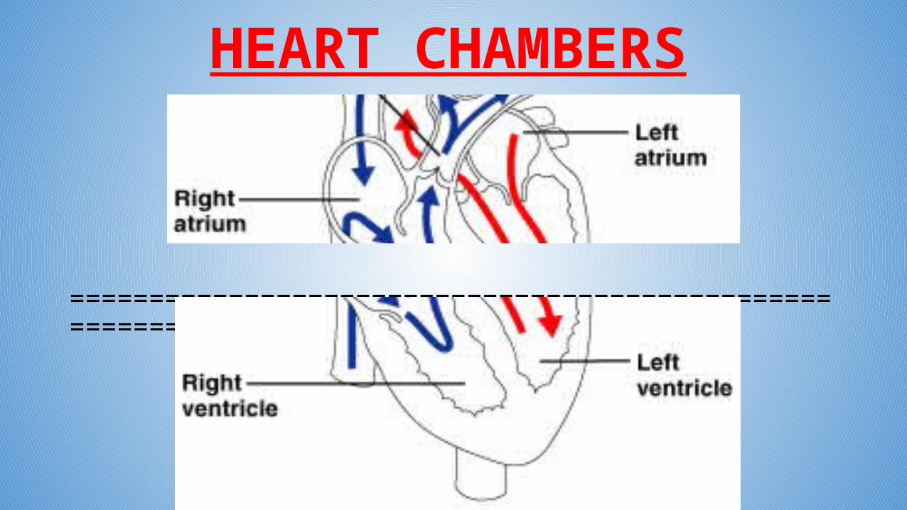
HEART CHAMBERS
=======================================================
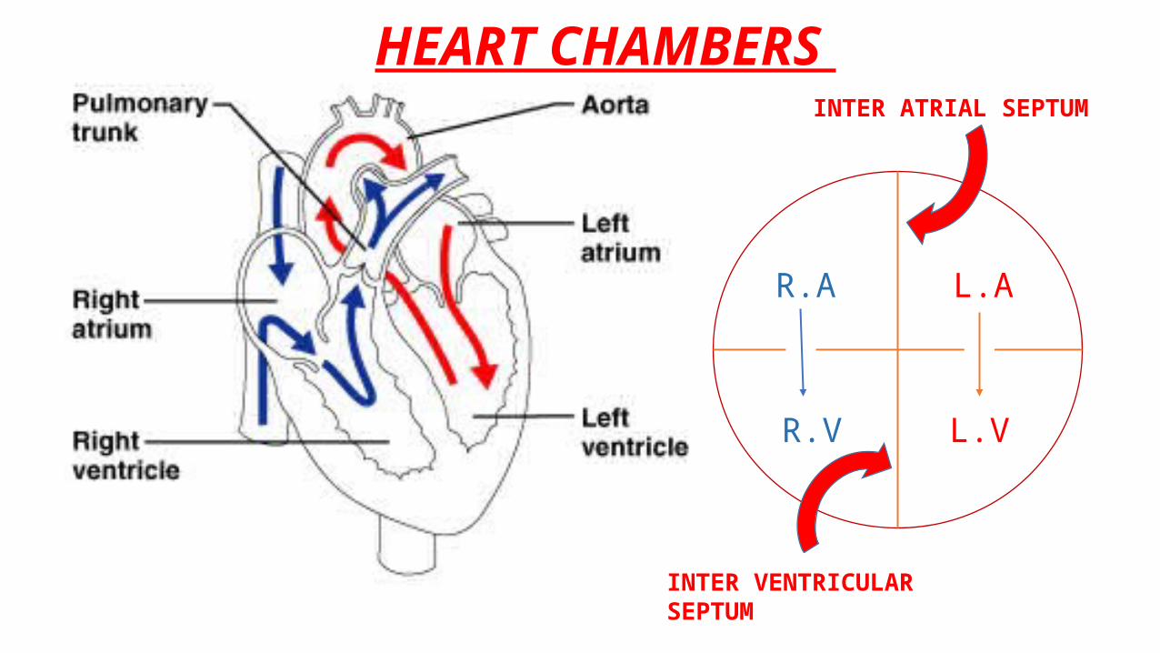
R.A
R.V
L.A
L.V
R R
INTER VENTRICULAR SEPTUM
INTER ATRIAL SEPTUM
HEART CHAMBERS
HEART VALVES:
Four Valves in the heart.Open and close (heart beating)
Right AV-Valve
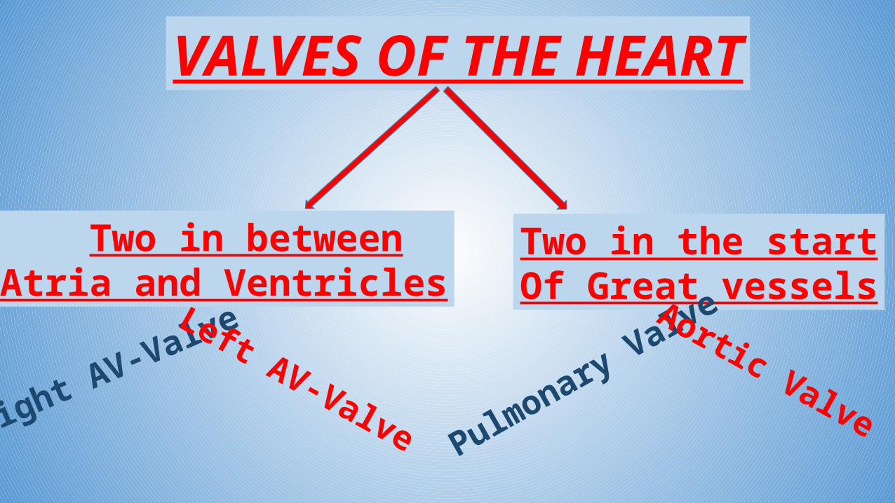
VALVES OF THE HEART
Two in betweenAtria and Ventricles
Two in the startOf Great vessels
Left AV-Valve Pulmonary ValveAortic Valve
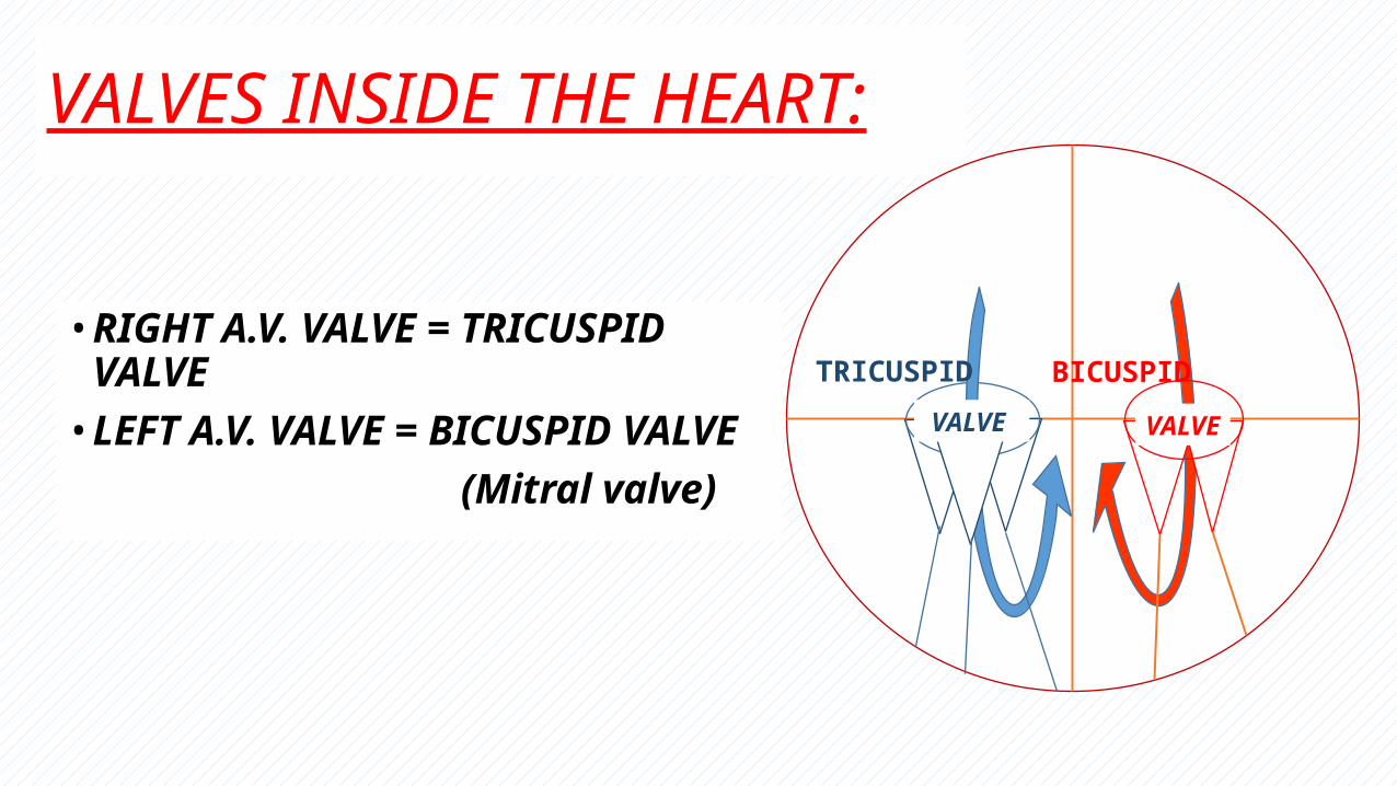
VALVES INSIDE THE HEART:
• RIGHT A.V. VALVE = TRICUSPID VALVE• LEFT A.V. VALVE = BICUSPID VALVE (Mitral valve)
R RVALVE VALVE
TRICUSPID BICUSPID
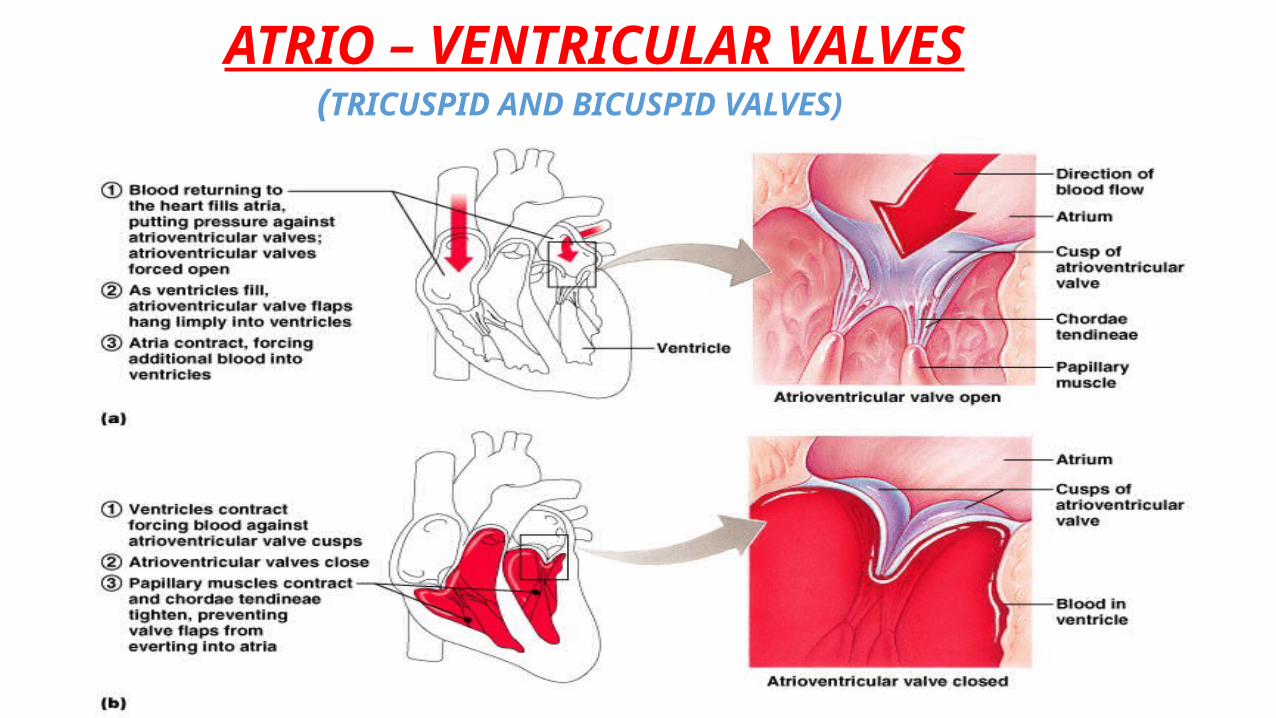
ATRIO – VENTRICULAR VALVES(TRICUSPID AND BICUSPID VALVES)
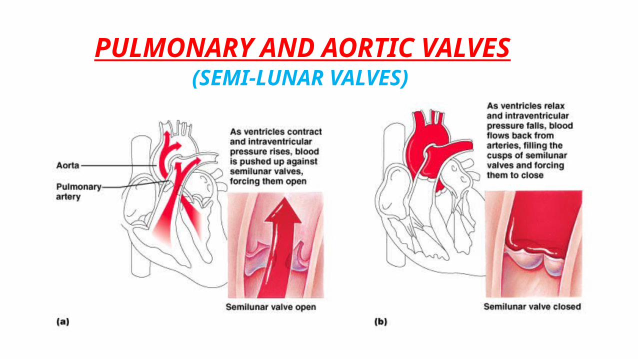
PULMONARY AND AORTIC VALVES(SEMI-LUNAR VALVES)
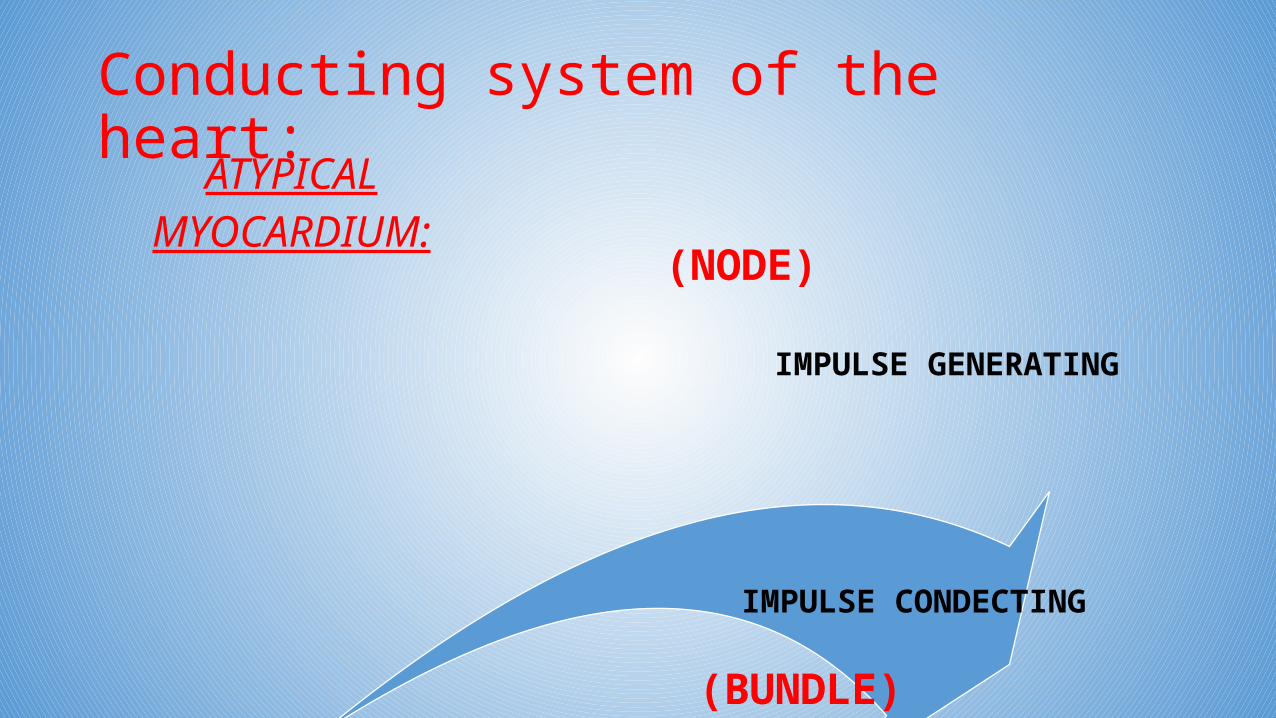
Conducting system of the heart:ATYPICAL
MYOCARDIUM:
IMPULSE GENERATING
IMPULSE CONDECTING
(NODE)
(BUNDLE)
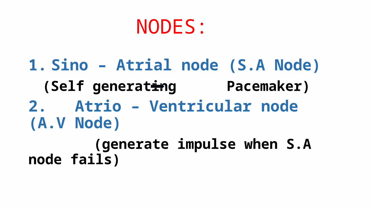
NODES:
1. Sino – Atrial node (S.A Node)(Self generating Pacemaker)
2. Atrio – Ventricular node (A.V Node) (generate impulse when S.A node fails)
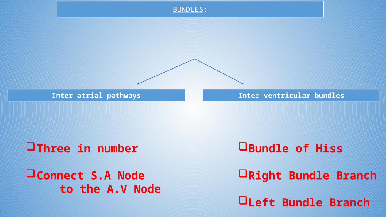
BUNDLES:
Inter atrial pathways Inter ventricular bundles
Three in number
Connect S.A Node to the A.V Node
Bundle of Hiss
Right Bundle Branch
Left Bundle Branch
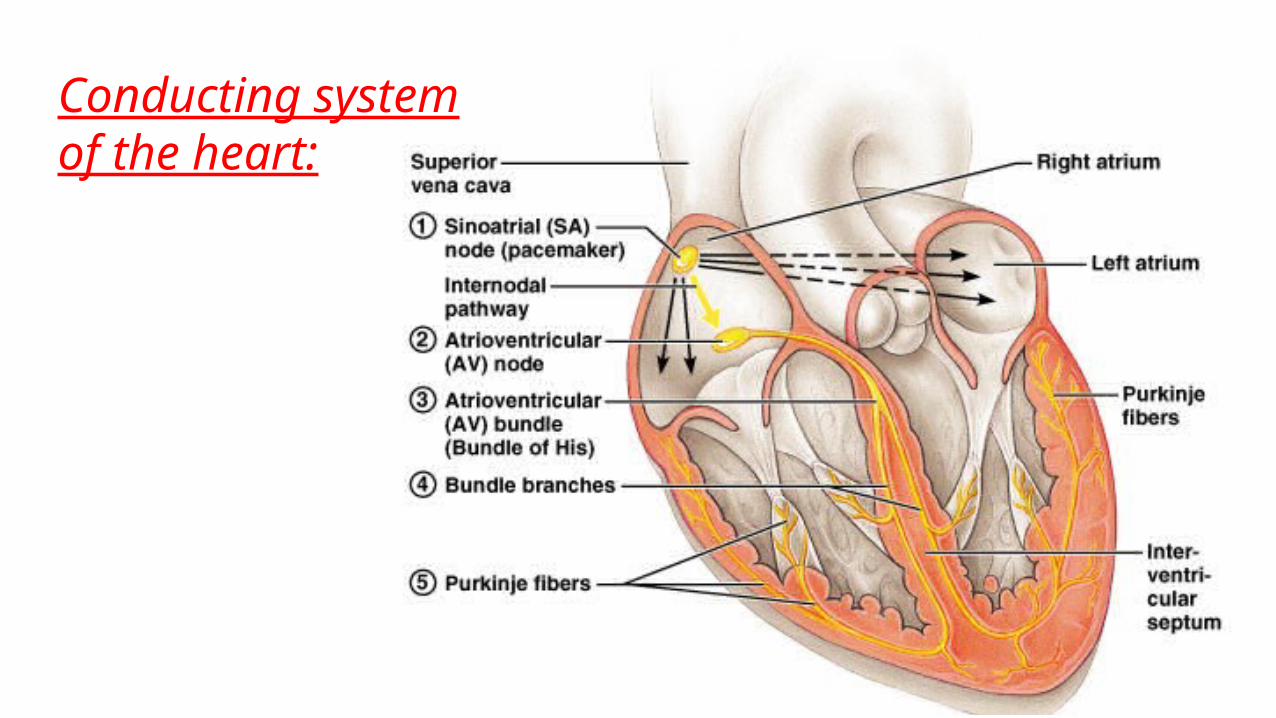
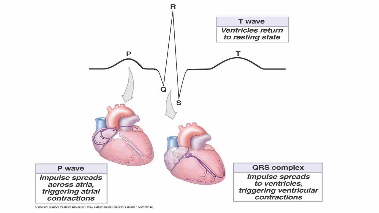
Conducting systemof the heart:
BLOOD SUPLY OF THE HEART
1. RIGHT CORONARY ARTERY
2. LEFT CORONARY ARTERY
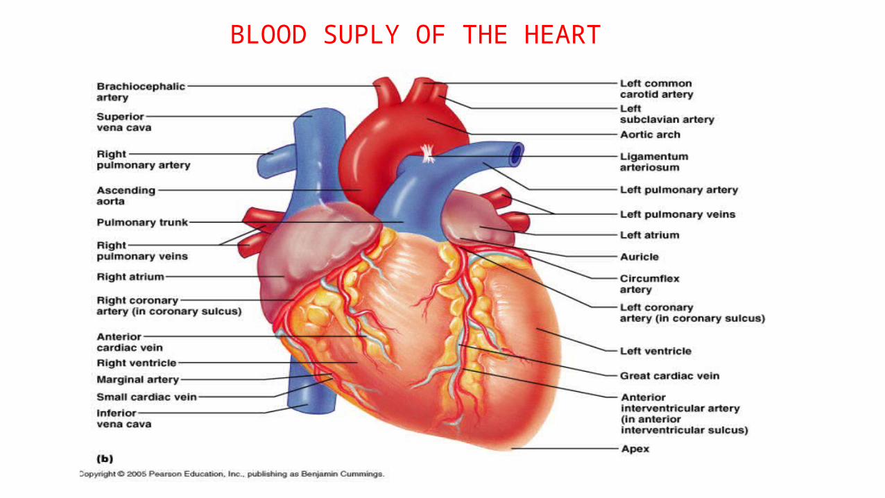
BLOOD SUPLY OF THE HEART
BLOODSUPLYOF THEHEART

MEDONLINE
thank you for attention.
THE END!

![-ravichandran@uiowa.edu] CVS Health (CVS) September … · Through the above service, CVS helps clients in designing ... Improvement, and Modernization ... prescriptions at CVS Pharmacy](https://img.pdfslide.net/doc/110x75/5b5140327f8b9a056a8bdae7/-ravichandranuiowaedu-cvs-health-cvs-september-through-the-above-service.jpg)


![CARDIO-VASCULAR SYSTEM [CVS] FUNCTIONAL ANATOMY OF HEART](https://img.pdfslide.net/doc/110x75/56813d0f550346895da6c769/cardio-vascular-system-cvs-functional-anatomy-of-heart.jpg)