Embed Size (px)
Citation preview
Immunity
Dr. TAREK NASRALA
AL AZHAR
What is Immunity?
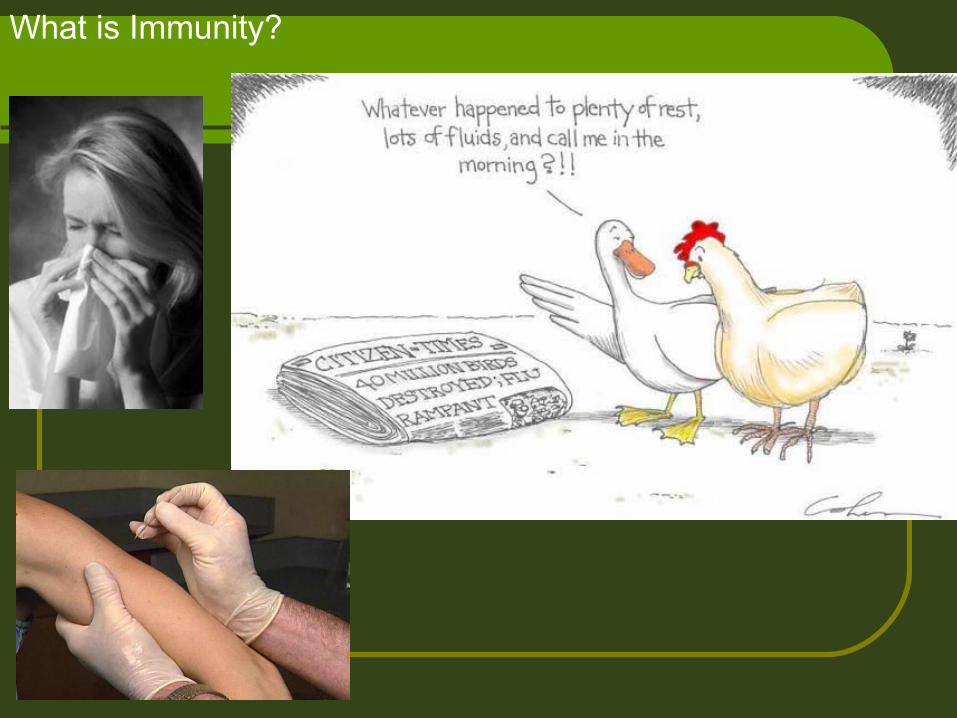
The Immune System
An animal must defend itself from the many dangerous pathogens it may encounter in the environment.
The Immune System
A host is susceptible to a parasite if it can’t eliminate a parasite before it becomes established.The parasite is infective.
The host is resistant if it is able to prevent establishment of the parasite.The parasite is noninfective.
The Immune System
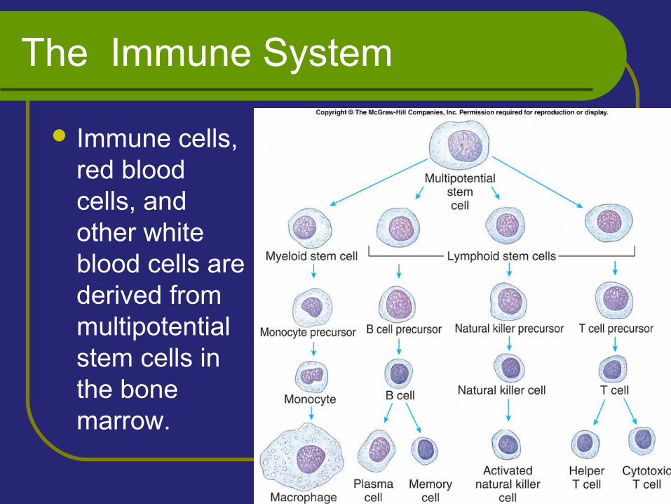
Immune cells, red blood cells, and other white blood cells are derived from multipotential stem cells in the bone marrow.
The Immune System
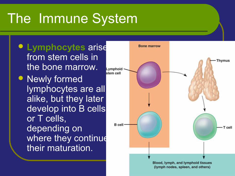
Lymphocytes arise from stem cells in the bone marrow.
Newly formed lymphocytes are all alike, but they later develop into B cells or T cells, depending on where they continue their maturation.
The Immune System
As B and T cells are maturing in the bone marrow and thymus, their antigen receptors are tested for possible self-reactivity.
Lymphocytes bearing receptors for antigens already present in the body are destroyed by apoptosis or rendered nonfunctional.
The Immune System
Two major kinds of immunity have evolved that counter these invaders: Innate immunityAcquired immunity
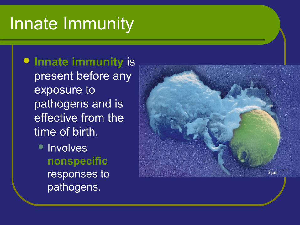
Innate Immunity
Innate immunity is present before any exposure to pathogens and is effective from the time of birth. Involves
nonspecific responses to pathogens.
Acquired Immunity
Acquired immunity develops only after exposure to inducing agents such as microbes, toxins, or other foreign substances. Involves a very specific response to
pathogens.
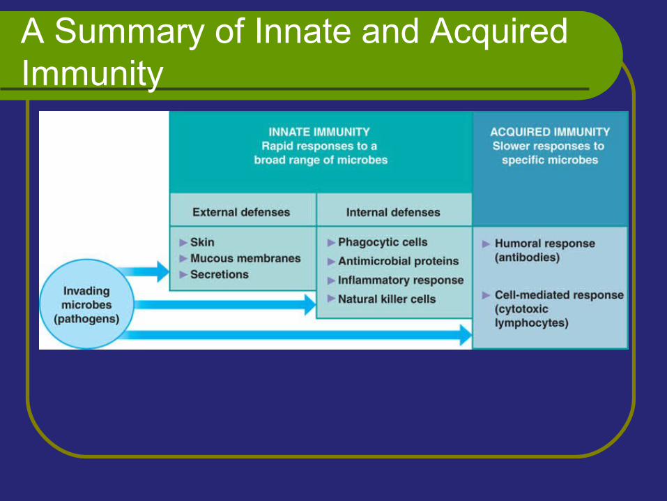
A Summary of Innate and Acquired Immunity
External Defenses
Intact skin and mucous membranes form physical barriers that block the entry of microorganisms and viruses.
Certain cells of the mucous membranes produce mucus - a viscous fluid that traps microbes and other particles.
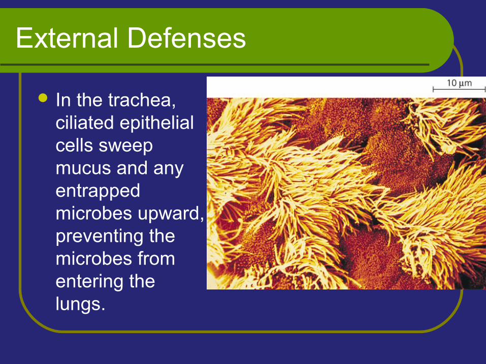
External Defenses
In the trachea, ciliated epithelial cells sweep mucus and any entrapped microbes upward, preventing the microbes from entering the lungs.
External Defenses
Secretions from the skin give the skin a pH between 3 and 5, which is acidic enough to prevent colonization of many microbes.Also include proteins such as lysozyme, an
enzyme that digests the cell walls of many bacteria.
Internal Cellular and Chemical Defenses
Internal cellular defenses depend mainly on phagocytosis.
Phagocytes are types of white blood cells that: Ingest invading microorganisms. Initiate the inflammatory response.
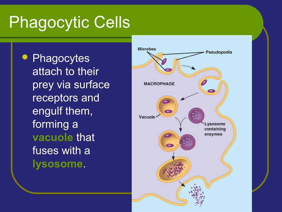
Phagocytic Cells
Phagocytes attach to their prey via surface receptors and engulf them, forming a vacuole that fuses with a lysosome.
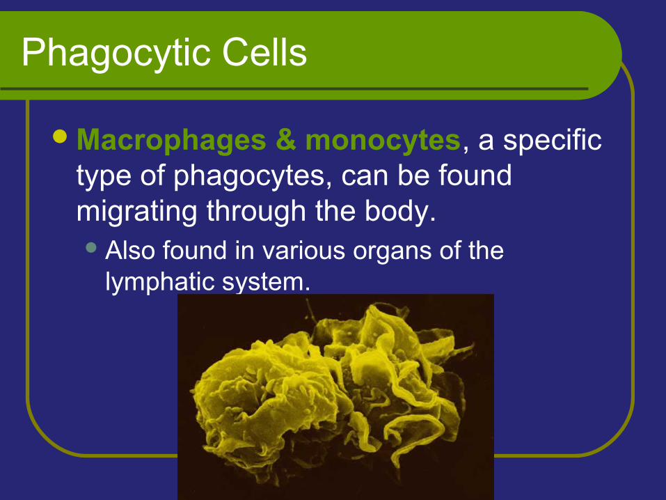
Phagocytic Cells
Macrophages & monocytes, a specific type of phagocytes, can be found migrating through the body.Also found in various organs of the
lymphatic system.
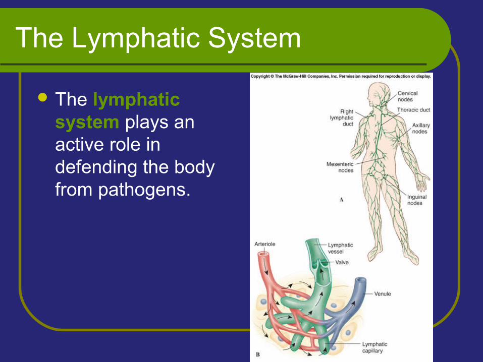
The Lymphatic System
The lymphatic system plays an active role in defending the body from pathogens.
Antimicrobial Proteins
Numerous proteins function in innate defense by attacking microbes directly or by impeding their reproduction.
Antimicrobial Proteins
About 30 proteins make up the complement system, which can cause lysis of invading cells and help trigger inflammation.
Interferons provide innate defense against viruses and help activate macrophages.
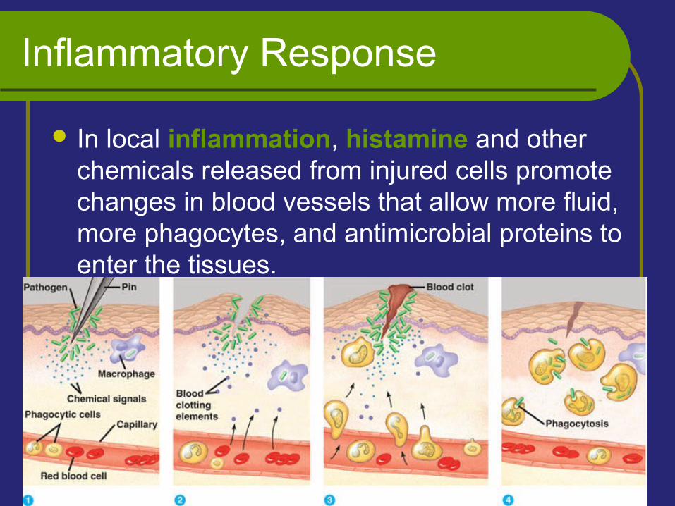
Inflammatory Response
In local inflammation, histamine and other chemicals released from injured cells promote changes in blood vessels that allow more fluid, more phagocytes, and antimicrobial proteins to enter the tissues.
Natural Killer Cells
Natural killer (NK) cells patrol the body and attack virus-infected body cells and cancer cells.Trigger apoptosis (programmed cell death)
in the cells they attack.
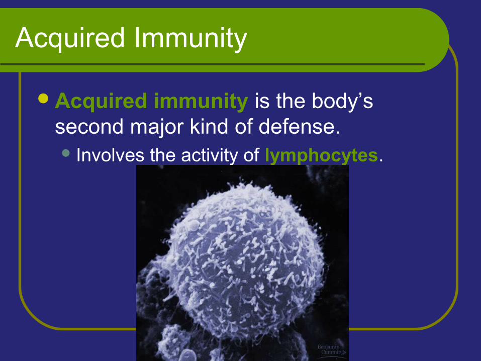
Acquired Immunity
Acquired immunity is the body’s second major kind of defense. Involves the activity of lymphocytes.
Acquired Immunity
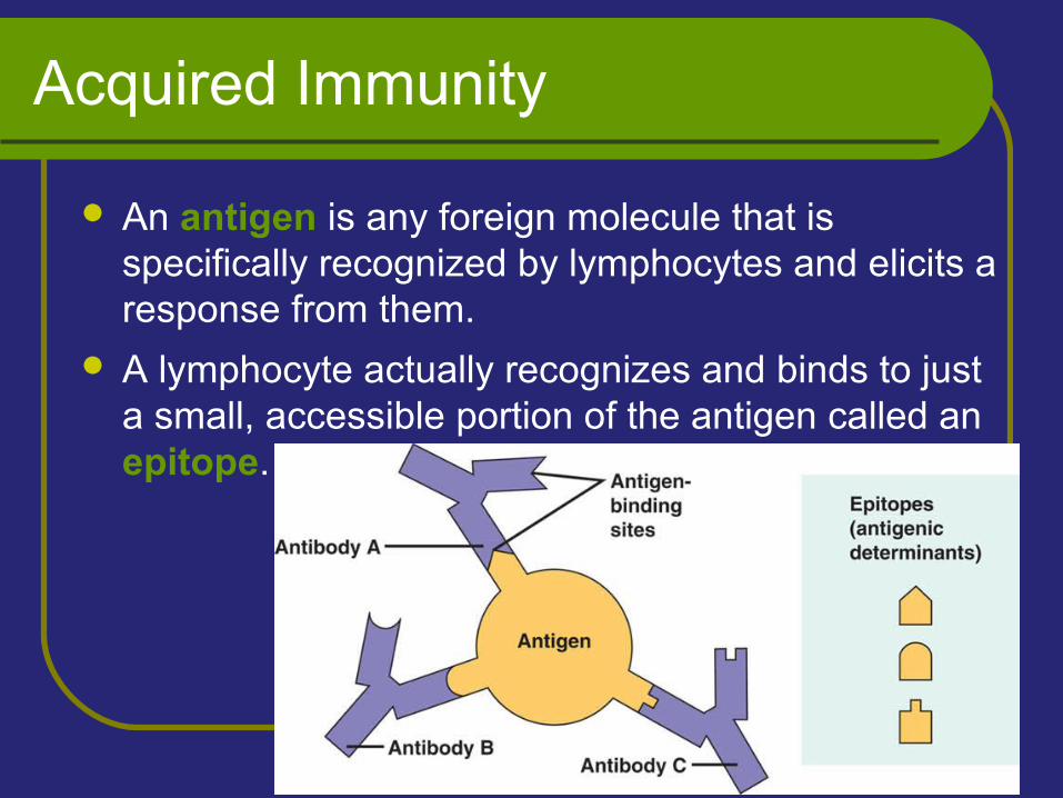
An antigen is any foreign molecule that is specifically recognized by lymphocytes and elicits a response from them.
A lymphocyte actually recognizes and binds to just a small, accessible portion of the antigen called an epitope.
Antigen Recognition by Lymphocytes
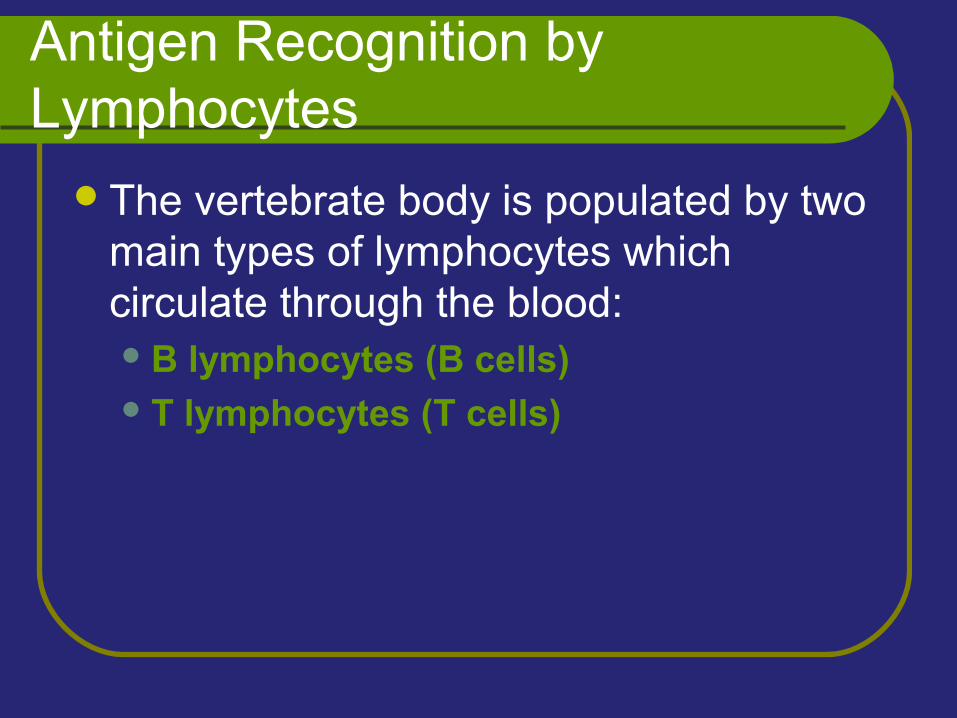
The vertebrate body is populated by two main types of lymphocytes which circulate through the blood:B lymphocytes (B cells) T lymphocytes (T cells)
B Cell Receptors for Antigens
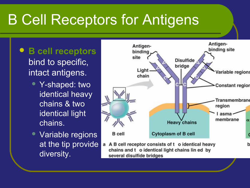
B cell receptors bind to specific, intact antigens. Y-shaped: two
identical heavy chains & two identical light chains.
Variable regions at the tip provide diversity.
T Cell Receptors for Antigens and the Role of the MHC
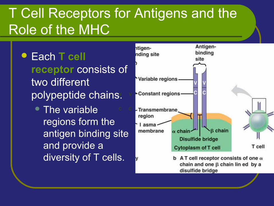
Each T cell receptor consists of two different polypeptide chains. The variable
regions form the antigen binding site and provide a diversity of T cells.
V V
C C
T Cell Receptors for Antigens and the Role of the MHC
T cells bind to small fragments of antigens that are bound to normal cell-surface proteins called MHC molecules.
MHC molecules are encoded by a family of genes called the major histocompatibility complex.
T Cell Receptors for Antigens and the Role of the MHC
Infected cells produce MHC molecules which bind to antigen fragments and then are transported to the cell surface in a process called antigen presentation.
A nearby T cell can then detect the antigen fragment displayed on the cell’s surface.
T Cell Receptors for Antigens and the Role of the MHC
Depending on their source, peptide antigens are handled by different classes of MHC molecules.
T Cell Receptors for Antigens and the Role of the MHC
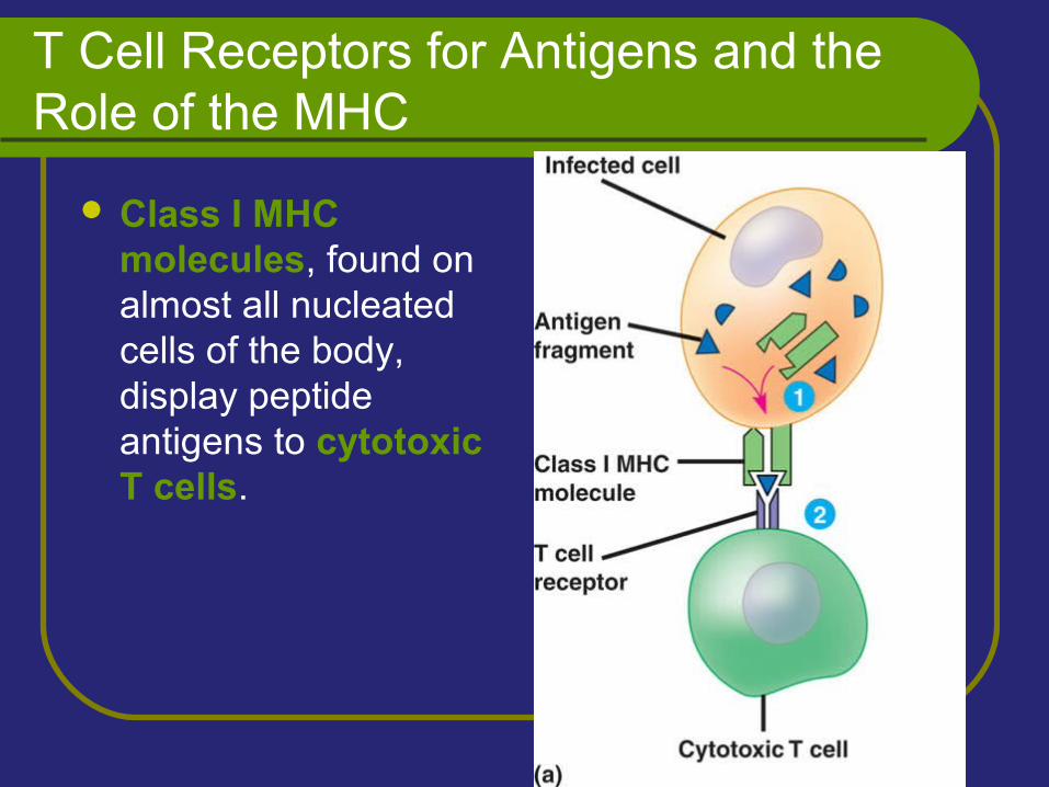
Class I MHC molecules, found on almost all nucleated cells of the body, display peptide antigens to cytotoxic T cells.
T Cell Receptors for Antigens and the Role of the MHC
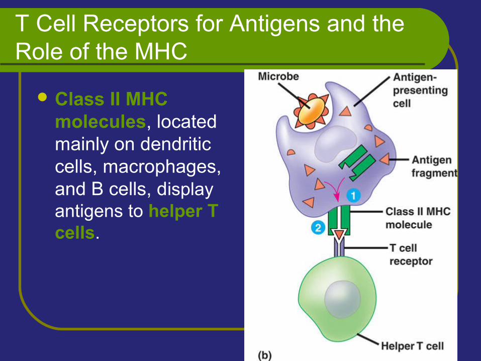
Class II MHC molecules, located mainly on dendritic cells, macrophages, and B cells, display antigens to helper T cells.
Clonal Selection of Lymphocytes
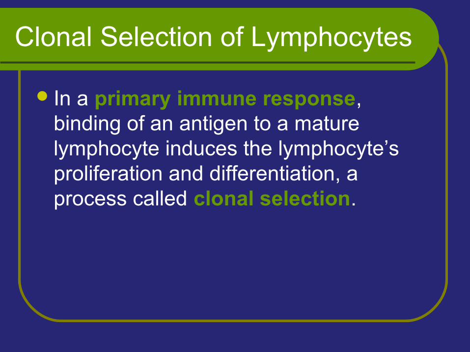
In a primary immune response, binding of an antigen to a mature lymphocyte induces the lymphocyte’s proliferation and differentiation, a process called clonal selection.
Clonal Selection of Lymphocytes
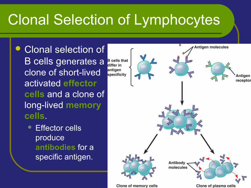
Clonal selection of B cells generates a clone of short-lived activated effector cells and a clone of long-lived memory cells. Effector cells
produce antibodies for a specific antigen.
Clonal Selection of Lymphocytes
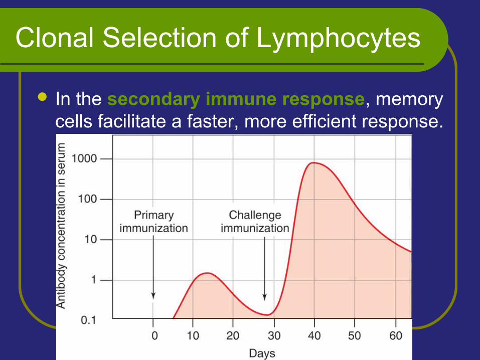
In the secondary immune response, memory cells facilitate a faster, more efficient response.
Humoral vs. Cell-Mediated Response
Acquired immunity includes two branches:The humoral immune response involves
the activation and clonal selection of B cells, resulting in the production of secreted antibodies.
The cell-mediated immune response involves the activation and clonal selection of cytotoxic T cells.
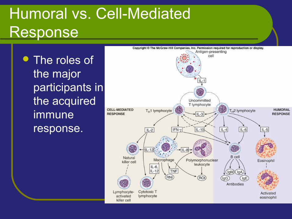
Humoral vs. Cell-Mediated Response
The roles of the major participants in the acquired immune response.
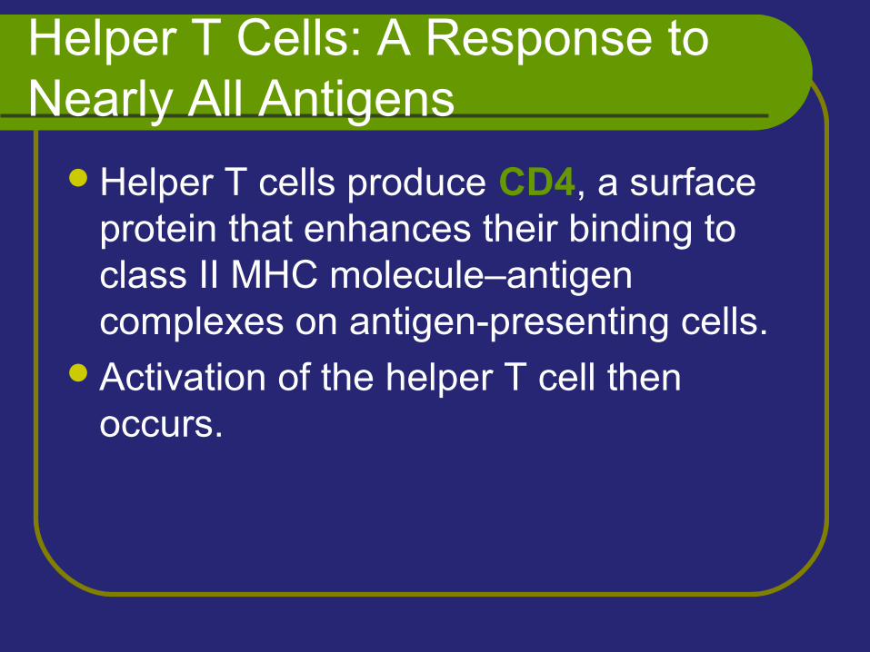
Helper T Cells: A Response to Nearly All Antigens
Helper T cells produce CD4, a surface protein that enhances their binding to class II MHC molecule–antigen complexes on antigen-presenting cells.
Activation of the helper T cell then occurs.
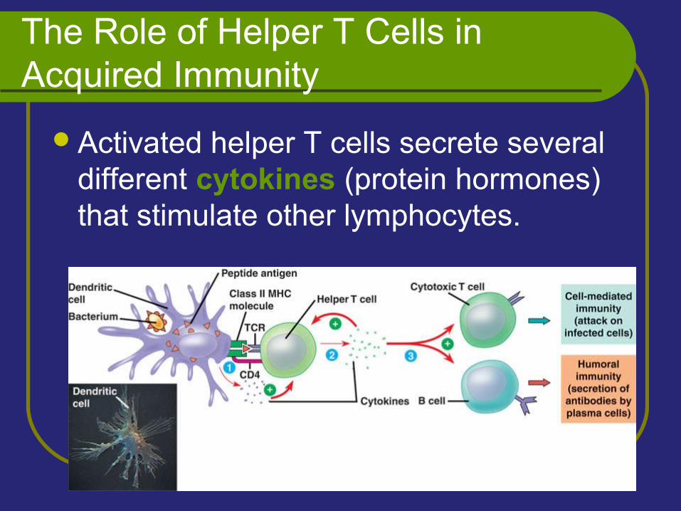
The Role of Helper T Cells in Acquired Immunity
Activated helper T cells secrete several different cytokines (protein hormones) that stimulate other lymphocytes.
Cytotoxic T Cells: A Response to Infected Cells and Cancer Cells
Cytotoxic T cells make CD8 - a surface protein that greatly enhances the interaction between a target cell and a cytotoxic T cell.
The Role of Helper T Cells in Acquired Immunity
Cytotoxic T cells bind to infected cells, cancer cells, and transplanted tissues.
Binding to a class I MHC complex on an infected body cell activates a cytotoxic T cell and differentiates it into an active killer.
The Role of Helper T Cells in Acquired Immunity
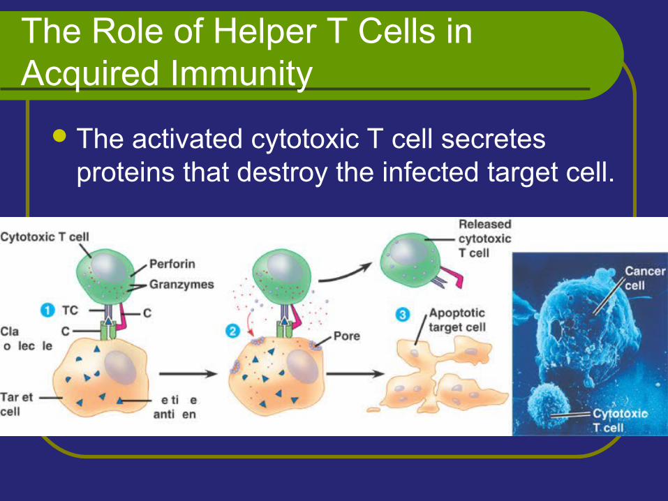
The activated cytotoxic T cell secretes proteins that destroy the infected target cell.
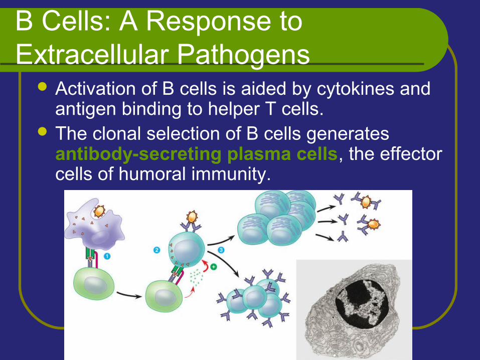
B Cells: A Response to Extracellular Pathogens
Activation of B cells is aided by cytokines and antigen binding to helper T cells.
The clonal selection of B cells generates antibody-secreting plasma cells, the effector cells of humoral immunity.
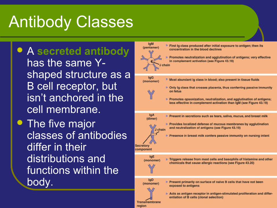
Antibody Classes
A secreted antibody has the same Y-shaped structure as a B cell receptor, but isn’t anchored in the cell membrane.
The five major classes of antibodies differ in their distributions and functions within the body.
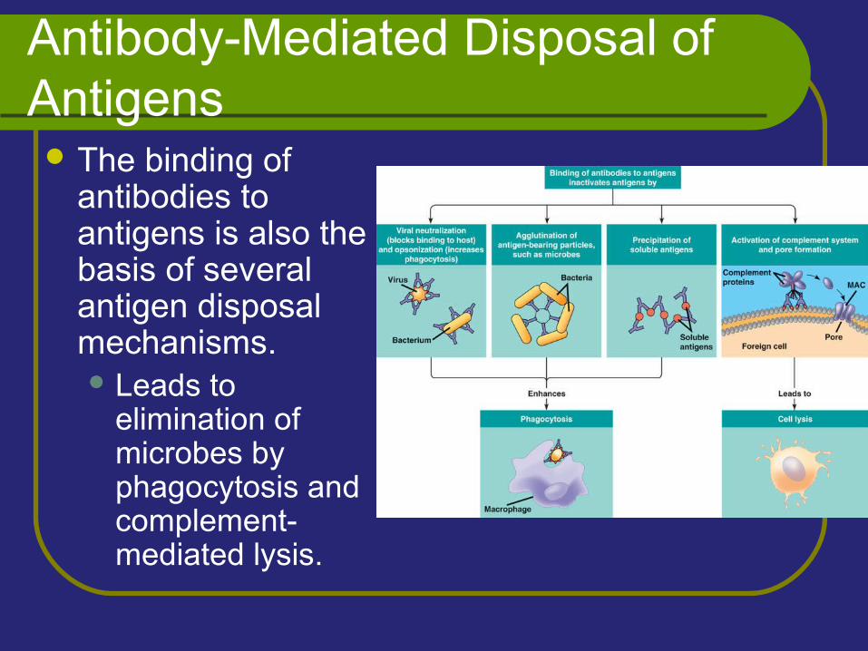
Antibody-Mediated Disposal of Antigens The binding of
antibodies to antigens is also the basis of several antigen disposal mechanisms. Leads to
elimination of microbes by phagocytosis and complement-mediated lysis.
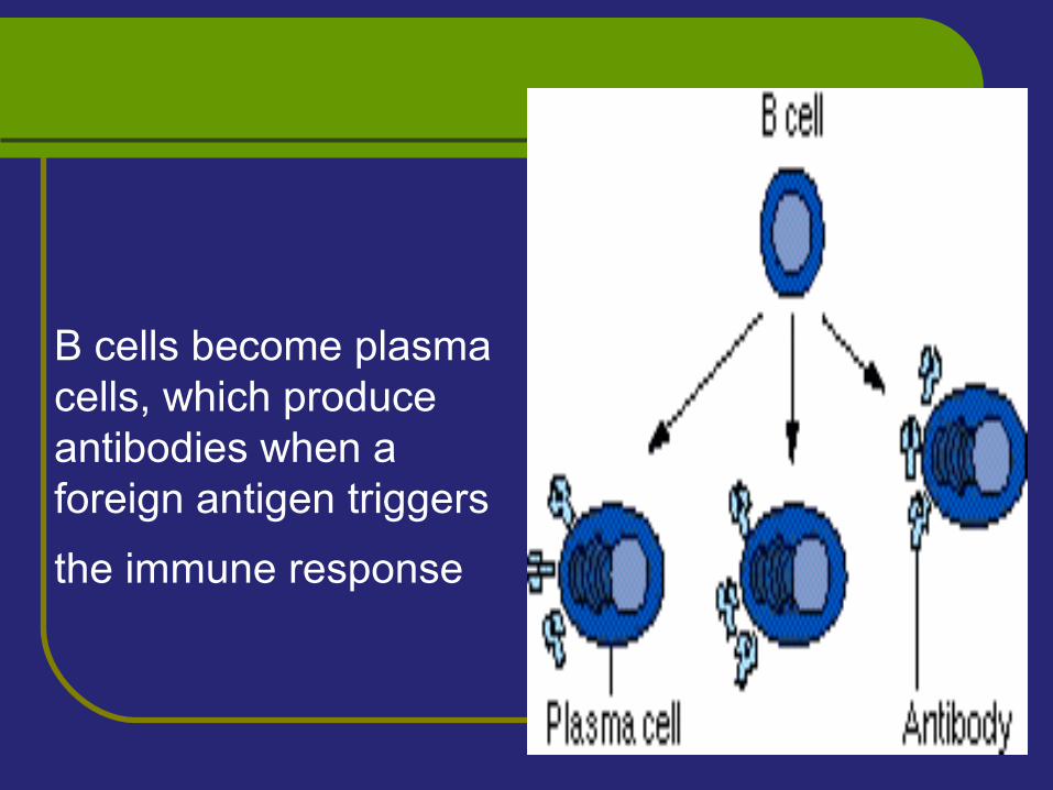
B cells become plasma cells, which produce antibodies when a foreign antigen triggers
the immune response
B-lymphocytesin bon marrow The lymphoid stem cells differentiate into B cellsB-cells precursors mature, differentiate into
immunocomptent B-cells with a single antigen specificity
Immature B-cells that express high affinity receptors for self antigens, die or fail to mature
i.e negative selection or clonal deletionThis process induces central self tolerance and
reduces autoimmune diseases
B - lymphocytes
Immature B cells express IgM receptors on the surface
Mature B cells express IgM, IgD molecules on surfaces
IgM and IgD molecules serve as receptors for antigens
Memory B-cells express IgG or IgA or IgE on the surface
B-cells bear receptors for Fc portion of IgG and a receptor for C3 component of the complement
They express an array of molecules on their surfaces that are important in B-cells interactions with other cells such as MHC II, B7 and CD40
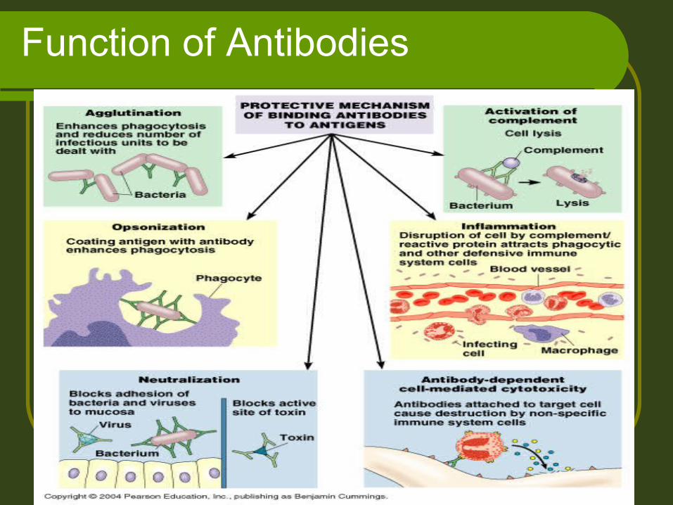
Function of Antibodies
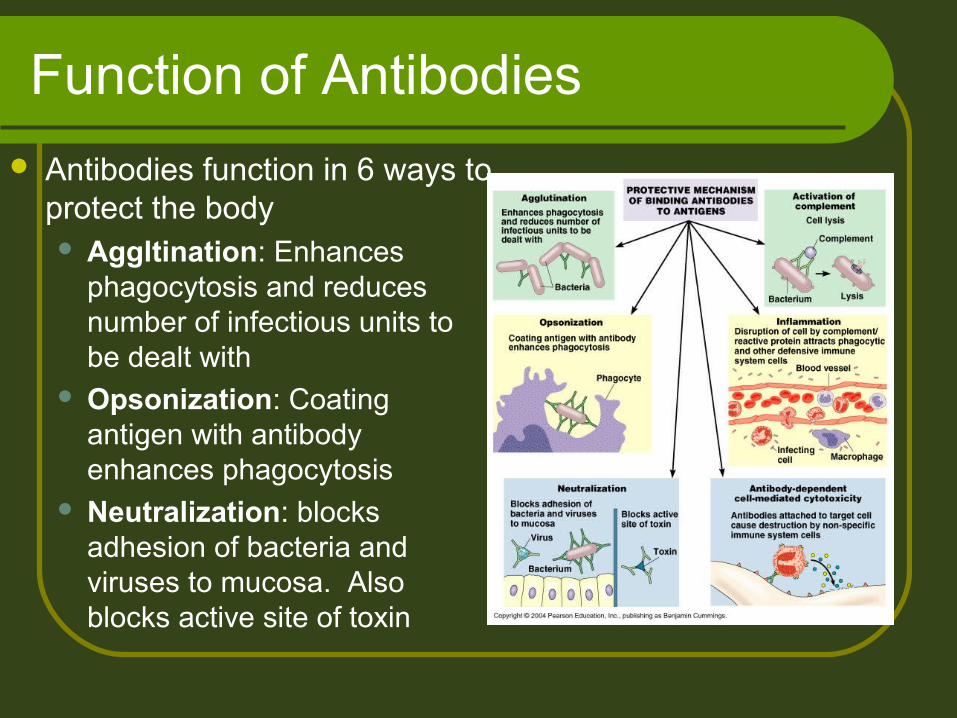
Function of Antibodies
Antibodies function in 6 ways to protect the body Aggltination: Enhances
phagocytosis and reduces number of infectious units to be dealt with
Opsonization: Coating antigen with antibody enhances phagocytosis
Neutralization: blocks adhesion of bacteria and viruses to mucosa. Also blocks active site of toxin
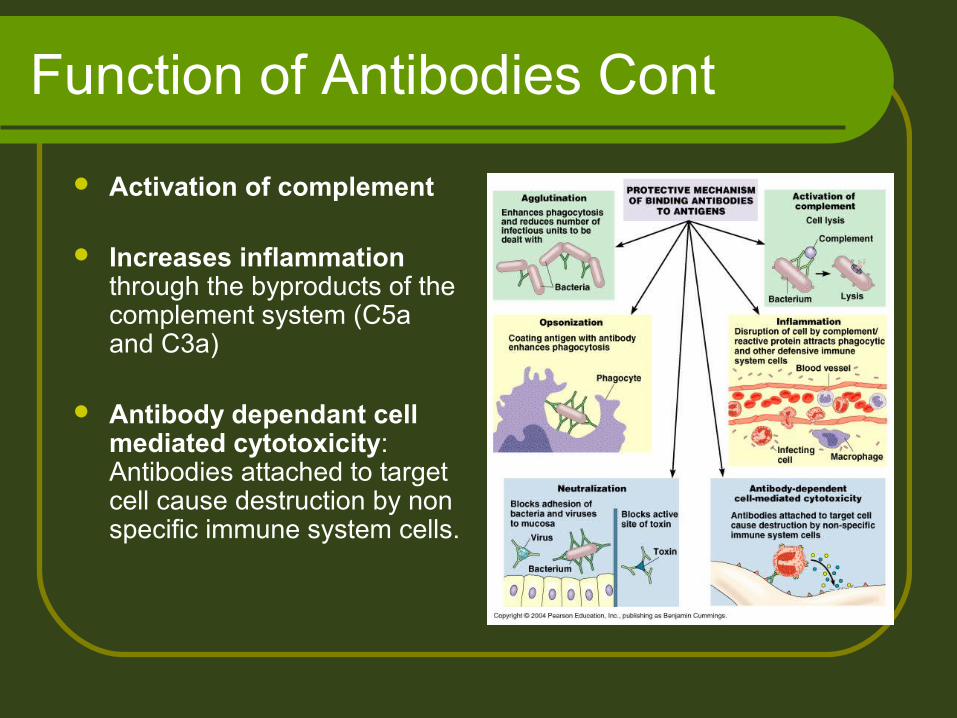
Function of Antibodies Cont
Activation of complement
Increases inflammation through the byproducts of the complement system (C5a and C3a)
Antibody dependant cell mediated cytotoxicity: Antibodies attached to target cell cause destruction by non specific immune system cells.
Mechanism of Humoral immunity
Antibodies induce resistance through:
1) Antitoxin neutralize bacterial toxins (diphtheria , tetanus)
Antitoxin are developed actively as a result of:
a- Previous infection
b- Artificial immunization
c- Transferred passively as antiserum
Neutralization of toxin with antitoxin prevents a combination with tissue cells
Mechanism of Humoral immunity
2) Antibodies attach to the surface of bacteria and
a- act as opsonins and enhance phagocytosisd
b- prevent the adherence of microorganisms to
their target cells, e.g. IgA in the gut
c- Activate the complement and lead to bacterial lysis
d- Clump bacteria (agglutination) leading to
phagocytosis
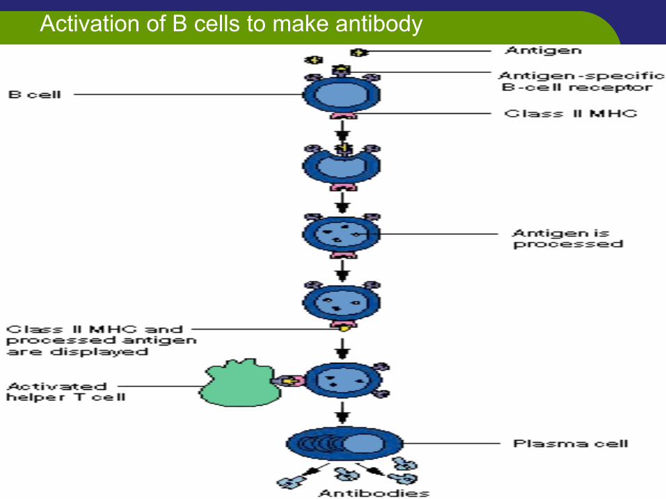
Activation of B cells to make antibody
T-Lmphocytes
T- lmphocytes migrate from bon marrow to enter thymus
1) In the outer cortex of thymus:
- T-lymphocytes acquire specific receptors (TCRs)
- This receptor commit lymphocyte to a single antigen
specificity
- Responding by proliferation and production of a
clone of cells (clonal selection)
- They differentiate to express CD3, both CD4 and
CD8 co receptors (double positive cells)
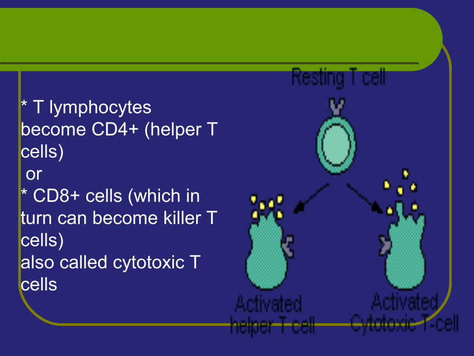
* T lymphocytes become CD4+ (helper T cells) or* CD8+ cells (which in turn can become killer T cells)also called cytotoxic T cells
T- Lymphocytes
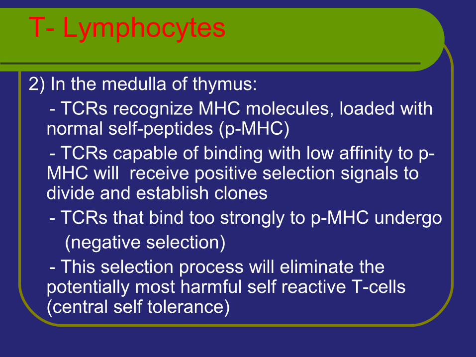
2) In the medulla of thymus: - TCRs recognize MHC molecules, loaded with
normal self-peptides (p-MHC) - TCRs capable of binding with low affinity to p-
MHC will receive positive selection signals to divide and establish clones
- TCRs that bind too strongly to p-MHC undergo (negative selection) - This selection process will eliminate the
potentially most harmful self reactive T-cells (central self tolerance)
T-Lmphocytes
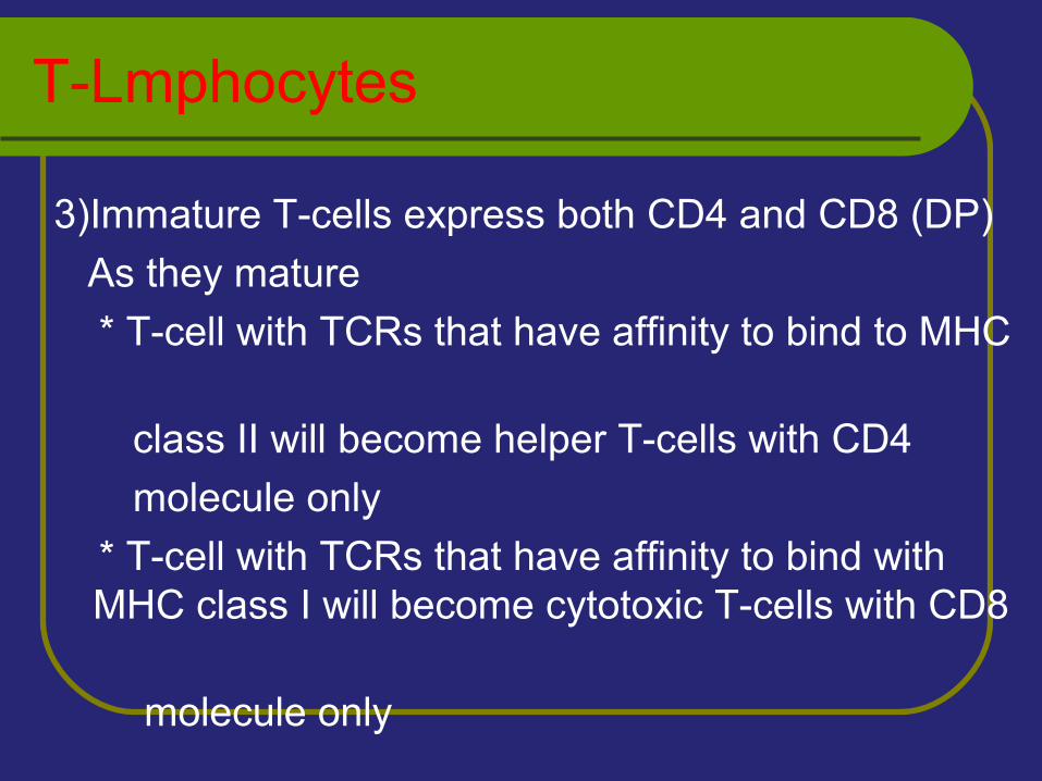
3)Immature T-cells express both CD4 and CD8 (DP)
As they mature
* T-cell with TCRs that have affinity to bind to MHC
class II will become helper T-cells with CD4
molecule only
* T-cell with TCRs that have affinity to bind with MHC class I will become cytotoxic T-cells with CD8
molecule only
T-Lmphocytes
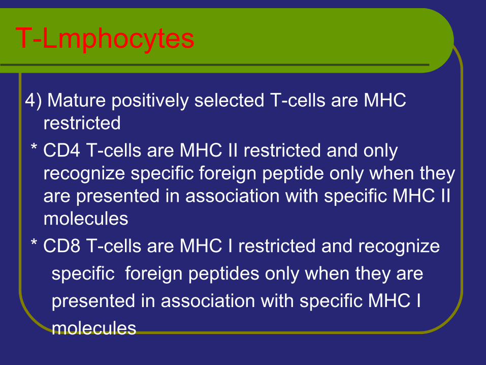
4) Mature positively selected T-cells are MHC restricted
* CD4 T-cells are MHC II restricted and only recognize specific foreign peptide only when they are presented in association with specific MHC II molecules
* CD8 T-cells are MHC I restricted and recognize
specific foreign peptides only when they are
presented in association with specific MHC I
molecules
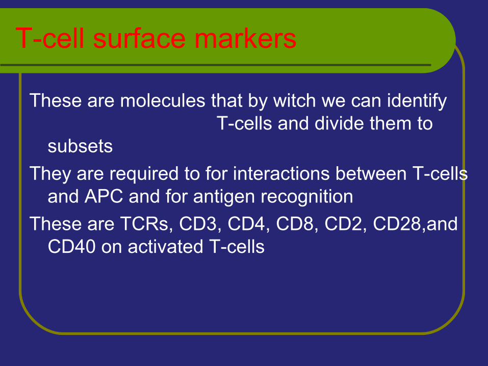
T-cell surface markers
These are molecules that by witch we can identify T-cells and divide them to subsets
They are required to for interactions between T-cells and APC and for antigen recognition
These are TCRs, CD3, CD4, CD8, CD2, CD28,and CD40 on activated T-cells
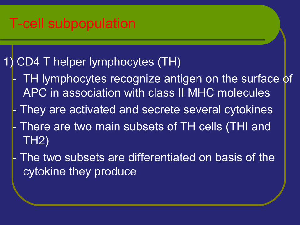
T-cell subpopulation
1) CD4 T helper lymphocytes (TH)
- TH lymphocytes recognize antigen on the surface of APC in association with class II MHC molecules
- They are activated and secrete several cytokines
- There are two main subsets of TH cells (THI and TH2)
- The two subsets are differentiated on basis of the cytokine they produce
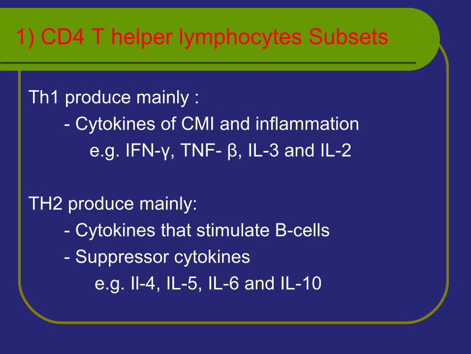
1) CD4 T helper lymphocytes Subsets
Th1 produce mainly :
- Cytokines of CMI and inflammation
e.g. IFN-γ, TNF- β, IL-3 and IL-2
TH2 produce mainly:
- Cytokines that stimulate B-cells
- Suppressor cytokines
e.g. Il-4, IL-5, IL-6 and IL-10
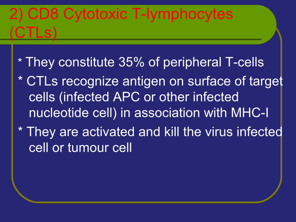
2) CD8 Cytotoxic T-lymphocytes (CTLs)
* They constitute 35% of peripheral T-cells
* CTLs recognize antigen on surface of target cells (infected APC or other infected nucleotide cell) in association with MHC-I
* They are activated and kill the virus infected cell or tumour cell
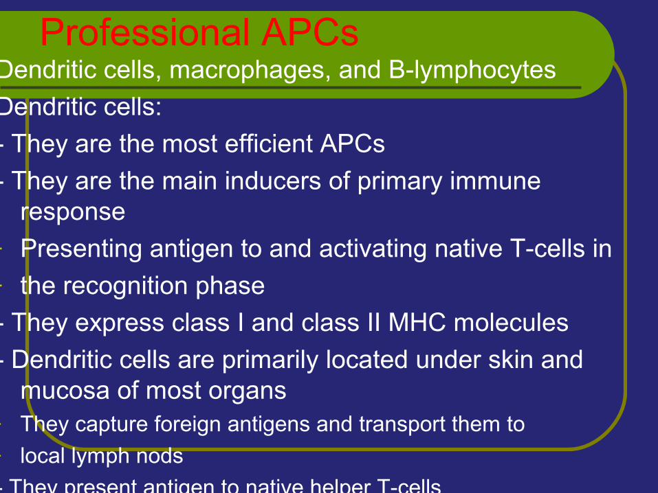
Professional APCsDendritic cells, macrophages, and B-lymphocytes
Dendritic cells:
- They are the most efficient APCs
- They are the main inducers of primary immune response
- Presenting antigen to and activating native T-cells in - the recognition phase
- They express class I and class II MHC molecules
- Dendritic cells are primarily located under skin and mucosa of most organs
- They capture foreign antigens and transport them to - local lymph nods
- They present antigen to native helper T-cells
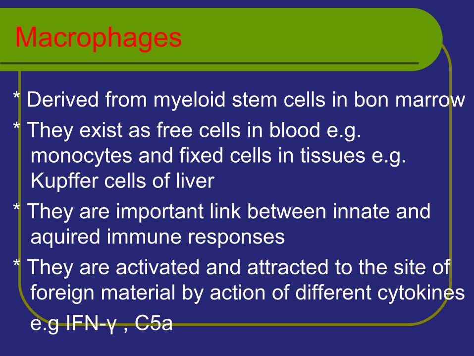
Macrophages
* Derived from myeloid stem cells in bon marrow
* They exist as free cells in blood e.g. monocytes and fixed cells in tissues e.g. Kupffer cells of liver
* They are important link between innate and aquired immune responses
* They are activated and attracted to the site of foreign material by action of different cytokines
e.g IFN-γ , C5a
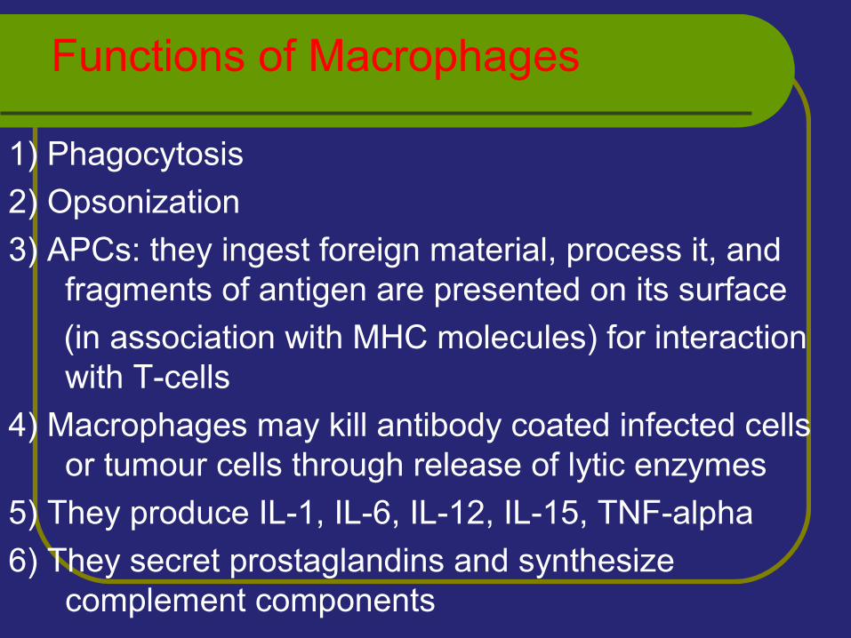
Functions of Macrophages
1) Phagocytosis
2) Opsonization
3) APCs: they ingest foreign material, process it, and fragments of antigen are presented on its surface
(in association with MHC molecules) for interaction with T-cells
4) Macrophages may kill antibody coated infected cells or tumour cells through release of lytic enzymes
5) They produce IL-1, IL-6, IL-12, IL-15, TNF-alpha
6) They secret prostaglandins and synthesize complement components
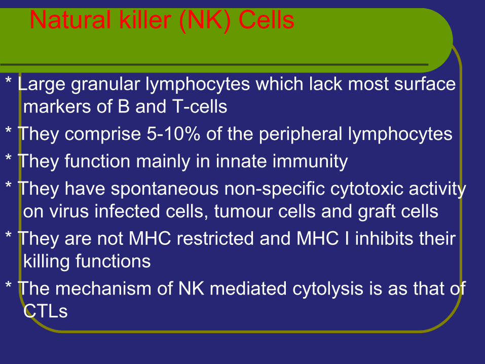
Natural killer (NK) Cells
* Large granular lymphocytes which lack most surface markers of B and T-cells
* They comprise 5-10% of the peripheral lymphocytes
* They function mainly in innate immunity
* They have spontaneous non-specific cytotoxic activity on virus infected cells, tumour cells and graft cells
* They are not MHC restricted and MHC I inhibits their killing functions
* The mechanism of NK mediated cytolysis is as that of CTLs
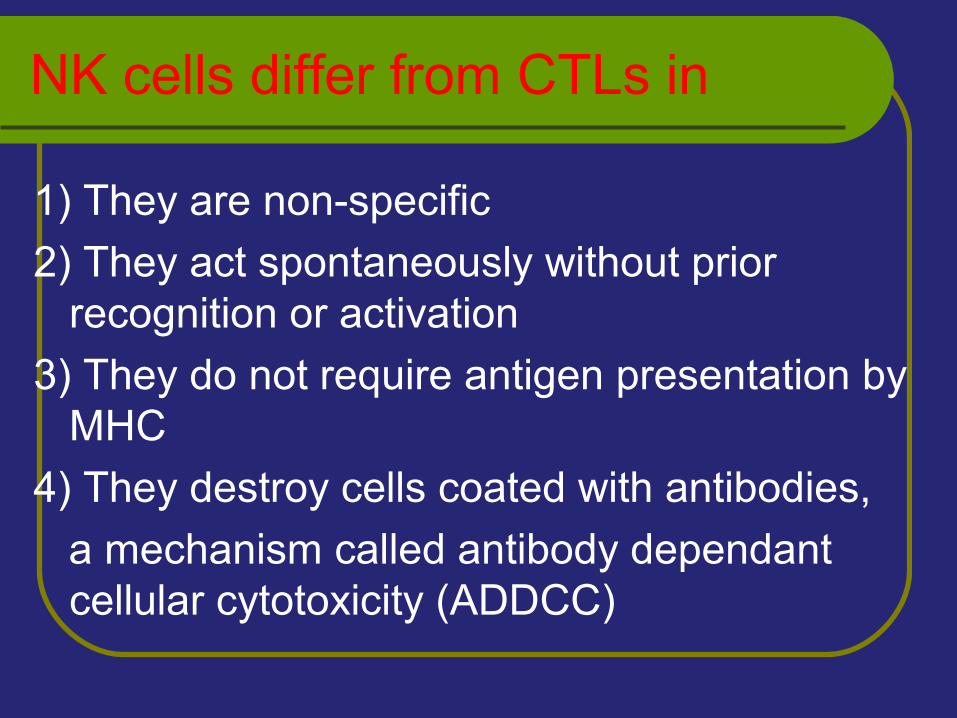
NK cells differ from CTLs in
1) They are non-specific
2) They act spontaneously without prior recognition or activation
3) They do not require antigen presentation by MHC
4) They destroy cells coated with antibodies,
a mechanism called antibody dependant cellular cytotoxicity (ADDCC)
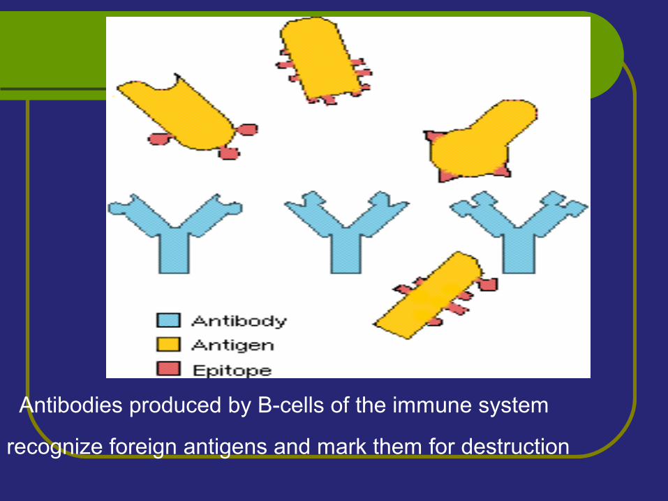
Antibodies produced by B-cells of the immune system
recognize foreign antigens and mark them for destruction
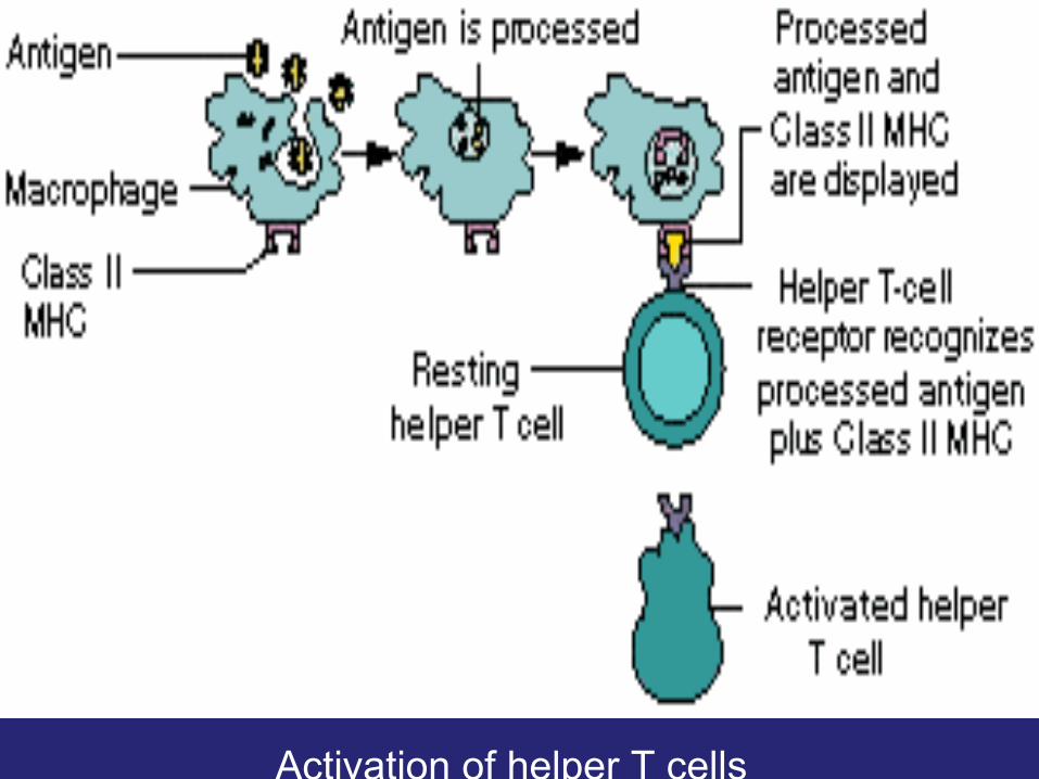
Activation of helper T cells
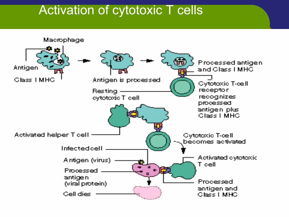
Activation of cytotoxic T cells
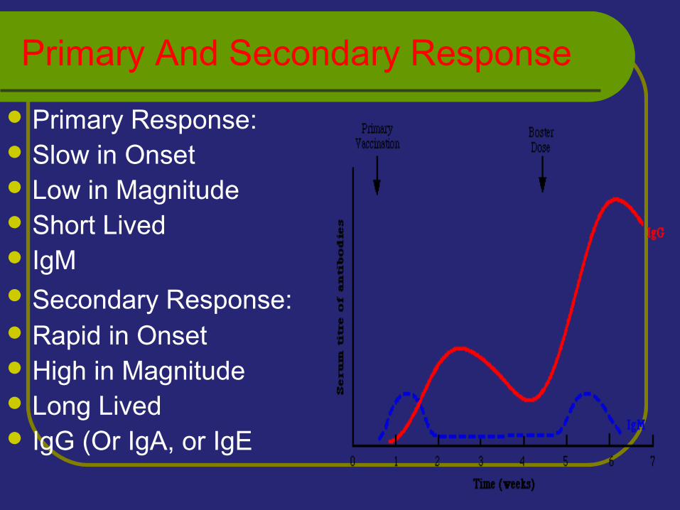
Primary And Secondary Response
Primary Response: Slow in Onset Low in Magnitude Short Lived IgM Secondary Response: Rapid in Onset High in Magnitude Long Lived IgG (Or IgA, or IgE

Thanks