Embed Size (px)
Citation preview
EYELIDSLecture one
Dr.Muqdad Fuad
Assistant professor
College of medicine/university of Dyiala/Iraq
The lecture contain demonstrating videos
Eyelids
The eyelids protect the eye by preventing contact with foreign materials
and by preventing excessive drying of the cornea and conjunctiva.
The palpebral fissure must be wide enough to allow light to enter the pupil
and should close sufficiently to provide protection and moisture to the
globe.
The lid contours and palpebral fissures should be symmetric to avoid
cosmetic deformity.
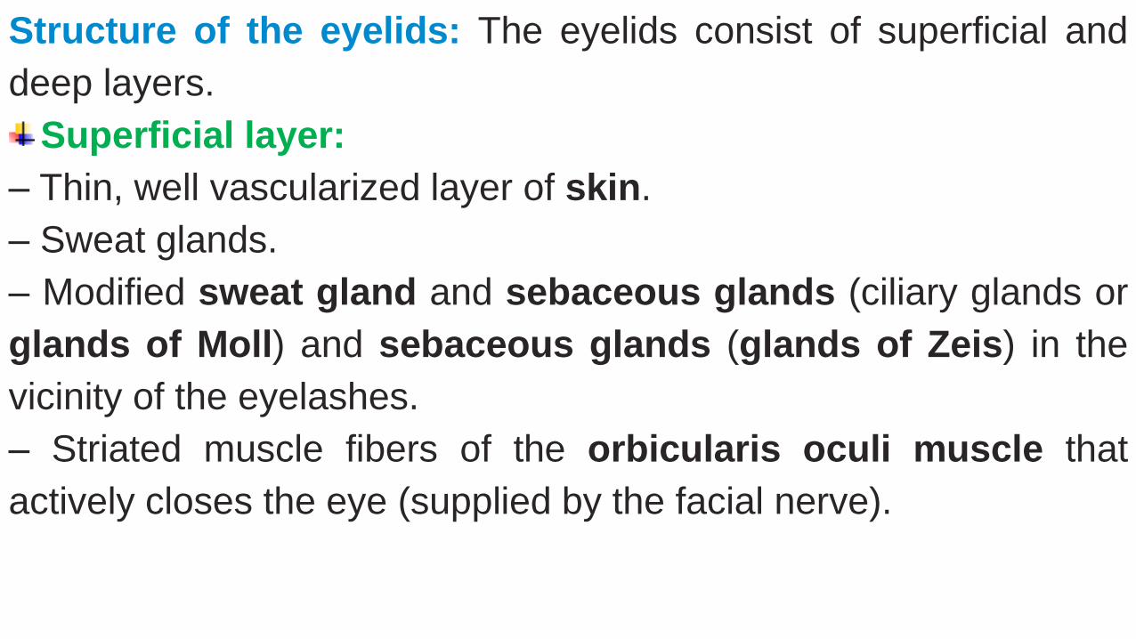
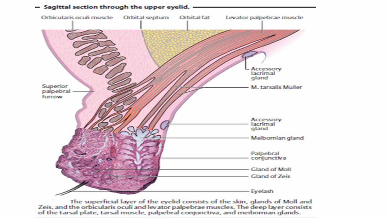
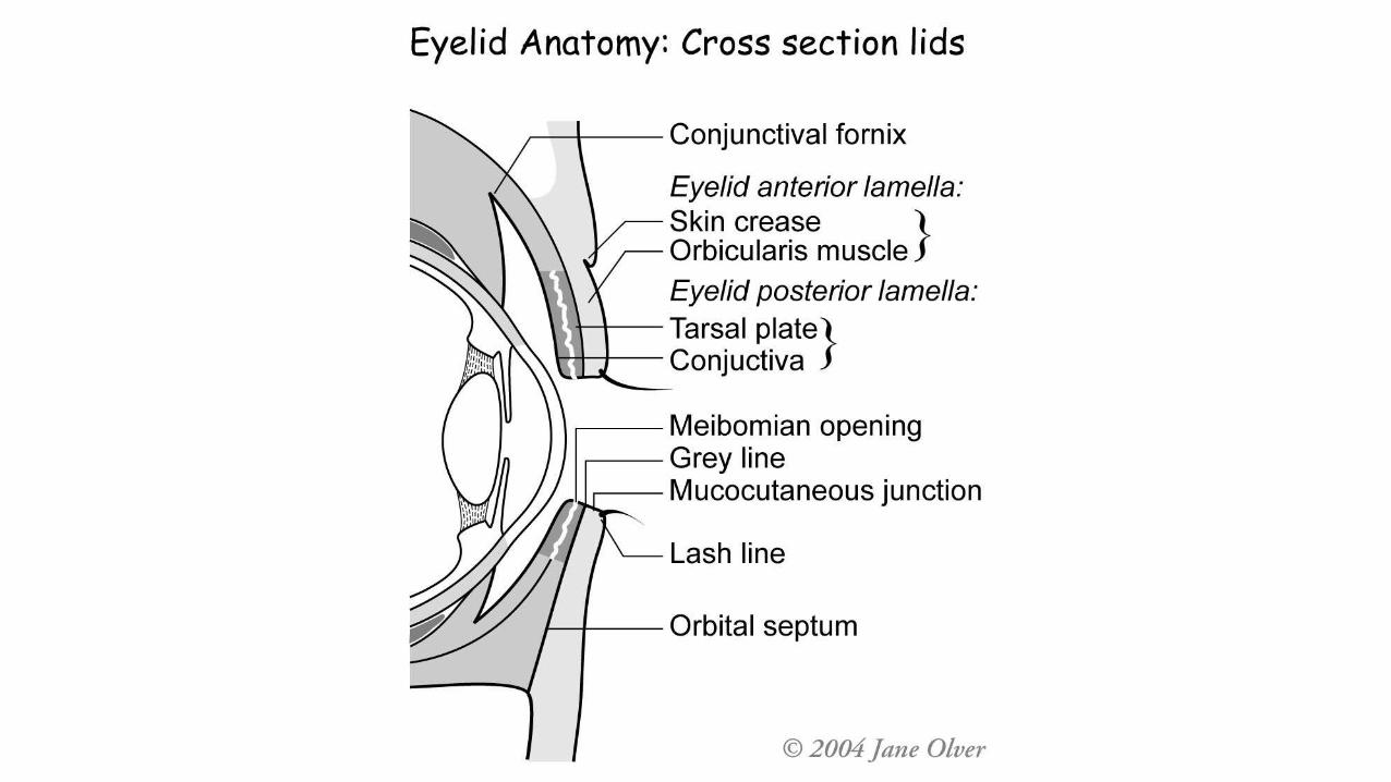
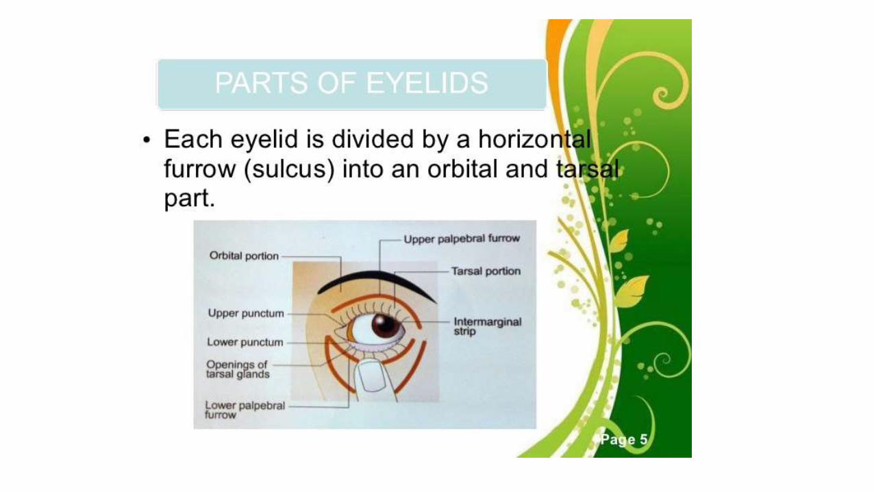
Structure of the eyelids: The eyelids consist of superficial and
deep layers.
Superficial layer:
– Thin, well vascularized layer of skin.
– Sweat glands.
– Modified sweat gland and sebaceous glands (ciliary glands or
glands of Moll) and sebaceous glands (glands of Zeis) in the
vicinity of the eyelashes.
– Striated muscle fibers of the orbicularis oculi muscle that
actively closes the eye (supplied by the facial nerve).
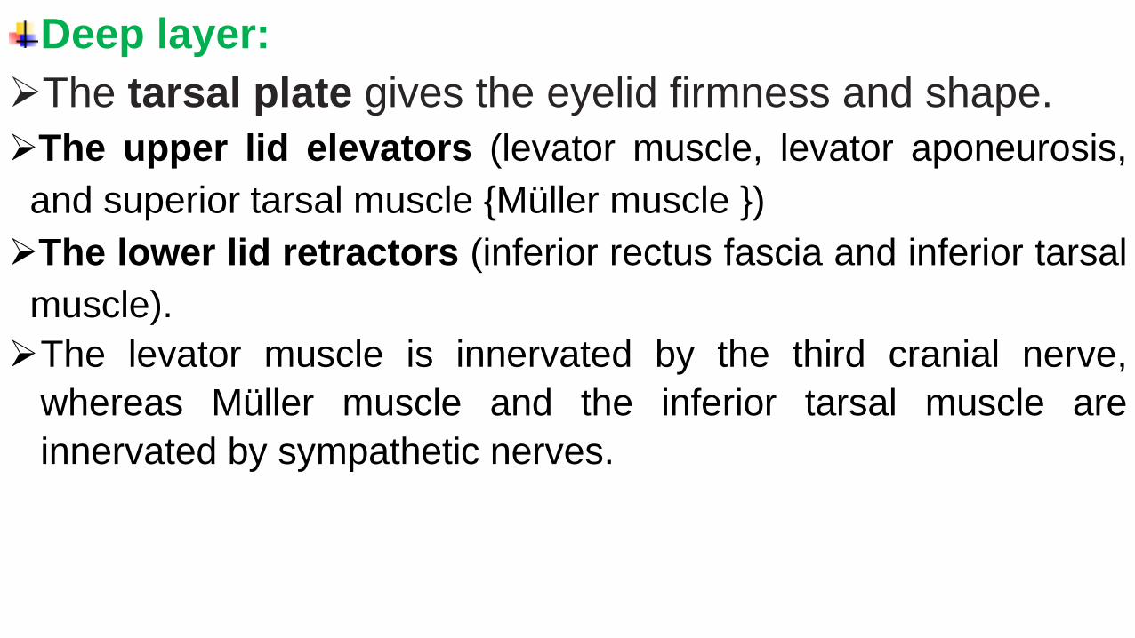
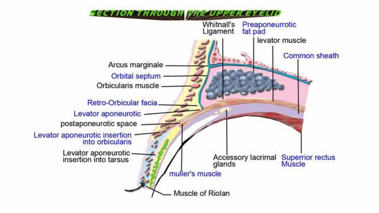
Deep layer:
The tarsal plate gives the eyelid firmness and shape.
The upper lid elevators (levator muscle, levator aponeurosis,
and superior tarsal muscle {Müller muscle })
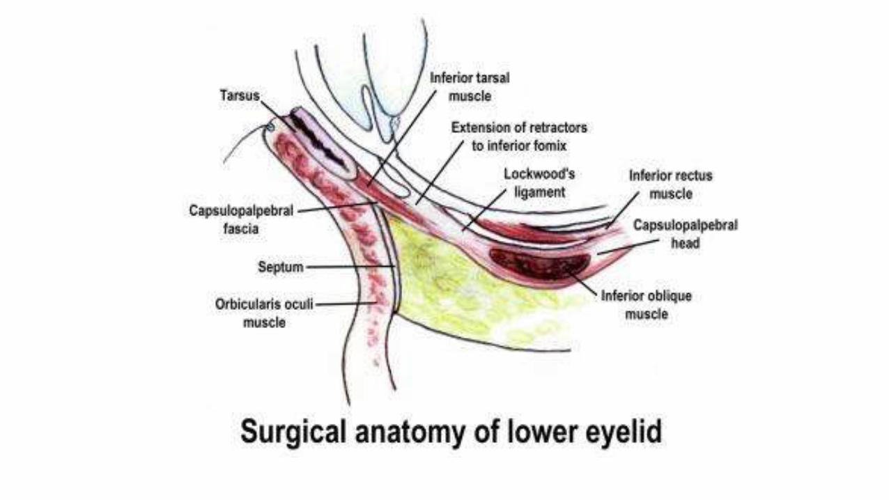
The lower lid retractors (inferior rectus fascia and inferior tarsal
muscle).
The levator muscle is innervated by the third cranial nerve,
whereas Müller muscle and the inferior tarsal muscle are
innervated by sympathetic nerves.
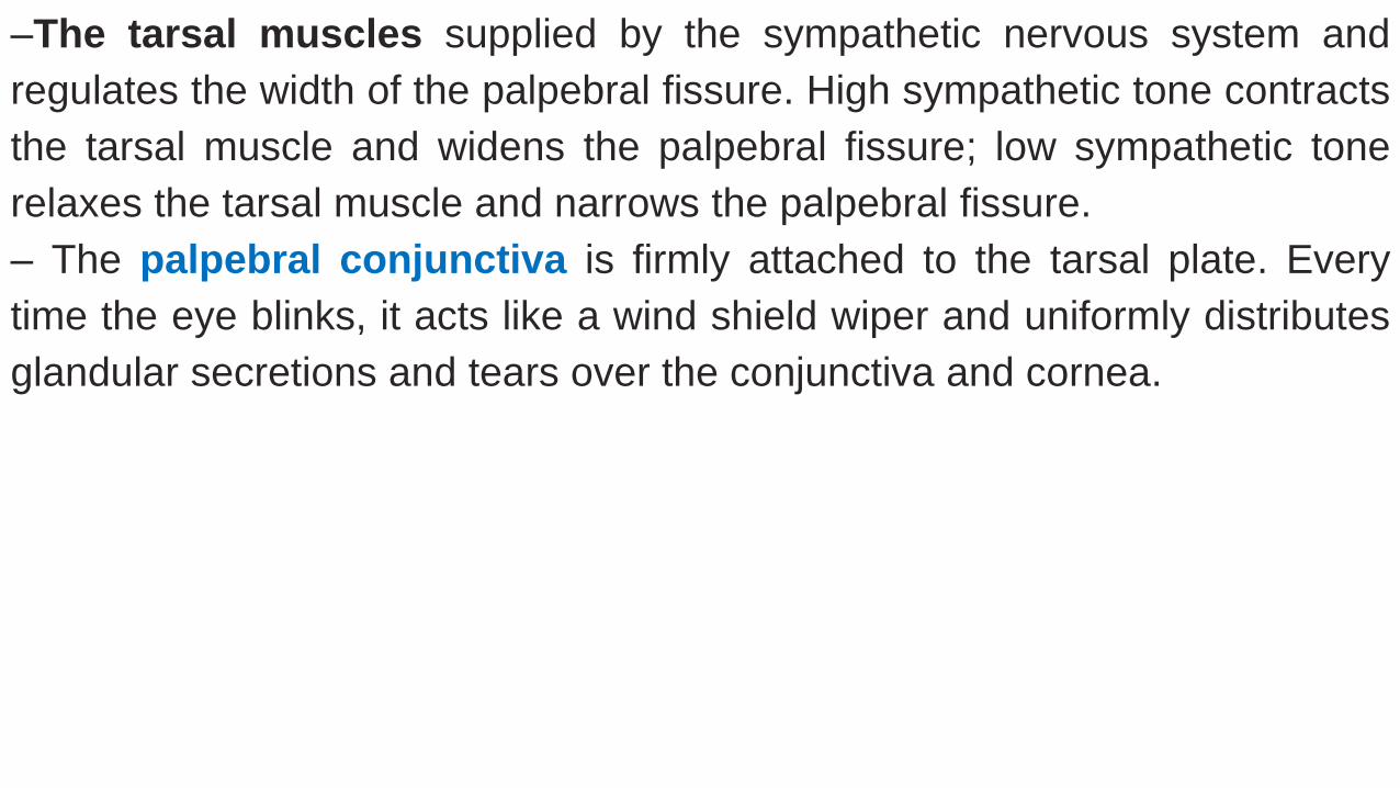
–The tarsal muscles supplied by the sympathetic nervous system and
regulates the width of the palpebral fissure. High sympathetic tone contracts
the tarsal muscle and widens the palpebral fissure; low sympathetic tone
relaxes the tarsal muscle and narrows the palpebral fissure.
– The palpebral conjunctiva is firmly attached to the tarsal plate. Every
time the eye blinks, it acts like a wind shield wiper and uniformly distributes
glandular secretions and tears over the conjunctiva and cornea.
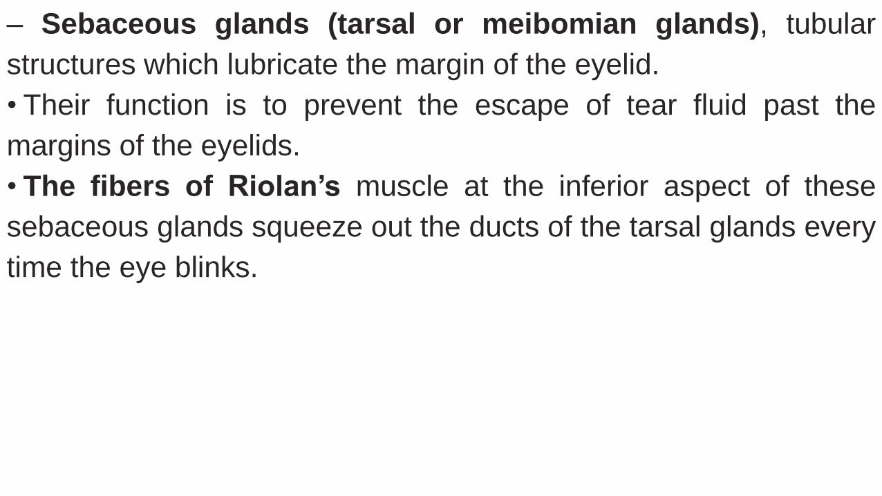
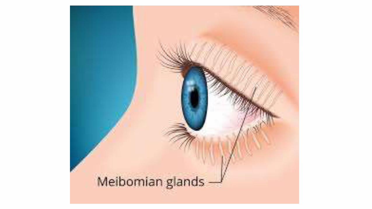
– Sebaceous glands (tarsal or meibomian glands), tubular
structures which lubricate the margin of the eyelid.
• Their function is to prevent the escape of tear fluid past the
margins of the eyelids.
• The fibers of Riolan’s muscle at the inferior aspect of these
sebaceous glands squeeze out the ducts of the tarsal glands every
time the eye blinks.
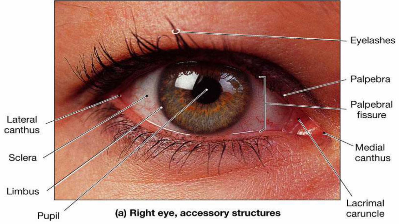
The lids and palpebral fissures are maintained in a stable
position by periosteal attachments provided by the medial
and lateral canthal tendons.
The palpebral fissure is closed by the orbicularis muscle,
which is innervated by the seventh cranial nerve.
The eyelashes project from the anterior aspect of the
margin of the eyelid. On the upper eyelid, approximately
150 eyelashes are arranged in three or four rows; on the
lower eyelid there are about 75 in two rows.
Like the eyebrows, the eyelashes help prevent dust and
sweat from entering the eye. The orbital septum is located
between the tarsal plate and the margin of the orbit. It is a
membranous sheet of connective tissue attached to the
margin of the orbit that retains the orbital fat.
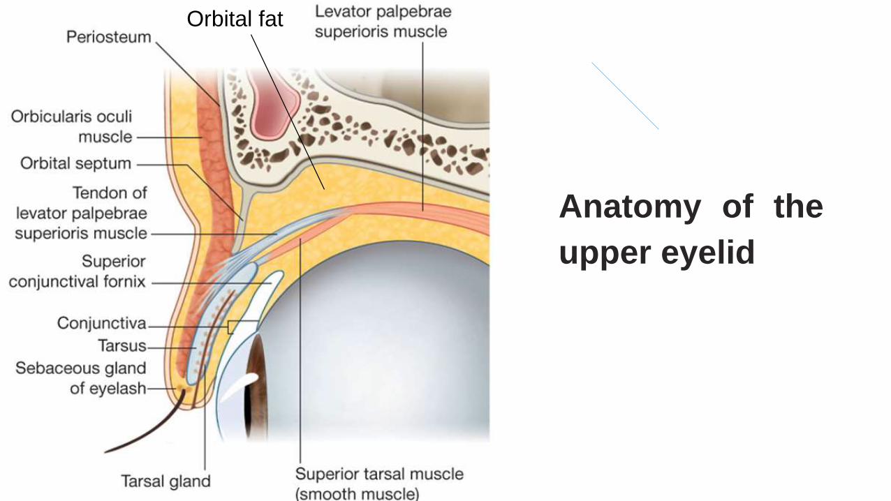
Anatomy of the
upper eyelid
Orbital fat
Congenital and developmental eyelid anomalies
Ptosis
It is paralysis of the levator palpebrae muscle with resulting drooping of
one or both upper eyelids
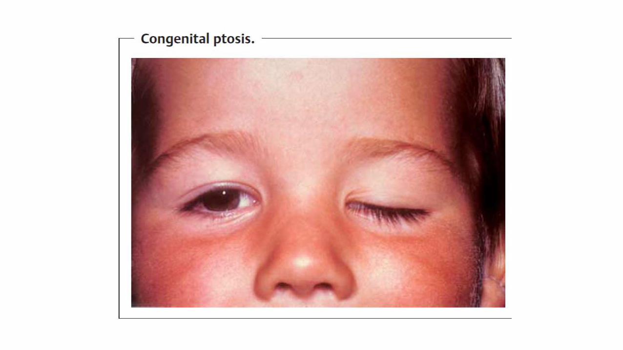
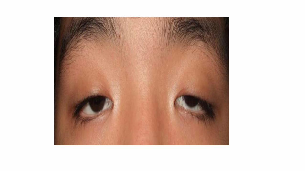
Congenital ptosis
It is usually unilateral, although approximately one-fourth of the cases
involve both upper eyelids. It may be associated with other abnormalities
The disorder is usually hereditary and is primarily autosomal dominant.
Most congenital ptosis is caused by is aplasia in the core of the
oculomotor nerve (neurogenic) that supplies the levator palpebrae
muscle
less frequently it is attributable to an underdeveloped levator palpebrae
muscle (myogenic)
Many cases of congenital ptosis are associated with other
developmental abnormalities such as:
blepharophimosis and epicanthus inversus (ptosis syndrome)
Marcus Gunn “jaw-winking” syndrome, in which the ptotic eyelid
is elevated with movement of the mandible.
Extraocular muscle palsies, particularly those involving the
superior rectus and inferior oblique muscles ipsilateral to the
ptosis.
Blepharophimos is a generalized narrowing of the palpebral fissure. This
abnormality is frequently associated with congenital ptosis and
epicanthus.
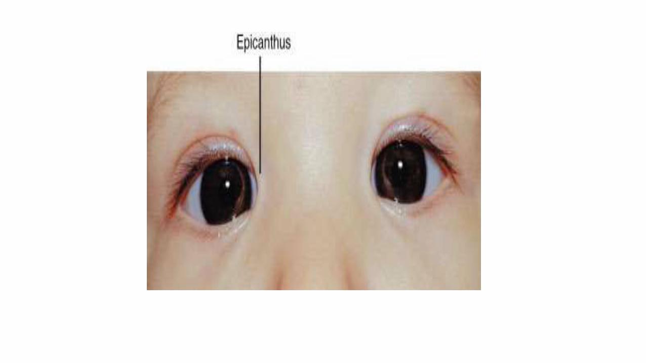
Epicanthus is a semilunar fold of skin that crosses the medial canthus.
Blepharophimosis and epicanthus should usually be repaired prior to
surgical correction of ptosis.
Treatment of congenital ptosis usually requires resection of part of the
weak levator muscle and aponeurosis (surgical retraction), or suspension
of the lids from the frontalis muscle (brow).
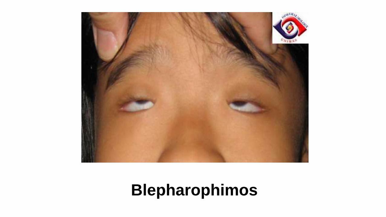
Blepharophimos
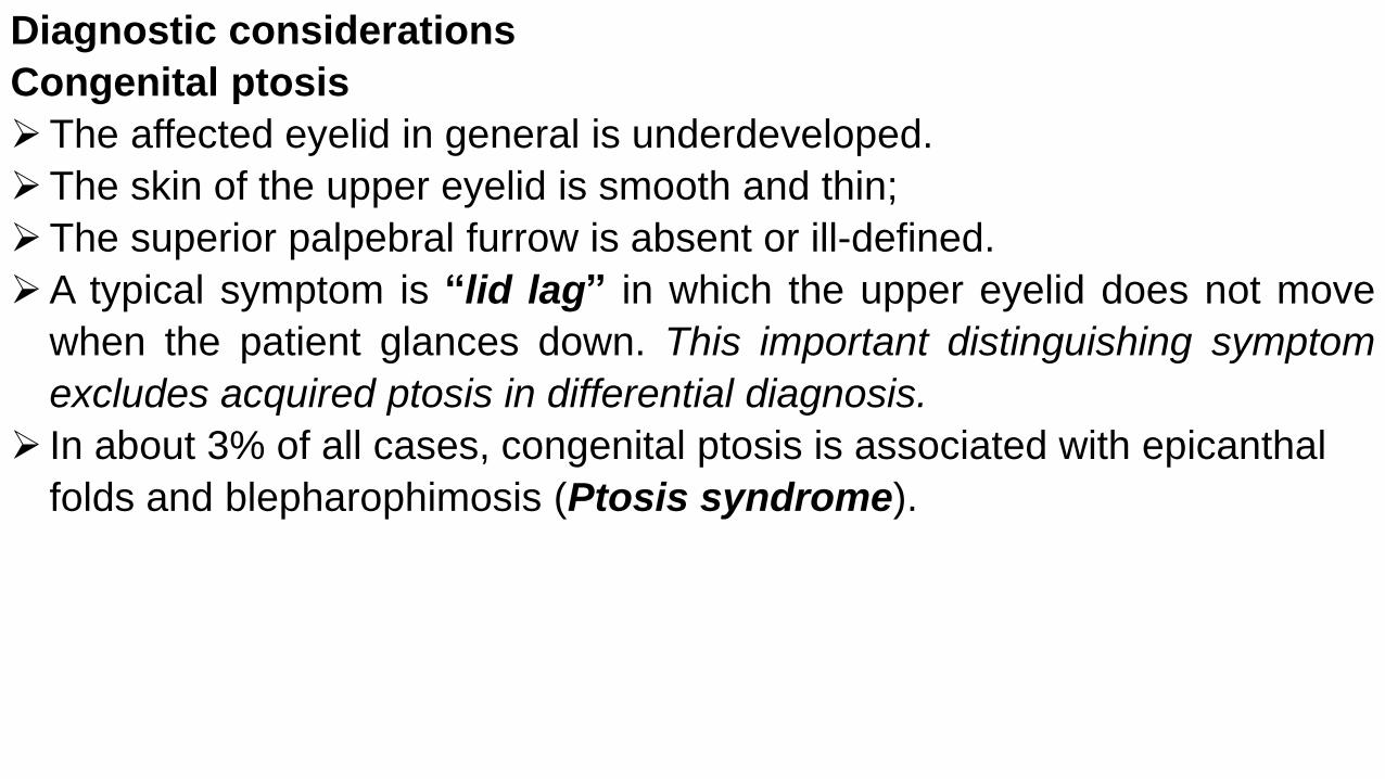
Diagnostic considerations
Congenital ptosis
The affected eyelid in general is underdeveloped.
The skin of the upper eyelid is smooth and thin;
The superior palpebral furrow is absent or ill-defined.
A typical symptom is “lid lag” in which the upper eyelid does not move
when the patient glances down. This important distinguishing symptom
excludes acquired ptosis in differential diagnosis.
In about 3% of all cases, congenital ptosis is associated with epicanthal
folds and blepharophimosis (Ptosis syndrome).
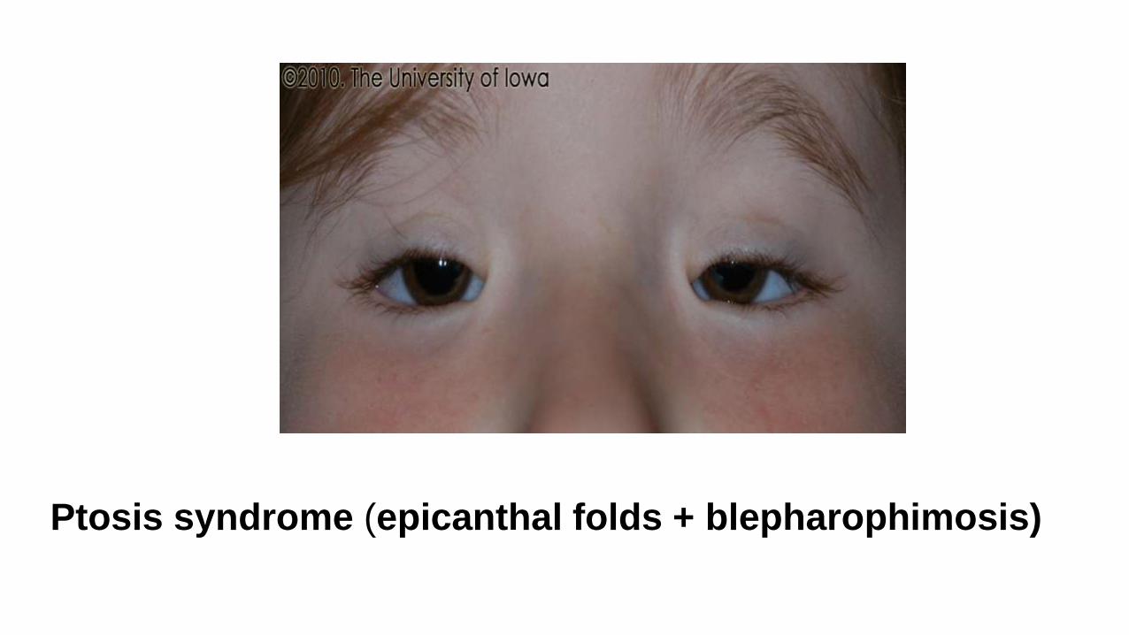
Ptosis syndrome (epicanthal folds + blepharophimosis)
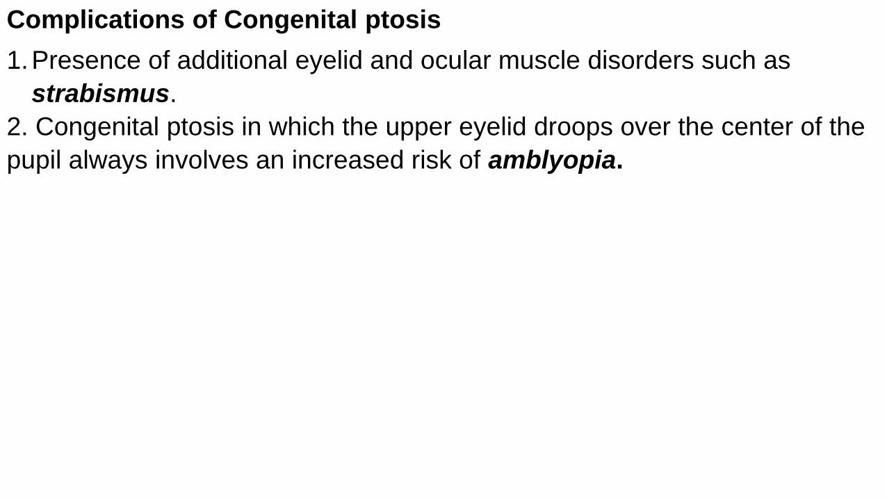
Complications of Congenital ptosis
1.Presence of additional eyelid and ocular muscle disorders such as
strabismus.
2. Congenital ptosis in which the upper eyelid droops over the center of the
pupil always involves an increased risk of amblyopia.
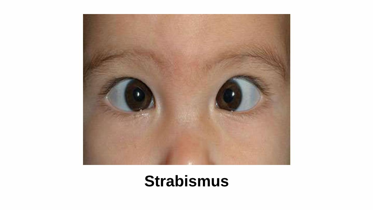
Strabismus
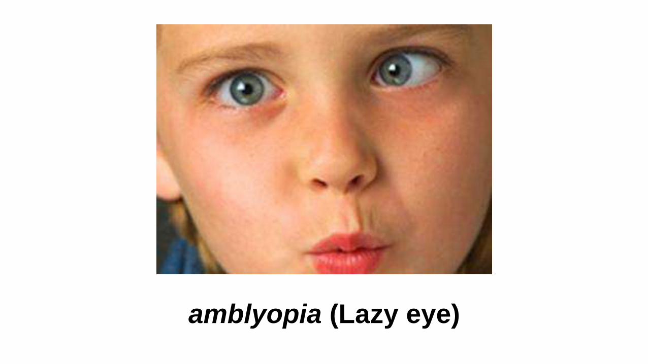
amblyopia (Lazy eye)
Strabismus is a disorder in which both eyes do not line up in the
same direction (misalignment), so they do not look at the same
object at the same time. The condition is more commonly known
as "crossed eyes.“
Types of strabismus:
• Hypertropia is a condition of misalignment of the eyes
(strabismus), whereby the visual axis of one eye is higher than the
fellow fixating eye.
• Hypotropia is a condition with the visual axis of one eye is lower
than the fellow fixating eye.
• Esotropia is a form of strabismus, or “squint,” in which one or
both eyes turns inward
• Exotropia is a form of strabismus where the eyes are deviated
outward. It is the opposite of esotropia.
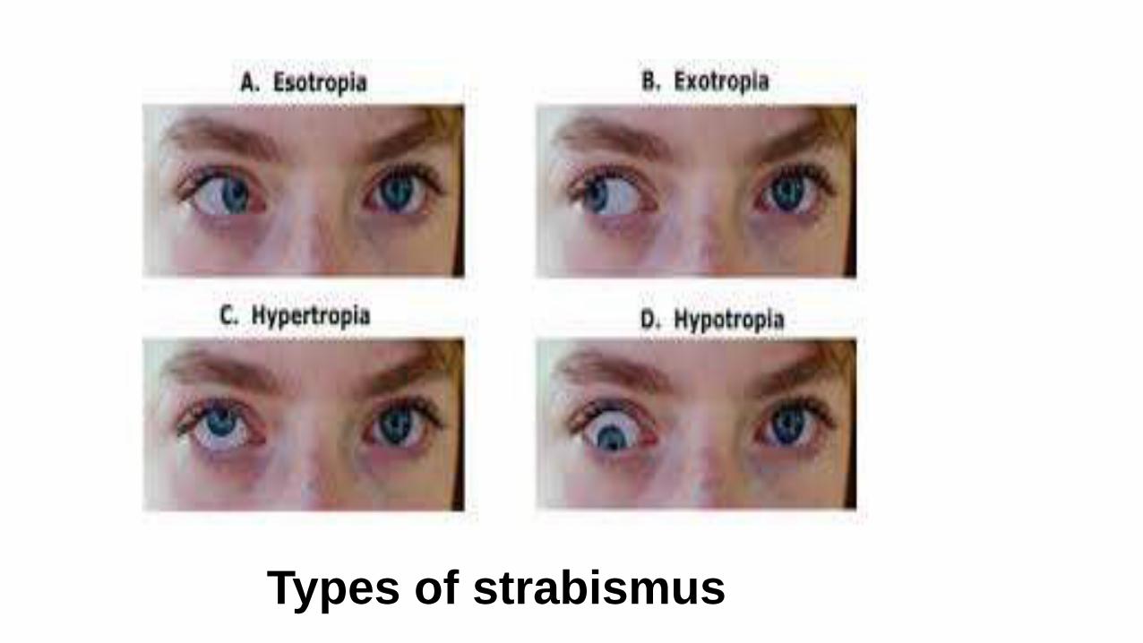
Types of strabismus
Amblyopia, or "lazy eye" is the most common cause of
visual impairment in children. It happens when an eye fails
to work properly with the brain. The eye may look normal,
but the brain favors the other eye. It can be hard to
diagnose amblyopia. It is often found during a routine
vision exam.
• In some cases, it can affect both eyes.
Causes of amblyopia include:
Strabismus - a disorder in which the two eyes don't line up in the same
direction
Refractive error in an eye - when one eye cannot focus as well as the
other, because of a problem with its shape. This includes
nearsightedness, farsightedness, and astigmatism.
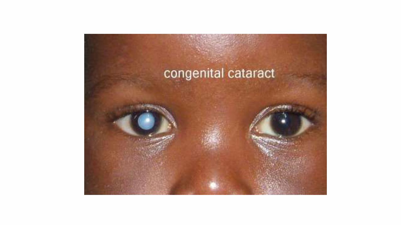
Cataract - a clouding in the lens of the eye
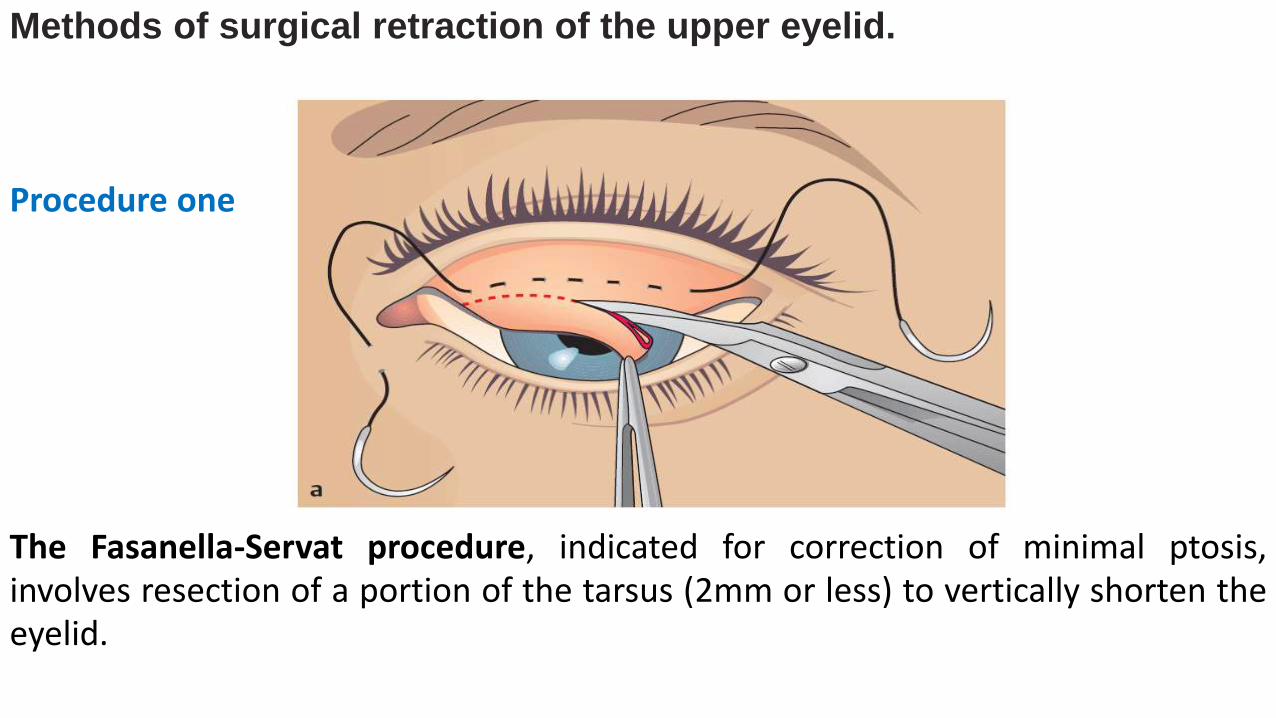
Methods of surgical retraction of the upper eyelid.
Procedure one
The Fasanella-Servat procedure, indicated for correction of minimal ptosis,involves resection of a portion of the tarsus (2mm or less) to vertically shorten theeyelid.

The Fasanella-Servat procedure
The Fasanella-Servat procedure
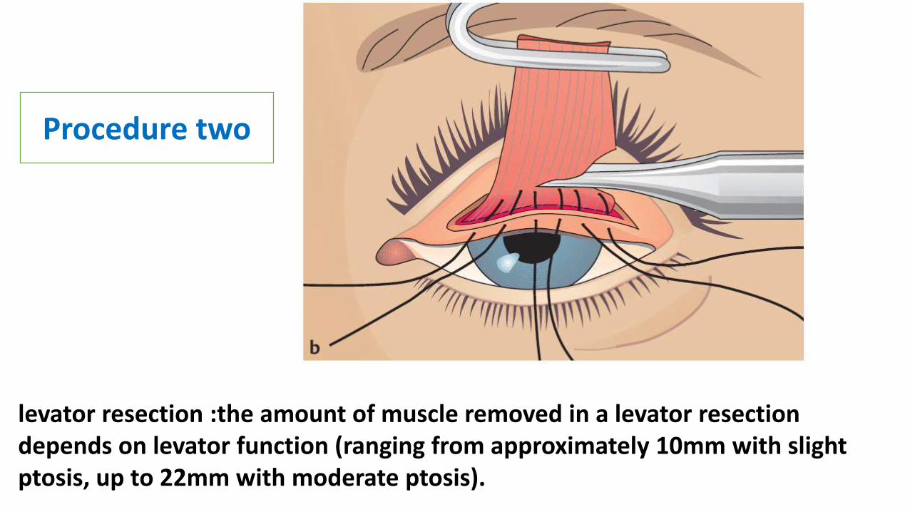
levator resection :the amount of muscle removed in a levator resection depends on levator function (ranging from approximately 10mm with slight ptosis, up to 22mm with moderate ptosis).
Procedure two
levator resection
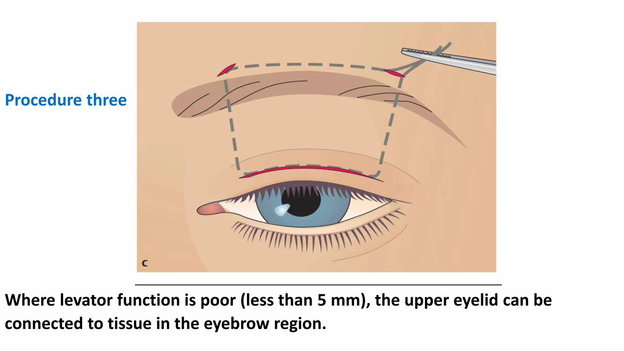
Procedure three
Where levator function is poor (less than 5 mm), the upper eyelid can be
connected to tissue in the eyebrow region.
Acquired ptosis:
Involutional ptosis is the most common ptosis encountered, often
involves both upper lids of older patients, and may occur following
cataract extraction. This is the most common form of acquired ptosis and
is caused by stretching of the levator aponeurosis or disinsertion of
the levator muscle from its insertion onto the tarsus.
Paralytic ptosis in oculomotor palsy is usually unilateral with thedrooping eyelid covering the whole eye. Often there will be other signs ofpalsy in the area supplied by the oculomotor nerve.
In external oculomotor palsy, only the extraocular muscles are affected(mydriasis will not be present), whereas in complete oculomotor palsy,the inner ciliary muscle and the sphincter pupillae muscle are alsoaffected (internal ophthalmoplegia with loss of accommodation, mydriasis,and complete loss of pupillary light reflexes).
Myasthenia gravis (myogenic ptosis that is often bilateral and may beasymmetrical) is associated with abnormal fatigue of the striatedextraocular muscles. Ptosis typically becomes more severe as the daygoes on.
Sympathetic ptosis occurs in Horner’s palsy (ptosis, miosis, andenophthalmos).
Traumatic ptosis can occur after injuries.
Mechanical ptosis may be associated with lid tumors such as
neurofibromas and may result from scars or foreign bodies.
Note: Rapidly opening and closing the eyelids provokes ptosis in
myasthenia gravis and simplifies the diagnosis.
Treatment of acquired ptosis:
Depends on the cause.
As palsies often resolve spontaneously, the patient should be observed
before resorting to surgical intervention.
Conservative treatment with special eyeglasses may be sufficient even in
irreversible cases.
Because of the risk of overcorrecting or undercorrecting the disorder,
several operations may be necessary.
Repair of the levator aponeurosis (tendon) if possible.
In more severe cases, the levator aponeurosis may be suspended
from the frontalis muscle if levator function is poor.

To be continued…………