Embed Size (px)
Citation preview
1
Neurogenesis in the hippocampus of C57BL/6J mice at early adulthood (56 days post-natal) following
chronic prenatal alcohol exposure
Oladiran I. Olateju1, Muhammad A. Spocter1,2, Nina Patzke1, Amadi O. Ihunwo1,
Paul R. Manger1
1 School of Anatomical Sciences, Faculty of Health Sciences, University of the Witwatersrand, 7 York Road,
Parktown, 2193, Johannesburg, South Africa.
2 Department of Anatomy, Des Moines University, Des Moines, IA 50312, USA
Corresponding Author: Oladiran I. Olateju; School of Anatomical Sciences, Faculty of Health Sciences,
University of the Witwatersrand, 7 York Road, Parktown, 2193, Johannesburg, Republic of South Africa.
[email protected]; Ph: +27 11 717 2763 Fax: +27 11 717 2422.
2
Abstract:
We examined the effect of chronic prenatal alcohol exposure on the process of adult neurogenesis in
C57BL/6J mice at early adulthood (PND 56). Pregnant mice, and their in utero litters, were exposed to alcohol,
through oral gavage, on gestational days 7 – 16, with recorded blood alcohol concentrations averaging 184
mg/dL (CA group). Two control groups, sucrose (CAc) and non-treated (NTc) control groups were also
examined. At PND 56, the pups from each group were sacrificed and the left hemisphere of the brain sectioned
in a sagittal plane, and stained for Nissl substance with cresyl violet, and immunostained for Ki-67 which labels
proliferative cells and doublecortin (DCX) for immature neurons. Morphologically, the neurogenic pattern was
identical in all three groups studied, and similar to arrangements in mammals and laboratory rodents.
Populations of Ki-67 immunopositive cells in the dentate gyrus were not statistically significantly different
between the experimental groups. Thus, the prenatal alcohol exposure in this study does not appear to have a
strong effect on the proliferative process in the adult hippocampus. In contrast, the numbers of immature
neurons, labeled with DCX, was statistically significantly lower in the mice exposed to prenatal alcohol
compared with the two control groups. This indicates that the prenatal alcohol exposure appears to lower either
the rate of conversion of proliferative cells to immature neurons or the survival of immature neurons within the
hippocampus. This lowered number of immature neurons, and their associated function, appears to mirror the
memory dysfunctions observed in FASD children.
Keywords: adult neurogenesis; adult hippocampal neurogenesis; fetal alcohol spectrum disorder; memory
dysfunction; proliferation; immature neurons.
3
Introduction:
Neurogenesis is the process of the generation of new neural cells from proliferating adult stem cells, and
this process persists throughout life in most mammals (Gil-Perotin et al. 2009; Patzke et al. 2015; Lazarov and
Hollands 2016; Opendak et al. 2016). Within the hippocampus, adult hippocampal neurogenesis begins with the
progenitor or stem cells found in the subgranular zone (SGZ), which is located deep to the granule cell layer
(GCL) of the dentate gyrus. The pluripotent stem cells in the SGZ undergo proliferation, with some of the newly
born cells remaining as progenitor stem cells, some differentiating into glia cells, while some differentiate into
neurons (Gil-Perotin et al. 2009). The cells destined to become neurons migrate a short distance to the GCL and
eventually integrate into the existing hippocampal circuitry where they augment hippocampal functions (Gil-
Perotin et al. 2009). The neurogenic activity in the hippocampal dentate gyrus can be readily modulated by both
internal (growth factors, neurotransmitters and humoral factors) and external factors (drugs, environmental and
social conditions, stress, starvation and physical activity) (Åberg et al. 2005; Cameron and Glover 2015;
Opendak et al. 2016).
Fetal exposure to alcohol has been shown to impact neurogenesis during brain development and
maturation, causing neurodevelopmental and neurobehavioural problems that are termed fetal alcohol syndrome
(FAS) (Sulik 2005; Niccols 2007; Incerti et al. 2010; Gil-Mohapel et al. 2010). Memory impairment and
learning difficulties have been reported in children with FAS (Uecker and Nadel 1996), pointing towards
pathology of the hippocampus, a brain region involved in the formation and retrieval of memories (Gil-Mohapel
et al. 2010). Fetal alcohol exposure appears to cause a reduction in the volume of hippocampus, which is
thought to result from either an overall reduction in cell proliferation or an increase in cell death, leading to
impairments in memory functions (Ikonomidou et al. 2000; Klintsova et al. 2007; Gil-Mohapel et al. 2010).
The effect of alcohol on the brain is diverse, while some studies investigated the postnatal effect of
alcohol on the brain, others have explored its effect on the developing fetus. Studies by Nixon and Crew (2002)
and Ieraci and Herrera (2007) both found that alcohol administered postnatally significantly reduced the
population of the progenitor cells in the hippocampus of the laboratory rodents. Likewise, hippocampal
proliferative activity was reduced in rats exposed to binge alcohol paradigm postnatally (Day 4–9) via
intragastric tubes (Klintsova et al. 2007). In studies on the effect of alcohol on the developing fetal brain, Choi
et al. (2005) reported that the population of progenitor cells in the dentate gyrus was unaffacted following
4
exposure of mice to voluntary alcohol consumption. This is similar to the report by Gil-Mohapel et al. (2014)
that found no disruption to hippocampal neurogenesis or the formation of new neurons in early adolescent rats
exposed to a liquid alcohol diet prenatally but a significant decrease in hippocampal neurogenesis and, more
severely, in the population of newly formed neurons was observed in the senescent rats. However, Redila et al.
(2006) reported a prolonged damaging effect of prenatal alcohol (administered voluntarily) on hippocampal
neurogenesis which was noticed in the rat at PND 35 and PND 50.
From the different reports in the literature, it seems the interpretation of the effects of prenatal alcohol on
hippocampal activity may be complicated by experiemental design. Variations in the experimental design such
as alcohol dosage, resulting BAC level, mode of alcohol administration, gestational timing of alcohol treatment
and age of animal at which neurogenic activity is assessed have all been reported to impact the outcome (see
review by Gil-Mohapel et al. 2010). For example, Miller (1995) reported contrasting effects of alcohol in both
prenatal and postnatal treatments on the population of dentate gyrus neurons. It was suggested that the contrast
in alcohol effects could be due to differential effects of alcohol on different proliferative areas of the
hippocampus, misalignment of prenatal alcohol treatment with the development of affective or targeted brain
region or as a result of differences in varying blood alcohol levels in the animals used for the development of the
FAS model. However, earlier studies on FAS rodent models revealed that the presentation of FAS-related
defects depends on the gestational day of alcohol exposure, not the mode of exposure (Sulik et al. 1981;
Webster et al. 1983; Cudd 2005).
Given this diverse range of findings, the present study aimed to provide further experimental data by
investigating the effect of prenatal alcohol exposure, using oral-gavage, on adult hippocampal neurogenesis of
mice at early adulthood (post-natal day 56). Unlike most studies of FAS models that used coronal brain sections
(for example Miller 1995; Choi et al. 2005; Redila et al. 2006; Gil-Mohapel et al. 2014), the present study
explored the neurogenic activity on sagittal sections of mice hippocampi allowing for micro-structural
comparison of the hippocampi of the different experimental groups and to quantify the neurogenic activity of
the entire dentate gyrus as different regions of the hippocampus are known to be functionally and structurally
distinct and have been reported to respond differently to alcohol treatment (Bannerman et al. 2003; Ieraci and
Herera 2007).
5
Materials and methods:
Breeding and prenatal ethanol exposure
All animals were treated and used according to the guidelines of the University of the Witwatersrand
Animal Ethics Committee (Clearance No. 2012/15/2B), which parallel those of the NIH for the care and use of
animals in scientific experimentation. 12 week-old female C57BL/6J mice (Mus musculus) were allocated into
three experimental groups: Chronic Alcohol exposure (CA), control for Chronic Alcohol exposure (CAc), and a
Non-Treatment control group (NTc). For effective mating, 1–2 female mice were introduced into the cage of a
C57BL/6J male mouse for 12 hours, which was considered gestational day 0 (GD 0). In all, a total of 14 female
mice (4–5 mice assigned to each experimental group) and 8 male mice were used to generate the required
numbers of pups used in the present study.
For the CA group, each pregnant mouse received a dose of 7.5 µL/g of 50 % alcohol in distilled water
(5.9 g/kg) per day (Haycock and Ramsay 2009; Knezovich and Ramsay 2012) for 10 consecutive days by oral
gavage, starting from GD 7 (Webster et al. 1980; Sulik et al. 1981; Webster et al. 1983; Choi et al. 2005; Redila
et al. 2006), while each pregnant mouse in the CAc group received an equivalent dose of isocaloric sucrose (704
g/L) by oral gavage over the same period (Haycock and Ramsay 2009; Knezovich and Ramsay 2012). To
control for the possible influence of stress in the pregnant mice, pregnant mice in the NTc group did not undergo
any oral gavage. Food and water was provided ad libitum to the mice, except in the control groups (CAc and
NTc), where it was withheld for two hours post-gavage in order to partially control for the reduction in feeding
during the period of peak intoxication of the alcohol-treated dams (Haycock and Ramsay 2009). The pups were
weaned 21 days after birth and then the male and female pups separated. Three pups of the same sex from each
experimental group were kept in separate cages (cage dimensions: 200 x 200 x 300 mm) with adequate food and
water supplies until post-natal day (PND) 56.
Blood alcohol concentration (BAC) assay in the pregnant mice
On the last day (10th day) of oral gavage (GD 16), a small lesion was made at the site of the saphenous
vein on the left hind legs of all the pregnant mice in the CA and CAc experimental groups. The saphenous
bleeding procedure was performed on the pregnant mice in the sucrose group in order to mimic the effects of the
6
bleeding on the alcohol exposed pregnant mice. The non-treatment pregnant mice served as controls for the
possible effects of the bleeding and/or the oral gavage procedures. 50 µL of blood was drawn into heparinized
capillary tubes at 30 min post-gavage (Bielawski and Abel 1997) to determine the BAC. The blood samples
from the FAS model and the sucrose control were stored at 4°C overnight after which they were centrifuged
with Vivaspin500 100 µm membrane tubes (Biotech, South Africa) for 30 min before the serum was extracted
and the BAC analyzed using an EnzyChrom™ Ethanol Assay Kit (BioVision, South Africa). The pregnant mice
belonging to the CA group that were administered alcohol had an average BAC of 184 mg/dL (s.e. = 39), which
is above the pharmacologically significant level of 100 mg/dL reported by Rhodes et al. (2005) and Sulik
(2005).
Sacrifice and tissue processing
At PND 56, when the prenatally-alcohol-exposed mice reached adulthood, a total number of six mice
(50% sex ratio) from each experimental group (1–2 mice randomly selected from each litter set) were weighed
and then euthanized (Eutha-naze 1 mL/kg, contains sodium pentobarbitone 100 mg/mL, intra-peritoneally)
before being perfused trans-cardially with 0.9% cold (4°C) saline followed immediately by cold 4%
paraformaldehyde in 0.1 M phosphate buffer (PB). The brain was removed from the skull, weighed and post-
fixed for 24 h in 4% paraformaldehyde in 0.1 M PB at 4°C. The brains were then cryoprotected by immersion in
30 % buffered sucrose solution in 0.1 M PB at 4°C until they equilibrated. The left cerebral hemisphere of all 18
individual mice was dissected from the remainder of the brain, frozen in crushed dry ice, and sectioned in a
sagittal plane at 50 µm thickness using a sliding microtome. A one in three series of sections was stained for
Nissl substance (cresyl violet) to reveal possible differences in the cytoarchitectural features, Ki-67
immunostaining, and doublecortin (DCX) immunostaining using free-floating immunohistochemistry.
Immunohistochemistry protocol
The sections used for immunohistochemical staining were initially incubated in an endogenous
peroxidase inhibitor solution (1.6% of 30% H2O2, 49.2% methanol, 49.2% 0.1M PB) for 30 min to reduce
endogenous peroxidase activity, which was followed by three 10 min rinses in 0.1M PB. To block unspecific
binding sites the sections were then pre-incubated for 2 h, at room temperature, in blocking buffer (3% normal
goat serum or 3% normal rabbit serum, 2% bovine serum albumin and 0.25% Triton X-100 in 0.1 M PB).
7
Thereafter, the sections were incubated for 48 h at 4°C in the primary antibody solution (1:1000, rabbit anti-Ki-
67, NCL-Ki-67p Dako in blocking buffer or 1:300, goat anti-doublecortin, DCX, SC-18 Santa Cruz Biotech in
blocking buffer) under gentle agitation. After incubation the sections were rinsed for 10 min in 0.1 M PB three
times and then incubated in a secondary antibody solution (1:1000 dilution of biotinylated goat anti-rabbit IgG,
BA1000, 3% normal goat serum, and 2% bovine serum albumin in 0.1 M PB or 1:1000 dilution of biotinylated
rabbit anti-goat IgG, BA 5000, Vector Labs, in 3% normal rabbit serum and 2% bovine serum albumin in 0.1 M
PB) for 2 h at room temperature. This was followed by three 10 min rinses in 0.1 M PB, after which sections
were incubated for 1 h in an avidin-biotin solution (1:125; Vector Labs), followed by a further three 10 min
rinses in 0.1 M PB. Sections were then placed in a solution containing 0.05% diaminobenzidine (DAB) in 0.1 M
PB for 5 min, followed by the addition of 3.3 µL of 30% hydrogen peroxide per 1 mL of DAB solution for 2
min. Development was stopped by placing sections in 0.1 M PB for 10 min, followed by two more 10 min
rinses in this solution. Sections were then mounted on 0.5% gelatine coated glass slides, dried overnight,
dehydrated in a graded series of alcohols, cleared in xylene and coverslipped with DPX. To rule out non-specific
staining of the immunohistochemical protocol, we ran tests on sections where we omitted the primary antibody,
and sections where we omitted the secondary antibody. In both cases no staining was observed (results not
shown).
Quantification of cell numbers and statistical analysis
The Nissl stained sections were used to determine and qualitatively compare the distribution of cells in
the hippocampus between experimental groups. Digital photomicrographs of these cell distributions were
captured using Zeiss Axioshop and Axiovision software. No pixilation adjustments or manipulation of the
captured images was undertaken, except for the adjustment of contrast, brightness, and levels using Adobe
Photoshop 7.
To quantify the number of Ki-67 immunolabelled cells in the left hippocampus of each mouse, was
exhaustively counted all positively immunolabelled cells with a clearly defined nuclear boundary observed in
the SGZ along the whole length of the dentate gyrus using a 40x objective (Erasso et al. 2012) on an Axiovision
light microscope. The total number of labelled cells in the left dentate gyrus of each brain was determined by
multiplying the number of labelled cells counted by three (Mouton et al. 2012). To reduce bias, the Ki-67
immunolabelled nuclei were counted again under blinded conditions in a sub-sample after a two-week interval.
8
No significant differences were observed between the counts recorded in the two time intervals, and thus only
the total counts are presented.
For the quantification of DCX immunopositive cells, a modified unbiased stereological procedure was
used as described previously by Segi-Nishida et al. (2008) and Noori and Fornal (2011). Total (absolute) cell
counts of immunopositive DCX cells in the SGZ of the dentate gyrus were counted at 40x magnification using
an Olympus BX-60 light microscope equipped with a video camera. Cells were included if the cells lay within,
or touched, the SGZ. The SGZ was defined as the area from one cell diameter within the GCL from the hilus-
GCL border and two cell diameters below the hilus-GCL border (Eriksson et al. 1998). Cells were excluded if
the cell was more than two cell diameters from the GCL, focusing through the thickness of the section according
to optical dissector principle (Gundersen et al. 1988; West 1993; Coggeshall and Lekan 1996) to avoid errors
due to oversampling. Similar to the Ki-67 analysis, every section was counted throughout the whole length of
the hippocampus and the sum was multiplied by 3 to provide an estimate of the total number of immunopositive
DCX cells in the left hippocampus of each mouse (Mouton et al. 2012). Our rationale for modifying the
stereological design by quantifying absolute cell counts, is in alignment with that given by other authors (e.g.,
Noori & Fornal, 2011) highlighting the inappropriateness of a random sampling design when applied to the
question of neurogenesis. These critical evaluations highlight the point that labelled cells in studies of
neurogenesis are heterogenous distributed through the hippocampus and violate stereological assumptions of a
uniform cell distribution (Noori & Fornal, 2011). Absolute counts have also been shown to achieve higher
efficiency in the quantification of cell proliferation than the use of unbiased stereological estimators (Noori &
Fornal, 2011).
An analysis of variance was performed on the data to evaluate group (CA, CAc and NTc) differences in
mean Ki-67 or DCX counts. All statistical analyses were performed using SPSS Inc programme (version 22.0).
A significance level of 5% was used as an indicator of significance difference for all the statistical analyses.
Results:
General observations on the body, brain, and patterns of adult neurogenesis
9
The pups that experienced chronic prenatal alcohol exposure (CA group) showed no overt signs of
FAS, in that no craniofacial abnormalities were readily apparent and there was no evident reduction in overall
body mass. At the time of sacrifice, the average body masses of the mice were: CA male – 18.96 g (s.e. 0.57 g),
CA female – 15.31 g (s.e. 0.29 g); CAc male – 20.53 g (s.e. 0.29 g), CAc female – 16.06 g (s.e. 0.16 g); NTc
male – 19.93 g (s.e. 0.25 g), NTc female – 15.91 g (s.e. 0.40 g). No statistically significant differences were
observed between experimental groups in terms of body mass.
In addition, there were no observable differences in the general morphology of the brains of mice treated
with alcohol (CA group), sucrose (CAc group) or the non-treated control group (NTc). The average brain
masses recorded for each group were: CA male – 0.406 g (s.e. 0.003 g), CA female – 0.397 g (s.e. 0.002 g);
CAc male – 0.409 g (s.e. 0.002 g), CAc female – 0.388 g (s.e. 0.002 g); NTc male – 0.403 g (s.e. 0.002 g), NTc
female – 0.393 g (s.e. 0.003 g). No statistically significant differences were observed between groups in terms of
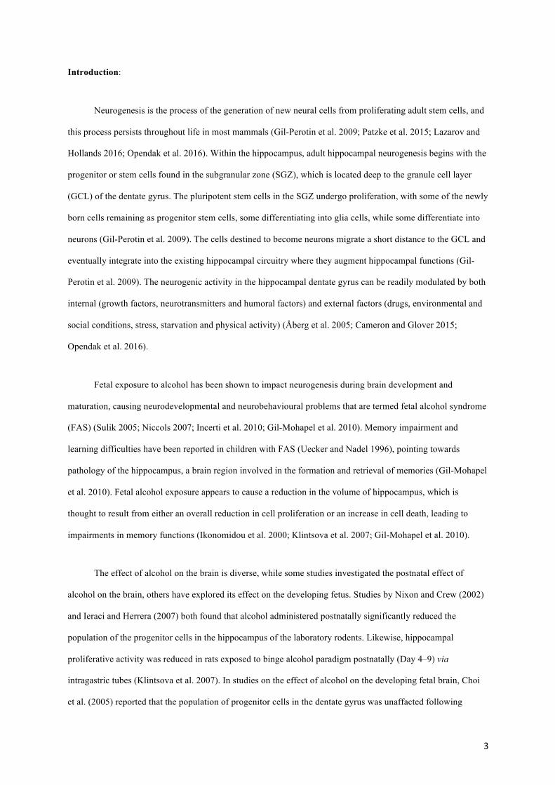
brain mass. In addition, Nissl staining revealed no observable abnormalities in the neuronal distribution pattern
in the brains of all the experimental mice, and specifically so in the dentate gyrus of the hippocampus (Fig. 1).
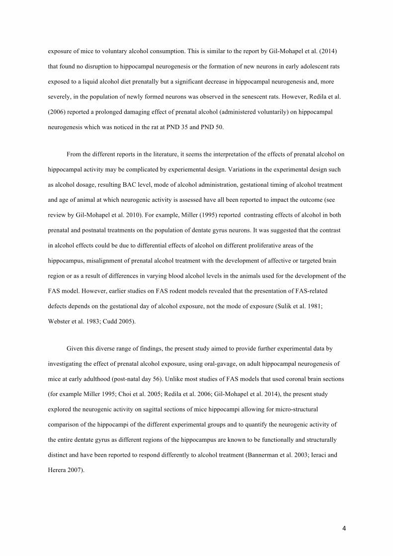
For all mice studied there were no marked differences in the shape or size of the Ki-67 immunopositive
cells in either the subgranular layer of the dentate gyrus or the subventricular zone of the lateral ventricle. In all
18 individual mice, irrespective of the experimental group, we found that the distribution of cells
immunopositive for Ki-67 was very similar. We observed Ki-67 immunopositive cells in two distinct
proliferative regions – the dentate gyrus of the hippocampus, and the subventricular zone of the lateral ventricle
(Figs. 2, 3). A distinct subgranular zone, located between the granular cell layer and the hilus of the dentate
gyrus of the hippocampus was evident in all mice examined (Fig. 2). Numerous Ki-67 immunopositive cells
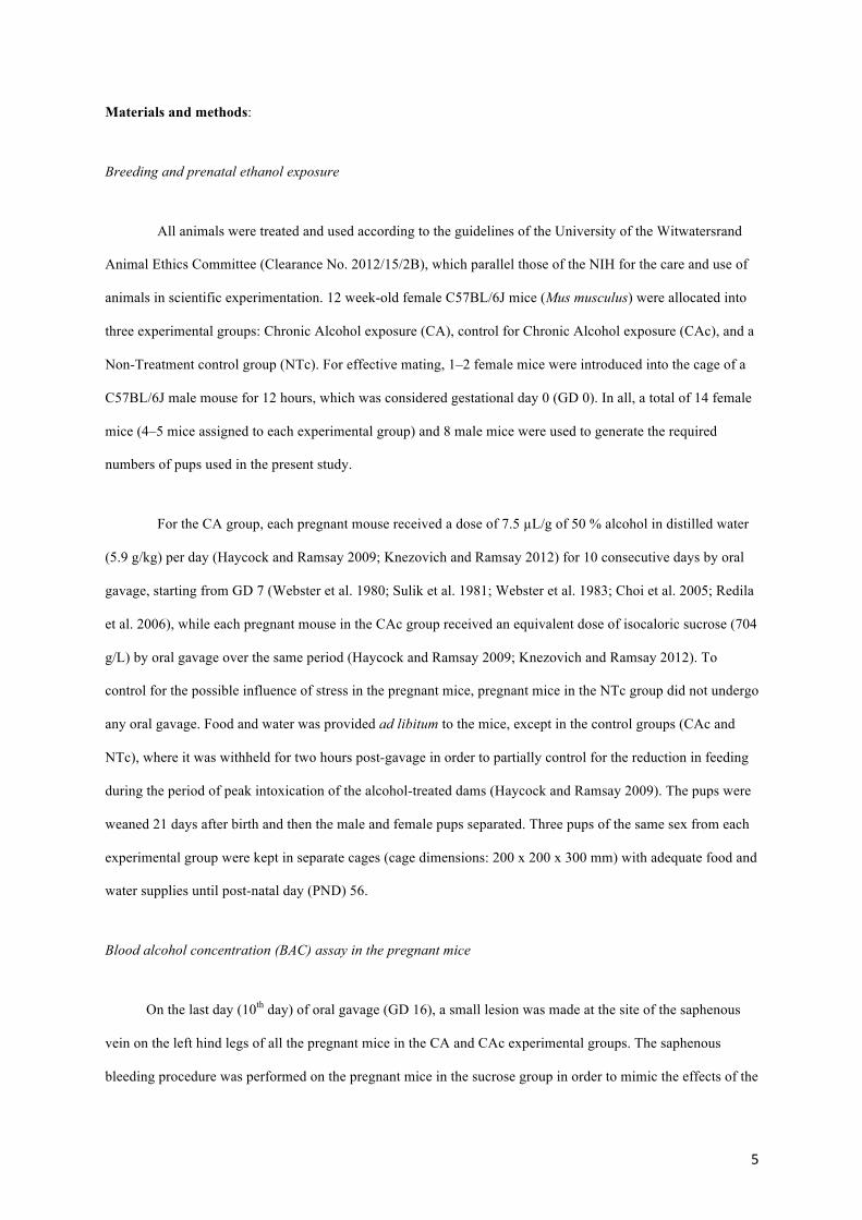
were observed in the subventricular zone of the lateral ventricle in all the mice studied. From the SVZ two
streams of Ki-67 immunopositive cells could be observed, one that ran dorsal and then rostral to the caudate
nucleus, and one that ran caudal to the caudate nucleus (Fig. 3). The stream that ran dorsal and anterior to the
caudate nucleus was observed to turn, in a rostral direction, into the olfactory tract to infuse the olfactory bulb
with Ki-67 immunopositive neurons (Fig. 3). The stream that ran caudal to the caudate nucleus was observed to
extend ventrally to the anterior commissure, from where it made a lateral extension around the globus pallidus
towards the piriform cortex.
10
In all 18 mice investigated, the distribution of cells immunopositive for DCX was very similar. A
distinct subgranular zone, located between the granular cell layer and the hilus of the dentate gyrus in the
hippocampus, was evident displaying DCX immunopositive cells (Fig. 2). The majority of these DCX positive
cells exhibited processes that extended into the molecular layer of the dentate gyrus. In all mice clusters of DCX
positive cells and processes were present in the subventricular zone (SVZ) with the highest density of
immunolabelled structures observed towards the rostral end of the lateral ventricle (Fig. 3). The labelled cells
were characterized by relatively short unipolar and or/bipolar processes. From the SVZ a stream of DCX
immunopositive cells could be observed – the rostral migratory stream (RMS). DCX immunopositive cells were
found between the rostrodorsal aspects of the caudate nucleus and the subcortical white matter, as well as on the
caudal aspect of the caudate nucleus. At the rostroventral pole of the caudate nucleus, the “stream” of
immunolabelled cells turned in a rostral direction with the stream ending in the olfactory bulb (Fig. 3). The
DCX immunopositive cells in the RMS were often obscured by the numerous tangentially oriented fibres of the
stream, but when readily viewable were found to be fusiform in shape, small in size and displayed bipolar
processes. The DCX immunopositive cells and processes found along the caudal aspect of the caudate nucleus
were observed to join the RMS where it turned to enter the olfactory bulb.
In the main olfactory bulb (MOB) DCX immunoreactivity was evident in all layers in all mice. The
majority of DCX-expressing cells were located in the granular cell layer (GCL), exhibiting radially orientated
DCX-positive cells and processes. Most of these cells were bipolar and ovoid in shape. The external plexiform
layer of the olfactory bulb (EPL) presented with distinct radial fibres, while the glomerular layer (GL) displayed
sparsely distributed DCX immunopositive cells that presumably represent periglomerular cells (Fig. 3). In
addition to these two main regions of DCX immunopositive structures, cells were observed in layer II of the
piriform cortex, and the occasional cell was observed in the amygdala. Weakly labelled DCX immunopositive
neurons were occasionally observed in the rostral half of the cerebral neocortex in some individuals.
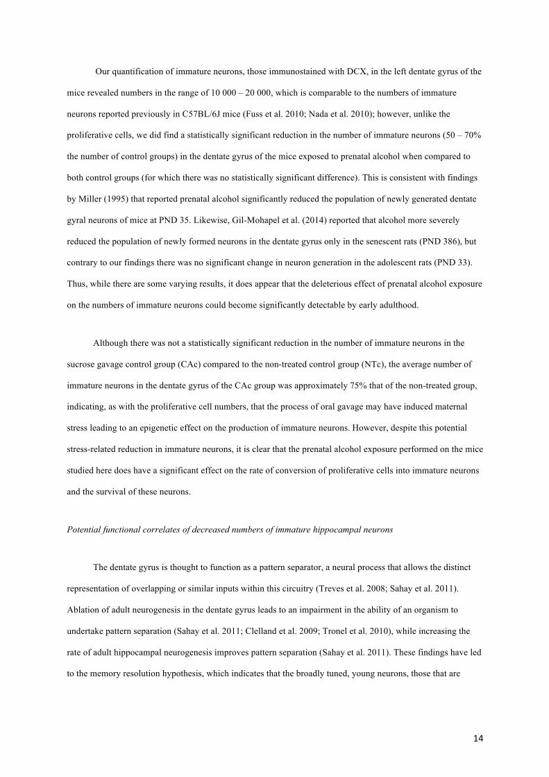
Quantification of proliferative cells (Ki-67 immunopositive) in the dentate gyrus of the hippocampus
Our quantitative analysis of the numbers of Ki-67 immunopositive cells, presumably proliferative cells,
in the dentate gyrus of the left hippocampus of mice from the three different experimental groups revealed
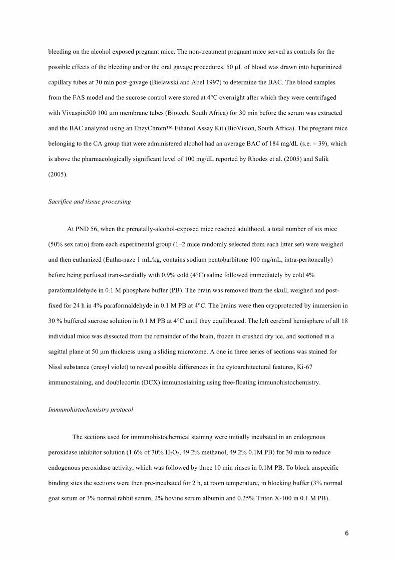
homogeneity in numbers between individuals in the same group, and between groups. For the CA group, the
average number of Ki-67 immunopositive cells was 1653.5 (s.e. 126.3), for the CAc group it was 1622.5 (s.e.
11
123.1) and for the NTc group it was 1349.0 (s.e. 90.3) (Fig. 4a). Thus, while there is a trend for the two groups
that underwent oral gavage procedures, irrespective of the presence of ethanol, to have higher numbers of
proliferative cells in the dentate gyrus, statistical analyses revealed that the higher number of proliferative cells
in the CA group was not statistically significantly different from the CAc group (p = 0.980) or the NTc group (p
= 0.178). A comparison between CAc and NTc groups was also not statistically significantly different (p =
0.241).
Quantification of immature neurons (DCX immunopositive) in the dentate gyrus of the hippocampus
In contrast to the quantification of Ki-67 immunopositive cells, our quantification of the numbers of
DCX immunopositive cells, presumably immature neurons, in the left hippocampus of mice from the three
experimental groups did reveal significant inter-group differences. For the CA group, the average number of
DCX immunopositive cells was 10 441.0 (s.e. 913.7), for the CAc group it was 15 238.5 (s.e. 1019.8) and for
the NTc group it was 19 963.0 (s.e. 817.3) (Fig. 4b). Thus, it is apparent that the group exposed to prenatal
alcohol, the CA group, had substantially lower numbers of DCX immunopositive cells in the dentate gyrus.
Statistical analyses confirmed that the estimated number of DCX immunopositive cells was significantly lower
in the CA group when compared to the CAc group (p = 0.006) or the NTc group (p < 0.0001), but there was not
statistically significant difference between the CAc and NTc groups (p = 0.404).
Discussion:
In the present study, the effect of chronic prenatal ethanol exposure, using an oral gavage method, on the
process of adult neurogenesis was explored in C57BL/6J mice at postnatal day 56. The fetal mice were exposed
to maternal blood alcohol concentrations (BAC) slightly above those considered pharmacologically significant
(Rhodes et al. 2005; Sulik 2005) from gestational days 7 – 16 inclusive (10 days in total), which appears to be
equivalent to the later stages of the first trimester and the early stages of the second trimester of humans (Patten
et al. 2014). Our results indicate that the location of cells associated with adult neurogenesis, and their
distribution throughout the brain, does not appear to be affected by this prenatal alcohol exposure. In addition,
the rate of proliferation of newly born cells in the dentate gyrus of the hippocampus does not appear to be
affected, but the numbers of immature hippocampal neurons in the dentate gyrus is significantly lower in the
mice exposed to ethanol compared to the control groups.
12
Methodological considerations
In this study, we utilized a chronic alcohol paradigm using an oral gavage method to develop a fetal
alcohol spectrum disorder (FASD) mouse model. The oral gavage method was utilized to ensure that the
pregnant mice had adequate doses of alcohol of pharmacological significance (Rhodes et al. 2005; Sulik 2005),
and to maintain uniform alcohol consumption amongst the pregnant mice (Åberg et al. 2005; Gil-Mohapel et al.
2014). The average BAC level obtained from the pregnant mice in this study was slightly higher than the
pharmacologically significant level reported by Miller (1995), Rhodes et al. (2005) and Sulik (2005).
Precautionary measures, as described by Turner et al. (2011), were taken during the oral gavage procedure in
order to reduce stress in the pregnant mothers. In addition, the pregnant mothers were housed alone, weighed
daily and blood samples were collected only on the last day of either alcohol or sucrose administration.
Subsequently, the weaned litters from these pregnant mice were housed in groups of three per cage. These
precautions were necessary because it has been reported that many factors can impact adult hippocampal
neurogenesis (Cameron and Glover 2015; Opendak et al. 2016).
Distribution of Ki-67 and doublecortin (DCX) immunopositive cells
Across mammals, the endogenous proteins Ki-67 and DCX have been shown to be accurate markers of
cells at two different phases in the process of adult neurogenesis, the proliferative phase (Ki-67) and the
immature neuron phase (DCX) (Balthazart and Ball 2014). In mammals generally, and in mice specifically, two
proliferative regions, the subventricular and subgranular zone, are consistently reported. The cells within the
subventricular zone migrate through the rostral migratory stream to infuse mostly in the olfactory bulb, but may
also enter other brain regions (Gil-Perotin et al. 2009). The cells of the subgranular zone migrate a short distance
to the granular layer of the dentate gyrus of the hippocampus, where many of these cells become integrated into
the hippocampal circuitry (Pawlak et al. 2002; Åberg et al. 2005). During the migratory phase, the proliferative
cells (immunopositive to Ki-67 antibodies) become immature migrating neurons (immunopositive to DCX
antibodies). This general pattern of adult neurogenesis did not appear to be affected by the prenatal ethanol
exposure in the sagittal sections of mice studied, with the overall pattern of adult neurogenesis being identical
across experimental groups, and to previous studies of adult neurogenesis in mice (Klintsova et al. 2007).
Quantification of proliferative (Ki-67 immunoreactive) cells in the dentate gyrus of the hippocampus
13
Our quantification of proliferative cells, those immunostained with Ki-67, in the left dentate gyrus of the
mice revealed numbers in the range of 1300 – 1650, which is comparable to the numbers of proliferative cells
reported previously in C57BL/6J mice (Fuss et al. 2010; Nada et al. 2010); however, we did not find any
statistically significant effect of prenatal ethanol exposure on the numbers of proliferative cells in the dentate
gyrus. This finding is in agreement with previous reports indicating that postnatal alcohol exposure does not
have an effect on the rate of cell proliferation in the dentate gyrus of hippocampus, especially at early adulthood
in laboratory rodent models (Wozniak et al. 2004; Klintsova et al. 2007; Gil-Mohapel et al. 2014). In contrast, in
senescent Sprague-Dawley rats subjected to prenatal alcohol exposure a decrease in proliferation rates was
observed (Gil-Mohapel et al. 2014). Thus, our study corroborates previous reports (Choi et al. 2005; Ieraci and
Herrera 2007; Klintsova et al. 2007; Gil-Mohapel et al. 2014) indicating that the deleterious effect of prenatal
alcohol exposure could be less noticeable during early adulthood, or indeed that this exposure had no specific
effect on the proliferation of cells within the early adult hippocampus.
Despite this, the number of Ki-67 immunoreactive cells in the hippocampi of the two experimental
groups that underwent oral gavage was higher (by ~300 cells, or 1.2 times, on average), though not statistically
significantly, than the non-treated control group. It has been shown that factors, such as chronic stress during the
development of the brain, may cause a permanent change in the rate of cell proliferation in the hippocampus
(Lee et al. 2000; Mirescu et al. 2004; Åberg et al. 2005; Ieraci and Herrera 2007). In the present study, standard
animal handling precautions (Turner et al. 2011) were strictly adhered to, to reduce the stress level in the mice
during the oral gavage procedure, but it is possible that stress-induced gavage and saphenous bleeding may have
had an effect on the neurogenic outcomes. In some cases, an increase in cell proliferation is thought to be a
compensatory mechanism for a significant increase in cell death in the dentate gyrus due to the damaging effect
of alcohol (Pawlak et al. 2002; Zharkovsky et al. 2003; Åberg et al. 2005), although we did not examine cell
death in the current study so cannot confirm this hypothesis. In the present study, the use of the oral gavage
method for delivery of the alcohol and/or saphenous bleeding may have caused, epigenetically through maternal
stress, the slight, but non-significant, increase in cell proliferation rates observed in the two groups that
underwent this treatment.
Quantification of immature (DCX immunoreactive) neurons in the dentate gyrus of the hippocampus
14
Our quantification of immature neurons, those immunostained with DCX, in the left dentate gyrus of the
mice revealed numbers in the range of 10 000 – 20 000, which is comparable to the numbers of immature
neurons reported previously in C57BL/6J mice (Fuss et al. 2010; Nada et al. 2010); however, unlike the
proliferative cells, we did find a statistically significant reduction in the number of immature neurons (50 – 70%
the number of control groups) in the dentate gyrus of the mice exposed to prenatal alcohol when compared to
both control groups (for which there was no statistically significant difference). This is consistent with findings
by Miller (1995) that reported prenatal alcohol significantly reduced the population of newly generated dentate
gyral neurons of mice at PND 35. Likewise, Gil-Mohapel et al. (2014) reported that alcohol more severely
reduced the population of newly formed neurons in the dentate gyrus only in the senescent rats (PND 386), but
contrary to our findings there was no significant change in neuron generation in the adolescent rats (PND 33).
Thus, while there are some varying results, it does appear that the deleterious effect of prenatal alcohol exposure
on the numbers of immature neurons could become significantly detectable by early adulthood.
Although there was not a statistically significant reduction in the number of immature neurons in the
sucrose gavage control group (CAc) compared to the non-treated control group (NTc), the average number of
immature neurons in the dentate gyrus of the CAc group was approximately 75% that of the non-treated group,
indicating, as with the proliferative cell numbers, that the process of oral gavage may have induced maternal
stress leading to an epigenetic effect on the production of immature neurons. However, despite this potential
stress-related reduction in immature neurons, it is clear that the prenatal alcohol exposure performed on the mice
studied here does have a significant effect on the rate of conversion of proliferative cells into immature neurons
and the survival of these neurons.
Potential functional correlates of decreased numbers of immature hippocampal neurons
The dentate gyrus is thought to function as a pattern separator, a neural process that allows the distinct
representation of overlapping or similar inputs within this circuitry (Treves et al. 2008; Sahay et al. 2011).
Ablation of adult neurogenesis in the dentate gyrus leads to an impairment in the ability of an organism to
undertake pattern separation (Sahay et al. 2011; Clelland et al. 2009; Tronel et al. 2010), while increasing the
rate of adult hippocampal neurogenesis improves pattern separation (Sahay et al. 2011). These findings have led
to the memory resolution hypothesis, which indicates that the broadly tuned, young neurons, those that are
15
identified with DCX immunostaining, interact with the specifically tuned mature neurons to increase the fidelity
of spatial and contextual discrimination (Aimone et al. 2011).
The results of the current investigation indicate that in FASD children there is a potential downregulation
of the number of immature neurons in the dentate gyrus. This appears to correlate with reports of executive
dysfunctions and the inability to recall more objects than in normal children in FASD children (Connor et al.
2000; Fuglestad et al. 2015). In addition, the ability to recall acquired information (retrospective memory)
(Willoughby et al. 2008), to perform delayed intentions (prospective memory) (Kliegel et al. 2008) and to
temporarily store and handle information (working memory) (Burden et al. 2005) were similarly dysfunctional
in children that were exposed to prenatal alcohol. These are indications that learning and memory are adversely
affected after exposure to prenatal alcohol (Kaemingk et al. 2003; Lewis et al. 2016; Mattson and Roebuck
2002), and that this in part may be the result of potentially fewer immature neurons within the hippocampus of
FASD children, as observed in the current mouse model.
16
References:
Åberg E, Hofstetter CP, Olson L, Brené S (2005) Moderate ethanol consumption increases hippocampal cell proliferation and neurogenesis in the adult mouse. Int J Neuropsychoph 8:557–567. doi:10.1017/S1461145705005286
Aimone JB, Deng W, Gage FH (2011) Resolving new memories: a critical look at the dentate gyrus, adult neurogenesis, and pattern separation. Neuron 70:589–596. doi:10.1016/j.neuron.2011.05.010
Balthazart J, Ball GF (2014) Endogenous versus exogenous markers of adult neurogenesis in canaries and other birds: advantages and disadvantages. J Comp Neurol 522:4100–4120. doi:10.1002/cne.23661
Bielawski DM, Abel EL (1997) Acute treatment of paternal alcohol exposure produces malformations in offspring. Alcohol 14:397–401. doi:10.1016/S0741-8329(97)87951-7
Burden MJ, Jacobson SW, Sokol RJ, Jacobson JL (2005) Effects of prenatal alcohol exposure on attention and working memory at 7.5 year of age. Alcohol Clin Exp Res 29: 443–452. doi:10.1097/01.ALC.0000156125.50577.EC
Cameron HA, Glover LR (2015) Adult neurogenesis: beyond learning and memory. Annu Rev Psychol 66:53–81. doi:10.1146/annurev-psych-010814-015006
Choi IY, Allan AM, Cunningham LA (2005) Moderate fetal alcohol exposure impairs the neurogenic response to an enriched environment in adult mice. Alcohol Clin Exp Res 29:2053–2062. doi:10.1097/01.alc.0000187037.02670.59
Clelland CD, Choi M, Romberg C, Clemenson GD, Fragniere A, Tyers P, Jessberger S, Saksida LM, Barker RA, Gage FH, Bussey TJ (2009) A functional role for adult hippocampal neurogenesis in spatial pattern separation. Science 325:210–213. doi:10.1126/science.1173215
Coggeshall RE, Lekan HA (1996) Methods for determining numbers of cells and synapses: a case for more uniform standards of review. J Comp Neurol 364:6–15. doi:10.1002/cne.903690102
Connor PD, Sampson PD, Bookstein FL, Barr HM, Streissguth AP (2000) Direct and indirect effects of prenatal alcohol damage on executive function. Dev Neuropsychol 18:331–354. doi:10.1207/S1532694204Connor
Cudd TA (2005) Animal model systems for the study of alcohol teratology. Exp Biol Med 230:389–393. doi:10.1177/15353702-0323006-06
Erasso DM, Chaparro RE, del Rio CEQ, Karlnoski R, Camporesi EM, Saporta S (2012) Quantitative assessment of new cell proliferation in the dentate gyrus and learning after isoflurane or propofol anesthesia in young and aged rats. Brain Res 1441:38–46. doi:10.1016/j.brainres.2011.11.025
Eriksson PS, Perfilieva E, Björk-Eriksson T, Alborn AM, Nordborg C, Peterson DA, Gage FH (1998) Neurogenesis in the adult human hippocampus. Nat Med 4:1313–1317.
17
Fuglestad AJ, Whitely ML, Carlson SM, Boys CJ, Eckerle JK, Fink BA, Wozniak JR (2015) Executive functioning deficits in preschool children with fetal alcohol spectrum disorders. Child Neuropsychol 21:716–731. doi:10.1080/09297049.2014.933792
Fuss J, Ben Abdullah NMB, Vogt MA, Touma C, Pacifici PG, Palme R, Witzemann V, Hellweg R, Gass P (2010) Voluntary exercise induces anxiety-like behavior in adult C57BL/6J mice correlating with hippocampal neurogenesis. Hippocampus 20:364–376. doi:10.1002/hipo.20634
Gil-Mohapel J, Boehme F, Kainer L, Christie BR (2010) Hippocampal cell loss and neurogenesis after fetal alcohol exposure: insights from different rodent models. Brain Res Rev 64:283–303. doi:10.1016/j.brainresrev.2010.04.011
Gil-Mohapel J, Titterness A, Patten A, Taylor S, Ratzlaff A, Ratzlaff T, Helfer J, Christie B (2014) Prenatal ethanol exposure differentially affects hippocampal neurogenesis in the adolescent and aged brain. Neuroscience 273:174–188. doi:10.1016/j.neuroscience.2014.05.012
Gil-Perotin SG, Alvarez-Buylla A, García-Verdugo JM (2009) Identification and Characterization of Neural Progenitor Cells in the Adult Mammalian Brain: 203 Advances in Anatomy, Embryology and Cell Biology. Springer New York.
Gundersen H, Bagger P, Bendtsen T, Evans S, Korbo L, Marcussen N, Møller A, Nielsen K, Nyengaard J, Pakkenberg B (1988) The new stereological tools: disector, fractionator, nucleator and point sampled intercepts and their use in pathological research and diagnosis. Apmis 96:857–881. doi:10.1111/j.1699-0463.1988.tb00954.x
Haycock PC, Ramsay M (2009) Exposure of mouse embryos to ethanol during preimplantation development: effect on DNA methylation in the h19 imprinting control region. Biol Reprod 81:618–627. doi:10.1095/biolreprod.108.074682
Ieraci A, Herrera DG (2007) Single alcohol exposure in early life damages hippocampal stem/progenitor cells and reduces adult neurogenesis. Neurobiol Dis 26:597–605. doi:10.1016/j.nbd.2007.02.011
Ikonomidou C, Bittigau P, Ishimaru MJ, Wozniak DF, Koch C, Genz K, Price MT, Stefovska V, Hörster F, Tenkova T, Dikranian K (2000) Ethanol-induced apoptotic neurodegeneration and fetal alcohol syndrome. Science 287:1056–1060. doi:10.1126/science.287.5455.1056
Incerti M, Vink J, Roberson R, Wood L, Abebe D, Spong CY (2010) Reversal of alcohol-induced learning deficits in the young adult in a model of fetal alcohol syndrome. Obstet Gynecol 115:350–356. doi:10.1097/AOG.0b013e3181cb59da
Kaemingk KL, Mulvaney S, Halverson PT (2003) Learning following prenatal alcohol exposure: Performance on verbal and visual multitrial tasks. Arch Clin Neuropsychy 18:33–47. doi:10.1016/S0887-6177(01)00182-2
Kliegel MA, Jager T, Altgassen M, Shum D (2008) Clinical neuropsychology of prospective memory. In: Kliegel M, McDaniel MA, Einstein GO (ed) Prospective Memory: Cognitive, Neuroscience, Developmental, and Applied Perspectives, Lawrence Erlbaum Associates, New York, pp 283–303
18
Klintsova AY, Helfer JL, Calizo LH, Dong WK, Goodlett CR, Greenough WT (2007) Persistent impairment of hippocampal neurogenesis in young adult rats following early postnatal alcohol exposure. Alcohol Clin Exp Res 31:2073–2082. doi:10.1111/j.1530-0277.2007.00528.x
Knezovich JG, Ramsay M (2012) The effect of preconception paternal alcohol expisure on epigenetic remodeling of the H19 and Rasgrf1 imprinting control regions in mouse offspring. Front Genet 3:10. doi:10.3389/fgene.2012.00010
Lazarov O, Hollands C (2016) Hippocampal neurogenesis: learning to remember. Prog Neurobiol 138:1-18. doi:10.1016/j.pneurobio.2015.12.006
Lee J, Duan W, Long JM, Ingram DK, Mattson MP (2000) Dietary restriction increases the number of newly generated neural cells, and induces BDNF expression, in the dentate gyrus of rats. J Mol Neurosci 15:99–108. doi:10.1385/JMN:15:2:99
Lewis CE, Thomas KG, Molteno CD, Kliegel M, Meintjes EM, Jacobson JL, Jacobson SW (2016) Prospective memory impairment in children with prenatal alcohol exposure. Alcohol Clin Exp Res 40:969–978. doi: 10.1111/acer.13045
Mattson SN, Roebuck TM (2002) Acquisition and retention of verbal and nonverbal information in children with heavy prenatal alcohol exposure. Alcohol Clin Exp Res 26:875–882. doi:10.1111/j.1530-0277.2002.tb02617.x
Miller MW (1995) Generation of neurons in the rat dentate gyrus and hippocampus: effects of prenatal and postnatal treatment with ethanol. Alcohol Clin Exp Res 19:1500–1509. doi:10.1111/j.1530-0277.1995.tb01014.x
Mirescu C, Peters JD, Gould E (2004) Early life experience alters response of adult neurogenesis to stress. Nat Neurosci 7:841–846. doi:10.1038/nn1290
Mouton PR, Kelley-Bell B, Tweedie D, Spangler EL, Perez E, Carlson OD, Short RG, de Cabo R, Chang J, Ingram DK, Li Y, Greig NH (2012) The effects of age and lipopolysaccharide (LPS)-mediated peripheral inflammation on numbers of central catecholaminergic neurons. Neurobiol Aging 33:423.e27–423.e36. doi:10.1016/j.neurobiolaging.2010.09.025
Nada MB, Slomianka L, Vyssotski AL, Lipp HP (2010) Early age-related changes in adult hippocampal neurogenesis in C57 mice. Neurobiol Aging 31:151–161. doi:10.1016/j.neurobiolaging.2008.03.002
Niccols A (2007) Fetal alcohol syndrome and the developing socio-emotional brain. Brain Cognition 65:135–142. doi:10.1016/j.bandc.2007.02.009
Nixon K, Crews FT (2002) Binge ethanol exposure decreases neurogenesis in adult rat hippocampus. J Neurochem 83:1087–1093. doi:10.1046/j.1471-4159.2002.01214.x
Noori HR, Fornal CA (2011) The appropriateness of unbiased optical fractionators to assess cell proliferation in the adult hippocampus. Front Neurosci 5:140. doi:10.3389/fnins.2011.00140
19
Opendak M, Briones BA, Gould E (2016) Social behavior, hormones and adult neurogenesis. Front Neuroendocrinol 41:71–86. doi:10.1016/j.yfrne.2016.02.002
Patten AR, Fontaine CJ, Christie BR (2014) A comparison of the different animal models of fetal alcohol spectrum disorders and their use in studying complex behaviors. Front Pediatr 2:1–19. doi:10.3389/fped.2014.00093
Patzke N, Spocter MA, Karlsson KÆ, Bertelsen MF, Haagensen M, Chawana R, Streicher S, Kaswera C, Gilissen E, Alagaili AN, Mohammed OB, Reep RL, Bennett NC, Siegel JM, Ihunwo AO, Manger PR (2015) In contrast to many other mammals, cetaceans have relatively small hippocampi that appear to lack adult neurogenesis. Brain Struct Funct 220:361–383. doi:10.1007/s00429-013-0660-1
Pawlak R, Skrzypiec A, Sulkowski S, Buczko W (2002) Ethanol-induced neurotoxicity is counterbalanced by increased cell proliferation in mouse dentate gyrus. Neurosci Lett 327:83–86. doi:10.1016/S0304-3940(02)00369-5
Redila VA, Olson AK, Swann SE, Mohades G, Webber AJ, Weinberg J, Christie BR (2006) Hippocampal cell proliferation is reduced following prenatal ethanol exposure but can be rescued with voluntary exercise. Hippocampus 16:305–311. doi:10.1002/hipo.20164
Rhodes JS, Best K, Belknap JK, Finn DA, Crabbe JC (2005) Evaluation of a simple model of ethanol drinking to intoxication in C57BL/6J mice. Physiol Behav 84:53–63. doi:10.1016/j.physbeh.2004.10.007
Sahay A, Scobie KN, Hill AS, O’Carroll CM, Kheirbek MA, Burghardt NS, Fenton AA, Dranovsky A, Hen R (2011) Increasing adult hippocampal neurogenesis is sufficient to improve pattern separation. Nature 472:466–470. doi:10.1038/nature09817
Segi-Nishida E, Warner-Schmidt JL, Duman RS (2008) Electroconvulsive seizure and VEGF increase the proliferation of neural stem-like cells in rat hippocampus. Proc Natl Acad Sci U S A 105:11352–11357. doi:10.1073/pnas.0710858105
Sulik KK (2005) Genesis of alcohol-induced craniofacial dysmorphism. Exp Biol Med 230:366–375. doi:10.1177/15353702-0323006-04
Sulik KK, Johnston MC, Webb MA (1981) Fetal alcohol syndrome: embryogenesis in a mouse model. Science 214:936–938. doi:10.1126/science.6795717
Treves A, Tashiro A, Witter MP, Moser EI (2008) What is the mammalian dentate gyrus good for? Neuroscience 154:1155–1172. doi:10.1016/j.neuroscience.2008.04.073
Tronel S, Fabre A, Charrier V, Oliet SH, Gage FH, Abrous DN (2010) Spatial learning sculpts the dendritic arbor of adult-born hippocampal neurons. Proc Natl Acad Sci U S A 107:7963–7969. doi:10.1073/pnas.0914613107
Turner PV, Brabb T, Pekow C, Vasbinder MA (2011) Administration of substances to laboratory animals: routes of administration and factors to consider. J Am Assoc Lab Anim Sci 50:600–613
20
Uecker A, Nadel L (1996) Spatial locations gone awry: object and spatial memory deficits in children with fetal alcohol syndrome. Neuropsychologia 34:209–223. doi:10.1016/0028-3932(95)00096-8
Webster W, Walsh D, McEwen SE, Lipson A (1983) Some teratogenic properties of ethanol and acetaldehyde in C57BL/6J mice: implications for the study of the fetal alcohol syndrome. Teratology 27:231–243. doi:10.1002/tera.1420270211
Webster WS, Walsh DA, Lipson AH, McEwen SE (1980) Teratogenesis after acute alcohol exposure in inbred and outbred mice. Neurotoxicol Teratol 2:227–234.
West MJ (1993) New stereological methods for counting neurons. Neurobiol Aging 14:275–285. doi:10.1016/0197-4580(93)90112-O
Willoughby KA, Sheard ED, Nash K, Rovet J (2008) Effects of prenatal alcohol exposure on hippocampal volume, verbal learning, and verbal and spatial recall in late childhood. J Int Neuropsychol Soc 14:1022–1033. doi:10.1017/S1355617708081368
Wozniak DF, Hartman RE, Boyle MP, Vogt SK, Brooks AR, Tenkova T, Young C, Olney JW, Muglia LJ (2004) Apoptotic neurodegeneration induced by ethanol in neonatal mice is associated with profound learning/memory deficits in juveniles followed by progressive functional recovery in adults. Neurobiol Dis 17:403–414. doi:10.1016/j.nbd.2004.08.006
Zagron G, Weinstock M (2006) Maternal adrenal hormone secretion mediates behavioural alterations induced by prenatal stress in male and female rats. Behav Brain Res 175:323–328. doi:10.1016/j.bbr.2006.09.003
Zharkovsky T, Kaasik A, Jaako K, Zharkovsky A (2003) Neurodegeneration and production of the new cells in the dentate gyrus of juvenile rat hippocampus after a single administration of ethanol. Brain Res 978:115–123. doi:10.1016/S0006-8993(03)02796-3
21
Figure Legends:
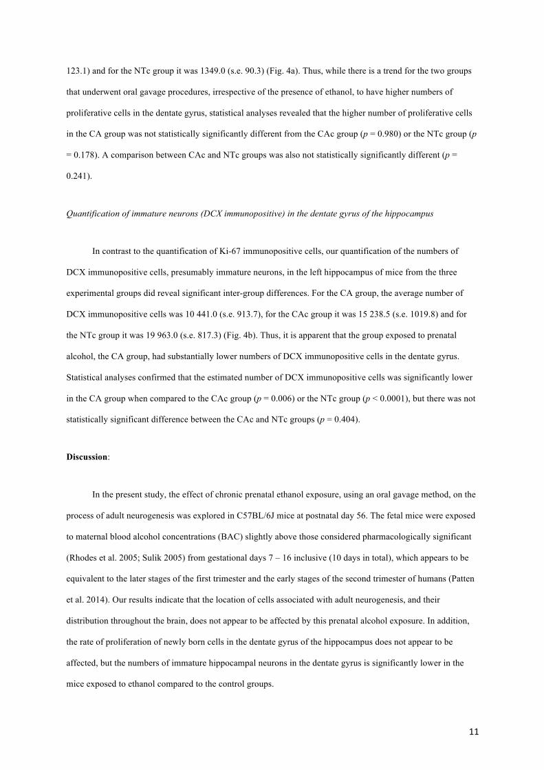
Figure 1: Photomicrographs of the dorsal left hippocampus of the mouse in the sagittal plane, showing the
general morphology of the dentate gyrus (DG) and cornu ammonis (CA1, CA3) regions in the three different
groups analyzed in the present study, the group exposed to chronic prenatal alcohol (CA) (a), the prenatal
gavage control group (CAc) (b) and the non-treated control group (NTc) (c). Note the lack of observable
differences in the structure of these hippocampal regions between groups. In all images dorsal is to the top and
rostral to the left. Scale bar in c = 500 µm and applies to all.
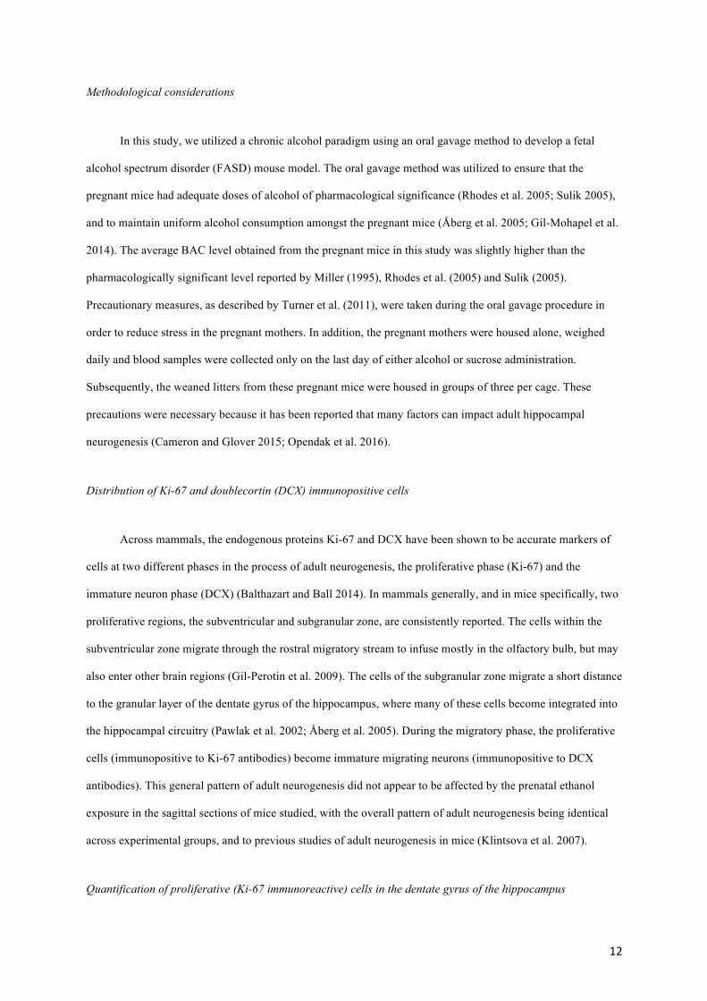
Figure 2: Photomicrographs of the dorsal left hippocampus of the mouse in the sagittal plane immunostained for
Ki-67 (a, c, e) or doublecortin (DCX) (b, d, f) in the three different groups analyzed in the present study, the
group exposed to chronic prenatal alcohol (CA) (a, b), the prenatal gavage control group (CAc) (c, d) and the
non-treated control group (NTc) (e, f). The insets in the upper right corner of each image represent higher
magnification photomicrographs of each region to demonstrate cellular morphology. Qualitatively, the density
of both Ki-67 and DCX immunostained cells and structures appears similar, but quantification (see Fig. 4)
revealed differences. In all images dorsal is to the top and rostral to the left. Scale bar in f = 500 µm and applies
to a – f. Scale bar in inset e = 100 µm, and applies to all insets.
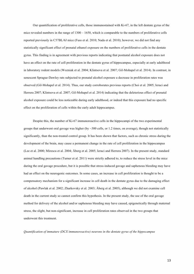
Figure 3: Photomicrographs of Ki-67 (a, b) and doublecortin (DCX) (c, d) immunostained sections showing
different aspects of the rostral migratory stream (RMS) in the mice studied. a. Ki-67 immunostained cells of the
RMS rostral to the caudate nucleus (c) and the flexure of this stream as it enters the olfactory bulb (OB). Scale
bar in a = 500 µm. b. Higher power photomicrograph of Ki-67 immunostained cells in the RMS showing their
typical clustered appearance. Scale bar in b = 100 µm. C. DCX immunostained structures in the subventricular
zone (SVZ) of the lateral ventricle (LV) located caudal and dorsal to the caudate nucleus (c), and giving rise to
the RMS. Scale bar in C = 250 µm. d. DCX immunostained cells and dendrites surrounding the glomeruli found
in the glomerular layer (GL) of the olfactory bulb. Scale bar in d = 100 µm. In all images dorsal is to the top
and rostral to the left.
Figure 4: Graphs showing the average numbers of Ki-67 (a) and doublecortin (DCX) (b) immunoreactive cells
in the left hippocampus of three different groups (n = 6 per group) analyzed in the present study, the group
exposed to chronic prenatal alcohol (CA), the prenatal gavage control group (CAc) and the non-treated control
group (NTc). Note that while the two groups that underwent gavage treatment (CA and CAc) appear to have
slightly higher numbers of Ki-67 immunoreactive cells than the untreated group (NTc) (a), these are not
statistically significant differences. The number of DCX immunoreactive cells (b) was statistically significantly
(*) lower in the CA group compared to the two control groups (CAc and NTc). The vertical bars represent
standard error bars.