Embed Size (px)
Citation preview
INTESTINAL OBSTRUCTION
BY OSMAN SALAH ALTOHAMY – A MEDICAL STUDENT
UNIVERSITY OF GEZIRA – WAD MEDANI - SUDAN
DEFINITION
Arrest of downward
propulsion of
intestinal contents.
CLASSIFICATION
There are different types of classification of
intestinal obstruction.
According to the pathological nature.
According to the level of the obstruction.
According to the onset and the course of
the obstruction.
According to the pathological nature.
1. Mechanical obstruction: is
caused by an organic block.
2. Paralytic ileus (Adynamic): is
due to loss of propulsive peristalsis
leading to functional obstruction.
According to the level of the obstruction.
1. High small bowel obstruction.
2. Low small bowel obstruction.
3. Large bowel obstruction.
ACCORDING TO THE ONSET
1. Acute .
2. Chronic: e.g., colon cancer, the symptoms are
insidious and slowly progressive. The patient has
constipation and distension.
3. Acute on chronic: A chronic obstruction may
develop acute symptoms as the obstruction suddenly
becomes complete when a narrowed lumen becomes
totally occluded.
ACUTE MECHANICAL INTESTINAL OBSTRUCTION
AETIOLOGY
1. In the lumen, e.g., faecal impaction, gallstone and
parasitic infestation.
2. In the wall, e.g., congenital atresia, tumours, Crohn's
disease, chronic diverticulitis and mesenteric vascular
occlusion.
3. Outside the wall, e.g., adhesions (commonly post-
operative), strangulated hernia, and volvulus.
PATHOLOGY
Distal to the obstruction the intestine empties and becomes
collapsed.
Proximally the intestine becomes distended by gas and fluids.
Gaseous distension is due to swallowed air, diffusion from
congested vessels and bacterial fermentation.
GI secretions (8 litres per day) accumulate above the site of
obstruction.
As intraluminal pressure rises, absorption ceases.
The stretched smooth muscles undergo hyperperistalsis
in an attempt to overcome the obstruction.
Distension impairs blood supply, and may end in
ulceration and perforation.
This is evident in cases of colon obstruction with a
competent ileocaecal valve (closed loop obstruction).
The rising pressure in the closed proximal colon causes
perforation of the caecum.
SYMPTOMS
1. Pain is usually the first symptom. It is colicky and is caused by the
hyperperistalsis.
2. Distension: is marked in large bowel obstruction (mainly in the
flanks). It is less marked and is central in small bowel obstruction.
3. Constipation: is an early symptom in large bowel obstruction but
late in small bowel obstruction.
4. Vomiting occurs early in small bowel obstruction but it is late or
even absent when the large bowel is obstructed.
SIGNS
On General examination: Evidence of dehydration as tachycardia,
oliguria, dry tongue, or even hypotension may be present.
On Abdominal inspection: Distension, visible peristalsis.
Scars of previous abdominal surgery may denote intraabdominal adhesions.
On Abdominal palpation: A mass may be felt (tumour or intussusception).
On Auscultation: Accentuated intestinal sounds.
Rectal examination reveals an empty rectum, or the finding of a hard
faecal mass, especially in elderly bed-ridden patient causing faecal
impaction.
Strangulation is suspected with:
Fever.
Evidence of blood loss as pallor, tachycardia,
and hypotension.
Pain that is not relieved by naso-gastric suction.
Marked tenderness, rebound tenderness, and
rigidity.
INVESTIGATIONS
Aimed to :-
Confirm the diagnosis
Define its level.
Define the aetiology.
Estimate the severity of water and electrolyte
imbalance.
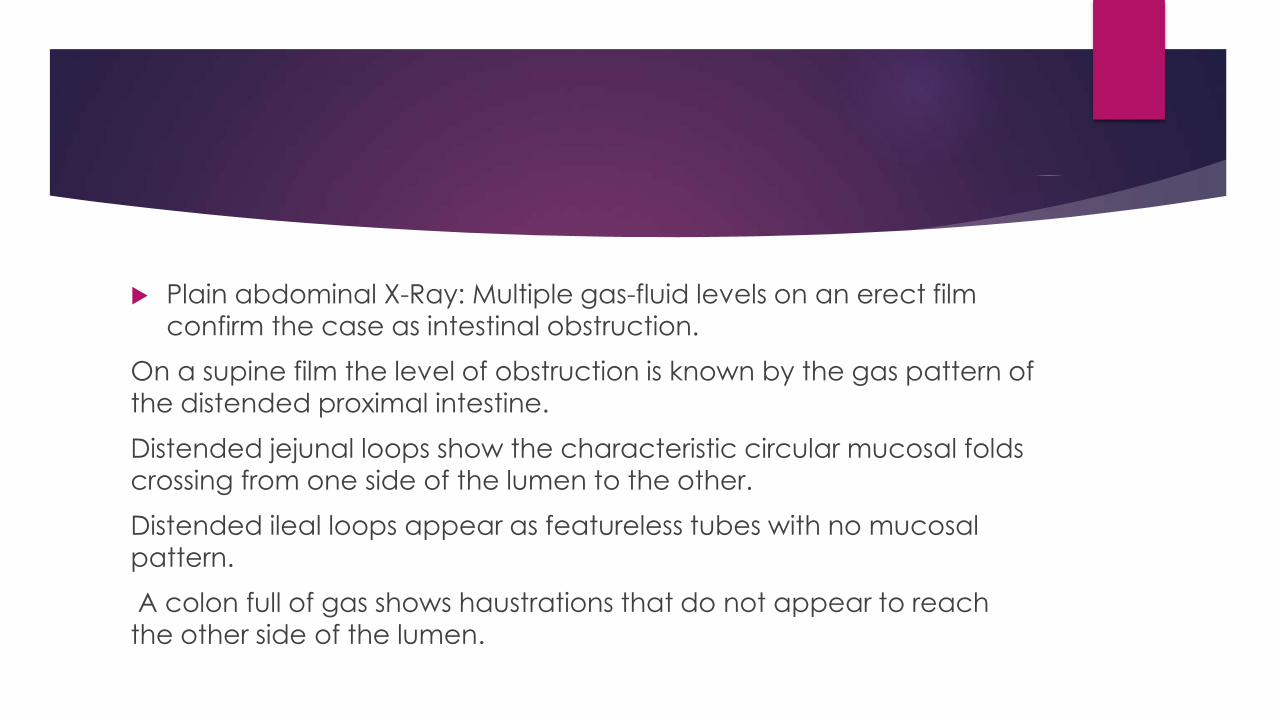
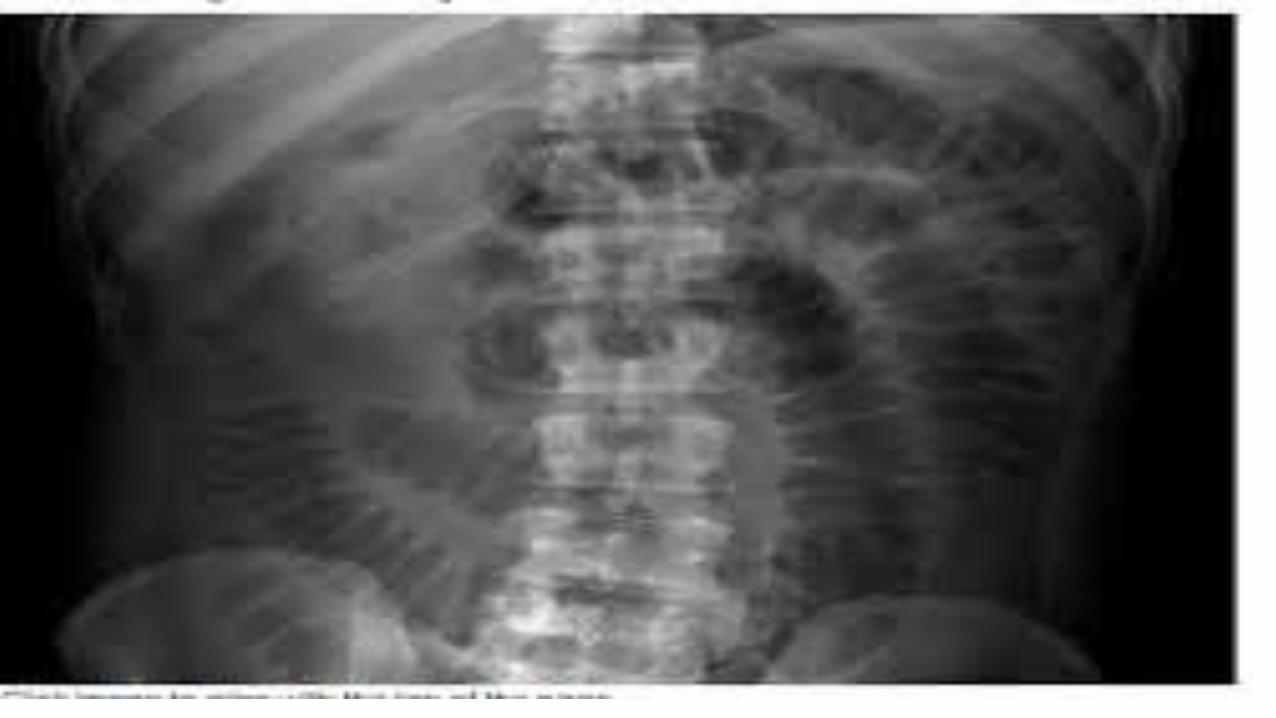
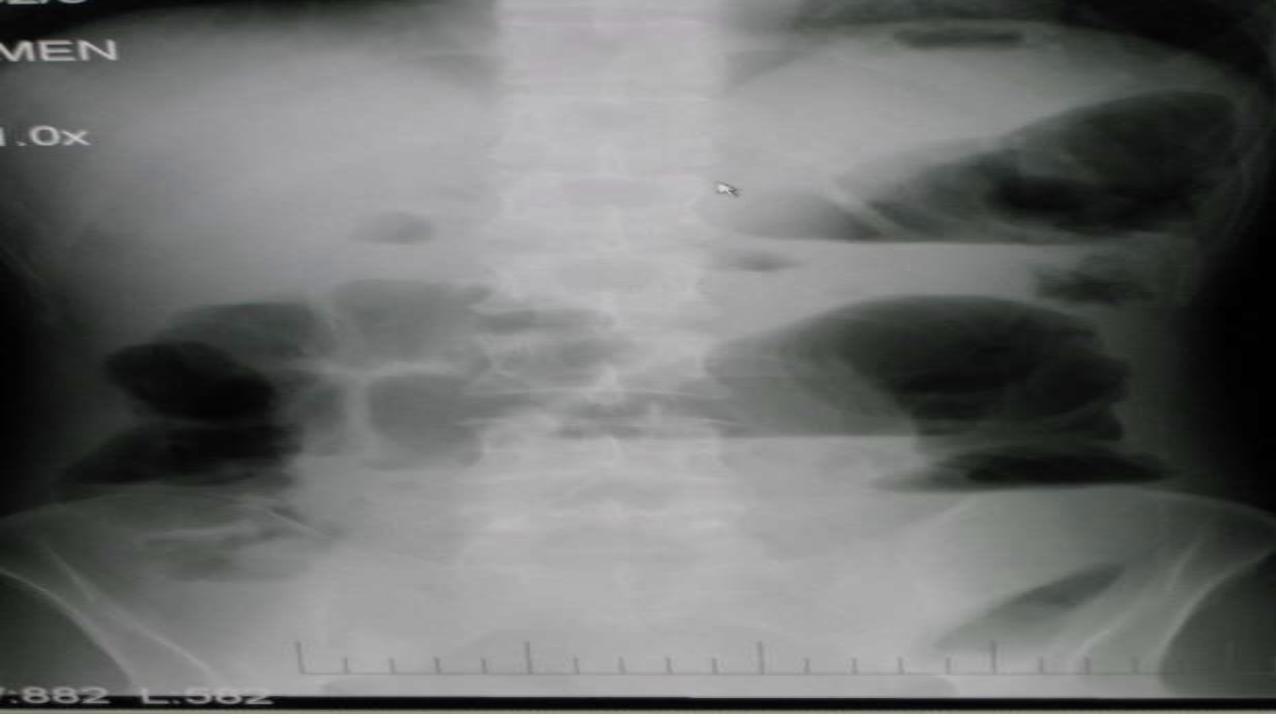
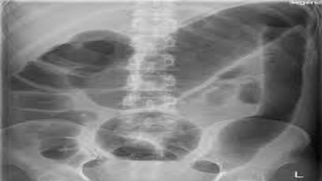
Plain abdominal X-Ray: Multiple gas-fluid levels on an erect film
confirm the case as intestinal obstruction.
On a supine film the level of obstruction is known by the gas pattern of
the distended proximal intestine.
Distended jejunal loops show the characteristic circular mucosal folds
crossing from one side of the lumen to the other.
Distended ileal loops appear as featureless tubes with no mucosal
pattern.
A colon full of gas shows haustrations that do not appear to reach the other side of the lumen.
CBC
Blood urea and electrolytes.
Ultrasound examination can reveal distended
bowel loops. A mass of intussusception can also
be demonstrated.
CT scan with contrast has a sensitivity of 80-90%
for the diagnosis of bowel obstruction.

MANAGEMENT
urgent relief of obstruction,
usually by surgery, after
adequate preoperative
preparation.
PREOPERATIVE PREPERATION
Intravenous replacement of fluid and electrolytes
together with blood and plasma if the patient is
shocked. Ringer's lactate solution is needed.
Gastric aspiration by a nasogastric tube to decompress
the bowel and to reduce the risk of aspiration during
induction of anaesthesia.
Antibiotics are given if there is a possibility of
strangulation.
A Foley catheter is inserted to check the urine output.
THE OPERATION
A longitudinal exploratory incision. In the the case of strangulated
hernia the incision is placed directly over it.
Look at the caecum.
If it is collapsed, this denotes small gut obstruction. If it is distended,
this means large bowel obstruction.
Determine the level of obstruction which is the junction of dilated
and collapsed bowe loops.
Relief of obstruction is attained by division of adhesive bands,
reduction and repair ofa hernia, or untwisting of volvulus.
Assess bowel viability.
Non-viable bowel is known by
Loss of peristalsis,
Loss of normal lustre.
Colour change to greenish or black bowel.
Loss of pulsations in the mesentery.
Resect the Non-viable intestine.
Anastomosis.
CONSERVATIVE
may be successful in certains situations provided thatthere is no clinical
evidence of ischaemia.
Adhesive intestinal obstruction may be relieved by IV drip and nasogastric
suction.
lleocaecal intussusception may be reduced by the hydrostatic effect of a
barium enema.
Sigmoid volvulus. Untwisting may be attempted using a rectal tube passed
through a sigmoidoscope.
Faecal impaction is treated by enema to dissolve the obstructing hard
faecal mass.
* Failure of conservative treatment and the suspicion of strangulation are
indications for surgery.
PARALYTIC ILEUS
is a form of adynamic obstruction in
which there is failure of the peristaltic
waves of the intestine due to failure of the
neuromuscular mechanism.
Paralytic ileus is most common after major
abdominal surgery.
AETIOLOGY
Reflex inhibition of intestinal motility following abdominal
operations retroperitoneal haemorrhage.
Metabolic abnormalities as hypokalaemia, uraemia,
and diabetic ketoacidosis.
Peritonitis induces paralytic ileus due the direct toxic
effect on the nerve plexuses of the intestine.
Drugs as anticholenergics, and tricyclic antidepressants
may produce paralytic ileus if taken in large doses.
CLINICAL FEATURES
Symptoms: Abdominal distension, absolute
constipation, and projectile vomiting . no colicky
abdominal pain but there is only a sense of fullness and
discomfort.
Signs: Abdominal distension and inaudible intestinal
sounds (silent abdomen) .
There may be evidence of localized or generalized
peritonitis.
PREVENTION
Prevention and correction of electrolyte
disturbances guided by serum level estimation.
Gentle handling of the intestine during surgery.
For major abdominal surgery, a naso-gastric
tube is used to decompress the bowel
postoperatively.
MANAGEMENT
Intravenous replacement of the lost fluids and
electrolytes.
Naso-gastric suction.
Correct underlying metabolic abnormalities
Occasionally in resistant cases a
parasympathomimetic, e.g., prostgimine, may
be useful.