Embed Size (px)
Citation preview
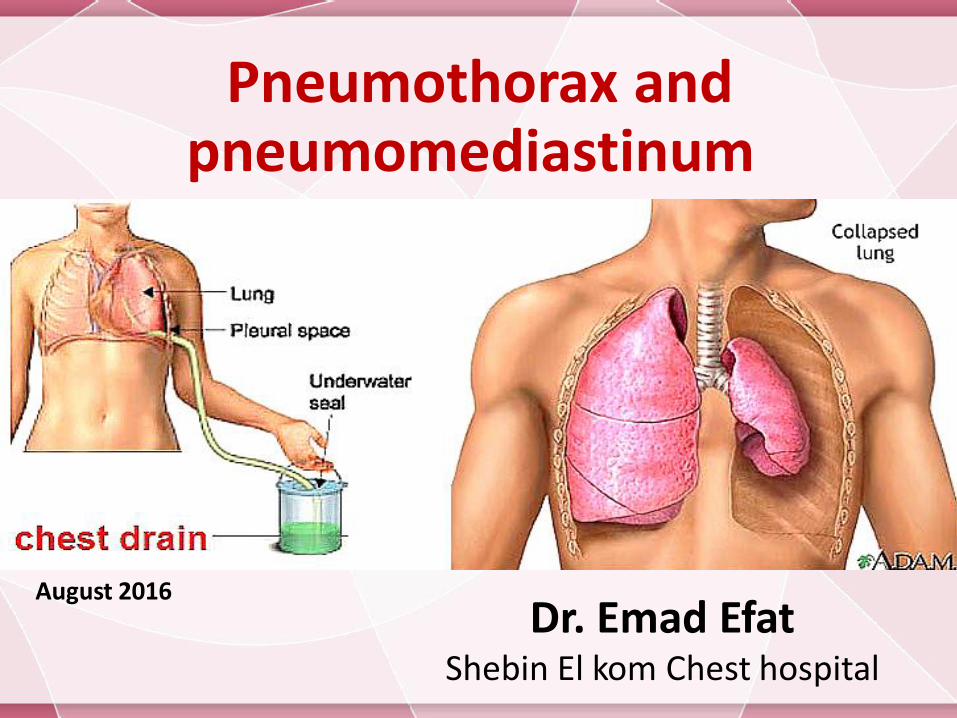
Pneumothorax and pneumomediastinum
Dr. Emad EfatShebin El kom Chest hospital
August 2016
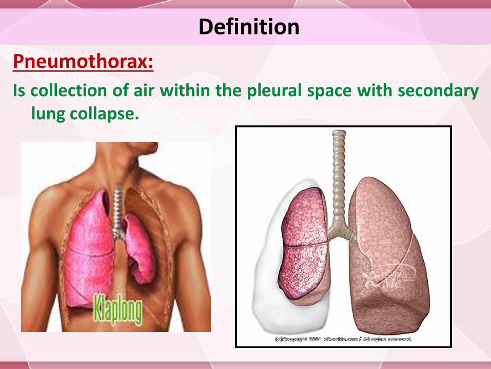
Definition
Pneumothorax:
Is collection of air within the pleural space with secondarylung collapse.
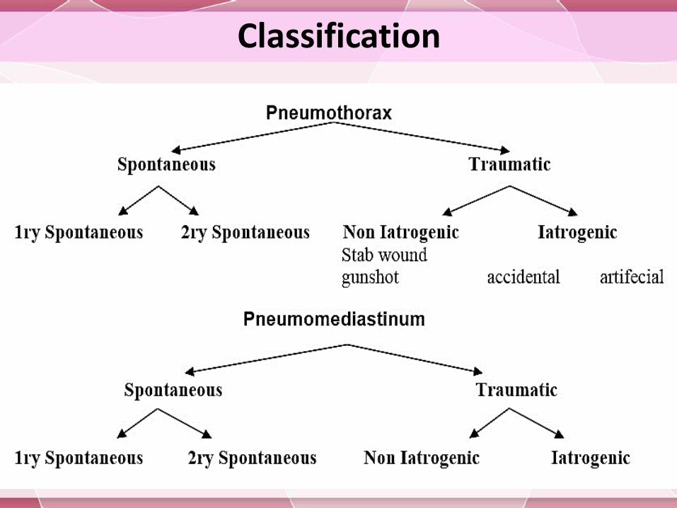
Classification
SyndromesSpontaneous pneumothorax:
Pneumothorax in the absence of iatrogenic or traumatic injury to the chest or lung.
Classified into :
1. 1ry spontaneous pneumothorax: usually occurs at rest without any prior lung disorders or diseases.
2. 2ry spontaneous pneumothorax: can occur as a complication of underlying lung disease.
Traumatic pneumothorax:Results from blunt or penetrating injury that disrupts the parietal or visceral pleura. Iatrogenic pneumothorax is secondary to diagnostic or therapeutic medical intervention
SyndromesTension pneumothorax :
Is a life-threatening condition caused by air within the pleural space that is under pressure; displacing mediastinalstructures and compromising cardiopulmonary function.
Artificial pneumothorax :
It is introduction of measured volume of air into pleura by needle using device
Indication:-
Treatment of pulmonary TB in the era before antituberculous therapy, but now obsolete
Diagnostically ; in thoracoscopic exam.
SyndromesCatamenial pneumothorax
Occurs in conjunction with menstruations & is usually recurrent.
It is rare phenomenon which the usual way in which the thoracic endometriosis declares itself
It generally occurs in women aged 30-40 years.
It frequently begins 1-3 days after menses onset.
The majority (90-95% ) affect the Right hemithorax, but isolated Left side or bilateral pneumothorax has been reported.
Catamenial pneumothorax is usually treated by:
1. Oral contraceptive or danazol (weak androgen) in few cases to suppress ovulation.
2. Surgical menopause by hysterectomy with bilateral oopherectomy
3. Thoracotomy with pleural abrasion or pleurectomy
SyndromesPneumomediastinum :
(Air in mediastinal tissue or mediastinal emphysema)
1. 1ry spontaneous Pneumomediastinum : usually occurs without any prior lung disorders or diseases
2. 2ry spontaneous Pneumomediastinum : can occur as a complication of underlying lung or mediastinal diseases , most often any cause of 2ry spontaneous Pneumomediastinum .
Bilateral spontaneous pneumothorax:
Is rare & may be rapidly fatal if occurring , may be due to:
1.Rupture bilateral apical blebs simultaneously
2. Patient with extensive bilateral emphysema or cystic lung disease
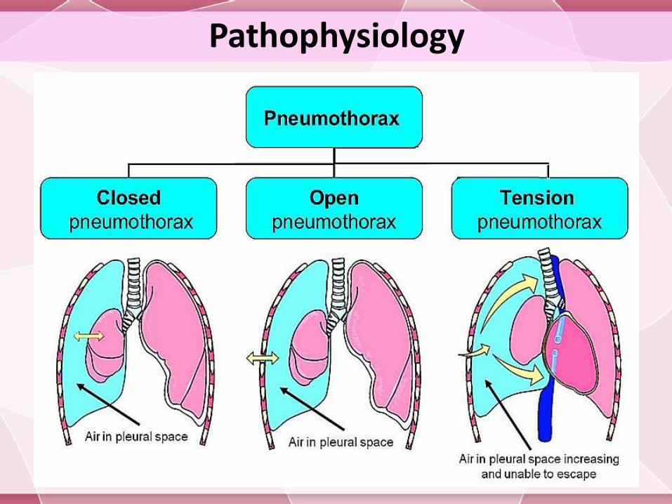
PathophysiologyIf the air enters the pleural cavity from:
The outside (open pneumothorax)
from the lung (closed pneumothorax)
Primary spontaneous pneumothoraces (PSP):
Results from apical pleural blebs related to airway inflammation from cigarette smoking in many patients & it is dose-dependent.
Secondary spontaneous pneumothoraces (SSP):
Occurs in the presence of lung disease, e.g. COPD . Air enter the pleural space via distended, damaged, or compromised alveoli.
Pathophysiology
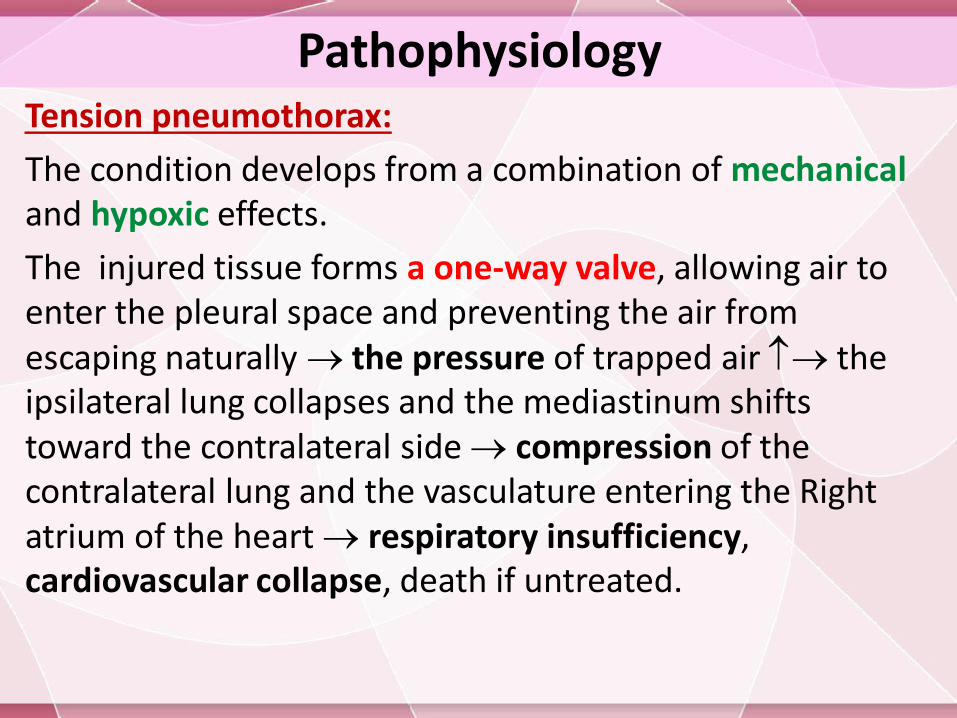
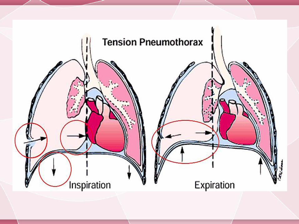
PathophysiologyTension pneumothorax:
The condition develops from a combination of mechanicaland hypoxic effects.
The injured tissue forms a one-way valve, allowing air to enter the pleural space and preventing the air from escaping naturally the pressure of trapped air the ipsilateral lung collapses and the mediastinum shifts toward the contralateral side compression of the contralateral lung and the vasculature entering the Right atrium of the heart respiratory insufficiency, cardiovascular collapse, death if untreated.
PathophysiologyPneumomediastinum
Air escapes into the mediastinum from:
Rupture of alveoli bordering the mediastinum.
Esophageal trauma or elevated airway pressures.
Air may then travel superiorly into the visceral, retropharyngeal, and subcutaneous spaces of the neck. From the neck, the subcutaneous compartment is continuous throughout the body; thus, air can diffuse widely (Subcutaneous emphysema).
Mediastinal air can also pass inferiorly into the retroperitoneum and other extraperitoneal compartments.
The mediastinal parietal pleura may rupture and cause a pneumothorax.
Causes1ry Spontaneous pneumothorax
The male-to-female ratio is about 6:1 .
most likely to occur during the fall or winter months.
Occurs most often in persons early in the third decade of life .
Occurs from the rupture of subpleural apical emphysematous blebs or bullae .
Smoking the risk by more than 22 fold in men and by nearly 10-fold in women. The risk is directly dose related to smoking
PSP is typically observed in tall people due to increased shear forces in the apex.
Familial tendency has been noted
Causes2ry spontaneous pneumothorax
Occur as a complication of underlying lung disease:
Diseases of the airways: COPD, cystic fibrosis, and status asthmaticus ,………..etc.
Interstitial lung diseases : (sarcoidosis, fibrosis, tuberous sclerosis,………..etc)
Infectious diseases : Pneumonia (especially with Staph. Pn, Klebsiella, Pseudomonas, and Pneumocystis species), tuberculosis, pertussis, lung abscess ,………..etc.
Malignancies: Sarcoma, lung cancer .
Pneumoconiosis .
Connective tissue diseases .
Chemotherapy for malignancy .
Radiation therapy .
CausesIatrogenic pneumothorax Transthoracic needle aspiration procedures (most common
cause, accounting for 32-37% of cases)
Transbronchial lung biopsy, Pleural biopsy, liver biopsy or surgery
Thoracentesis
Tracheostomy
Mechanical ventilation (directly related to peak airway pressures) , central venous cannulation; hyperbaric oxygen therapy.
Cardiopulmonary resuscitation
Subclavian and supraclavicular cannulation
Intercostal nerve block .
Unsuccessful attempts to convert an open pneumothorax to a simple pneumothorax in which the occlusive dressing functions as a 1-way valve can lead to a tension pneumothorax.
CausesTraumatic pneumothorax
Penetrating (Open pneumothorax ) and non penetrating injury .
Rib fracture .
Thoracic endometriosis :
Leading to catamenial pneumothorax .
Other causes:
Tall, thin stature in a healthy person or a person with Marfansyndrome.
High-risk occupation (e.g., diving, flying) .
Acupuncture
Tension Pneumothorax
Any condition that leads to pneumothorax can cause a tension pneumothorax
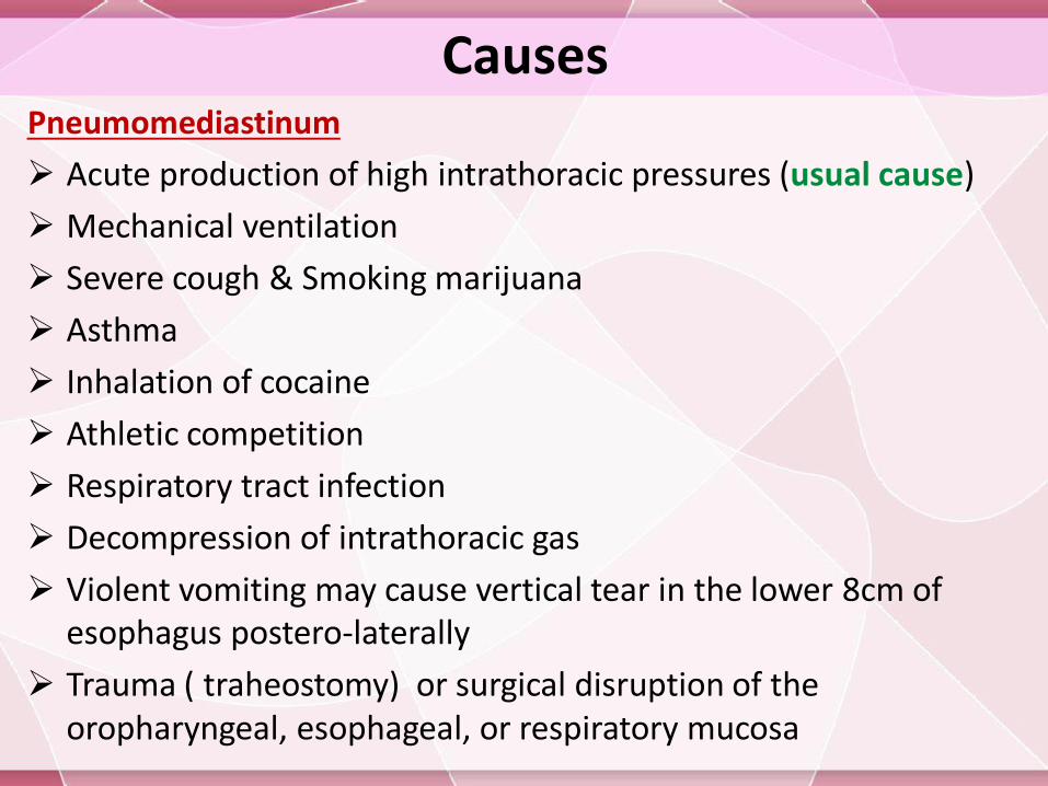
CausesPneumomediastinum
Acute production of high intrathoracic pressures (usual cause)
Mechanical ventilation
Severe cough & Smoking marijuana
Asthma
Inhalation of cocaine
Athletic competition
Respiratory tract infection
Decompression of intrathoracic gas
Violent vomiting may cause vertical tear in the lower 8cm of esophagus postero-laterally
Trauma ( traheostomy) or surgical disruption of the oropharyngeal, esophageal, or respiratory mucosa
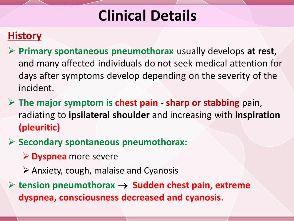
Clinical DetailsHistory
Primary spontaneous pneumothorax usually develops at rest, and many affected individuals do not seek medical attention for days after symptoms develop depending on the severity of the incident.
The major symptom is chest pain - sharp or stabbing pain, radiating to ipsilateral shoulder and increasing with inspiration(pleuritic)
Secondary spontaneous pneumothorax:
Dyspnea more severe
Anxiety, cough, malaise and Cyanosis
tension pneumothorax Sudden chest pain, extreme dyspnea, consciousness decreased and cyanosis.
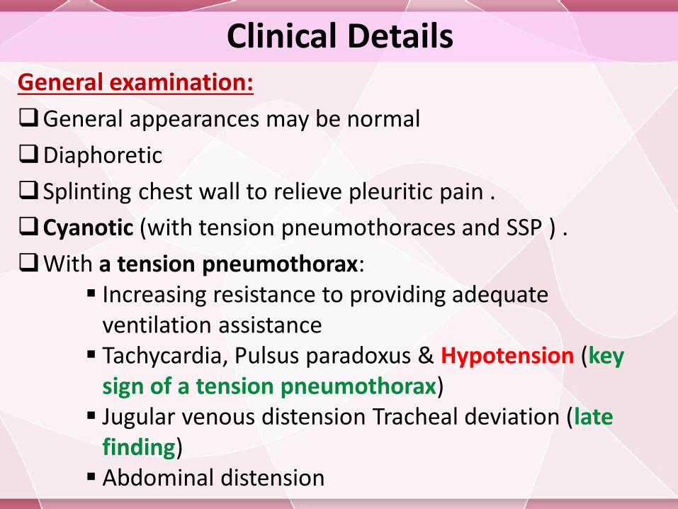
Clinical DetailsGeneral examination:
General appearances may be normal
Diaphoretic
Splinting chest wall to relieve pleuritic pain .
Cyanotic (with tension pneumothoraces and SSP ) .
With a tension pneumothorax: Increasing resistance to providing adequate
ventilation assistance Tachycardia, Pulsus paradoxus & Hypotension (key
sign of a tension pneumothorax) Jugular venous distension Tracheal deviation (late
finding) Abdominal distension
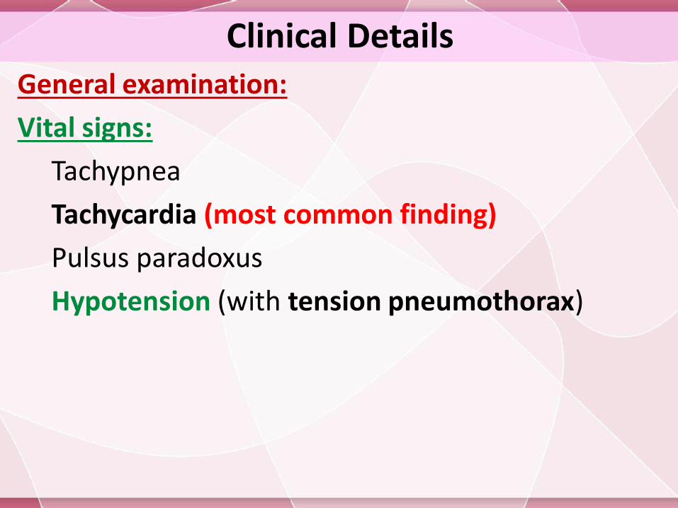
Clinical DetailsGeneral examination:
Vital signs:
Tachypnea
Tachycardia (most common finding)
Pulsus paradoxus
Hypotension (with tension pneumothorax)
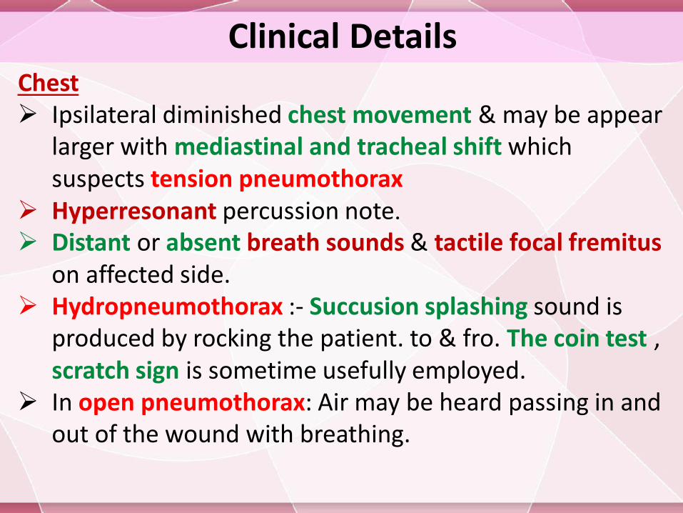
Clinical DetailsChest Ipsilateral diminished chest movement & may be appear
larger with mediastinal and tracheal shift which suspects tension pneumothorax
Hyperresonant percussion note. Distant or absent breath sounds & tactile focal fremitus
on affected side. Hydropneumothorax :- Succusion splashing sound is
produced by rocking the patient. to & fro. The coin test , scratch sign is sometime usefully employed.
In open pneumothorax: Air may be heard passing in and out of the wound with breathing.
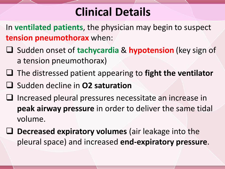
Clinical DetailsIn ventilated patients, the physician may begin to suspect tension pneumothorax when:
Sudden onset of tachycardia & hypotension (key sign of a tension pneumothorax)
The distressed patient appearing to fight the ventilator
Sudden decline in O2 saturation
Increased pleural pressures necessitate an increase in peak airway pressure in order to deliver the same tidal volume.
Decreased expiratory volumes (air leakage into the pleural space) and increased end-expiratory pressure.
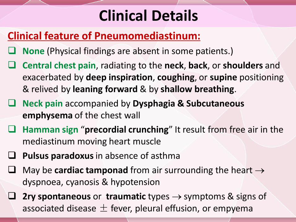
Clinical DetailsClinical feature of Pneumomediastinum:
None (Physical findings are absent in some patients.)
Central chest pain, radiating to the neck, back, or shoulders and exacerbated by deep inspiration, coughing, or supine positioning & relived by leaning forward & by shallow breathing.
Neck pain accompanied by Dysphagia & Subcutaneous emphysema of the chest wall
Hamman sign “precordial crunching” It result from free air in the mediastinum moving heart muscle
Pulsus paradoxus in absence of asthma
May be cardiac tamponad from air surrounding the heart dyspnoea, cyanosis & hypotension
2ry spontaneous or traumatic types symptoms & signs of associated disease ± fever, pleural effusion, or empyema
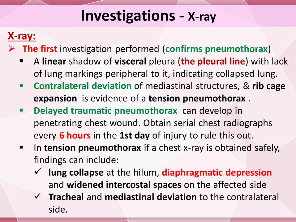
Investigations - X-ray
X-ray: The first investigation performed (confirms pneumothorax) A linear shadow of visceral pleura (the pleural line) with lack
of lung markings peripheral to it, indicating collapsed lung. Contralateral deviation of mediastinal structures, & rib cage
expansion is evidence of a tension pneumothorax . Delayed traumatic pneumothorax can develop in
penetrating chest wound. Obtain serial chest radiographs every 6 hours in the 1st day of injury to rule this out.
In tension pneumothorax if a chest x-ray is obtained safely, findings can include: lung collapse at the hilum, diaphragmatic depression
and widened intercostal spaces on the affected side Tracheal and mediastinal deviation to the contralateral
side.
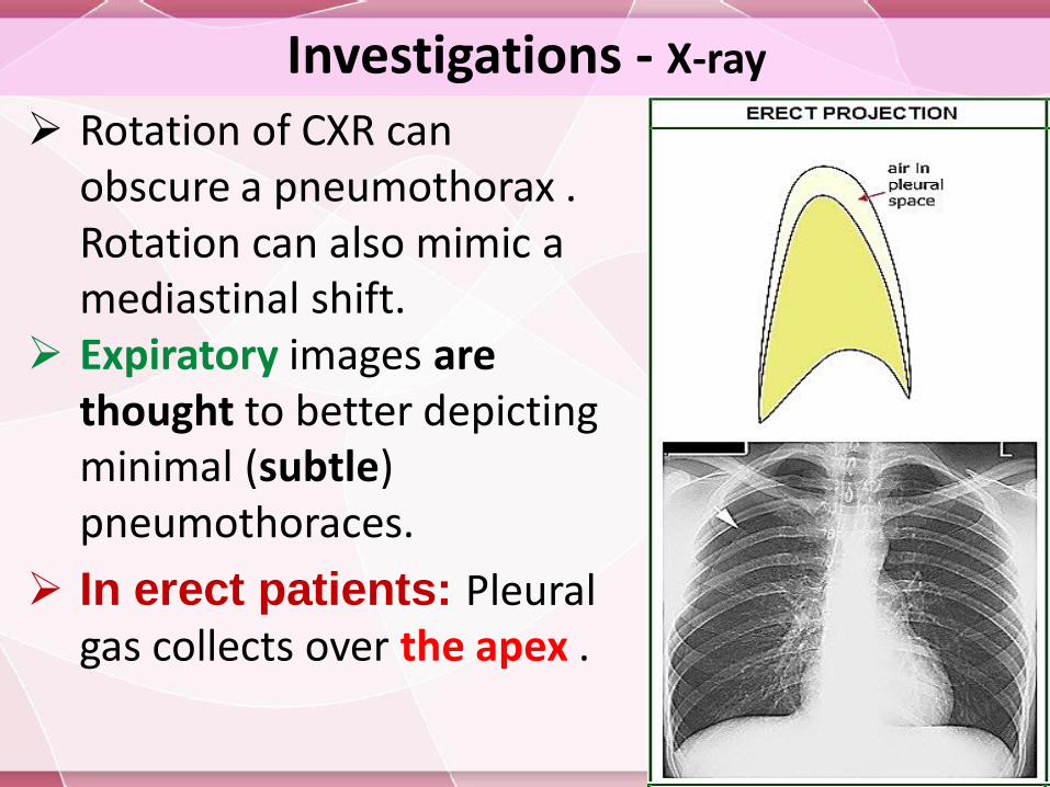
Investigations - X-ray
Rotation of CXR can obscure a pneumothorax . Rotation can also mimic a mediastinal shift.
Expiratory images are thought to better depicting minimal (subtle) pneumothoraces.
In erect patients: Pleural gas collects over the apex .
Investigations - X-ray
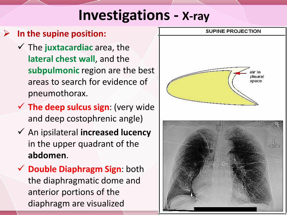
In the supine position:
The juxtacardiac area, the lateral chest wall, and the subpulmonic region are the best areas to search for evidence of pneumothorax.
The deep sulcus sign: (very wide and deep costophrenic angle)
An ipsilateral increased lucencyin the upper quadrant of the abdomen.
Double Diaphragm Sign: both the diaphragmatic dome and anterior portions of the diaphragm are visualized
Investigations - X-ray
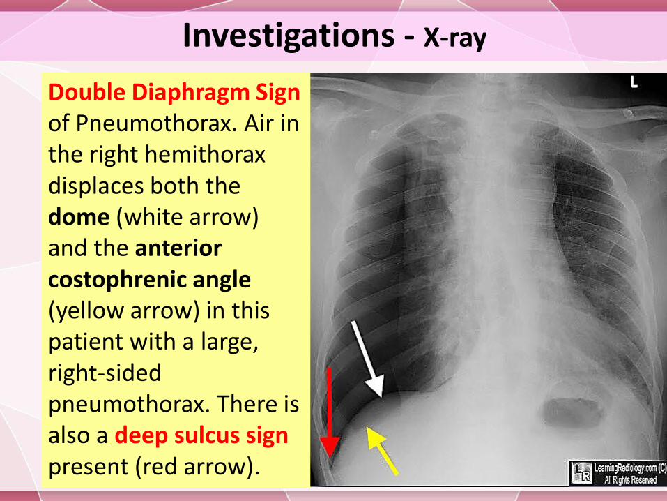
Double Diaphragm Sign of Pneumothorax. Air in the right hemithoraxdisplaces both the dome (white arrow) and the anterior costophrenic angle (yellow arrow) in this patient with a large, right-sided pneumothorax. There is also a deep sulcus sign present (red arrow).
Investigations - X-ray
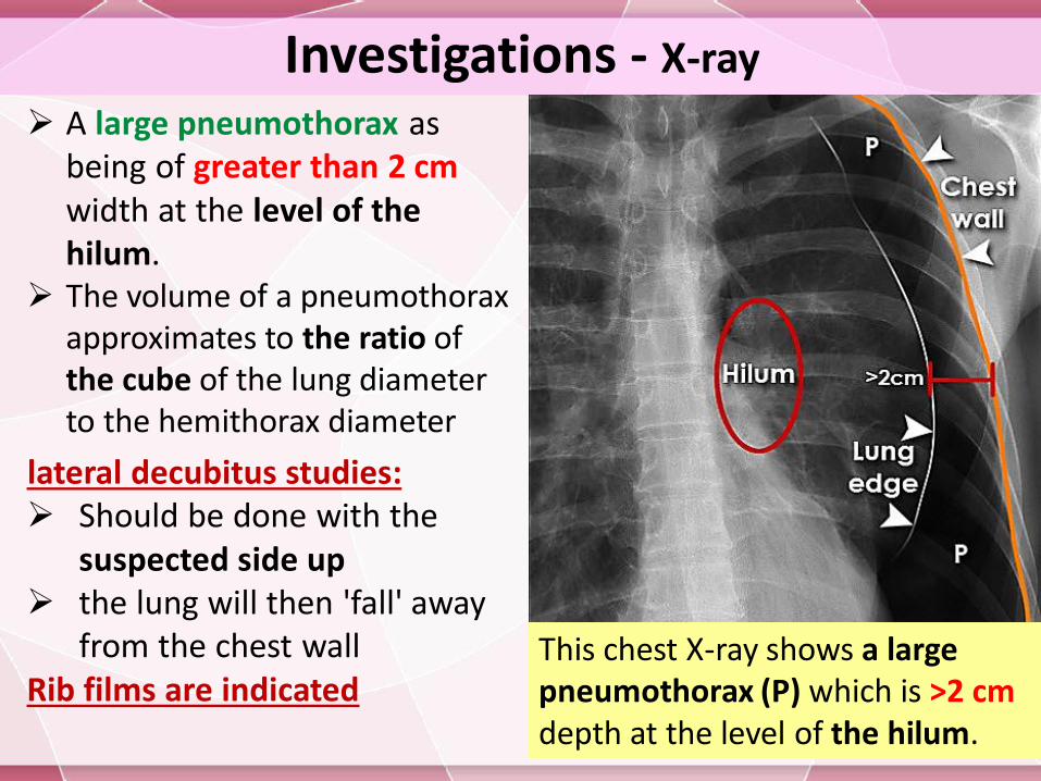
A large pneumothorax as being of greater than 2 cm width at the level of the hilum.
The volume of a pneumothorax approximates to the ratio of the cube of the lung diameter to the hemithorax diameter
lateral decubitus studies: Should be done with the
suspected side up the lung will then 'fall' away
from the chest wall Rib films are indicated
This chest X-ray shows a large pneumothorax (P) which is >2 cm depth at the level of the hilum.
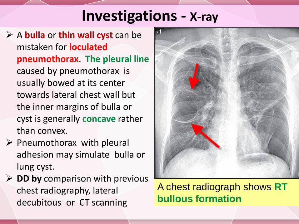
Investigations - X-ray
A bulla or thin wall cyst can be mistaken for loculatedpneumothorax. The pleural linecaused by pneumothorax is usually bowed at its center towards lateral chest wall but the inner margins of bulla or cyst is generally concave rather than convex.
Pneumothorax with pleural adhesion may simulate bulla or lung cyst.
DD by comparison with previous chest radiography, lateral decubitous or CT scanning
A chest radiograph shows RT
bullous formation
Investigations - X-ray
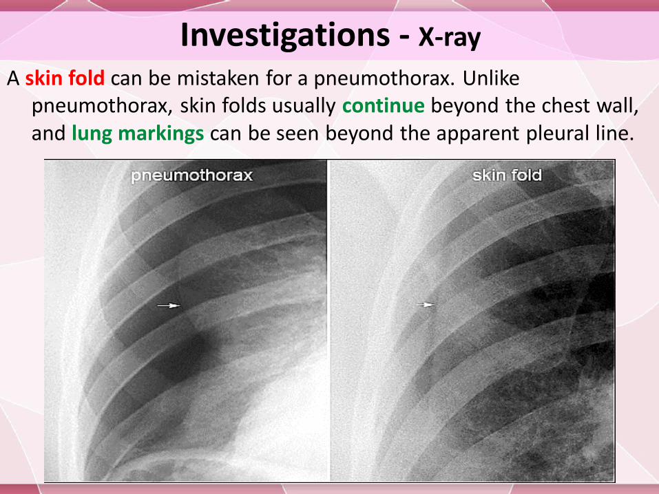
A skin fold can be mistaken for a pneumothorax. Unlike pneumothorax, skin folds usually continue beyond the chest wall, and lung markings can be seen beyond the apparent pleural line.
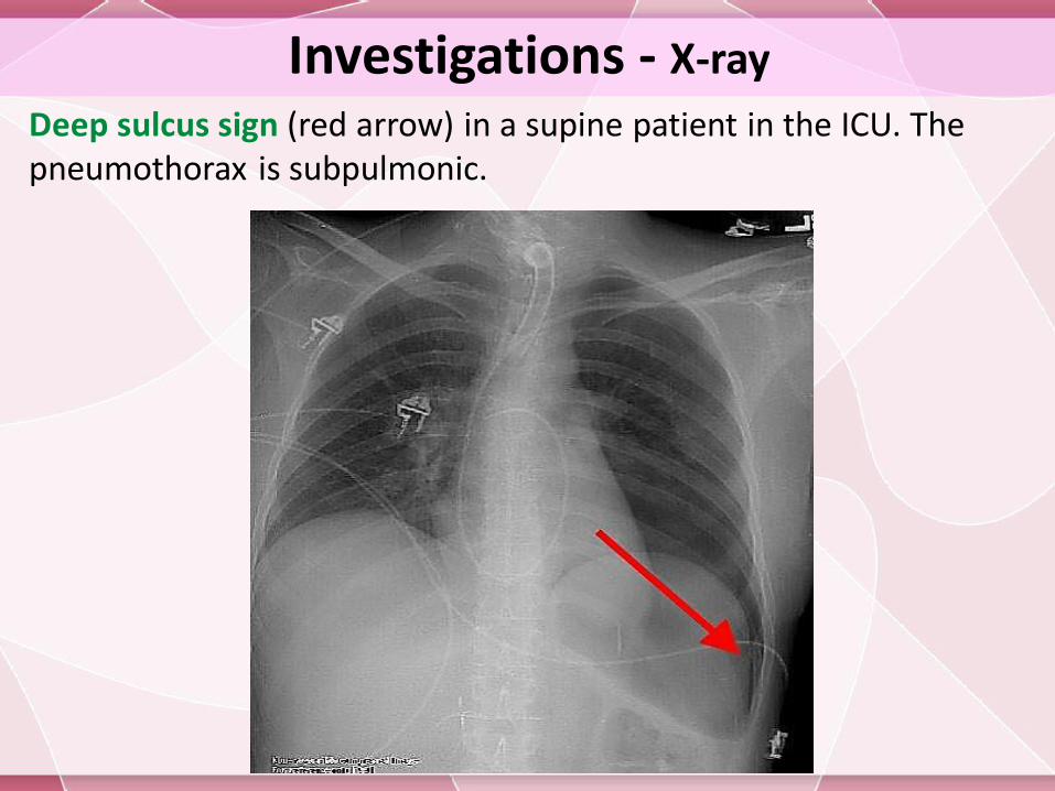
Deep sulcus sign (red arrow) in a supine patient in the ICU. The pneumothorax is subpulmonic.
Investigations - X-ray
Investigations - X-ray
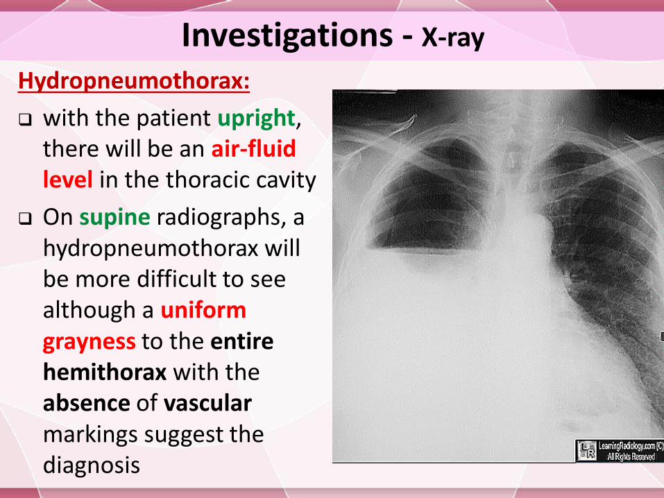
Hydropneumothorax:
with the patient upright, there will be an air-fluid level in the thoracic cavity
On supine radiographs, a hydropneumothorax will be more difficult to see although a uniform grayness to the entire hemithorax with the absence of vascularmarkings suggest the diagnosis
Investigations - X-ray
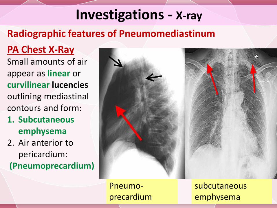
Radiographic features of Pneumomediastinum
PA Chest X-RaySmall amounts of air appear as linear or curvilinear lucenciesoutlining mediastinalcontours and form:1. Subcutaneous
emphysema2. Air anterior to
pericardium:(Pneumoprecardium)
Pneumo-precardium
subcutaneous emphysema
Investigations - X-ray
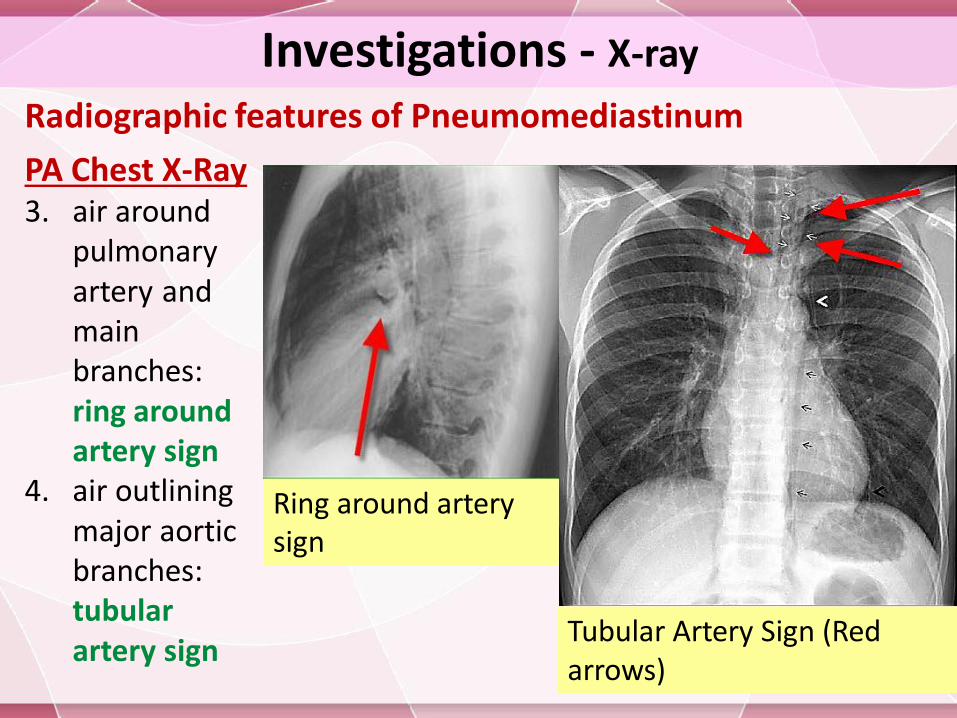
Radiographic features of Pneumomediastinum
PA Chest X-Ray3. air around
pulmonary artery and main branches: ring around artery sign
4. air outlining major aortic branches: tubular artery sign
Tubular Artery Sign (Red arrows)
Ring around artery sign
Investigations - X-ray
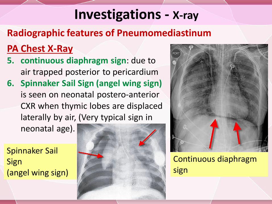
Radiographic features of Pneumomediastinum
PA Chest X-Ray5. continuous diaphragm sign: due to
air trapped posterior to pericardium6. Spinnaker Sail Sign (angel wing sign)
is seen on neonatal postero-anterior CXR when thymic lobes are displaced laterally by air, (Very typical sign in neonatal age).
Spinnaker Sail Sign (angel wing sign)
Continuous diaphragm sign
Investigations - X-ray
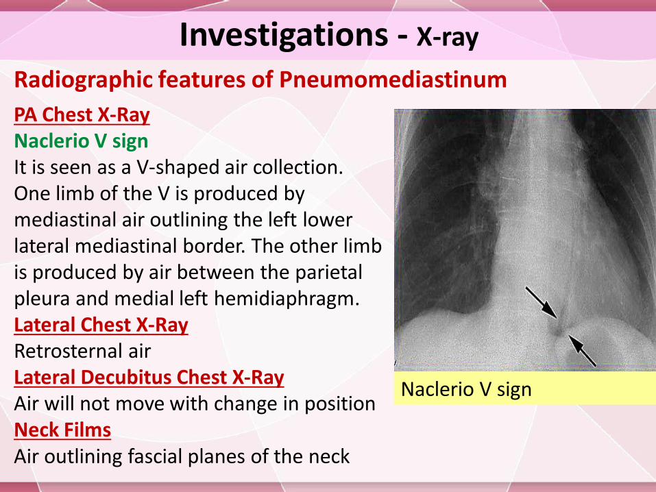
Radiographic features of Pneumomediastinum
PA Chest X-RayNaclerio V signIt is seen as a V-shaped air collection. One limb of the V is produced by mediastinal air outlining the left lower lateral mediastinal border. The other limb is produced by air between the parietal pleura and medial left hemidiaphragm.Lateral Chest X-RayRetrosternal airLateral Decubitus Chest X-RayAir will not move with change in positionNeck FilmsAir outlining fascial planes of the neck
Naclerio V sign
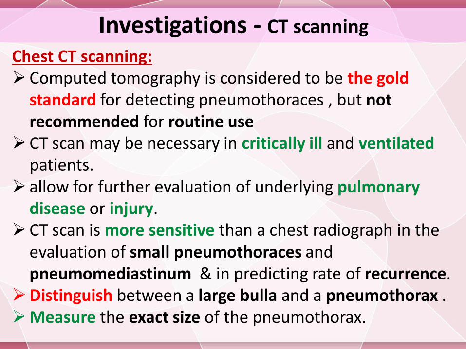
Investigations - CT scanning
Chest CT scanning: Computed tomography is considered to be the gold
standard for detecting pneumothoraces , but not recommended for routine use
CT scan may be necessary in critically ill and ventilatedpatients.
allow for further evaluation of underlying pulmonary disease or injury.
CT scan is more sensitive than a chest radiograph in the evaluation of small pneumothoraces and pneumomediastinum & in predicting rate of recurrence.
Distinguish between a large bulla and a pneumothorax .Measure the exact size of the pneumothorax.
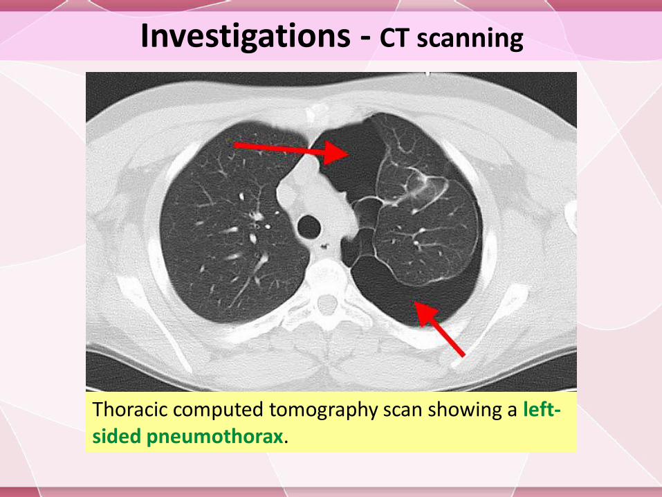
Investigations - CT scanning
Thoracic computed tomography scan showing a left-sided pneumothorax.
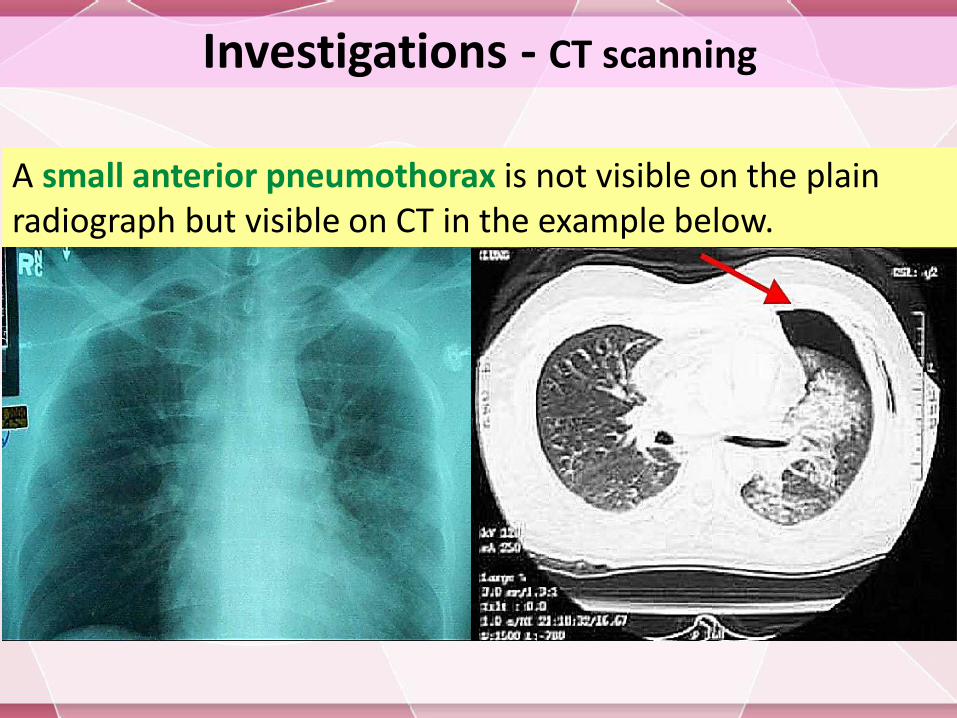
Investigations - CT scanning
A small anterior pneumothorax is not visible on the plain radiograph but visible on CT in the example below.
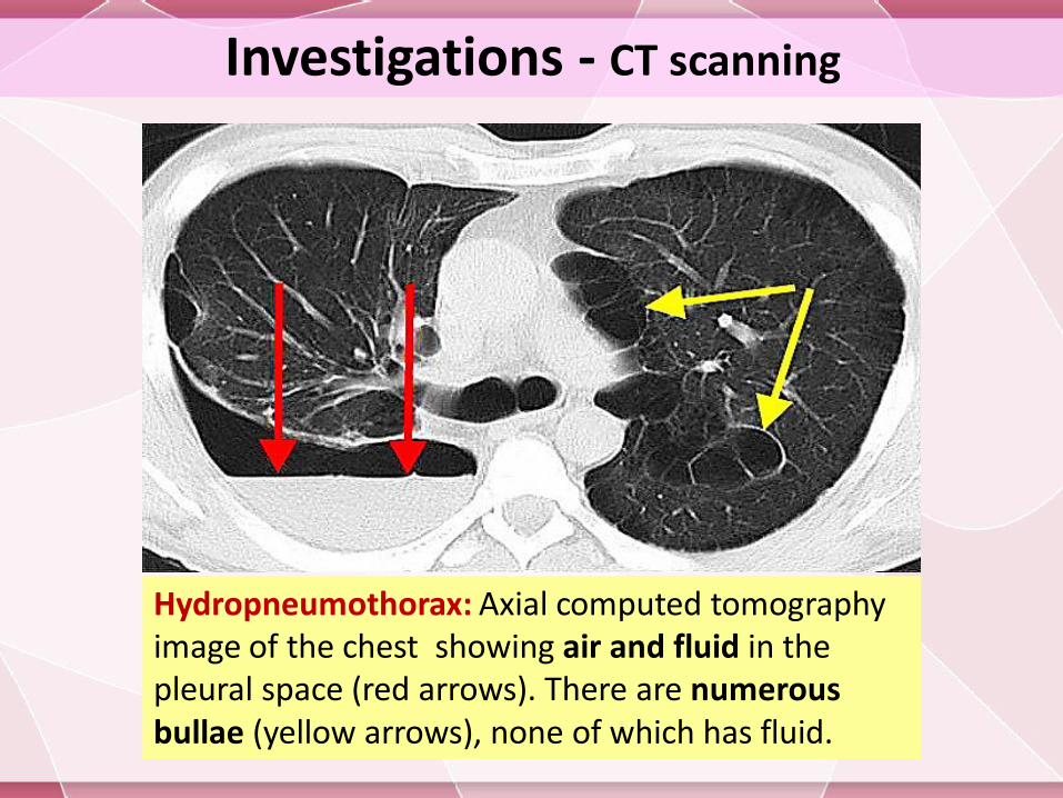
Hydropneumothorax: Axial computed tomography image of the chest showing air and fluid in the pleural space (red arrows). There are numerous bullae (yellow arrows), none of which has fluid.
Investigations - CT scanning
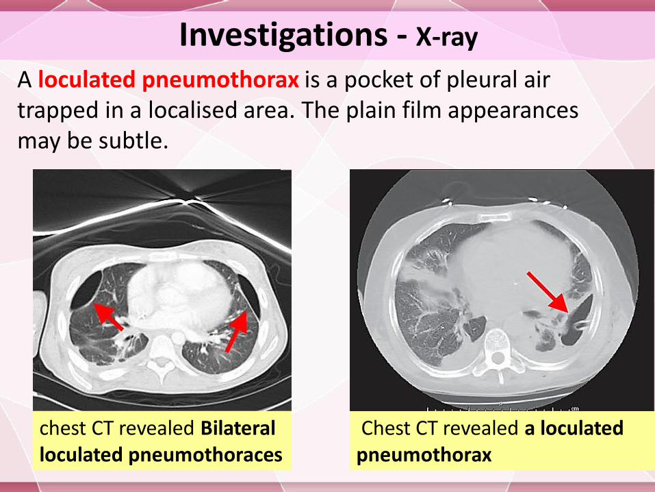
Investigations - X-ray
A loculated pneumothorax is a pocket of pleural air trapped in a localised area. The plain film appearances may be subtle.
Chest CT revealed a loculated pneumothorax
chest CT revealed Bilateral loculated pneumothoraces
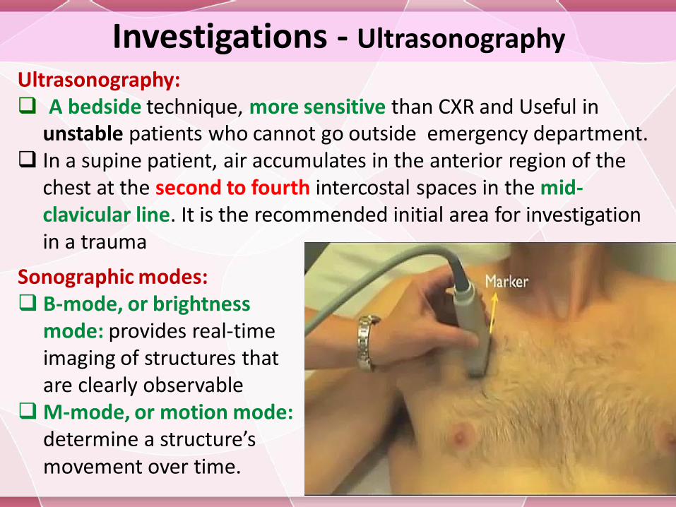
Investigations - Ultrasonography
Ultrasonography: A bedside technique, more sensitive than CXR and Useful in
unstable patients who cannot go outside emergency department. In a supine patient, air accumulates in the anterior region of the
chest at the second to fourth intercostal spaces in the mid-clavicular line. It is the recommended initial area for investigation in a trauma
Sonographic modes: B-mode, or brightness
mode: provides real-time imaging of structures that are clearly observable
M-mode, or motion mode: determine a structure’s movement over time.
Investigations - Ultrasonography
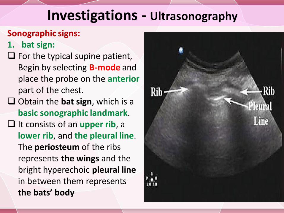
Sonographic signs:1. bat sign: For the typical supine patient,
Begin by selecting B-mode and place the probe on the anteriorpart of the chest.
Obtain the bat sign, which is a basic sonographic landmark.
It consists of an upper rib, a lower rib, and the pleural line. The periosteum of the ribs represents the wings and the bright hyperechoic pleural line in between them represents the bats’ body
Investigations - Ultrasonography
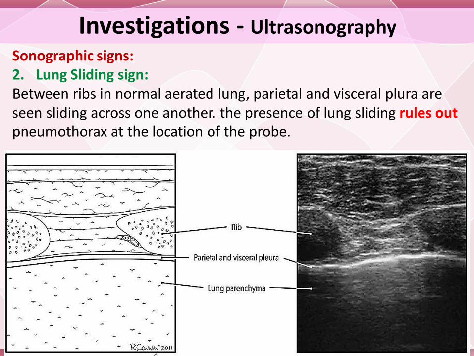
Sonographic signs:2. Lung Sliding sign:Between ribs in normal aerated lung, parietal and visceral plura are seen sliding across one another. the presence of lung sliding rules out pneumothorax at the location of the probe.
Investigations - Ultrasonography
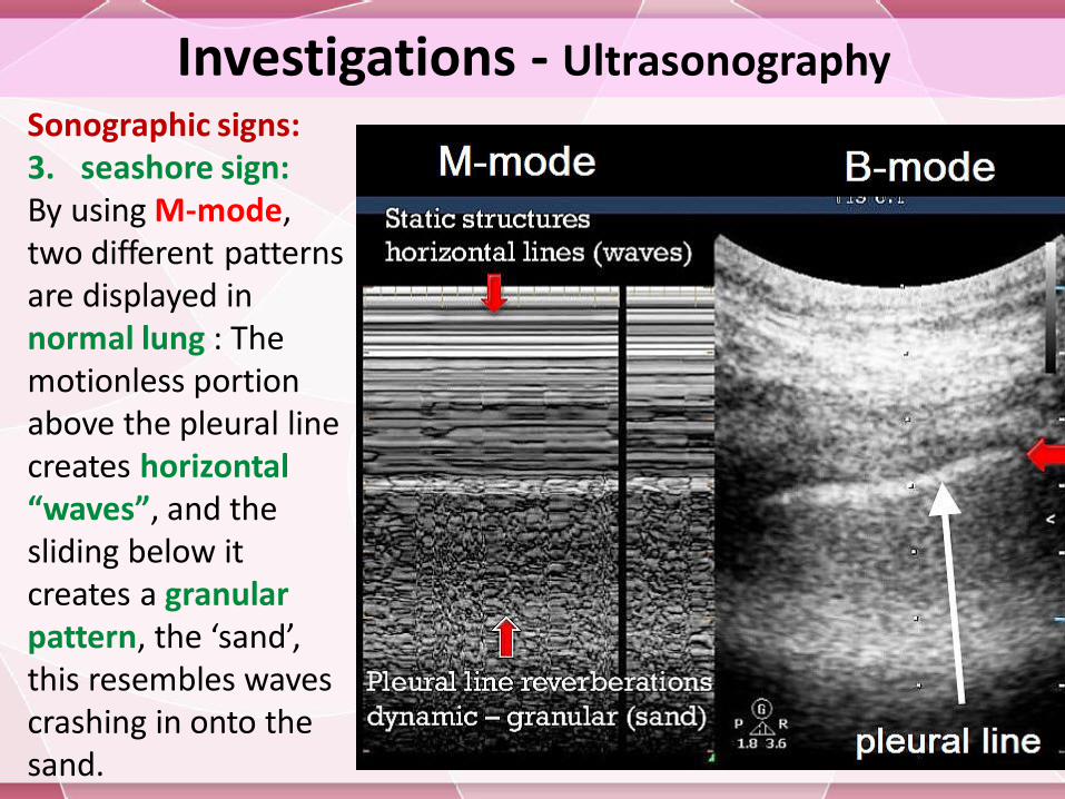
Sonographic signs:3. seashore sign:By using M-mode, two different patterns are displayed in normal lung : The motionless portion above the pleural line creates horizontal “waves”, and the sliding below it creates a granular pattern, the ‘sand’, this resembles waves crashing in onto the sand.
Investigations - Ultrasonography
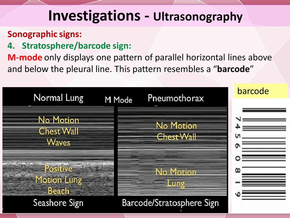
Sonographic signs:4. Stratosphere/barcode sign:M-mode only displays one pattern of parallel horizontal lines above and below the pleural line. This pattern resembles a “barcode”
barcode
Investigations - Ultrasonography
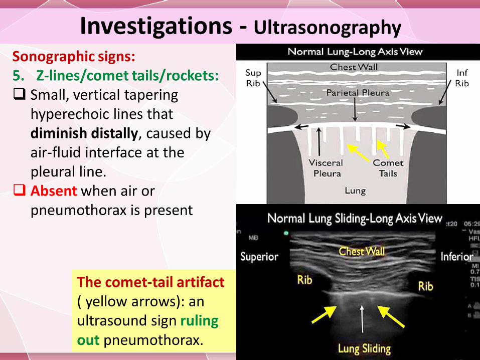
Sonographic signs:5. Z-lines/comet tails/rockets: Small, vertical tapering
hyperechoic lines that diminish distally, caused by air-fluid interface at the pleural line.
Absent when air or pneumothorax is present
The comet-tail artifact ( yellow arrows): an ultrasound sign ruling out pneumothorax.
Investigations - Ultrasonography
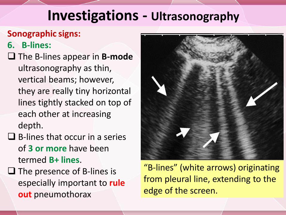
Sonographic signs:6. B-lines: The B-lines appear in B-mode
ultrasonography as thin, vertical beams; however, they are really tiny horizontal lines tightly stacked on top of each other at increasing depth.
B-lines that occur in a series of 3 or more have been termed B+ lines.
The presence of B-lines is especially important to rule out pneumothorax
“B-lines” (white arrows) originating from pleural line, extending to the edge of the screen.
Investigations - Ultrasonography
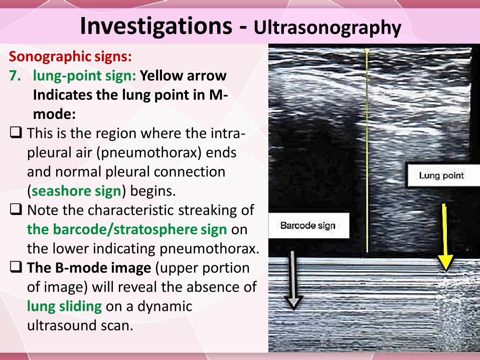
Sonographic signs:7. lung-point sign: Yellow arrow
Indicates the lung point in M-mode:
This is the region where the intra-pleural air (pneumothorax) ends and normal pleural connection (seashore sign) begins.
Note the characteristic streaking of the barcode/stratosphere sign on the lower indicating pneumothorax.
The B-mode image (upper portion of image) will reveal the absence of lung sliding on a dynamic ultrasound scan.
InvestigationsContrast-enhanced esophagogram: If emesis or retching is the precipitating event, an esophagogramshould be obtained to evaluate Boerhaave syndrome (an esophageal tear), which has a high mortality rate . The electrocardiogram (ECG):Patient with left pneumothorax may shows changes suggesting antero lateral myocardial infarction ( A right axis deviation, poor R wave progression, ↓QRS amplitude & precordial T wave inversion ).
Lab Studies
Arterial blood gas
In patients with severe lung disease
In those with persistent respiratory distress despite treatment
ABG analysis may be useful in evaluating the following: Hypoxia, Hypercarbia and respiratory acidosis
Differential diagnosis Pulmonary embolismDissecting aortic aneurysm, rupture Esophageal spasm, Perforation, Rupture and Tears. Acute pericarditis Acute myocardial infarction Acute Coronary Syndrome Congestive Heart Failure and Pulmonary EdemaHaemothorax Pleural effusion ARDS Asthma Foreign Bodies, Trachea Adult Diaphragmatic Injuries
Pneumothorax Treatment
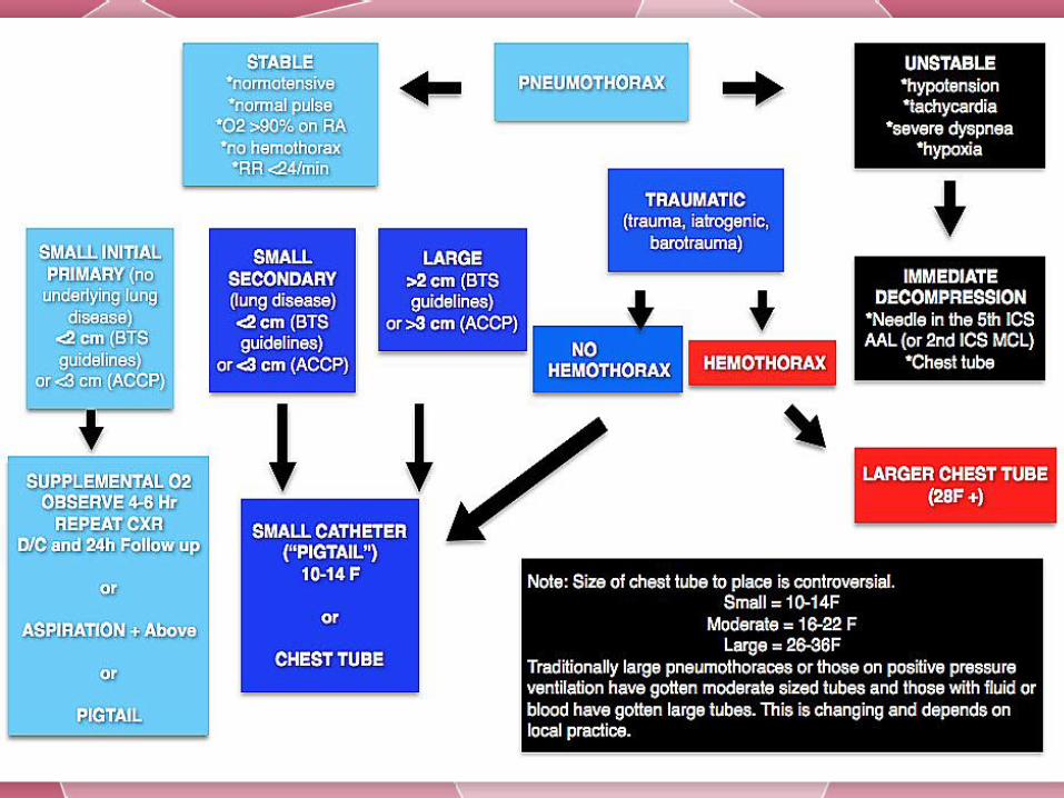
Treatment Based on Risk Stratification (Patient presentation ):The following are possible presentations of patients with pneumothorax: Asymptomatic (incidental finding): Treatment decisions are guided
by estimate of long-term recurrence risk. Symptomatic but clinically stable:
The British Thoracic Society (BTS) advocates for simple aspiration and deferring hospitalization in PSP
A small-bore catheter or chest tube placement is recommended by the American College of Chest Physicians (ACCP)
Clinically fragile: air evacuation and observation; comorbid conditions may preclude observation
Life-threatening: must be treated immediately with tube thoracostomy

Pneumothorax Treatment
Selection of site of patient careOutpatient care: This can occur in asymptomatic patients
or those with a small pneumothorax and reliable follow-upEmergency department (ED) care: when Prolonged periods
of observation are inefficient and clinically suboptimal; manual aspiration and placement of one-way valves are performed.
Inpatient care: when high flow Oxygen is needed, the pneumothorax is larger but the patient is stable, or associated comorbidities ; the average hospital stay is 2-8 days
Intensive care unit (ICU): for patients who are unstable or intubated
Pneumothorax Treatment
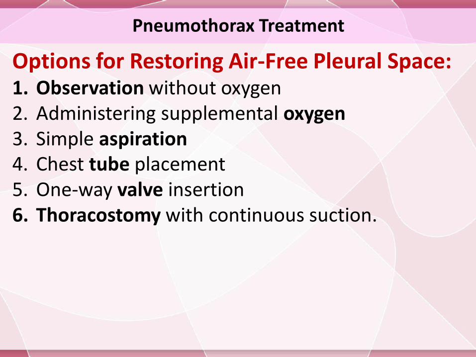
Options for Restoring Air-Free Pleural Space:1. Observation without oxygen2. Administering supplemental oxygen3. Simple aspiration4. Chest tube placement5. One-way valve insertion6. Thoracostomy with continuous suction.
Pneumothorax Treatment
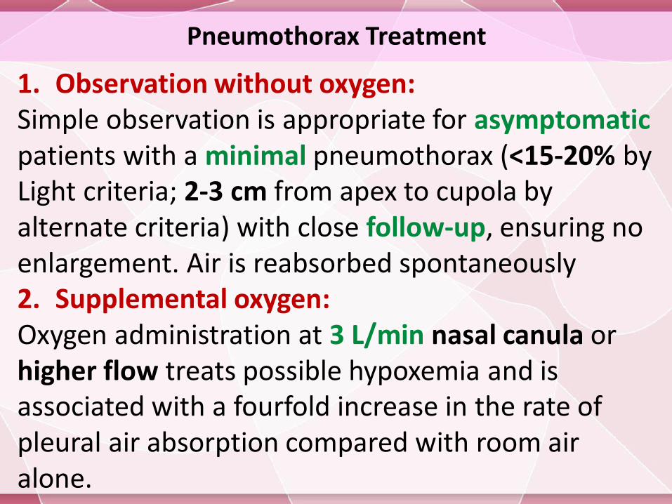
1. Observation without oxygen:Simple observation is appropriate for asymptomaticpatients with a minimal pneumothorax (<15-20% by Light criteria; 2-3 cm from apex to cupola by alternate criteria) with close follow-up, ensuring no enlargement. Air is reabsorbed spontaneously2. Supplemental oxygen:Oxygen administration at 3 L/min nasal canula or higher flow treats possible hypoxemia and is associated with a fourfold increase in the rate of pleural air absorption compared with room air alone.

Pneumothorax Treatment
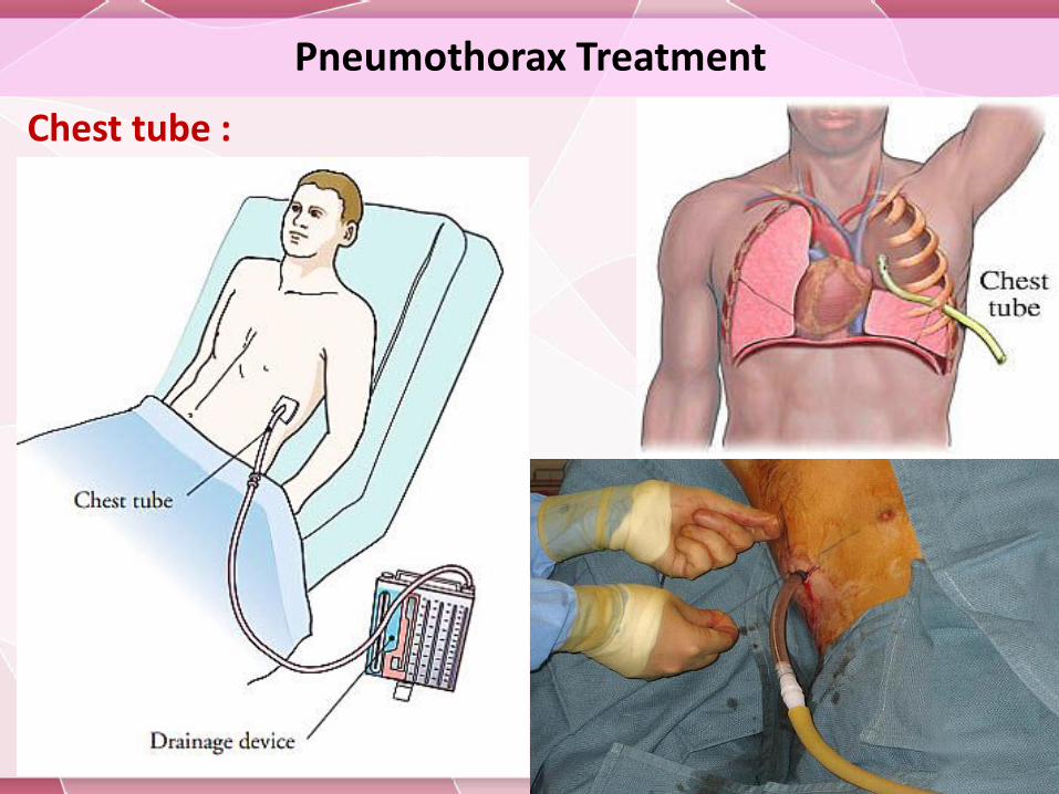
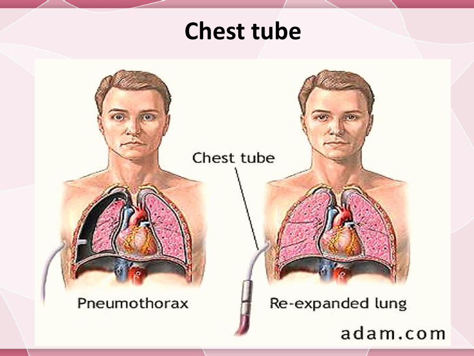
3. Simple aspiration:A more recent ED study supports needle aspiration as safe and effective as chest tube placement for PSP, conferring the additional benefits of shorter length of stay and fewer hospital admissions4. Chest tube placement:A tube inserted into the pleural space is connected to a device with one-way flow for air removal. Examples of such devices are Heimlich valves or water seal canisters, and tubes connected to wall suction devices.
Pneumothorax Treatment
Chest tube :

Pneumothorax Treatment
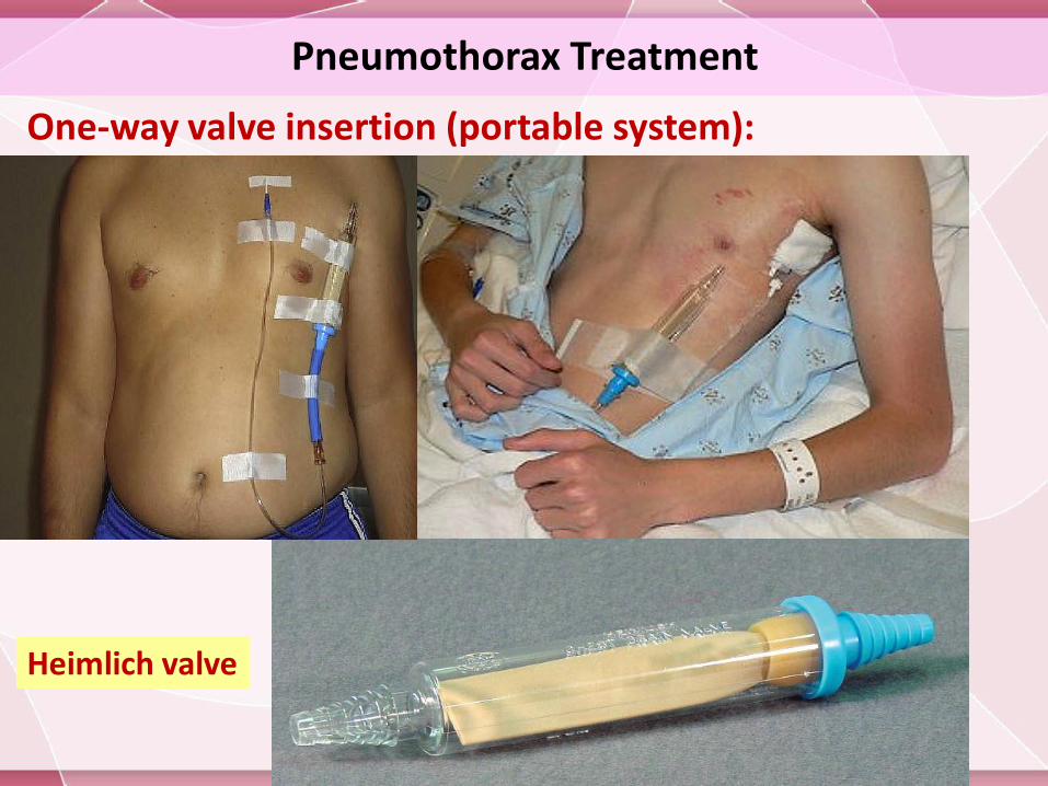
5. One-way valve insertion (portable system):The typical goal of inserting one-way valve
systems is to avoid hospital admission and still treat the spontaneous pneumothorax. One-way valves may be used during transport of an injured patient.
A Heimlich valve is a one-way, rubber flutter valve that allows for complete evacuation of air that is not under tension
Effective as simple manual needle aspiration or a conventional chest tube thoracotomy.
Pneumothorax Treatment
One-way valve insertion (portable system):
Heimlich valve
Pneumothorax Treatment
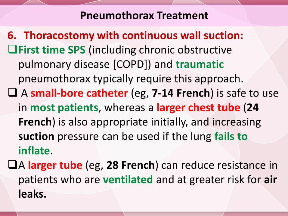
6. Thoracostomy with continuous wall suction:First time SPS (including chronic obstructive
pulmonary disease [COPD]) and traumaticpneumothorax typically require this approach.
A small-bore catheter (eg, 7-14 French) is safe to use in most patients, whereas a larger chest tube (24 French) is also appropriate initially, and increasing suction pressure can be used if the lung fails to inflate.
A larger tube (eg, 28 French) can reduce resistance in patients who are ventilated and at greater risk for air leaks.
Pneumothorax Treatment
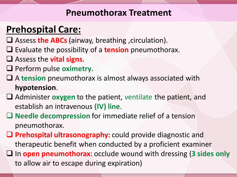
Prehospital Care: Assess the ABCs (airway, breathing ,circulation). Evaluate the possibility of a tension pneumothorax. Assess the vital signs. Perform pulse oximetry. A tension pneumothorax is almost always associated with
hypotension. Administer oxygen to the patient, ventilate the patient, and
establish an intravenous (IV) line. Needle decompression for immediate relief of a tension
pneumothorax. Prehospital ultrasonography: could provide diagnostic and
therapeutic benefit when conducted by a proficient examiner In open pneumothorax: occlude wound with dressing (3 sides only
to allow air to escape during expiration)
Pneumothorax Treatment
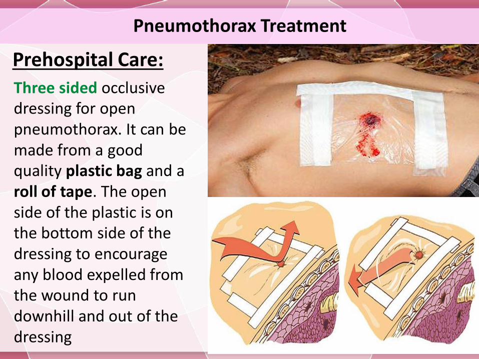
Prehospital Care:
Three sided occlusive dressing for open pneumothorax. It can be made from a good quality plastic bag and a roll of tape. The open side of the plastic is on the bottom side of the dressing to encourage any blood expelled from the wound to run downhill and out of the dressing
Pneumothorax Treatment
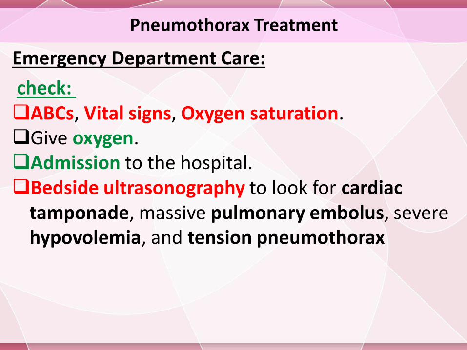
Emergency Department Care:
check: ABCs, Vital signs, Oxygen saturation.Give oxygen.Admission to the hospital.Bedside ultrasonography to look for cardiac
tamponade, massive pulmonary embolus, severe hypovolemia, and tension pneumothorax
Pneumothorax Treatment
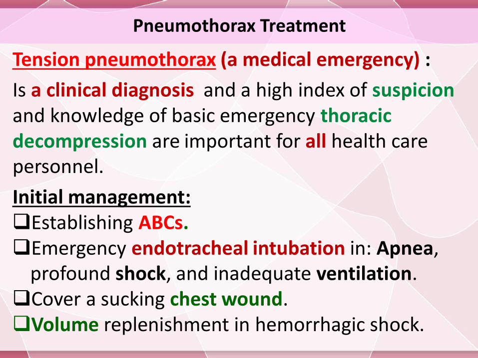
Tension pneumothorax (a medical emergency) :
Is a clinical diagnosis and a high index of suspicionand knowledge of basic emergency thoracic decompression are important for all health care personnel.
Initial management:Establishing ABCs.Emergency endotracheal intubation in: Apnea,
profound shock, and inadequate ventilation.Cover a sucking chest wound.Volume replenishment in hemorrhagic shock.
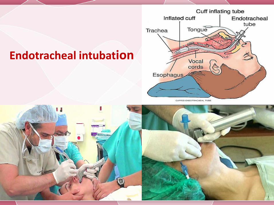
Endotracheal intubation
Pneumothorax Treatment
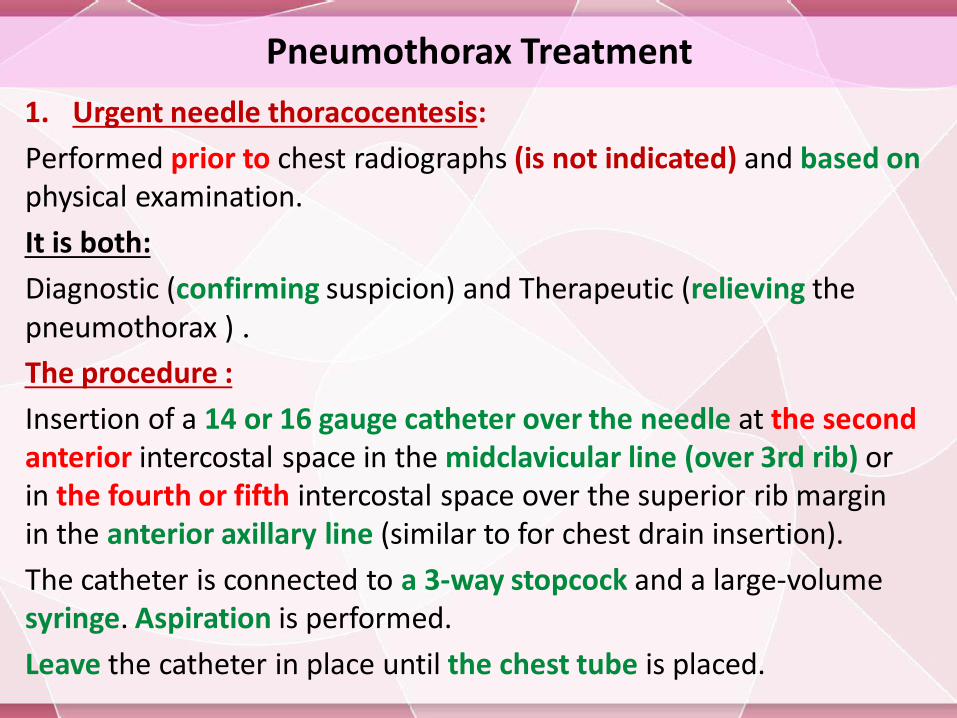
1. Urgent needle thoracocentesis:
Performed prior to chest radiographs (is not indicated) and based onphysical examination.
It is both:
Diagnostic (confirming suspicion) and Therapeutic (relieving the pneumothorax ) .
The procedure :
Insertion of a 14 or 16 gauge catheter over the needle at the secondanterior intercostal space in the midclavicular line (over 3rd rib) or in the fourth or fifth intercostal space over the superior rib margin in the anterior axillary line (similar to for chest drain insertion).
The catheter is connected to a 3-way stopcock and a large-volume syringe. Aspiration is performed.
Leave the catheter in place until the chest tube is placed.
Pneumothorax Treatment
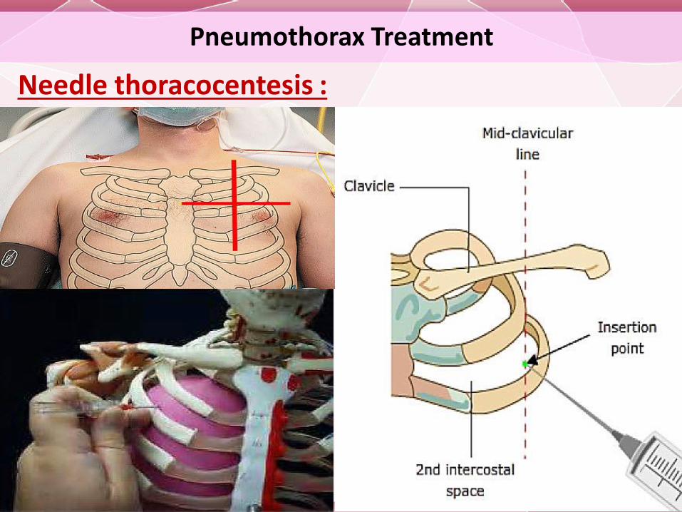
Needle thoracocentesis :
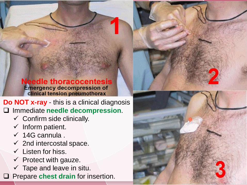
Do NOT x-ray - this is a clinical diagnosis
Immediate needle decompression.
Confirm side clinically.
Inform patient.
14G cannula .
2nd intercostal space.
Listen for hiss.
Protect with gauze.
Tape and leave in situ.
Prepare chest drain for insertion.
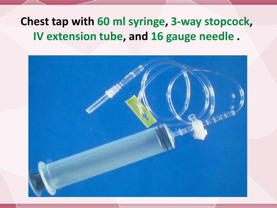
Chest tap with 60 ml syringe, 3-way stopcock, IV extension tube, and 16 gauge needle .
Pneumothorax Treatment
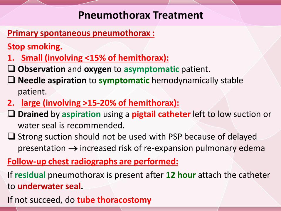
Primary spontaneous pneumothorax :
Stop smoking.1. Small (involving <15% of hemithorax): Observation and oxygen to asymptomatic patient. Needle aspiration to symptomatic hemodynamically stable
patient.2. large (involving >15-20% of hemithorax): Drained by aspiration using a pigtail catheter left to low suction or
water seal is recommended. Strong suction should not be used with PSP because of delayed
presentation increased risk of re-expansion pulmonary edema
Follow-up chest radiographs are performed:
If residual pneumothorax is present after 12 hour attach the catheter to underwater seal.
If not succeed, do tube thoracostomy
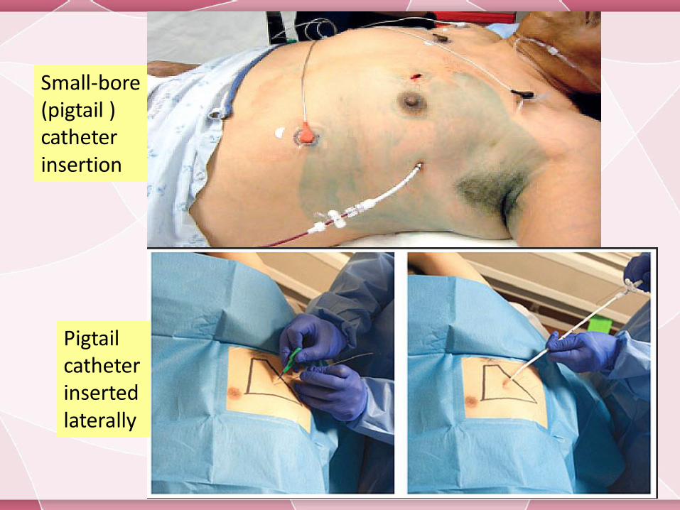
Small-bore (pigtail ) catheter insertion
Pigtail catheter inserted laterally
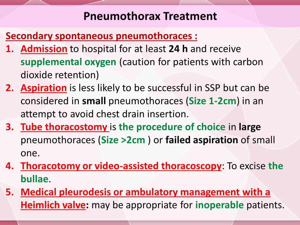
Pneumothorax Treatment
Secondary spontaneous pneumothoraces :1. Admission to hospital for at least 24 h and receive
supplemental oxygen (caution for patients with carbon dioxide retention)
2. Aspiration is less likely to be successful in SSP but can be considered in small pneumothoraces (Size 1-2cm) in an attempt to avoid chest drain insertion.
3. Tube thoracostomy is the procedure of choice in largepneumothoraces (Size >2cm ) or failed aspiration of small one.
4. Thoracotomy or video-assisted thoracoscopy: To excise the bullae.
5. Medical pleurodesis or ambulatory management with a Heimlich valve: may be appropriate for inoperable patients.
Pneumothorax Treatment
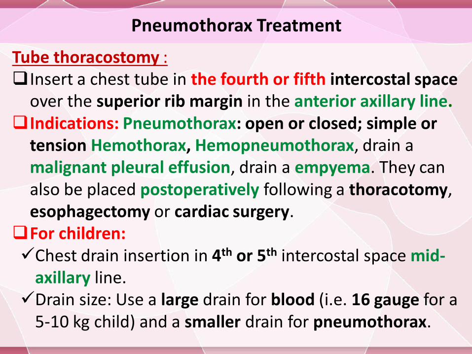
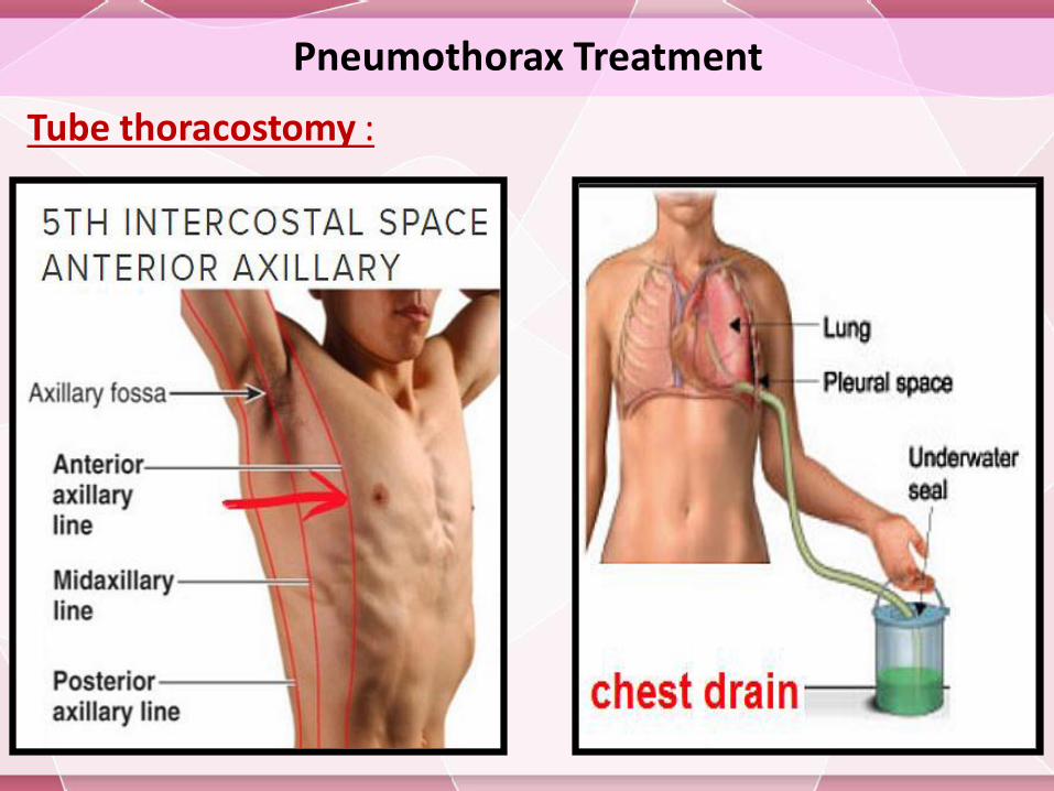
Tube thoracostomy :Insert a chest tube in the fourth or fifth intercostal space
over the superior rib margin in the anterior axillary line.Indications: Pneumothorax: open or closed; simple or
tension Hemothorax, Hemopneumothorax, drain a malignant pleural effusion, drain a empyema. They can also be placed postoperatively following a thoracotomy, esophagectomy or cardiac surgery.
For children:Chest drain insertion in 4th or 5th intercostal space mid-
axillary line.Drain size: Use a large drain for blood (i.e. 16 gauge for a
5-10 kg child) and a smaller drain for pneumothorax.
Pneumothorax Treatment
Tube thoracostomy :
Chest tube
Pneumothorax Treatment
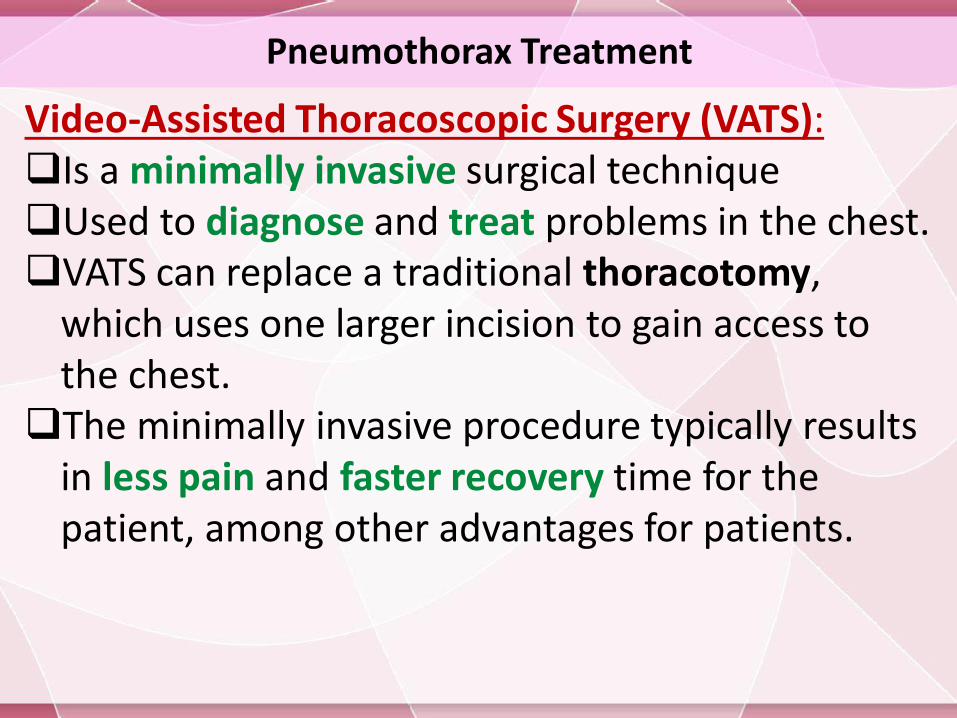
Video-Assisted Thoracoscopic Surgery (VATS):Is a minimally invasive surgical technique Used to diagnose and treat problems in the chest.VATS can replace a traditional thoracotomy,
which uses one larger incision to gain access to the chest.
The minimally invasive procedure typically results in less pain and faster recovery time for the patient, among other advantages for patients.
Pneumothorax Treatment
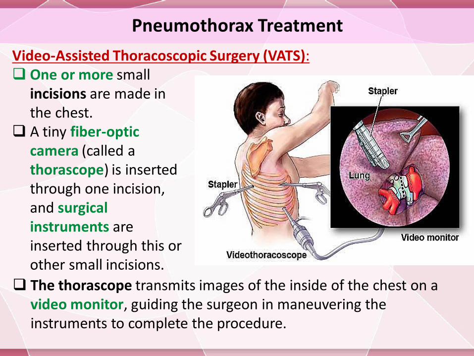
One or more small incisions are made in the chest.
A tiny fiber-optic camera (called a thorascope) is inserted through one incision, and surgical instruments are inserted through this or other small incisions.
Video-Assisted Thoracoscopic Surgery (VATS):
The thorascope transmits images of the inside of the chest on a video monitor, guiding the surgeon in maneuvering the instruments to complete the procedure.
Pneumothorax Treatment
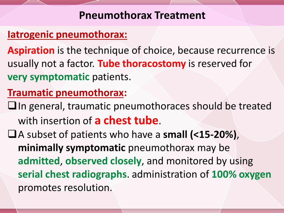
Iatrogenic pneumothorax:
Aspiration is the technique of choice, because recurrence is usually not a factor. Tube thoracostomy is reserved for very symptomatic patients.
Traumatic pneumothorax:In general, traumatic pneumothoraces should be treated
with insertion of a chest tube.A subset of patients who have a small (<15-20%),
minimally symptomatic pneumothorax may be admitted, observed closely, and monitored by using serial chest radiographs. administration of 100% oxygen promotes resolution.
Pneumothorax Treatment
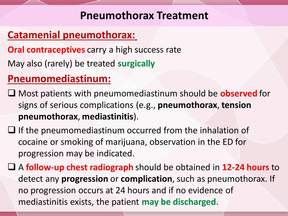
Catamenial pneumothorax:
Oral contraceptives carry a high success rate
May also (rarely) be treated surgically
Pneumomediastinum:
Most patients with pneumomediastinum should be observed for signs of serious complications (e.g., pneumothorax, tension pneumothorax, mediastinitis).
If the pneumomediastinum occurred from the inhalation of cocaine or smoking of marijuana, observation in the ED for progression may be indicated.
A follow-up chest radiograph should be obtained in 12-24 hours to detect any progression or complication, such as pneumothorax. If no progression occurs at 24 hours and if no evidence of mediastinitis exists, the patient may be discharged.
Pneumothorax Treatment
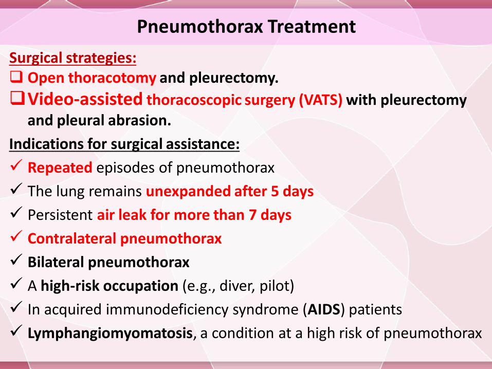
Surgical strategies: Open thoracotomy and pleurectomy.
Video-assisted thoracoscopic surgery (VATS) with pleurectomyand pleural abrasion.
Indications for surgical assistance:
Repeated episodes of pneumothorax
The lung remains unexpanded after 5 days
Persistent air leak for more than 7 days
Contralateral pneumothorax
Bilateral pneumothorax
A high-risk occupation (e.g., diver, pilot)
In acquired immunodeficiency syndrome (AIDS) patients
Lymphangiomyomatosis, a condition at a high risk of pneumothorax

Pneumothorax Treatment
Drug treatment: Local Anesthetics (Lidocaine hydrochloride): Used for analgesia,
for thoracentesis and chest tube placement.Opiate Analgesics (Fentanyl citrate and Morphine): Used for
pain control, ensures patient comfort, and promotes pulmonary toilet.
Benzodiazepines (Midazolam and Lorazepam ): Used for conscious sedation,in premedication before pleurodesis, sclerotherapy or placement of a thoracostomy tube.
Antibiotics (Doxycycline and Cefazolin): In patients with repeated pneumothoraces who are not good candidates for surgery, pleurodesis (or sclerotherapy) may be necessary.
Prophylactic antibiotics are not recommended for the placement of chest tubes in non-traumatic causes.
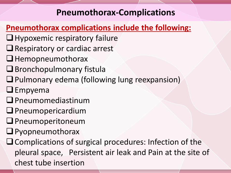
Pneumothorax-Complications
Pneumothorax complications include the following:Hypoxemic respiratory failureRespiratory or cardiac arrestHemopneumothoraxBronchopulmonary fistulaPulmonary edema (following lung reexpansion)EmpyemaPneumomediastinumPneumopericardiumPneumoperitoneumPyopneumothoraxComplications of surgical procedures: Infection of the
pleural space, Persistent air leak and Pain at the site of chest tube insertion
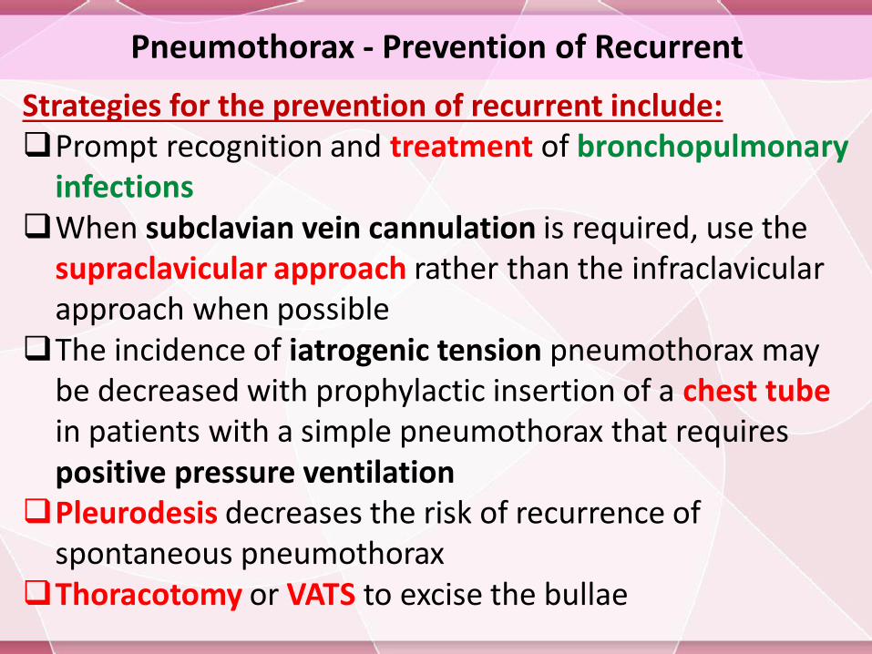
Pneumothorax - Prevention of Recurrent
Strategies for the prevention of recurrent include:Prompt recognition and treatment of bronchopulmonary
infections When subclavian vein cannulation is required, use the
supraclavicular approach rather than the infraclavicularapproach when possible
The incidence of iatrogenic tension pneumothorax may be decreased with prophylactic insertion of a chest tube in patients with a simple pneumothorax that requires positive pressure ventilation
Pleurodesis decreases the risk of recurrence of spontaneous pneumothorax
Thoracotomy or VATS to excise the bullae


![BilateralSpontaneousPneumothorax, Pneumomediastinum ... · as pneumothorax [2]. The ratio of simultaneous bilateral spontaneous pneumothorax is approximately 1.3% among all cases](https://img.pdfslide.net/doc/110x75/5cc1981888c993110a8c6f12/bilateralspontaneouspneumothorax-pneumomediastinum-as-pneumothorax-2.jpg)


![BilateralSpontaneousPneumothorax, Pneumomediastinum…downloads.hindawi.com/journals/criem/2012/242579.pdf · 2019-07-31 · as pneumothorax [2]. The ratio of simultaneous bilateral](https://img.pdfslide.net/doc/110x75/5f4072a3171ef02d0d32a564/bilateralspontaneouspneumothorax-pneumo-2019-07-31-as-pneumothorax-2-the-ratio.jpg)