Embed Size (px)
Citation preview

Questions Femur
The highlighted region is the
1 Greater tuberosity
2 Adductor tubercle
3 Greater trochanter
4 Linea aspera
5 Lesser trochanter
Question 1 – Femur
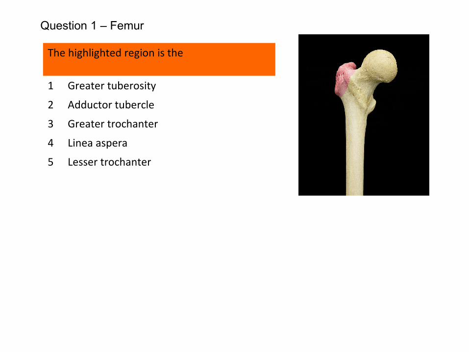
The highlighted region is the
1 Greater tuberosity F
2 Adductor tubercle F
3 Greater trochanter T
4 Linea aspera F
5 Lesser trochanter F
The greater tuberosity is part of the proximal humerus, not femur!The adductor tubercle and linea aspera are found distally on the femur.
Question 1 – Femur
The following are attached to the greater trochanter of femur:
1 tendon of psoas major
2 gluteus maximus
3 gluteus minimus
4 gluteus medius
5 iliacus
Question 2 -– Femur
The following are attached to the greater trochanter of femur:
1 tendon of psoas major F
2 gluteus maximus F
3 gluteus minimus T
4 gluteus medius T
5 iliacus F
The tendons of psoas major and iliacus are attached to the lesser trochanter.
Gluteus maximus is inserted mainly into the ilio-tibial tract with a small portion of the muscle attaching to the gluteal tuberosity on the back of the upper femur.
Question 2 -– Femur
The following structures attach to the highlighted prominence of the distal femur
1 Tibial collateral ligament
2 Lateral head of gastrocnemius
3 Adductor magnus
4 Adductor longus
5 Vastus lateralis
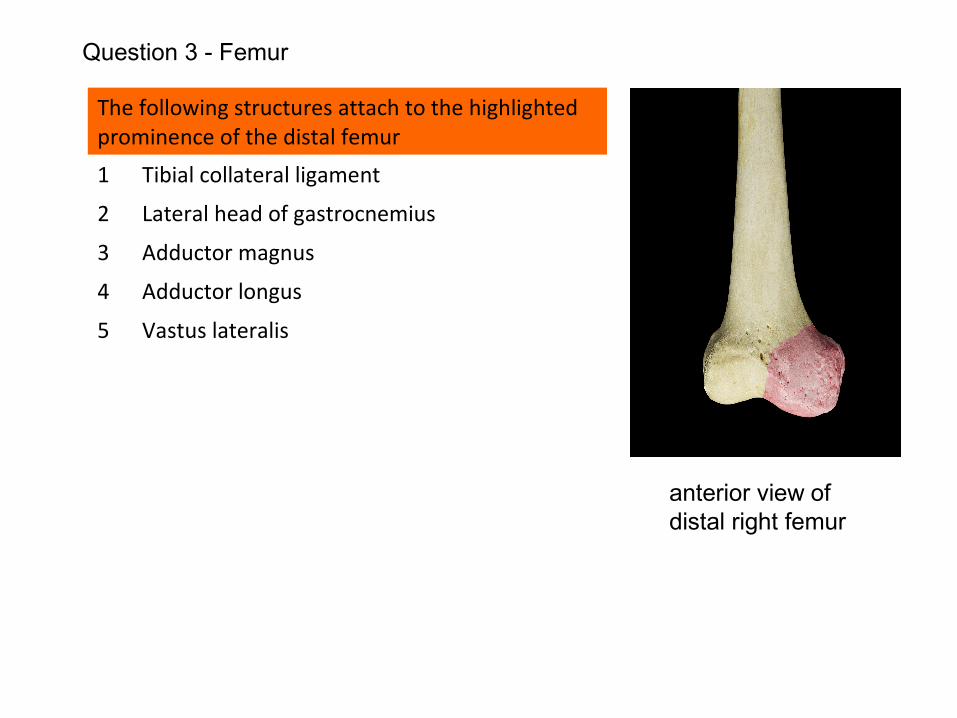
Question 3 - Femur
anterior view of distal right femur
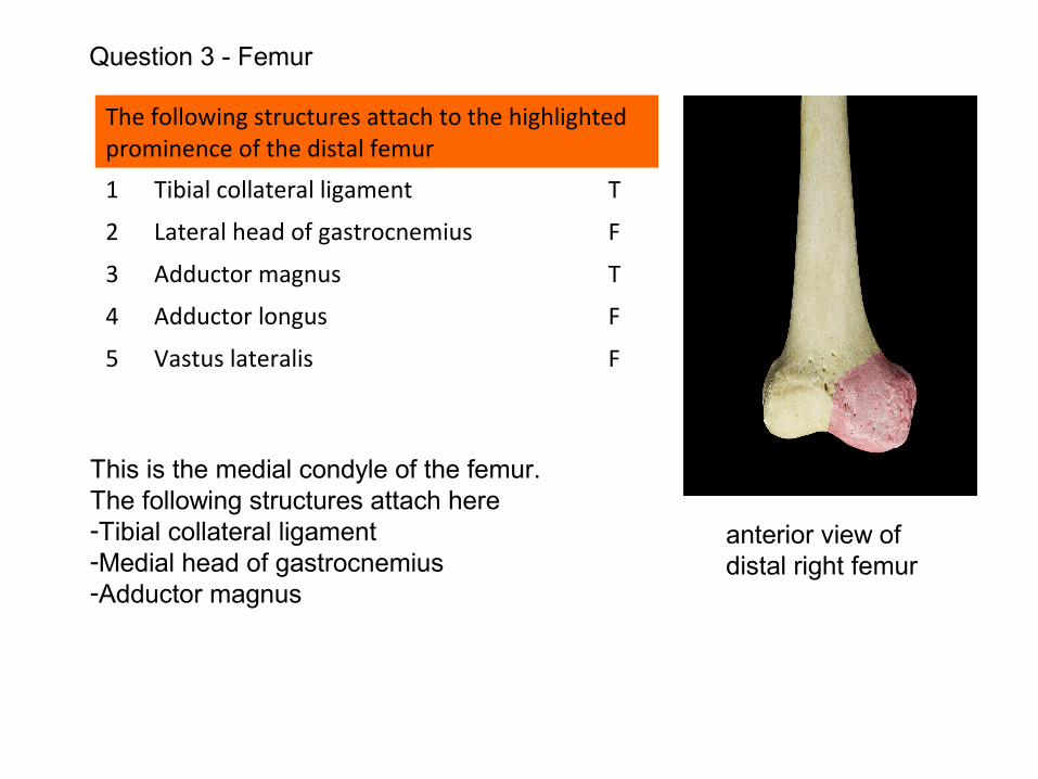
The following structures attach to the highlighted prominence of the distal femur
1 Tibial collateral ligament T
2 Lateral head of gastrocnemius F
3 Adductor magnus T
4 Adductor longus F
5 Vastus lateralis F
This is the medial condyle of the femur.The following structures attach here-Tibial collateral ligament-Medial head of gastrocnemius-Adductor magnus
Question 3 - Femur
anterior view of distal right femur
The following structures attach to the linea aspera on the posterior surface of the femoral shaft:
1 semimembranosus
2 semitendinosus
3 adductor longus
4 vastus lateralis
5 long head of biceps femoris
Question 4 - Femur
The following structures attach to the linea aspera on the posterior surface of the femoral shaft:
1 semimembranosus F
2 semitendinosus F
3 adductor longus T
4 vastus lateralis T
5 long head of biceps femoris F
Neither semimembranosus nor semitendinosus have any attachment to the femur.
The short head of biceps femoris is attached to the linea aspera; not the long head.
Question 4 - Femur
With regard to the lower end of the femur:
1 the adductor tubercle is a bony prominence on the lateral femoral condyle
2 the medial femoral epicondyle gives attachment to the tendon of popliteus
3 the anterior cruciate ligament is attached to the lateral femoral condyle
4 the adductor tubercle receives the insertion of adductor longus
5 the intercondylar notch lies outside the capsular attachment of the knee joint
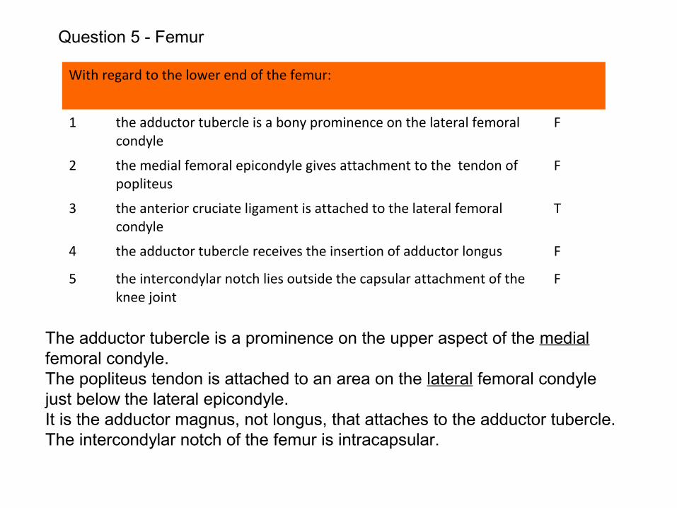
Question 5 - Femur
With regard to the lower end of the femur:
1 the adductor tubercle is a bony prominence on the lateral femoral condyle
F
2 the medial femoral epicondyle gives attachment to the tendon of popliteus
F
3 the anterior cruciate ligament is attached to the lateral femoral condyle
T
4 the adductor tubercle receives the insertion of adductor longus F
5 the intercondylar notch lies outside the capsular attachment of the knee joint
F
The adductor tubercle is a prominence on the upper aspect of the medial femoral condyle.The popliteus tendon is attached to an area on the lateral femoral condyle just below the lateral epicondyle. It is the adductor magnus, not longus, that attaches to the adductor tubercle.The intercondylar notch of the femur is intracapsular.
Question 5 - Femur
The following structures attach to the highlighted region
1 adductor brevis
2 adductor longus
3 psoas major
4 iliacus
5 obturator internus
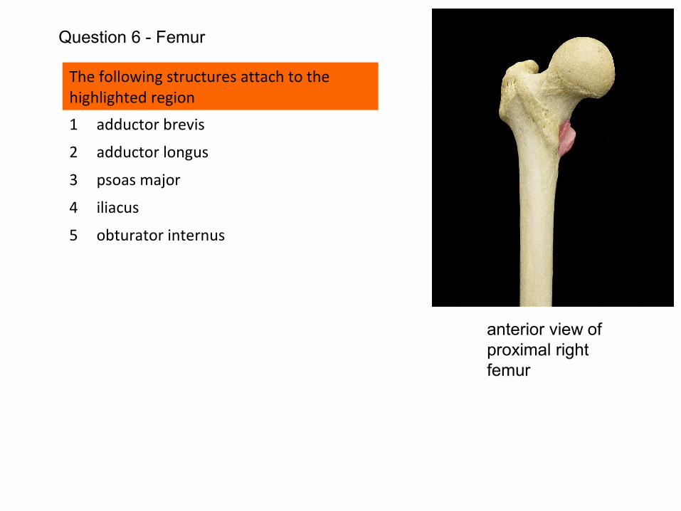
Question 6 - Femur
anterior view of proximal right femur
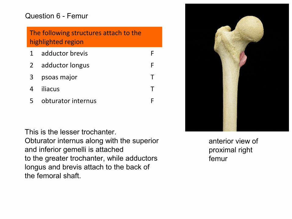
The following structures attach to the highlighted region
1 adductor brevis F
2 adductor longus F
3 psoas major T
4 iliacus T
5 obturator internus F
This is the lesser trochanter.Obturator internus along with the superior and inferior gemelli is attachedto the greater trochanter, while adductors longus and brevis attach to the back of the femoral shaft.
Question 6 - Femur
anterior view of proximal right femur
With regard to the highlighted area on the proximal femur
1 This is the linea aspera
2 The hip capsule attaches here
3 This is the intertrochanteric line
4 Gluteus maximus attaches here
5 Vastus medialis attaches here
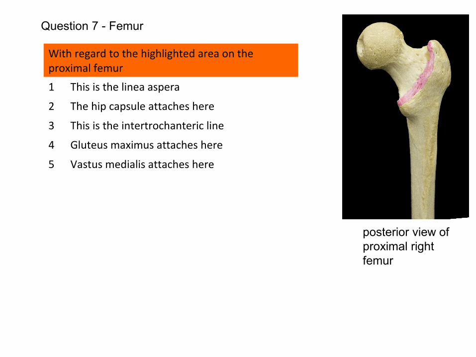
Question 7 - Femur
posterior view of proximal right femur
With regard to the highlighted area on the proximal femur
1 This is the linea aspera F
2 The hip capsule attaches here F
3 This is the intertrochanteric line F
4 Gluteus maximus attaches here F
5 Vastus medialis attaches here F
This is the posterior view of the proximal femur. The highlighted region is the intertrochanteric crest.The hip joint capsule attaches 2cm supero-medial to the intertrochanteric crest on the back of the femoral neck, but attaches to the intertrochanteric line, anteriorly.Gluteus maximus attaches to the gluteal tuberosity, more distally, on the posterior aspect of the femur.Vastus medialis attaches to the lower aspect of the intertrochanteric line on the anterior aspect of the proximal femur.
Question 7 - Femur
posterior view of proximal right femur
With regard to the proximal end of the femur
1 the intertrochanteric line gives attachment to the capsule of the hip joint
2 the lesser trochanter is an intracapsular structure
3 the blood supply to the femoral head in the elderly individual is principally from the artery that accompanies the ligament of the head of femur
4 fractures through the base of the femoral neck jeopardise the blood supply of the femoral head more than do subcapital femoral fractures
5 the upper end of the femur is the ‘growing end’
Question 8 - Femur
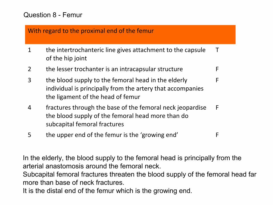
With regard to the proximal end of the femur
1 the intertrochanteric line gives attachment to the capsule of the hip joint
T
2 the lesser trochanter is an intracapsular structure F
3 the blood supply to the femoral head in the elderly individual is principally from the artery that accompanies the ligament of the head of femur
F
4 fractures through the base of the femoral neck jeopardise the blood supply of the femoral head more than do subcapital femoral fractures
F
5 the upper end of the femur is the ‘growing end’ F
In the elderly, the blood supply to the femoral head is principally from the arterial anastomosis around the femoral neck.Subcapital femoral fractures threaten the blood supply of the femoral head far more than base of neck fractures.It is the distal end of the femur which is the growing end.
Question 8 - Femur
The following muscles attach to the highlighted region
1 Gluteus minimus
2 Piriformis
3 Obturator externus
4 Pectineus
5 Adductor brevis
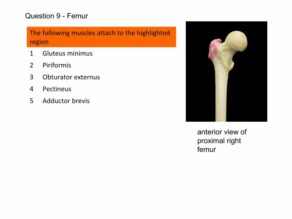
Question 9 - Femur
anterior view of proximal right femur
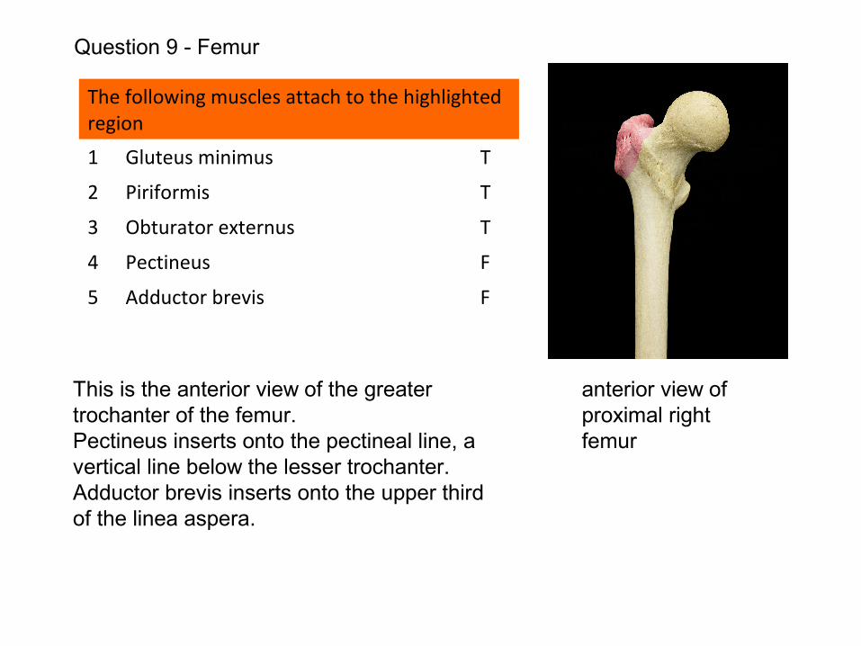
The following muscles attach to the highlighted region
1 Gluteus minimus T
2 Piriformis T
3 Obturator externus T
4 Pectineus F
5 Adductor brevis F
This is the anterior view of the greater trochanter of the femur.Pectineus inserts onto the pectineal line, a vertical line below the lesser trochanter.Adductor brevis inserts onto the upper third of the linea aspera.
Question 9 - Femur
anterior view of proximal right femur
The following muscles attach to the proximal femur
1 Sartorius
2 Quadratus femoris
3 Vastus lateralis
4 Gracilis
5 Iliacus
Question 10 -Femur
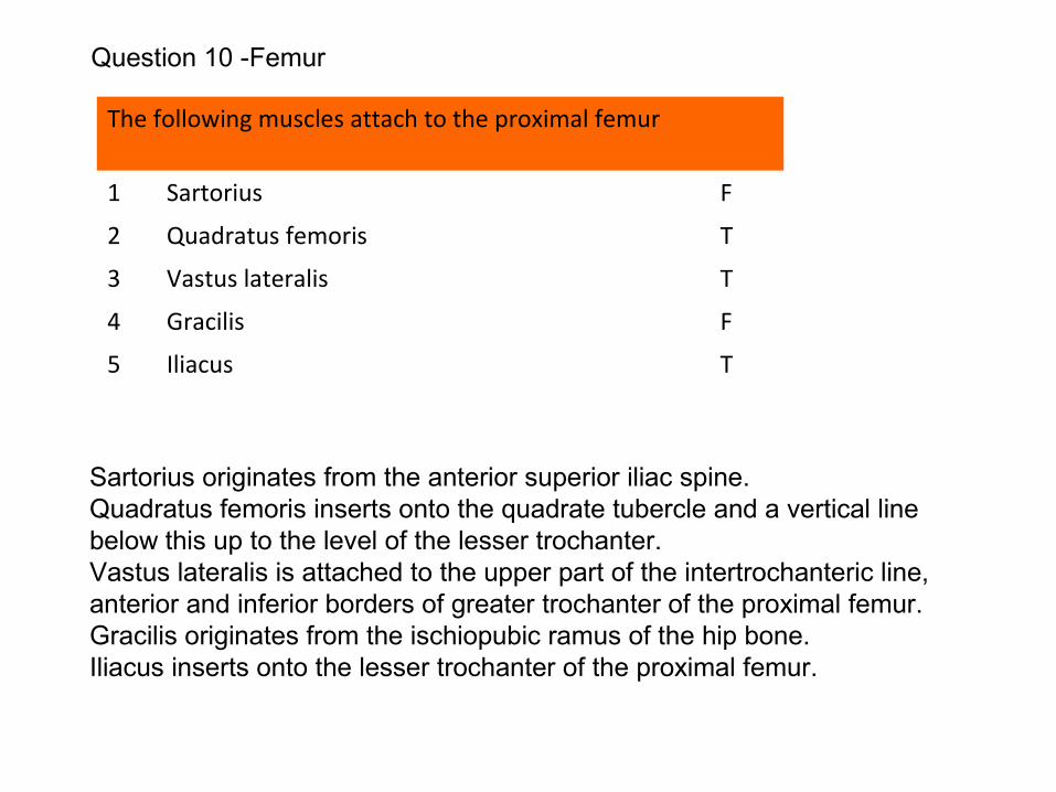
The following muscles attach to the proximal femur
1 Sartorius F
2 Quadratus femoris T
3 Vastus lateralis T
4 Gracilis F
5 Iliacus T
Sartorius originates from the anterior superior iliac spine.Quadratus femoris inserts onto the quadrate tubercle and a vertical line below this up to the level of the lesser trochanter.Vastus lateralis is attached to the upper part of the intertrochanteric line, anterior and inferior borders of greater trochanter of the proximal femur.Gracilis originates from the ischiopubic ramus of the hip bone.Iliacus inserts onto the lesser trochanter of the proximal femur.
Question 10 -Femur