Embed Size (px)
Citation preview
RADIOLOGY OF NOSE AND
PARANASAL SINUSES
ANDREA R SALINS
Paranasal sinuses
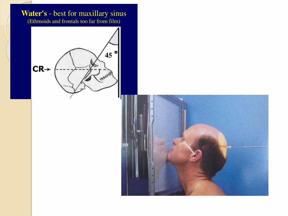
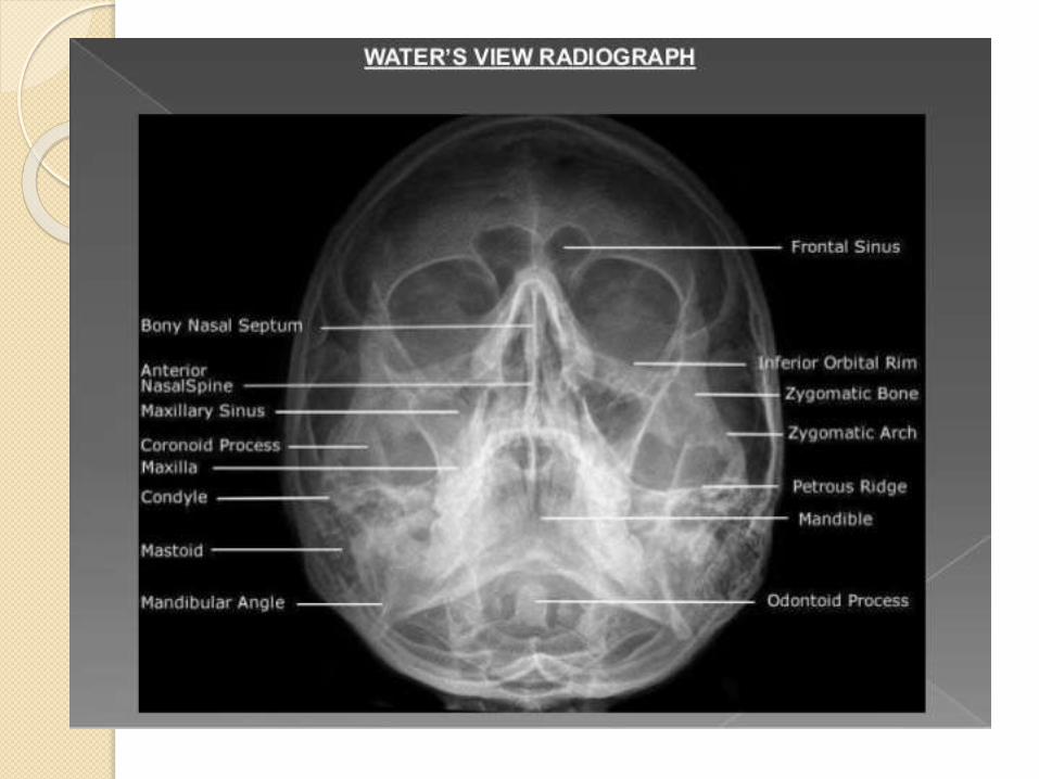
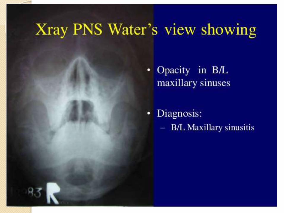
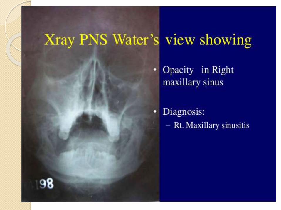
Water’s view
Caldwell view
Lateral view
Submentovertical view
Right and left vertical view
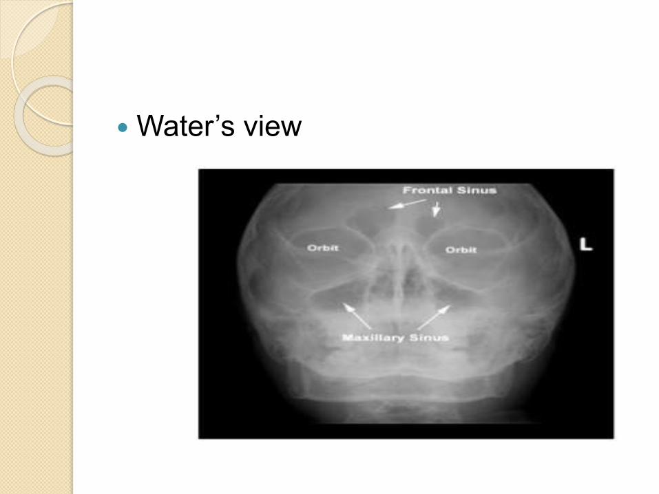
Water’s view
Also called as occipitomental view or nose-chin position
In 1914,Dr C.A.Waters and C.W.Waldron,two British radiologists introduced this view
Nose and chin touch the film and X-ray beam is projected from occipital side
Open mouth view shows sphenoid sinus
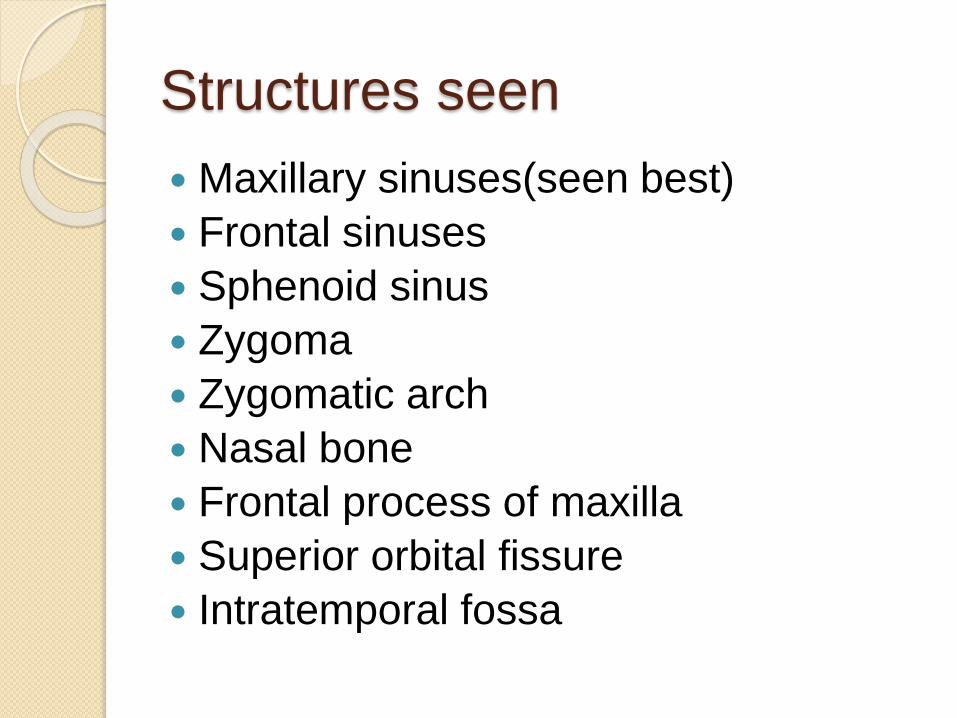
Structures seen
Maxillary sinuses(seen best)
Frontal sinuses
Sphenoid sinus
Zygoma
Zygomatic arch
Nasal bone
Frontal process of maxilla
Superior orbital fissure
Intratemporal fossa
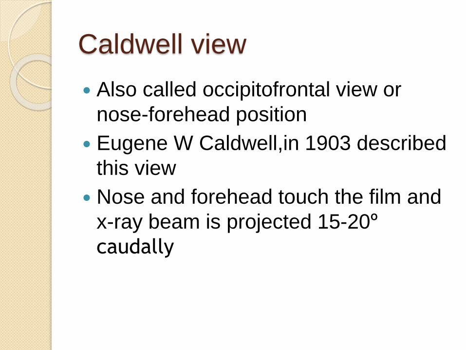
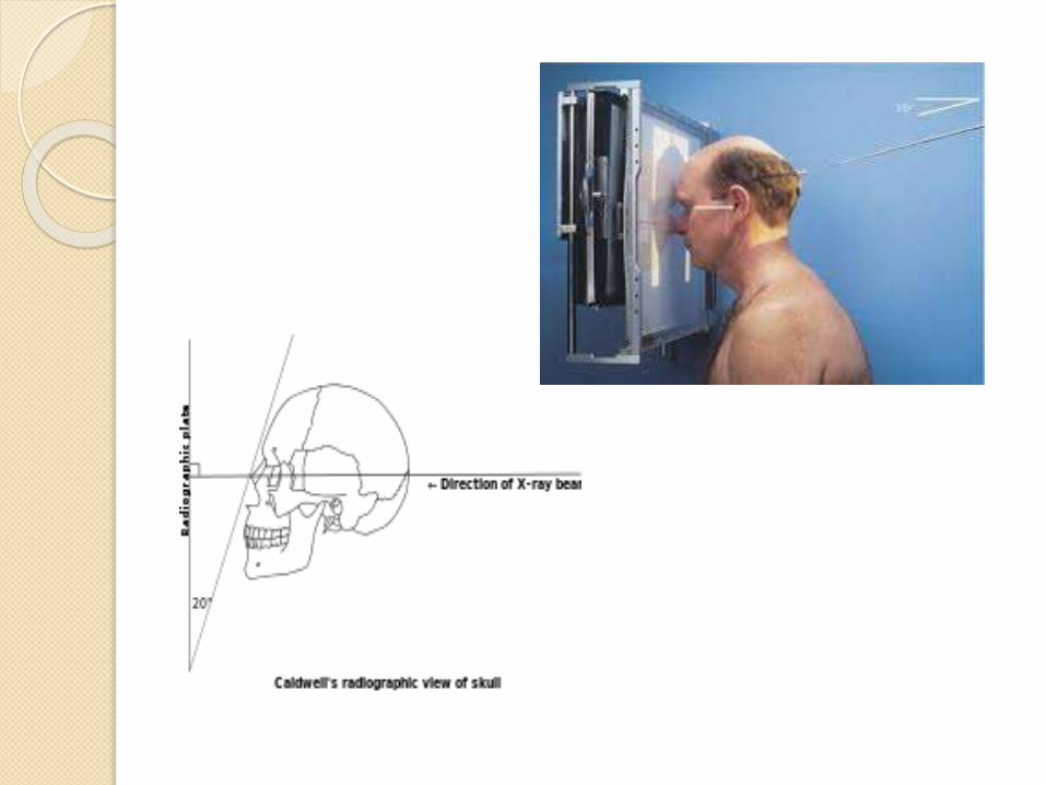
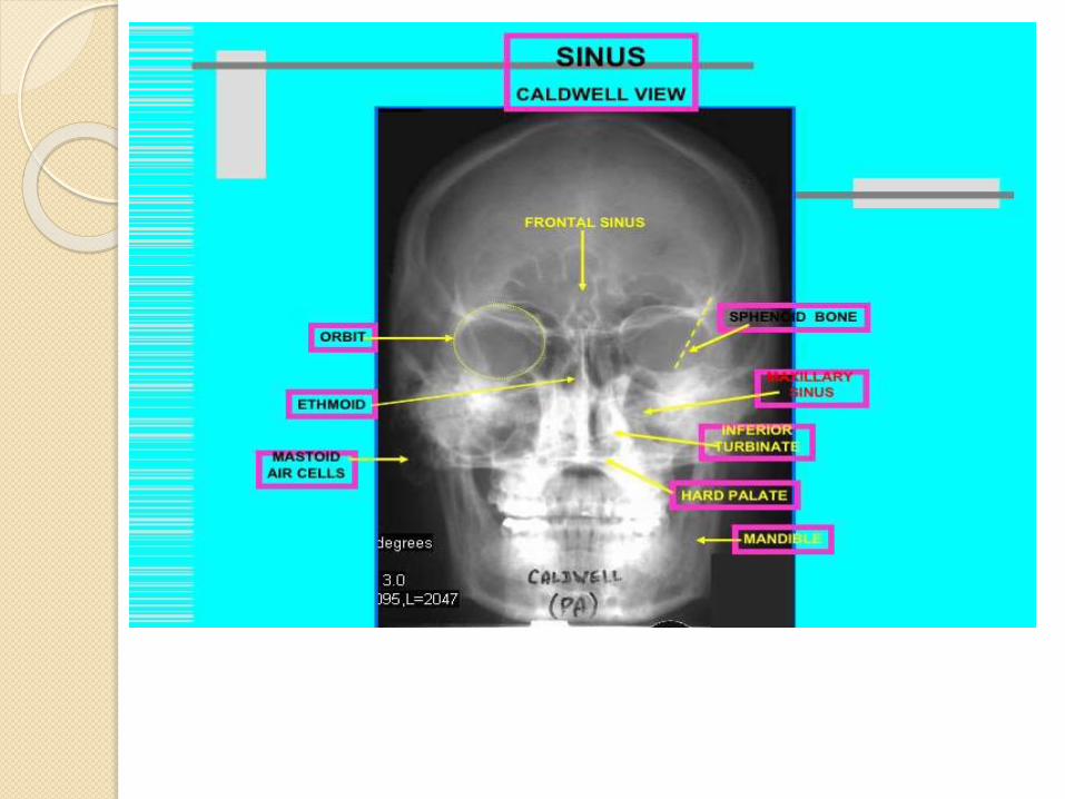
Caldwell view
Also called occipitofrontal view or
nose-forehead position
Eugene W Caldwell,in 1903 described
this view
Nose and forehead touch the film and
x-ray beam is projected 15-20º
caudally
Structures seen
Frontal sinuses(best)
Ethmoid sinuses,maxillary sinuses
Frontal process of of zygoma and
zygomatic process of frontal bone
Superior margin of orbit and lamina
papyracea
Superior orbital fissure
Foramen rotundum
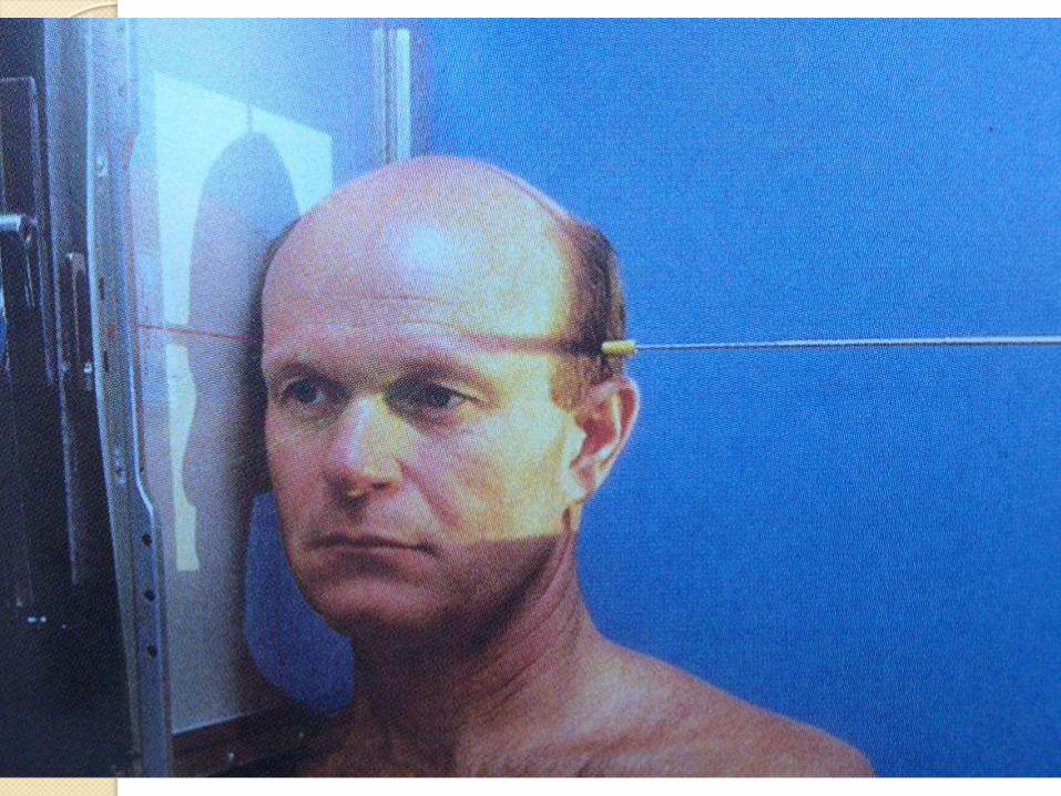
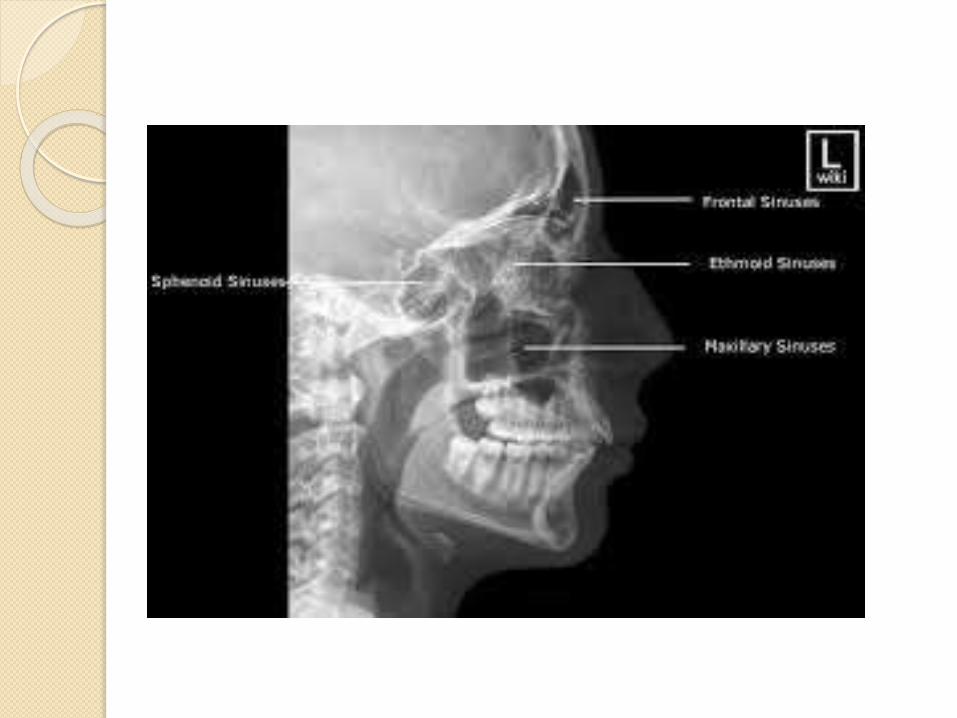
Lateral view
Lateral side of skull lies against the
film and x-ray beam is projected
perpendicular from the other side
Fluid levels of all sinuses can be seen
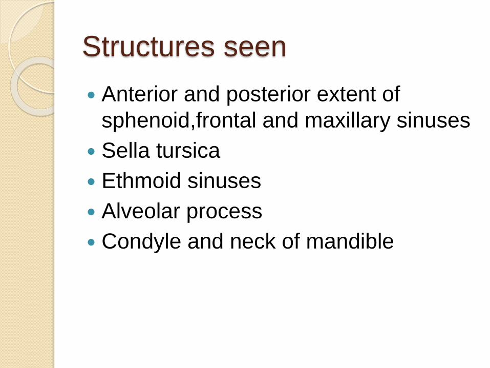
Structures seen
Anterior and posterior extent of
sphenoid,frontal and maxillary sinuses
Sella tursica
Ethmoid sinuses
Alveolar process
Condyle and neck of mandible
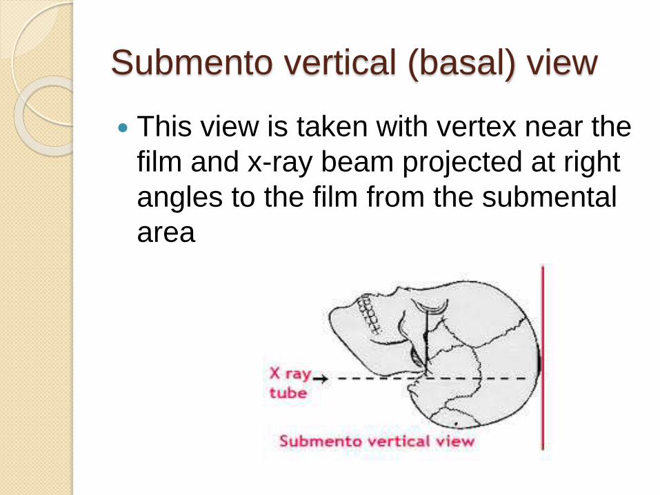
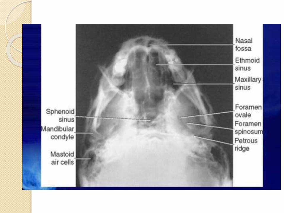
Submento vertical (basal) view
This view is taken with vertex near the
film and x-ray beam projected at right
angles to the film from the submental
area
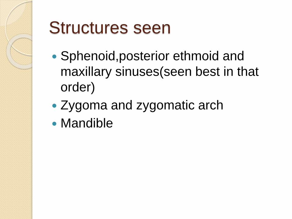
Structures seen
Sphenoid,posterior ethmoid and
maxillary sinuses(seen best in that
order)
Zygoma and zygomatic arch
Mandible

Right and left oblique view
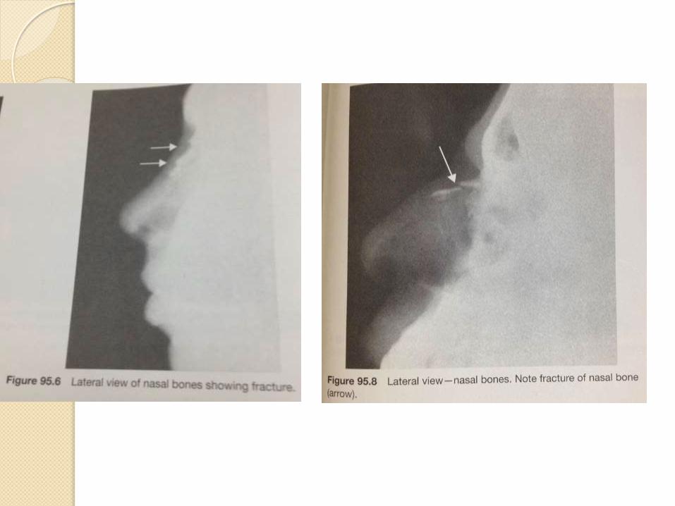
X-rays for nasal fractures
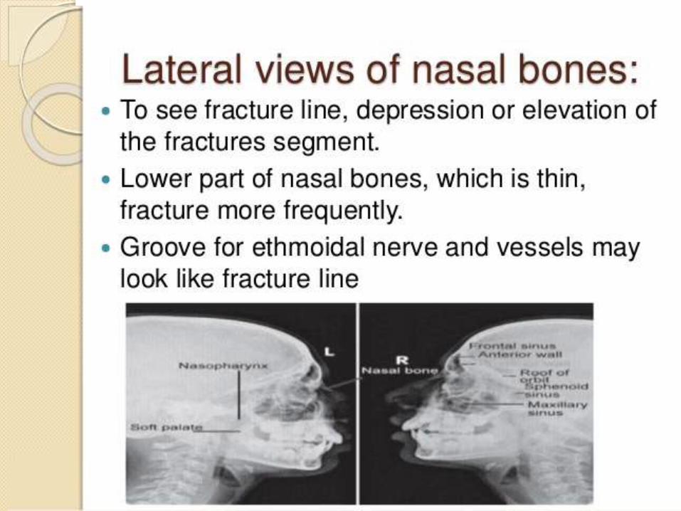
Lateral view of nasal bones
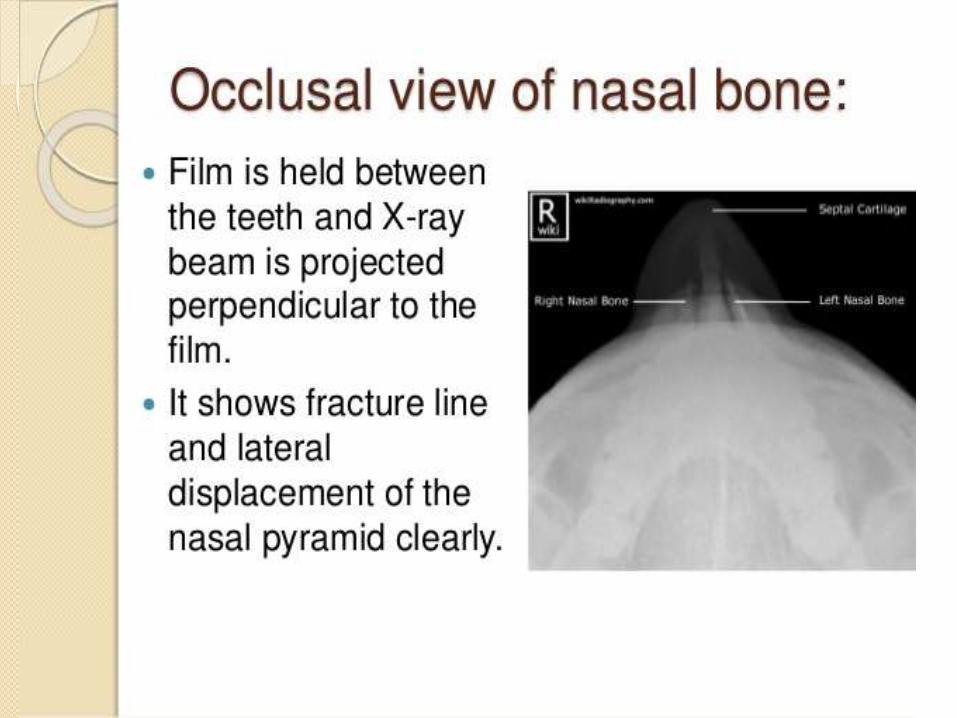
Occlusal view of nasal bone
Water’s view
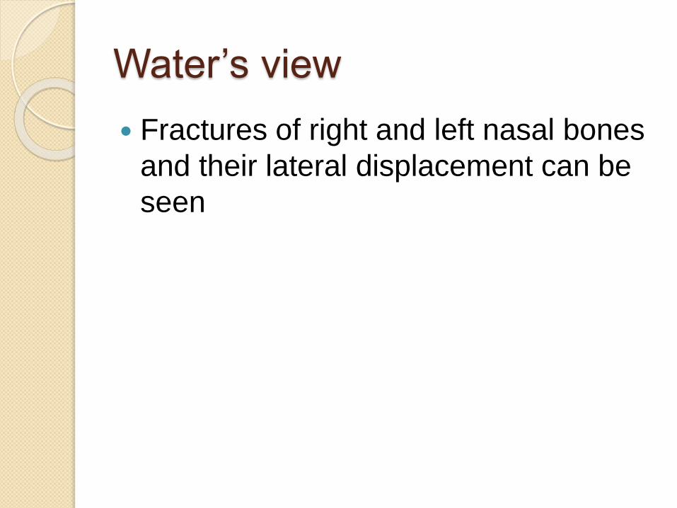
Water’s view
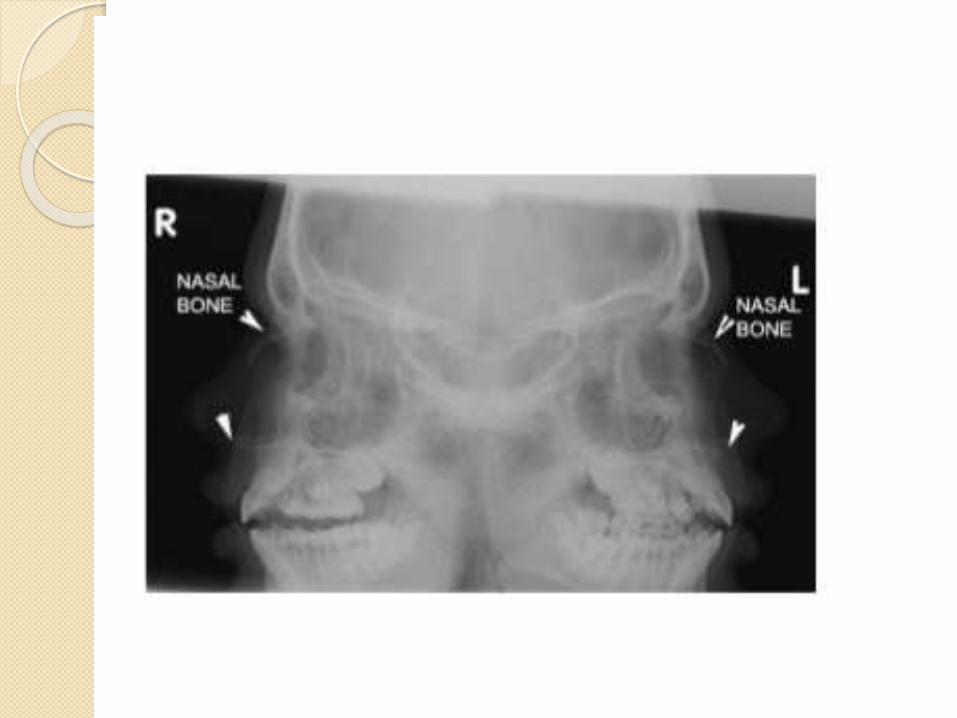
Fractures of right and left nasal bones
and their lateral displacement can be
seen
Water’s view
Advantages of X-ray imaging
Cost effectiveness
Easy availability

Disadvantages
Plain X-ray have false positivity of 4%
False negativity-more than 30%
Currently available digital imaging
techniques provide better bone and
soft tissue resolution when compared
to conventional X-ray

THANK YOU