Embed Size (px)
Citation preview
RADIOULNAR SYNOSTOSIS
Lamyaa Anwar Alghafli
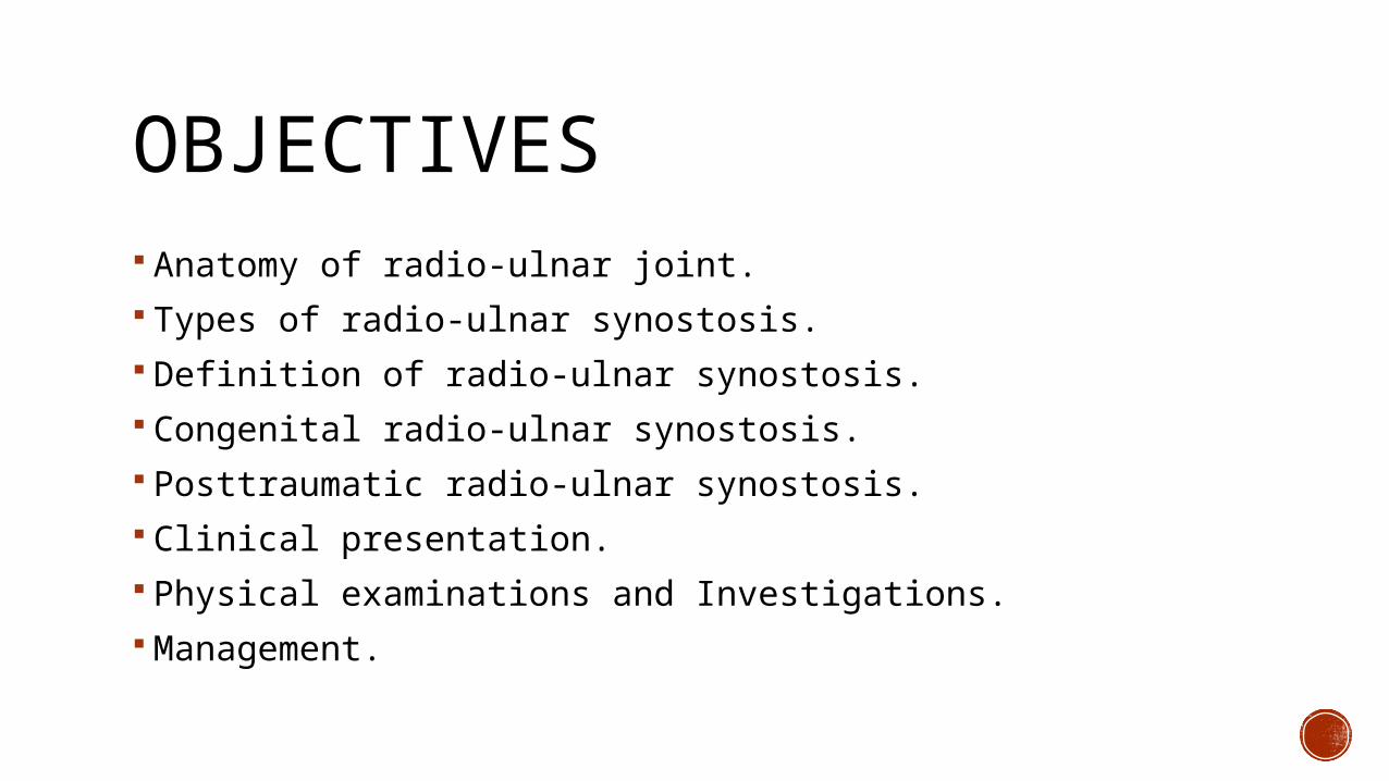
OBJECTIVES Anatomy of radio-ulnar joint. Types of radio-ulnar synostosis. Definition of radio-ulnar synostosis. Congenital radio-ulnar synostosis. Posttraumatic radio-ulnar synostosis. Clinical presentation. Physical examinations and Investigations. Management.
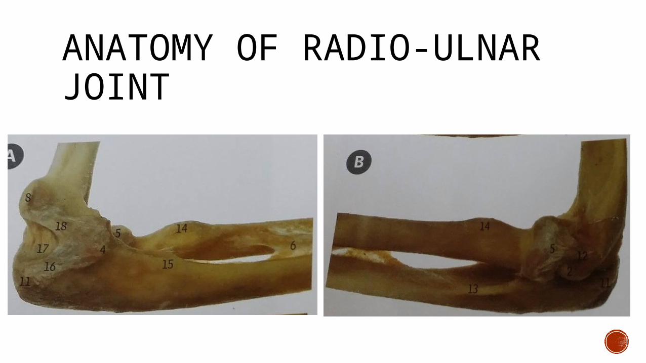
ANATOMY OF RADIO-ULNAR JOINT
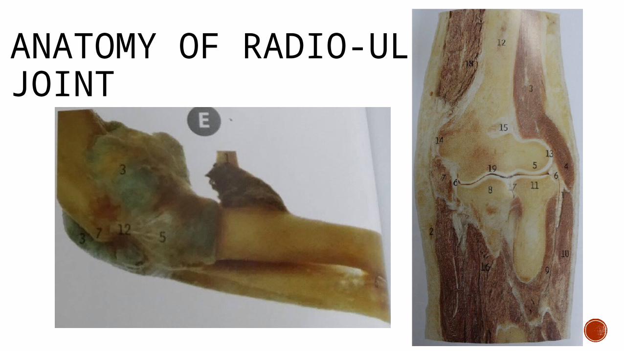
ANATOMY OF RADIO-ULNAR JOINT
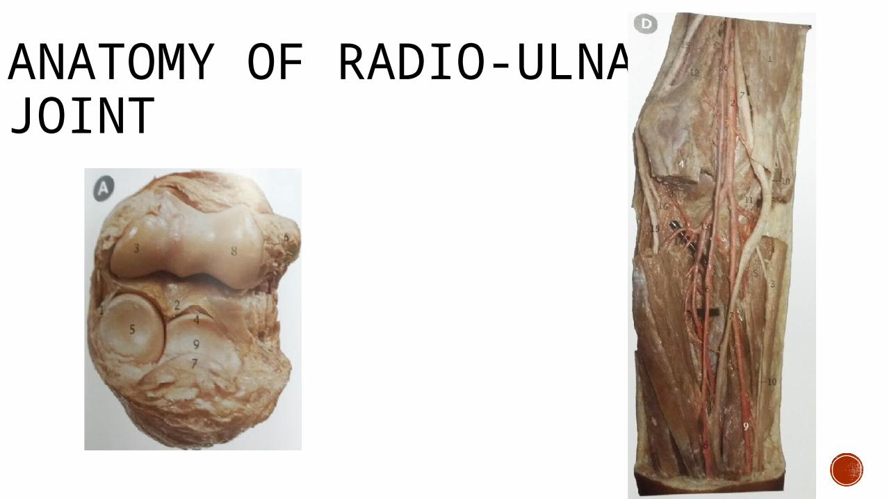
ANATOMY OF RADIO-ULNAR JOINT
DEFINITION OF RADIO-ULNAR SYNOSTOSIS
A radio-ulnar synostosis is a rare condition which is upper limb skeletal malformation characterized by bony fusion of the radius and ulna.
In 1793, Sandifort provided the initial description of congenital type.
Gros described posttraumatic radioulnar synostosis in 1864.
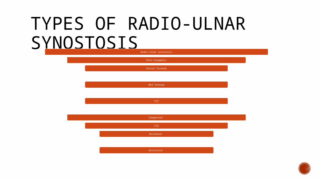
TYPES OF RADIO-ULNAR SYNOSTOSIS
Radio-ulnar synostosis
Post-traumatic
Distal forearm
Mid forearm
Proximalforearm
Congenital
Proximalforearm
Bilateral
Unilateral
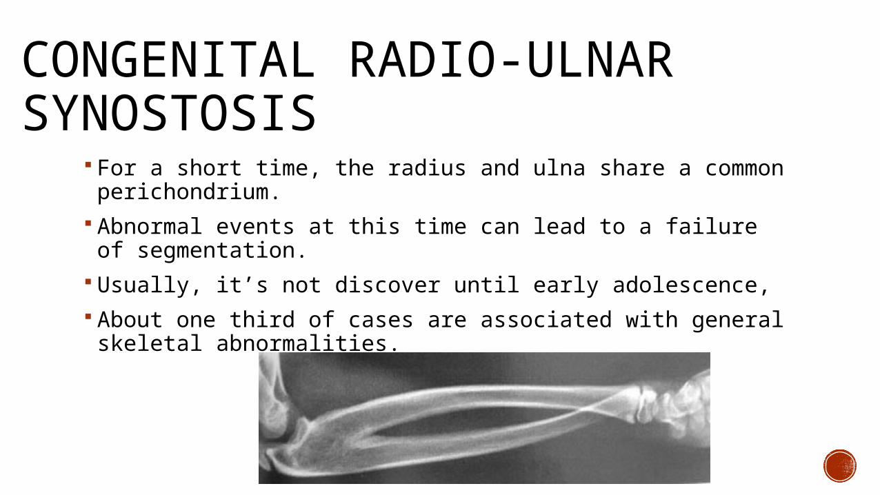
CONGENITAL RADIO-ULNAR SYNOSTOSIS
For a short time, the radius and ulna share a common perichondrium.
Abnormal events at this time can lead to a failure of segmentation.
Usually, it’s not discover until early adolescence, About one third of cases are associated with general skeletal abnormalities.
POSTTRAUMATIC RADIOULNAR SYNOSTOSIS The most common cause: operatively treated forearm fracture. Patients with high-energy, comminuted, open fractures. Monteggia and proximal forearm fractures. The use of bone graft and screws protruding through the opposite cortex.
Any trauma causing hematoma formation.
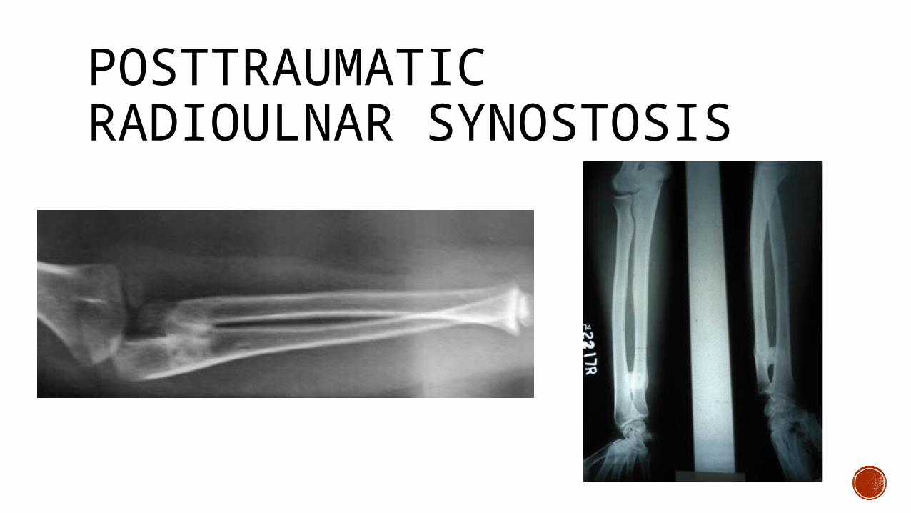
POSTTRAUMATIC RADIOULNAR SYNOSTOSIS
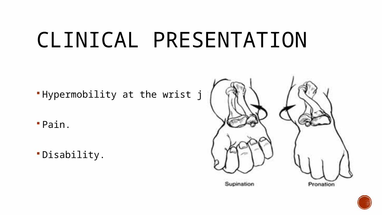
CLINICAL PRESENTATION
Hypermobility at the wrist joint.
Pain.
Disability.
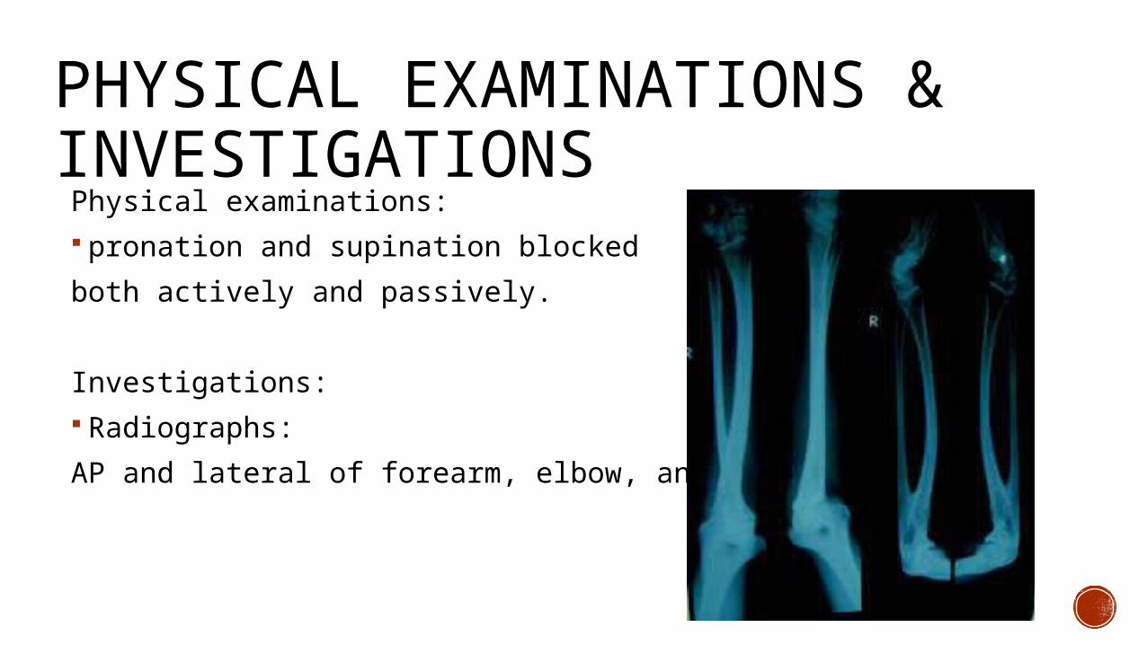
PHYSICAL EXAMINATIONS & INVESTIGATIONSPhysical examinations: pronation and supination blocked
both actively and passively.
Investigations: Radiographs:
AP and lateral of forearm, elbow, and wrist.
MANAGEMENT non operative:
Observation.
Operative: osteotomy with fusion. surgical resection of synostosis, irradiation, and indomethacin.
proximal radial excision.
Postoperative rehabilitation: Splinting in maximum pronation and supination between passive and active physiotherapy.
SOURCES Clinical ATLAS of human anatomy, Peter H, Johannes M, Jonathan D.
Radioulnar synostosis : A case report, Ramakrishna Avadhani , Bindhu S. , Vikram , Dhanesh Kumar K.U.& Arunachalam Kumar
http://emedicine.medscape.com/article/1240467-overview#a0102
http://www.orthobullets.com/trauma/1026/radioulnar-synostosis
http://www.sciencedirect.com/science/article/pii/S1877056812001582

SOURCES http://www.ncbi.nlm.nih.gov/pubmed/23789710
http://www.wheelessonline.com/ortho/radioulnar_synostosis
http://www.childrenshospital.org/conditions-and-treatments/conditions/r/radioulnar-synostosis/treatments
http://radiopaedia.org/articles/proximal-radio-ulnar-synostosis