Embed Size (px)
Citation preview
SECONDARY HYPERTENSION-EVALUATION
CASE PRESENTATION
A 17 YEARS OLD MALE COLLEGE STUDENT-CAME TO THE HOSPITAL WITH CHIEF COMPLAINTS OF :
HEADACHE,
BLURRING OF VISION AND
VOMITINGS SINCE 2 DAYS
PRESENT HISTORY
HE WAS APPARENTLY ASYMPTOMATIC 2 DAYS BACK TO START WITH HE HAD BLURRING OF VISION IN BOTH EYES ,
HEADACHE OVER OCCIPETAL REGION WHICH IS ASSOCIATED WITH 2 EPISODES OF NON PROJECTILE, NON BILIOUS VOMITINGS CONTAINING INGESTED FOOD PARTICLES SINCE 2 DAYS.
HEADACHE IS MORE SEVERE IN EARLY MORNING HOURS
BOWEL AND BLADDER HABITS ARE REGULAR
NO H/O LOSS OF CONSCIOUSNESS,HEAD INJURY,LOOSE MOTIONS,FEVER,PALPITATIONS,PERIORBITAL EDEMA,HEMATURIA,WEIGHT LOSS/GAIN,COLD/HEAT INTOLERANCE,SNORING,DAY TIME SOMNALENCE,BURNING MICTURITION.
PAST HISTORYH/o similar complaints 3 months back and diagnosed as hypertension and advised antihypertensives but he is not taking medicines regularly.
No h/o diabetes mellitus ,renal disease and other chronic illness
Not a known smoker/alcoholic
Near vision (myopia) since 7years wearing spectacles of power -2.0 in both eyes
Family historyBorn out of consanguineous marriage
One brother- both had hypertrophic pyloric stenosis –operated (ramstead pyloroplasty) at 3 months of age.
His Father is an essential hypertensive on treatment.
TREATMENT HISTORY: He is not taking any drug regularly.
RELEVANT POSITIVE HISTORY
Young male with occipital headache,blurring of vision in both eyes and non projectile vomitings since 2 daysHeadache over occipital region and more in early morning hours.H/o similar complaints 3months back and diagnosed as hypertension on irregular treatment.Myopic(power,-2.0) on spectacles since 7 yearsBorn out of consanguineous marriage.One brother- both had hypertrophic pyloric stenosis at the age of 3 months and operated (ram stead pyloroplasty).His father is an essential hypertensive on treatment.
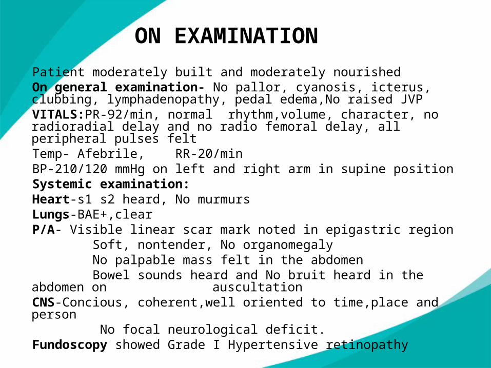
ON EXAMINATION
Patient moderately built and moderately nourishedOn general examination- No pallor, cyanosis, icterus, clubbing, lymphadenopathy, pedal edema,No raised JVPVITALS:PR-92/min, normal rhythm,volume, character, no radioradial
delay and no radio femoral delay, all peripheral pulses feltTemp- Afebrile, RR-20/minBP-210/120 mmHg on left and right arm in supine positionSystemic examination:Heart-s1 s2 heard, No murmursLungs-BAE+,clearP/A- Visible linear scar mark noted in epigastric region Soft, nontender, No organomegaly No palpable mass felt in the abdomen Bowel sounds heard and No bruit heard in the abdomen on auscultationCNS-Concious, coherent,well oriented to time,place and person No focal neurological deficit.Fundoscopy showed Grade I Hypertensive retinopathy
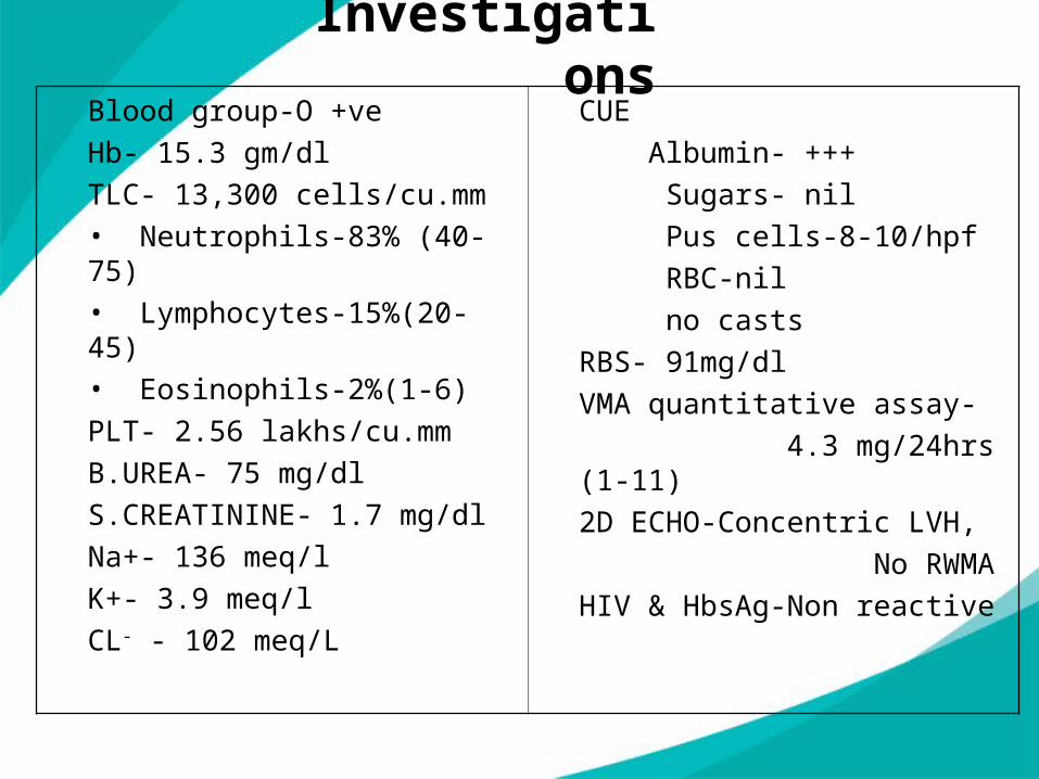
InvestigationsBlood group-O +ve
Hb- 15.3 gm/dl
TLC- 13,300 cells/cu.mm• Neutrophils-83% (40-75)• Lymphocytes-15%(20-45)• Eosinophils-2%(1-6)
PLT- 2.56 lakhs/cu.mm
B.UREA- 75 mg/dl
S.CREATININE- 1.7 mg/dl
Na+- 136 meq/l
K+- 3.9 meq/l
CL- - 102 meq/L
CUE
Albumin- +++
Sugars- nil
Pus cells-8-10/hpf
RBC-nil
no casts
RBS- 91mg/dl
VMA quantitative assay-
4.3 mg/24hrs (1-11)
2D ECHO-Concentric LVH,
No RWMA
HIV & HbsAg-Non reactive
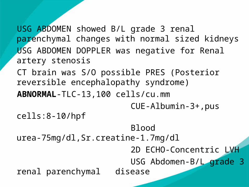
USG ABDOMEN showed B/L grade 3 renal parenchymal changes with normal sized kidneys
USG ABDOMEN DOPPLER was negative for Renal artery stenosis
CT brain was S/O possible PRES (Posterior reversible encephalopathy syndrome)
ABNORMAL-TLC-13,100 cells/cu.mm
CUE-Albumin-3+,pus cells:8-10/hpf
Blood urea-75mg/dl,Sr.creatine-1.7mg/dl
2D ECHO-Concentric LVH
USG Abdomen-B/L grade 3 renal parenchymal disease
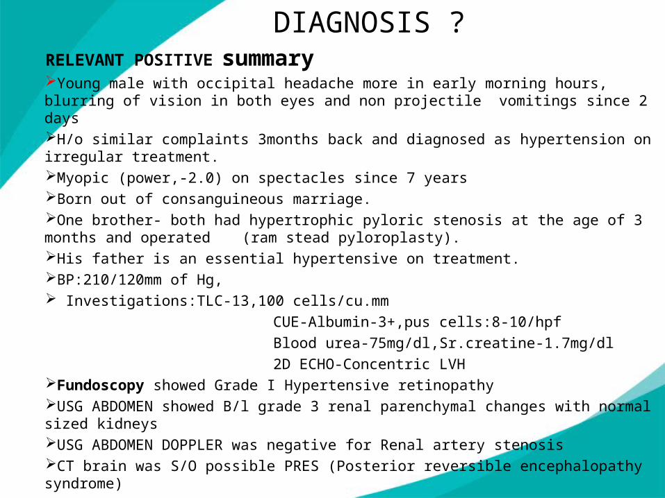
DIAGNOSIS ?RELEVANT POSITIVE summaryYoung male with occipital headache more in early morning hours, blurring of vision in both eyes and non projectile vomitings since 2 daysH/o similar complaints 3months back and diagnosed as hypertension on irregular treatment.Myopic (power,-2.0) on spectacles since 7 yearsBorn out of consanguineous marriage.One brother- both had hypertrophic pyloric stenosis at the age of 3 months and operated (ram stead pyloroplasty).His father is an essential hypertensive on treatment.BP:210/120mm of Hg, Investigations:TLC-13,100 cells/cu.mm
CUE-Albumin-3+,pus cells:8-10/hpf
Blood urea-75mg/dl,Sr.creatine-1.7mg/dl
2D ECHO-Concentric LVHFundoscopy showed Grade I Hypertensive retinopathyUSG ABDOMEN showed B/l grade 3 renal parenchymal changes with normal sized kidneysUSG ABDOMEN DOPPLER was negative for Renal artery stenosisCT brain was S/O possible PRES (Posterior reversible encephalopathy syndrome)
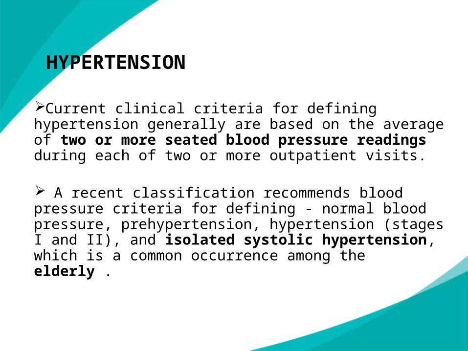
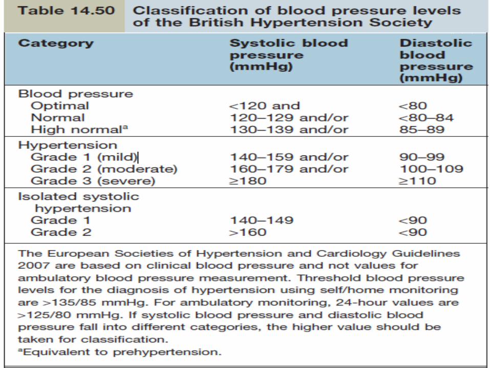
HYPERTENSION
Current clinical criteria for defining hypertension generally are based on the average of two or more seated blood pressure readings during each of two or more outpatient visits.
A recent classification recommends blood pressure criteria for defining - normal blood pressure, prehypertension, hypertension (stages I and II), and isolated systolic hypertension, which is a common occurrence among the elderly .
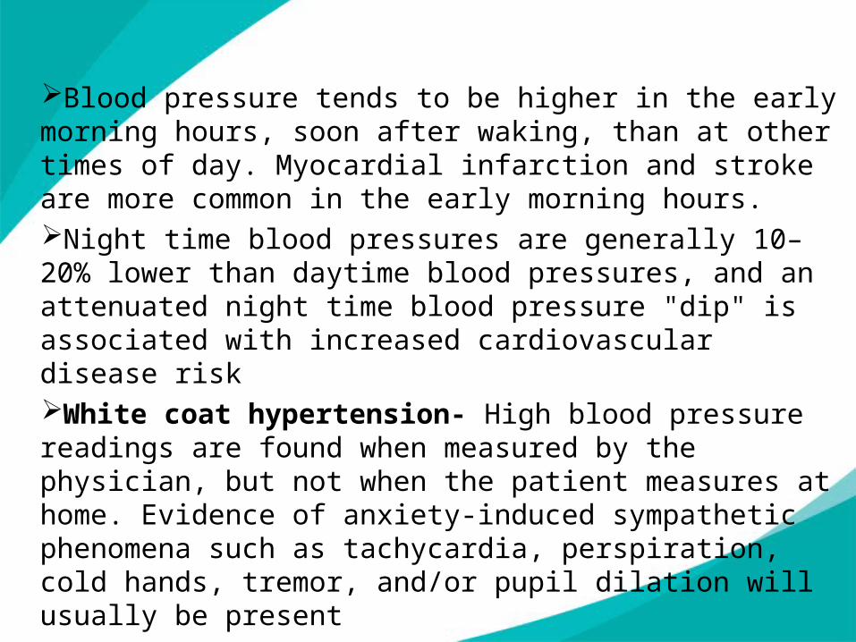
Blood pressure tends to be higher in the early morning hours, soon after waking, than at other times of day. Myocardial infarction and stroke are more common in the early morning hours.Night time blood pressures are generally 10–20% lower than daytime blood pressures, and an attenuated night time blood pressure "dip" is associated with increased cardiovascular disease risk White coat hypertension- High blood pressure readings are found when measured by the physician, but not when the patient measures at home. Evidence of anxiety-induced sympathetic phenomena such as tachycardia, perspiration, cold hands, tremor, and/or pupil dilation will usually be present
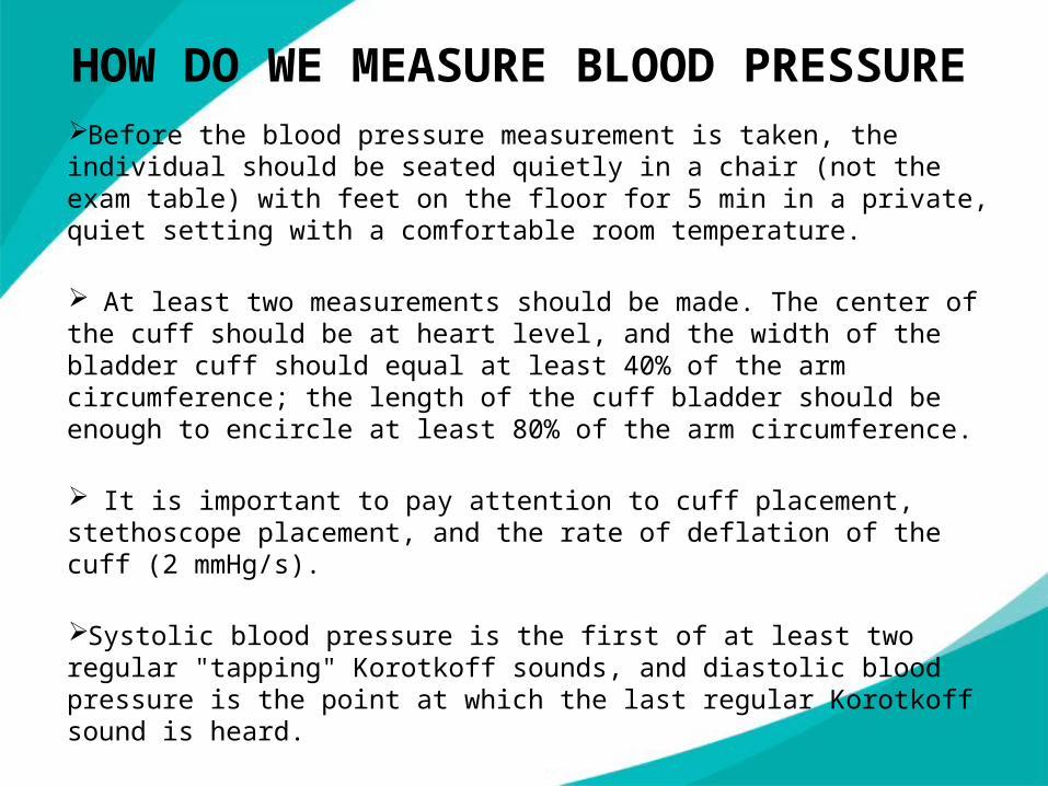
Before the blood pressure measurement is taken, the individual should be seated quietly in a chair (not the exam table) with feet on the floor for 5 min in a private, quiet setting with a comfortable room temperature.
At least two measurements should be made. The center of the cuff should be at heart level, and the width of the bladder cuff should equal at least 40% of the arm circumference; the length of the cuff bladder should be enough to encircle at least 80% of the arm circumference.
It is important to pay attention to cuff placement, stethoscope placement, and the rate of deflation of the cuff (2 mmHg/s).
Systolic blood pressure is the first of at least two regular "tapping" Korotkoff sounds, and diastolic blood pressure is the point at which the last regular Korotkoff sound is heard.
HOW DO WE MEASURE BLOOD PRESSURE
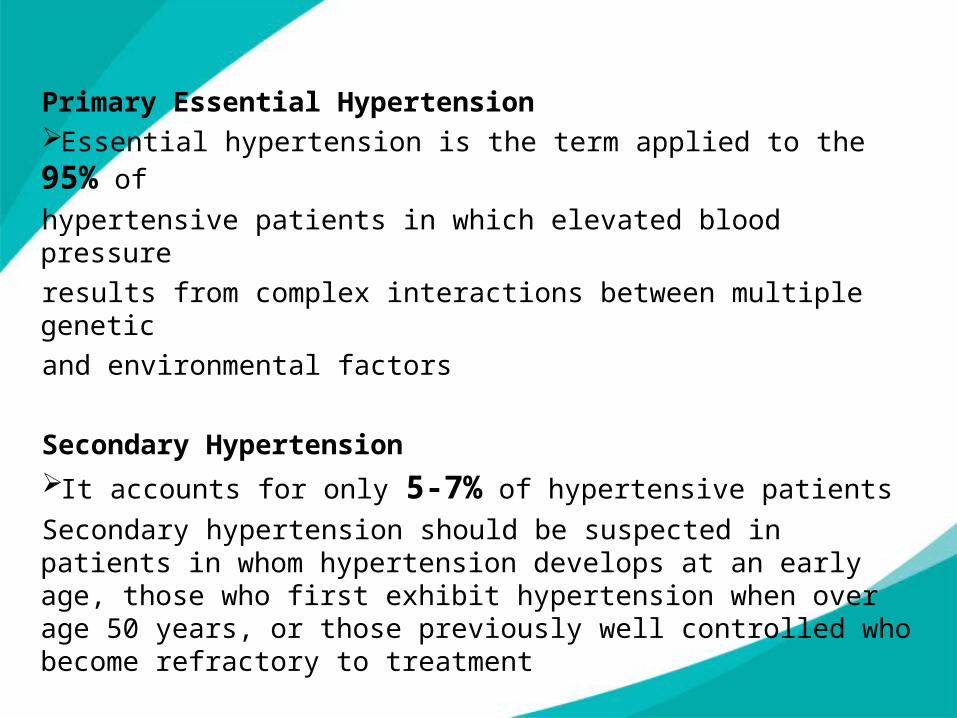
Primary Essential HypertensionEssential hypertension is the term applied to the 95% of
hypertensive patients in which elevated blood pressure
results from complex interactions between multiple genetic
and environmental factors
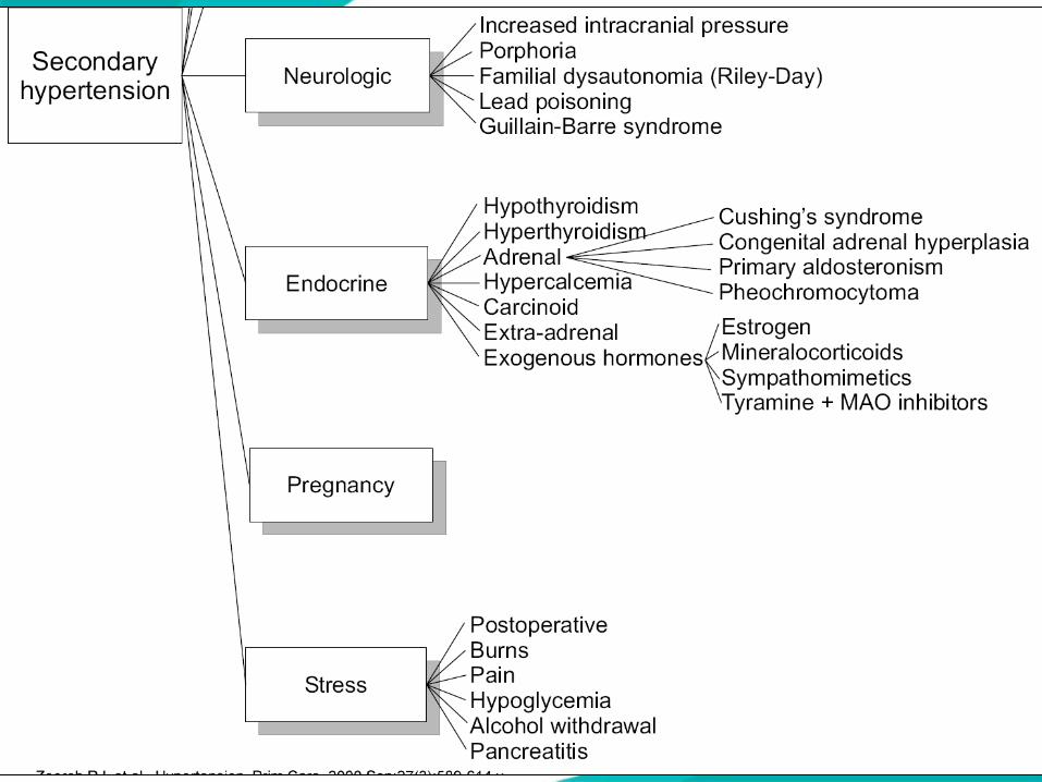
Secondary HypertensionIt accounts for only 5-7% of hypertensive patients
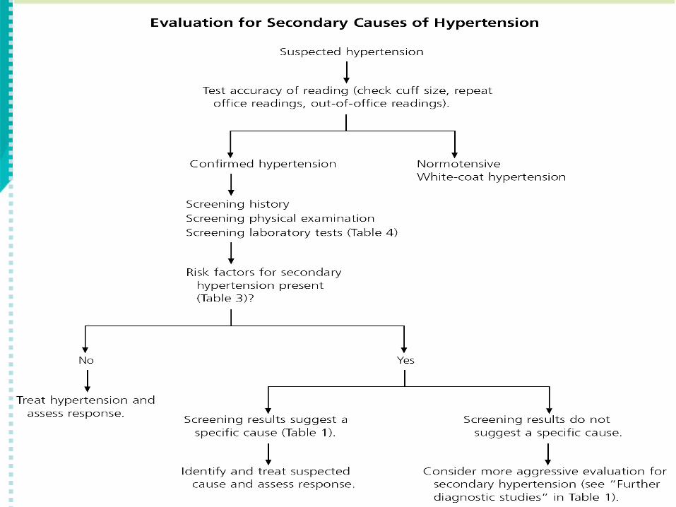
Secondary hypertension should be suspected in patients in whom hypertension develops at an early age, those who first exhibit hypertension when over age 50 years, or those previously well controlled who become refractory to treatment
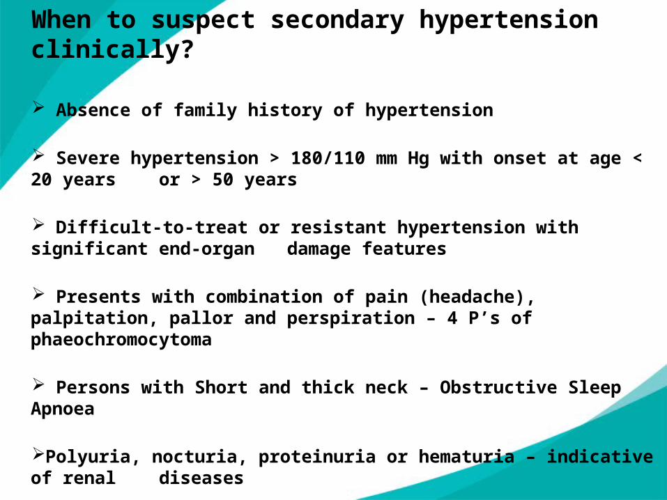
When to suspect secondary hypertension clinically?
Absence of family history of hypertension
Severe hypertension > 180/110 mm Hg with onset at age < 20 years or > 50 years
Difficult-to-treat or resistant hypertension with significant end-organ damage features
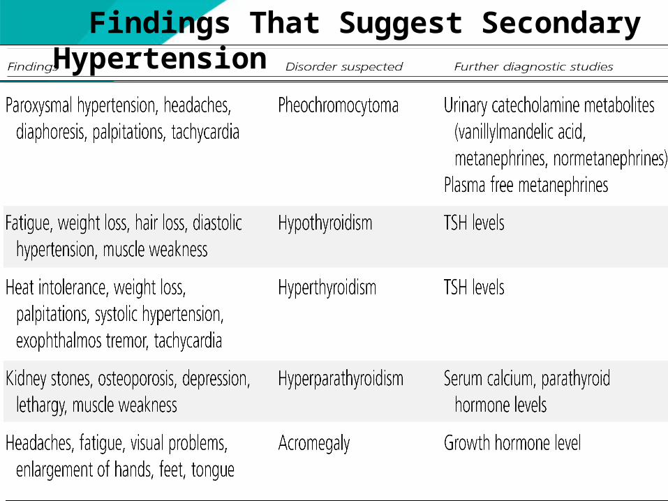
Presents with combination of pain (headache), palpitation, pallor and perspiration – 4 P’s of phaeochromocytoma
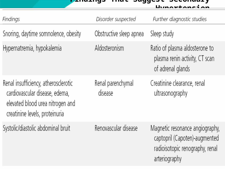
Persons with Short and thick neck – Obstructive Sleep Apnoea
Polyuria, nocturia, proteinuria or hematuria – indicative of renal diseases
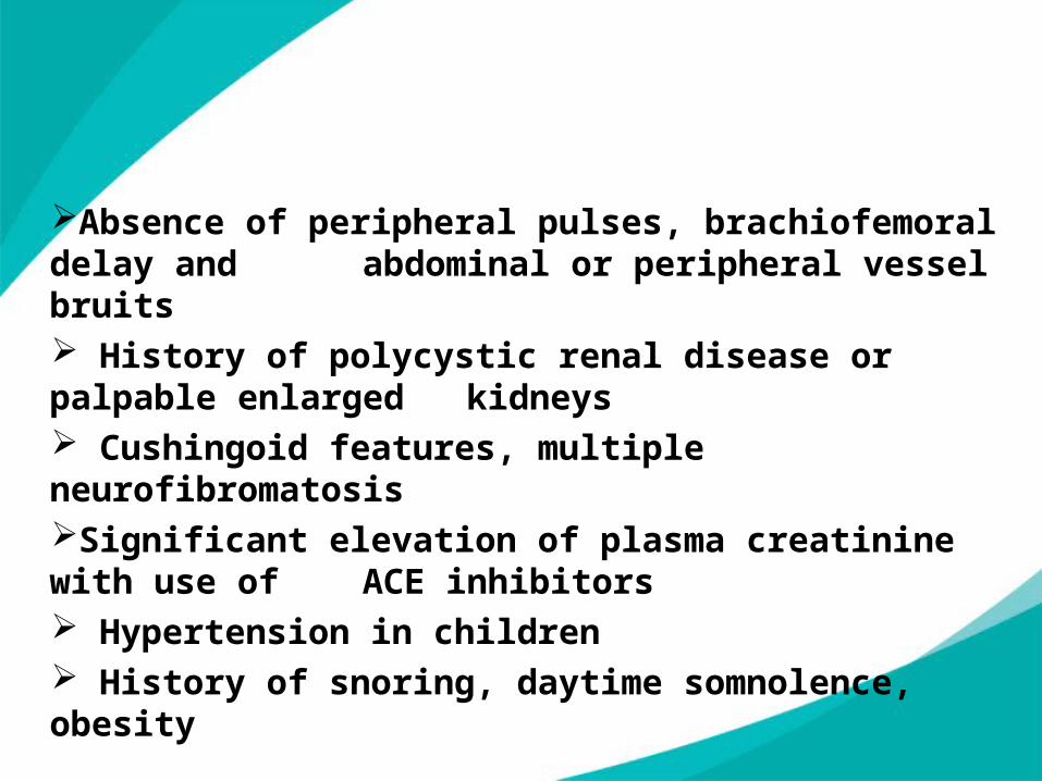
Absence of peripheral pulses, brachiofemoral delay and abdominal or peripheral vessel bruits
History of polycystic renal disease or palpable enlarged kidneys
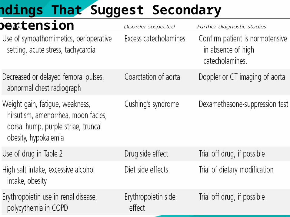
Cushingoid features, multiple neurofibromatosisSignificant elevation of plasma creatinine with use of ACE inhibitors Hypertension in children History of snoring, daytime somnolence, obesity
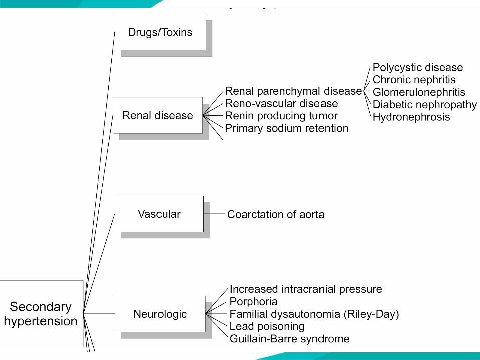
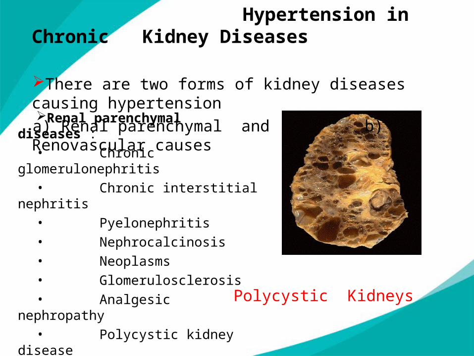
Renal parenchymal diseases :• Chronic glomerulonephritis• Chronic interstitial nephritis• Pyelonephritis • Nephrocalcinosis • Neoplasms • Glomerulosclerosis • Analgesic nephropathy• Polycystic kidney disease• Gout with renal failure• Obstructive nephropathy
Polycystic Kidneys
Hypertension in Chronic Kidney Diseases
There are two forms of kidney diseases causing hypertensiona) Renal parenchymal and b) Renovascular causes
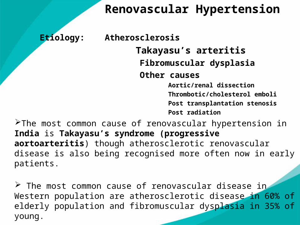
Renovascular Hypertension
Etiology: Atherosclerosis
Takayasu’s arteritis Fibromuscular dysplasia
Other causes Aortic/renal dissection
Thrombotic/cholesterol emboli
Post transplantation stenosis
Post radiation
The most common cause of renovascular hypertension in India is Takayasu’s syndrome (progressive aortoarteritis) though atherosclerotic renovascular disease is also being recognised more often now in early patients.
The most common cause of renovascular disease in Western population are atherosclerotic disease in 60% of elderly population and fibromuscular dysplasia in 35% of young.
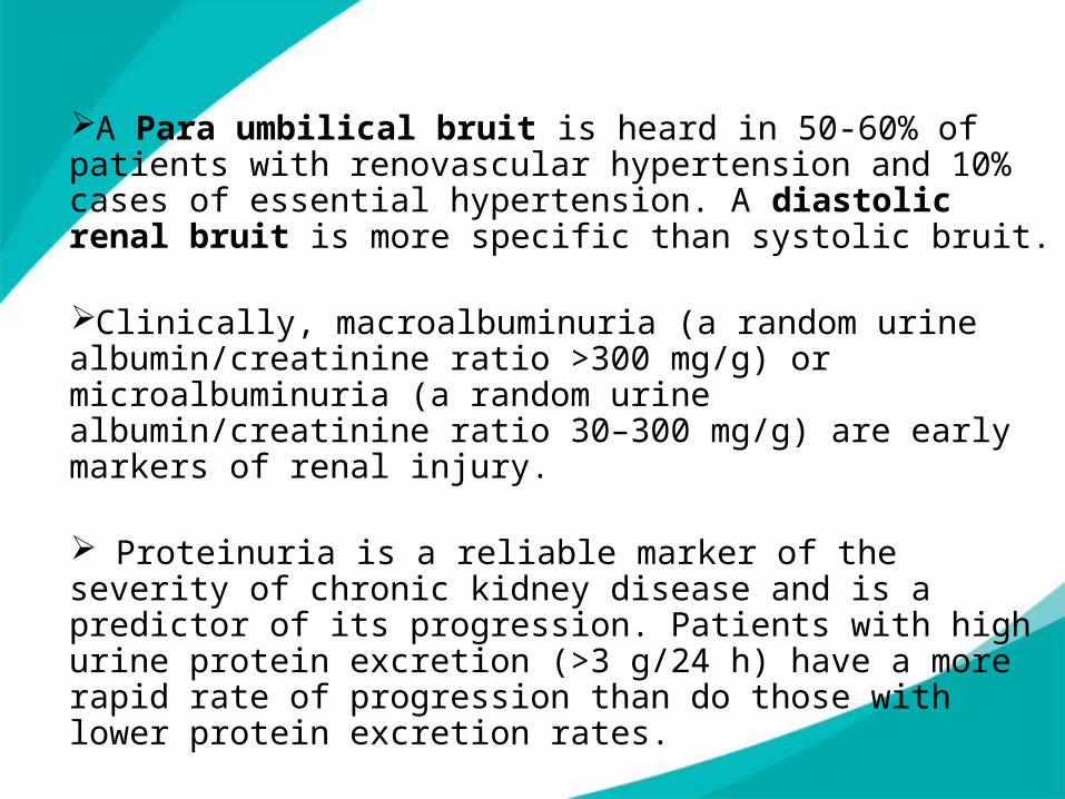
A Para umbilical bruit is heard in 50-60% of patients with renovascular hypertension and 10% cases of essential hypertension. A diastolic renal bruit is more specific than systolic bruit.
Clinically, macroalbuminuria (a random urine albumin/creatinine ratio >300 mg/g) or microalbuminuria (a random urine albumin/creatinine ratio 30–300 mg/g) are early markers of renal injury.
Proteinuria is a reliable marker of the severity of chronic kidney disease and is a predictor of its progression. Patients with high urine protein excretion (>3 g/24 h) have a more rapid rate of progression than do those with lower protein excretion rates.
Investigations :
In patients with moderate degree of suspicion of renovascular hypertension, non-invasive tests are recommended initially which are--1)Colour Doppler Ultrasound (CDUS)-to know the renal artery stenosis2)CT angiography and3)MRI angiography. 4)99Tc – DTPA and 123 I – Hippuran scan. These tests give functional status of CKD. MRI angiography has higher sensitivity (90%) and specificity (92%).In patients with high degree of suspicion, direct selective renal arteriography is recommended.Conventional angiography, though invasive, is the gold standard test.
Other tests:Captopril Screening Test-Test sensitivity is excellent but specificity is poor Method: Patient maintains normal salt intake and receives no diuretics and withdraw all antihypertensives 3 weeks before the test, if possible· Patient should be seated for at least 30 minutes; draw venous blood sample and measure baseline plasma renin activity· Dilute 50 mg of captopril in 10 mL of water; patient immediately drinks the solution and after 60 minutes, draw venous blood samples and measure stimulated plasma renin activityInterpretationTest is positive in the presence of:· Stimulated plasma renin activity of 12 ng/mL/hr or more and· Absolute increase in plasma renin activity of 10 ng.mL.hr or more
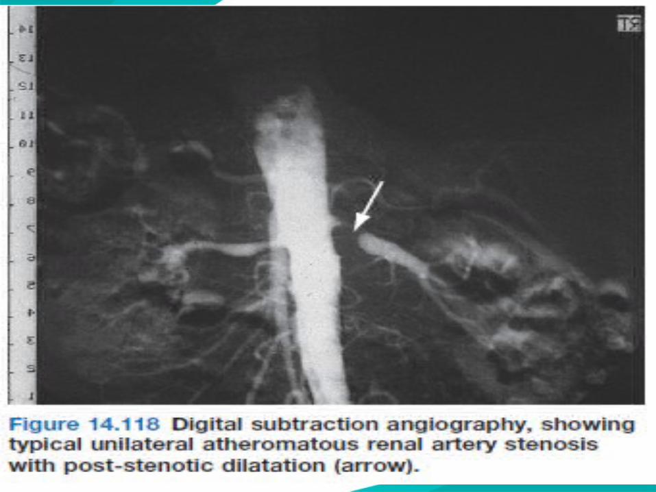
Intra-arterial injection with digital subtraction angiography (DSA) may be used.
Once the diagnosis is confirmed, renal angioplasty with stenting is the treatment of choice.
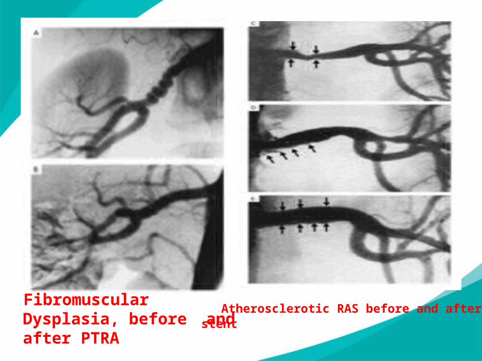
Fibromuscular Dysplasia, before and after PTRA
Atherosclerotic RAS before and after stent
Endocrine causes
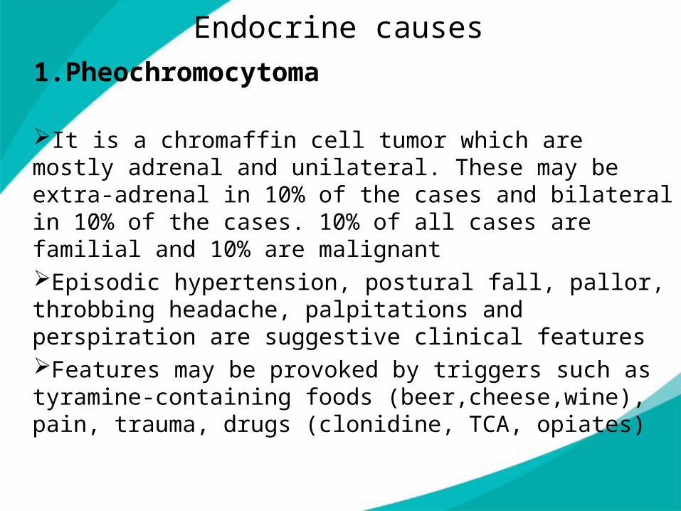
1.Pheochromocytoma
It is a chromaffin cell tumor which are mostly adrenal and unilateral. These may be extra-adrenal in 10% of the cases and bilateral in 10% of the cases. 10% of all cases are familial and 10% are malignantEpisodic hypertension, postural fall, pallor, throbbing headache, palpitations and perspiration are suggestive clinical featuresFeatures may be provoked by triggers such as tyramine-containing foods (beer,cheese,wine), pain, trauma, drugs (clonidine, TCA, opiates)
Investigations
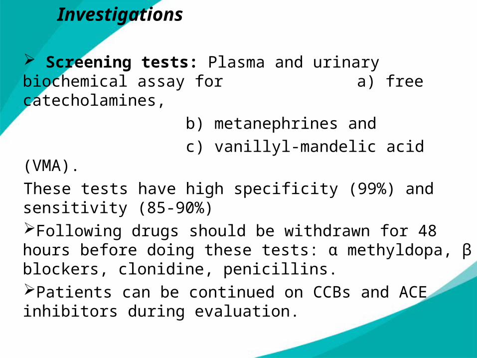
Screening tests: Plasma and urinary biochemical assay for a) free catecholamines,
b) metanephrines and
c) vanillyl-mandelic acid (VMA).
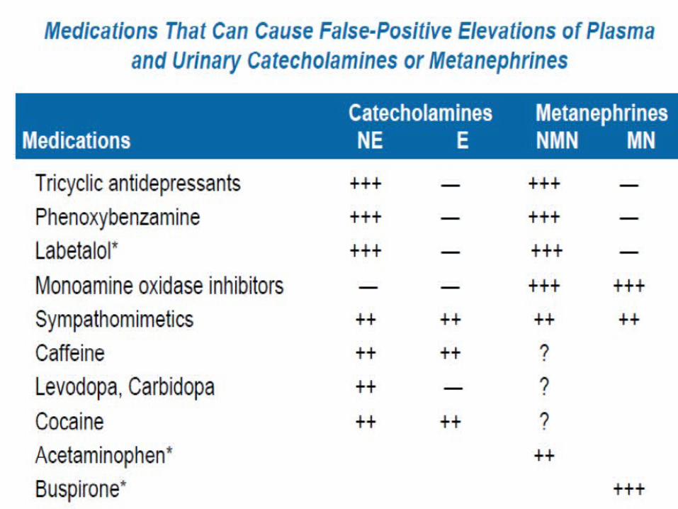
These tests have high specificity (99%) and sensitivity (85-90%)Following drugs should be withdrawn for 48 hours before doing these tests: α methyldopa, β blockers, clonidine, penicillins. Patients can be continued on CCBs and ACE inhibitors during evaluation.
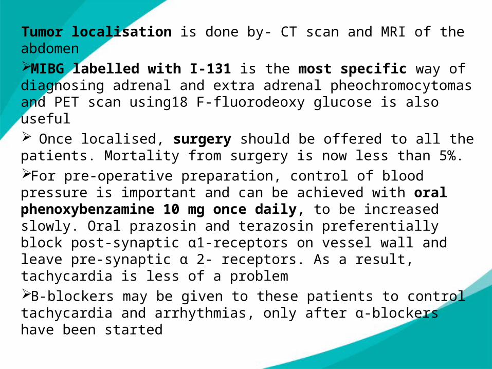
Tumor localisation is done by- CT scan and MRI of the abdomenMIBG labelled with I-131 is the most specific way of diagnosing adrenal and extra adrenal pheochromocytomas and PET scan using18 F-fluorodeoxy glucose is also useful Once localised, surgery should be offered to all the patients. Mortality from surgery is now less than 5%. For pre-operative preparation, control of blood pressure is important and can be achieved with oral phenoxybenzamine 10 mg once daily, to be increased slowly. Oral prazosin and terazosin preferentially block post-synaptic α1-receptors on vessel wall and leave pre-synaptic α 2- receptors. As a result, tachycardia is less of a problemΒ-blockers may be given to these patients to control tachycardia and arrhythmias, only after α-blockers have been started

2. Primary Aldosteronism
Primary aldosteronism is due to excess aldosterone secretion by the adrenal adenomas and occasionally due to bilateral adrenocortical hyperplasiaClinical presentation
•May be asymptomatic; headache, muscle cramps, polyuria•Hypokalemia (K normal in 40%), metabolic alkalosis•Retinopathy
This is suspected in a case of hypertension showing persistent hyokalaemic metabolic alkalosis in the absence of diuretic therapy
InvestigationsPlasma Aldosterone to Plasma Renin Activity (PRA) ratio more than 20 to 25 (normal < 10) is 95% sensitive and 75% specific for Primary Hyper AldosteronismConfirmatory test: by demonstrating failure to suppress plasma aldosterone to <277 pmol/L (<10 ng/dL) after IV infusion of 2 L of isotonic saline over 4 h;. Alternative confirmatory tests include failure to suppress aldosterone in response to an oral NaCl load, fludrocortisone, or captopril. It is usually diagnosed by imaging techniques- CT Scan, adrenal scintigraphy with 6 beta-[I131]iodomethyl-19-nor cholesterol after dexamethasone suppression (0.5 mg every 6 h for 7 days)
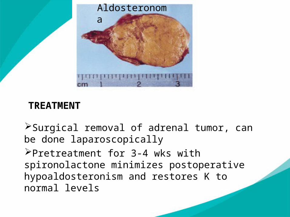
TREATMENT
Surgical removal of adrenal tumor, can be done laparoscopicallyPretreatment for 3-4 wks with spironolactone minimizes postoperative hypoaldosteronism and restores K to normal levels
Aldosteronoma
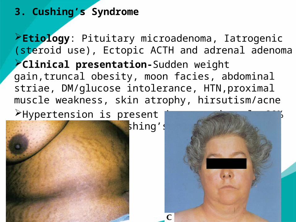
3. Cushing’s Syndrome
Etiology: Pituitary microadenoma, Iatrogenic (steroid use), Ectopic ACTH and adrenal adenomaClinical presentation-Sudden weight gain,truncal obesity, moon facies, abdominal striae, DM/glucose intolerance, HTN,proximal muscle weakness, skin atrophy, hirsutism/acneHypertension is present in approximately 80% of patients with Cushing’s syndrome.
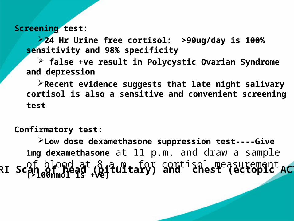
Screening test:24 Hr Urine free cortisol: >90ug/day is 100% sensitivity and
98% specificity false +ve result in Polycystic Ovarian Syndrome and
depressionRecent evidence suggests that late night salivary cortisol is
also a sensitive and convenient screening test
Confirmatory test:Low dose dexamethasone suppression test----Give 1mg
dexamethasone at 11 p.m. and draw a sample of blood at 8 a.m. for cortisol measurement (>100nmol is +ve)
CT/MRI Scan of head (pituitary) and chest (ectopic ACTH tumor)
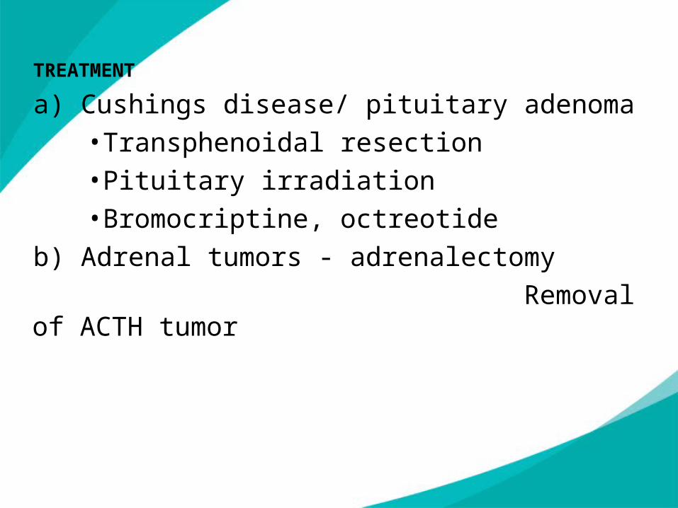
TREATMENT
a) Cushings disease/ pituitary adenoma•Transphenoidal resection•Pituitary irradiation•Bromocriptine, octreotide
b) Adrenal tumors - adrenalectomy
Removal of ACTH tumor
4. Coarctation of the aorta
Coarctation of the aorta consists of localized narrowing of the aortic arch just distal to the origin of the left subclavian artery. Collateral circulation develops around the coarctation through the intercostal arteries and the branches of the subclavian arteries and can result in a lower transcoarctation gradient by enabling blood flow to bypass the obstruction. Congenital defect, male>femaleClinical presentation:
•Differential systolic BP in arms and legs (=DBP)•May have differential BP in arms if defect is proximal to Left
subclavian artery•Diminished/absent femoral artery pulse•Often asymptomatic•Associated with Turners syndrome, bicuspid Aortiv valve
If uncorrected, 67% will develop LV failure by age 40 and 75% will die by age 50
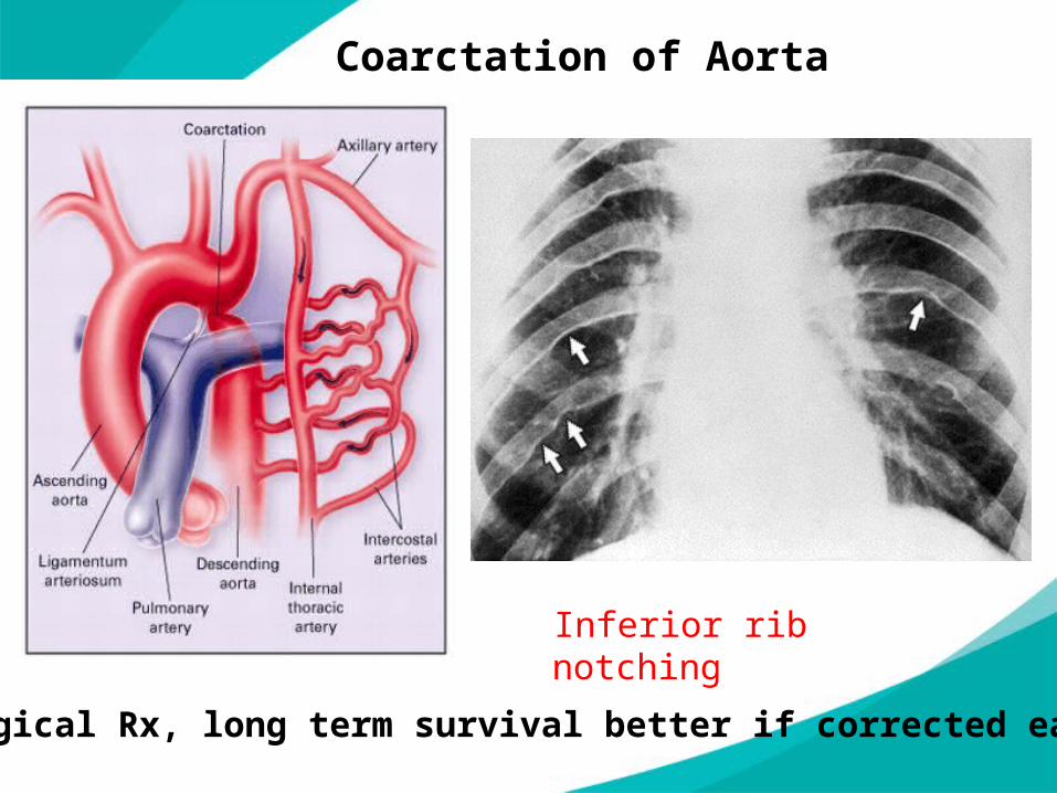
Inferior rib notching
Coarctation of Aorta
Surgical Rx, long term survival better if corrected early
5. Hypertension associated with pregnancy
During pregnancy, a blood pressure of 140/90 mmHg is considered to be abnormally elevated and is associated with an increase in perinatal morbidity and mortality.
In all pregnant women, the measurement of blood pressure should be performed in the sitting position, because the lateral recumbent position may result in a blood pressure lower than that recorded in the sitting position.
The diagnosis of hypertension requires the measurement of two elevated blood pressures, at least 6 h apart.
Hypertension during pregnancy is usually caused by pre eclampsia, chronic hypertension, gestational hypertension, or renal disease.
Pre eclampsia: new onset of hypertension (blood pressure >140/90 mmHg) and proteinuria (>300 mg/24 h) after 20 weeks of gestation
Severe preeclampsia :Marked elevation of blood pressure (>160/110 mmHg), severe proteinuria (>5 g/24 h), or evidence of central nervous system (CNS) dysfunction (headaches, blurred vision, seizures, coma), renal dysfunction (oliguria or creatinine > 1.5 mg/dL), pulmonary edema, hepatocellular injury (ALT > 2-fold the upper limits of normal), hematologic dysfunction (platelet count < 100,000/L or disseminated intravascular coagulation), or placental dysfunction (oligohydramnios or severe intrauterine growth restriction).
HELLP syndrome: Hemolysis, elevated liver enzymes, low platelets

6. Estrogen use / oral contraceptives
A small increase in blood pressure occurs in most women taking oral contraceptives.
This is caused by volume expansion due to increased hepatic synthesis of angiotensinogen and consequent activation of the renin–angiotensin–aldosterone system.
Postmenopausal estrogen does not generally cause hypertension, but rather maintains endothelium-mediated vasodilation.
Other important secondary causes :
Thyroid disorders: hypothyroidism and hyperthyroidismSleep apnea syndrome is one of the common causes of reversible hypertension. Polysomnography is diagnostic. No specific drugs have proven superior in sleep apnea but use of C-PAP improves the hypertensionAcute stressful situations cause intense sympathetic discharge and may temporarily induce hypertension. Common conditions include acute mental stress, hypoglycaemia, acute intermittent porphyria, exposure to cold, burns, perioperative period and post head injury.
Immunosuppressive agents-Cyclosporine, tacrolimus, corticosteroids
NSAID’s--Ibuprofen, naproxen, piroxicamCOX-2 inhibitors--Celecoxib, rofecoxib, valdecoxibEstrogensWeight-loss agents--Sibutramine, phentermine,ephedrineMinerocorticosteroids—FludrocortisoneAntiparkinsonian—BromocriptineMonoamine oxidase inhibitors—PhenelazineAnabolic steroids—TestosteroneSympathomimetics—PseudoephedrineStimulants--Nicotine, amphetamines
Drugs that Raises Blood Pressure

complications
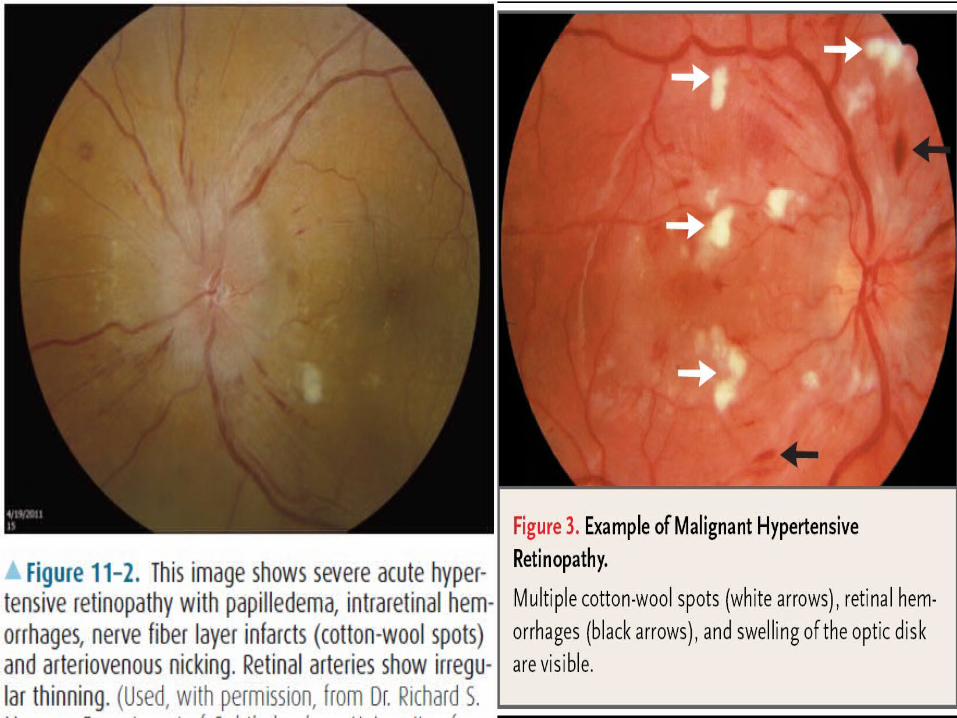
Fundoscopy is an essential part of the examination of any hypertensive patient . The abnormalities are graded according to the Keith-Wagener classification:
Grade 1 – Tortuosity of the retinal arteries with increased reflectiveness (silver wiring)
Grade 2 – grade 1 plus the appearance of arteriovenous nipping produced when thickened retinal arteries pass over the retinal veins Grade 3 – grade 2 plus flame-shaped haemorrhages and soft
(‘cotton wool’) exudates actually due to small infarcts Grade 4 – grade 3 plus papilloedema (blurring of the margins of the
optic disc)Grades 3 and 4 are diagnostic of malignant hypertension.
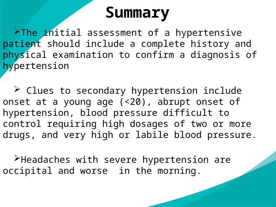
Summary The initial assessment of a hypertensive patient should
include a complete history and physical examination to confirm a diagnosis of hypertension
Clues to secondary hypertension include onset at a young age (<20), abrupt onset of hypertension, blood pressure difficult to control requiring high dosages of two or more drugs, and very high or labile blood pressure.
Headaches with severe hypertension are occipital and worse in the morning.

STRESS
PREGNANCY
RENALPARENCHYMAL
2-3%
DRUGS
COARCTATION
OF
AORTA
THYROID
DISORDERS
0.5%
SLEEP
APNOEA
ENDOCRINE
CAUSES
0.3-1%
RENOVASCULAR
1-2%
SECONDARY
HYPERTENSION
5-7%
Findings That Suggest Secondary Hypertension
Findings That Suggest Secondary Hypertension
Findings That Suggest Secondary Hypertension
Thank you..

![VA/DoD Hypertension Clinical Practice Guideline · ] Secondary hypertension is high blood pressure that results from an underlying and identifiable cause. [3] Main causes o f secondary](https://img.pdfslide.net/doc/110x75/5dd0e688d6be591ccb633edb/vadod-hypertension-clinical-practice-guideline-secondary-hypertension-is-high.jpg)