Embed Size (px)
Citation preview
Prepared by : Hossam Mohammed MahmoudSupervised by : Dr . Naser Zaghloul
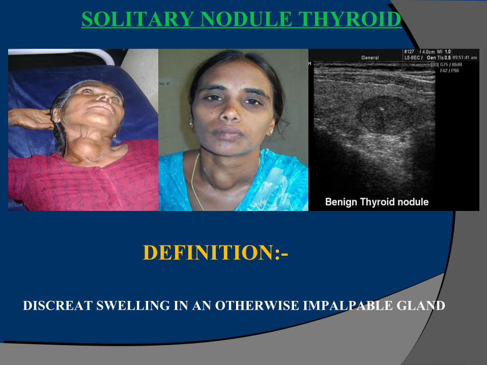
SOLITARY NODULE THYROID
DEFINITION:-
DISCREAT SWELLING IN AN OTHERWISE IMPALPABLE GLAND
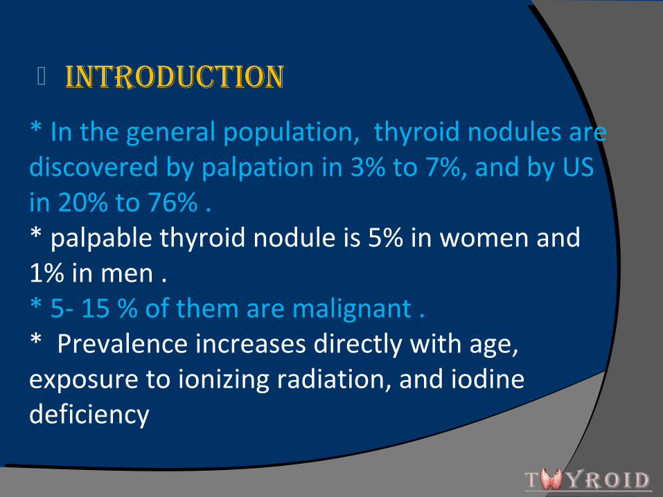
IntroductIon
* In the general population, thyroid nodules are discovered by palpation in 3% to 7%, and by US in 20% to 76% . * palpable thyroid nodule is 5% in women and 1% in men . * 5- 15 % of them are malignant . * Prevalence increases directly with age, exposure to ionizing radiation, and iodine deficiency
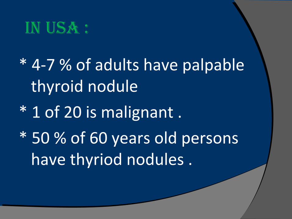
* 4-7 % of adults have palpable thyroid nodule
* 1 of 20 is malignant .
* 50 % of 60 years old persons have thyriod nodules .
In uSA :
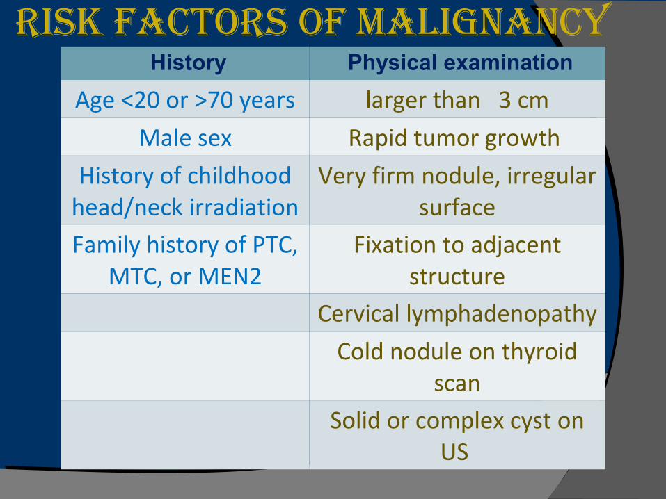
rISk fActorS of mAlIgnAncy History Physical examination
Age <20 or >70 years larger than 3 cm
Male sex Rapid tumor growth
History of childhood head/neck irradiation
Very firm nodule, irregular surface
Family history of PTC, MTC, or MEN2
Fixation to adjacent structure
Cervical lymphadenopathy
Cold nodule on thyroid scan
Solid or complex cyst on US
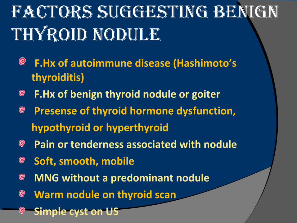
fActorS SuggeStIng benIgn thyroId nodule
F.Hx of autoimmune disease (Hashimoto’s thyroiditis)
F.Hx of benign thyroid nodule or goiter
Presense of thyroid hormone dysfunction,
hypothyroid or hyperthyroid
Pain or tenderness associated with nodule
Soft, smooth, mobile
MNG without a predominant nodule
Warm nodule on thyroid scan
Simple cyst on US
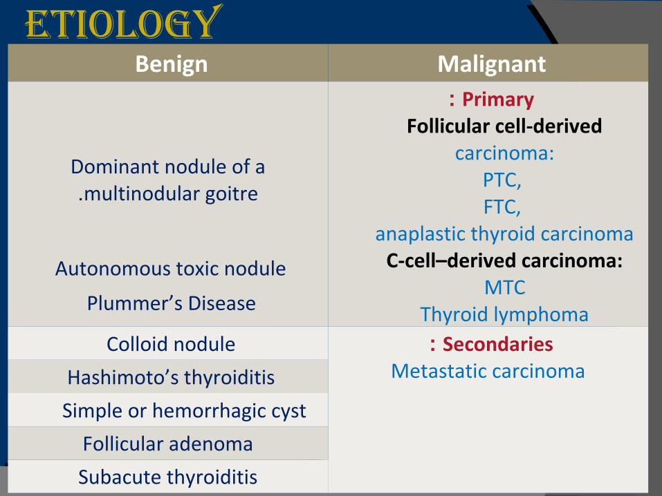
Etiology Benign Malignant
Dominant nodule of a multinodular goitre.
Primary : Follicular cell-derived
carcinoma:PTC, FTC,
anaplastic thyroid carcinomaC-cell–derived carcinoma:
MTCThyroid lymphoma
Colloid nodule Secondaries : Metastatic carcinomaHashimoto’s thyroiditis
Simple or hemorrhagic cyst
Follicular adenoma
Subacute thyroiditis
Autonomous toxic nodule
Plummer’s Disease
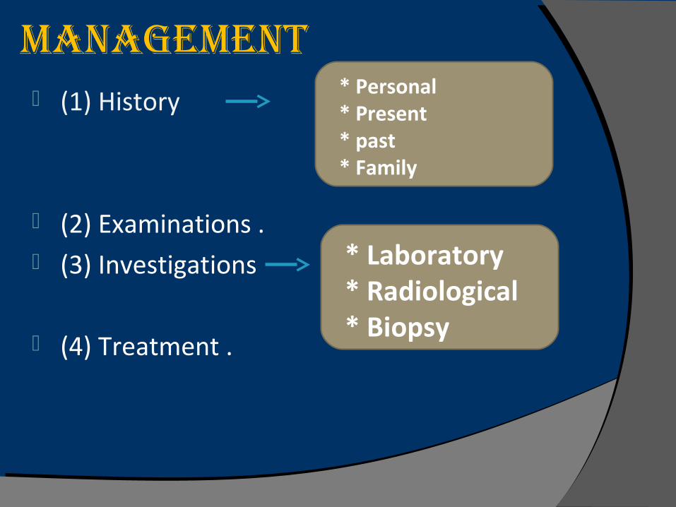
ManagEMEnt (1) History
(2) Examinations . (3) Investigations
(4) Treatment .
* Personal * Present * past * Family
* Laboratory * Radiological * Biopsy
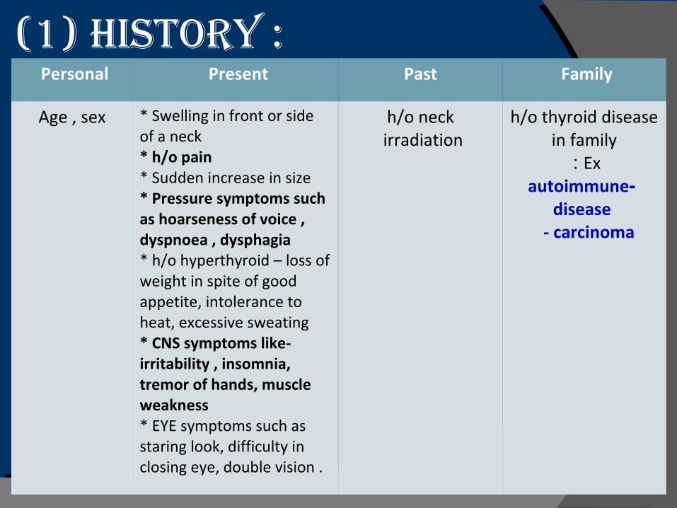
(1) History :Personal Present Past Family
Age , sex * Swelling in front or side of a neck* h/o pain* Sudden increase in size* Pressure symptoms such as hoarseness of voice , dyspnoea , dysphagia* h/o hyperthyroid – loss of weight in spite of good appetite, intolerance to heat, excessive sweating* CNS symptoms like- irritability , insomnia, tremor of hands, muscle weakness* EYE symptoms such as staring look, difficulty in closing eye, double vision .
h/o neck irradiation
h/o thyroid disease in family
Ex : - autoimmune
disease - carcinoma
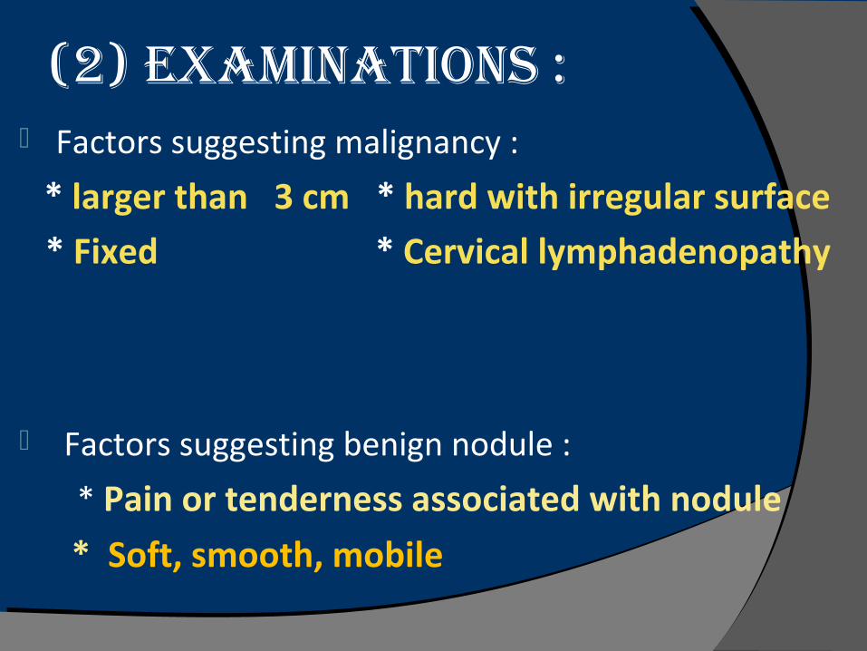
(2) ExaMinations : Factors suggesting malignancy :
* larger than 3 cm * hard with irregular surface * Fixed * Cervical lymphadenopathy
Factors suggesting benign nodule :
* Pain or tenderness associated with nodule
* Soft, smooth, mobile
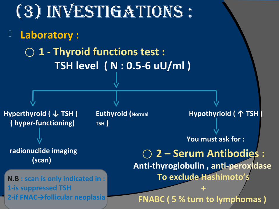
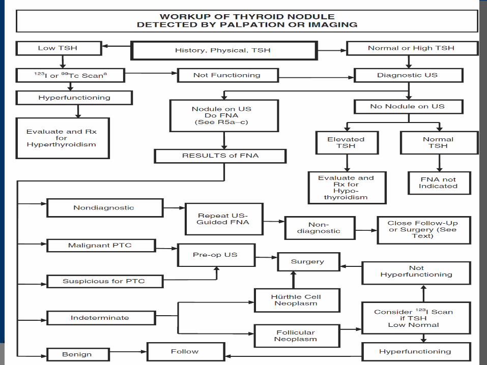
(3) invEstigations : Laboratory :
⃝ 1 - Thyroid functions test : TSH level ( N : 0.5-6 uU/ml )
Hyperthyroid ( ↓ TSH )( hyper-functioning)
radionuclide imaging(scan)
Euthyroid (Normal
TSH )
Hypothyrioid ( ↑ TSH )
You must ask for :
⃝ 2 – Serum Antibodies :Anti-thyroglobulin , anti-peroxidase
To exclude Hashimoto’s+
FNABC ( 5 % turn to lymphomas )
N.B : scan is only indicated in : 1-is suppressed TSH2-if FNAC→follicular neoplasia
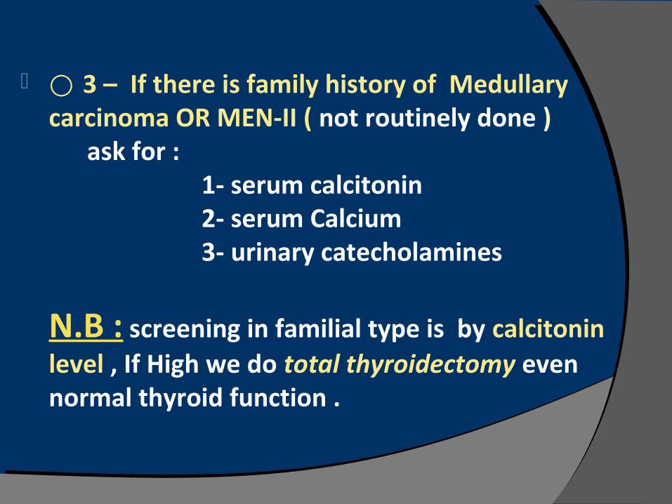
⃝ 3 – If there is family history of Medullary carcinoma OR MEN-II ( not routinely done ) ask for : 1- serum calcitonin 2- serum Calcium 3- urinary catecholamines
N.B : screening in familial type is by calcitonin level , If High we do total thyroidectomy even normal thyroid function .
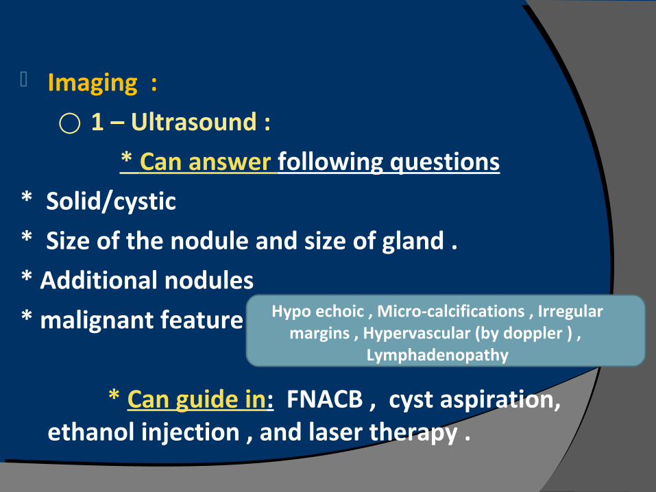
Imaging :
⃝ 1 – Ultrasound :
* Can answer following questions
* Solid/cystic
* Size of the nodule and size of gland .
* Additional nodules
* malignant feature
* Can guide in: FNACB , cyst aspiration, ethanol injection , and laser therapy .
Hypo echoic , Micro-calcifications , Irregular margins , Hypervascular (by doppler ) ,
Lymphadenopathy
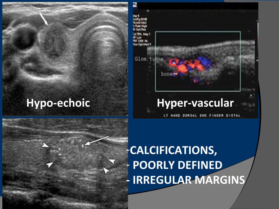
Hypo-echoic Hyper-vascular
-CALCIFICATIONS,- POORLY DEFINED- IRREGULAR MARGINS
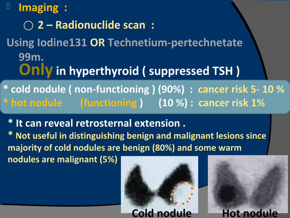
Imaging :
⃝ 2 – Radionuclide scan :
Using Iodine131 OR Technetium-pertechnetate 99m.
* cold nodule ( non-functioning ) (90%) : cancer risk 5- 10 %* hot nodule (functioning ) (10 %) : cancer risk 1%
Only in hyperthyroid ( suppressed TSH )
* Not useful in distinguishing benign and malignant lesions since majority of cold nodules are benign (80%) and some warm nodules are malignant (5%)
* It can reveal retrosternal extension .
Cold nodule Hot nodule
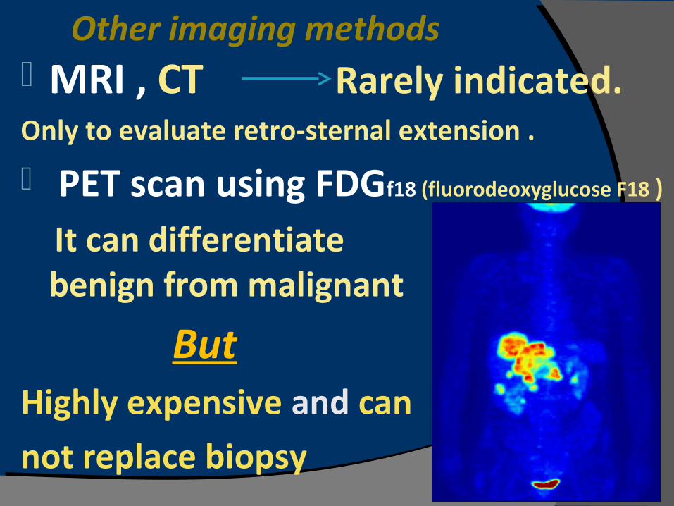
Other imaging methods MRI , CT Rarely indicated.Only to evaluate retro-sternal extension .
PET scan using FDGf18 (fluorodeoxyglucose F18 )
It can differentiate benign from malignant
But
Highly expensive and can
not replace biopsy
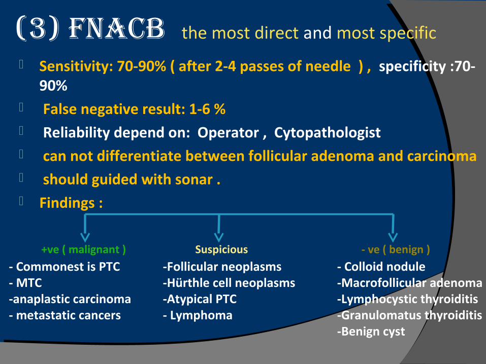
(3) FNACB the most direct and most specific Sensitivity: 70-90% ( after 2-4 passes of needle ) , specificity :70-
90% False negative result: 1-6 % Reliability depend on: Operator , Cytopathologist can not differentiate between follicular adenoma and carcinoma should guided with sonar . Findings :
+ve ( malignant ) - ve ( benign )
- Commonest is PTC- MTC-anaplastic carcinoma- metastatic cancers
- Colloid nodule-Macrofollicular adenoma-Lymphocystic thyroiditis-Granulomatus thyroiditis -Benign cyst
Suspicious
-Follicular neoplasms -Hürthle cell neoplasms -Atypical PTC - Lymphoma

ImmuNohIstoChemICAl mArkers
HBME-1 (Hector Battifora mesothelial cell -1 )
monoclonal antibody stains papillary cancer positively but
does not stain benign follicular tumors Galectin-3
acts as a cell-death suppressordistinguish benign from malignant
thyroid follicular tumors
TreaTmenT opTions : 1- Levothyroxine : ( in benign nodule ) to keep TSH below
0.1 mU/L
Have many Side effects , so not recommended
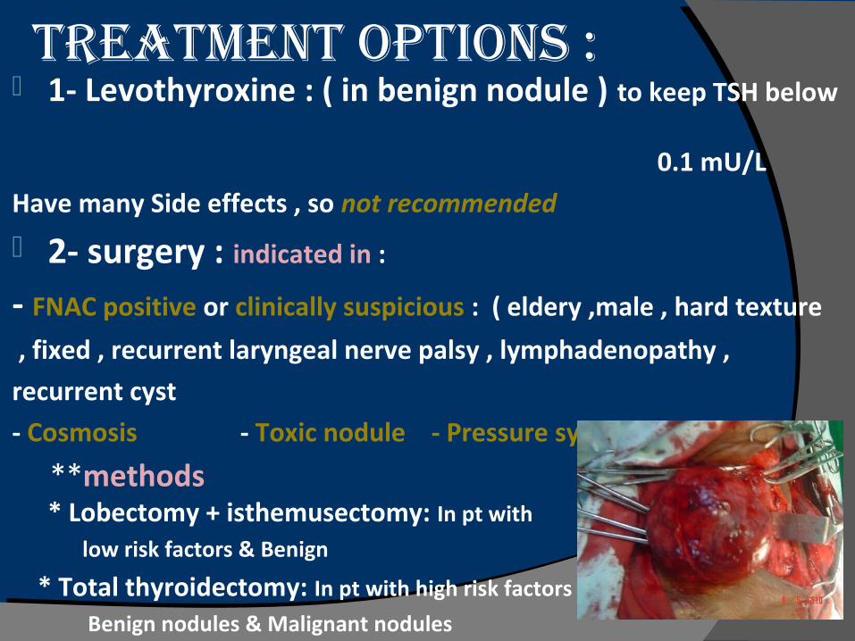
2- surgery : indicated in :
- FNAC positive or clinically suspicious : ( eldery ,male , hard texture
, fixed , recurrent laryngeal nerve palsy , lymphadenopathy ,
recurrent cyst
- Cosmosis - Toxic nodule - Pressure symptoms
**methods * Lobectomy + isthemusectomy: In pt with
low risk factors & Benign
* Total thyroidectomy: In pt with high risk factors
Benign nodules & Malignant nodules
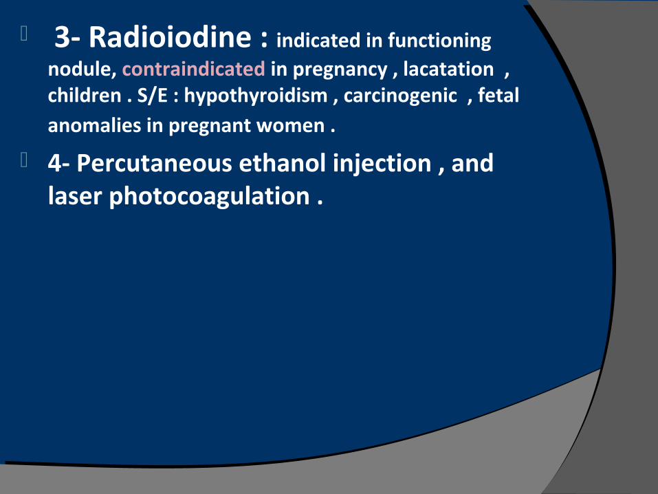
3- Radioiodine : indicated in functioning nodule, contraindicated in pregnancy , lacatation , children . S/E : hypothyroidism , carcinogenic , fetal anomalies in pregnant women .
4- Percutaneous ethanol injection , and laser photocoagulation .
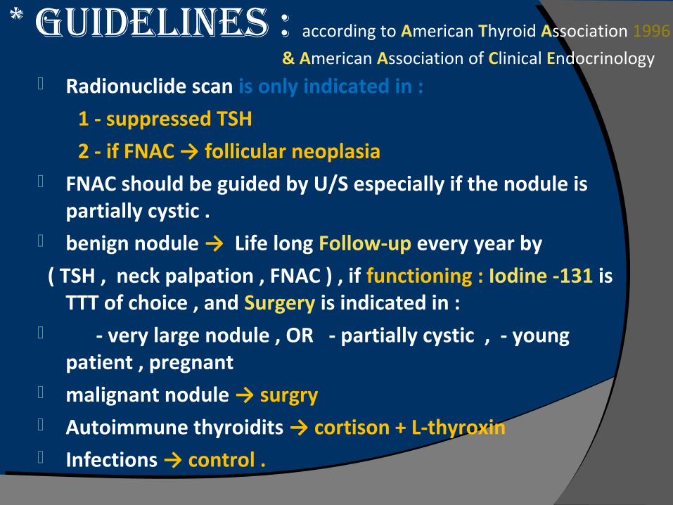
* Guidelines : according to American Thyroid Association 1996
& American Association of Clinical Endocrinology Radionuclide scan is only indicated in :
1 - suppressed TSH
2 - if FNAC → follicular neoplasia FNAC should be guided by U/S especially if the nodule is
partially cystic . benign nodule → Life long Follow-up every year by
( TSH , neck palpation , FNAC ) , if functioning : Iodine -131 is TTT of choice , and Surgery is indicated in :
- very large nodule , OR - partially cystic , - young patient , pregnant
malignant nodule → surgry Autoimmune thyroidits → cortison + L-thyroxin Infections → control .
Revised American Thyroid
Association Guidelines
2009