Embed Size (px)
Citation preview
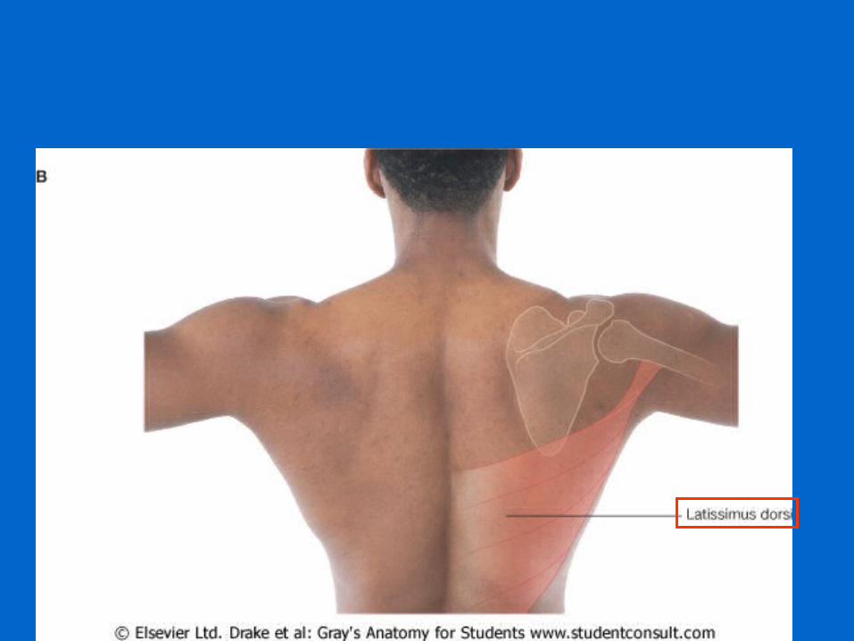
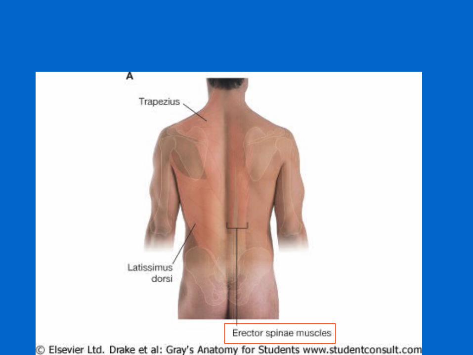
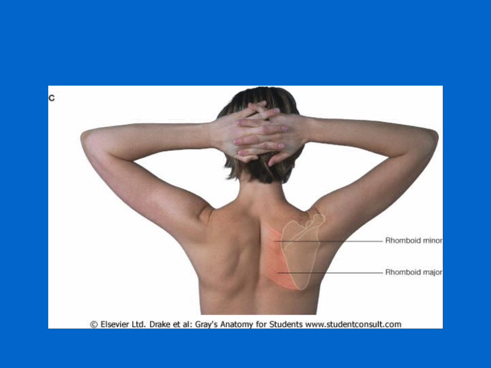
Surface anatomy of the back
Prepared by: Zariifard n
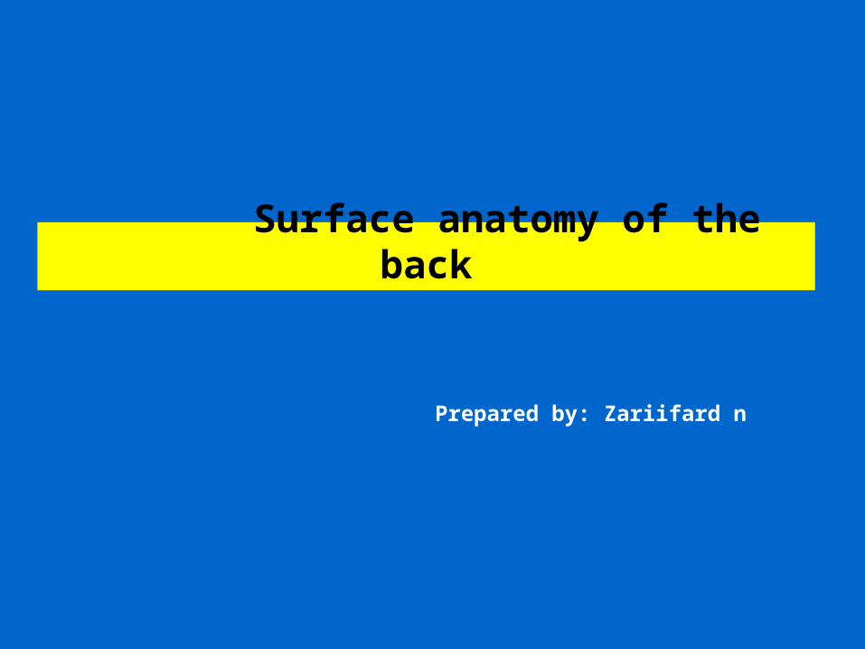
Length60-70
neck: 12cm thoracic:28cmlumbar:12cmpelvis:18cm
2cm
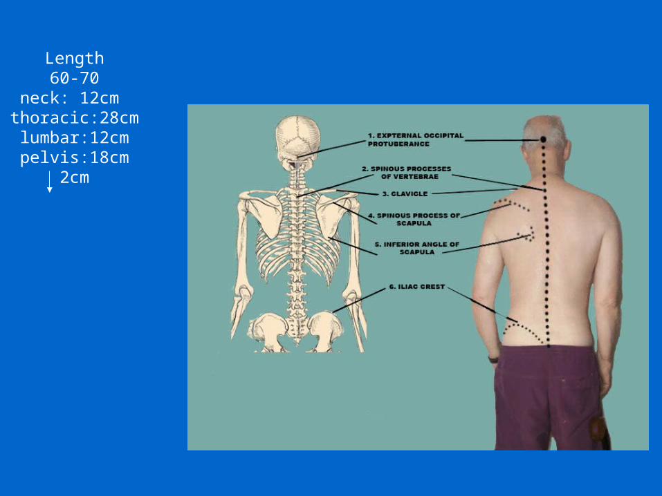
2/5 length of body
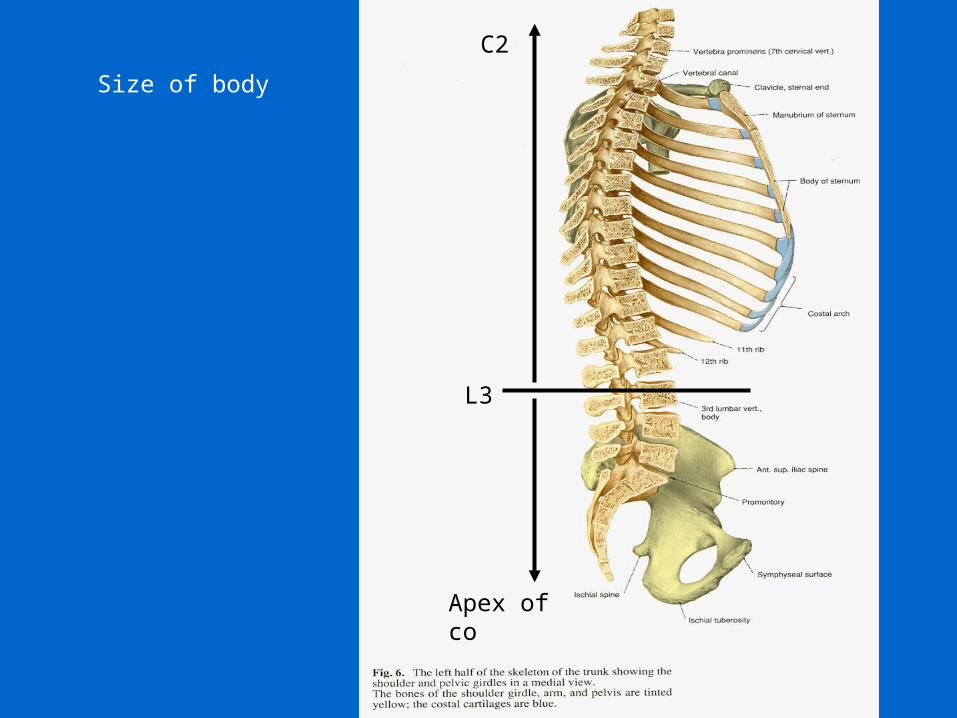
Size of body
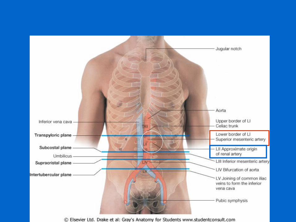
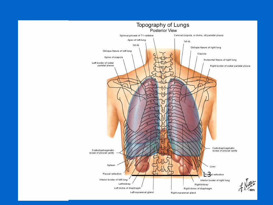
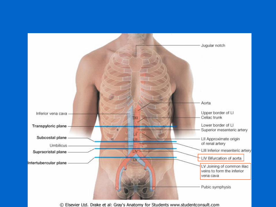
L3
C2
Apex of co
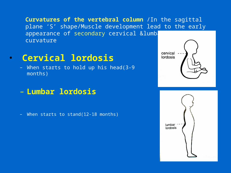
• Cervical lordosis– When starts to hold up his head(3-9 months)
– Lumbar lordosis
– When starts to stand(12-18 months)
Curvatures of the vertebral column /In the sagittal plane ‘S’ shape/Muscle development lead to the early appearance of secondary cervical &lumbar spinal curvature
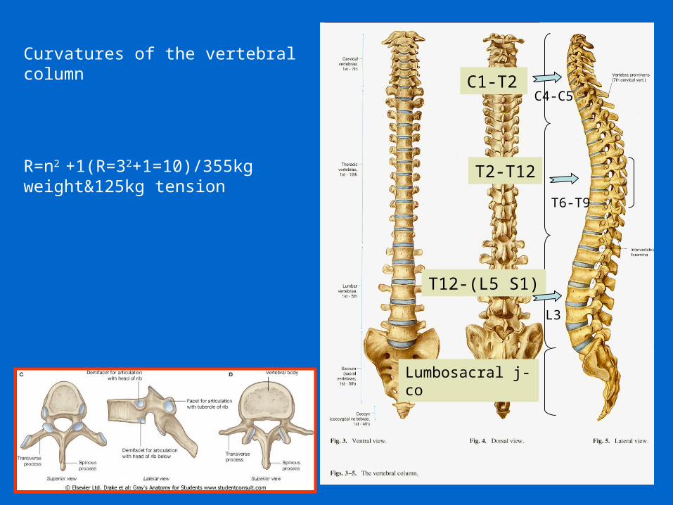
Curvatures of the vertebral column
R=n2 +1(R=32+1=10)/355kg weight&125kg tension
C1-T2
T2-T12
T12-(L5 S1)
C4-C5
T6-T9
L3
Lumbosacral j-co
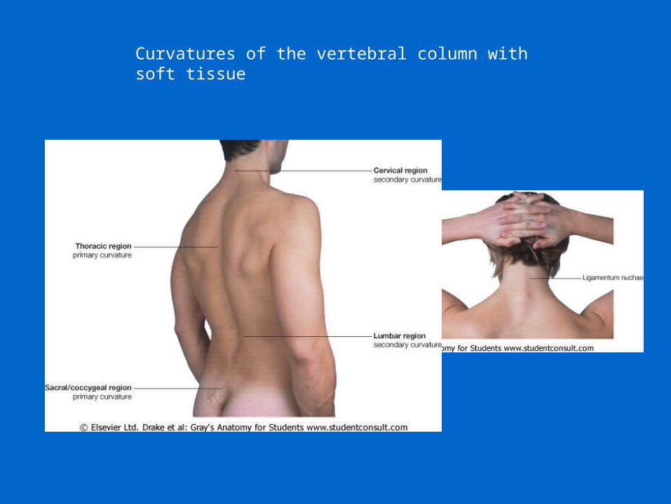
Curvatures of the vertebral column with soft tissue
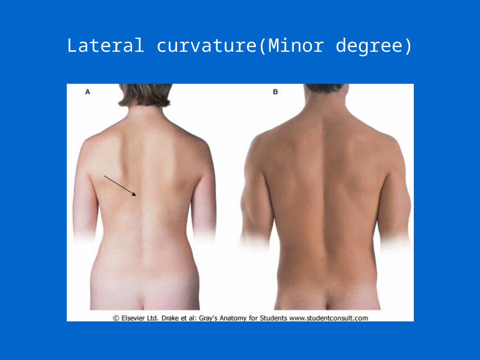
Lateral curvature(Minor degree)
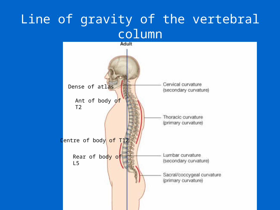
Line of gravity of the vertebral column
Dense of atlas
Ant of body of T2
Centre of body of T12
Rear of body of L5
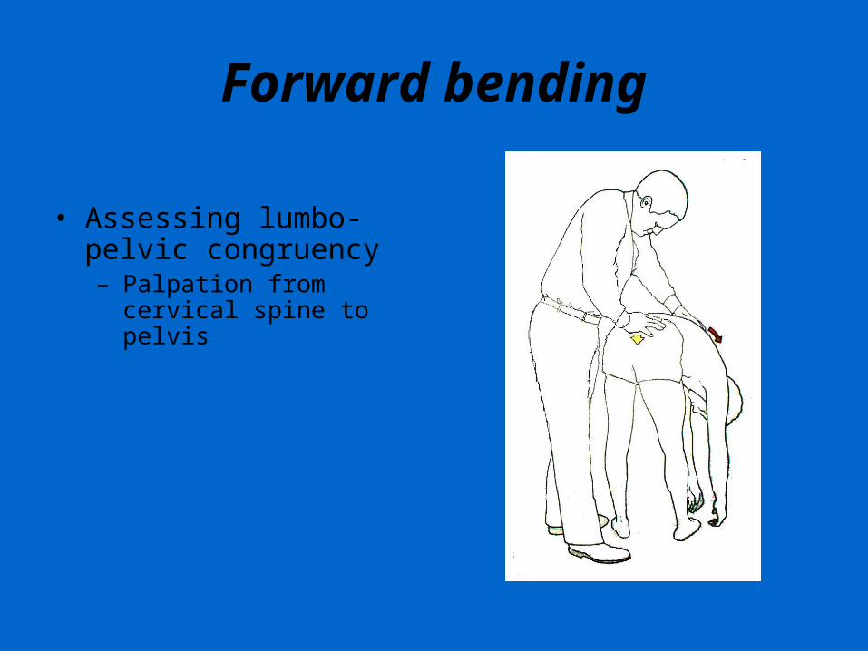
Forward bending
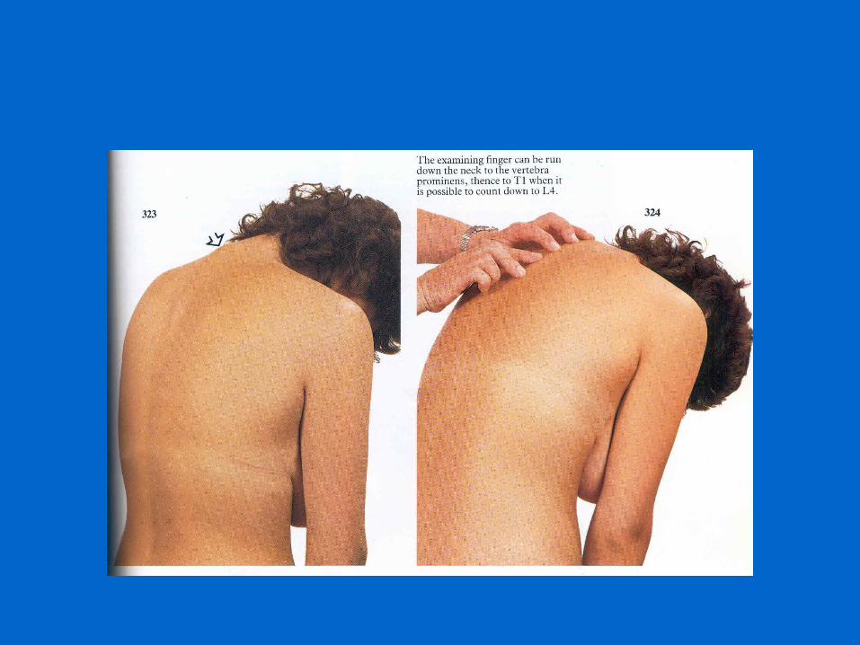
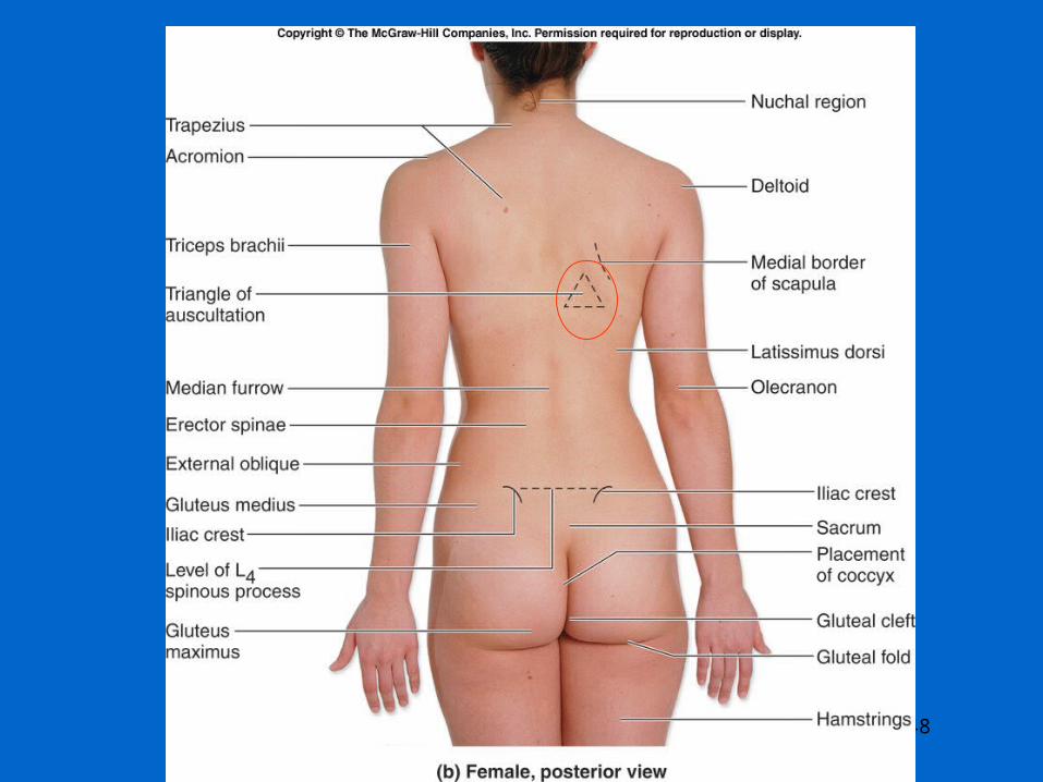
• Assessing lumbo-pelvic congruency– Palpation from cervical
spine to pelvis
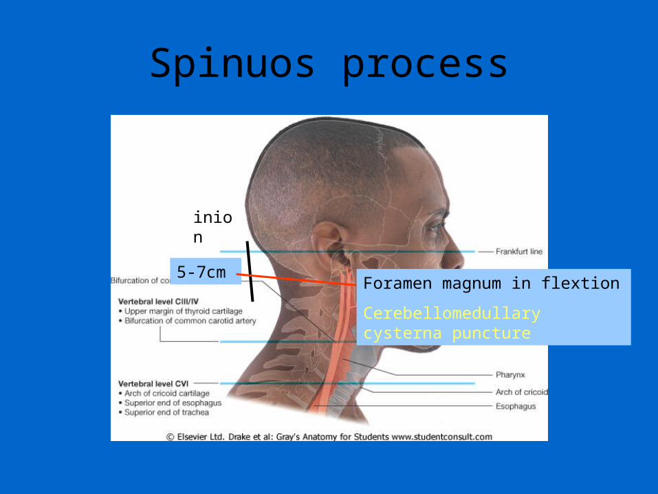
Spinuos process
5-7cm
inion
Foramen magnum in flextion
Cerebellomedullary cysterna puncture
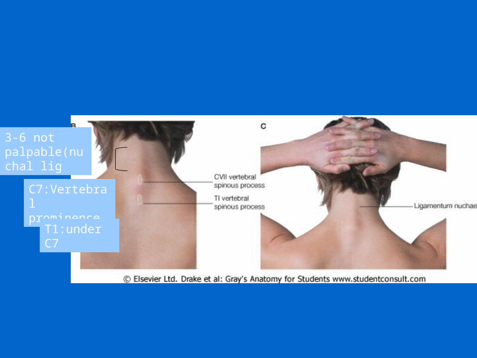
3-6 not palpable(nuchal lig
C7:Vertebral prominence
T1:under C7
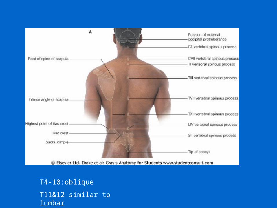
T4-10:oblique
T11&12 similar to lumbar
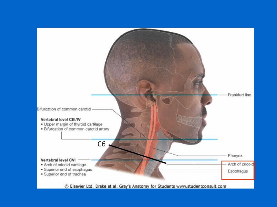
C6
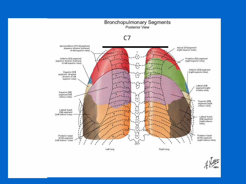
C7
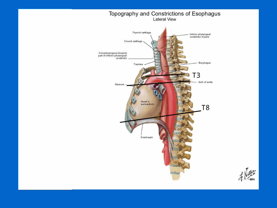
T3
T8
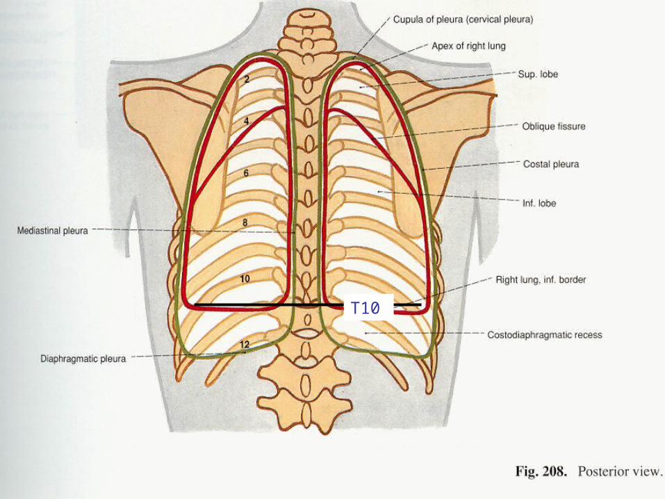
T10
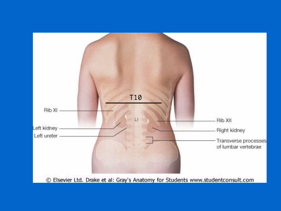
T10
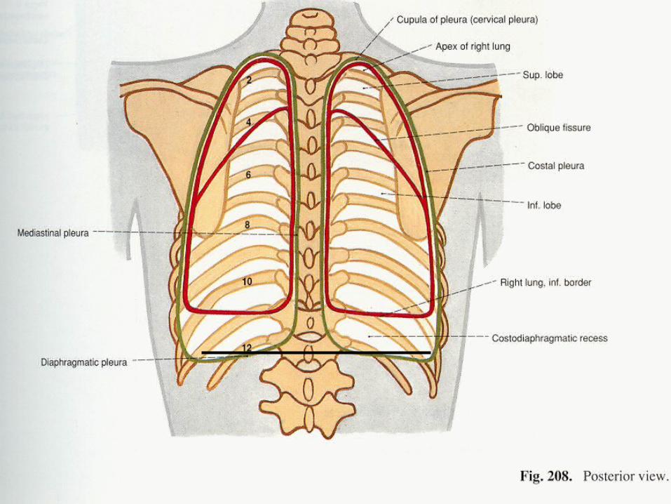
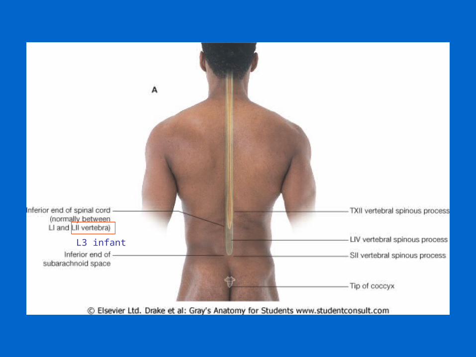
L3 infant
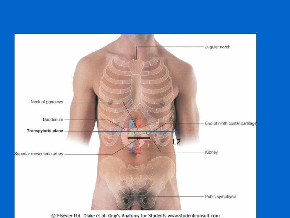
L2
L3
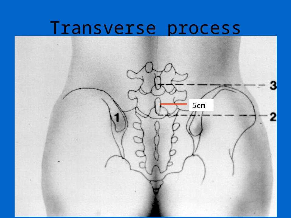
Transverse process
5cm
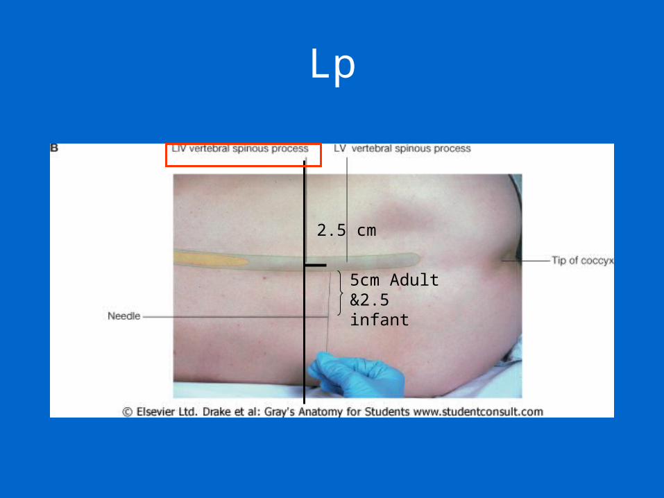
Lp
2.5 cm
5cm Adult &2.5 infant
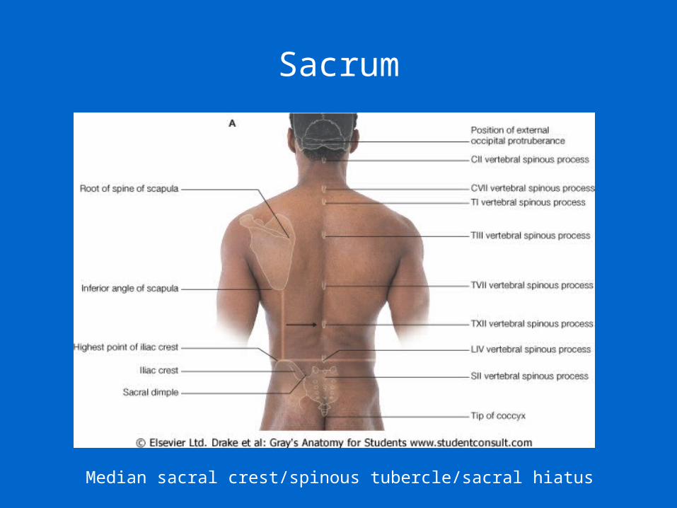
Sacrum
Median sacral crest/spinous tubercle/sacral hiatus
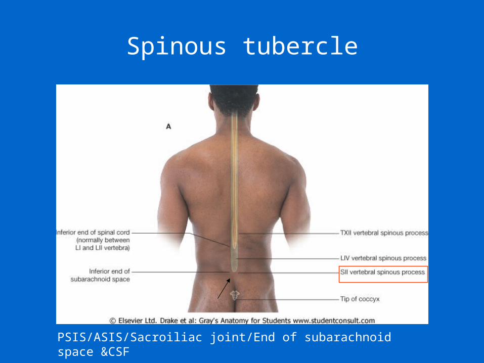
Spinous tubercle
PSIS/ASIS/Sacroiliac joint/End of subarachnoid space &CSF
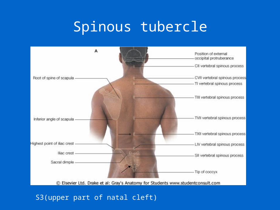
Spinous tubercle
S3(upper part of natal cleft)
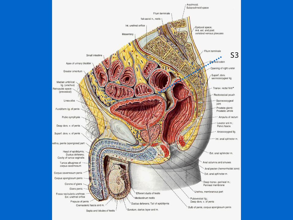
S3
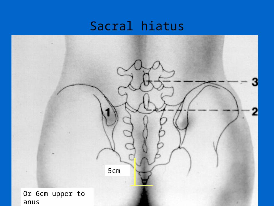
Sacral hiatus
5cm
Or 6cm upper to anus

Posterior sacral foramina4 n
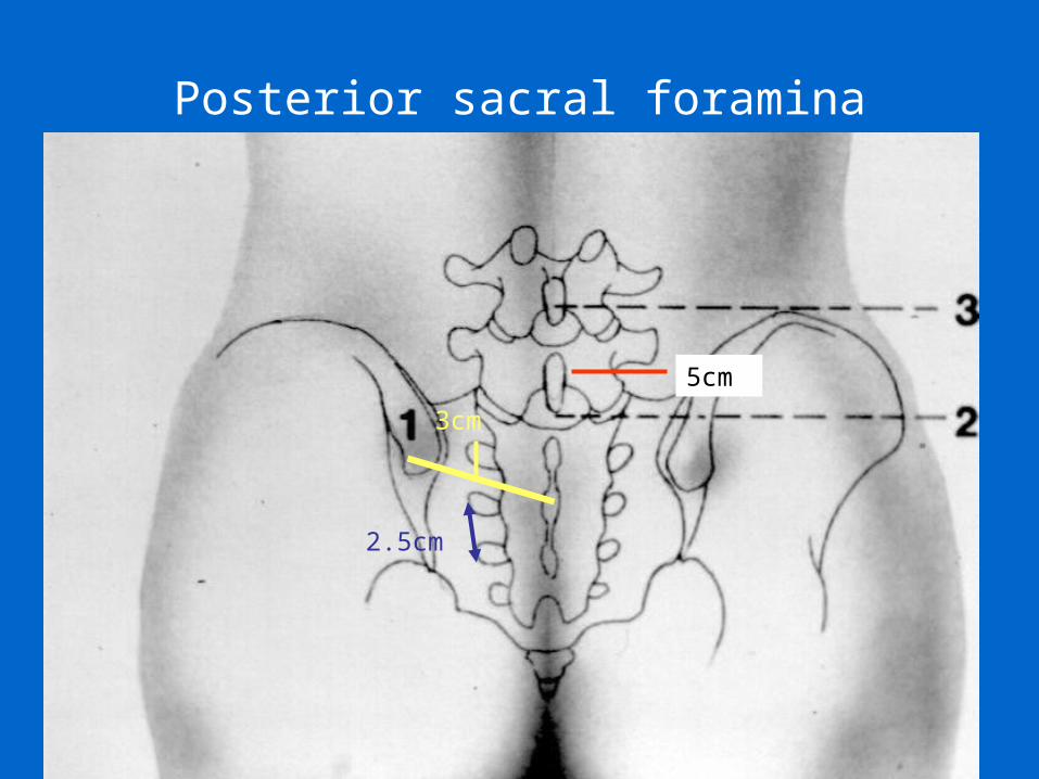
Posterior sacral foramina
5cm
3cm
2.5cm
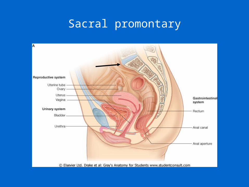
Sacral promontary
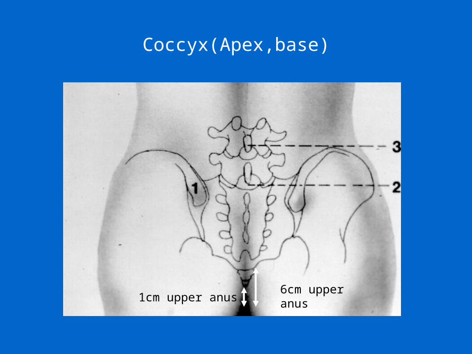
Coccyx(Apex,base)
1cm upper anus6cm upper anus
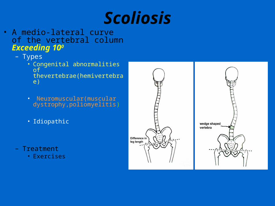
Scoliosis• A medio-lateral curve of the
vertebral column Exceeding 100
– Types• Congenital abnormalities of
thevertebrae(hemivertebrae)
• Neuromuscular(muscular dystrophy,poliomyelitis)
• Idiopathic
– Treatment• Exercises
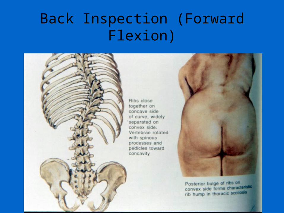
Back Inspection (Forward Flexion)
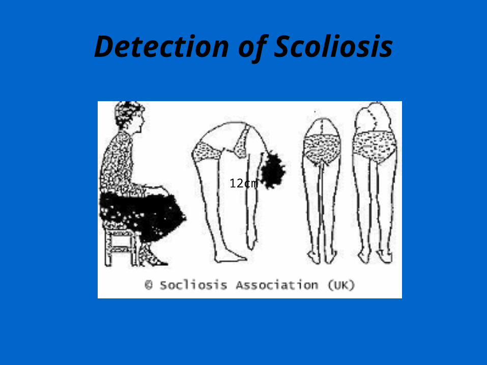
Detection of Scoliosis
12cm
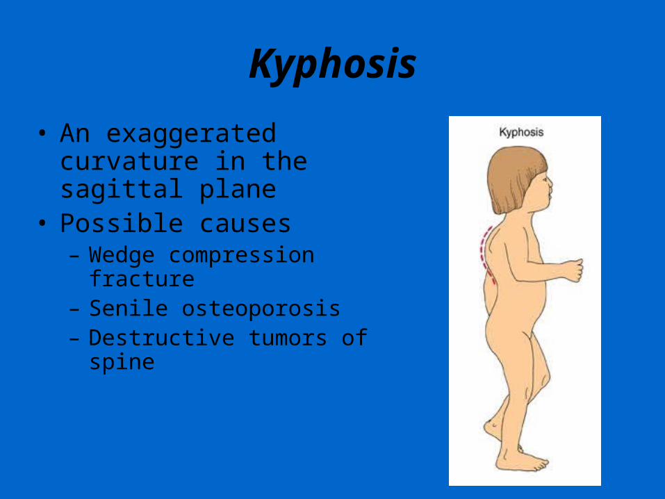
Kyphosis• An exaggerated curvature in the
sagittal plane• Possible causes
– Wedge compression fracture– Senile osteoporosis– Destructive tumors of spine
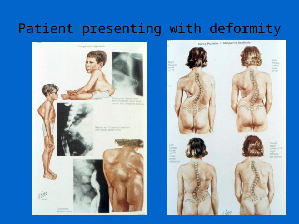
Patient presenting with deformity
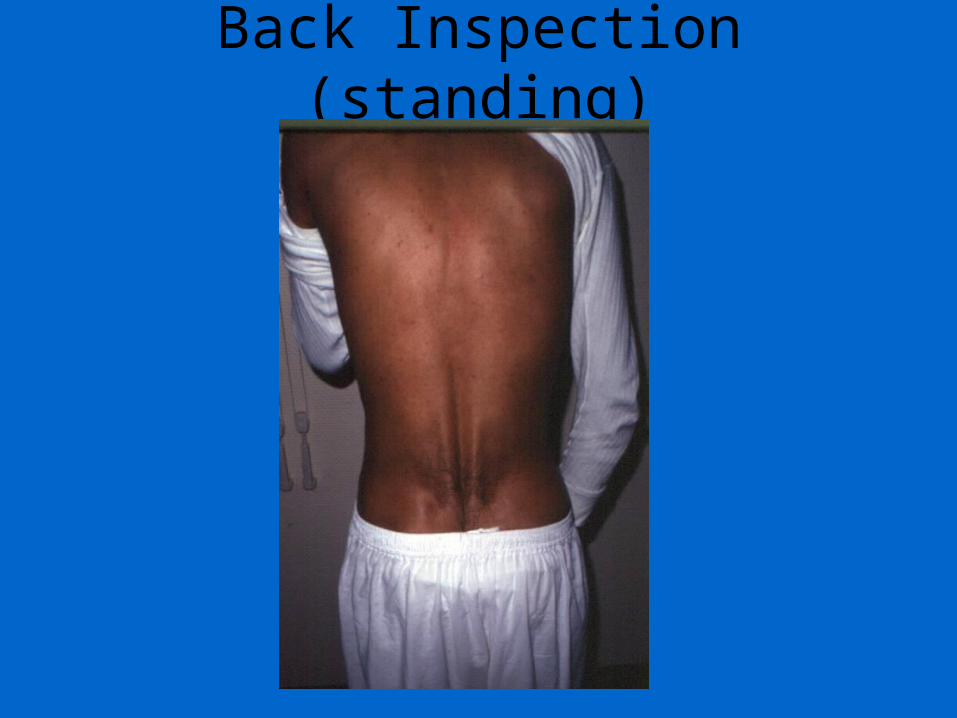
Back Inspection (standing)
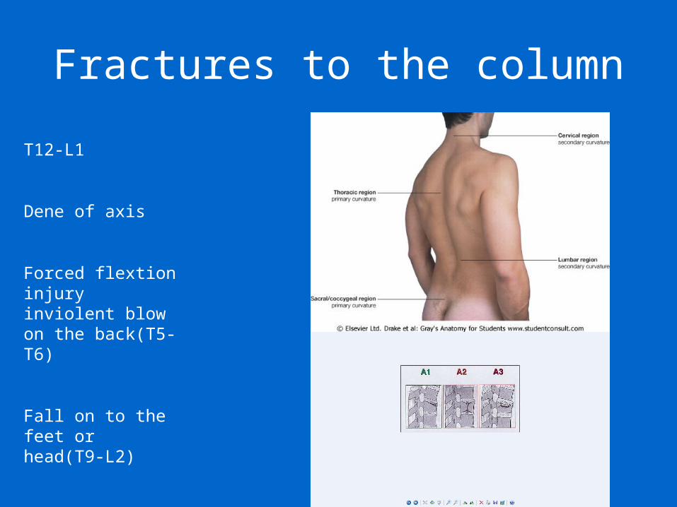
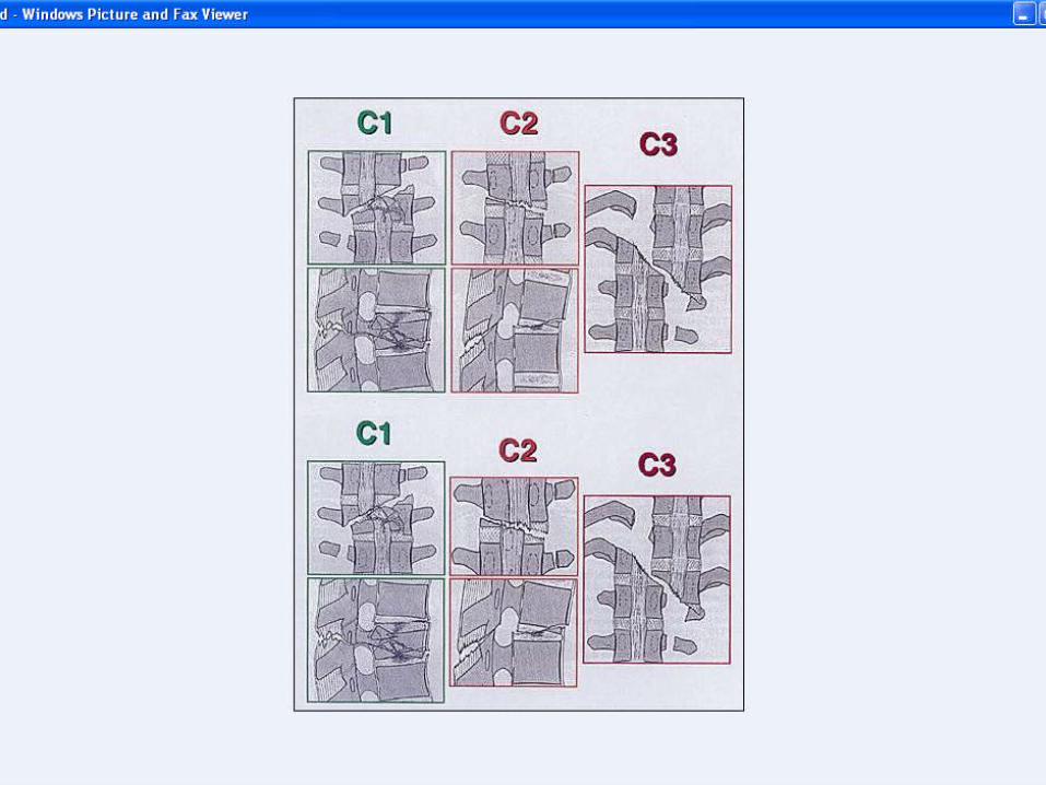
Fractures to the column
T12-L1
Dene of axis
Forced flextion injury inviolent blow on the back(T5-T6)
Fall on to the feet or head(T9-L2)
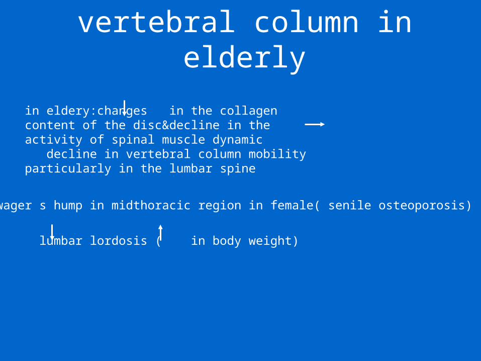
vertebral column in elderly
– Dowager s hump in midthoracic region in female( senile osteoporosis)
– lumbar lordosis ( in body weight)
in eldery:changes in the collagen content of the disc&decline in the activity of spinal muscle dynamic decline in vertebral column mobility particularly in the lumbar spine
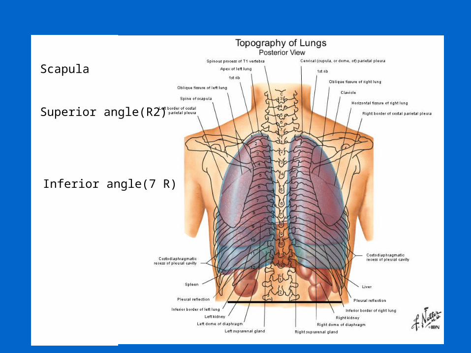
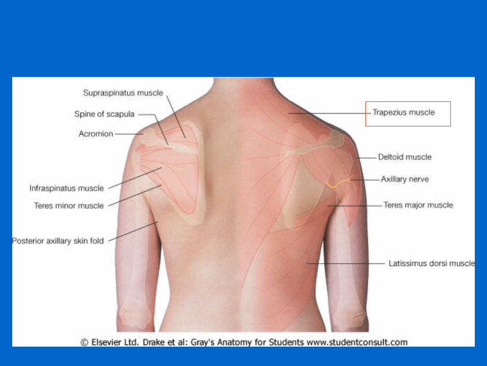
Inferior angle(7 R)
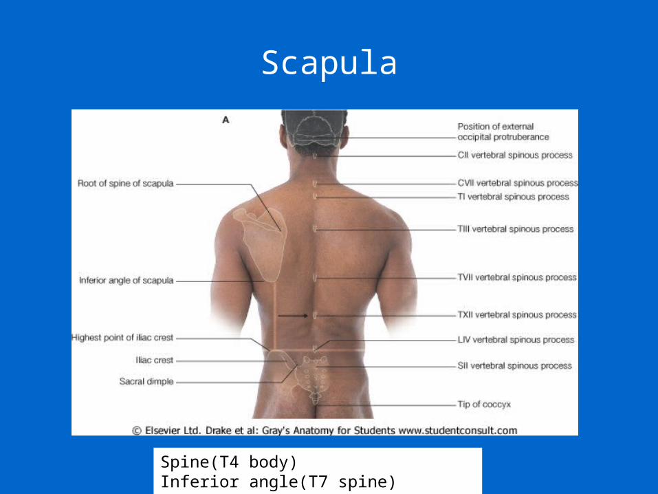
Scapula
Superior angle(R2)
Scapula
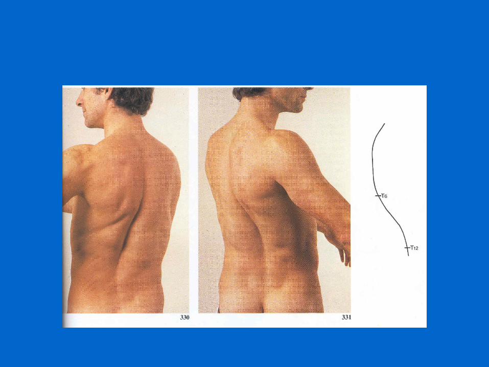
Spine(T4 body)Inferior angle(T7 spine)
48
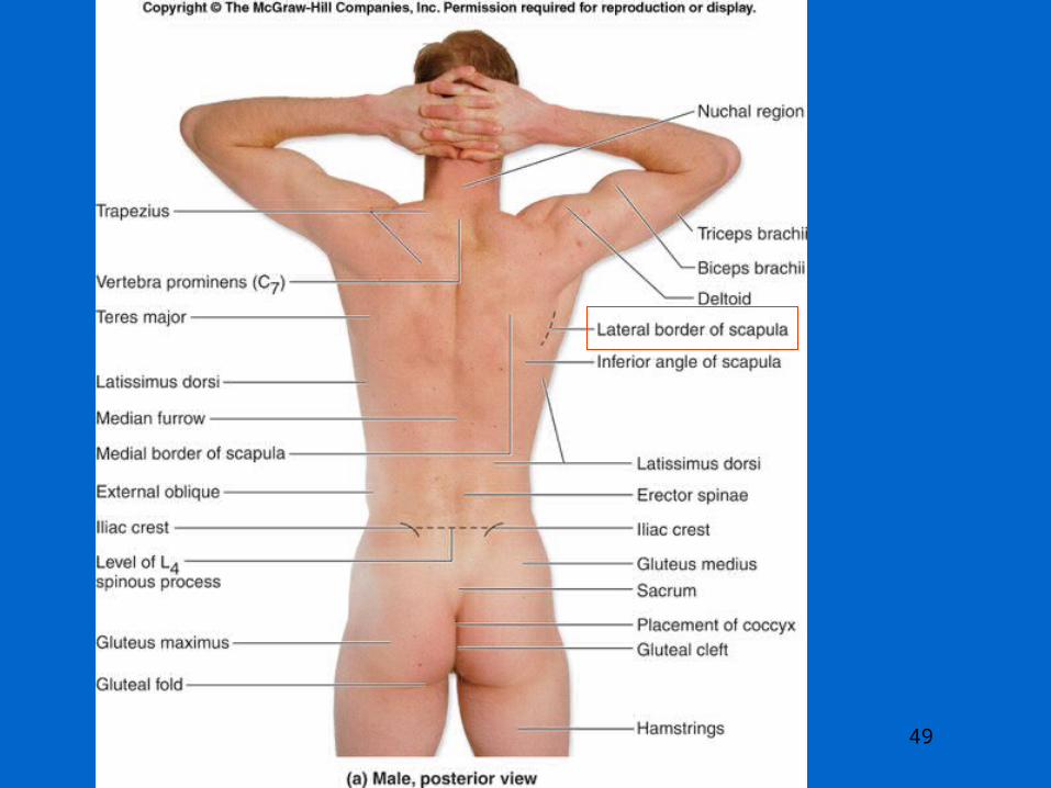
49
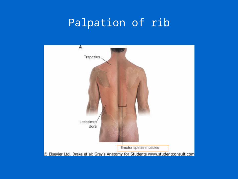
Palpation of rib
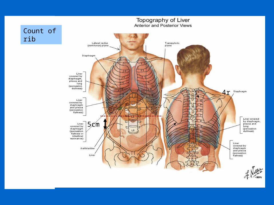
Count of rib
5cm
4r
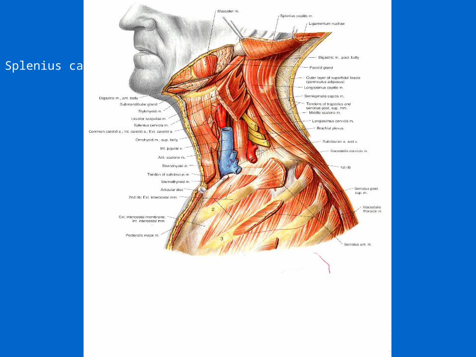
Splenius capitis
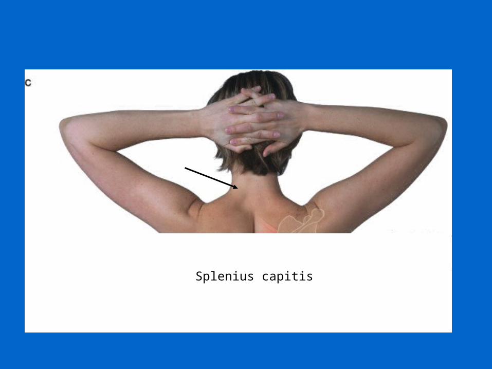
Splenius capitis
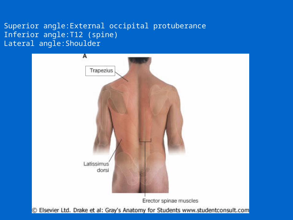
Superior angle:External occipital protuberanceInferior angle:T12 (spine)Lateral angle:Shoulder
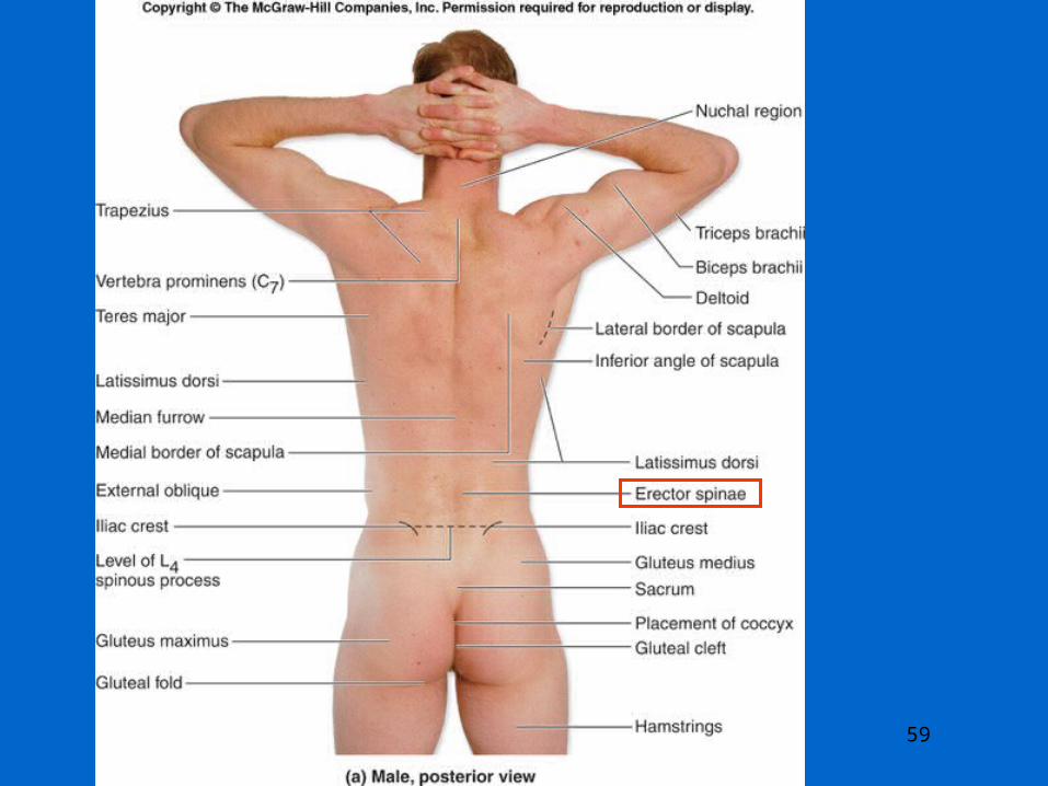
59
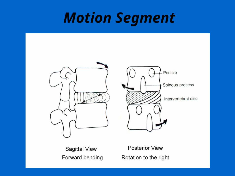
Motion Segment
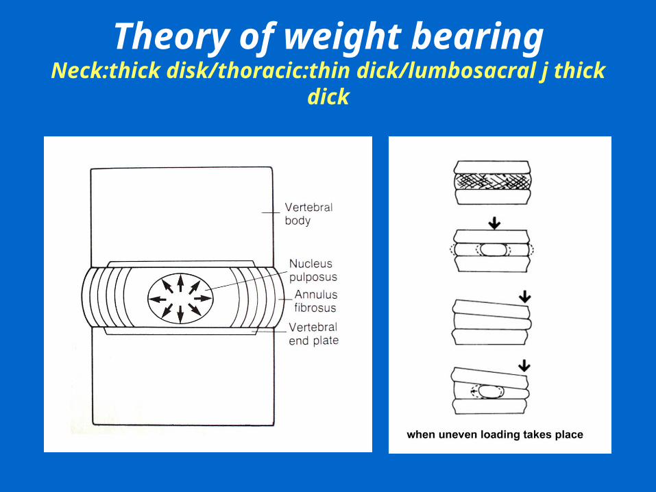
Theory of weight bearingNeck:thick disk/thoracic:thin dick/lumbosacral j thick dick
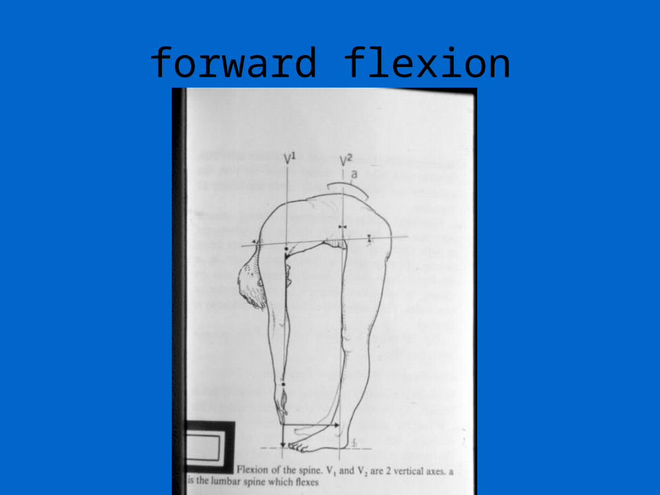
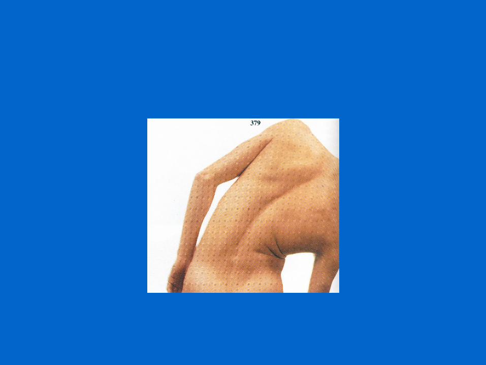
forward flexion
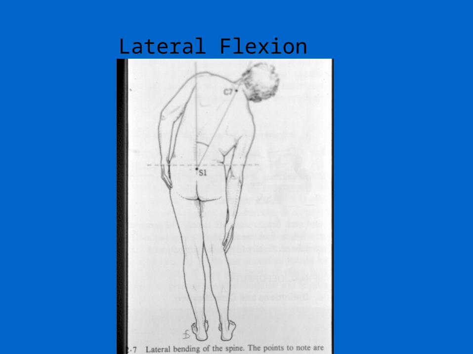
Lateral Flexion
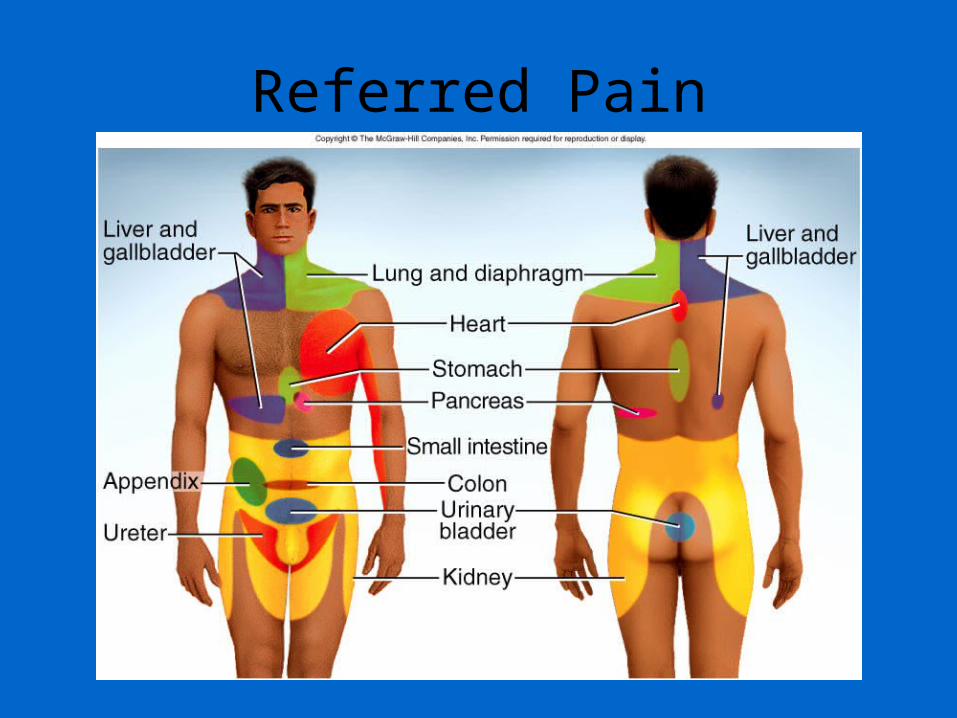
Referred Pain


![[PPT]Chapter 11 Surface Anatomy - Gavilan College -> …hhh.gavilan.edu/rmorales/documents/ch12lect_000.ppt · Web viewChapter 12 Surface Anatomy Surface Anatomy of Head Surface Anatomy](https://img.pdfslide.net/doc/110x75/5b29e6677f8b9ad8298b5149/pptchapter-11-surface-anatomy-gavilan-college-hhh-web-viewchapter-12.jpg)