Embed Size (px)
Citation preview
The Adrenal GlandThe Adrenal Gland
Atif Hassan Khirelsied, Ph.D,
Department of BiochemistryFaculty of Medicine
International University of Africa
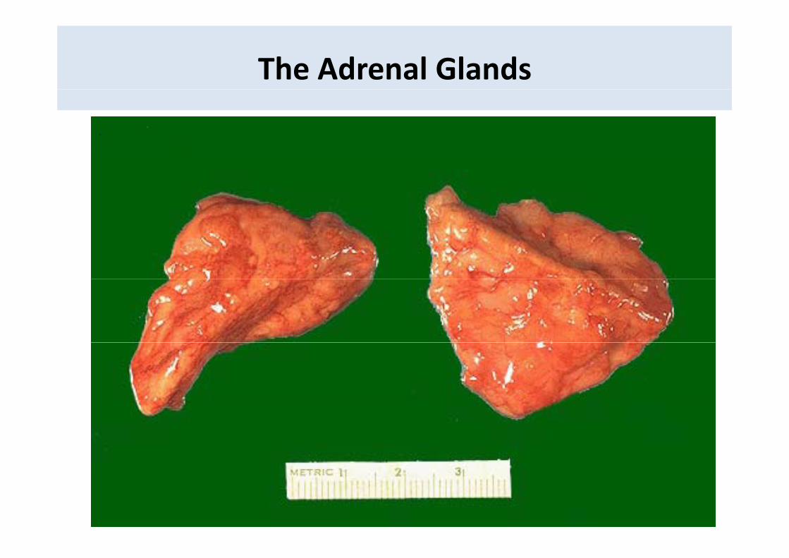
The Adrenal Glands
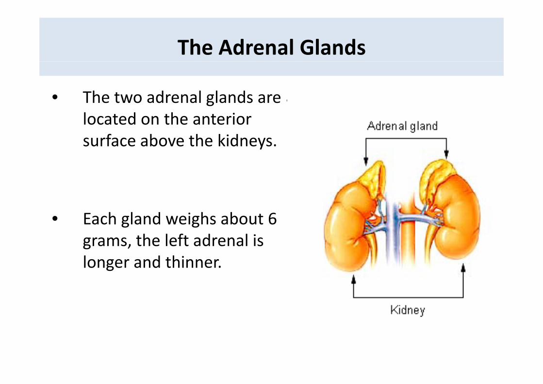
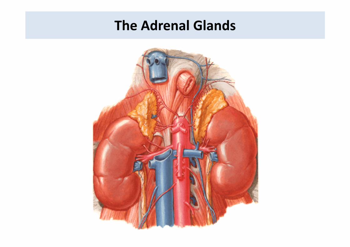
• The two adrenal glands are glocated on the anterior surface above the kidneys.y
• Each gland weighs about 6 th l ft d l igrams, the left adrenal is
longer and thinner.
The Adrenal Glands
The Adrenal Glands
The Adrenal Glands
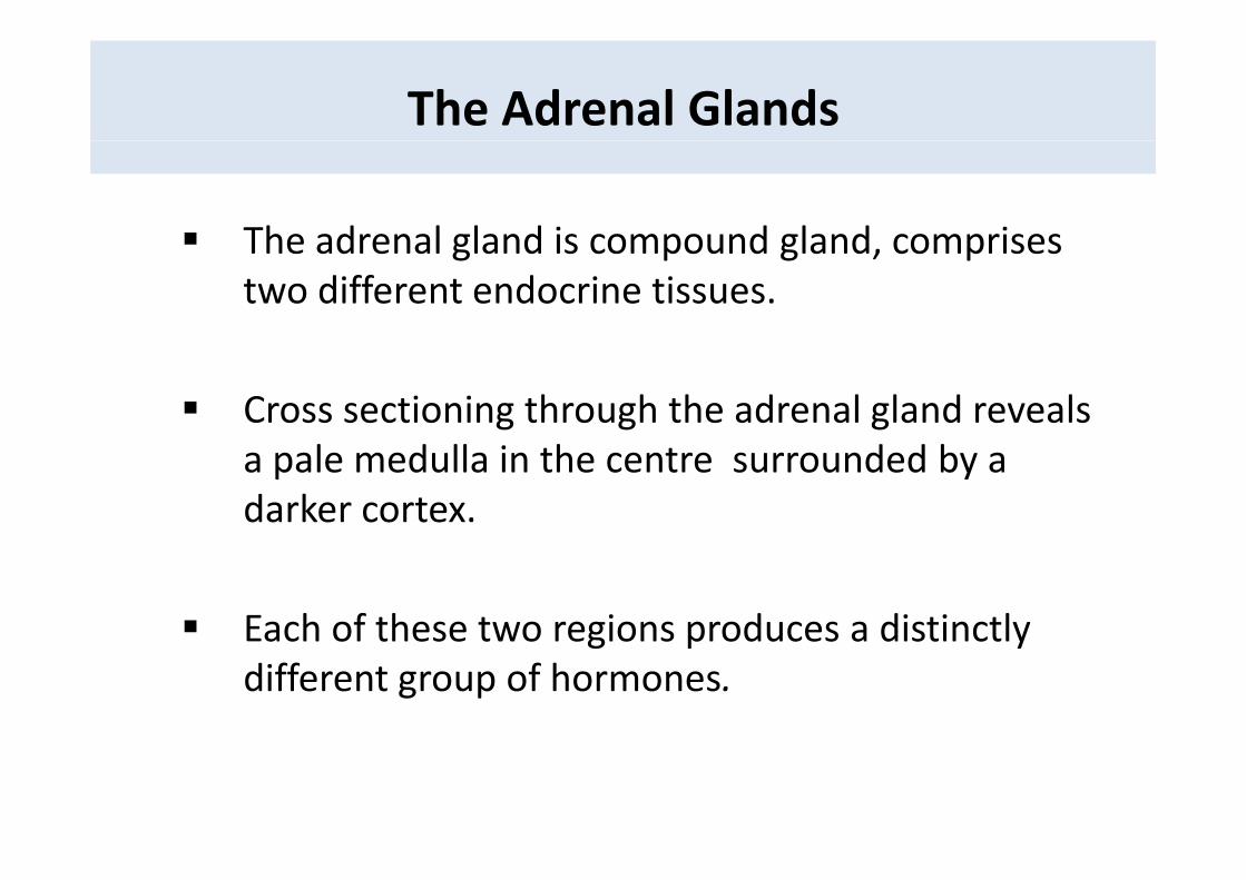
The adrenal gland is compound gland comprisesThe adrenal gland is compound gland, comprises two different endocrine tissues.
Cross sectioning through the adrenal gland reveals a pale medulla in the centre surrounded by a darker cortex.
Each of these two regions produces a distinctlyEach of these two regions produces a distinctly different group of hormones.
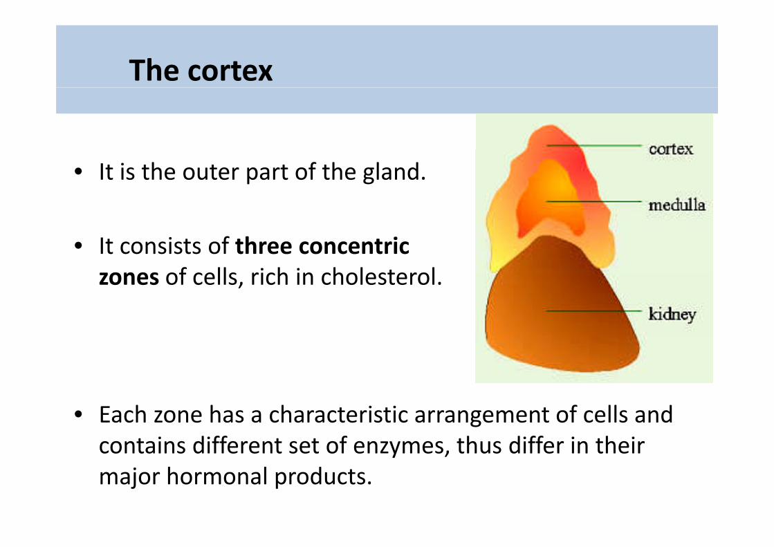
The cortex
• It is the outer part of the gland.
• It consists of three concentric zones of cells rich in cholesterolzones of cells, rich in cholesterol.
• Each zone has a characteristic arrangement of cells and contains different set of enzymes, thus differ in their major hormonal products.
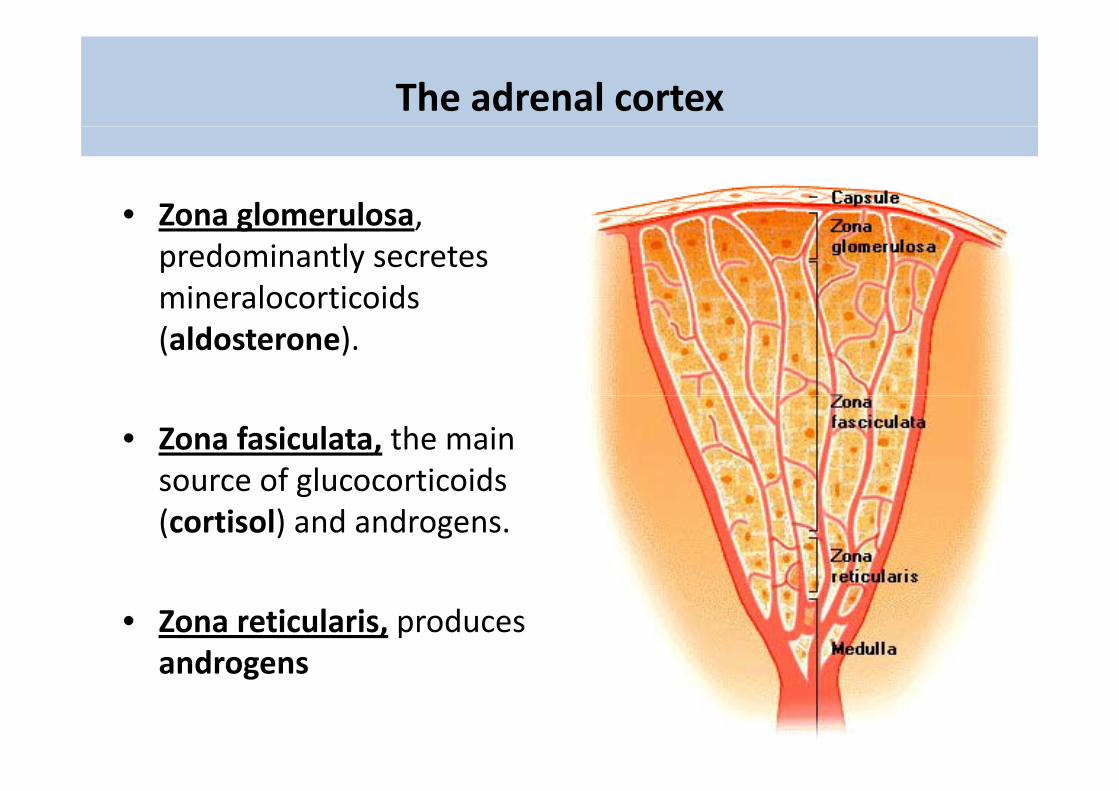
The adrenal cortex
• Zona glomerulosa• Zona glomerulosa, predominantly secretes mineralocorticoidsmineralocorticoids(aldosterone).
• Zona fasiculata, the main source of glucocorticoidssource of glucocorticoids(cortisol) and androgens.
• Zona reticularis, produces androgensandrogens
The Adrenal Gland
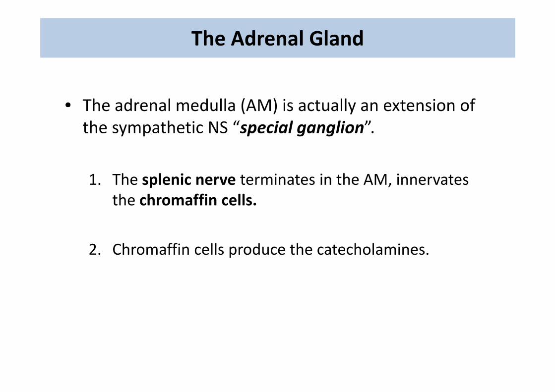
• The adrenal medulla (AM) is actually an extension ofThe adrenal medulla (AM) is actually an extension of the sympathetic NS “special ganglion”.
1. The splenic nerve terminates in the AM, innervates th h ffi llthe chromaffin cells.
2. Chromaffin cells produce the catecholamines.
The hormones of the adrenal medulla
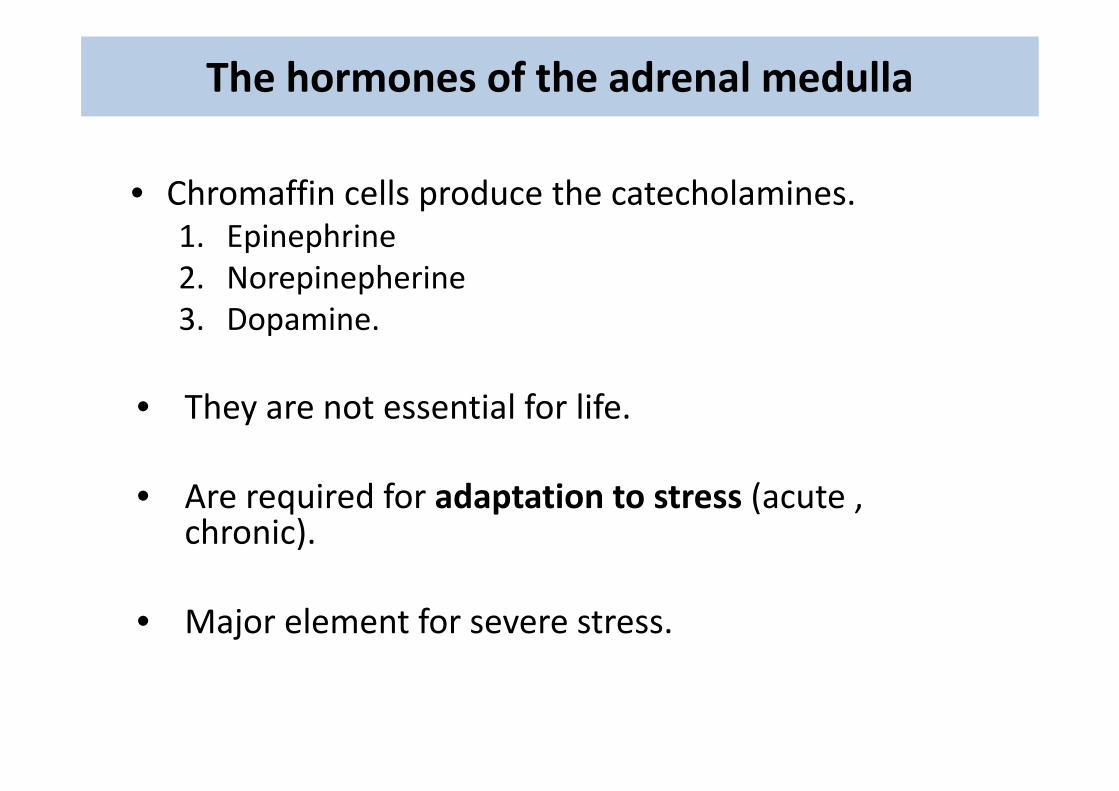
• Chromaffin cells produce the catecholamines.1. Epinephrine2. Norepinepherine3. Dopamine.
Th i l f lif• They are not essential for life.
A i d f d i (• Are required for adaptation to stress (acute , chronic).
• Major element for severe stress.
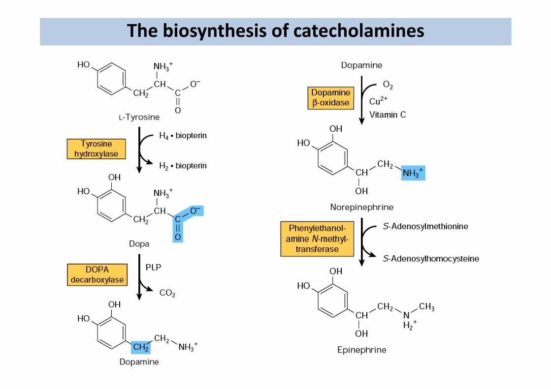
The biosynthesis of catecholamines
The biosynthesis of catecholamines
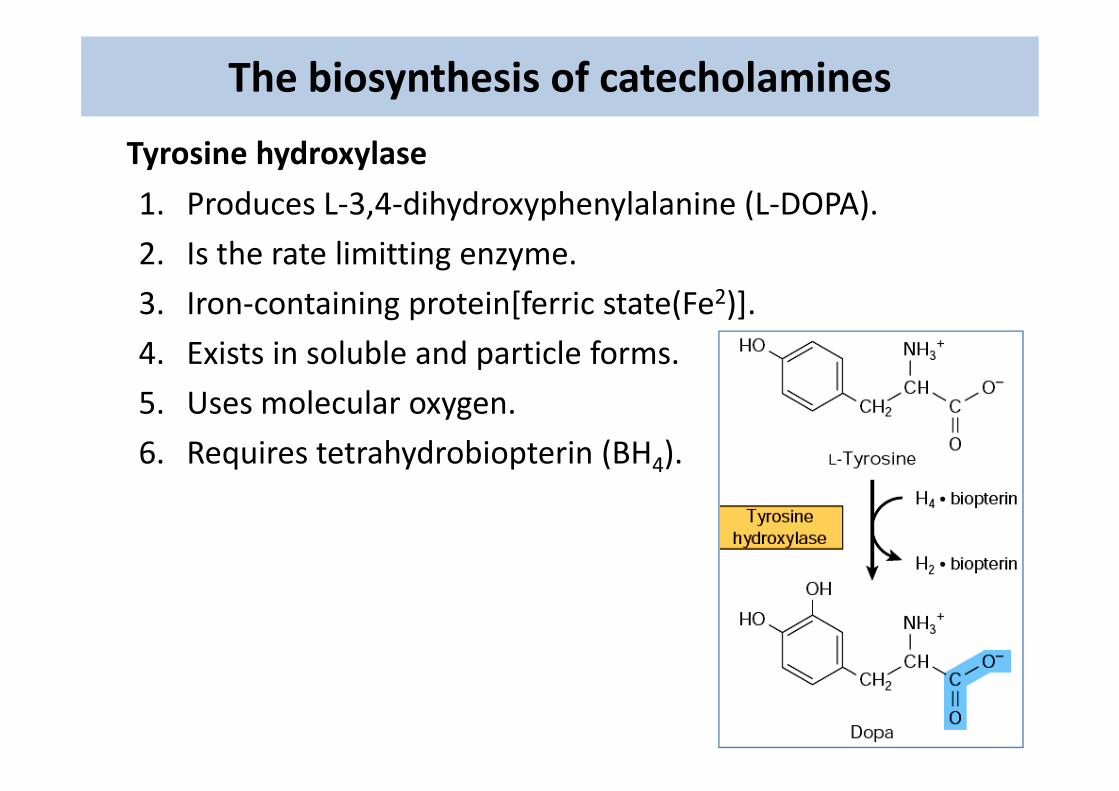
Tyrosine hydroxylase
1. Produces L‐3,4‐dihydroxyphenylalanine (L‐DOPA). , y yp y ( )
2. Is the rate limitting enzyme.
3 Iron‐containing protein[ferric state(Fe2)]3. Iron‐containing protein[ferric state(Fe )].
4. Exists in soluble and particle forms.
5 U l l5. Uses molecular oxygen.
6. Requires tetrahydrobiopterin (BH4).
The biosynthesis of catecholamines
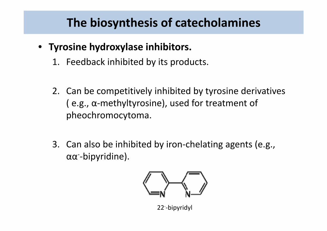
• Tyrosine hydroxylase inhibitors.1 Feedback inhibited by its products1. Feedback inhibited by its products.
2 Can be competiti el inhibited b t rosine deri ati es2. Can be competitively inhibited by tyrosine derivatives ( e.g., α‐methyltyrosine), used for treatment of pheochromocytomapheochromocytoma.
3 C l b i hibit d b i h l ti t (3. Can also be inhibited by iron‐chelating agents (e.g., αα‐‐bipyridine).
22‐‐bipyridyl
The biosynthesis of catecholamines
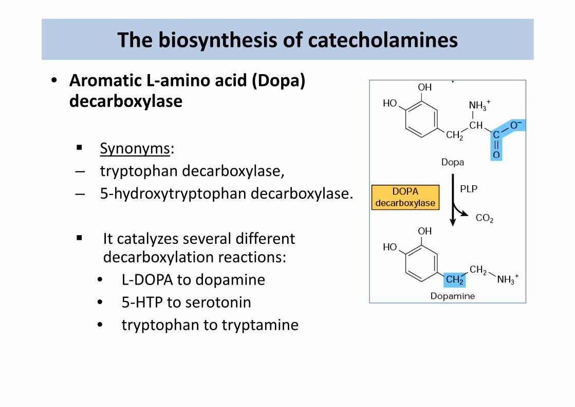
• Aromatic L‐amino acid (Dopa) decarboxylase
Synonyms: – tryptophan decarboxylase, – 5‐hydroxytryptophan decarboxylase.
It catalyzes several different decarboxylation reactions:
• L‐DOPA to dopamine• 5‐HTP to serotonin• tryptophan to tryptamine
The biosynthesis of catecholamines
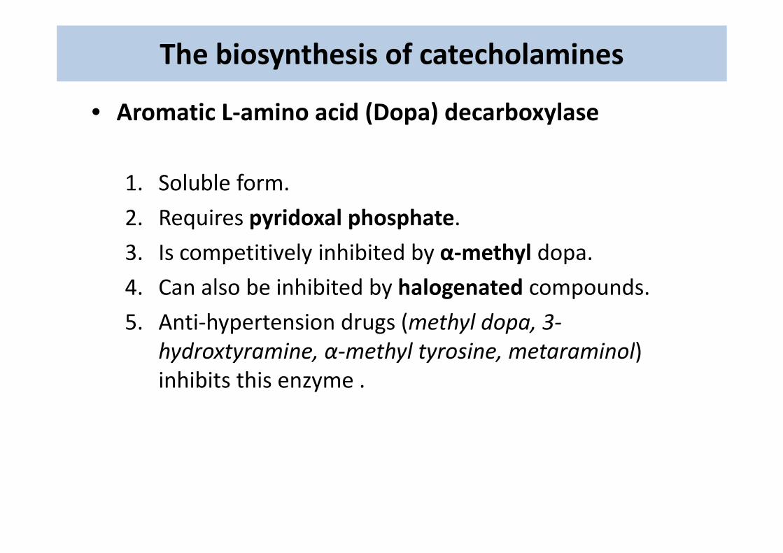
• Aromatic L‐amino acid (Dopa) decarboxylase
1. Soluble form.
2 Req ires pyridoxal phosphate2. Requires pyridoxal phosphate.
3. Is competitively inhibited by α‐methyl dopa.
4. Can also be inhibited by halogenated compounds.
5. Anti‐hypertension drugs (methyl dopa, 3‐hydroxtyramine, α‐methyl tyrosine, metaraminol) inhibits this enzyme .
The biosynthesis of catecholamines
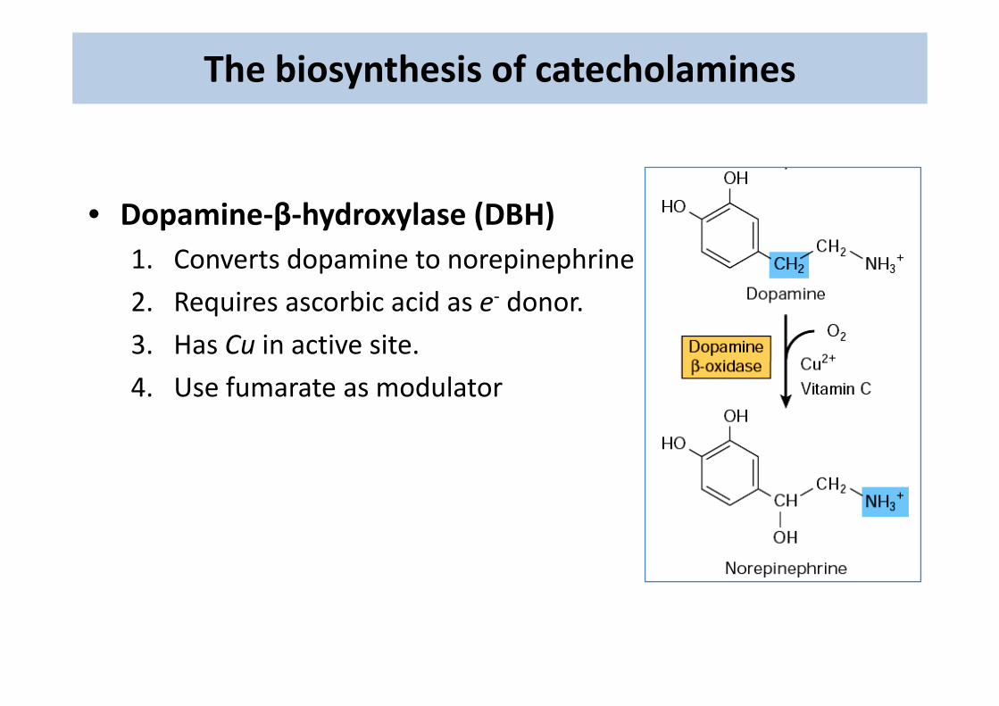
• Dopamine‐β‐hydroxylase (DBH)1. Converts dopamine to norepinephrinep p p
2. Requires ascorbic acid as e‐ donor.
3. Has Cu in active site.3. Has Cu in active site.
4. Use fumarate as modulator
The biosynthesis of catecholamines
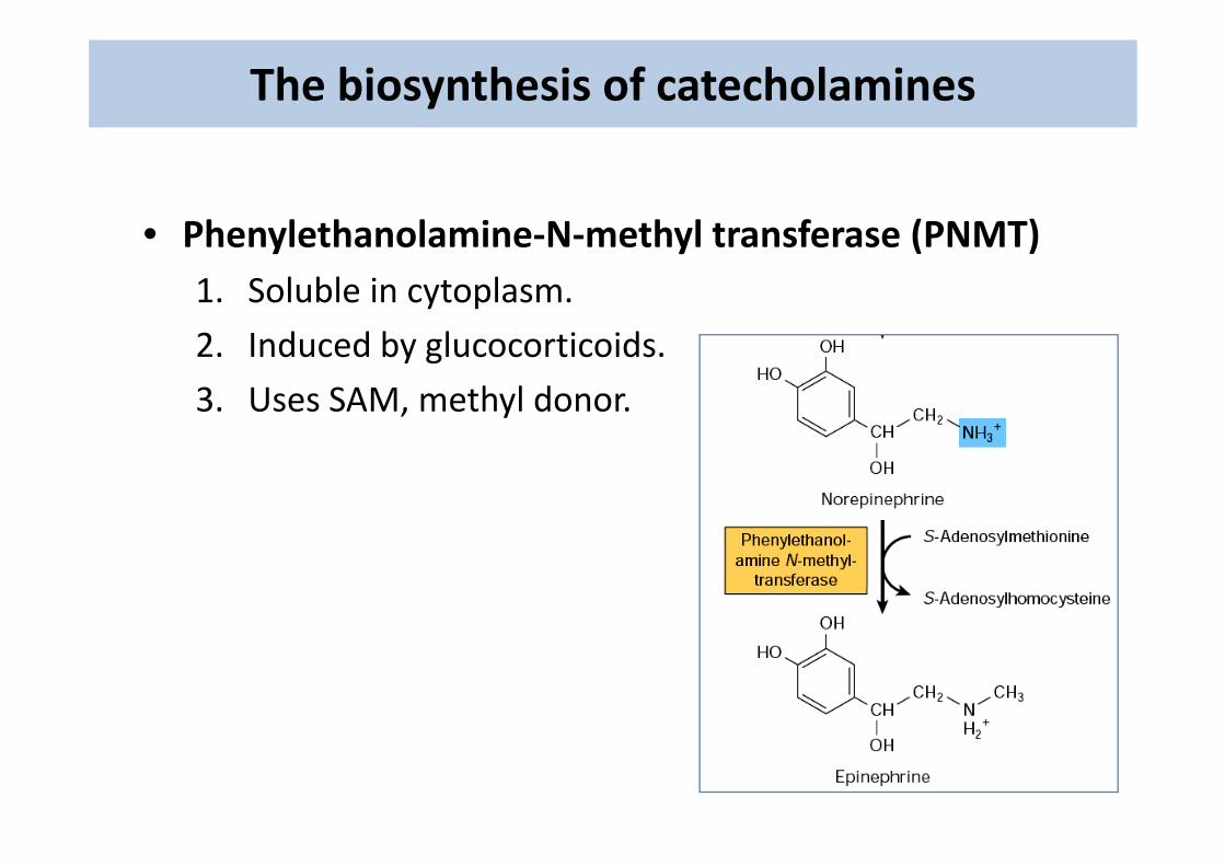
• Phenylethanolamine‐N‐methyl transferase (PNMT)• Phenylethanolamine‐N‐methyl transferase (PNMT)1. Soluble in cytoplasm.
2 I d d b l ti id2. Induced by glucocorticoids.
3. Uses SAM, methyl donor.
The regulation of catecholamines synthesis
1 Stimulated by splanchnic nerve1. Stimulated by splanchnic nerve.
2. Increases after acute stress by activation of enzymes.
3 En mes are ind ced b chronic stress (corticoids)3. Enzymes are induced by chronic stress (corticoids).
The storage, release and uptake of catecholamines
• Storage .1. Stored in the chromaffin granules2. Associated with ATP‐Mg2+ and Ca2+
• Release .1. By exocytosis (Ca2+‐dependent)2. Stimulated by cholinergic and β‐adrenergic3. Inhibited by α‐adrenergic
• Uptake.Neuronal uptake of the hormone is necessary for:1. Conservation of the hormone2. Termination of signal
The catecholamines receptors
• α1 .1. Acts via calcium.2. Increases glycogenolysis.3. Smooth muscle contraction (blood vessels, urinogenital
tract)tract).
• α2.α2.1. Inhibits cAMP formation.2. Smooth muscle relaxation (GIT)3. Smooth muscle contraction (some vascular beds)4. Inhibits:
1 li l i1. lipolysis2. Renine release3. Platelets aggregation4. Insulin secretion
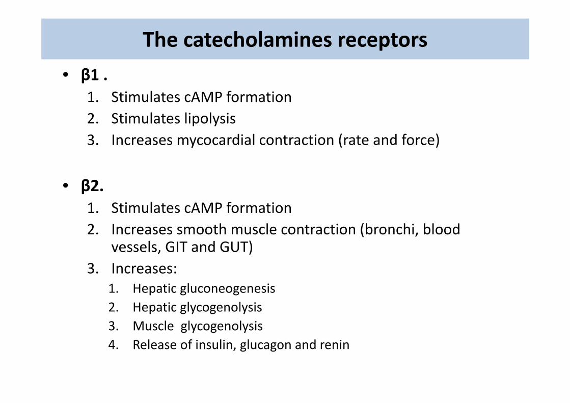
The catecholamines receptors
• β1 .1. Stimulates cAMP formation2. Stimulates lipolysis3. Increases mycocardial contraction (rate and force)
• β2.1. Stimulates cAMP formation2. Increases smooth muscle contraction (bronchi, blood
vessels GIT and GUT)vessels, GIT and GUT)3. Increases:
1. Hepatic gluconeogenesisp g g2. Hepatic glycogenolysis3. Muscle glycogenolysis4. Release of insulin, glucagon and renin
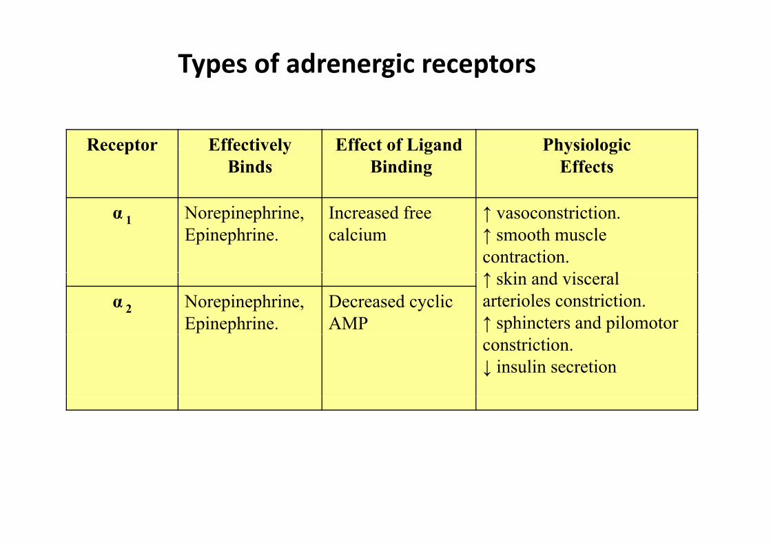
Types of adrenergic receptors
Receptor Effectively Effect of Ligand PhysiologicReceptor Effectively Binds
Effect of LigandBinding
Physiologic Effects
α Norepinephrine Increased free ↑ vasoconstrictionα 1 Norepinephrine,Epinephrine.
Increased free calcium
↑ vasoconstriction. ↑ smooth muscle contraction.↑ ki d i l↑ skin and visceral arterioles constriction.↑ sphincters and pilomotor
α 2 Norepinephrine,Epinephrine.
Decreased cyclic AMP
constriction.↓ insulin secretion
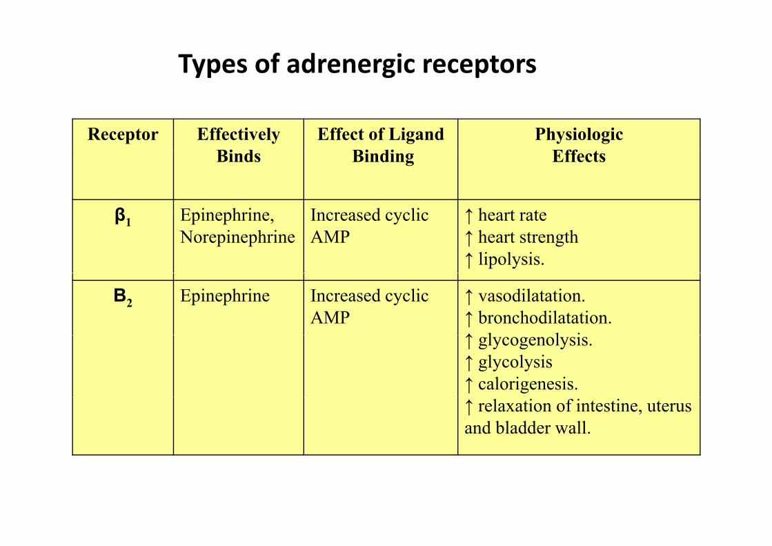
Types of adrenergic receptors
Receptor Effectively Effect of Ligand Physiologic Binds Binding Effects
β E i h i I d li ↑ h t tβ1 Epinephrine, Norepinephrine
Increased cyclic AMP
↑ heart rate↑ heart strength↑ lipolysis.
Β2 Epinephrine Increased cyclic AMP
↑ vasodilatation.↑ bronchodilatation.↑ l l i↑ glycogenolysis.↑ glycolysis↑ calorigenesis.↑ relaxation of intestine, uterus and bladder wall.
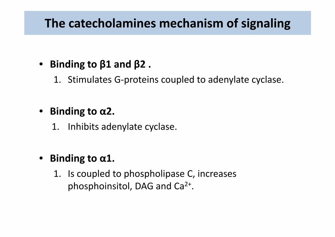
The catecholamines mechanism of signaling
• Binding to β1 and β2• Binding to β1 and β2 .1. Stimulates G‐proteins coupled to adenylate cyclase.
• Binding to α2.1. Inhibits adenylate cyclase.
• Binding to α1.1 Is coupled to phospholipase C increases1. Is coupled to phospholipase C, increases
phosphoinsitol, DAG and Ca2+.
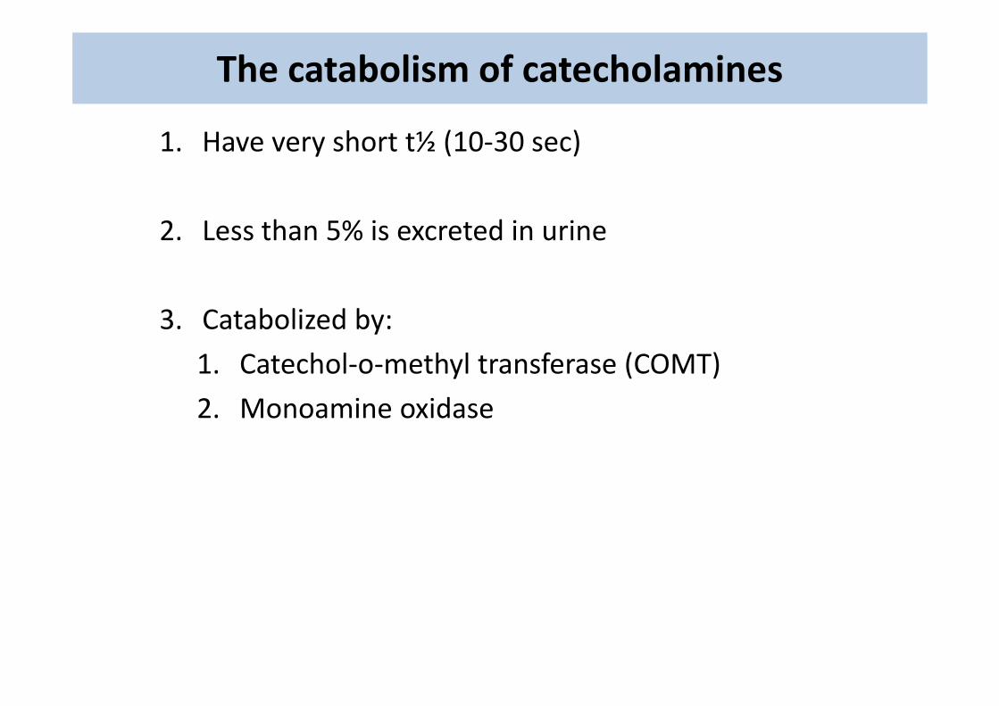
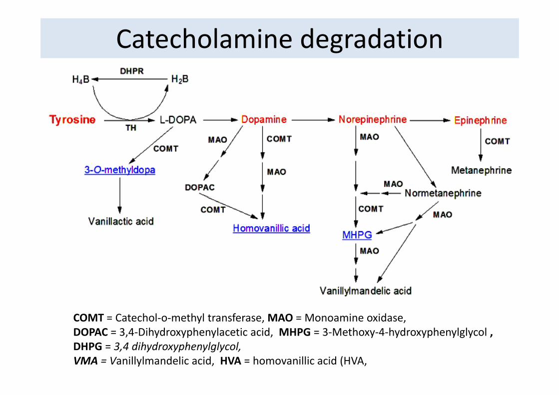
The catabolism of catecholamines
1. Have very short t½ (10‐30 sec)
2. Less than 5% is excreted in urine
3. Catabolized by:
h l h l f ( )1. Catechol‐o‐methyl transferase (COMT)
2. Monoamine oxidase
Catecholamine degradation
COMT = Catechol‐o‐methyl transferase, MAO = Monoamine oxidase, DOPAC = 3 4 Dihydroxyphenylacetic acid MHPG = 3 Methoxy 4 hydroxyphenylglycolDOPAC = 3,4‐Dihydroxyphenylacetic acid, MHPG = 3‐Methoxy‐4‐hydroxyphenylglycol , DHPG = 3,4 dihydroxyphenylglycol, VMA = Vanillylmandelic acid, HVA = homovanillic acid (HVA,