Embed Size (px)
Citation preview
The B30.2 domain of pyrin, the familialMediterranean fever protein, interacts directlywith caspase-1 to modulate IL-1� productionJae Jin Chae*†, Geryl Wood*, Seth L. Masters‡, Katharina Richard*§, Grace Park¶, Brian J. Smith‡, and Daniel L. Kastner*
*Genetics and Genomics Branch and ¶Office of the Clinical Director, National Institute of Arthritis and Musculoskeletal and Skin Diseases, National Institutesof Health, Bethesda, MD 20892; and ‡The Walter and Eliza Hall Institute of Medical Research, 1G Royal Parade, Parkville, Victoria 3050, Australia
Edited by Charles A. Dinarello, University of Colorado Health Sciences Center, Denver, CO, and approved May 19, 2006 (received for review March 14, 2006)
Familial Mediterranean fever (FMF) is a recessively inherited au-toinflammatory disorder with high carrier frequencies in the Mid-dle East. Pyrin, the protein mutated in FMF, regulates caspase-1activation and consequently IL-1� production through cognateinteraction of its N-terminal PYRIN motif with the ASC adaptorprotein. However, the preponderance of mutations reside inpyrin’s C-terminal B30.2 domain. Here we demonstrate directinteraction of this domain with caspase-1. In lysates from cells notexpressing ASC, reciprocal GST pull-downs demonstrated the in-teraction of pyrin with the p20 and p10 catalytic subunits ofcaspase-1. Coimmunoprecipitations of pyrin and caspase-1 fromTHP-1 human monocytic cells were consistent with the interactionof endogenous proteins. The C-terminal B30.2 domain of pyrin isnecessary and sufficient for the interaction, and binding wasreduced by FMF-associated B30.2 mutations. Full-length pyrin at-tenuated IL-1� production in cells transfected with a caspase-1�IL-1� construct, an effect diminished by FMF-associated B30.2mutations and in B30.2 deletion mutants. Modeling of the crystalstructure of caspase-1 with the deduced structure of the pyrinB30.2 domain corroborated both the interaction and the impor-tance of M694V and M680I pyrin mutations. Consistent with a netinhibitory effect of pyrin on IL-1� activation, small interfering RNA(siRNA)-mediated pyrin knockdown in THP-1 cells augmented IL-1�production in response to bacterial LPS. Moreover, the IL-1 receptorantagonist anakinra suppressed acute-phase proteins in a patientwith FMF and amyloidosis. Our data support a direct, ASC-inde-pendent effect of pyrin on IL-1� activation and suggest heightenedIL-1 responsiveness as one factor selecting for pyrin mutations.
autoinflammatory disorder � ASC � siRNA � anakinra � structure
Familial Mediterranean fever (FMF, MIM249100) is the pro-totype of a group of disorders, termed systemic autoinflam-
matory diseases, characterized by seemingly unprovoked epi-sodes of inf lammation without evidence of high-titerautoantibodies or antigen-specific T cells (1, 2). FMF is reces-sively inherited and presents with 1- to 3-day attacks of feveraccompanied by sterile peritonitis, pleurisy, rash, and�or arthri-tis (3). During attacks there is a substantial influx of polymor-phonuclear leukocytes into the affected tissues. In some patientsthe ectopic deposition of misfolded fragments of serum amyloidA leads to renal failure and death.
The gene mutated in FMF (designated MEFV for Mediterra-nean fever) was identified by positional cloning (4, 5), and to date�50 FMF-associated MEFV mutations have been identified (3).FMF carrier frequencies in the range of 1:3 to 1:5 have beendocumented in a number of populations, including Jews, Arabs,Turks, Armenians, and Italians (3). The fact that these highcarrier frequencies are found in multiple populations, withdifferent mutations predominating in the various ethnic groups,strongly favors heterozygote selection over genetic drift as thelikely explanation.
The protein product of MEFV, denoted pyrin (or marenos-trin), is a 781-aa protein expressed in granulocytes, cytokine-
activated monocytes, and serosal and synovial fibroblasts (6–8).A large percentage of FMF-associated pyrin mutations reside inthe �200-residue C-terminal B30.2 (PRYSPRY, rfp) domain.B30.2 domains in other proteins are thought to mediate protein–protein interactions (9–12), although the precise role of pyrin’sB30.2 domain is unknown. In contrast, the �90 aa at the Nterminus of pyrin define a motif, variously called the PYRINdomain (13, 14), PYD (15), PAAD (16), or DAPIN (17), that israrely mutated in FMF, and assumes a six �-helical death-foldstructure similar to death domains, death effector domains, andcaspase recruitment domains (CARDs). The death-fold confor-mation permits cognate interactions, and a number of experi-mental systems have established that pyrin interacts with anadaptor protein denoted apoptosis-associated speck-like proteinwith a CARD (ASC) through homotypic interaction of theirrespective PYRIN domains (18, 19). ASC, in turn, plays animportant role in regulating cytokine secretion, NF-�B activa-tion, and apoptosis (20–22).
Of particular note, ASC has recently been shown to assemblein macromolecular complexes denoted ‘‘inflammasomes’’ (23–25), which contain one of at least three different members of theNALP (NACHT, leucine-rich repeat, PYRIN) protein family toactivate pro-caspase-1 [IL-1�-converting enzyme (ICE)]. In theinflammasome, ASC interacts with one of the NALP proteinsthrough cognate PYRIN domain interactions, and with pro-caspase-1 through homotypic CARD interactions. The inflam-masome complex brings two molecules of pro-caspase-1 intoclose proximity, leading to autocatalysis and the subsequentrelease of the active catalytic p20 and p10 domains of caspase-1.Caspase-1, in turn, cleaves the 31-kDa precursor form of IL-1�into its biologically active 17-kDa fragment, a potent mediator offever and inflammation.
The role of pyrin in IL-1� activation is controversial. Our ownlaboratory has provided several lines of evidence that pyrin caninhibit IL-1� activation (19). Transfection of full-length mousepyrin into the murine RAW monocytic cell line suppresses IL-1�secretion, and peritoneal macrophages from mice expressing atruncated pyrin exhibit increased caspase-1 activation and IL-1�production, relative to WT controls. Moreover, in GST pull-down experiments, mouse ASC bound pyrin preferentially tocaspase-1. These findings suggest that pyrin negatively regulates
Conflict of interest statement: No conflicts declared.
This paper was submitted directly (Track II) to the PNAS office.
Freely available online through the PNAS open access option.
Abbreviations: FMF, familial Mediterranean fever; CARD, caspase recruitment domain; ICE,IL-1�-converting enzyme; siRNA, small interfering RNA.
†To whom correspondence should be addressed at: Genetics and Genomics Branch, Build-ing 10, Room 9N-214, 10 Center Drive MSC 1820, Bethesda, MD 20892-1820. E-mail:[email protected].
§Present address: Department of Cell Biology and Molecular Genetics, University of Mary-land, Microbiology Building, Room 1109, College Park, MD 20742.
© 2006 by The National Academy of Sciences of the USA
9982–9987 � PNAS � June 27, 2006 � vol. 103 � no. 26 www.pnas.org�cgi�doi�10.1073�pnas.0602081103
inflammasome activity by competing for ASC. On the otherhand, in transfected 293T human embryonic kidney cells, pyrinmay actually activate caspase-1 and IL-1� (26).
Whether pyrin inhibits or activates IL-1� through ASC,neither formulation adequately explains the proinflammatoryeffect and high frequency of mutations in the C-terminal B30.2domain of pyrin. In this study we investigated the potentialinteraction of the B30.2 domain of pyrin with caspase-1 at abiochemical, structural, and functional level. We further ex-plored the role of pyrin in IL-1 regulation through knockdownexperiments and studies of an FMF patient receiving an IL-1blocking agent. Taken together, our data provide a possiblemolecular explanation for the high frequency of pyrin B30.2mutations in some populations.
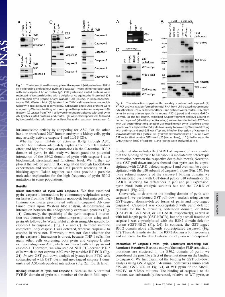
ResultsDirect Interaction of Pyrin with Caspase-1. We first examinedpyrin–caspase-1 interactions by coimmunoprecipitation assayson lysates from the THP-1 human monocytic leukemia cell line.Immune complexes precipitated with anti-caspase-1 Ab con-tained pyrin upon Western blot analysis, demonstrating aninteraction between the endogenously expressed proteins (Fig.1A). Conversely, the specificity of the pyrin–caspase-1 interac-tion was demonstrated by coimmunoprecipitation using anti-pyrin Ab followed by Western blot analysis using Abs specific forcaspase-1 to caspase-10 (Fig. 1 B and C). In these immunecomplexes, only caspase-1 was detected, whereas caspase-2 tocaspase-10 were not. However, it was not clear whether thepyrin–caspase-1 interaction is direct, because THP-1 cells, likemany other cells expressing both pyrin and caspase-1, alsoexpress endogenous ASC, which can interact with both pyrin andcaspase-1. Therefore, we also studied NIH 3T3-derived PT67cells, which do not express ASC even by sensitive RT-PCR (Fig.2A). In vivo GST pull-down analysis of lysates from PT67 cellscotransfected with GST–pyrin and myc-tagged caspase-1 dem-onstrated ASC-independent interaction (Fig. 2B, fourth lane).
Binding Domains of Pyrin and Caspase-1. Because the N-terminalPYRIN domain of pyrin is a member of the death-fold super-
family that also includes the CARD of caspase-1, it was possiblethat the binding of pyrin to caspase-1 is mediated by heterotypicinteraction between the respective death-fold motifs. Neverthe-less, GST pull-down analysis showed that pyrin can be copre-cipitated with CARD-deleted caspase-1 and even can be copre-cipitated with the p20 subunit of caspase-1 alone (Fig. 2B). Formore refined mapping of the caspase-1 binding domain, wecotransfected pyrin with GST-fused p20 or p10 subunits or theCARD. Allowing for differences in p20 and p10 expression,pyrin binds both catalytic subunits but not the CARD ofcaspase-1 (Fig. 2C).
Conversely, to determine the binding domain of pyrin withcaspase-1, we performed GST pull-down analysis using variousGST-tagged, domain-deleted forms of pyrin and myc-taggedcaspase-1. Caspase-1 was coprecipitated with pyrin deletionmutants for the N terminus, coiled-coil domain, or B-box(GST-BCR, GST-NBR, or GST-NCR, respectively), as well aswith full-length pyrin (GST-NBCR), but only a small fraction ofcaspase-1 was coprecipitated with the B30.2 domain deletionmutant (GST-NBC) (Fig. 3A). In contrast, the GST-taggedB30.2 domain alone efficiently coprecipitated caspase-1 (Fig.3B). These data indicate that the B30.2 domain is both necessaryand sufficient for the direct interaction of pyrin with caspase-1.
Interaction of Caspase-1 with Pyrin Constructs Harboring FMF-Associated Mutations. Because many of the major FMF-associatedmutations are clustered in the B30.2 domain of pyrin, weconsidered the possible effect of these mutations on the bindingto caspase-1. We first examined the binding by GST pull-downanalysis using GST-tagged C-terminal constructs (amino acids375–781; GST-BCR in Fig. 3A) of WT pyrin or the M680I,M694V, or V726A mutants. The binding of caspase-1 to themutants was substantially decreased, relative to WT pyrin, as
Fig. 1. The interaction of human pyrin with caspase-1. (A) Lysates from THP-1cells expressing endogenous pyrin and caspase-1 were immunoprecipitatedwith anti-caspase-1 Ab or control IgG. Cell lysates and eluted proteins weresubjected to Western blotting with a polyclonal Ab against the N-terminal 374aa of human pyrin (Upper) or anti-caspase-1 Ab (Lower). IP, immunoprecipi-tation; WB, Western blot. (B) Lysates from THP-1 cells were immunoprecipi-tated with anti-pyrin Ab or control IgG. Cell lysates and eluted proteins wereanalyzed by Western blotting with anti-pyrin Ab (Upper) or anti-caspase-1 Ab(Lower). (C) Lysates from THP-1 cells were immunoprecipitated with anti-pyrinAb. Lysates, eluted proteins, and control IgG were electrophoresed, followedby Western blotting with anti-pyrin Ab or Abs against caspase-1 to caspase-10.
Fig. 2. The interaction of pyrin with the catalytic subunits of caspase-1. (A)RT-PCR analysis was performed on total RNA from LPS-treated mouse mono-cytes (first lane), PT67 cells (second lane), and distilled water control (DW, thirdlane) by using primers specific to mouse ASC (Upper) and mouse GAPDH(Lower). (B) The full-length, combined p20p10 fragment and p20 subunit ofhuman caspase-1 (all with myc epitope tags) were cotransfected into PT67 cellswith GST vector (first three lanes) or GST-fused human pyrin (last three lanes).Lysates were subjected to GST pull-down assay followed by Western blottingwith anti-myc and anti-GST Abs (Top and Middle). Expression of caspase-1 isshown in Bottom (cell lysates). (C) Pyrin was cotransfected into PT67 cells withGST vector (first lane) or GST-fused p20 (second lane), p10 (third lane), or theCARD (fourth lane) of caspase-1, and lysates were analyzed as in B.
Chae et al. PNAS � June 27, 2006 � vol. 103 � no. 26 � 9983
MED
ICA
LSC
IEN
CES
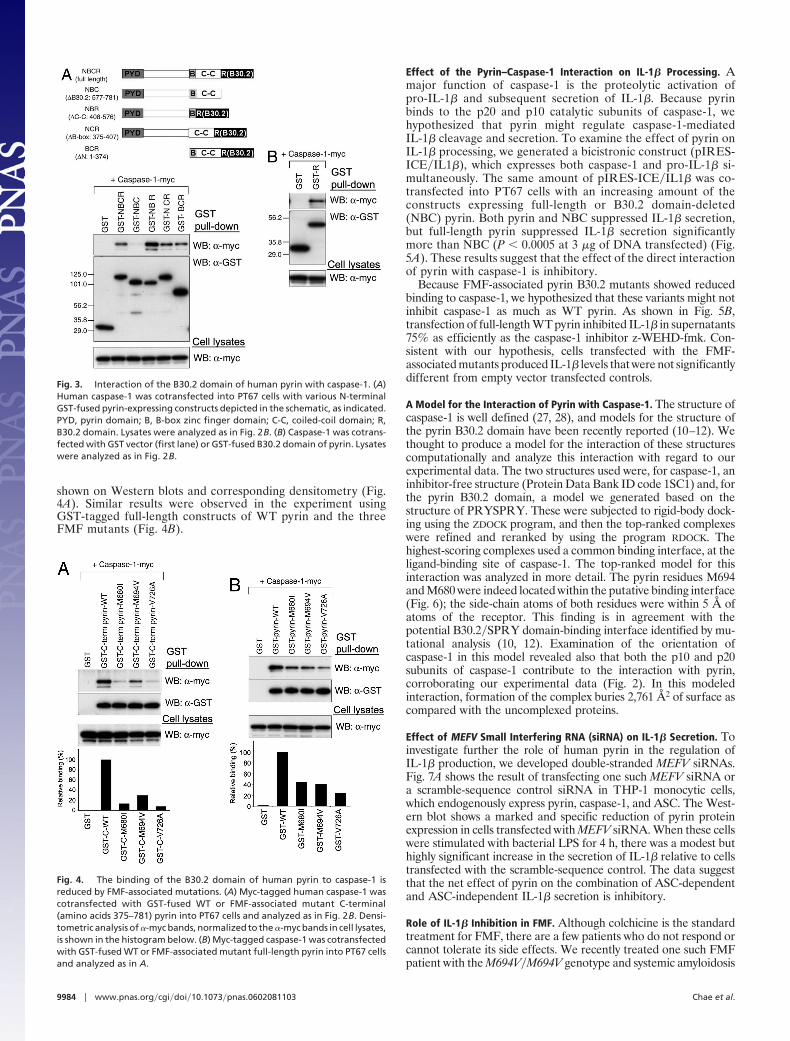
shown on Western blots and corresponding densitometry (Fig.4A). Similar results were observed in the experiment usingGST-tagged full-length constructs of WT pyrin and the threeFMF mutants (Fig. 4B).
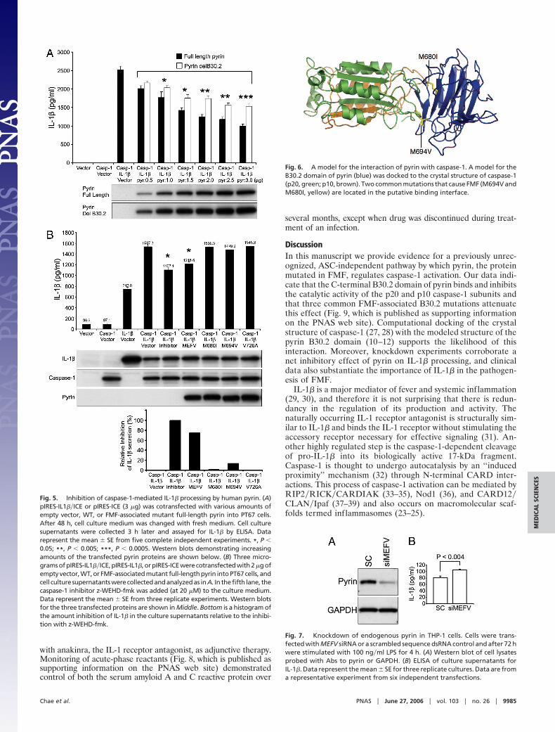
Effect of the Pyrin–Caspase-1 Interaction on IL-1� Processing. Amajor function of caspase-1 is the proteolytic activation ofpro-IL-1� and subsequent secretion of IL-1�. Because pyrinbinds to the p20 and p10 catalytic subunits of caspase-1, wehypothesized that pyrin might regulate caspase-1-mediatedIL-1� cleavage and secretion. To examine the effect of pyrin onIL-1� processing, we generated a bicistronic construct (pIRES-ICE�IL1�), which expresses both caspase-1 and pro-IL-1� si-multaneously. The same amount of pIRES-ICE�IL1� was co-transfected into PT67 cells with an increasing amount of theconstructs expressing full-length or B30.2 domain-deleted(NBC) pyrin. Both pyrin and NBC suppressed IL-1� secretion,but full-length pyrin suppressed IL-1� secretion significantlymore than NBC (P � 0.0005 at 3 �g of DNA transfected) (Fig.5A). These results suggest that the effect of the direct interactionof pyrin with caspase-1 is inhibitory.
Because FMF-associated pyrin B30.2 mutants showed reducedbinding to caspase-1, we hypothesized that these variants might notinhibit caspase-1 as much as WT pyrin. As shown in Fig. 5B,transfection of full-length WT pyrin inhibited IL-1� in supernatants75% as efficiently as the caspase-1 inhibitor z-WEHD-fmk. Con-sistent with our hypothesis, cells transfected with the FMF-associated mutants produced IL-1� levels that were not significantlydifferent from empty vector transfected controls.
A Model for the Interaction of Pyrin with Caspase-1. The structure ofcaspase-1 is well defined (27, 28), and models for the structure ofthe pyrin B30.2 domain have been recently reported (10–12). Wethought to produce a model for the interaction of these structurescomputationally and analyze this interaction with regard to ourexperimental data. The two structures used were, for caspase-1, aninhibitor-free structure (Protein Data Bank ID code 1SC1) and, forthe pyrin B30.2 domain, a model we generated based on thestructure of PRYSPRY. These were subjected to rigid-body dock-ing using the ZDOCK program, and then the top-ranked complexeswere refined and reranked by using the program RDOCK. Thehighest-scoring complexes used a common binding interface, at theligand-binding site of caspase-1. The top-ranked model for thisinteraction was analyzed in more detail. The pyrin residues M694and M680 were indeed located within the putative binding interface(Fig. 6); the side-chain atoms of both residues were within 5 Å ofatoms of the receptor. This finding is in agreement with thepotential B30.2�SPRY domain-binding interface identified by mu-tational analysis (10, 12). Examination of the orientation ofcaspase-1 in this model revealed also that both the p10 and p20subunits of caspase-1 contribute to the interaction with pyrin,corroborating our experimental data (Fig. 2). In this modeledinteraction, formation of the complex buries 2,761 Å2 of surface ascompared with the uncomplexed proteins.
Effect of MEFV Small Interfering RNA (siRNA) on IL-1� Secretion. Toinvestigate further the role of human pyrin in the regulation ofIL-1� production, we developed double-stranded MEFV siRNAs.Fig. 7A shows the result of transfecting one such MEFV siRNA ora scramble-sequence control siRNA in THP-1 monocytic cells,which endogenously express pyrin, caspase-1, and ASC. The West-ern blot shows a marked and specific reduction of pyrin proteinexpression in cells transfected with MEFV siRNA. When these cellswere stimulated with bacterial LPS for 4 h, there was a modest buthighly significant increase in the secretion of IL-1� relative to cellstransfected with the scramble-sequence control. The data suggestthat the net effect of pyrin on the combination of ASC-dependentand ASC-independent IL-1� secretion is inhibitory.
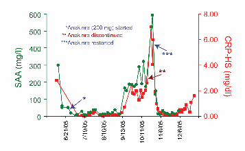
Role of IL-1� Inhibition in FMF. Although colchicine is the standardtreatment for FMF, there are a few patients who do not respond orcannot tolerate its side effects. We recently treated one such FMFpatient with the M694V�M694V genotype and systemic amyloidosis
Fig. 3. Interaction of the B30.2 domain of human pyrin with caspase-1. (A)Human caspase-1 was cotransfected into PT67 cells with various N-terminalGST-fused pyrin-expressing constructs depicted in the schematic, as indicated.PYD, pyrin domain; B, B-box zinc finger domain; C-C, coiled-coil domain; R,B30.2 domain. Lysates were analyzed as in Fig. 2B. (B) Caspase-1 was cotrans-fected with GST vector (first lane) or GST-fused B30.2 domain of pyrin. Lysateswere analyzed as in Fig. 2B.
Fig. 4. The binding of the B30.2 domain of human pyrin to caspase-1 isreduced by FMF-associated mutations. (A) Myc-tagged human caspase-1 wascotransfected with GST-fused WT or FMF-associated mutant C-terminal(amino acids 375–781) pyrin into PT67 cells and analyzed as in Fig. 2B. Densi-tometric analysis of �-myc bands, normalized to the �-myc bands in cell lysates,is shown in the histogram below. (B) Myc-tagged caspase-1 was cotransfectedwith GST-fused WT or FMF-associated mutant full-length pyrin into PT67 cellsand analyzed as in A.
9984 � www.pnas.org�cgi�doi�10.1073�pnas.0602081103 Chae et al.
with anakinra, the IL-1 receptor antagonist, as adjunctive therapy.Monitoring of acute-phase reactants (Fig. 8, which is published assupporting information on the PNAS web site) demonstratedcontrol of both the serum amyloid A and C reactive protein over
several months, except when drug was discontinued during treat-ment of an infection.
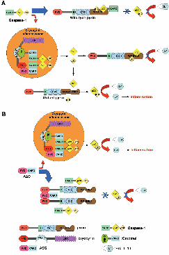
DiscussionIn this manuscript we provide evidence for a previously unrec-ognized, ASC-independent pathway by which pyrin, the proteinmutated in FMF, regulates caspase-1 activation. Our data indi-cate that the C-terminal B30.2 domain of pyrin binds and inhibitsthe catalytic activity of the p20 and p10 caspase-1 subunits andthat three common FMF-associated B30.2 mutations attenuatethis effect (Fig. 9, which is published as supporting informationon the PNAS web site). Computational docking of the crystalstructure of caspase-1 (27, 28) with the modeled structure of thepyrin B30.2 domain (10–12) supports the likelihood of thisinteraction. Moreover, knockdown experiments corroborate anet inhibitory effect of pyrin on IL-1� processing, and clinicaldata also substantiate the importance of IL-1� in the pathogen-esis of FMF.
IL-1� is a major mediator of fever and systemic inflammation(29, 30), and therefore it is not surprising that there is redun-dancy in the regulation of its production and activity. Thenaturally occurring IL-1 receptor antagonist is structurally sim-ilar to IL-1� and binds the IL-1 receptor without stimulating theaccessory receptor necessary for effective signaling (31). An-other highly regulated step is the caspase-1-dependent cleavageof pro-IL-1� into its biologically active 17-kDa fragment.Caspase-1 is thought to undergo autocatalysis by an ‘‘inducedproximity’’ mechanism (32) through N-terminal CARD inter-actions. This process of caspase-1 activation can be mediated byRIP2�RICK�CARDIAK (33–35), Nod1 (36), and CARD12�CLAN�Ipaf (37–39) and also occurs on macromolecular scaf-folds termed inflammasomes (23–25).
Fig. 5. Inhibition of caspase-1-mediated IL-1� processing by human pyrin. (A)pIRES-IL1��ICE or pIRES-ICE (3 �g) was cotransfected with various amounts ofempty vector, WT, or FMF-associated mutant full-length pyrin into PT67 cells.After 48 h, cell culture medium was changed with fresh medium. Cell culturesupernatants were collected 3 h later and assayed for IL-1� by ELISA. Datarepresent the mean � SE from five complete independent experiments. *, P �0.05; **, P � 0.005; ***, P � 0.0005. Western blots demonstrating increasingamounts of the transfected pyrin proteins are shown below. (B) Three micro-grams of pIRES-IL1��ICE, pIRES-IL1�, or pIRES-ICE were cotransfected with 2 �g ofempty vector, WT, or FMF-associated mutant full-length pyrin into PT67 cells, andcell culture supernatantswerecollectedandanalyzedas in A. In thefifth lane, thecaspase-1 inhibitor z-WEHD-fmk was added (at 20 �M) to the culture medium.Data represent the mean � SE from three replicate experiments. Western blotsfor the three transfected proteins are shown in Middle. Bottom is a histogram ofthe amount inhibition of IL-1� in the culture supernatants relative to the inhibi-tion with z-WEHD-fmk.
Fig. 6. A model for the interaction of pyrin with caspase-1. A model for theB30.2 domain of pyrin (blue) was docked to the crystal structure of caspase-1(p20, green; p10, brown). Two common mutations that cause FMF (M694V andM680I, yellow) are located in the putative binding interface.
Fig. 7. Knockdown of endogenous pyrin in THP-1 cells. Cells were trans-fected with MEFV siRNA or a scrambled sequence dsRNA control and after 72 hwere stimulated with 100 ng�ml LPS for 4 h. (A) Western blot of cell lysatesprobed with Abs to pyrin or GAPDH. (B) ELISA of culture supernatants forIL-1�. Data represent the mean � SE for three replicate cultures. Data are froma representative experiment from six independent transfections.
Chae et al. PNAS � June 27, 2006 � vol. 103 � no. 26 � 9985
MED
ICA
LSC
IEN
CES

Under various experimental conditions (19, 26), pyrin itself caneither inhibit or accentuate caspase-1 activity through the interac-tion of its N-terminal death-fold with ASC (19, 26), a key moleculein the inflammasome (23–25). This article demonstrates that pyrinalso modulates caspase-1 activation through a second interactioninvolving the binding of its C-terminal B30.2 domain to the catalyticdomains of caspase-1. The B30.2 domain is found in �500 differentproteins of various functions and is thought to mediate protein–protein interactions (9–12). The B30.2 domain, in turn, comprisesan �139-aa C-terminal SPRY segment (named after the dual-purpose splA kinase and the ryanodine receptor) and an �61-aaN-terminal PRY subdomain.
Recently, the structure of the SPRY domain of SSB-2 wasdetermined by NMR (10), and the structures of a second SPRYdomain and a B30.2 domain were solved by crystallography (11,12). All three analyses indicated a �-sandwich structure formedby antiparallel �-sheets in the SPRY domain and showed that theresidues equivalent to the M694V and M680I mutations in thepyrin B30.2 domain fall in distinct loops comprising a commonbinding surface. The computational docking studies reportedhere indicate that this surface of pyrin’s B30.2 domain can in factinteract with caspase-1 and that FMF-associated mutationsdisrupt this interaction. Our modeling did not predict a signif-icant role for the V726A mutation in abrogating the pyrin–caspase-1 interaction. However, this mutation has been associ-ated with a milder phenotype than the other two variants (3, 4),and it is possible that the effects we see in our biochemical andfunctional assays are the result of structural perturbations thatare more apparent in vitro than in vivo.
Our knockdown data indicate that the overall effect of pyrinon IL-1� processing in human monocytes, taking into accountboth ASC-dependent and ASC-independent pathways, is inhib-itory. We had previously proposed an ASC-dependent inhibitoryeffect of pyrin on IL-1� processing based on studies of mousepyrin. Consistent with this hypothesis, others had shown in a293T transfection system that human pyrin inhibits the interac-tion of ASC with cryopyrin and that pyrin inhibits the interactionof human ASC with caspase-8 (40, 41).
More recently, Yu et al. (26), using a 293T cell-based ASC–caspase-1 transfection system similar to the two previous studies,but stably expressing a more physiologic level of ASC, did notobserve that pyrin inhibits cryopyrin-induced caspase-1 activa-tion. Moreover, under these experimental conditions WT pyrinactually induced ASC-dependent caspase-1 activation and IL-1�secretion, but FMF-associated mutations did not increase ordecrease this effect. It is noteworthy that another laboratory,also using the 293T transfection system, observed a potentiatingeffect of pyrin on caspase-1 activation when ASC was present,but an inhibitory effect of pyrin when pyrin was coexpressed withcaspase-1 and IL-1� but without ASC (42).
Human embryonic kidney 293T cells may afford a number ofadvantages in their ease of transfection and passage in tissueculture. However, even if they are stably transfected withphysiologic levels of ASC, they may not recapitulate all of therelevant interactions of endogenous proteins in leukocytes. Inour previous work we demonstrated that transfection of MEFVinto human U937 myeloid cells, which endogenously expressASC, pro-caspase-1, and IL-1�, inhibited LPS-induced IL-1�production relative to cells transfected with empty vector (19).In the present study we used THP-1, another human monocyticcell line that endogenously expresses all of the relevant molec-ular machinery of IL-1� secretion. Using siRNA specific forMEFV we observed dramatic and specific reduction of pyrinexpression and a concomitant increase in LPS-induced IL-1�secretion.
Our data are consistent with a model in which WT pyrininhibits caspase-1 activation, perhaps as part of a homeostaticloop induced by proinflammatory stimuli. By this formulation,
FMF-associated mutations have less of an inhibitory effect andmay have been selected in human history because of the resultingincrease in innate immune responses. The beneficial effect ofanakinra in an otherwise refractory FMF patient heightensinterest in the role of pyrin in the regulation of cytokineprocessing.
Materials and MethodsGST Pull-Down Assay, Coimmunoprecipitation, and Western Blots. Toperform coimmunoprecipitation of endogenous pyrin andcaspase-1, THP-1 cells were lysed with Mild lysis buffer (Cyto-Signal) and incubated with a polyclonal Ab against the N-terminal 374 aa of human pyrin or anti-caspase-1 Ab (Santa CruzBiotechnology), which were crosslinked to protein A agarosebeads (Pierce) for 16 h at 4°C. After washing the beads, boundproteins were eluted and subjected to Western blotting. For invivo GST pull-down, WT, FMF-associated mutant, and domain-deleted MEFV and genes encoding each domain of caspase-1were cloned into a mammalian GST expression vector (pEGST)and pcDNA3.1-myc�His (Invitrogen). Each GST-fused expres-sion construct for pyrin and caspase-1 was transiently cotrans-fected into RetroPack PT67 cells (CLONTECH) with myc-tagged caspase-1 and pyrin expression constructs, respectively,using Lipofectamine 2000 reagent (Invitrogen). After 24 h, cellswere lysed with Strong lysis buffer (CytoSignal) and incubatedwith GST beads (Amersham Pharmacia) for 16 h at 4°C. Celllysates or eluted samples obtained from GST pull-down andcoimmunoprecipitation were subjected to Western blotting.Western blots were performed by using primary Abs: anti-pyrinAb; anti-caspase-1, -4, -5, and -6 Abs (Santa Cruz Biotechnol-ogy); anti-caspase-2, -3, -7, -8, -9, and -10 Abs (R & D Systems);anti-GST Ab (Amersham Pharmacia); and anti-myc Ab (SantaCruz Biotechnology).
RT-PCR. RT-PCR analysis was performed on total RNA fromPT67 cells and LPS-treated mouse monocytes using a forwardprimer (5�-ATGGGGCGGGCACGAGATG-3�) and a reverseprimer (5�-GCTCTGCTCCAGGTCCATCAC-3�) for the detec-tion of mouse ASC expression and mouse G3PDH controlamplimer set (CLONTECH).
Measurement of IL-1� Secretion. Both or each of the genes encod-ing IL-1� and caspase-1 were subcloned into the bicistronicmammalian expression vector pIRES (CLONTECH) and de-noted pIRES-IL1��ICE, pIRES-IL1�, or pIRES-ICE. Theseconstructs were cotransfected into PT67 cells with variousamounts of pMEFV-Myc and pNBC-Myc or 2 �g of mutantpyrin constructs (pM680I-Myc, pM694V-Myc, and pV726A-Myc), as indicated. After 48 h, cell culture medium was changedwith fresh medium, and cell culture supernatants were collected3 h later for IL-1� ELISA (R & D Systems), performedaccording to the manufacturer’s instructions.
siRNA Studies. siRNA duplex (sequence CTCTGCTGGTCAC-CTACTA) corresponding to pyrin was designed by Dharmacon.The negative control scramble-sequence siRNA was purchasedfrom Ambion. THP-1 cells were transfected with siRNA (100nM) by using Lipofectamine 2000 (Invitrogen), incubated for72 h, and then cultured an additional 4 h with LPS (100 ng�ml).Cell lysates and supernatants were then collected for Westernblotting and IL-1� ELISA, respectively.
Computational Analysis of the Interaction of Pyrin with Caspase-1.The MODELLER program (version 6v2) (43) was used toproduce homology models of the pyrin B30.2 domain based onthe structure of PRYSPRY (Protein Data Bank ID code2FBE) as a template. An alignment for the sequences of theseproteins was obtained by using the program FUGUE (44).
9986 � www.pnas.org�cgi�doi�10.1073�pnas.0602081103 Chae et al.

Twenty-five models were then created, and the model withlowest MODELLER objective function was used for docking withthe receptor. The ZDOCK (45) program was used to performrigid-body docking of the pyrin B30.2 domain model as theligand, with caspase-1 as the receptor, producing 2,000 possi-ble interactions ranked according to their structural andelectrostatic complementarity. The C-terminal residues pro-jecting from the p10 subunit of caspase-1 (V292 to D297) wereremoved because this region is likely to be f lexible. The
alanine mutation at position 285 was converted to the nativecysteine. The highest-scoring initial 200 models were subjectedto molecular mechanics refinement and redocking using theRDOCK approach (46). The interaction energy in the refinedmodels was evaluated by using a scoring function includingdesolvation and electrostatic terms. The buried surface areawas calculated from the difference in solvent-accessible sur-face areas of the uncomplexed and complexed structures byusing the program NACCESS (version 2.1.1) (47).
1. Galon, J., Aksentijevich, I., McDermott, M. F., O’Shea, J. J. & Kastner, D. L.(2000) Curr. Opin. Immunol. 12, 479–486.
2. Stojanov, S. & Kastner, D. L. (2005) Curr. Opin. Rheumatol. 17, 586–599.3. Kastner, D. L. & Aksentijevich, I. (2005) in Arthritis and Allied Conditions, eds.
Koopman, W. J. & Moreland, L. W. (Lippincott Williams and Wilkins,Philadelphia), pp. 1411–1461.
4. International FMF Consortium (1997) Cell 90, 797–807.5. French FMF Consortium (1997) Nat. Genet. 17, 25–31.6. Centola, M., Wood, G., Frucht, D. M., Galon, J., Aringer, M., Farrell, C.,
Kingma, D. W., Horwitz, M. E., Mansfield, E., Holland, S. M., et al. (2000)Blood 95, 3223–3231.
7. Matzner, Y., Abedat, S., Shapiro, E., Eisenberg, S., Bar-Gil-Shitrit, A.,Stepensky, P., Calco, S., Azar, Y. & Urieli-Shoval, S. (2000) Blood 96, 727–731.
8. Diaz, A., Hu, C., Kastner, D. L., Schaner, P., Reginato, A. M., Richards, N. &Gumucio, D. L. (2004) Arthritis Rheum. 50, 3679–3689.
9. Henry, J., Mather, I. H., McDermott, M. F. & Pontarotti, P. (1998) Mol. Biol.Evol. 15, 1696–1705.
10. Masters, S. L., Yao, S., Willson, T. A., Zhang, J. G., Palmer, K. R., Smith, B. J.,Babon, J. J., Nicola, N. A., Norton, R. S. & Nicholson, S. E. (2006) Nat. Struct.Mol. Biol. 13, 77–84.
11. Grutter, C., Briand, C., Capitani, G., Mittl, P. R., Papin, S., Tschopp, J. &Grutter, M. G. (2006) FEBS Lett. 580, 99–106.
12. Woo, J. S., Imm, J. H., Min, C. K., Kim, K. J., Cha, S. S. & Oh, B. H. (2006)EMBO J. 25, 1353–1363.
13. Bertin, J. & DiStefano, P. S. (2000) Cell Death Differ. 7, 1273–1274.14. Fairbrother, W. J., Gordon, N. C., Humke, E. W., O’Rourke, K. M., Staro-
vasnik, M. A., Yin, J. P. & Dixit, V. M. (2001) Protein Sci. 10, 1911–1918.15. Martinon, F., Hofmann, K. & Tschopp, J. (2001) Curr. Biol. 11, R118–R120.16. Pawlowski, K., Pio, F., Chu, Z., Reed, J. C. & Godzik, A. (2001) Trends
Biochem. Sci. 26, 85–87.17. Staub, E., Dahl, E. & Rosenthal, A. (2001) Trends Biochem. Sci. 26, 83–85.18. Richards, N., Schaner, P., Diaz, A., Stuckey, J., Shelden, E., Wadhwa, A. &
Gumucio, D. L. (2001) J. Biol. Chem. 276, 39320–39329.19. Chae, J. J., Komarow, H. D., Cheng, J., Wood, G., Raben, N., Liu, P. P. &
Kastner, D. L. (2003) Mol. Cell 11, 591–604.20. Masumoto, J., Taniguchi, S., Ayukawa, K., Sarvotham, H., Kishino, T.,
Niikawa, N., Hidaka, E., Katsuyama, T., Higuchi, T. & Sagara, J. (1999) J. Biol.Chem. 274, 33835–33838.
21. Srinivasula, S. M., Poyet, J. L., Razmara, M., Datta, P., Zhang, Z. & Alnemri,E. S. (2002) J. Biol. Chem. 277, 21119–21122.
22. Stehlik, C., Fiorentino, L., Dorfleutner, A., Bruey, J. M., Ariza, E. M., Sagara,J. & Reed, J. C. (2002) J. Exp. Med. 196, 1605–1615.
23. Martinon, F., Burns, K. & Tschopp, J. (2002) Mol. Cell 10, 417–426.
24. Agostini, L., Martinon, F., Burns, K., McDermott, M. F., Hawkins, P. N. &Tschopp, J. (2004) Immunity 20, 319–325.
25. Martinon, F. & Tschopp, J. (2004) Cell 117, 561–574.26. Yu, J. W., Wu, J., Zhang, Z., Datta, P., Ibrahimi, I., Taniguchi, S., Sagara, J.,
Fernandes-Alnemri, T. & Alnemri, E. S. (2006) Cell Death Differ. 13, 236–249.27. Wilson, K. P., Black, J. A., Thomson, J. A., Kim, E. E., Griffith, J. P., Navia,
M. A., Murcko, M. A., Chambers, S. P., Aldape, R. A., Raybuck, S. A., et al.(1994) Nature 370, 270–275.
28. Romanowski, M. J., Scheer, J. M., O’Brien, T. & McDowell, R. S. (2004)Structure (London) 12, 1361–1371.
29. Dinarello, C. A. (2004) J. Endotoxin Res. 10, 201–222.30. Dinarello, C. A. (2005) Crit. Care Med. 33, S460–S462.31. Arend, W. P., Malyak, M., Guthridge, C. J. & Gabay, C. (1998) Annu. Rev.
Immunol. 16, 27–55.32. Salvesen, G. S. & Dixit, V. M. (1999) Proc. Natl. Acad. Sci. USA 96,
10964–10967.33. Inohara, N., del Peso, L., Koseki, T., Chen, S. & Nunez, G. (1998) J. Biol. Chem.
273, 12296–12300.34. McCarthy, J. V., Ni, J. & Dixit, V. M. (1998) J. Biol. Chem. 273, 16968–16975.35. Thome, M., Hofmann, K., Burns, K., Martinon, F., Bodmer, J. L., Mattmann,
C. & Tschopp, J. (1998) Curr. Biol. 8, 885–888.36. Yoo, N. J., Park, W. S., Kim, S. Y., Reed, J. C., Son, S. G., Lee, J. Y. & Lee,
S. H. (2002) Biochem. Biophys. Res. Commun. 299, 652–658.37. Damiano, J. S., Stehlik, C., Pio, F., Godzik, A. & Reed, J. C. (2001) Genomics
75, 77–83.38. Geddes, B. J., Wang, L., Huang, W. J., Lavellee, M., Manji, G. A., Brown, M.,
Jurman, M., Cao, J., Morgenstern, J., Merriam, S., et al. (2001) Biochem.Biophys. Res. Commun. 284, 77–82.
39. Poyet, J. L., Srinivasula, S. M., Tnani, M., Razmara, M., Fernandes-Alnemri,T. & Alnemri, E. S. (2001) J. Biol. Chem. 276, 28309–28313.
40. Dowds, T. A., Masumoto, J., Chen, F. F., Ogura, Y., Inohara, N. & Nunez, G.(2003) Biochem. Biophys. Res. Commun. 302, 575–580.
41. Masumoto, J., Dowds, T. A., Schaner, P., Chen, F. F., Ogura, Y., Li, M., Zhu,L., Katsuyama, T., Sagara, J., Taniguchi, S., et al. (2003) Biochem. Biophys. Res.Commun. 303, 69–73.
42. Stehlik, C., Lee, S. H., Dorfleutner, A., Stassinopoulos, A., Sagara, J. & Reed,J. C. (2003) J. Immunol. 171, 6154–6163.
43. Sali, A. & Blundell, T. L. (1993) J. Mol. Biol. 234, 779–815.44. Shi, J., Blundell, T. L. & Mizuguchi, K. (2001) J. Mol. Biol. 310, 243–257.45. Chen, R., Li, L. & Weng, Z. (2003) Proteins 52, 80–87.46. Li, L., Chen, R. & Weng, Z. (2003) Proteins 53, 693–707.47. Hubbard, S. L. & Thornton, J. M. (1993) NACCESS (University College London,
London), version 2.1.1.
Chae et al. PNAS � June 27, 2006 � vol. 103 � no. 26 � 9987
MED
ICA
LSC
IEN
CES