Embed Size (px)
Citation preview

Radiographic evaluation of mid face fracture
Definition
• Fracture of the mid facial region may be limited to the maxilla alone or may be quite variable but often follow the general patterns classified by Leon le fort.
classification
• Horizontal fracture le fort 1• Pyramidal fracture le fort 2• Craniofacial disjunction le fort 3• Zygomatic fracture
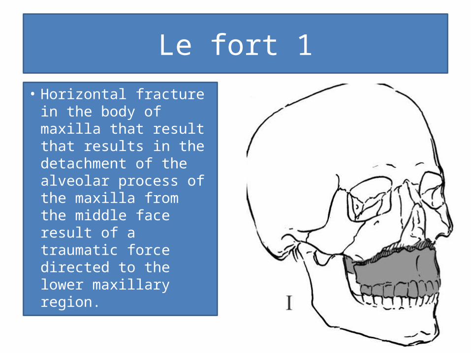
Le fort 1• Horizontal fracture
in the body of maxilla that result that results in the detachment of the alveolar process of the maxilla from the middle face result of a traumatic force directed to the lower maxillary region.
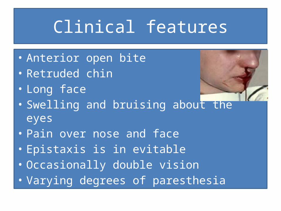
Clinical features
• Anterior open bite • Retruded chin• Long face• Swelling and bruising about the eyes • Pain over nose and face• Epistaxis is in evitable • Occasionally double vision • Varying degrees of paresthesia
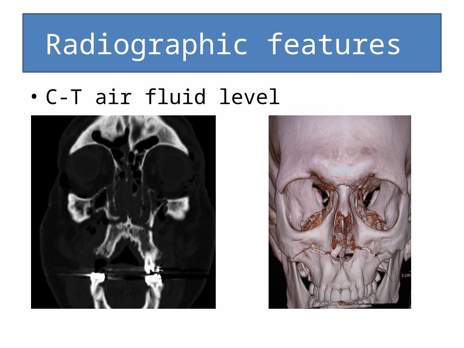
Radiographic features
• C-T air fluid level
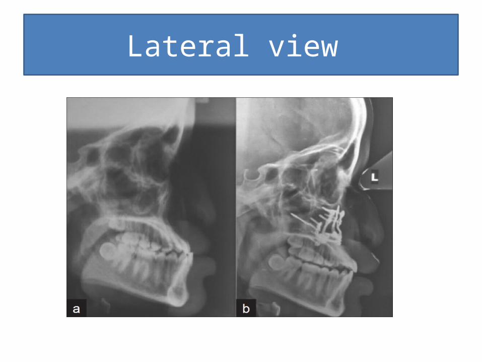
Lateral view
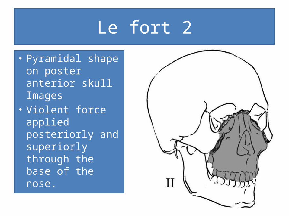
Le fort 2
• Pyramidal shape on poster anterior skull Images
• Violent force applied posteriorly and superiorly through the base of the nose.
Clinical features
• Edema • Swelling of the middle third of the face• Ecchymosis• Cerebrospinal fluid rhinorrhea• Double vision• Variable degrees of paresthesia
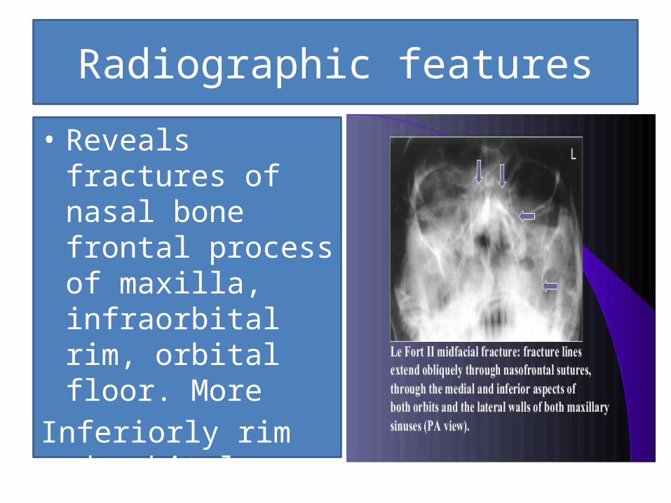
Radiographic features
• Reveals fractures of nasal bone frontal process of maxilla, infraorbital rim, orbital floor. More
Inferiorly rim and orbital floor.
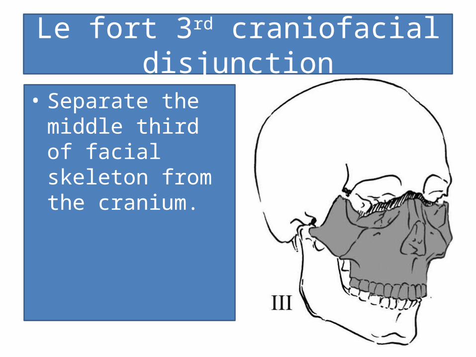
Le fort 3rd craniofacial disjunction
• Separate the middle third of facial skeleton from the cranium.
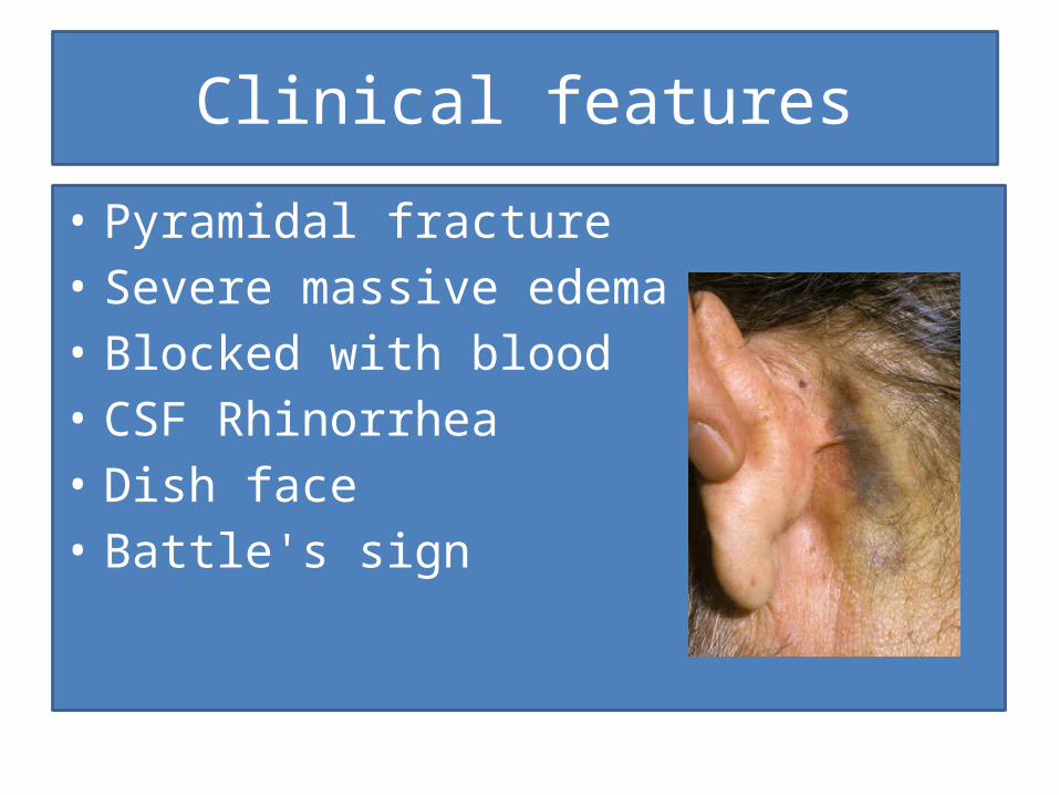
Clinical features
• Pyramidal fracture• Severe massive edema• Blocked with blood• CSF Rhinorrhea• Dish face• Battle's sign
Radiographic features
• Multiple fractures with plain films.• Radiopaque air fluid levels with mucosal
thickening.
Examination after mid face trauma
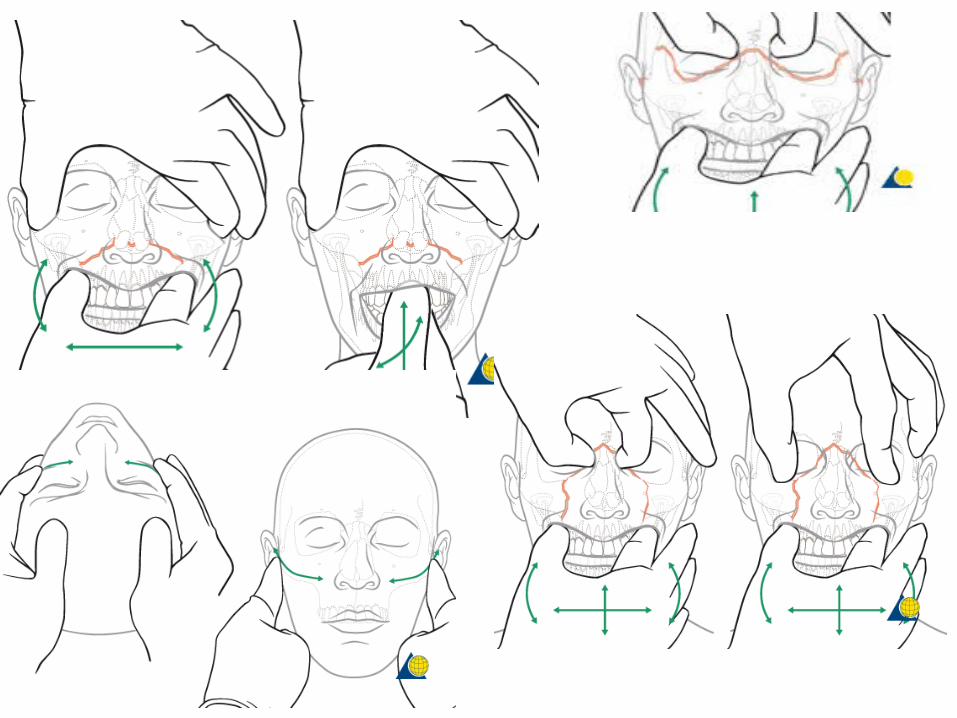
• To clinically evaluate possible midfacial injuries a standard examination protocol is strongly recommended and has to include full examination of the head, eyes, ears, nose, throat, and neck.
• For the experienced surgeon, assessment of midfacial injuries does not take very long.
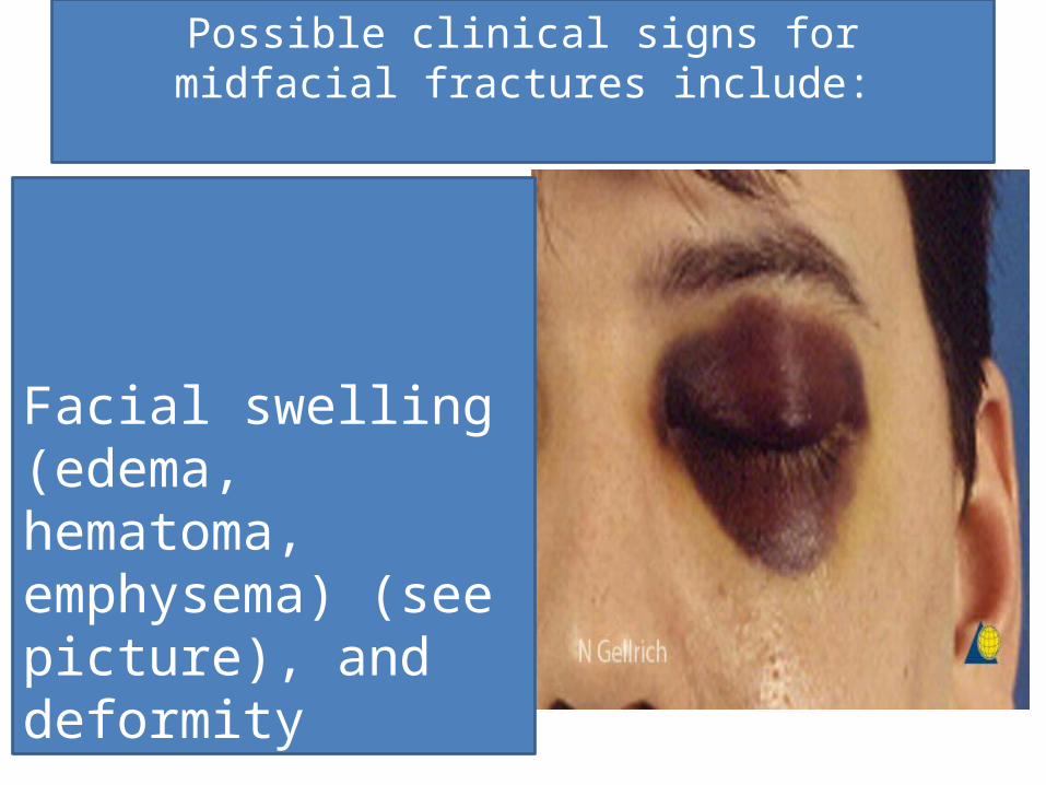
Possible clinical signs for midfacial fractures include:
Facial swelling (edema, hematoma, emphysema) (see picture), and deformity
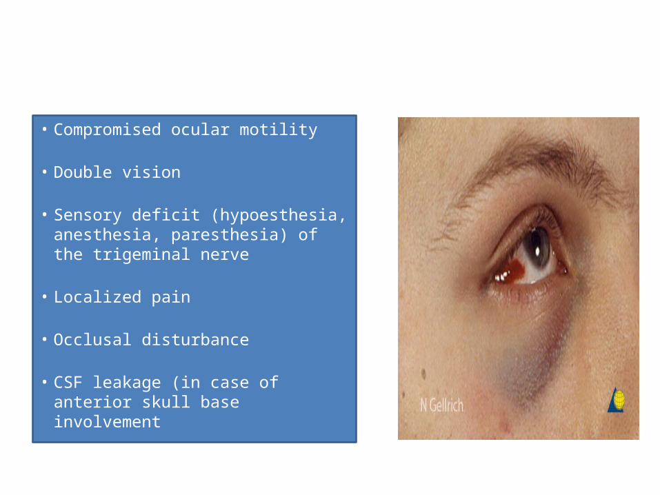
• Compromised ocular motility
• Double vision
• Sensory deficit (hypoesthesia, anesthesia, paresthesia) of the trigeminal nerve
• Localized pain
• Occlusal disturbance
• CSF leakage (in case of anterior skull base involvement
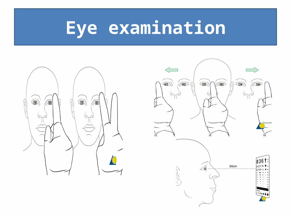
Eye examination
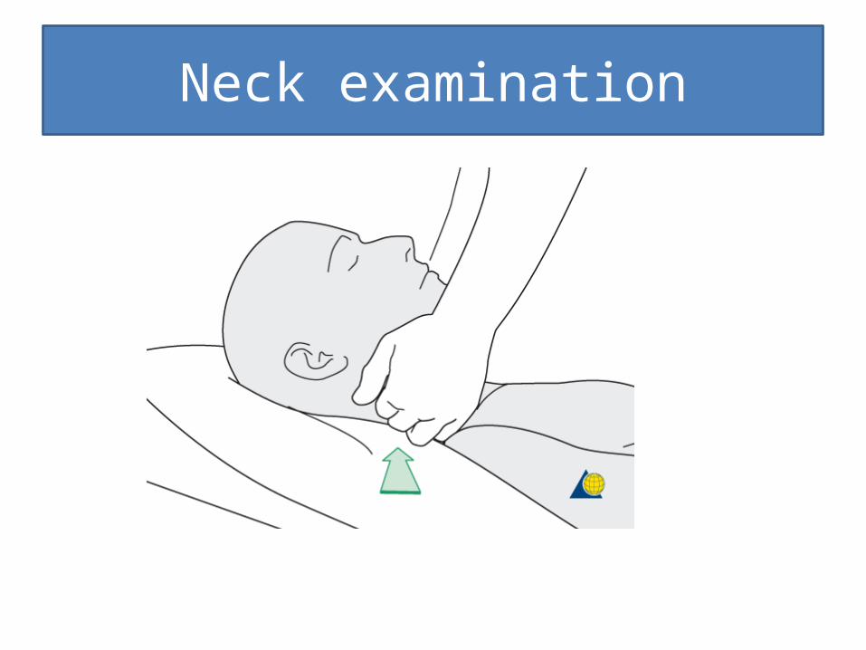
Neck examination
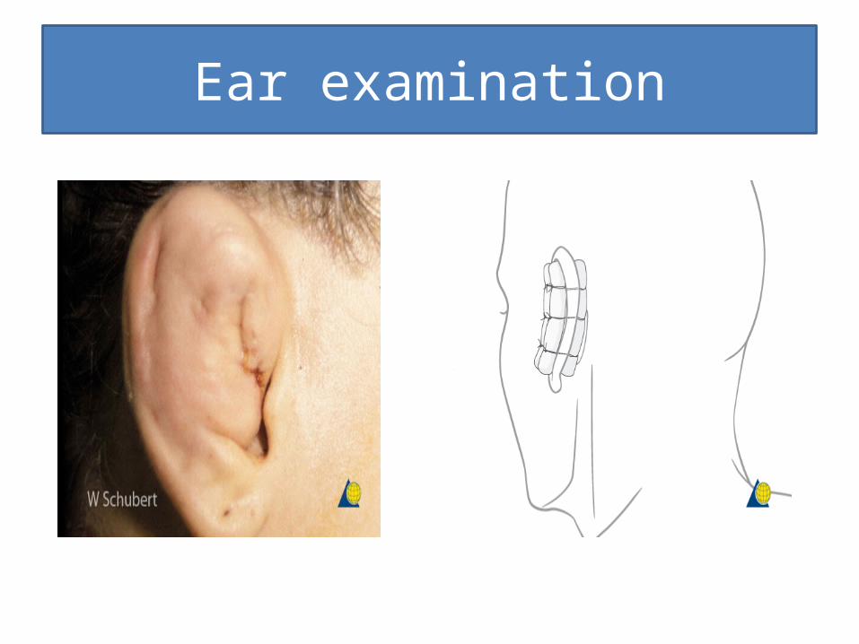
Ear examination
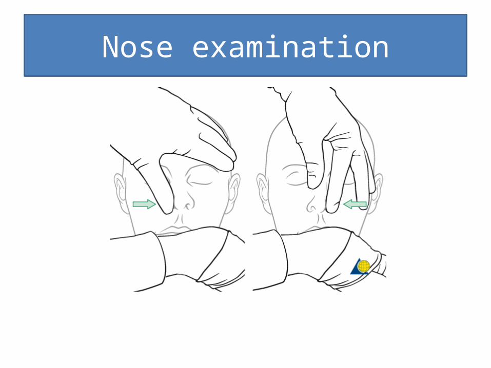
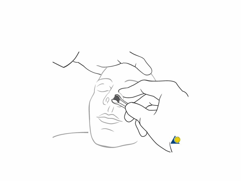
Nose examination
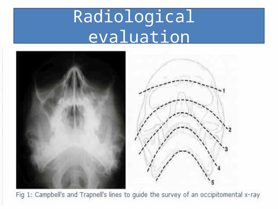
Radiological evaluation
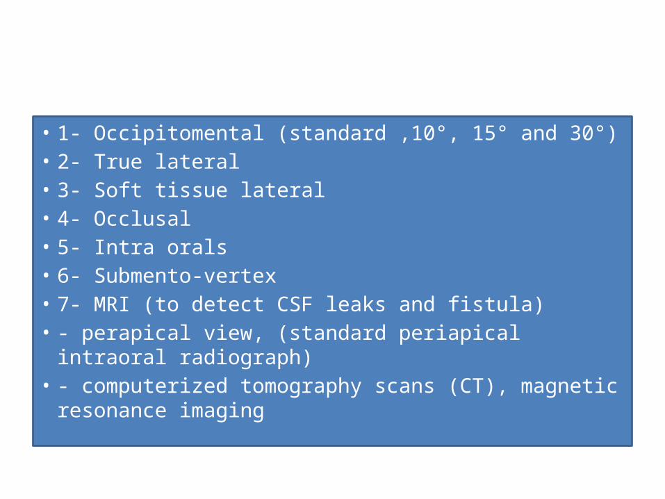
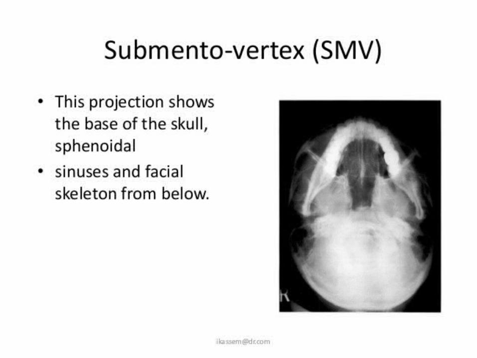
• 1- Occipitomental (standard ,10°, 15° and 30°)• 2- True lateral• 3- Soft tissue lateral• 4- Occlusal• 5- Intra orals• 6- Submento-vertex• 7- MRI (to detect CSF leaks and fistula)• - perapical view, (standard periapical intraoral radiograph)• - computerized tomography scans (CT), magnetic
resonance imaging
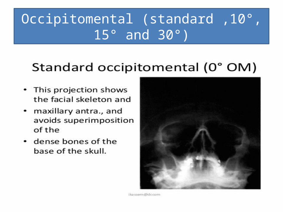
Occipitomental (standard ,10°, 15° and 30°)
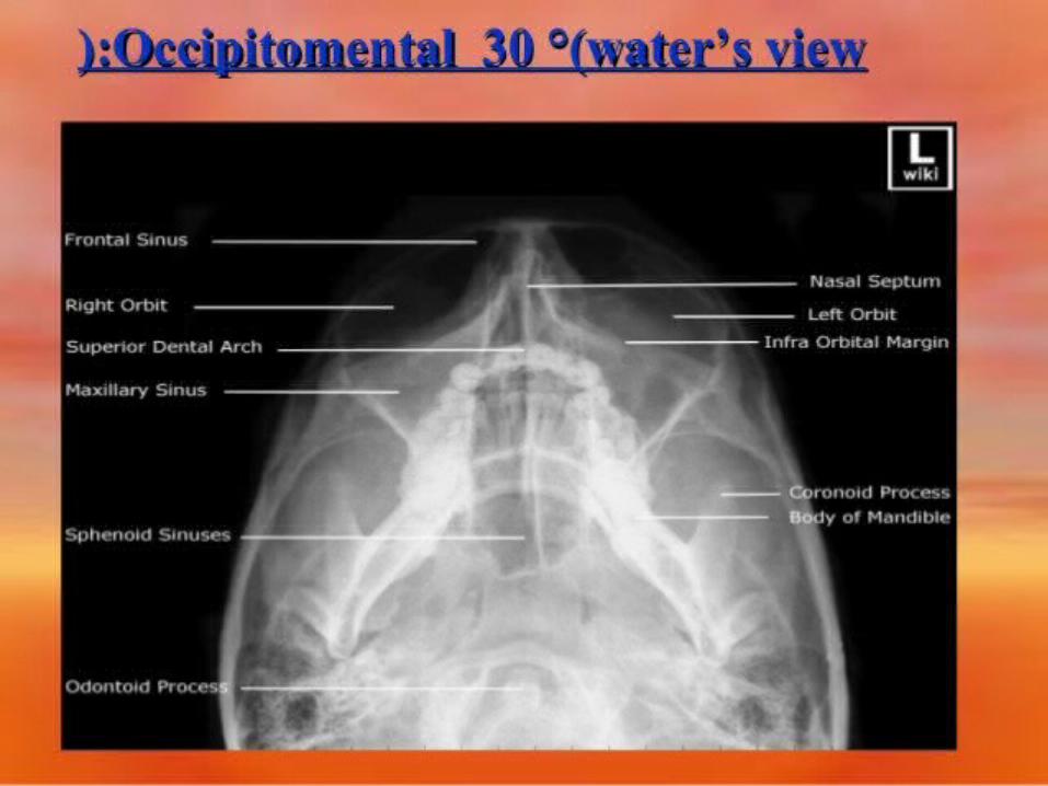
Occipitomental 30degreewater’s view
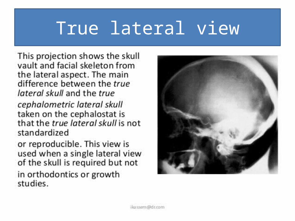
True lateral view
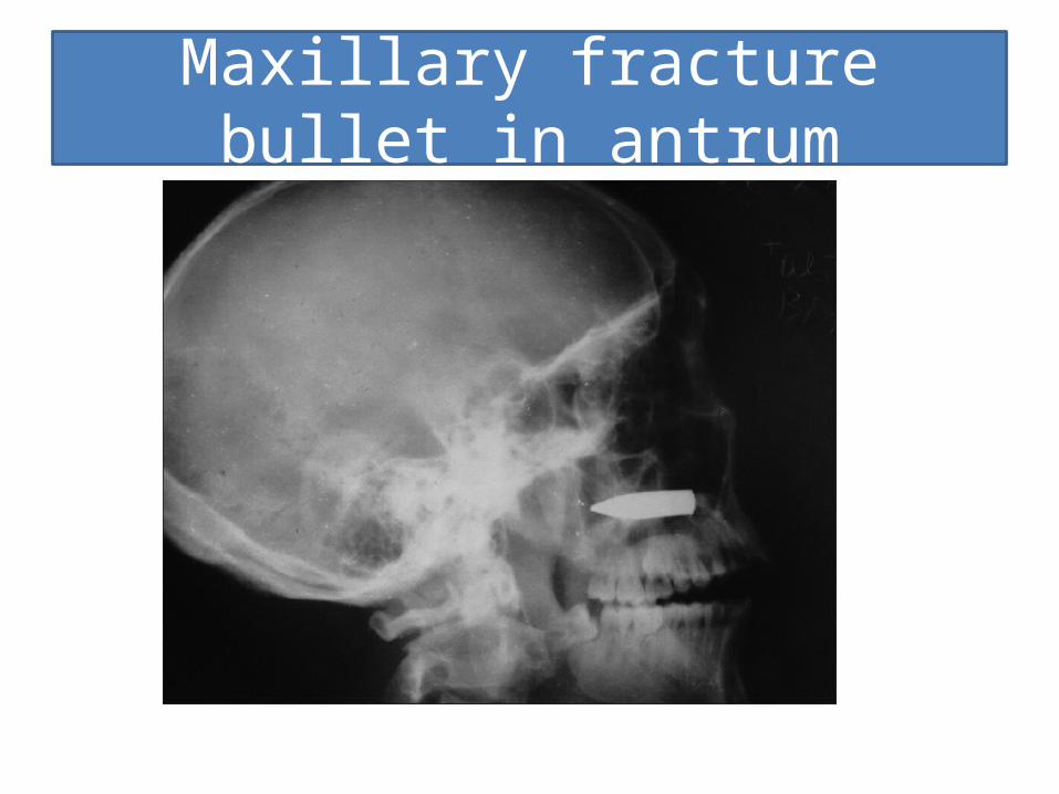
Maxillary fracture bullet in antrum
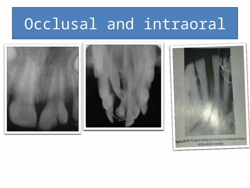
Occlusal and intraoral
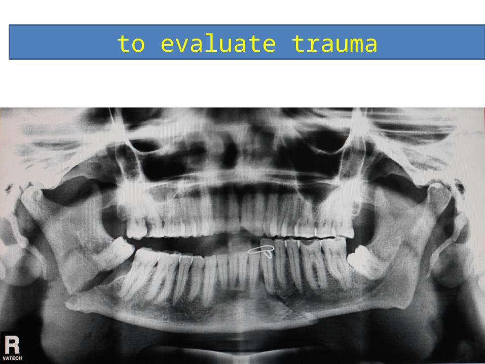
to evaluate trauma
CBCT
• CBCT has become increasingly important in treatment planning and diagnosis in implant dentistry, interventional radiology (IR), among other things. Perhaps because of the increased access to such technology, CBCT scanners are now finding many uses in dentistry, such as in the fields of endodontics and orthodontics, as well. IN TRAUMA
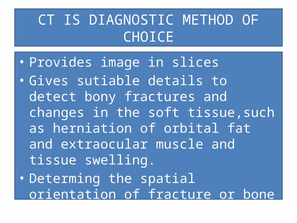
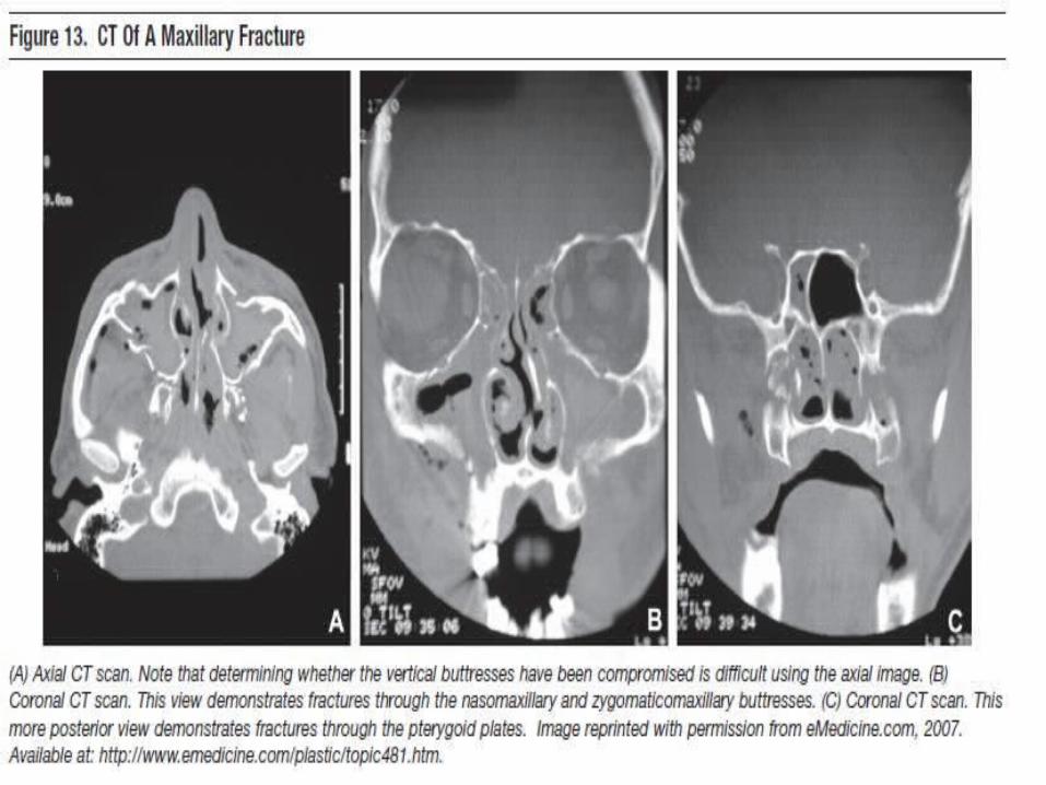
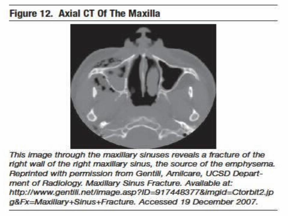
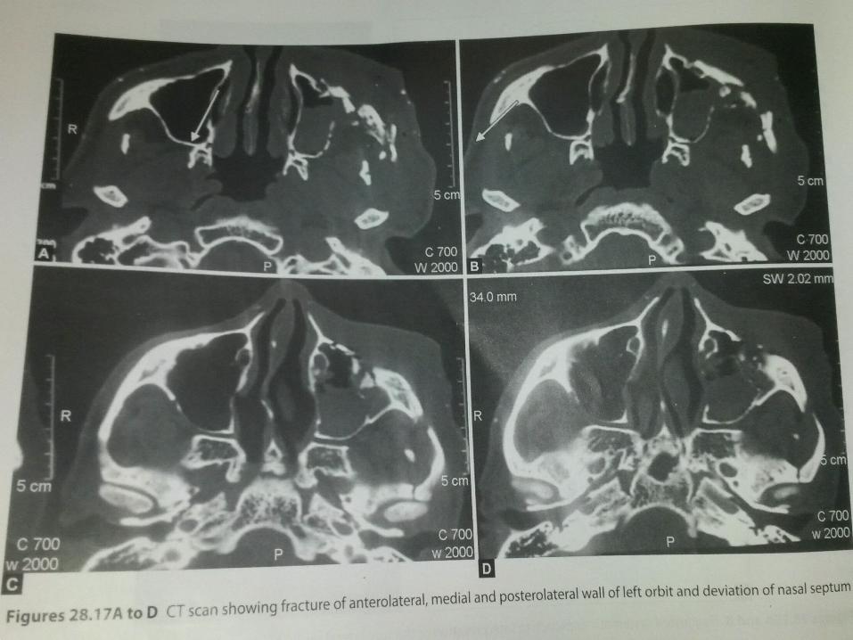
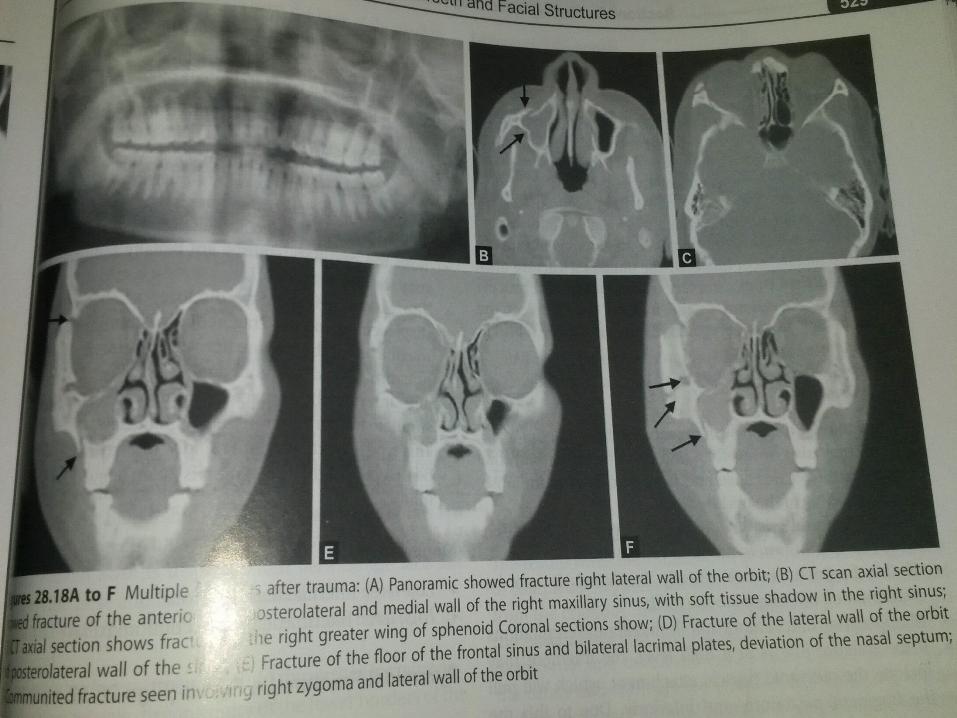
CT IS DIAGNOSTIC METHOD OF CHOICE
• Provides image in slices• Gives sutiable details to detect bony fractures
and changes in the soft tissue,such as herniation of orbital fat and extraocular muscle and tissue swelling.
• Determing the spatial orientation of fracture or bone fragments
• Reformatted in three dimensional images
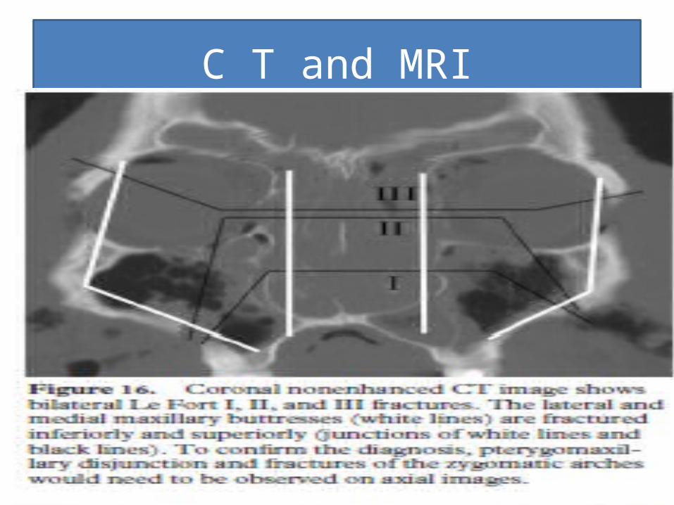
C T and MRI
conclusion
• Radiography play important role in the diagnosis ,location and determination of the extent of injury in cases of traumatic injuries however there are serious limitation in the study of bone and teeth for evidence of fracturebut with the advent of newer imaging modalites like CT and MRI AND CBCT detection of minute fracture is posssible
• PRINCIPLES OF DENTAL IMAGING – by Langland and Langlais
• ORAL RADIOLOGY PRINCIPLES AND INTERPRETATIONS –by White and Pharaoh
• TEXTBOOK OF DENTAL AND MAXILLOFACIAL RADIOLOGY –by Freny R. Karjodkar
REFERENCES