Embed Size (px)
Citation preview
https://kat.cr/user/Blink99/

Smart Study SeriesObstetrics and Gynecology

This page intentionally left blank
https://kat.cr/user/Blink99/

Smart Study SeriesObstetrics and Gynecology
Third Edition
Punit S BhojaniMD, DNB, FCPS, DGO, DFP
Consultant Obstetrician and Gynecologist Mumbai
ELSEVIERA division of
Reed Elsevier India Private Limited
Smart Study Series: Obstetrics and Gynecology, 3/eBhojani
© 2014 Reed Elsevier India Private Limited
All rights reserved. No part of this publication may be reproduced or transmitted in any form or by any means, electronic or mechanical, including photocopying, recording, or any information storage and retrieval system, without permission in writing from the Publisher.
This book and the individual contributions contained in it are protected under copyright by the Publisher (other than as may be noted herein).
ISBN: 978-81-312-3767-0
Notices
Knowledge and best practice in this fi eld are constantly changing. As new research and experience broaden our understand-ing, changes in research methods, professional practices, or medical treatment may become necessary.
Practitioners and researchers must always rely on their own experience and knowledge in evaluating and using any informa-tion, methods, compounds, or experiments described herein. In using such information or methods they should be mindful of their own safety and the safety of others, including parties for whom they have a professional responsibility.
With respect to any drug or pharmaceutical products identifi ed, readers are advised to check the most current information provided (i) on procedures featured or (ii) by the manufacturer of each product to be administered, to verify the recommended dose or formula, the method and duration of administration, and contraindications. It is the responsibility of practitioners, relying on their own experience and knowledge of their patients, to make diagnoses, to determine dosages and the best treat-ment for each individual patient, and to take all appropriate safety precautions.
To the fullest extent of the law, neither the Publisher nor the authors, contributors, or editors, assume any liability for any injury and/or damage to persons or property as a matter of products liability, negligence or otherwise, or from any use or operation of any methods, products, instructions, or ideas contained in the material herein.
Please consult full prescribing information before issuing prescription for any product mentioned in this publication.
The Publisher
Published by Reed Elsevier India Private LimitedRegistered Office: 305, Rohit House, 3 Tolstoy Marg, New Delhi-110 001Corporate Office: 14th Floor, Building No. 10B, DLF Cyber City, Phase II, Gurgaon-122 002, Haryana, India
Content Strategist: Anubhuti KalaProject Managers: Anand K Jha & Nayagi AthmanathanCopy Editor: Isha BaliManager - Publishing Operations: Sunil KumarManager - Production: N C PantCover Designer: Raman Kumar
Typeset by TNQ Books & Journals, Chennai, IndiaPrinted and bound at
https://kat.cr/user/Blink99/

Dedicated tomy Teachersmy Students
my Parents and my Wife

This page intentionally left blank
https://kat.cr/user/Blink99/
vii
Foreword
It is said that true study of mankind is in books. Historically speaking books are the most convenient form for extra-corporeal memory allowing the reader to benefit from knowledge and work of experts in any selected field.
Today there has been a veritable explosion in scientific information which often leaves students overwhelmed and confused.
This particular book in Obstetrics and Gynecology which is a part of “Smart Study Series” is published to address the special need of both undergraduate and postgraduate medical students. While it is particularly useful while preparing for entrance into postgraduation, in my opinion information which is presented with clarity and lucidity could be of use to any practitioner of Obstetrics and Gynecology.
I have known the author Dr Punit S Bhojani as a student and seen him evolve into being an inspiring teacher. That the book is already into its third edition is ample testimony to its success.
I wish the students reading this book the very best and hope that they can achieve academic and professional success and also enjoy the journey.
Dr Nozer K SheriarMD, DNB, FICOG, FCPS, DGO
Consultant Obstetrician and Gynecologist, Breach Candy, Hinduja, Holy Family and Masina Hospitals, Mumbai
President Mumbai Obstetrics and Gynecological SocietyPostgraduate teacher for DNB

This page intentionally left blank
https://kat.cr/user/Blink99/
ix
Foreword
“Smart Study Series in Obstetrics and Gynecology” has been written by Dr Punit Bhojani keeping in mind the needs of medical graduates who are aiming to do their postgraduation. This text is a handy reference for those who are preparing to appear in postgraduate entrance examinations. The book covers all aspects of obstetrics and gynecol-ogy and forms a fair part of the MCQ-based qualifying examination. It gives a comprehensive, brief and yet lucid account of the subject.
Dr Bhojani has considerable experience in guiding the PG aspirants and now he has compiled all his knowledge in this volume with the objective to benefit many more students as well. The book will also prove a quick tool for revision to both undergraduate and postgraduate medical students.
Dr Vinita Salvi Consultant Obstetrician and Gynecologist,
Seven Hills Hospital, Mumbai Ex-Professor and Head of Unit,
Seth GS Medical College and King Edward Memorial Hospital, Mumbai
Ex-Officer in-charge, ICMR, Regional Centre for Research in Reproduction
Mumbai, India

This page intentionally left blank
https://kat.cr/user/Blink99/

xi
Preface
It gives me great pleasure to present to you the third edition of “Smart Study Series: Obstetrics and Gynecology”. The tremendous success of the first two editions and an overwhelming response from the students, have been the driving force for this edition.
I am very happy that the book has delivered what it had promised—a sure success in all the entrance examinations.It feels fantastic to hear from my students that the first two editions have stood the test of all exams conducted
over the past years. As per the feedback of my students, all questions in OBGYN, including the NEET and the recently conducted AIIMS and AIPGE were directly or indirectly from this book.
I am very confident that this third edition will tremendously benefit the students.Mastering the book is more than enough preparation for the subject. I have designed it to be a one-stop prepara-
tion source for OBGYN.The third edition is even bigger and better. MCQs have been updated. Recent advances have been added which
will become important in the future examinations. I have tried to keep repetition (between the theory and MCQ section) to the minimum. Hence, I would urge the students to master both sections before taking any entrance examination.
Each and every line in the book is a potential MCQ for the exam.Though this book is principally for students preparing for postgraduate entrance exams, I am pretty confident that
final year MBBS and postgraduate students will find it extremely handy for rapid revisions before exams.Also, extreme care has been taken to authenticate each statement made in this book based on postgraduate text-
books like “Williams 22nd/e”, “Speroff 7th/e”, “Novak’s 14th/e”, “TeLinde’s 9th/e”, etc.Lastly, I urge you to see my video lectures on my website www.drmentors.com. This is India’s biggest website,
containing more than 200 hours of pre-recorded video lectures by best faculties, useful for any PG entrance exam. You can see the lectures multiple times as per your convenience.
Suggestions, queries and corrections are always welcome. You can personally contact me at [email protected] you all the success for the exams and your postgraduate career.
Punit S Bhojani

This page intentionally left blank
https://kat.cr/user/Blink99/

xiii
Acknowledgments
I would like to thank ELSEVIER publications for once again giving me an opportunity for the third edition of this project.
Thank you Dr Anubhooti Kala for your patience and invaluable help throughout this journey. I would also like to thank Mr Anand K Jha and Mr Vikas Kapoor.
I take this opportunity to thank all my teachers for molding my career. A very special thanks to my mentors Dr Vinita S Salvi and Dr Nozer Sheriar for writing the Forewords.
My sincere thanks to all my dear students who have been a great motivational force.Last but not the least, I express my profound sense of gratitude to my parents and my wife Dr Resham Bhojani for
their unconditional love, help and support; without whom this would not have been possible.

This page intentionally left blank
https://kat.cr/user/Blink99/

xv
List of Referred Books
1. Williams Obstetrics, 22nd Ed. 2. Speroff. Clinical Gynecologic Endocrinology and Infertility, 7th Ed. 3. TeLinde’s Operative Gynecology, 9th Ed. 4. Novak’s Gynecology, 14th Ed. 5. Chaudhuri SK. Practice of Fertility Control, 7th Ed. 6. Dutta DC. Textbook of Obstetrics, 6th Ed. 7. Dutta DC. Textbook of Gynecology, 5th Ed. 8. Callen. USG in Obstetrics and Gynecology, 4th Ed. 9. Park. Preventive and Social Medicine, 18th Ed. 10. Robbins Pathologic Basis of Disease, 6th Ed. 11. Tripathi KD. Essentials of Medical Pharmacology, 4th Ed.

This page intentionally left blank
https://kat.cr/user/Blink99/

xvii
List of Abbreviations
ACOG American College of Obstetrics and GynecologyA/W associated withB/W betweenBOH bad obstetric historyCPD cephalopelvic disproportionDOC drug of choiceFHR fetal heart rateFHS fetal heart soundIUFD intrauterine fetal deathIUGR intrauterine growth restrictionLSCS lower segment cesarean sectionMC most commonMSAF meconium stained amniotic fluidMSAFP maternal serum alpha fetoproteinNST non stress testO/E on examinationPV per vaginum

This page intentionally left blank
https://kat.cr/user/Blink99/

xix
Contents
Foreword vii & ix
Preface xi
Acknowledgments xiii
List of Referred Books xv
List of Abbreviations xvii
1. Antepartum 1
2. Intrapartum 35
3. Obstetric Complications 69
4. Medical and Surgical Complications in Pregnancy 127
5. Puerperium 161
6. Contraception 167
7. Reproductive Physiology, Endocrinology, and Infertility 197
8. Menstrual Disorders, Menopause and HRT 235
9. Prolapse, Urogynecology and Infections 251
10. Oncology and Fibroids 273
11. Pictorial Questions 309
Readers’ Reviews 315

This page intentionally left blank
https://kat.cr/user/Blink99/
1
1Antepartum
ANATOMY
Uterus
• The prepubertal uterus varies in length from 2.5 to 3.5 cm. The uterus of adult nulliparous women is from 6 to 8 cm in length and that of multiparous women is from 9 to 10 cm. Uteri of nulliparous women average 50–70 g and those of parous women average 80 g.
• The cervix-to-corpus ratio is2:1 before puberty1:2 at puberty1:3 in adults
• Pregnancy-induced uterine changes: Pregnancy stimulates remarkable uterine growth due to hypertrophy of muscle fibers. The weight of uterus increases from 70 g to about 1100 g at term. Its total volume averages about 5 liters.
Cervix
Before childbirth, the external cervical os is a small, regular, oval opening. After childbirth, the orifice is con-verted into a transverse slit that is divided such that there are the so-called anterior and posterior lips of the cervix. The mucosa of the cervical canal is composed of a single layer of very high ciliated columnar epithelium that rests on a thin basement membrane.
The cervical glands secrete alkaline mucus with pH of 7.8. The mucus is rich in fructose, glycoprotein, and mucopolysaccharides. It also contains sodium chloride.
Fallopian TubeTotal length = 10 cm
Parts Length (cm) Diameter of lumen (mm)
Intramural 1.25 1
Isthmus 2.5 2.5
Ampulla 5 6
Infundibulum 1.25 6
Mucous membrane is lined by columnar epithelium, partly ciliated, others secretory nonciliated and ‘Peg’ cells.
Ovary • 3 cm (L) × 2 cm (B) × 1 cm (T) • They lie on the ovarian fossa on the lateral pelvic wall.
Relations of ovarian fossa:
• Superior: External iliac vein • Posterior: Ureter and internal iliac vessels • Lateral: Obturator vessels and nerve
C H A P T E R
2 SMART STUDY SERIES: OBSTETRICS AND GYNECOLOGY
Vagina • The canal is directed upward and backward, forming an angle of 45° with the horizontal in erect posture. • Looks ‘H’ shaped on transverse section. • Length of anterior wall: 7 cm • Length of posterior wall: 9 cm
Period pH
Birth–2 weeks 4–5
2 weeks–prepuberty >7
Puberty Shifts from alkaline to acid
Reproductive period 4–5
Postmenopause Neutral or alkaline 6 to >7
Uteroplacental Blood Flow
Uteroplacental blood flow increases progressively during pregnancy, ranging from approximately 700 to 900 mL/ min near term.
Branches of the Internal Iliac Artery
Anterior Division Posterior Division
Uterine Superior gluteal
Obliterated umbilical Lateral sacral
Superior & inferior vesical Iliolumbar
Obturator
Internal pudendal
Inferior gluteal
Middle rectal
Vaginal
Uterine artery is a branch of anterior division of internal iliac artery. In cases of severe hemorrhage when the internal iliac artery ligation is done, the anterior division should be ligated.
Principle of Internal Iliac Artery LigationWhenever internal iliac artery ligation is done, the pulse pressure across the ligated vessel decreases by 80% and this converts an arterial system to venous system (the blood now flows as in veins) and thus the blood begins to clot and hemostasis is achieved.
Blood Supply
Organ Arterial Venous
Vagina • Cervicovaginal branch of uterine • Vaginal • Middle rectal • Internal pudendal • Azygos (anterior, posterior)
Internal iliac and internal pudendal veins
Uterus • Uterine artery • Ovarian and vaginal arteries
Uterine vein → internal iliac vein
Fallopian tube • Uterine artery • Ovarian
Pampiniform plexus → ovarian veins
Ovary Ovarian artery (branch of abdominal aorta)
Left ovarian vein → left renal vein Right ovarian vein → IVC
https://kat.cr/user/Blink99/
ANTEPARTUM 3
Lymphatic Drainage
Organ Lymphatic drainage
Uterus (fundus) Along ovarian lymphatics>superior lumbar (para-aortic)
Uterus (cornu) Along round ligament superficial inguinal (horizontal group)
Uterus (body) External Iliac
Cervix Parametrial (paracervical)Internal iliacObturatorExternal iliacPresacralCommon iliacSuperior lumbar
Fallopian tube Same as uterine fundus
Ovaries Para-aortic and preaortic
Vagina
Upper 2/3rd Same as cervixLower 1/3rd Inguinal and ext iliac
Vulva
L. Majora (anterior ½) Superficial inguinalL. Majora (posterior ½) Superficial inguinal → deep inguinal → external iliacL. Minora and prepuce of clitoris Superficial inguinalGlans of clitoris Deep inguinal and ext iliacBartholin’s glands Superficial inguinal and anorectal
PHYSIOLOGY OF PREGNANCY
Placenta
• Human placenta is discoid, hemochoroidal, deciduateFetal component—chorion frondosumMaternal component—decidua basalis
• The development of the placenta begins at 6th week of gestation and is well established by the 12th week • The placenta at term:
Diameter = 15–20 cmThickness = 2.5 cmWeight = 500 gBirth weight-to-placenta weight ratio = 6:1
• At term, four-fifths of the placenta is of fetal origin. • Only the decidua basalis and the blood in the intervillus space are of maternal origin. • Line of separation of placenta is through the decidua spongiosum. • Nitabuch’s membrane is the fibrinoid deposition in the outer syncytiotrophoblast. It limits the further invasion
of the deciduas by the trophoblast. Absence of the membrane causes placenta accreta. • During the early weeks of pregnancy, there is a space between the decidua capsularis and decidua parietalis
because the gestational sac does not fill the entire uterine cavity. By 14–16 weeks, the expanding sac has enlarged enough to fill the uterine cavity.
• The uteroplacental circulation is established 9–10 days after fertilization. • Fetoplacental circulation is established 21 days post fertilization. • Chorionic villi can first be distinguished in the human placenta on about the 12th day after fertilization. • FFN (fetal fibronectin) has been called trophoblast glue to suggest a critical role for this protein in the migration
and attachment of trophoblasts to maternal decidua. • The presence of FFN in cervical or vaginal fluid can be used as a prognostic indicator for preterm labor.
4 SMART STUDY SERIES: OBSTETRICS AND GYNECOLOGY
NOTE: The tumor which can metastasize to placenta is melanoma.
Decidual Spiral Artery Invasion by Trophoblast • The timing of the development of the uteroplacental vessels has been described in waves, or stages, over
the course of gestation. The first wave occurs before 12 weeks postfertilization and consists of invasion and modification of the spiral arteries of the decidua. Between 12 and 16 weeks postfertilization, the second wave occurs. This involves invasion of the intramyometrial parts of the spiral arteries, converting narrow lumen, muscular spiral arteries into dilated, low-resistance uteroplacental vessels. If this fails to happen, the mother is more prone to develop preeclampsia (theory of improper trophoblastic invasion) and fetus may develop IUGR.
• Hofbauer cells, representing fetal macrophages, increase in numbers and maturation state as pregnancy progresses. Although phagocyte, they have an immunosuppressive phenotype.
Variations of Placenta 1. Placentomegaly (big placenta) is seen in a. multiple pregnancies b. diabetes mellitus c. macrosomy d. hydrops fetalis (immune and nonimmune) e. syphilis (due to inflammation and edema) 2. Small placentas are seen in a. postdatism b. IUGR c. placental infarcts 3. Succenturiate lobes: There is presence of one or more small accessory lobes that develop in the membranes at a
distance from periphery of the main placenta. The accessory lobe may sometimes be retained in the uterus after delivery and may cause serious hemorrhage. In some cases, an accompanying vasa previa may cause dangerous fetal hemorrhage at delivery.
4. Membranaceous placenta: Very rarely, all of the fetal membranes are covered by functioning villi, and the placenta develops as a thin membranous structure occupying the entire periphery of the chorion. This finding is called placenta membranacea and is also referred to as placenta diffusa. Diagnosis often can be made using sonography. It may occasionally give rise to serious hemorrhage because of associated placenta previa or accreta.
5. Circumvallate placenta: When the chorionic plate, which is on the fetal side of the placenta, is smaller than the basal plate, which is located on the maternal side, the placental periphery is uncovered and leads to extrachorial placenta. If the fetal surface of such a placenta presents a central depression surrounded by a thickened, grayish-white ring, it is called a circumvallate placenta. This ring is composed of a double fold of amnion and chorion, with degenerated decidua and fibrin in between. There is an increased risk with circumvallate placentas of antepartum hemorrhage—both from placental abruption and from fetal hemorrhage—as well as of preterm delivery, perinatal mortality, and fetal malformations and IUGR.
6. Placental infarctions: These are the most common placental lesions, and their presence is a continuum from normal changes to extensive and pathological involvement. If they are numerous, placental insufficiency may develop. When they are thick, centrally located and randomly distributed, they may be associated with preeclampsia or lupus anticoagulant. These arise after occlusion of the decidual artery interrupts blood flow to the intervillus space. If decidual artery occlusion is followed by hemorrhage, then placental abruption results.
Umbilical Cord
• The average length of umbilical cord is 37–50 cm.The cord has three vessels: 1 vein and 2 arteries. The right vein disappears (the left is left).
• The O2 supply to the fetus is at the rate of 5 mL/kg/min and this is achieved with cord blood flow of 165–330 mL/min.
https://kat.cr/user/Blink99/
ANTEPARTUM 5
Variations of Umbilical Cord • Cord length at term has appreciable variation, and extremes range from no cord (achordia) to lengths up to 300 cm. • Short umbilical cords may be a/w: a. Fetal growth restriction b. Abnormal lie/presentation c. Congenital malformations d. Premature placental separation • Excessively long cords are a/w: a. Cord prolapse b. Cord entanglement and true knots c. Nuchal cord (cord round the neck) d. Fetal distress e. Fetal anomalies • Single umbilical artery a. About one-fourth of all infants with only one umbilical artery have associated congenital anomalies. b. The incidence is increased considerably in women with diabetes, epilepsy, preeclampsia, antepartum
hemorrhage, oligohydramnios, and hydramnios. c. In many cases, a single umbilical artery is detected by routine ultrasound screening. The fetal prognosis
depends on whether the two-vessel cord is associated with other abnormalities or whether it is an isolated finding.
d. Coexistent fetal anomalies (renal aplasia, limb-reduction defects and atresia of hollow organs) detected by USG range from 10 to 50%.
e. When a two-vessel cord is a nonisolated finding, as many as half of fetuses are aneuploidy. • Battledore placenta: Cord insertion at the placental margin is referred to as a battledore placenta. • Velamentous insertion: The umbilical vessels separate in the membranes at a distance from the placental margin,
which they reach surrounded only by a fold of amnion. • Vasa previa a. This finding is associated with velamentous insertion when some of the fetal vessels in the membranes cross
the region of the cervical os below the presenting fetal part. b. Marginal cord insertions and bilobed or succenturiate-lobed placentas are also associated with vasa previa. c. Color Doppler is the investigation of choice. d. With vasa previa, there is considerable potential fetal danger because membrane rupture may be
accompanied by tearing of a fetal vessel. This is a/w very high perinatal mortality as there is exclusive fetal blood loss.
e. Low-lying placenta is a risk factor in 80% of cases. f Patients of vasa previa should be delivered by elective LSCS.
Amniotic Fluid
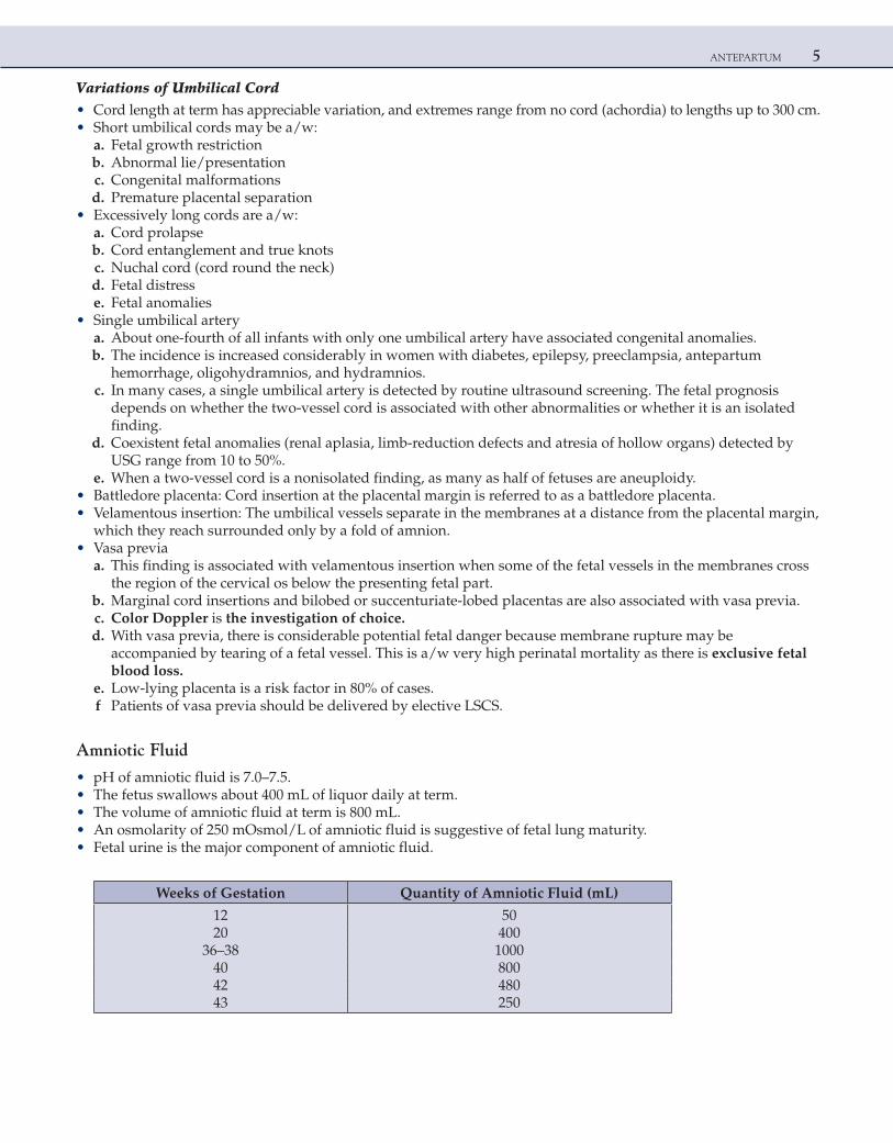
• pH of amniotic fluid is 7.0–7.5. • The fetus swallows about 400 mL of liquor daily at term. • The volume of amniotic fluid at term is 800 mL. • An osmolarity of 250 mOsmol/L of amniotic fluid is suggestive of fetal lung maturity. • Fetal urine is the major component of amniotic fluid.
Weeks of Gestation Quantity of Amniotic Fluid (mL)
12 5020 400
36–38 100040 80042 48043 250
6 SMART STUDY SERIES: OBSTETRICS AND GYNECOLOGY
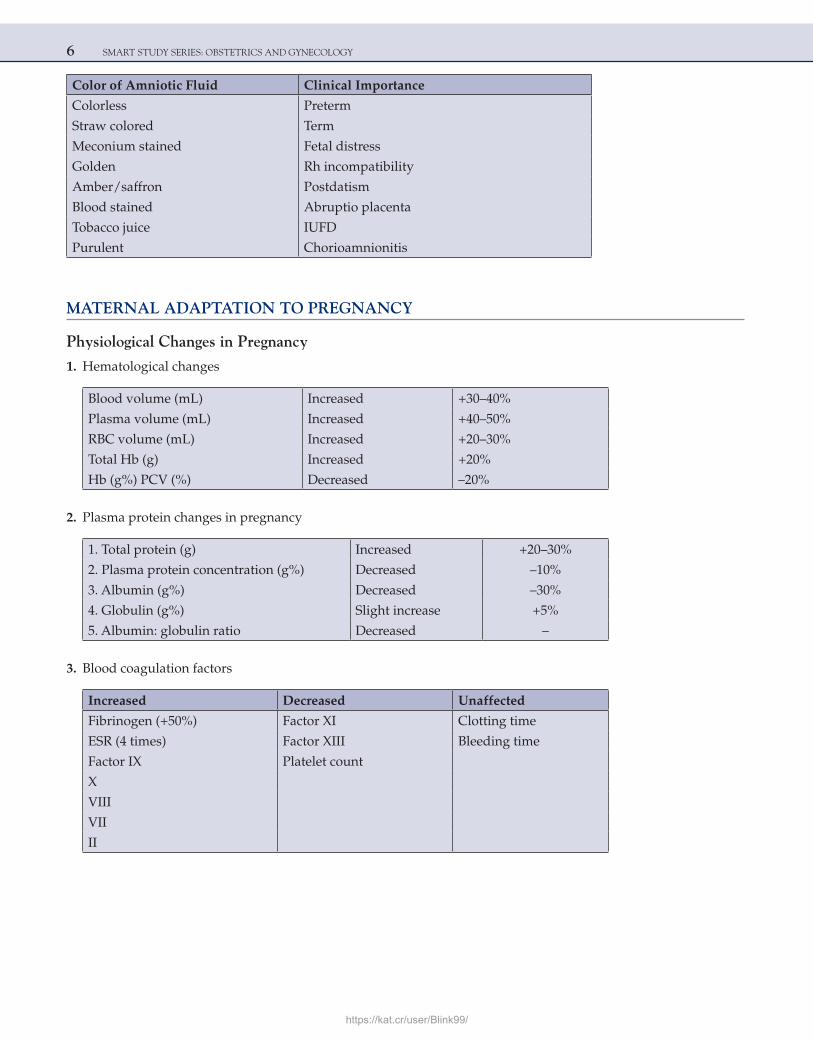
Color of Amniotic Fluid Clinical Importance
Colorless Preterm
Straw colored Term
Meconium stained Fetal distress
Golden Rh incompatibility
Amber/saffron Postdatism
Blood stained Abruptio placenta
Tobacco juice IUFD
Purulent Chorioamnionitis
MATERNAL ADAPTATION TO PREGNANCY
Physiological Changes in Pregnancy
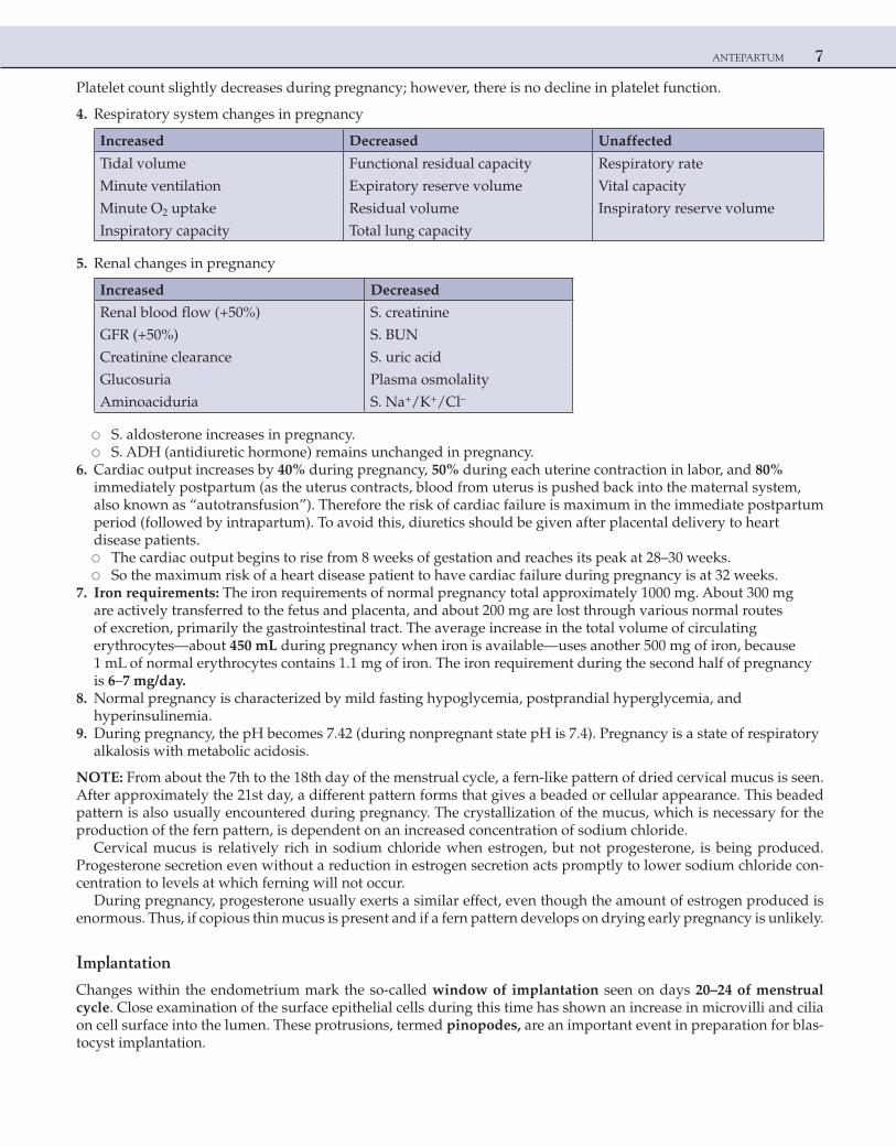
1. Hematological changes
Blood volume (mL) Increased +30–40%
Plasma volume (mL) Increased +40–50%
RBC volume (mL) Increased +20–30%
Total Hb (g) Increased +20%
Hb (g%) PCV (%) Decreased –20%
2. Plasma protein changes in pregnancy
1. Total protein (g) Increased +20–30%
2. Plasma protein concentration (g%) Decreased –10%
3. Albumin (g%) Decreased –30%
4. Globulin (g%) Slight increase +5%
5. Albumin: globulin ratio Decreased –
3. Blood coagulation factors
Increased Decreased Unaffected
Fibrinogen (+50%) Factor XI Clotting time
ESR (4 times) Factor XIII Bleeding time
Factor IX Platelet count
X
VIII
VII
II
https://kat.cr/user/Blink99/
ANTEPARTUM 7
Platelet count slightly decreases during pregnancy; however, there is no decline in platelet function.
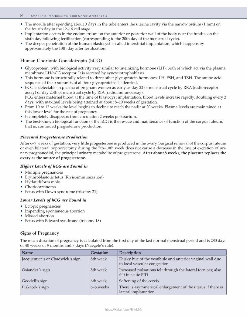
4. Respiratory system changes in pregnancy
Increased Decreased Unaffected
Tidal volume Functional residual capacity Respiratory rate
Minute ventilation Expiratory reserve volume Vital capacity
Minute O2 uptake Residual volume Inspiratory reserve volume
Inspiratory capacity Total lung capacity
5. Renal changes in pregnancy
Increased Decreased
Renal blood flow (+50%) S. creatinine
GFR (+50%) S. BUN
Creatinine clearance S. uric acid
Glucosuria Plasma osmolality
Aminoaciduria S. Na+/K+/Cl–
○ S. aldosterone increases in pregnancy. ○ S. ADH (antidiuretic hormone) remains unchanged in pregnancy. 6. Cardiac output increases by 40% during pregnancy, 50% during each uterine contraction in labor, and 80%
immediately postpartum (as the uterus contracts, blood from uterus is pushed back into the maternal system, also known as “autotransfusion”). Therefore the risk of cardiac failure is maximum in the immediate postpartum period (followed by intrapartum). To avoid this, diuretics should be given after placental delivery to heart disease patients.
○ The cardiac output begins to rise from 8 weeks of gestation and reaches its peak at 28–30 weeks. ○ So the maximum risk of a heart disease patient to have cardiac failure during pregnancy is at 32 weeks. 7. Iron requirements: The iron requirements of normal pregnancy total approximately 1000 mg. About 300 mg
are actively transferred to the fetus and placenta, and about 200 mg are lost through various normal routes of excretion, primarily the gastrointestinal tract. The average increase in the total volume of circulating erythrocytes—about 450 mL during pregnancy when iron is available—uses another 500 mg of iron, because 1 mL of normal erythrocytes contains 1.1 mg of iron. The iron requirement during the second half of pregnancy is 6–7 mg/day.
8. Normal pregnancy is characterized by mild fasting hypoglycemia, postprandial hyperglycemia, and hyperinsulinemia.
9. During pregnancy, the pH becomes 7.42 (during nonpregnant state pH is 7.4). Pregnancy is a state of respiratory alkalosis with metabolic acidosis.
NOTE: From about the 7th to the 18th day of the menstrual cycle, a fern-like pattern of dried cervical mucus is seen. After approximately the 21st day, a different pattern forms that gives a beaded or cellular appearance. This beaded pattern is also usually encountered during pregnancy. The crystallization of the mucus, which is necessary for the production of the fern pattern, is dependent on an increased concentration of sodium chloride.
Cervical mucus is relatively rich in sodium chloride when estrogen, but not progesterone, is being produced. Progesterone secretion even without a reduction in estrogen secretion acts promptly to lower sodium chloride con-centration to levels at which ferning will not occur.
During pregnancy, progesterone usually exerts a similar effect, even though the amount of estrogen produced is enormous. Thus, if copious thin mucus is present and if a fern pattern develops on drying early pregnancy is unlikely.
Implantation
Changes within the endometrium mark the so-called window of implantation seen on days 20–24 of menstrual cycle. Close examination of the surface epithelial cells during this time has shown an increase in microvilli and cilia on cell surface into the lumen. These protrusions, termed pinopodes, are an important event in preparation for blas-tocyst implantation.
8 SMART STUDY SERIES: OBSTETRICS AND GYNECOLOGY
• The morula after spending about 3 days in the tube enters the uterine cavity via the narrow ostium (1 mm) on the fourth day in the 12–16 cell stage.
• Implantation occurs in the endometrium on the anterior or posterior wall of the body near the fundus on the sixth day following fertilization (corresponding to the 20th day of the menstrual cycle).
• The deeper penetration of the human blastocyst is called interstitial implantation, which happens by approximately the 13th day after fertilization.
Human Chorionic Gonadotropin (hCG)
• Glycoprotein, with biological activity very similar to luteinizing hormone (LH), both of which act via the plasma membrane LH-hCG receptor. It is secreted by syncytiotrophoblasts.
• This hormone is structurally related to three other glycoprotein hormones: LH, FSH, and TSH. The amino acid sequence of the α-subunits of all four glycoproteins is identical.
• hCG is detectable in plasma of pregnant women as early as day 22 of menstrual cycle by RRA (radioreceptor assay) or day 25th of menstrual cycle by RIA (radioimmunoassay).hCG enters maternal blood at the time of blastocyst implantation. Blood levels increase rapidly, doubling every 2 days, with maximal levels being attained at about 8–10 weeks of gestation.
• From 10 to 12 weeks the level begins to decline to reach the nadir at 20 weeks. Plasma levels are maintained at this lower level for the rest of pregnancy.
• It completely disappears from circulation 2 weeks postpartum. • The best-known biological function of the hCG is the rescue and maintenance of function of the corpus luteum,
that is, continued progesterone production.
Placental Progesterone ProductionAfter 6–7 weeks of gestation, very little progesterone is produced in the ovary. Surgical removal of the corpus luteum or even bilateral oophorectomy during the 7th–10th week does not cause a decrease in the rate of excretion of uri-nary pregnanediol, the principal urinary metabolite of progesterone. After about 8 weeks, the placenta replaces the ovary as the source of progesterone.
Higher Levels of hCG are Found in • Multiple pregnancies • Erythroblastotic fetus (Rh isoimmunization) • Hydatidiform mole • Choriocarcinoma • Fetus with Down syndrome (trisomy 21)
Lower Levels of hCG are Found in • Ectopic pregnancies • Impending spontaneous abortion • Missed abortion • Fetus with Edward syndrome (trisomy 18)
Signs of Pregnancy
The mean duration of pregnancy is calculated from the first day of the last normal menstrual period and is 280 days or 40 weeks or 9 months and 7 days (Naegele’s rule).
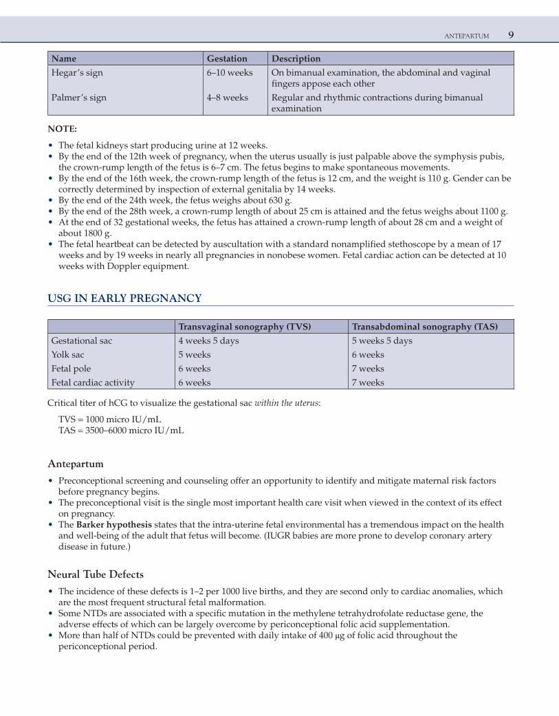
Name Gestation Description
Jacquemier’s or Chadwick’s sign 8th week Dusky hue of the vestibule and anterior vaginal wall due to local vascular congestion
Osiander’s sign 8th week Increased pulsations felt through the lateral fornices; also felt in acute PID
Goodell’s sign 6th week Softening of the cervix
Piskacek’s sign 6–8 weeks There is asymmetrical enlargement of the uterus if there is lateral implantation
https://kat.cr/user/Blink99/
ANTEPARTUM 9
Name Gestation Description
Hegar’s sign 6–10 weeks On bimanual examination, the abdominal and vaginal fingers appose each other
Palmer’s sign 4–8 weeks Regular and rhythmic contractions during bimanual examination
NOTE:
• The fetal kidneys start producing urine at 12 weeks. • By the end of the 12th week of pregnancy, when the uterus usually is just palpable above the symphysis pubis,
the crown-rump length of the fetus is 6–7 cm. The fetus begins to make spontaneous movements. • By the end of the 16th week, the crown-rump length of the fetus is 12 cm, and the weight is 110 g. Gender can be
correctly determined by inspection of external genitalia by 14 weeks. • By the end of the 24th week, the fetus weighs about 630 g. • By the end of the 28th week, a crown-rump length of about 25 cm is attained and the fetus weighs about 1100 g. • At the end of 32 gestational weeks, the fetus has attained a crown-rump length of about 28 cm and a weight of
about 1800 g. • The fetal heartbeat can be detected by auscultation with a standard nonamplified stethoscope by a mean of 17
weeks and by 19 weeks in nearly all pregnancies in nonobese women. Fetal cardiac action can be detected at 10 weeks with Doppler equipment.
USG IN EARLY PREGNANCY
Transvaginal sonography (TVS) Transabdominal sonography (TAS)
Gestational sac 4 weeks 5 days 5 weeks 5 days
Yolk sac 5 weeks 6 weeks
Fetal pole 6 weeks 7 weeks
Fetal cardiac activity 6 weeks 7 weeks
Critical titer of hCG to visualize the gestational sac within the uterus:
TVS = 1000 micro IU/mLTAS = 3500–6000 micro IU/mL
Antepartum
• Preconceptional screening and counseling offer an opportunity to identify and mitigate maternal risk factors before pregnancy begins.
• The preconceptional visit is the single most important health care visit when viewed in the context of its effect on pregnancy.
• The Barker hypothesis states that the intra-uterine fetal environmental has a tremendous impact on the health and well-being of the adult that fetus will become. (IUGR babies are more prone to develop coronary artery disease in future.)
Neural Tube Defects
• The incidence of these defects is 1–2 per 1000 live births, and they are second only to cardiac anomalies, which are the most frequent structural fetal malformation.
• Some NTDs are associated with a specific mutation in the methylene tetrahydrofolate reductase gene, the adverse effects of which can be largely overcome by periconceptional folic acid supplementation.
• More than half of NTDs could be prevented with daily intake of 400 μg of folic acid throughout the periconceptional period.
10 SMART STUDY SERIES: OBSTETRICS AND GYNECOLOGY
• A woman with a prior pregnancy complicated by a neural tube defect can reduce the 23% recurrence risk by more than 70% if she takes 4 mg of folic acid for the month before conception and for the first trimester of pregnancy.
Risk Factors for NTD
1. Family history of NTD 2. Past history of NTD 3. Diabetes mellitus 4. Hyperthermia 5. Drugs and medications (refer Teratogens) 6. Genetic factors 7. Production of antifolate receptor antibodies
Anencephaly
• Anencephaly is a lethal NTD characterized by absence of the brain and cranium above the base of the skull and orbits. It can be diagnosed as early as the first trimester on USG.
• 70% of fetuses are female. • Face presentation is the most common presentation. • Recurrence risk is 5% after one affected fetus and 13% after two affected fetuses. • Frog eyes are seen.
Polyhydramnios is commonly seen due to the following reasons: a. Transudation of fluid across the membranes b. Absence of swallowing c. Absent fetal pituitary (absence of ADH hormone implies that the baby passes more urine) • Postdatism is seen as fetal pituitary plays an important role in initiation of labor. • However preterm labor can also be there due to polyhydramnios. • Pseudoshoulder dystocia is seen as the soft head/face can slip through incompletely dilated cervix. Classically,
fetuses with spina bifida have one or more of the following cranial signs on USG: 1. Small biparietal diameter. 2. Ventriculomegaly 3. Frontal bone scalloping or the so-called lemon sign. 4. Elongation and downward displacement of the cerebellum, the so-called banana sign. 5. Effacement or obliteration of the cisterna magna. ○ The lateral ventricle is commonly measured at its atrium, which is the confluence of the temporal and occipi-
tal horns. The measurement is relatively constant at 7 mm, with standard deviation of 1 mm from 15 weeks onward.
○ Mild ventriculomegaly is diagnosed when the atrial width measures 10–15 mm and overt ventriculomegaly when it exceeds 15 mm. A dangling choroid plexus characteristically is found in severe cases.
MSAFP
Maternal serum alpha-fetoprotein (MSAFP) estimation is commonly done between 15 and 20 weeks of gestation.
Conditions Associated with Abnormal Maternal Serum Alpha-Fetoprotein Concentrations
Elevated Levels 1. Neural tube defects 2. Pilonidal cysts 3. Esophageal or intestinal obstruction 4. Liver necrosis 5. Cystic hygroma 6. Sacrococcygeal teratoma
https://kat.cr/user/Blink99/
ANTEPARTUM 11
7. Abdominal wall defects—omphalocele, gastroschisis 8. Urinary obstruction 9. Renal anomalies—polycystic or absent kidneys 10. Congenital nephrosis 11. Osteogenesis imperfecta 12. Congenital skin defects 13. Cloacal exstrophy 14. Chorioangioma of placenta 15. Placenta accreta 16. Oligohydramnios 17. Preeclampsia 18. Multifetal gestation 19. Low birthweight 20. Fetal death 21. Underestimated gestational age, decreased maternal weight 22. Maternal hepatoma or teratoma
Low Levels 1. Chromosomal trisomies 2. Gestational trophoblastic disease 3. Increased maternal weight 4. Overestimated gestational age ○ NTD is suspected if the maternal serum AFP is elevated, and if the ultrasonographic examination is nondiag-
nostic, then amniotic fluid AFP levels are measured. ○ An elevated amniotic fluid AFP level prompts assay of the same sample for acetylcholinesterase.
The presence of this enzyme 100% confirms that exposed neural tissue or another open fetal defect is present.
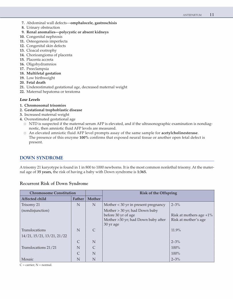
DOWN SYNDROME
A trisomy 21 karyotype is found in 1 in 800 to 1000 newborns. It is the most common nonlethal trisomy. At the mater-nal age of 35 years, the risk of having a baby with Down syndrome is 1:365.
Recurrent Risk of Down Syndrome
Chromosome Constitution Risk of the Offspring
Affected child Father Mother
Trisomy 21 N N Mother < 30 yr in present pregnancy 2–3%
(nondisjunction) Mother > 30 yr; had Down baby before 30 yr of ageMother >30 yr; had Down baby after 30 yr age
Risk at mothers age +1%Risk at mother’s age
Translocations N C 11.9%
14/21, 15/21, 13/21, 21/22
C N 2–3%
Translocations 21/21 N C 100%
C N 100%
Mosaic N N 2–3%C = carrier; N = normal.
12 SMART STUDY SERIES: OBSTETRICS AND GYNECOLOGY
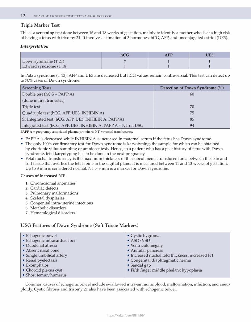
Triple Marker Test
This is a screening test done between 16 and 18 weeks of gestation, mainly to identify a mother who is at a high risk of having a fetus with trisomy 21. It involves estimation of 3 hormones: hCG, AFP, and unconjugated estriol (UE3).
Interpretation
hCG AFP UE3
Down syndrome (T 21)Edward syndrome (T 18)
↑↓
↓↓
↓↓
In Patau syndrome (T 13): AFP and UE3 are decreased but hCG values remain controversial. This test can detect up to 70% cases of Down syndrome.
Screening Tests Detection of Down Syndrome (%)
Double test (hCG + PAPP A) 60
(done in first trimester)
Triple test 70
Quadruple test (hCG, AFP, UE3, INHIBIN A) 75
Sr Integrated test (hCG, AFP, UE3, INHIBIN A, PAPP A) 85
Integrated test (hCG, AFP, UE3, INHIBIN A, PAPP A + NT on USG 94PAPP A = pregnancy-associated plasma protein A; NT = nuchal translucency.
• PAPP A is decreased while INHIBIN A is increased in maternal serum if the fetus has Down syndrome. • The only 100% confirmatory test for Down syndrome is karyotyping, the sample for which can be obtained
by chorionic villus sampling or amniocentesis. Hence, in a patient who has a past history of fetus with Down syndrome, fetal karyotyping has to be done in the next pregnancy.
• Fetal nuchal translucency is the maximum thickness of the subcutaneous translucent area between the skin and soft tissue that overlies the fetal spine in the sagittal plane. It is measured between 11 and 13 weeks of gestation. Up to 3 mm is considered normal. NT > 3 mm is a marker for Down syndrome.
Causes of increased NT:
1. Chromosomal anomalies 2. Cardiac defects 3. Pulmonary malformations 4. Skeletal dysplasias 5. Congenital intra-uterine infections 6. Metabolic disorders 7. Hematological disorders
USG Features of Down Syndrome (Soft Tissue Markers)
• Echogenic bowel • Echogenic intracardiac foci • Duodenal atresia • Absent nasal bone • Single umbilical artery • Renal pyelectasis • Exomphalos • Choroid plexus cyst • Short femur/humerus
• Cystic hygroma • ASD/VSD • Ventriculomegaly • Annular pancreas • Increased nuchal fold thickness, increased NT • Congenital diaphragmatic hernia • Sandal gap • Fifth finger middle phalanx hypoplasia
Common causes of echogenic bowel include swallowed intra-amnionic blood, malformation, infection, and aneu-ploidy. Cystic fibrosis and trisomy 21 also have been associated with echogenic bowel.
https://kat.cr/user/Blink99/
ANTEPARTUM 13
Duodenal atresia occurs in about 1 in 10,000 live births. The lesion may be diagnosed prenatally by the dem-onstration of the so-called double bubble sign, which represents distention of the stomach and the first part of the duodenum. About 30% of fetuses with duodenal atresia diagnosed antenatally have trisomy 21 and more than half have other anomalies.
INDICATIONS FOR CHORIONIC VILLUS SAMPLING/AMNIOCENTESIS (ACOG GUIDELINES)
• Singleton pregnancy at age over 35 years at delivery • Dizygotic twin pregnancy at age over 31 years at delivery • Previous autosomal trisomy birth • Previous 47, XXX or 47, XXY birth or triploidy birth • Patient or partner is carrier of chromosomal translocation/inversion • Some cases with repetitive early pregnancy losses • Patient or partner has aneuploidy • Major fetal structural defect identified by ultrasound
Amniocentesis
• Traditionally done between 16 and 20 weeks of gestation • Early amniocentesis is done between 12 and 14 weeks of gestation • It is done under USG guidance • Risk of gestational loss (abortion) is 0.3–0.5% • Other complications include chorioamnionitis, PROM, fetal trauma
Chorionic Villus Sampling
• Can be done through transabdominal or transcervical route • As per ACOG guidelines, CVS should be done only after 10 completed weeks (after 70 days) • Complications a/w early CVS are limb reduction defects and oro-mandibular defects • Risk of gestational loss with CVS is 0.8–1%
CORDOCENTESIS (PERCUTANEOUS UMBILICAL BLOOD SAMPLING)
It is done after 18 weeks of gestation. Risk of gestational loss is 1–5%.
Indications
1. Rapid karyotyping in fetuses with structural anomalies on USG 2. Fetal hemolytic disease (diagnosis as well as management by intra-uterine transfusion) 3. Suspected fetal thrombocytopenia/hemoglobinopathy 4. Suspected fetal viral infection 5. Diagnosis of twin-to-twin transfusion syndrome
Features of Trisomy 18 (Edward Syndrome)
1. IUGR 2. Prominent occiput 3. Rotated and malformed auricles, short palpebral fissures, small mouth 4. Cardiac defects (VSD/ASD/PDA) 5. Horseshoe kidney 6. Radial aplasia, hemivertebrae
14 SMART STUDY SERIES: OBSTETRICS AND GYNECOLOGY
7. Clenched hands and overlapping fingers, syndactyly 8. Hernias, imperforate anus 9. Severe MR 10. Rocker bottom feet
Features of Trisomy 13 (Patau Syndrome)
1. Cardiac defects 2. Holoprosencephaly, moderate microcephaly, microphthalmia 3. Cleft lip/palate, abnormal ears 4. Omphalocele 5. Polycystic kidneys 6. Radial aplasia 7. Cutis aplasia 8. Polydactyly
Features of Turner Syndrome (45XO)
1. Short stature 2. Broad chest, widely spaced nipples 3. Congenital lymphedema 4. Cubitus valgus 5. Webbed posterior neck 6. High arched palate 7. Ovarian dysgenesis and infertility (90%) 8. Aortic coarctation or bicuspid aortic valves 9. Normal intelligence 10. Hypoplastic uterus (due to lack of estrogen)
Aneuploidy Risk Associated with Major Structural Fetal Malformations
Defect Aneuploidy risk (%)
Cystic hygroma 60–75Hydrops 30–80Hydrocephalus 3–8Holoprosencephaly 40–60Cardiac defects 5–30Diaphragmatic hernia 20–25Omphalocele 30–40Gastroschisis MinimalDuodenal atresia 20–30Facial cleft 1Clubfoot 20–30Limb reduction 8
TERATOLOGY
A teratogen is any agent that acts during embryonic or fetal development to produce a permanent alteration of form or function.
https://kat.cr/user/Blink99/
ANTEPARTUM 15
• The word teratogen is derived from the Greek “teratos,” meaning monster. Because this derivation implies obvious visible defects, a teratogen is most properly defined as an agent that produces structural abnormalities.
• A hadegen—after Hades, (the god who possessed a helmet conferring invisibility)—is an agent that interferes with normal maturation and function of an organ.
• Identical defects with different etiologies are called phenocopies. • Exposures within the first 8 weeks result in an embryopathy and after 8 weeks in a fetopathy. • The preimplantation period is 2 weeks from fertilization to implantation and has traditionally been called the
“all or none” period. The zygote undergoes cleavage and cells divide into an outer and inner cell mass. An insult damaging a large number of cells usually causes death of the embryo. If only a few cells are injured, compensation is usually possible with continued normal development.
Proven Human Teratogens
Drug Adverse Effect on Fetus
1. Phenytoin Fetal hydantoin syndrome (craniofacial defects, limb defects, MR)
2. Valproic acids Spina bifida (1–2% lumbosacral type)
3. Warfarin Nasal hypoplasia, stippled vertebral and femoral epiphyses, agenesis of corpus callosum, Dandy Walker malformation, midline cerebellar atrophy, microphthalmia, optic atrophy, blindness, MR (Conradi’s syndrome)
4. ACE inhibitors Oligohydramnios, renal anomalies, neonatal renal failure, pulmonary hypoplasia, hypocalvaria, growth restriction, death
5. Isotretinoin Craniofacial defects, cleft palate, cardiac defects, hydrocephalus, thymic defects
6. DES Clear cell adenocarcinoma of cervix/vagina, ectropion and adenosis, hypoplastic T-shaped uterus, cervical collars, hoods, septa, withered fallopian tubes; in male fetuses epididymal cysts, microphallus, cryptorchidism, testicular hypoplasia, hypospadias
7. Cyclophosphamide Missing/hypoplastic digits, cleft palate, single coronary artery, imperforate anus, IUGR microcephaly
8. Methotrexate IUGR failure of calvarial ossification, craniosynostosis, hypoplastic supraorbital ridges, small posteriorly rotated ears, micrognathia, severe limb abnormalities
9. Tetracyclines Yellowish brown discoloration of deciduous teeth
10. Streptomycin VIII cranial nerve damage (i.e., ototoxicity)
11. Griseofulvin Conjoint twins
12. Tobacco IUGR, subfertility, spontaneous abortion, abruption and preterm delivery, cleft lip and palate, Poland sequence
13. Cocaine Placental abruption, abortions, stillbirth, skull defects, cutis aplasia, porencephaly, ileal atresia, cardiac anomalies and visceral infarcts, urinary defects, periventricular leukoma-lacia, prune-belly syndrome
14. Thalidomide Phocomelia
15. Misoprostol Moebius syndrome
16 SMART STUDY SERIES: OBSTETRICS AND GYNECOLOGY
Features of Fetal Alcohol Syndrome
Growth restriction Craniofacial anomalies
Behavioral disturbances Absence of hypoplastic philtrum
Brain defects Broad upper lip
Cardiac defects Flattened nasal bridge
Spinal defects Hypoplastic upper lip vermillion
Microphthalmia Micrognathia
Short nose
Short palpebral tissues
Methods for Assessment of Fetal Well-being
Antepartum Intrapartum Postpartum
Nonstress test (NST)Biophysical profile (BPP)Vibroacoustic stimulation test (VSAT)Contraction stress test/oxytocin challenge test (CST/ OCT)Fetal kick countColor Doppler USG
CTG (cardiotocography)Fetal heart rate (Doppler)Fetal scalp electrode monitoringFetal pulse oximetryFetal scalp pH monitoring
Apgar scoreUmbilical cord pH
• There is a decrease in baseline fetal heart rate of 24 beats/min between 16 weeks and term; or approximately 1 beat/min per week. This normal gradual slowing corresponds to maturation of parasympathetic (vagal) heart control.
• Bradycardia: The baseline fetal heart rate lesser than 110 beats/min. • Tachycardia: The baseline fetal heart rate greater than 160 beats/min. • Fetal hypoxia and hypercapnia can modulate the heart rate as it is also under the control of arterial
chemoreceptors. More severe and prolonged hypoxia, with a rising blood lactate level and severe metabolic acidemia, induces a prolonged fall of heart rate due to direct effects on the myocardium.
• Some causes of fetal bradycardia include congenital heart block and serious fetal compromise (hypoxia/ acidosis).
• The most common explanation for fetal tachycardia is maternal fever. • Other causes of fetal tachycardia include fetal compromise, cardiac arrhythmia, and maternal administration of
atropine or terbutaline.
Beat-to-Beat Variability • Normal beat to beat variability should be 6–25 beats/minute. • Diminished beat-to-beat variability can be an ominous sign and may indicate a seriously compromised fetus. • Loss of beat-to-beat variability along with decelerations is associated with fetal acidemia. • A common cause of diminished beat-to-beat variability is analgesic drugs given during labor. • A large variety of CNS depressant drugs like narcotics, barbiturates, phenothiazines, tranquilizers, general
anesthetics, and magnesium sulfate can cause transient diminished beat-to-beat variability.
Sinusoidal Heart Rate • A true sinusoidal pattern is seen with serious fetal anemia, whether from D-isoimmunization, ruptured vasa
previa, fetomaternal hemorrhage, parvo virus infection, or twin-to-twin transfusion. Insignificant sinusoidal patterns have been reported following administration of morphine.
• A sinusoidal pattern also has been described with chorioamnionitis, fetal distress (asphyxia), and umbilical cord occlusion.
https://kat.cr/user/Blink99/
ANTEPARTUM 17
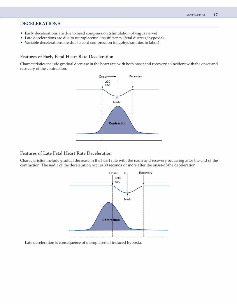
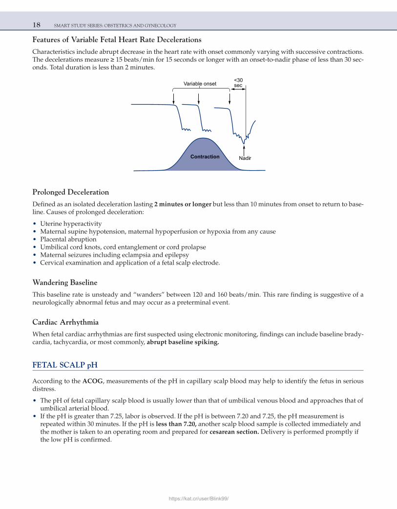
DECELERATIONS
• Early decelerations are due to head compression (stimulation of vagus nerve) • Late decelerations are due to uteroplacental insufficiency (fetal distress/hypoxia) • Variable decelerations are due to cord compression (oligohydramnios in labor)
Features of Early Fetal Heart Rate Deceleration
Characteristics include gradual decrease in the heart rate with both onset and recovery coincident with the onset and recovery of the contraction.
Onset Recovery
>30sec
Contraction
Nadir
Features of Late Fetal Heart Rate Deceleration
Characteristics include gradual decrease in the heart rate with the nadir and recovery occurring after the end of the contraction. The nadir of the deceleration occurs 30 seconds or more after the onset-of-the deceleration.
Onset Recovery
>30sec
Contraction
Nadir
Late deceleration is consequence of uteroplacental-induced hypoxia.
18 SMART STUDY SERIES: OBSTETRICS AND GYNECOLOGY
Features of Variable Fetal Heart Rate Decelerations
Characteristics include abrupt decrease in the heart rate with onset commonly varying with successive contractions. The decelerations measure ≥ 15 beats/min for 15 seconds or longer with an onset-to-nadir phase of less than 30 sec-onds. Total duration is less than 2 minutes.
<30secVariable onset
NadirContraction
Prolonged Deceleration
Defined as an isolated deceleration lasting 2 minutes or longer but less than 10 minutes from onset to return to base-line. Causes of prolonged deceleration:
• Uterine hyperactivity • Maternal supine hypotension, maternal hypoperfusion or hypoxia from any cause • Placental abruption • Umbilical cord knots, cord entanglement or cord prolapse • Maternal seizures including eclampsia and epilepsy • Cervical examination and application of a fetal scalp electrode.
Wandering Baseline
This baseline rate is unsteady and “wanders” between 120 and 160 beats/min. This rare finding is suggestive of a neurologically abnormal fetus and may occur as a preterminal event.
Cardiac Arrhythmia
When fetal cardiac arrhythmias are first suspected using electronic monitoring, findings can include baseline brady-cardia, tachycardia, or most commonly, abrupt baseline spiking.
FETAL SCALP pH
According to the ACOG, measurements of the pH in capillary scalp blood may help to identify the fetus in serious distress.
• The pH of fetal capillary scalp blood is usually lower than that of umbilical venous blood and approaches that of umbilical arterial blood.
• If the pH is greater than 7.25, labor is observed. If the pH is between 7.20 and 7.25, the pH measurement is repeated within 30 minutes. If the pH is less than 7.20, another scalp blood sample is collected immediately and the mother is taken to an operating room and prepared for cesarean section. Delivery is performed promptly if the low pH is confirmed.
https://kat.cr/user/Blink99/
ANTEPARTUM 19
FETAL PULSE OXIMETRY
Using technology similar to that of adult pulse oximetry, instrumentation has been developed that may allow assess-ment of fetal oxyhemoglobin saturation once the membranes are ruptured. A unique pad-like sensor is inserted through the cervix and positioned against the fetal face, where it is held in place by the uterine wall.
The lower limit for normal fetal oxygen saturation is generally considered to be 30% by most investigators.
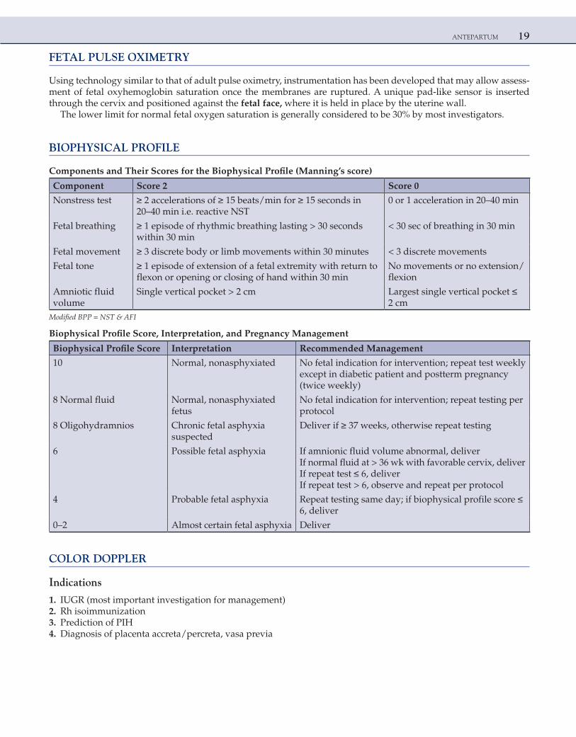
BIOPHYSICAL PROFILE
Components and Their Scores for the Biophysical Profile (Manning’s score)
Component Score 2 Score 0
Nonstress test ≥ 2 accelerations of ≥ 15 beats/min for ≥ 15 seconds in 20–40 min i.e. reactive NST
0 or 1 acceleration in 20–40 min
Fetal breathing ≥ 1 episode of rhythmic breathing lasting > 30 seconds within 30 min
< 30 sec of breathing in 30 min
Fetal movement ≥ 3 discrete body or limb movements within 30 minutes < 3 discrete movements
Fetal tone ≥ 1 episode of extension of a fetal extremity with return to flexon or opening or closing of hand within 30 min
No movements or no extension/flexion
Amniotic fluid volume
Single vertical pocket > 2 cm Largest single vertical pocket ≤ 2 cm
Modified BPP = NST & AFI
Biophysical Profile Score, Interpretation, and Pregnancy Management
Biophysical Profile Score Interpretation Recommended Management
10 Normal, nonasphyxiated No fetal indication for intervention; repeat test weekly except in diabetic patient and postterm pregnancy (twice weekly)
8 Normal fluid Normal, nonasphyxiated fetus
No fetal indication for intervention; repeat testing per protocol
8 Oligohydramnios Chronic fetal asphyxia suspected
Deliver if ≥ 37 weeks, otherwise repeat testing
6 Possible fetal asphyxia If amnionic fluid volume abnormal, deliverIf normal fluid at > 36 wk with favorable cervix, deliverIf repeat test ≤ 6, deliverIf repeat test > 6, observe and repeat per protocol
4 Probable fetal asphyxia Repeat testing same day; if biophysical profile score ≤ 6, deliver
0–2 Almost certain fetal asphyxia Deliver
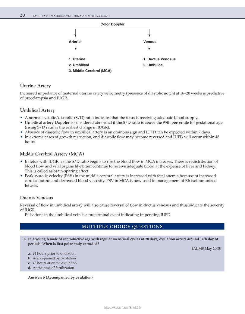
COLOR DOPPLER
Indications
1. IUGR (most important investigation for management) 2. Rh isoimmunization 3. Prediction of PIH 4. Diagnosis of placenta accreta/percreta, vasa previa
20 SMART STUDY SERIES: OBSTETRICS AND GYNECOLOGY
relppoDroloC
suoneVlairetrA
susoneVsutcuD.1eniretU.1
lacilibmU.2lacilibmU.2
3. Middle Cerebral (MCA)
Uterine Artery
Increased impedance of maternal uterine artery velocimetry (presence of diastolic notch) at 16–20 weeks is predictive of preeclampsia and IUGR.
Umbilical Artery
• A normal systolic/diastolic (S/D) ratio indicates that the fetus is receiving adequate blood supply. • Umbilical artery Doppler is considered abnormal if the S/D ratio is above the 95th percentile for gestational age
(rising S/D ratio is the earliest change in IUGR). • Absence of diastolic flow in umbilical artery is an ominous sign and IUFD can be expected within 7 days. • In extreme cases of growth restriction, end diastolic flow may become reversed and IUFD will occur within 48
hours.
Middle Cerebral Artery (MCA)
• In fetus with IUGR, as the S/D ratio begins to rise the blood flow in MCA increases. There is redistribution of blood flow and vital organs like brain continue to receive adequate blood at the expense of liver and kidney. This is called as brain-sparing effect.
• Peak systolic velocity (PSV) in the middle cerebral artery is increased with fetal anemia because of increased cardiac output and decreased blood viscosity. PSV in MCA is now used in management of Rh isoimmunized fetuses.
Ductus Venosus
Reversal of flow in umbilical artery will also cause reversal of flow in ductus venosus and thus indicate the severity of IUGR.
Pulsations in the umbilical vein is a preterminal event indicating impending IUFD.
MULTIPLE CHOICE QUESTIONS
1. In a young female of reproductive age with regular menstrual cycles of 28 days, ovulation occurs around 14th day of periods. When is first polar body extruded?
[AIIMS May 2005] a. 24 hours prior to ovulation b. Accompanied by ovulation c. 48 hours after the ovulation d. At the time of fertilization
Answer: b (Accompanied by ovulation)
https://kat.cr/user/Blink99/
ANTEPARTUM 21
Explanation:In the ovary, a single oocyte is formed from the two meiotic divisions of the oogonium, with excess genetic material con-
tained in two polar bodies, each extruded as a result of the one meiotic division.The first polar body contains 23 chromosomes, each with two strands of DNA, while the second polar body contains 23
chromosomes, each with one strand of DNA.Meiosis begins in the ovary between the third month of gestation and shortly after birth. Meiosis consists of four steps: pro-
phase, metaphase, anaphase, and telophase. The prophase of meiosis I (prophase I) is further subdivided into five individual stages: the leptotene, zygotene, pachytene, diplotene, and diakinesis.
The oocyte reaches the diplotene stage just before or shortly after birth. The meiotic process is arrested at this point, and the oocyte remains at this stage just prior to ovulation.
In the oocyte, LH stimulation results in resumption of meiosis. The diplotene stage leads to diakinesis and prophase I is completed. Oocyte then progresses to metaphase I, anaphase I, and telophase I, and then cell division occurs. The oocyte retains the vast majority of the ooplasm but the chromatin is divided equally between the oocyte and the polar body.
Thus, the first polar body is extruded accompanied by ovulation, while the second polar body is extruded after fertilization of the ovum by the sperm.
References: 1. Williams, 22nd Ed., Pg. 52. 2. Mischell, 4th Ed., Pg. 175–7.
2. The finding of a single umbilical artery on examination of the umbilical cord after delivery is: a. Insignificant b. Occurs in 10% of newborns c. An indicator of considerably increased incidence of major malformation of the fetus d. Equally common in newborn of diabetic and nondiabetic mothers
Answer: c (An indicator of considerably increased incidence of major malformation of the fetus)
Explanation:The absence of one umbilical artery occurs in 0.7–0.8% of all umbilical cords of singletons, in 2.5% of all abortuses, and
in approximately 5% of at least one twin. The incidence of a single artery is significantly increased in newborns of diabetic mothers, and it occurs in white infants twice as often as in newborns of black women. The incidence of major fetal malforma-tions, when only one artery is identified, has been reported to be as high as 18%, and there is an increased incidence of overall fetal mortality. The finding is an indication to offer amniocentesis, or chorionic villus sampling to study fetal chromosomes, although there is debate about whether this should be done when there is only a truly isolated finding of single umbilical artery.
Reference: 1. Williams, 22nd Ed., Pg. 626.
3. Which of the following is the investigation of choice in a pregnant lady at 18 weeks of pregnancy, with past history of delivering a baby with Down syndrome?
[All India 2004] a. Triple screen test b. Amniocentesis c. Chorionic villous biopsy d. Ultrasonography
Answer: b (Amniocentesis)
Explanation:Because there is a past history of Down syndrome, a confirmatory test should be done.Amniocentesis and karyotyping is the best choice here. It is generally done around 14–18 weeks, and gives confirmatory
results.Triple marker is only a screening test and not a confirmatory test. Similarly, USG can pick up soft tissue markers of Down
syndrome, but it is not confirmatory and USG can be normal in a fetus with Down syndromeIf the same patient presents at 11–12 weeks, then the answer is CVS and karyotyping.
22 SMART STUDY SERIES: OBSTETRICS AND GYNECOLOGY
Reference:
1. Williams, 22Ed., Pg. 314.
4. Minimum HCG levels at which gestational sac can be detected by transvaginal sonography is------micro IU/mL: [All India 2013] a. 500 b. 1000 c. 2000 d. 4000
Answer: b (1000)
Explanation:An intra-uterine GS should be seen by TVS when the maternal serum beta hCG level is 1000–1200 micro IU/mL and by
TAS with the level of hCG 3500–6000 micro IU/mL.Gestational sac (GS) is eccentric in position within the endometrium of fundus or body of the uterus and is seen at 4 weeks
5 days on TVS.Double decidua sign of the gestational sac is due to the interface between the decidua and the chorion, which appears as
two distinct layers of the wall of the gestational sac.
Reference:
1. Williams, 22nd Ed., Pg. 259.
5. Fetal hydronephrosis is diagnosed in a mother at 34 weeks gestation. The amniotic fluid is normal. Which of the following is the most appropriate management?
a. Fetal intervention to decompress hydronephrotic kidney b. Premature termination of pregnancy, followed by pyeloplasty c. Delivery at term, followed by radiological evaluation d. Delivery at term followed by early pyeloplasty
Answer: c (Delivery at term, followed by radiological evaluation)
Explanation:The USG diagnostic criteria for fetal hydronephrosis are:
1. A-P diameter of fetal kidneys >10 mm 2. Dilated pelvicalyceal system 3. Cortico/medullary ratio < 0.50
Causes of fetal hydronephrosis are:
Obstructive Nonobstructive
Pelvic ureteric junction obstruction Multicystic dysplastic kidney
Uretero-vesical junction obstruction Autosomal recessive polycystic kidney
Ectopic ureterocele Autosomal dominant polycystic kidney
Posterior urethral valves
Duplex ureter
In the above clinical scenario, since there is adequate liquor, the fetal kidneys seem to be functionally normal. Hence from 34 weeks onward, fetal surveillance using NST and USG sequentially to monitor well-being is the ideal management option.
In the absence of any fetal distress, delivery at term is indicated. Fetal hydronephrosis (mild/moderate) is seen to resolve spontaneously postnatally. Hence, postnatal USG to confirm resolution is indicated.
Invasive procedures like pyelocentesis and pyeloplasty are not indicated in the presence of fetal well-being. Also prema-ture delivery is not indicated in the presence of fetal well-being.
Reference:
1. Williams, 22nd Ed., Pg. 400.
https://kat.cr/user/Blink99/
ANTEPARTUM 23
6. The best time to do chorionic villous sampling is: [AIIMS May 2005, 2008] a. 6–8 weeks b. 7–9 weeks c. 9–11 weeks d. 11–13 weeks
Answer: d (11–13 weeks)
Explanation:As per ACOG guidelines, chorionic villus biopsy should be done only after 10 weeks of gestation. This is done to avoid fetal
risks of limb reduction defects and oromandibular defects and to ensure retrieving adequate sample for processing. It can be done by abdominal route or vaginal route. Chorionic villus sampling below 10 weeks is criticized due to its adverse fetal effects.
Reference:
1. Williams, 22nd Ed., Pg. 329–30.
7. Fetal pulmonary maturity can be evaluated by phospholipids’ activity in amniotic fluid. In which of the following pregnancies does the fetus have the least chance of developing respiratory distress syndrome (RDS)?
[AIIMS Nov 2005] a. Normal pregnancy: amniotic fluid L/S is 1.8:1, phosphatidyl glycerol (PG) is absent b. Diabetic pregnancy: amniotic fluid L/S is 2:1, PG is absent c. Diabetic pregnancy: amniotic fluid L/S is 2:1, PG is present d. All of the above
Answer: c (Diabetic pregnancy: amniotic fluid L/S is 2:1, PG is present)
Explanation:The lecithin-to-sphingomyelin (L/S) ratio in amniotic fluid is close to 1 until about 34 weeks of gestation, when the concen-
tration of lecithin begins to rise. For pregnancies of unknown duration but otherwise uncomplicated, the risk of respiratory distress syndrome (RDS) is relatively minor when the L/S is at least 2:1. Maternal hypertensive disorders and fetal growth retardation may accelerate the rate of fetal pulmonary maturation, possibly as a result of chronic fetal stress.
A delay in fetal pulmonary maturation is observed in pregnancies complicated by maternal diabetes or erythroblastosis feta-lis. There is a substantial risk of RDS when the L/S ratio is <1.5. When the L/S ratio is >2, the risk of RDS is minimal. However, when the fetus is likely to have a serious metabolic compromise at birth (e.g., diabetic pregnancy or sepsis) RDS may develop even with a mature L/S ratio (>2.0). This may be explained by lack of phosphatidyl glycerol (PG), a phospholipid that enhances surfactant properties. The presence of PG in amniotic fluid provides considerable reassurance that RDS will not develop.
Besides amniotic fluid contamination by blood, meconium, or vaginal secretions will not alter PG measurements.
Reference:
1. Williams, 22nd Ed., Pg. 651–2.
8. With reference to fetal heart rate, a nonstress test is considered reactive when: [AIIMS Nov 2003, All India 2013] a. Two fetal heart rate accelerations are noted in 20 minutes b. One fetal heart rate acceleration is noted in 20 minutes c. Two fetal heart rate accelerations are noted in 10 minutes d. Three fetal heart rate accelerations are noted in 30 minutes
Answer: a (Two fetal heart rate accelerations are noted in 20 minutes)
Explanation:In a nonstress test, a continuous electronic monitoring of the fetal heart rate along with recording of fetal movements is
undertaken. There is an observed association of FHR acceleration with fetal movements, which, when present, indicates a healthy fetus. It can reliably be used as a screening test. The accelerations of the FHR associated with fetal movements are presumably reflex mediated. It takes into account the overall uteroplacental function on the central nervous system of the fetus. Apart from fetal hypoxia, depression of FHR associated with fetal movements is observed in fetal acidosis and when narcotic drugs are used by the mother.
24 SMART STUDY SERIES: OBSTETRICS AND GYNECOLOGY
Inferences:Reactive—When two or more accelerations of more than 15 beats per minute above the baseline and longer than 15 seconds
in duration are present in 20 minutes observation.Nonreactive—Absence of the two accelerations in the two observations period.
Reference:
1. Williams, 22nd Ed., Pg. 378–80.
9. In early pregnancy, the clinical sign of soft cervix is: a. Hegar sign b. Chadwick sign c. Goodell sign d. Osiander sign
Answer: c (Goodell sign)
Explanation:Signs of pregnancy in first trimester:
1. Goodell’s sign—cervix becomes soft as early as in the sixth week 2. Osiander’s sign—increased pulsation felt through the lateral fornices at 8 weeks 3. Jacquemier’s or Chadwick’s sign—it is the dusky hue of the vestibule and anterior vaginal wall visible at about eight
weeks of pregnancy 4. Piskacek’s sign—asymmetrical enlargement of the uterus if there is a lateral implantation 5. Palmer’s sign—regular and rhythmic contraction can be elicited during bimanual examination as early as 4–8 weeks 6. Hegar’s sign—demonstrated between 6 and 10 weeks. On bimanual examination there is approximation of fingers
Reference:
1. Dutta, 5th Ed., Pg. 67–8.
10. Average material weight gain in full term pregnancy is: a. 10–12 kg b. 12–14 kg c. 14–16 kg d. 6–8 kg
Answer: a (10–12 kg)
Explanation:The total weight gain during a singleton pregnancy averages 11 kg (24 lb). This is distributed as 1 kg in first trimester and
5 kg each in second and third trimesters. The total weight gain at term is as follows:
Reproductive weight gain: 6 kg Net maternal weight gain: 6 kg
• Fetus 3.3 kg • Liquor 0.8 kg • Placenta 0.6 kg • Uterus 0.9 kg; breasts 0.4 kg
• Increase in blood volume 1.3 kg • Increase in extracellular fluid 12 kg • Accumulation of fat and protein 3.5 kg
Reference:
1. Dutta, 5th Ed., Pg. 51.
11. A 30-year-old nonpregnant woman has a BMI of 28 kg/m2. What is the recommended weight gain for her during pregnancy, when she becomes pregnant?
a. 5–6 kg b. 8–11 kg c. 10–13 kg d. 14–16 kg
Answer: b (8–11 kg)
https://kat.cr/user/Blink99/
ANTEPARTUM 25
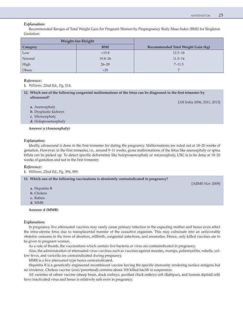
Explanation:Recommended Ranges of Total Weight Gain for Pregnant Women by Prepregnancy Body Mass Index (BMI) for Singleton
Gestation:
Weight-for-HeightRecommended Total Weight Gain (kg)Category BMI
Low <19.8 12.5–18
Normal 19.8–26 11.5–14
High 26–29 7–11.5
Obese >29 7
Reference:
1. Williams, 22nd Ed., Pg. 214.
12. Which one of the following congenital malformations of the fetus can be diagnosed in the first trimester by ultrasound?
[All India 2006, 2011, 2013] a. Anencephaly b. Dysplastic kidneys c. Microcephaly d. Holoprosencephaly
Answer: a (Anencephaly)
Explanation:Ideally, ultrasound is done in the first trimester for dating the pregnancy. Malformations are ruled out at 18–20 weeks of
gestation. However, in the first trimester, i.e., around 9–11 weeks, gross malformations of the fetus like anencephaly or spina bifida can be picked up. To detect specific deformities like holoprosencephaly or microcephaly, USG is to be done at 18–20 weeks of gestation and not in the first trimester.
Reference:
1. Williams, 22nd Ed., Pg. 394, 989.
13. Which one of the following vaccinations is absolutely contraindicated in pregnancy? [AIIMS Nov 2009] a. Hepatitis B b. Cholera c. Rabies d. MMR
Answer: d (MMR)
Explanation:In pregnancy, live attenuated vaccines may rarely cause primary infection in the expecting mother and hence even affect
the intra-uterine fetus due to transplacental transfer of the causative organism. This may culminate into an unfavorable obstetric outcome in the form of abortion, stillbirth, congential infections, and anomalies. Hence, only killed vaccines are to be given to pregnant women.
As a rule of thumb, the vaccinations which contain live bacteria or virus are contraindicated in pregnancy.Also, the administration of attenuated virus vaccines such as vaccines against measles, mumps, poliomyelitis, rubella, yel-
low fever, and varicella are contraindicated during pregnancy.MMR is a live attenuated type hence contraindicated.Hepatitis B is a genetically engineered recombinant vaccine having the specific-immunity rendering surface antigens but
no virulence. Cholera vaccine (oral/parenteral) contains about 109 killed bacilli in suspension.All varieties of rabies vaccine (sheep brain, duck embryo, purified chick embryo cell (Rabipur), and human diploid cell)
have inactivated virus and hence is relatively safe even in pregnancy.
26 SMART STUDY SERIES: OBSTETRICS AND GYNECOLOGY
Reference:
1. Williams, 22nd Ed., Pg. 1282–3.
14. Ideal time to perform USG to measure nuchal translucency is _____ weeks of gestation. [All India 2007] a. 8–10 b. 11–13 c. 14–16 d. 18–20
Answer: b (11–13)
Explanation:Nuchal fold is seen as a sonolucency at the back of the fetal neck in the midsagittal plane. Although its precise etiology is
unknown, it may represent one end of the spectrum of lymphatic obstruction sequence.NT is measured between 11 and 13 weeks. Up to 3 mm is considered normal. More than 3 mm NT is one of the markers of
Down syndrome on USG [All India 2010].
Reference:
1. Williams, 22nd Ed., Pg. 391.
15. All are features of Down syndrome on USG except: a. Duodenal atresia b. Cystic hygroma c. Echogenic intracardiac foci d. Short femur
Explanation:This is a DUMMY question. It means either all four options are correct or all four are wrong. In the above question, all four
options are correct. All are features of Down syndrome on USG.Every year in All India/AIIMS, there can be one or two dummy questions, which you are suppose to leave blank. Do not
attempt these questions. In entrance exams where there are no negative markings, you can mark any of the option.Cystic hygroma is seen in both Turner and Down syndrome.
Features of Down syndrome on USG (soft tissue markers) are:
• Echogenic bowel • Echogenic intracardiac foci • Duodenal atresia • Absent nasal bone • Single umbilical artery • Renal pyelectasis (dilatation of renal pelvicalyceal system) • Exomphalos • Choroid plexus cyst • Short femur/humerus
• Cystic hygroma • ASD/VSD • Ventriculomegaly • Annular pancreas • Increased nuchal fold thickness, increased NT (>3 mm) • Congenital diaphragmatic hernia • Sandal gap • Fifth finger middle phalanx hypoplasia
Reference:
1. Williams, 22nd Ed., Pg. 290.
16. Maximum permissible dose of radiation in pregnancy is: a. 0.05 rads b. 0.5 rads c. 5 rads d. 10 rads
Answer: c (5 rads)
https://kat.cr/user/Blink99/
ANTEPARTUM 27
Explanation:The harmful effects of radiation exposure are direct or indirect:
1. Cell death, which affects embryogenesis 2. Growth restriction 3. Congenital malformations 4. Carcinogenesis (controversial) 5. Microcephaly and mental retardation 6. Sterility
The harmful fetal effects of ionizing radiation have been extensively studied for cell damage with resultant dysfunction of embryogenesis.
The risk is greatest at 8–15 weeks, and larger doses are necessary at 16–25 weeks to cause an equivalent proportion of cases of mental retardation.
Current evidence suggests that there is no increased risk of malformations, growth restriction, or abortion from a radiation dose of 5 rads or less.
MRI uses nonionizing radiation and is very safe. The most common fetal indication for MRI is suspected brain anomaly.
Reference:
1. Williams, 22nd Ed., Pg. 977–9.
17. Most common tumor to show metastasis to placenta is: a. Ca breast b. Ca lung c. Melanoma d. No tumor can metastasize to placenta
Answer: c (Melanoma)
Explanation:Malignant tumors rarely metastasize to the placenta. Of those that do, melanoma accounts for nearly one-third of reported
cases, and leukemias and lymphomas comprise another third.
Reference:
1. Williams, 22nd Ed., Pgs. 624, 1264.
18. Oxygenated blood from the placenta reaches the fetal heart in utero via: a. Umbilical arteries b. Umbilical vein c. Ductus venosus d. Ductus arteriosus
Answer: c (Ductus venosus)
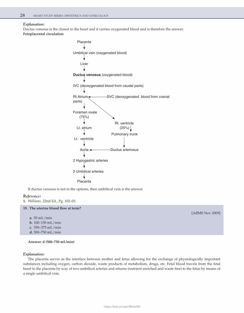
28 SMART STUDY SERIES: OBSTETRICS AND GYNECOLOGY
Explanation:Ductus venosus is the closest to the heart and it carries oxygenated blood and is therefore the answer.Fetoplacental circulation
Placenta
Umbilical vein (oxygenated blood)
Liver
Ductus venosus (oxygenated blood)
IVC (deoxygenated blood from caudal parts)
Rt Atrium SVC (deoxygenated blood from cranial parts)
Foramen ovale (75%)
Rt. ventricle Lt. atrium (25%)
Pulmonary trunk Lt . ventricle
Aorta Ductus arteriosus
2 Hypogastric arteries
2 Umbilical arteries
Placenta
If ductus venosus is not in the options, then umbilical vein is the answer.
Reference:
1. Williams, 22nd Ed., Pg. 102–03.
19. The uterine blood flow at term? [AIIMS Nov 2009] a. 50 mL/min b. 100–150 mL/min c. 350–375 mL/min d. 500–750 mL/min
Answer: d (500–750 mL/min)
Explanation:The placenta serves as the interface between mother and fetus allowing for the exchange of physiologically important
substances including oxygen, carbon dioxide, waste products of metabolism, drugs, etc. Fetal blood travels from the fetal heart to the placenta by way of two umbilical arteries and returns (nutrient enriched and waste free) to the fetus by means of a single umbilical vein.
https://kat.cr/user/Blink99/
ANTEPARTUM 29
Uterine blood flow is one critical determinant of the proper functioning of the placenta and thus the health of the fetus. Uterine blood flow is not autoregulated and as a result the flow is proportional to uterine perfusion pressure (arterial pressure minus uterine venous pressure). Flow is also inversely related to uterine vascular resistance.
Uteroplacental blood flow increases progressively during pregnancy and ranges from 500–800 mL/min at term.
Reference:
1. Williams, 22nd Ed., Pgs. 97, 123.
20. Maternal weight gain in pregnancy depends on all of the following except: [All India 2010, 2011] a. Smoking b. Ethnicity c. Socio-economic status d. Prepregnancy weight
Answer: a (Smoking)
Explanation:Average maternal weight gain during pregnancy is 11–12 kgs. Factors which affect maternal weight gain during pregnancy are:
a) Prepregnancy weight: if the prepregnancy weight is more than normal (obese ), there is a tendency to gain excessive weight during pregnancy
b) Race and ethnicity: American women tend to put on more weight during pregnancy compared to Asians & Africans c) Socio-economic status: women from high socio-economic group have more weight gain compared to women from low
socio-economic group. Malnutrition prevents optimum weight gain d) Women with gestational /overt diabetes mellitus, twins and polyhydramnios have higher weight gain during
pregnancy
Smoking does not affect maternal weight gain during pregnancy. Smoking affects fetal weight gain. It is one of the causes of IUGR.
References:
1. Williams, 22nd Ed., Pgs. 213, 1012. 2. Maternal Nutrition: Kamini Rao, Pg. 21–3.
21. If mother received lithium treatment for bipolar disorder during pregnancy, the fetus is likely to show: [AIIMS Nov 2010] a. Neural tube defects b. Facial defects c. Urogenital defects d. Cardiac defect
Answer: d (Cardiac defect)
Explanation:Lithium is pregnancy category D drug. Lithium is known to cause various cardiac anomalies, especially Ebstein anomaly.
There is an increased risk of this anomaly in babies exposed to lithium during the teratogenic period as compared with the normal frequency of Ebstein anomaly which is about 1 in 20,000 births.Teratogenic effects of some drugs:
1. Phenytoin = Fetal hydantoin syndrome 2. Valproic acids = Spina bifida 3. Warfarin = Nasal hypoplasia, stippled vertebral and femoral epiphyses, agenesis of corpus callosum, Dandy-Walker
malformation, midline cerebellar atrophy, micro-ophthalmia, optic atrophy, blindness (Conradi’s syndrome) 4. ACE inhibitors = Oligohydramnios, renal anomalies, neonatal renal failure, pulmonary hypoplasia, hypocalvaria, growth
restriction, death.
Reference:
1. Williams, 22nd Ed., Pg. 347–52.
30 SMART STUDY SERIES: OBSTETRICS AND GYNECOLOGY
22. A 32-year-old woman at 9 weeks of gestation has a son of 10 years age with Down syndrome. She doesn’t want to have another child with Down. As a doctor, what would you advise?
[AIIMS Nov 2010] a. Maternal blood examination can diagnose Down at this time of pregnancy b. Ultrasound can diagnose at this time of pregnancy c. Can do CVS, which can definitely diagnose Down d. No need to do any investigation as there is minimal risk since her age is <35 years
Answer: c (Can do CVS, which can definitely diagnose Down)
Explanation:Triple marker test (maternal blood examination) is a screening test and cannot diagnose Down syndrome. Similarly, a
detailed anomaly scan at 18–20 weeks would pick up soft tissue markers of Down syndrome, but again, this is not confirma-tory as USG can be normal in a fetus with Down syndrome.
The only 100% confirmatory test for Down syndrome is karyotyping, the sample for which can be obtained by chorionic villus sampling (CVS) or amniocentesis. Hence, in a patient who has a past history of fetus with Down syndrome, fetal karyo-typing has to be done in current pregnancy.
Because there is a past history of Down syndrome, a confirmatory test should be done.Chorionic villus sampling and karyotyping should be done for this patient after 10 weeks of gestation as it will detect
Down syndrome in 100% cases.As the patient is 9 weeks, patient should be called back after 1 week for CVS. Chorionic villus sampling done before 10
weeks is a/w increased risk of limb reduction defects and oromandibular defects.Amniocentesis is generally done around 14–18 weeks and also gives confirmatory results.
Reference:
1. Williams, 22nd Ed., Pg. 314.
23. Fetal karyotyping can be done by all, EXCEPT: [AIIMS Nov 2011] a. Cordocentesis b. Amniocentesis c. CVS d. Fetal skin biopsy
Answer: d (Fetal skin biopsy)
Explanation:Amniocentesis is an invasive, relatively safe, and accurate procedure performed between 14 weeks and 20 weeks of preg-
nancy for detecting the fetal karyotype.It is performed under USG guidance. A 22-gauge needle is passed into the amniotic cavity and 10–20 mL of amniotic fluid
that contains cells from amnion, fetal skin, lungs, and urinary tract epithelium are collected.The amniotic fluid can also be analyzed for determination of fetal lung maturity (L/S ratio) in third trimester.CVS is performed ideally after 10 weeks’ gestation (11–12 weeks). In CVS, under USG guidance a catheter is passed
through the cervix or through the abdominal wall into the uterus, and a sample of chorionic villi surrounding the sac is obtained. Chromosome analysis is carried out to determine the fetal karyotype.
DNA can be extracted from the cells for molecular analysis. DNA analysis of CVS specimens is helpful for early diagnosis of hemoglobinopathies.
Percutaneous umbilical blood sampling (PUBS) is also known as cordocentesis is performed after 16 weeks’ gestation. Under USG guidance a needle is inserted into umbilical vein. This technique apart from karyotyping is also useful for evaluat-ing fetal metabolism and hematologic abnormalities.
Percutaneous skin biopsy is done under USG guidance between 17–20 weeks of gestation. The skin disorders, which can be diagnosed are anhidrotic ectodermal dysplasia, epidermolysis bullosa letalis, epidermolysis bullosa dystrophica, hypohi-drotic ectodermal dysplasia, oculocutaneous albinism, and genetic forms of ichthyosis.
Reference:
1. Williams, 22nd Ed., Pg. 328–31.
https://kat.cr/user/Blink99/
ANTEPARTUM 31
24. Best prenatal treatment for Congenital Adrenal Hyperplasia (CAH) is: [AIIMS Nov 2011] a. Dexamethasone b. Betamethasone c. Prednisone d. Hydrocortisone
Answer: a (Dexamethasone)
Explanation:CAH is an autosomal recessive disease. Most children with CAH are born to parents and with no family history. Each
child has 25% chances of being born with the CAH. The aim is to minimize the degree of virilization of a girl child. There is no known prenatal harm to a male fetus from CAH, so treatment can begin at birth.
By the 9th week of gestation, the adrenal glands of female fetuses with CAH begin producing excess testosterone. The most important aspects of virilization (urogenital closure and phallic urethra) occur between 8 and 12 weeks. So, if enough glucocorticoid can be supplied to the fetus it will reduce fetal adrenal testosterone production and virilization would be prevented.
Dexamethasone is the drug of choice. If it is taken by a pregnant woman, it can cross the placenta and suppress fetal adre-nal function. The current strategy is to start dexamethasone as soon as a pregnancy has been confirmed.
Then by doing CVS or amniocentesis, the fetal sex can be determined (these are the western guidelines and this is not legally allowed in India). If the fetus is a male, then dexamethasone can be discontinued. If the fetus is a female, fetal DNA is analyzed to see if she carries one of the known abnormal alleles of the CYP21 gene. If so, dexamethasone is continued for the remainder of the pregnancy at a dose of about 1–1.5 mg daily.
Reference:
1. Speroff, 7th Ed., Pg. 330–8.
25. Basic emergency obstetric services includes all, EXCEPT: a. Parenteral oxytocics b. Antibiotics and anticonvulsants c. Manual extraction of the placenta d. Blood transfusions
Answer: d (Blood tranfusions)
Explanation:Basic emergency obstetric services include :
• Parenteral oxytocics • Antibiotics and anticonvulsants • Assisted deliveries • Manual extraction of the placenta • Removal of retained products
Comprehensive emergency obstetric services include:
• Basic services • Cesarean sections • Blood transfusions
Reference:
1. WHO Bulletin, http://www.who.int/bulletin/volumes/87/1/07-047076/en/index.html.
32 SMART STUDY SERIES: OBSTETRICS AND GYNECOLOGY
26. A female has just given birth. The most appropriate time for starting Kegel exercise is: [All India 2012] a. Immediately after delivery b. 3–6 weeks after delivery c. She should have started in 3rd trimester itself d. After cesarean section only
Answer: c (She should have started in 3rd trimester itself)
Explanation:Kegel exercise (named after Dr. Arnold Kegel), consists of repeatedly contracting and relaxing the muscles that form part
of the pelvic floor.The aim of this exercise is to improve muscle tone by strengthening the pubococcygeus muscles. Pregnant women are
advised this exercise to prepare the pelvic floor for physiological stress of vaginal childbirth.Kegel exercises are also found to be good for treating very early vaginal prolapse and preventing uterine prolapse.These exercises may be beneficial in treating urinary incontinence in both men and women. Kegel exercises may also
increase sexual gratification and aid in reducing premature ejaculation.
Reference:
1. Williams, 22nd Ed., Pg. 219.
27. Least likely to be seen in a normal pregnancy is: [All India 2012, 2013] a. Increase in blood volume b. Increase in cardiac output c. Increase in heart rate d. Decrease in systolic pressure
Answer: d (Decrease in systolic pressure)
Explanation:To meet the increased metabolic demands of the mother and fetus there are certain changes in the cardiovascular system
during pregnancy.Blood volume increases from 6 to 8 weeks’ gestation and reaches a maximum at approximately 30 weeks. There will be no
evidence of circulatory overload in the healthy pregnant woman and most of the added volume of blood is accounted for by an increased capacity of the uterine, breast, renal, striated muscle, and cutaneous vascular systems. Plasma volume increases by 40–50%, and this is relatively greater than the increase in that of red cell mass (20–30%), resulting in hemodilution and a decrease in hemoglobin concentration.
The increased blood volume facilitates maternal and fetal exchanges of respiratory gases, nutrients, etc and it also reduces the impact of maternal blood loss during delivery.
Cardiac output is 40% higher than that in the non-pregnant state. This is primarily, due to an increase in stroke volume (35%) and, to a lesser extent, to a more rapid heart rate (15%). The systemic vascular resistance (SVR) decreases which contrib-utes toward the hyperdynamic circulation observed in pregnancy.
Blood pressure (BP): In spite of increased cardiac output, the BP remains almost within normal values. Systemic arterial pressure is never increased during normal gestation. In fact, by midpregnancy, a slight decrease in diastolic pressure can be recognized.
Reference:
1. Williams, 22nd Ed., Pg. 132–4.
https://kat.cr/user/Blink99/
ANTEPARTUM 33
28. Moebius syndrome in fetus occurs due to maternal intake of: [All India 2012] a. Mifepristone b. Misoprostol c. DES d. Methotrexate
Answer: b (Misoprostol)
Explanation:Moebius syndrome is an extremely rare congenital neurological disorder. It is characterized by facial paralysis and there
is inability to move the eyes from side to side. Most people with this syndrome are born with complete facial paralysis and cannot close their eyes or form facial expressions. Limb and chest wall abnormalities sometimes occur with the syndrome. Most people have normal intelligence.
Moebius syndrome results from the underdevelopment of the VI and VII cranial nerves.Genetic predisposition and vascular interruption may contribute to this. There is a strong association between Moebius
syndrome and antenatal use of misoprostol. Misoprostol is thought to cause an ischemic event in the embryonic brainstem early in gestation.
Exposure to ergotamine, thalidomide, cocaine during early fetal development has also been implicated in several cases of Moebius syndrome.
References:
1. Williams, 22nd Ed., Pg. 354–5. 2. www.emedicine.com.
29. Highest cardiac output is seen: [AIIMS May 2012, AIIMS Nov 2013] a. After delivery b. Second trimester c. First trimester d. Near term
Answer: a (After delivery)
Explanation:Cardiac output increases by 40% during pregnancy, 50% during each uterine contraction in labor, and 80% immediately
postpartum (as the uterus contracts, blood from uterus is pushed back into the maternal system, also known as ‘autotransfu-sion’). Therefore, the risk of cardiac failure in heart disease patients is maximum in the immediate postpartum period (fol-lowed by intrapartum period). To avoid this, diuretics should be given after placental delivery to heart disease patients.
Reference:
1. Williams, 22nd Ed. Pg. 1020.
30. Primigravida with full term, complains of faintness on lying down and she feels well when turns to side or sitting position. This is due to:
[AIIMS May 2012] a. Increased abdominal pressure b. IVC compression c. Increased intracranial pressure d. After heavy lunch
Answer: b (IVC compression)

34 SMART STUDY SERIES: OBSTETRICS AND GYNECOLOGY
Explanation:When a pregnant woman lies on her back, i.e. in the supine position there is the compression of the abdominal aorta and
inferior vena cava by the gravid uterus. It is a frequent cause of maternal hypotension which can result in loss of conscious-ness and in extreme circumstances fetal demise.
This aortocaval compression is thought to be the cause of supine hypotensive syndrome characterized by pallor, brady-cardia, sweating, nausea, hypotension and dizziness and occurs when a pregnant woman lies on her back and resolves when she is turned on her side.
Reference:
1. Dutta DC. Obstetrics, 7th Ed. Pg. 53.
https://kat.cr/user/Blink99/
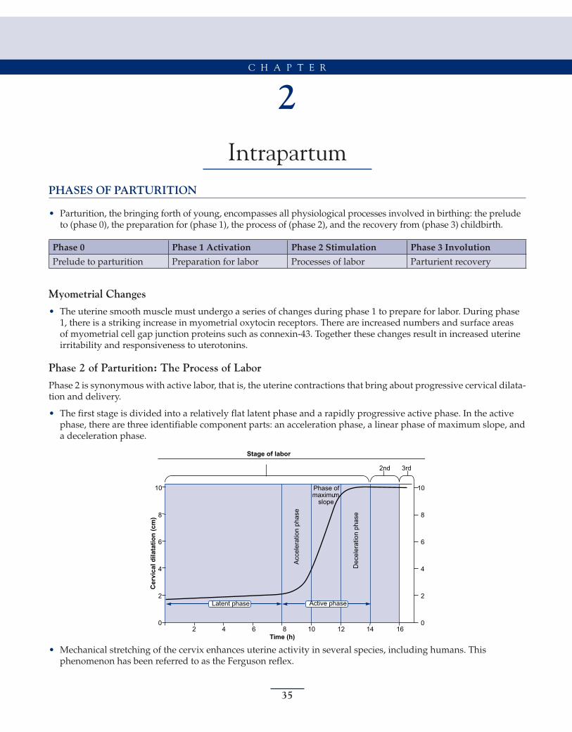
35
2Intrapartum
PHASES OF PARTURITION
• Parturition, the bringing forth of young, encompasses all physiological processes involved in birthing: the prelude to (phase 0), the preparation for (phase 1), the process of (phase 2), and the recovery from (phase 3) childbirth.
Phase 0 Phase 1 Activation Phase 2 Stimulation Phase 3 Involution
Prelude to parturition Preparation for labor Processes of labor Parturient recovery
Myometrial Changes
• The uterine smooth muscle must undergo a series of changes during phase 1 to prepare for labor. During phase 1, there is a striking increase in myometrial oxytocin receptors. There are increased numbers and surface areas of myometrial cell gap junction proteins such as connexin-43. Together these changes result in increased uterine irritability and responsiveness to uterotonins.
Phase 2 of Parturition: The Process of Labor
Phase 2 is synonymous with active labor, that is, the uterine contractions that bring about progressive cervical dilata-tion and delivery.
• The first stage is divided into a relatively flat latent phase and a rapidly progressive active phase. In the active phase, there are three identifiable component parts: an acceleration phase, a linear phase of maximum slope, and a deceleration phase.
Stage of labor
2nd 3rd
Phase ofmaximum
slope
Dec
eler
atio
n ph
ase
Acc
eler
atio
n ph
ase
10
8
6
4
2
0
10
8
6
4
2
02 4 6 8 10 12 14 16
Cer
vica
l dila
tatio
n (c
m)
Time (h)
Active phaseLatent phase
• Mechanical stretching of the cervix enhances uterine activity in several species, including humans. This phenomenon has been referred to as the Ferguson reflex.
C H A P T E R
36 SMART STUDY SERIES: OBSTETRICS AND GYNECOLOGY
Endothelin
• Endothelins are very powerful inducers of myometrial smooth muscle contraction, and endothelin receptors are demonstrable in myometrial tissue. Enkephalinase catalyzes the degradation of endothelin-1.
The Key Factors Thought to Regulate the Phases of Parturition
Phase 0 (Quiescence)
Phase 1 (Activation)
Phase 2 (Stimulation)
Phase 3 (Involution)
ProgesteroneProstacyclinRelaxinNitric oxide
EstrogenProgesteroneUterine stretchProstaglandins
ProstaglandinsOxytocin
Oxytocin
PARTS OF FETAL SKULL
Part Location
Bregma Anterior fontanelle
Brow Between bregma and root of nose
Face Between root of nose and supraorbital ridges and junction of the floor of the mouth with neck
Occiput Bony prominence behind lambda
Vertex Diamond prominence behind anterior and posterior fontanelles and parietal eminences
Fetal Skull: Molding
• Due to descent, the frontal bones slip under parietal bones, resulting in molding • Parietal bones can also slip under each other or under occipital bone • Molding reduces head circumference • Degree of molding (assessed vaginally): a. 0 : Suture lines separate b. +1: Suture lines meet c. +2: Suture lines overlap but are reduced d. +3: Suture lines overlap but are irreducible
Varieties of Cephalic Presentations in Different Attitudes
Diameters Attitude of Head Presentation
1. Suboccipitobregmatic—9.5 cm, extends from the nape of the neck to the center of the bregma
Complete flexion Vertex
2. Suboccipitofrontal—10 cm, extends from the nape of the neck to the anterior end of the anterior fontanelle or center of the sinciput
Incomplete flexion Vertex
3. Occipitofrontal—11.5 cm, extends from the occipital eminence to the root of the nose (glabella)
Marked deflexion Vertex
4. Mentovertical—14 cm, extends from the midpoint of the chin to the highest point on the sagittal suture
Partial extension Brow
5. Submentovertical—11.5 cm, extends from junction of floor of the mouth and neck to the highest point on sagittal suture
Incomplete extension Face
6. Submentobregmatic—9.5 cm, extends from junction of floor of the mouth and neck to the center of the bregma
Complete extension Face
https://kat.cr/user/Blink99/
INTRAPARTUM 37
Important Diameters of Pelvis
Diameter Measurement (cm)
True/anatomical conjugate 11
Obstetric conjugate 10
Diagonal conjugate 12
Transverse (inlet) 13
Oblique 12
Sacrocotyloid 9.5
AP (cavity) 12
Transverse (cavity) 12
Bispinous 10.5
Posterior sagittal (obstetrical outlet) 5
Posterior sagittal (anatomical outlet) 8.5
Intertuberous 11
• The fetus enters the pelvis in the left occiput transverse (LOT) position in 40% of labors and in the right occiput transverse (ROT) position in 20%.
• In about 20% of labors, the fetus enters the pelvis in an occiput posterior (OP) position.
IMPORTANCE OF PLANE OF LEAST DIMENSION
1. Curve of carus bends forward 2. Origin of levator ani begins 3. Internal rotation occurs here 4. Station is ‘O’ at this plane 5. Pudendal block given here
STAGES OF LABOR
Stage Definition
First From the onset of true labor to full dilation of cervix
Second From full dilation of cervix to birth of the baby
Third From birth of the baby to delivery of the placenta
Fourth 1 h observation period following delivery of the placenta
Physiological chills are seen in the fourth stage of labor.The cardinal movements of labor are engagement, descent, flexion, internal rotation, extension, external rotation,
and expulsion.The mechanism by which the biparietal diameter, the greatest transverse diameter of the fetal head in occiput
presentations, passes through the pelvic inlet is designated as engagement.
Descent
The movement is the first requisite for the birth of a newborn.Descent is brought about by one or more of four forces:
1. Pressure of the amniotic fluid 2. Direct pressure of the fundus upon the breech with contractions
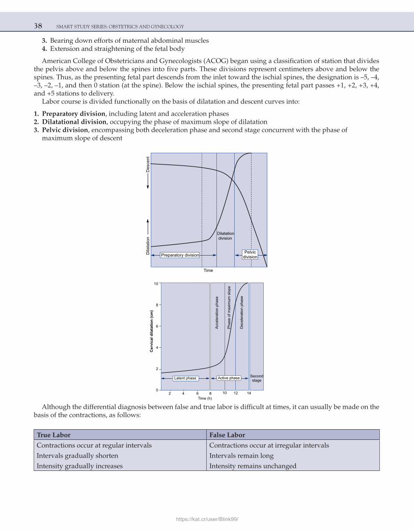
38 SMART STUDY SERIES: OBSTETRICS AND GYNECOLOGY
3. Bearing down efforts of maternal abdominal muscles 4. Extension and straightening of the fetal body
American College of Obstetricians and Gynecologists (ACOG) began using a classification of station that divides the pelvis above and below the spines into five parts. These divisions represent centimeters above and below the spines. Thus, as the presenting fetal part descends from the inlet toward the ischial spines, the designation is –5, –4, –3, –2, –1, and then 0 station (at the spine). Below the ischial spines, the presenting fetal part passes +1, +2, +3, +4, and +5 stations to delivery.
Labor course is divided functionally on the basis of dilatation and descent curves into:
1. Preparatory division, including latent and acceleration phases 2. Dilatational division, occupying the phase of maximum slope of dilatation 3. Pelvic division, encompassing both deceleration phase and second stage concurrent with the phase of
maximum slope of descent
Des
cent
Dila
tatio
n
Time
Dilatationdivision
Preparatory divisionPelvic
division
Cer
vica
l dila
tatio
n (c
m)
Acc
eler
atio
n ph
ase
Time (h)
Pha
se o
f max
imum
slo
pe
Dec
eler
atio
nph
ase
Secondstage
2 4 6 8 10 12 14
Latent phase
10
8
6
4
2
0
Active phase
Although the differential diagnosis between false and true labor is difficult at times, it can usually be made on the basis of the contractions, as follows:
True Labor False Labor
Contractions occur at regular intervals Contractions occur at irregular intervals
Intervals gradually shorten Intervals remain long
Intensity gradually increases Intensity remains unchanged
https://kat.cr/user/Blink99/
INTRAPARTUM 39
True Labor False Labor
Discomfort is in the back and abdomen Discomfort is chiefly in the lower abdomen
Cervix dilates Cervix does not dilate
Discomfort is not stopped by sedation Discomfort usually is relieved by sedation
Caldeyro-Barcia and Poseiro from Montevideo, Uruguay, were pioneers who have done much to elucidate the pat-terns of spontaneous uterine activity throughout pregnancy.
They also introduced the concept of Montevideo units to define uterine activity. By this definition, uterine per-formance is the product of the intensity (increased uterine pressure above baseline tone during contraction) in mil-limeters of mercury multiplied by contraction frequency per 10 min. For example, three contractions in 10 min, each of 50 mm Hg intensity, would equal 150 Montevideo units.
According to Caldeyro-Barcia and Poseiro, clinical labor usually commences when uterine activity reaches val-ues between 80 and 120 Montevideo units. This translates into approximately three contractions of 40 mm Hg every 10 min.
Origin and Propagation of Contractions
The normal contractile wave of labor originates near the uterine end of one of the fallopian tubes; thus, these areas act as “pacemakers.” The right pacemaker usually predominates over the left and starts the great majority of con-tractile waves. Contractions spread from the pacemaker area throughout the uterus at 2 cm/s, depolarizing the whole organ within 15 s. This depolarization wave propagates downward toward the cervix. Intensity is greatest in the fundus.
Partogram
Composite graphical record of key data (maternal and fetal) during labor entered against time on a single sheet of paper. It provides an accurate record of the progress of labor and any delay or deviation from normal may be detected quickly and treated accordingly. It was first devised by Freidman in 1954.
Components 1. Patient identification. 2. Time: It is recorded at an interval of one hour. For spontaneous labor zero time is the time of admission in the
labor ward while for induced labor, it is the time of induction. 3. Fetal heart rate: Recorded every thirty minutes. 4. Liquor color and membrane status: ‘I’ = intact membranes, ‘C’ = clear and ‘M’ = meconium stained liquor. 5. Cervical dilatation and descent of head. 6. Uterine contractions: Intensity and duration. 7. IV fluids and any drugs given. 8. Temperature record. 9. Blood pressure: At an interval of 2 hours. 10. Pulse rate: Every 30 minutes. 11. Oxytocin: Dose and concentration if used. 12. Urine analysis.
Advantages • Provides all important information on single sheet of paper. • Predicts any deviation from normal progress of labor. • Improvement in maternal and perinatal morbidity and mortality.
The concept of `alert line’ and `action line’ was introduced by Philpott and Castle in 1972. The action line can be placed at 2–4 hours interval, to the right and parallel to alert line. In partograms recommended by ‘WHO’ the dis-tance between the alert and action lines is 4 hours.
40 SMART STUDY SERIES: OBSTETRICS AND GYNECOLOGY
GUIDELINES FOR INTRAPARTUM FETAL HEART RATE SURVEILLANCE
Surveillance Low-Risk Pregnancies High-Risk Pregnancies
Acceptable methods
Intermittent auscultation Yes Yes
Continuous electronic monitoring (internal or external) Yes Yes
Evaluation intervals
First-stage labor (active) 30 min 15 min
Second-stage labor 15 min 5 min
Frequency of various presentations
Presentation Frequency (%)
Cephalic 96.5
Breech 2.7
Transverse 0.3
Compound 0.1
Face 0.05
Brow 0.01
• MC malpresentation = Breech • MC malposition = Occipitoposterior
RITGEN MANEUVER
When the head distends the vulva and perineum enough to open the vaginal introitus to a diameter of 5 cm or more, a towel-draped, gloved hand may be used to exert forward pressure on the chin of the fetus through the perineum just in front of coccyx. Concurrently, the other hand exerts pressure superiorly against the occiput. This maneuver allows controlled delivery of the head.
METHODS OF PLACENTAL SEPARATION
1) Schultz (more common) 2) Duncan Matthews
Schultz Mechanism
By far the most common mechanism of placental expulsion.Delivery of the placenta with the fetal side presenting. Results when separation begins centrally with correspond-
ing formation of a central retroplacental clot, which weights the placenta so the central portion descends first.This then inverts the placenta and amniotic sac and causes the membranes to peel-off the remainder of the decidua
and trail behind the placenta. Bleeding associated with Schultz mechanism is not visible until the placenta and mem-branes are delivered, since the inverted membranes hold and catch the blood.
Duncan Mechanism
Delivery of the placenta with the maternal side presenting. Results when separation first takes place at the mar-gin or periphery of the placenta. The placenta descends sideways and the amniotic sac, therefore, is not inverted
https://kat.cr/user/Blink99/
INTRAPARTUM 41
but trails behind the placenta for delivery. Blood escapes between the membranes and uterine wall and is visible externally.
Memory aid for remembering Schultz vs Duncan: Based on the appearance of the two different sides of the pla-centa. Fetal side is shiny and glistening because it is covered by membranes, therefore ‘shiny Schultz’. Maternal side is rough and red-looking, thus ‘dirty Duncan.’
Remember: S.S.C = Shiny Schultz Central.
BREECH
Varieties
Complete (20%)Incomplete: • Frank breech (70%) • Footling breech • Kneeling breech
Frank breech is the most common and is most suitable for vaginal delivery. Footling breech is the least common and has the highest risk of cord prolapse.
Etiology
MC cause = Prematurity
Fetal 1. Multiple pregnancy 2. Hydrocephalus/spina bifida 3. Polyhydramnios/oligohydramnios
Maternal 1. Congenital malformation of the uterus 2. Multiparity 3. CPD 4. Uterine fibroid/pelvic tumors 5. Past history
Placental 1. Placenta previa 2. Cornufundal attachment of placenta 3. Short cord
Prevalence of Breech Presentation by Gestational Age
Gestational Age (Weeks) Breech (%)
28 24
30 17
32 11
34 6
36 5
37–40 4
42 SMART STUDY SERIES: OBSTETRICS AND GYNECOLOGY
METHODS OF VAGINAL DELIVERY
There are three general methods of breech delivery through the vagina:
• Spontaneous breech delivery: The infant is expelled entirely spontaneously without any traction or manipulation other than support of the infant.
• Assisted breech delivery: The infant is delivered spontaneously as far as the umbilicus, but the remainder of the body is extracted or delivered with operator traction and assisted maneuvers, with or without maternal expulsive efforts. This is considered as the best method of vaginal breech delivery.
• Total breech extraction: The entire body of the infant is extracted by the obstetrician. This method is done only in cases of fetal distress.
The incidence of cord prolapse with frank breech presentation is about 0.5%. In contrast, the incidence of cord prolapse with complete breech presentation is 5%, and it is 15% with footling breeches.
Gynecoid and anthropoid pelves are favorable, but android and platypelloid pelvis are unfavorable for vaginal breech delivery.Various maneuvers for breech delivery:
1. Kristellar: suprapubic pressure 2. Pinnard’s: arrested lower limbs (put the fingers in popliteal fossa, flex the knee and grasp the foot) 3. Prague’s: dorsoposterior breech 4. Lovset’s: nuchal arm. The diagnosis is made by noting the winging of the scapula.
Maneuvers for delivering after-coming head of breech:
1. Mauriceau-Smellie-Veit: malar flexion and shoulder traction 2. Burns-Marshall: baby held by ankle and trunk is swung in upward and forward direction 3. Wigard-Martin: malar flexion and supra-pubic pressure 4. Pipers forceps: Piper’s forceps is the best method to deliver the after-coming head of breech because: (a) It is a controlled delivery, sudden decompression of the head is avoided (b) Undue traction on the neck is avoided, so the risk of brachial plexus injury is least
INDICATIONS FOR CESAREAN SECTION IN BREECH PRESENTATION
1. Primi with breech 2. Footling breech 3. Twins with first baby in breech 4. Previous LSCS with breech 5. Preterm breech (risk of intraventricular hemorrhage increases with vaginal delivery) 6. Stargazing/flying fetus
In perhaps 5% of term breech presentations, the fetal head may be in extreme hyperextension. This presentation is referred to as the stargazer fetus or the flying fetus. With such hyperextension, vaginal delivery may result in injury to the cervical spinal cord. In general, marked hyperextension after labor has begun is considered an indication for cesarean delivery.
Preterm infants undergoing cesarean delivery have a better prognosis.Occasionally, especially with small preterm fetuses, the incompletely dilated cervix will not allow vaginal delivery
of the after-coming head. In such cases, Dührssen incisions are usually necessary (cut the cervix at 10 and 2 o’clock positions).
EXTERNAL CEPHALIC VERSION
The ACOG recommends that efforts should be made to reduce breech presentation by external cephalic version (ECV) whenever possible.
The success rate for external cephalic version ranges from 35% to 85%, with an average of about 60%.
https://kat.cr/user/Blink99/
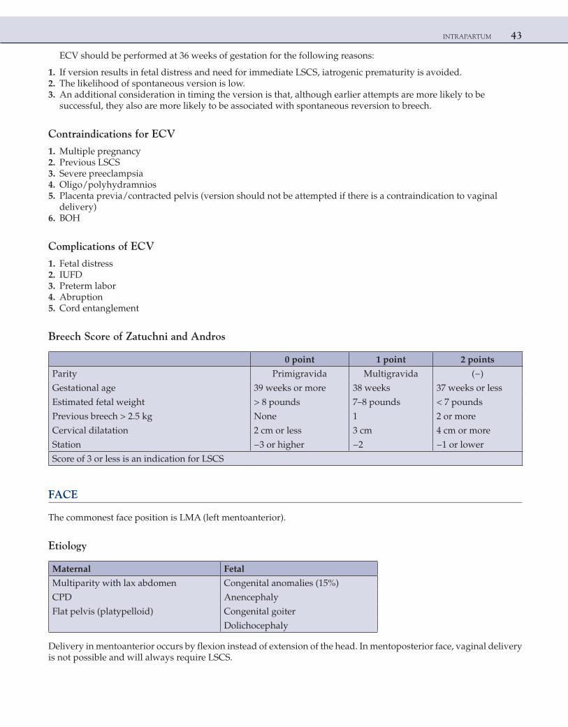
INTRAPARTUM 43
ECV should be performed at 36 weeks of gestation for the following reasons:
1. If version results in fetal distress and need for immediate LSCS, iatrogenic prematurity is avoided. 2. The likelihood of spontaneous version is low. 3. An additional consideration in timing the version is that, although earlier attempts are more likely to be
successful, they also are more likely to be associated with spontaneous reversion to breech.
Contraindications for ECV
1. Multiple pregnancy 2. Previous LSCS 3. Severe preeclampsia 4. Oligo/polyhydramnios 5. Placenta previa/contracted pelvis (version should not be attempted if there is a contraindication to vaginal
delivery) 6. BOH
Complications of ECV
1. Fetal distress 2. IUFD 3. Preterm labor 4. Abruption 5. Cord entanglement
Breech Score of Zatuchni and Andros
0 point 1 point 2 points
Parity Primigravida Multigravida (−)
Gestational age 39 weeks or more 38 weeks 37 weeks or less
Estimated fetal weight > 8 pounds 7–8 pounds < 7 pounds
Previous breech > 2.5 kg None 1 2 or more
Cervical dilatation 2 cm or less 3 cm 4 cm or more
Station −3 or higher −2 −1 or lower
Score of 3 or less is an indication for LSCS
FACE
The commonest face position is LMA (left mentoanterior).
Etiology
Maternal Fetal
Multiparity with lax abdomen Congenital anomalies (15%)
CPD Anencephaly
Flat pelvis (platypelloid) Congenital goiter
Dolichocephaly
Delivery in mentoanterior occurs by flexion instead of extension of the head. In mentoposterior face, vaginal delivery is not possible and will always require LSCS.
44 SMART STUDY SERIES: OBSTETRICS AND GYNECOLOGY
BROW
1. Brow is the rarest presentation. 2. Brow is commonly unstable and converts into either vertex or face. 3. Supraorbital ridges and anterior fontanelle can be palpated on P/v examination. 4. There is no mechanism of labor in persistent brow presentation. Delivery is by LSCS. 5. It is associated with contracted pelvis or fetal macrosomia.
TRANSVERSE LIE
1. The dorsoanterior position is most common (60%). 2. In dorsoposterior, the chance of fetal extension is common with increased risk of arm prolapse and cord prolapse.
Etiology
1. Multiparity 2. Prematurity 3. Multiple pregnancy 4. Polyhydramnios 5. Uterine anomalies 6. Placenta previa 7. Pelvic tumors (fibroids/ovarian cysts) 8. CPD
There is no mechanism of labor in transverse lie. Delivery is by LSCS.If the fetus is small (usually <800 g) and the pelvis is large, spontaneous delivery is possible in transverse lie. The
fetus is compressed with the head forced against the abdomen. A portion of the thoracic wall below the shoulder thus becomes the most dependent part, appearing at the vulva. The head and thorax then pass through the pelvic cavity at the same time, and the fetus, which is doubled upon itself, is expelled—this is referred to as conduplicato corpora.
OCCIPITOPOSTERIOR POSITION
Engaging diameter of the head; occipitofrontal-11.5 cm
Mechanism of labor in occipitoposterior positionDiameter of engagement—oblique diameter
or suboccipitofrontal-10 cm
Good uterine contractionfavorable pelvis
)%01(elbarovafnU)%09(elbarovaF
Weak pains
Increasing flexion with engagement
Long anterior internal rotation of the occiput(three-eight of circle) and normal delivery likeoccipitoanterior (MC outcome)
Android or anthropoid pelvis
Anterior rotation ofocciput (one-eight circle)
Nonrotationof occiput
Posterior rotationof occiput
Deep transversearrest
Occiput obliqueposterior arrest
Occipito sacralposition
Face to pubis delivery(ANTHROPOID PELVIS)
Occipito sacralarrest
Management of deep transverse arrest, oblique posterior arrest, and occipito sacral arrest in modern-day obstetrics is done by cesarean section.
https://kat.cr/user/Blink99/
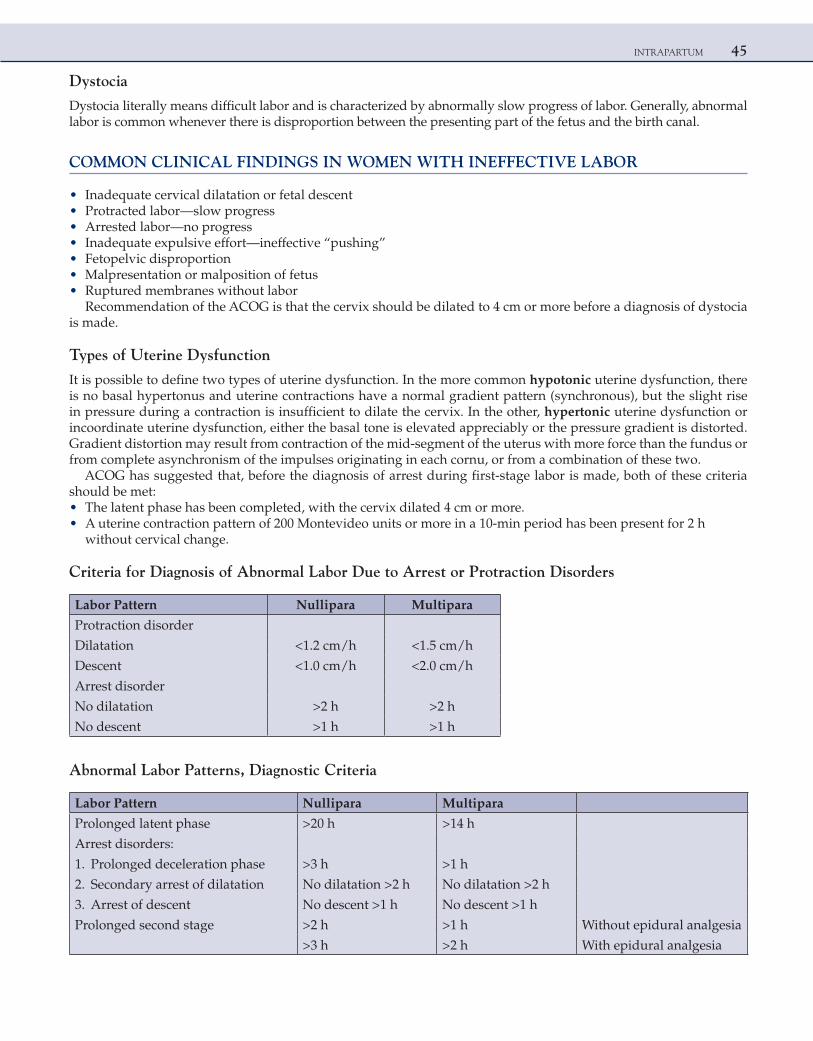
INTRAPARTUM 45
Dystocia
Dystocia literally means difficult labor and is characterized by abnormally slow progress of labor. Generally, abnormal labor is common whenever there is disproportion between the presenting part of the fetus and the birth canal.
COMMON CLINICAL FINDINGS IN WOMEN WITH INEFFECTIVE LABOR
• Inadequate cervical dilatation or fetal descent • Protracted labor—slow progress • Arrested labor—no progress • Inadequate expulsive effort—ineffective “pushing” • Fetopelvic disproportion • Malpresentation or malposition of fetus • Ruptured membranes without labor
Recommendation of the ACOG is that the cervix should be dilated to 4 cm or more before a diagnosis of dystocia is made.
Types of Uterine Dysfunction
It is possible to define two types of uterine dysfunction. In the more common hypotonic uterine dysfunction, there is no basal hypertonus and uterine contractions have a normal gradient pattern (synchronous), but the slight rise in pressure during a contraction is insufficient to dilate the cervix. In the other, hypertonic uterine dysfunction or incoordinate uterine dysfunction, either the basal tone is elevated appreciably or the pressure gradient is distorted. Gradient distortion may result from contraction of the mid-segment of the uterus with more force than the fundus or from complete asynchronism of the impulses originating in each cornu, or from a combination of these two.
ACOG has suggested that, before the diagnosis of arrest during first-stage labor is made, both of these criteria should be met: • The latent phase has been completed, with the cervix dilated 4 cm or more. • A uterine contraction pattern of 200 Montevideo units or more in a 10-min period has been present for 2 h
without cervical change.
Criteria for Diagnosis of Abnormal Labor Due to Arrest or Protraction Disorders
Labor Pattern Nullipara Multipara
Protraction disorder
Dilatation <1.2 cm/h <1.5 cm/h
Descent <1.0 cm/h <2.0 cm/h
Arrest disorder
No dilatation >2 h >2 h
No descent >1 h >1 h
Abnormal Labor Patterns, Diagnostic Criteria
Labor Pattern Nullipara Multipara
Prolonged latent phase >20 h >14 h
Arrest disorders:
1. Prolonged deceleration phase >3 h >1 h
2. Secondary arrest of dilatation No dilatation >2 h No dilatation >2 h
3. Arrest of descent No descent >1 h No descent >1 h
Prolonged second stage >2 h >1 h Without epidural analgesia
>3 h >2 h With epidural analgesia
46 SMART STUDY SERIES: OBSTETRICS AND GYNECOLOGY
SHOULDER DYSTOCIA
A head-to-body delivery time exceeding 60 sec is used to define shoulder dystocia.
• Risk factors include D, O, P, E.D = diabetes mellitusO = obesityP = postdatismE = excessive weight gain during pregnancy (mother or fetus)
• Postpartum hemorrhage, usually from uterine atony, but also from vaginal and cervical lacerations, is the major maternal risk, following shoulder dystocia.
• Shoulder dystocia may be associated with significant fetal morbidity and even mortality. • Transient Erb or Duchenne brachial plexus palsies are the most common injury, followed by clavicular fractures
and humeral fractures.
As per ACOG guidelines, planned cesarean delivery is to be considered for the nondiabetic woman carrying a fetus with an estimated fetal weight exceeding 5000 g or the diabetic woman whose fetus is estimated to weigh more than 4500 g to avoid the risk of shoulder dystocia.
Management of Shoulder Dystocia
1. Extend the episiotomy, remove the lithotomy position. Never give fundal pressure. Moderate suprapubic pressure can be applied by an assistant while downward traction is applied to the fetal head.
Check if it is a unilateral shoulder dystocia (posterior shoulder is in hollow of sacrum, anterior is above pelvic brim) or a bilateral shoulder dystocia (both shoulders above pelvic brim).
If it is bilateral shoulder dystocia, directly proceed to perform LSCS after doing the Zavanelli maneuver (cephalic replacement into the pelvis and then cesarean delivery).
The rest of the maneuvers can be tried for unilateral shoulder dystocia, and if they fail, then proceed for Zavanelli maneuver (cephalic replacement into the pelvis) and LSCS.
2. The McRoberts maneuver: The maneuver consists of removing the legs from the stirrups and sharply flexing them up onto the abdomen. This procedure causes straightening of the sacrum relative to the lumbar vertebrae, rotation of the symphysis pubis toward the maternal head, and a decrease in the angle of pelvic inclination.
3. Woods reported that, by progressively rotating the posterior shoulder 180° in a corkscrew fashion, the impacted anterior shoulder could be released. This is frequently referred to as the Woods corkscrew maneuver.
4. Delivery of the posterior shoulder. 5. Rubin maneuver. 6. Cleidotomy consists of cutting the clavicle with scissors or other sharp instruments and is usually used for a
dead fetus. Symphysiotomy has also been applied successfully. 7. Hibbard maneuver is not used, as it is associated with fetal orthopedic and neurological damage.
DIFFERENCE BETWEEN CONSTRICTION AND RETRACTION RINGS
Constriction Ring (Schroeder’s Ring) Retraction Ring (Bandl’s Ring)
Nature It is a manifestation of localized incoordinate uterine contraction
It is an end result of tonic uterine contrac-tion and retraction
Cause Undue irritability of the uterus Following obstructed labor
Situation Usually at the junction of upper and lower segment but may occur in other places. The position does not alter
Always situated at the junction of upper and lower segment. The position progres-sively moves upward
Uterus Upper segment contracts and retracts with relaxation in between, lower segment remains thick and loose
Upper segment is tonically contracted with no relaxation. The wall becomes thicker; lower segment becomes distended and thinned out
Maternal condition Almost unaffected unless the labor is prolonged Maternal distress is invariably present
https://kat.cr/user/Blink99/
INTRAPARTUM 47
Constriction Ring (Schroeder’s Ring) Retraction Ring (Bandl’s Ring)
Abdominal examination
1. Uterus feels normal and not tender 2. Fetal parts are easily felt 3. Ring is not felt 4. Round ligament is not felt 5. FHS is usually present
1. Uterus is tense and tender 2. Not easily felt 3. Ring is felt as a groove placed
obliquely 4. Taut and tender round ligaments are
felt 5. FHS is usually absent
Vaginal examination
1. The lower segment is not pressed by the presenting part
2. Ring is felt usually above the head 3. Features of obstructed labor are absent
1. Lower segment is very much pressed by the forcibly driven presenting part
2. Ring cannot be felt vaginally 3. Features of obstructed labor are present
End result 1. Exhaustion to the mother is a late feature 2. Fetal anoxia due to prolonged uterine
hypertonic state may appear late 3. Chance of uterine rupture is absent
1. Exhaustion and sepsis appear early 2. Fetal death is usually early due to
tonic contraction and exaggerated retraction
3. Ruptured uterus in multigravidas and uterine exhaustion in primigravidas are the common mode to terminations
Principle of treatment
To relax the ring followed by delivery of the baby
To relieve the obstruction by safe proce-dure after excluding ruptured uterus
BISHOP SCORING SYSTEM USED FOR ASSESSMENT OF INDUCIBILITY
Factor
Score Dilatation (cm) Effacement (%) Station Cervical Consistency Cervical Position
0 Closed 0–30 −3 Firm Posterior
1 1–2 40–50 −2 Medium Mid-position
2 3–4 60–70 −1 Soft Anterior
3 ≥5 >80 +1 to +2 – –
A score of 9 conveys a high likelihood for a successful induction. Score of 4 or less identifies unfavorable cervix and needs for cervical ripening.
Local application of prostaglandin E2 (dinoprostone) is commonly used for cervical ripening.ACOG has approved use of 25 μg vaginal misoprostol for cervical ripening. A vaginal dose of 50 μg is associated
with tachysystole/meconium passage aspiration.
OPERATIVE VAGINAL DELIVERY (FORCEPS AND VACUUM)
ACOG Classification of Forceps and Vacuum Delivery According to Station and Rotation
Procedure Criteria
Outlet 1. Scalp is visible at introitus without separating the labia (station ≥+3)
2. Fetal skull has reached pelvic floor
3. Sagittal suture is in anteroposterior diameter or right or left occiput anterior or posterior position
4. Fetal head is at or on the perineum
5. Rotation does not exceed 45°
48 SMART STUDY SERIES: OBSTETRICS AND GYNECOLOGY
Procedure Criteria
Low Leading point of fetal skull is at station ≥ +2, and not on pelvic floor
Rotation is 45° or less (left or right occiput anterior to occiput anterior, or left or right OP to OP)
Rotation is greater than 45°
Mid-pelvic Station above +2 cm, but head is engaged
High Not included in classification
In modern day obstetrics, forceps is not applied if station is above +2 (station should be at least +2 before applying forceps).
Prerequisites for Forceps Application
There are at least six prerequisites for successful application of forceps:
• The head must be engaged. • The fetus must present as a vertex or by the face with the chin anterior. The position of the fetal head must be
precisely known. • The cervix must be completely dilated. • The membranes must be ruptured. • There should be no suspected cephalic-pelvic disproportion.
With the application of forceps, the head of the fetus is perfectly grasped only when the long axis of blades cor-responds to occipitomental diameter.
Generally, the indications and prerequisites for the use of the vacuum extractor for delivery are the same as for forceps delivery.
Differences between forceps and vacuum
Forceps Vacuum
Traction force = +18 kg for primi, +13 kg for multi Negative pressure = 0.8 kg/cm2 (600 mm Hg)
Cervix should be fully dilated Minimum 7 cm dilation
Less fetal but more maternal complications More fetal but less maternal complications
Preferred in fetal distress Less preferred (as vacuum takes time to build up)
Rotation forceps not applied nowadays Vacuum causes rotation and extraction
Can be applied on face presentation and after-coming head of breech
Cannot be applied on face presentation and after com-ing head of breech
Can be applied on preterm fetus Contraindicated on preterm fetus (increased risk of intraventricular hemorrhage)
Can be applied in cases of fetal coagulopathy and if recent scalp blood sampling has been done
Contraindicated in cases of fetal coagulopathy and if recent scalp blood sampling has been done
Can be applied in cases of IUFD Should not be applied as chignon formation will not occur in IUFD
When using rigid cups, it is recommended that the vacuum be created gradually by increasing the suction by 0.2 kg/cm2 every 2 min until a negative pressure of 0.8 kg/cm2 (600 mm Hg) is reached. With soft cups, negative pressure can be increased to 0.8 kg/cm2 within 1 min.
Comparisons: Forceps Versus Vacuum
• There are significantly more third- and fourth-degree lacerations, in the forceps-delivered group. • Conversely, the incidence of shoulder dystocia and cephalohematomas are more in the vacuum group.
https://kat.cr/user/Blink99/
INTRAPARTUM 49
• Investigators have found decreased maternal trauma by vacuum compared with forceps. • Although retinal hemorrhage occasionally is seen with vacuum usage, it has no apparent long-term effects.
Johanson and Menon analyzed 10 randomized trials and confirmed that vacuum extraction was associated with less maternal but more fetal trauma, for example, cephalohematoma and retinal hemorrhage.
Definitions
Term Definition
Prophylactic forceps Forceps delivery only to shorten the second stage (e.g., heart disease patients)
Trial forceps It is a tentative attempt of forceps delivery in case of suspected mild CPD with a pre-amble declaration of abandoning it in favor of cesarean section if moderate traction fails to overcome the resistance. It is to be performed in the operation theater
Failed forceps When a deliberate attempt in vaginal delivery with forceps has resulted in significant fetal or maternal trauma
Causes of Failed Forceps
1. Failure of application 2. Failure of locking 3. Failure of extraction 4. Undue maternal/fetal trauma
CESAREAN SECTION
Most often the incision is made in the lower uterine segment transversely, as described by Kerr. Occasionally, a low-segment vertical incision, as described by Krönig, may be used. The classical incision is a vertical incision into the body of the uterus above the lower uterine segment and reaching the uterine fundus. This incision is seldom used today.
Indications
Absolute Relative
Central placenta previa CPD
Adherent placenta Previous LSCS
Severe degree of contracted pelvis Dystocia
Previous two LSCS Abruption
Classical CS IUGR
Fetal distress BOH
Transverse/oblique lie Elderly primi/grand multipara
Advanced carcinoma cervix MSAF
Preeclampsia/severe eclampsia
Indications for Classical Cesarean Section
1. Lower segment fibroid 2. Cervical cancer 3. Placenta percreta 4. Dense adhesions in lower pelvis 5. Severe kyphoscoliotic pelvis
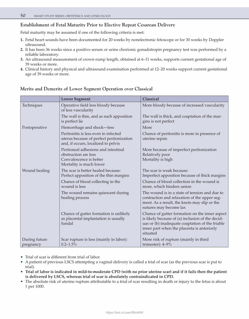
50 SMART STUDY SERIES: OBSTETRICS AND GYNECOLOGY
Establishment of Fetal Maturity Prior to Elective Repeat Cesarean Delivery
Fetal maturity may be assumed if one of the following criteria is met:
1. Fetal heart sounds have been documented for 20 weeks by nonelectronic fetoscope or for 30 weeks by Doppler ultrasound.
2. It has been 36 weeks since a positive serum or urine chorionic gonadotropin pregnancy test was performed by a reliable laboratory.
3. An ultrasound measurement of crown-rump length, obtained at 6–11 weeks, supports current gestational age of 39 weeks or more.
4. Clinical history and physical and ultrasound examination performed at 12–20 weeks support current gestational age of 39 weeks or more.
Merits and Demerits of Lower Segment Operation over Classical
Lower Segment Classical
Techniques Operative field less bloody because of less vascularity
More bloody because of increased vascularity
The wall is thin, and as such apposition is perfect lie
The wall is thick, and coaptation of the mar-gins is not perfect
Postoperative Hemorrhage and shock—less More
Peritonitis is less even in infected uterus because of perfect peritonization and, if occurs, localized to pelvis
Chance of peritonitis is more in presence of uterine sepsis
Peritoneal adhesions and intestinal obstruction are lessConvalescence is betterMortality is much lower
More because of imperfect peritonizationRelatively poorMortality is high
Wound healing The scar is better healed because:Perfect apposition of the thin margins
The scar is weak because:Imperfect apposition because of thick margins
Chance of blood collecting in the wound is less
Chance of blood collection in the wound is more, which hinders union
The wound remains quiescent during healing process
The wound is in a state of tension and due to contraction and relaxation of the upper seg-ment. As a result, the knots may slip or the sutures may become lax
Chance of gutter formation is unlikely as placental implantation is usually fundal
Chance of gutter formation on the inner aspect is likely because of (a) inclusion of the decid-uas or (b) inadequate coaptation of the friable inner part when the placenta is anteriorly situated
During future pregnancy
Scar rupture is less (mainly in labor): 0.2–1.5%
More risk of rupture (mainly in third trimester): 4–9%
• Trial of scar is different from trial of labor. • A patient of previous LSCS attempting a vaginal delivery is called a trial of scar (as the previous scar is put to
trial). • Trial of labor is indicated in mild-to-moderate CPD (with no prior uterine scar) and if it fails then the patient
is delivered by LSCS, whereas trial of scar is absolutely contraindicated in CPD. • The absolute risk of uterine rupture attributable to a trial of scar resulting in death or injury to the fetus is about
1 per 1000.
https://kat.cr/user/Blink99/
INTRAPARTUM 51
Recommendations of the ACOG Useful for the Selection of Candidates for Vaginal Birth after Cesarean Delivery
1. No more than one prior low-transverse cesarean delivery 2. Clinically adequate pelvis (no CPD) 3. No other uterine scars or previous rupture 4. Physician immediately available throughout active labor who is capable of monitoring labor and performing
emergency cesarean delivery 5. Availability of anesthesia and personnel for emergency cesarean delivery
Estimated Risks for Uterine Rupture in Women with a Prior Cesarean Delivery
Prior Uterine Incision Estimated Rupture (%)
Classical 4–9
T shaped 4–9
Low vertical 1–7
Low transverse 0.2–1.5
• In women with uterine malformations who have undergone cesarean delivery, the risks for uterine rupture in a subsequent pregnancy may be as high as with a classical incision.
• Women who have previously sustained a uterine rupture are at increased risk of recurrence. Those with a rupture confined to the lower segment have been reported to have a 6% recurrence risk in subsequent labor, whereas those whose prior rupture included the upper uterus have a 32% recurrence risk.
• The rate of uterine rupture is increased nearly fivefold in women with two previous cesarean deliveries compared with that in those only with one—3.7% versus 0.8%.
• Any previous vaginal delivery, either before or following a cesarean birth, significantly improves the prognosis for a subsequent successful vaginal birth after cesarean delivery (VBAC).
• The success rate for a trial of scar depends to some extent on the indication for the previous cesarean delivery. Generally, about 60–80% of trials after prior cesarean birth result in vaginal delivery, with success being maximum if previous cesarean section was because of breech presentation.
• Women attempting VBAC who had no previous vaginal deliveries, the relative risk of uterine rupture is more than doubled when the birth weight is at least 4000 g.
• As maternal weight increases, the rate of VBAC success decreases. • Any attempt to induce cervical ripening or to induce or augment labor increases the risk of uterine rupture in
women undergoing a trial of scar.Use of oxytocin to induce or augment labor has been implicated in uterine ruptures in women attempting VBAC.
• The American Academy of Pediatrics and the ACOG have concluded that oxytocin may be used for both labor induction and augmentation with close patient monitoring, in women with a prior cesarean delivery undergoing a trial of scar.
• Several prostaglandin preparations commonly are employed for cervical ripening or labor induction. Recent evidence indicates that their use in women attempting VBAC substantively increases the risk of uterine rupture.
• The ACOG discourages the use of prostaglandin cervical ripening agents for the induction of labor in women with previous LSCS.
RUPTURE UTERUS
Uterine rupture typically is classified as either complete (all layers of the uterine wall separated) or incomplete (uter-ine muscle separated but visceral peritoneum is intact). Incomplete rupture is also commonly referred to as uterine dehiscence.
The greatest risk factor for either complete or incomplete uterine rupture is prior cesarean delivery.Following uterine rupture the most common electronic fetal monitoring finding tends to be sudden, severe heart
rate decelerations that may evolve into late decelerations, bradycardia, and undetectable fetal heart action.
52 SMART STUDY SERIES: OBSTETRICS AND GYNECOLOGY
In some cases in which the fetal presenting part has entered the pelvis with labor, loss of station may be detected by pelvic examination. If the fetus is partly or totally extruded from the site of uterine rupture, abdominal palpation or vaginal examination may be helpful to identify the presenting part, which will have moved away from the pelvic inlet. A firm contracted uterus may at times be felt alongside the fetus.
With rupture and expulsion of the fetus into the peritoneal cavity, the chances for intact fetal survival are dismal, and reported mortality rates range from 50% to 75%.
Clinical Features of Ruptured Uterus
Impending Scar Rupture (Scar Dehiscence) Ruptured Uterus
Unexplained tachycardia Weak thready fast pulse
Hypotension Shock
Fetal tachycardia Persistent fetal bradycardia/IUFD
Uterine scar tenderness Hematuria
Bleeding pv Bleeding pv
Hematuria Recession of presenting part
Change in fetal heart rate (tachycardia/loss of beat to beat variability/decelerations) is the earliest sign of impending scar dehiscence, followed by maternal tachycardia.
MULTIPLE CHOICE QUESTIONS
1. Montevideo unit is: a. Uterine contraction in mm of Hg per 10 min b. Uterine contraction in cm of H2O per 10 min c. Uterine contraction in mm of Hg per hour d. Uterine contraction in cm of H2O per hour
Answer: a (Uterine contraction in mm of Hg per 10 min)
Explanation:
• Montevideo units (MVUs) refer to the strength of contractions in mm of Hg multiplied by the frequency per 10 min as measured by intra-uterine pressure transducer.
• The uterine contractile force produced must exceed 200 MVUs/10 min for active labor to be considered adequate.
For example, three contractions in 10 min such that each reaches a peak of 60 mmHg above the baseline, then the strength of contraction is 60 × 3 = 180 MVUs.
Reference: 1. Williams, 22nd Ed., Pg. 466.
2. Which of the following abnormalities of labor is associated with a significantly increased incidence of neonatal morbidity?
a. Prolonged latent phase b. Protracted descent c. Secondary arrest of dilation d. Protracted active-phase dilation
Answer: c (Secondary arrest of dilation)
Explanation:Three significant advances in the treatment of uterine dysfunction have reduced the risk of perinatal morbidity (PNM) and
mortality: (1) the avoidance of undue prolongation of labor, (2) the use of intravenous oxytocin in the treatment of some patterns of uterine dysfunction, and (3) the liberal use of cesarean section (rather than midforceps) to affect delivery when oxytocin fails.
https://kat.cr/user/Blink99/
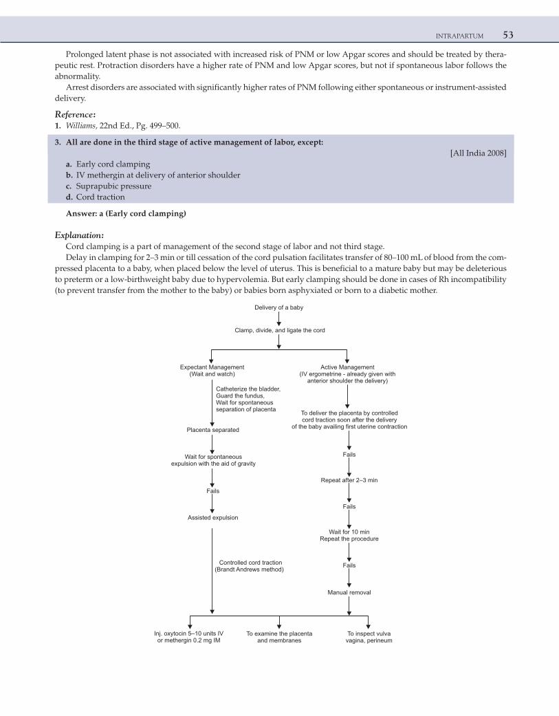
INTRAPARTUM 53
Prolonged latent phase is not associated with increased risk of PNM or low Apgar scores and should be treated by thera-peutic rest. Protraction disorders have a higher rate of PNM and low Apgar scores, but not if spontaneous labor follows the abnormality.
Arrest disorders are associated with significantly higher rates of PNM following either spontaneous or instrument-assisted delivery.
Reference: 1. Williams, 22nd Ed., Pg. 499–500.
3. All are done in the third stage of active management of labor, except: [All India 2008] a. Early cord clamping b. IV methergin at delivery of anterior shoulder c. Suprapubic pressure d. Cord traction
Answer: a (Early cord clamping)
Explanation:Cord clamping is a part of management of the second stage of labor and not third stage.Delay in clamping for 2–3 min or till cessation of the cord pulsation facilitates transfer of 80–100 mL of blood from the com-
pressed placenta to a baby, when placed below the level of uterus. This is beneficial to a mature baby but may be deleterious to preterm or a low-birthweight baby due to hypervolemia. But early clamping should be done in cases of Rh incompatibility (to prevent transfer from the mother to the baby) or babies born asphyxiated or born to a diabetic mother.
Delivery of a baby
Clamp, divide, and ligate the cord
Expectant Management(Wait and watch)
Active Management(IV ergometrine - already given with
anterior shoulder the delivery)Catheterize the bladder,Guard the fundus,Wait for spontaneousseparation of placenta
Placenta separated
To deliver the placenta by controlledcord traction soon after the delivery
of the baby availing first uterine contraction
Wait for spontaneousexpulsion with the aid of gravity
Fails
Assisted expulsion
Controlled cord traction(Brandt Andrews method)
Fails
Repeat after 2–3 min
Fails
Wait for 10 minRepeat the procedure
Fails
Manual removal
Inj. oxytocin 5–10 units IVor methergin 0.2 mg IM
To examine the placentaand membranes
To inspect vulvavagina, perineum
54 SMART STUDY SERIES: OBSTETRICS AND GYNECOLOGY
Reference: 1. Dutta DC. Obstetrics, 6th Ed. Pg. 140–3.
4. Hypertonic dysfunctional labor generally can be expected to: a. Be associated with rapid cervical dilation b. Occur in the active phase of labor c. React favorably to oxytocin stimulation d. Respond to sedation
Answer: d (Respond to sedation)
Explanation:Hypertonic uterine dysfunction is characterized by a lack of coordination of uterine contractions, possibly caused by disor-
ganization of the contraction gradient, which normally is greatest at the fundus and least at the cervix. This type of dysfunc-tion usually appears during the latent phase of labor and is responsive to sedation, not oxytocin stimulation. The disorder is accompanied by a great deal of discomfort with little cervical dilation. After being sedated for a few hours, affected women usually awaken in active labor.
Sedation is also given to differentiate whether the patient is in prolonged latent phase or in false labor: Patients in false labor sleep and awake without contractions, but patients in latent phase show cervical changes after a period of sleep.
Reference: 1. Williams, 22nd Ed., Pg. 500.
5. Commonest presentation in breech, in primigravida is: [All India 2001] a. Frank breech b. Complete breech c. Footling breech d. Knee breech
Answer: a (Frank breech)
Explanation:There are two varieties of breech presentation.
1. Complete (flexed breech): The normal attitude of full flexion is maintained. The thighs are flexed at the hips and the legs at the knees. The presenting part consists of the buttocks, external genitalia and the two feet. It is commonly present in multiparas.
2. Incomplete: This is due to varying degrees of extension of thighs or legs at the podalic pole. Three varieties are possible: a. Breech with extended legs (Frank breech) (MC variety): In this condition, the things are flexed on the trunk and the
legs are extended at the knee joints. The presenting part consists of the two buttocks and external genitalia only. It is commonly present in primigravidas. The increased prevalence in primigravidas is due to tight abdominal wall, good uterine tone and early engagement of breech.
b. Footling presentation: Both the thighs and legs are partially extended, bringing the legs to present at the brim. c. Knee presentation: Thighs are extended but the knees are flexed, bringing knees down to present at the brim. The two
latter varieties are not common.
Reference: 1. Williams, 22nd Ed., Pgs. 566–7.
6. A 25-year-old primigravida patient at 38 weeks complains of gross rupture of membranes and painful uterine contractions every 2–3 min. On digital examination, her cervix is 3 cm with fetal feet palpable through the cervix, and the fetal heart rate tracing is reactive. What is the best method to achieve delivery?
a. Deliver the fetus vaginally by breech extraction b. Deliver the baby vaginally after external cephalic version c. Perform an emergency cesarean section d. Perform an internal podalic version
Answer: c (Perform an emergency cesarean section)
https://kat.cr/user/Blink99/
INTRAPARTUM 55
Explanation:The patient described here has a fetus in the footling breech presentation. Because of the very high risk of cord prolapse,
it is recommended that fetuses with footling breech presentations undergo delivery by cesarean section. External cephalic version is a procedure by which the presentation of the fetus is changed from breech to cephalic, by manipulating the fetus externally through the abdominal wall. It is not indicated in this patient because the membranes are ruptured and the risk of cord prolapse is great. In addition, this procedure generally requires that the uterus be soft and relaxed, which is not the case with this patient in labor. Internal podalic version is a procedure used in the delivery of a second twin. It involves turning the fetus by inserting a hand into the uterus, grabbing both feet, and delivering the fetus by breech extraction.
Reference: 1. Williams, 22nd Ed., Pg. 571.
7. You are delivering an obese primigravida at 41 weeks. After 15 min of pushing, the baby’s head delivers spontaneously but then retracts back against the perineum. As you apply gently downward traction to the head, the baby’s anterior shoulder fails to deliver. All of the following are appropriate next steps in the management of this patient, except:
[AIIMS Nov 2008; All India 2010, 2011] a. Instruct the nurse to apply fundal pressure b. Cut a generous episiotomy c. Flex the maternal legs upon her abdomen d. Call for help
Answer: a (Instruct the nurse to apply fundal pressure)
Explanation:In this clinical scenario, a shoulder dystocia is encountered. A shoulder dystocia occurs when the fetal shoulders fail to
spontaneously deliver secondary to impaction of the anterior shoulder against the pubic bone, after delivery of the head has occurred. Shoulder dystocia is an obstetric emergency, and one should always call for help when such a situation is encoun-tered. A generous episiotomy should always be made to allow the obstetrician to have adequate room to perform a num-ber of manipulations to try to relieve the dystocia. Such maneuvers include the following: suprapubic pressure, McRoberts maneuver (flexing maternal legs upon the abdomen and abduct them), Wood’s corkscrew maneuver (rotating the posterior shoulder), and delivery of the posterior shoulder.
There is no role for fundal pressure because this action further impacts the shoulder against the pubic bone and makes the situation worse. Never give fundal pressure in cases of shoulder dystocia.
Reference: 1. Williams, 22nd Ed., Pgs. 514–5.
8. A 38-year-old G3P2L2 at 40 weeks gestational age presents with pain and regular uterine contractions every 4–5 min. On arrival, the patient is in a lot of pain and requesting relief immediately. You check her cervix and note that it is 5 cm dilated. What is the most appropriate method of pain control for this patient?
a. Intramuscular morphine b. Pudendal block c. Local block d. Epidural block
Answer: d (Epidural block)
Explanation:The most appropriate modality for pain control in this patient is administration of an epidural block. An epidural block
provides relief from the pain of uterine contractions and delivery. It is accomplished by injecting a local anesthetic agent into the epidural space at the level of the lumbar intervertebral space. An indwelling catheter can be left in place to provide con-tinuous infusion of an anesthetic agent throughout labor and delivery via a volumetric pump.
When delivery is imminent, as in the case of this patient, a rapidly acting agent can be administered through the epi-dural catheter to affect perineal anesthesia. In this patient, intramuscular narcotics such as morphine would not be preferred because these agents can cause respiratory depression in the newborn. A pudendal block involves local infiltration of the pudendal nerve, which provides anesthesia to the perineum for delivery but no pain relief for uterine contractions. A local block refers to infusing a local anesthetic to the area of an episiotomy.
56 SMART STUDY SERIES: OBSTETRICS AND GYNECOLOGY
Reference: 1. Williams, 22nd Ed., Pg. 483.
9. A 30-year-old primigravida at 39 weeks has been completely dilated and has been pushing for 3 h. She had taken epidural analgesia. She is exhausted and has a temperature of 37.8°C. The fetal heart rate is 170/min with decreased variability. The patient’s membranes have been ruptured for over 24 h. The patient’s cervix is completely dilated and effaced, and the fetal head is visible at the introitus between pushes. The fetal bones are at the +3 station. What is the most appropriate next step in the management of this patient?
a. Deliver the patient by cesarean section b. Encourage the patient to continue to push after a short rest c. Attempt operative delivery with forceps d. Apply fundal pressure
Answer: c (Attempt operative delivery with forceps)
Explanation:Indications for an operative vaginal delivery with a vacuum extractor or forceps occur in situations where the fetal
head is engaged, the cervix is completely dilated, and there is a prolonged second stage, suspicion of potential fetal compromise, or need to shorten the second stage for maternal benefit. In this situation, all the indications for operative delivery apply. This patient has been pushing for 3 h, which is the definition for prolonged second stage of labor in a nulliparous patient with an epidural. In addition, potential maternal and fetal compromise exists, since the patient has the clinical picture of chorioamnionitis and the fetal heart rate is not reassuring. It is best to avoid cesarean section, since it would take more time to achieve and since the patient is infected. At full dilatation and a suitable station, forceps is faster than LSCS.
Reference: 1. Williams, 22nd Ed., Pgs. 549–50.
10. A 24-year-old primigravida woman, at term, has been dilated to 9 cm for 3 h. The fetal vertex is in the right occiput posterior position, at +1 station. There have been mild late decelerations for the last 10 min. Twenty minutes ago the fetal scalp pH was 7.27; it is now 7.20. Next line of management is:
a. Wait and watch b. Repeat scalp pH after 15 min c. Midforceps rotation d. Low transverse cesarean section
Answer: d (Low transverse cesarean section)
Explanation:A woman who has been dilated 9 cm for 3 h is experiencing a secondary arrest in labor. The deteriorating fetal condition
(as evidenced e.g., by late decelerations and falling scalp pH) dictates immediate delivery.As per ACOG guidelines only, outlet or low forceps should be attempted.A forceps rotation would be inappropriate because the cervix is not fully dilated and, besides, in modern day obstetrics
LSCS is preferred over rotation forceps.Cesarean section would be the safest and quickest method.
Reference: 1. Williams, 22nd Ed., Pgs. 448, 458.
11. Long axis of forceps lies along which fetal diameter: [AIIMS May 2001, All India 2013] a. Mentovertical b. Suboccipitobregmatic c. Occipitofrontal d. Occipitomental
Answer: d (Occipitomental)
Explanation:Forceps are constructed so that their cephalic curve is closely adapted to the sides of the fetal head. The biparietal diameter
of the fetal head corresponds to the greatest distance between the appropriately applied blades. Consequently, the head of the
https://kat.cr/user/Blink99/
INTRAPARTUM 57
fetus is perfectly grasped only when the long axis of the blades corresponds to the occipitomental diameter, with the major portion of the blade lying over the face, while the concave margins of the blades are directed toward either the sagittal suture (occipitoanterior position) or the face (occipitoposterior position).
Reference: 1. Williams, 22nd Ed., Pg. 552.
12. The pelvic inlet usually is considered to be contracted if its shortest anteroposterior diameter is less than: [All India 2002] a. 12 cm b. 10 cm c. 8 cm d. 14 cm
Answer: b (10 cm)
Explanation:The pelvic inlet usually is considered to be contracted if its shortest anteroposterior diameter is less than 10 cm or if the
greatest transverse diameter is less than 12 cm. The anteroposterior diameter of the pelvic inlet is commonly approximated by manually measuring the diagonal conjugate, which is about 1.5 cm greater. Therefore, inlet contraction is usually defined as a diagonal conjugate of less than 11.5 cm.
In women with contracted pelves, face and shoulder presentations are encountered three times more frequently, and cord prolapse occurs 4–6 times more frequently.
The midpelvis is likely contracted when the sum of the interischial spinous and posterior sagittal diameters of the midpel-vis (normal, 10.5 + 5 cm = 15.5 cm) falls to 13.5 cm or below.
There is reason to suspect midpelvic contraction whenever the interischial spinous diameter is less than 10 cm. When it measures less than 8 cm, the midpelvis is contracted.
Contracted Pelvic OutletThis finding usually is defined as an interischial tuberous diameter of 8 cm or less.Outlet contraction without concomitant midplane contraction is rare.Müller and Hillis both described a clinical maneuver to predict dis-proportion.In an occiput presentation, the fetal brow and the suboccipital region are grasped through the abdominal wall with the
fingers and firm pressure is directed downward in the axis of the inlet. Fundal pressure by an assistant is usually helpful. The effect to the forces on the descent of the head can be evaluated by concomitant vaginal examination. If no disproportion exists, the head readily enters the pelvis, and vaginal delivery can be predicted. Inability to push the head into the pelvis, however, does not necessarily indicate that vaginal delivery is impossible. A clear demonstration of a flexed fetal head that overrides the symphysis pubis, however, is presumptive evidence of disproportion.
NOTE:Naegele’s pelvis: absent one ala of sacrumRobert’s pelvis: absent both ala of sacrum
Reference: 1. Williams, 22nd Ed., Pg. 503.
13. The prostaglandin most commonly used at term for induction of labor is: [All India 2003] a. PGI2 b. PGE1 c. PGE2 d. PGF2α
Answer: c (PGE2)
Explanation:PGF2α causes strong tetanic contractions of the whole of uterus (like methergin), so it is never used for induction of labor
as it will lead to fetal distress and IUFD. It is mainly used in prevention and treatment of atonic PPH.PGE1 and PGE2 cause cervical ripening, softening and uterine contractions and hence can be used for induction and aug-
mentation of labor.PGE2 is most commonly used at term for induction of labor. It is preferred over PGE1. It has got great collagenolytic prop-
erties and also sensitizes the myometrium to oxytocin.
58 SMART STUDY SERIES: OBSTETRICS AND GYNECOLOGY
PGE2 is available as gel or tablets.ACOG has only recently approved use of 25 μg vaginal PGE1 (misoprostol) for cervical ripening. A 50 μg vaginal dose is
associated with tachysystole/meconium passage aspiration/fetal distress.Prostaglandins are more effective than oxytocin in cases of intra-uterine death or early gestational period with unfavorable
cervix where oxytocin is less effective.
Various Uses of Prostaglandins in Obstetrics
Medical method of first trimester MTP (mifepristone followed by misoprostol)
Induction of labor (PGE2 and PGE1. PGE2 preferred as more safe)
Second trimester MTP (PGE1 and PGF2α, PGE1 preferred) Acceleration of labor (PGE2 and PGE1. PGE2 preferred as more safe)
Management of atonic postpartum hemorrhage (PGF2α and PGE1)
Medical management of tubal ectopic pregnancy (PGF2α was used in the past as direct injection into the ectopic sac. Not used nowadays)
Reference:
1. Williams, 22nd Ed., Pgs. 537–8.
14. Naegele’s asynclitism is more common in: a. Primigravida b. Multigravida c. Cervical stenosis d. In all labor cases
Answer: b (Multigravida)
Explanation:Deflection of fetal head in relation to the pelvic inlet is called asynclitism.When the sagittal suture lies anteriorly, the posterior parietal bone is the leading presenting part and is called poste-
rior or Litzman asynclitism. It is more common in primigravidas due to good uterine and abdominal wall tone.When the sagittal suture is more posterior and the anterior parietal bone is the leading presenting part, it is called anterior
or Naegele’s asynclitism. It is more common in multiparous patients.
Reference: 1. Williams, 22nd Ed., Pgs. 415–6.
15. Treatment of cord prolapse is based on all of the following factors, except: a. Fetal viability b. Fetal maturity c. Fetal weight d. Cervical dilatation
Answer: c (Fetal weight)
Explanation:Cord prolapse is an obstetric emergency. After cord prolapse on exposure to external environment the whole cord goes into
spasm, leading to severe decelerations in fetal heart rate and fetal distress. If the fetus is alive and mature enough for survival, immediate delivery should be done.Risk factors for cord prolapse include:
1. Long cord 2. Polyhydramnios 3. Abnormal lie (transverse, breech, and oblique) 4. Multiple pregnancies 5. Floating (unengaged) head
https://kat.cr/user/Blink99/
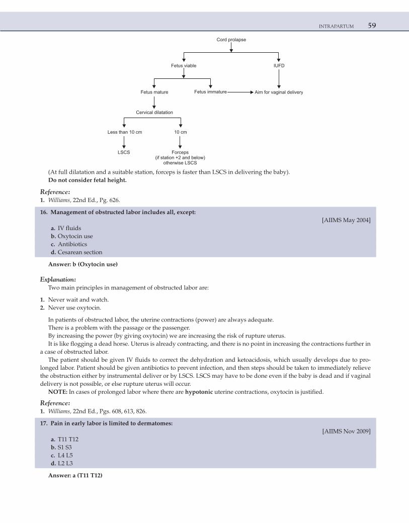
INTRAPARTUM 59
Cord prolapse
DFUIelbaivsuteF
Fetus mature Fetus immature Aim for vaginal delivery
Cervical dilatation
Less than 10 cm
LSCS
10 cm
Forceps(if station +2 and below)
otherwise LSCS
(At full dilatation and a suitable station, forceps is faster than LSCS in delivering the baby).Do not consider fetal height.
Reference: 1. Williams, 22nd Ed., Pg. 626.
16. Management of obstructed labor includes all, except: [AIIMS May 2004] a. IV fluids b. Oxytocin use c. Antibiotics d. Cesarean section
Answer: b (Oxytocin use)
Explanation:Two main principles in management of obstructed labor are:
1. Never wait and watch. 2. Never use oxytocin.
In patients of obstructed labor, the uterine contractions (power) are always adequate.There is a problem with the passage or the passenger.By increasing the power (by giving oxytocin) we are increasing the risk of rupture uterus.It is like flogging a dead horse. Uterus is already contracting, and there is no point in increasing the contractions further in
a case of obstructed labor.The patient should be given IV fluids to correct the dehydration and ketoacidosis, which usually develops due to pro-
longed labor. Patient should be given antibiotics to prevent infection, and then steps should be taken to immediately relieve the obstruction either by instrumental deliver or by LSCS. LSCS may have to be done even if the baby is dead and if vaginal delivery is not possible, or else rupture uterus will occur.
NOTE: In cases of prolonged labor where there are hypotonic uterine contractions, oxytocin is justified.
Reference: 1. Williams, 22nd Ed., Pgs. 608, 613, 826.
17. Pain in early labor is limited to dermatomes: [AIIMS Nov 2009] a. T11 T12 b. S1 S3 c. L4 L5 d. L2 L3
Answer: a (T11 T12)
60 SMART STUDY SERIES: OBSTETRICS AND GYNECOLOGY
Explanation:Pain during first stage of labor is generated largely from the uterus.Early in labor the pain of uterine contractions is transmitted predominantly through the T11 and T12 nerves.Pain with vaginal delivery arises from stimuli from lower genital tract.These are transmitted mainly through pudendal nerve (S 2,3,4)Motor pathway to the uterus leave the spinal cord at the level of T7 and T8.Complete analgesia from pain of labor and vaginal delivery necessitates a block from T10 to S5 dermatome
Reference: 1. Williams, 22nd Ed., Pg. 477.
18. The disadvantage of active management of third stage of labor is: [All India 2008, 2013] a. Increased blood loss b. Increased time interval c. Increased incidence of retained placenta d. All of the above
Answer: c (Increased incidence of retained placenta)
Explanation:Methergin can be given by IM or IV route.In routine management of third stage of labor IM methergin is given after placental delivery.Active management of third stage of labor includes giving IV methergin at the time of delivery of anterior shoulder. The
timing is very important because if given early it will give rise to shoulder dystocia.The principle of active management is to induce strong uterine contractions, so that the placenta separates and immedi-
ately follows the delivery of baby. This decreases the time duration and blood loss during third stage of labor.Absolute contraindications to the use of methergin are:
1. Chronic hypertension/preeclampsia/eclampsia 2. Heart disease in pregnancy 3. After the delivery of the first baby of the twins. (It can be given after 2nd baby delivery of twin.
Obviously the contraindications to the use of methergin are also the contraindications for active management of the 3rd stage of labor.
The only disadvantage of active management of third stage of labor is slightly increased incidence of retained placenta. This can happen because the placenta separates but the cervical os closes giving rise to trapped placenta.NOTE: Rh negative pregnancy is a relative (not absolute) contraindication for use of methergin.
Reference: 1. Williams, 22nd Ed., Pgs. 433–4.
19. Which of the following is the least common variety of pelvis? [All India 2013] a. Gynecoid b. Android c. Anthropoid d. Platypelloid
Answer: d (Platypelloid)
Explanation:Caldwell and Moloy classification of pelvis
Pelvis Incidence (%)
Gynecoid 50
Anthropoid 25
Android 20
Platypelloid 5
https://kat.cr/user/Blink99/
INTRAPARTUM 61
Android pelvis is a/w deep transverse arrest.Anthropoid pelvis is a/w occipitoposterior position and face to pubis delivery.Platypelloid pelvis is a/w face presentation.
Reference: 1. Williams, 22nd Ed., Pg. 35.
20. A patient presents with occipitoposterior position in labor. Management is: [AIIMS Nov 2009] a. Oxytocin drip b. Artificial rupture of membranes c. Wait and Watch d. Cesarean section
Answer: c (Wait and Watch)
Explanation:Occipitoposterior (OP) is the most common malposition. Anthropoid and android variety of pelvis favor this position.In cases of occipitoposterior position the best management is to wait and watch.In around 80% cases there is a long anterior rotation through 3/8th of a circle and normal delivery like occipito anterior
will take place.Only if there are inadequate uterine activity then oxytocin augmentation is required.Per say, OP is not an indication for LSCS.If there is a short anterior rotation then a deep transverse arrest will happen and then LSCS is required.Oblique posterior arrest and occipito sacral arrest are indications for LSCS.
Reference: 1. Williams, 22nd Ed., Pg. 415–8.
21. A G2P1L1 with previous LSCS presents with hematuria during labor. The most likely diagnosis is: [AIIMS Nov 2009] a. Impending rupture of scar b. Urethral trauma c. Prolong labor d. Cystitis
Answer: a (Impending rupture of scar)
Explanation:The main risk of trial of scar (V.B.A.C) is scar dehiscence & rupture uterus.Rupture of previous LSCS scar is the most common cause of rupture uterus.Hematuria is seen with rupture uterus and also with impending rupture of scar.
Clinical features of impending scar rupture (scar dehiscence)
Unexplained tachycardiaHypotensionFetal tachycardiaUterine scar tendernessBleeding PVHematuria
Reference: 1. Williams, 22nd Ed., Pg. 615.
22. All of the following are used for induction of labor, EXCEPT: [AIIMS May 2004] a. PGF2α tablet b. PGE2 tablet c. PGE2 gel d. Misoprostol
Answer: a (PGF2α tablet)
62 SMART STUDY SERIES: OBSTETRICS AND GYNECOLOGY
Explanation:Methods for induction of labor include:
1. Oxytocin infusion 2. Prostaglandins (PGE1 and PGE2): Prostaglandins act on the cervix to enable ripening by a number of different
mechanisms. They alter the extracellular ground substance of the cervix and PGE2 increases the activity of collagenase in the cervix. They cause an increase in elastase, glycosaminoglycan, dermatan sulfate, and hyaluronic acid levels in the cervix. A relaxation of cervical smooth muscle facilitates dilation. Finally, prostaglandins allow for an increase in intracellular calcium levels, causing contraction of myometrial muscles. Risks associated with the use of prostaglandins include uterine hyperstimulation and maternal side effects such as nausea, vomiting, diarrhea, and fever.PGE2 is available in the form of gel and tablets. Misoprostol (PGE1) tablets can also be used vaginally.
3. Mifepristone: Mifepristone is an antiprogesterone agent. Progesterone inhibits contractions of the uterus, while mifepristone counteracts this action.
PGF2α is not available in tablet form. It ia available as intramuscular injection.It has action similar to Methergin. It causes a strong titanic contraction of the entire uterus, and hence, it is used only for
prevention and treatment of PPH.It can never be used for induction of labor.
Reference: 1. Williams, 22nd Ed., Pg. 537–42.
23. On per vaginal examination, anterior fontanelle and supra-orbital ridge is felt in the second stage of labor. The presentation is:
[AIIMS May 2002] a. Brow b. Shoulder c. Vertex d. Face
Answer: a (Brow)
Explanation:
1. Brow is the rarest presentation. 2. Brow is commonly unstable and converts into either vertex or face. 3. Supra-orbital ridges and anterior fontanelle can be palpated on P/V examination. 4. There is no mechanism of labor in persistent brow presentation. Delivery is by LSCS. 5. It is associated with contracted pelvis or fetal macrosomia.
Reference: 1. Williams, 22nd Ed., Pg. 506–9.
24. All of the following are indicators of scar dehiscence in a case of previous LSCS, EXCEPT: [AIIMS Nov 2001, All India 2006] a. Presence of meconium b. Fetal bradycardia c. Vaginal bleeding d. Hematuria
Answer: a (Presence of meconium)
Explanation:Clinical features of impending scar rupture (scar dehiscence):
• Unexplained tachycardia • Hypotension • Fetal tachycardia followed by fetal distress (bradycardia) • Uterine scar tenderness
https://kat.cr/user/Blink99/
INTRAPARTUM 63
• Fresh bleeding PV • Hematuria (seen in both rupture and dehiscence)
Hematuria in case of rupture is due to injury to bladder and in case of dehiscence is due to pelvic congestion.Passage of meconium does not mean that there is fetal distress. It was an old concept that:Passage of meconium = Fetal distressBut this does not hold true anymore. Baby can pass meconium even without distress (e.g., postdatism).
Reference: 1. Williams, 22nd Ed., Pg. 615.
25. A primigravida presents with 37 weeks of gestation with 10 hours of duration of labor with 1 cm dilated cervix. How will you manage the case?
[All India 2011] a. Give sedation and watch b. Amniotomy c. Cesaration d. Oxytocin infusion
Answer: a (Give sedation and watch)
Explanation:Patient has presented at term and is in labor since 10 hours. This could be either prolonged latent phase of labor or false
labor. Best plan is to give sedation to the patient and wait and watch.Patients in false labor sleep and awake without contractions. But patients in latent phase show cervical changes and will
progress after period of sleep.Amniotomy (ARM) should be done in active labor. There is no need of doing LSCS at present.
Reference: 1. Williams, 22nd Ed., Pg. 500.
26. A patient with rheumatic heart disease has PPH. Which of the following drugs is contraindicated? [All India 2011] a. Mifepristone b. Methylergometrine c. Oxytocin d. Carboprost
Answer: b (Methylergometrine)
Explanation:Methylergometrine (Methergin) can be used in the prevention and treatment of PPH. Absolute contraindications to the
use of Methergin are:
1. Chronic hypertension/preeclampsia/eclampsia 2. Heart disease in pregnancy 3. After the delivery of the first baby of the twins. (It can be given after second baby delivery of twin.)
Hence, if the patient has a heart disease and develops PPH, Methergin is absolutely contraindicated. Obviously, the contra-indications to the use of Methergin are also the contraindications for active management of the third stage of labor.
Reference: 1. Williams, 22nd Ed., Pg. 430–4.
27. A 30-year-old female comes with obstructed labor and is febrile and dehydrated with IUFD and cephalic presentation. Which is the best way to manage?
[All India 2006, AIIMS May 2011] a. Craniotomy b. Decapitation c. Cesarean section d. Forceps extraction
Answer: c (Cesarean section)
64 SMART STUDY SERIES: OBSTETRICS AND GYNECOLOGY
Explanation:Two main principles in the management of obstructed labor are:
1. Never wait and watch 2. Never use oxytocin
In patients of obstructed labor, the uterine contractions (power) are always adequate.The patient should be given i.v. fluids to correct the dehydration and ketoacidosis, which usually develop due to pro-
longed labor. Patient should be given antibiotics to prevent infection and then steps should be taken to immediately relieve the obstruction by LSCS.
LSCS may have to be done (even if the baby is dead) if vaginal delivery is not possible, or else, rupture uterus will occur.In modern-day obstetrics, destructive operations (decapitation, craniotomy, evisceration, etc) are never to be performed
as they are more dangerous and can lead to complications like rupture uterus and bladder injury.LSCS is much safer than destructive operations.
NOTE: So remember that if vaginal delivery is not possible, then LSCS has to be done. Destructive operations never to be marked as the answer.
Reference: 1. Williams, 22nd Ed., Pg. 608, 613, 826.
28. All of the following will cause difficulty in delivery of ‘after-coming head of breech’, EXCEPT: [AIIMS Nov 2011] a. Placenta previa b. Extension head c. Hydrocephalus d. Incomplete dilatation of cervix
Answer: a (Placenta previa)
Explanation:Breech is the most common malpresentation.The most difficult and dangerous part in vaginal breech delivery is the delivery of the after-coming head.The breech and limbs being soft can easily deliver, but there is a danger of head getting entrapped, leading to perinatal
morbidity and mortality.Three types of vaginal breech deliveries are described, as follows:
• Spontaneous breech delivery: No traction or manipulation of the infant is used. This occurs predominantly in very preterm, often previable, deliveries.
• Assisted breech delivery: This is the most common type of vaginal breech delivery. The infant is allowed to spontaneously deliver up to the umbilicus, and then maneuvers are initiated to assist in the delivery of the remainder of the body, arms, and head.
• Total breech extraction: The fetal feet are grasped, and the entire fetus is extracted. Total breech extraction should be used only for a non-cephalic second twin or in cases of fetal distress; it should not be routinely used for a singleton fetus because the cervix may not be adequately dilated to allow passage of the fetal head.
Extension of head (stargazing fetus), hydrocephalus, and incomplete dilatation of cervix, all will create problems in deliv-ery of the fetal head, leading to difficult second stage and head entrapment and morbidity and mortality.
Question of vaginal delivery does not occur in case of placenta previa.Patients with placenta previa (placenta is in front of presenting part) are to be delivered by LSCS (whether it is vertex or breech).
There would be profuse hemorrhage leading to maternal mortality if vaginal delivery is attempted in cases of placenta previa.
Reference: 1. Williams, 22nd Ed., Pg. 566–70.
29. All are true about outlet forceps, EXCEPT: [AIIMS Nov 2011] a. Head at ‘0’ station b. Can be applied in vertex and face presentation in mentoanterior c. Caput succedaneum may be present d. Sagittal suture at 15° to anteroposterior diameter
Answer: a (Head at ‘0’ station)
https://kat.cr/user/Blink99/
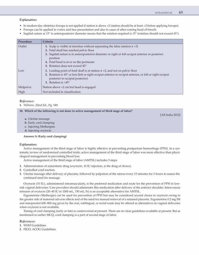
INTRAPARTUM 65
Explanation:
• In modern-day obstetrics forceps is not applied if station is above +2 (station should be at least +2 before applying forceps). • Forceps can be applied in vertex and face presentation and also in cases of after-coming head of breech. • Sagittal suture at 15° to anteroposterior diameter means that the rotation required is 15° (rotation should not exceed 45°).
Procedure Criteria
Outlet 1. Scalp is visible at introitus without separating the labia (station ≥ +3) 2. Fetal skull has reached pelvic floor 3. Sagittal suture is in anteroposterior diameter or right or left occiput anterior or posterior
position 4. Fetal head is at or on the perineum 5. Rotation does not exceed 45°
Low 1. Leading point of fetal skull is at station ≥ +2, and not on pelvic floor 2. Rotation is 45° or less (left or right occiput anterior to occiput anterior, or left or right occiput
posterior to occiput posterior) 3. Rotation is >45°
Midpelvic Station above +2 cm but head is engaged
High Not included in classification
Reference: 1. Williams, 22nd Ed., Pg. 549.
30. Which of the following is not done in active management of third stage of labor? [All India 2012] a. Uterine massage b. Early cord clamping c. Injecting Methergine d. Injecting oxytocin
Answer: b (Early cord clamping)
Explanation:Active management of the third stage of labor is highly effective at preventing postpartum hemorrhage (PPH). In a sys-
tematic review of randomized controlled trials, active management of the third stage of labor was more effective than physi-ological management in preventing blood loss.
Active management of the third stage of labor (AMTSL) includes 3 steps:
1. Administration of auterotonic drug (oxytocin, 10 IU injection, is the drug of choice). 2. Controlled cord traction. 3. Uterine massage after delivery of placenta, followed by palpation of the uterus every 15 minutes for 2 hours to assess the
continued need for massage.
Oxytocin (10 IU), administered intramuscularly, is the preferred medication and route for the prevention of PPH in low-risk vaginal deliveries. Care providers should administer this medication after delivery of the anterior shoulder. Intravenous infusion of oxytocin (20–40 IU in 1000 mL, 150 mL/h) is an acceptable alternative for AMTSL.
Ergometrine (Methergin) can be used for prevention of PPH but may be considered second choice to oxytocin owing to the greater risk of maternal adverse effects and of the need for manual removal of a retained placenta. Ergometrine 0.2 mg IM and misoprostol 600–800 mg given by the oral, sublingual, or rectal route may be offered as alternatives in vaginal deliveries when oxytocin is not available.
Timing of cord clamping (early or late) is controversial at present. There are no clear guidelines available at present. But as mentioned in earlier MCQ, cord clamping is a part of second stage of labor.
References: 1. WHO Guidelines. 2. FIGO, ACOG Guidelines.
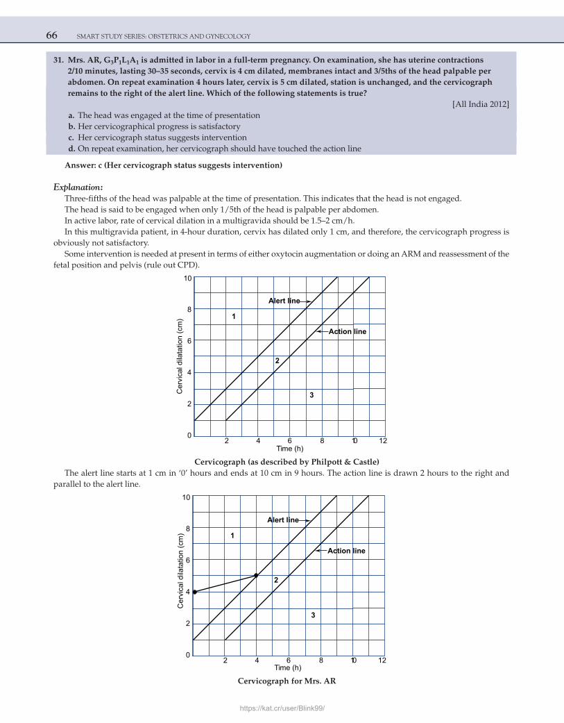
66 SMART STUDY SERIES: OBSTETRICS AND GYNECOLOGY
31. Mrs. AR, G3P1L1A1 is admitted in labor in a full-term pregnancy. On examination, she has uterine contractions 2/10 minutes, lasting 30–35 seconds, cervix is 4 cm dilated, membranes intact and 3/5ths of the head palpable per abdomen. On repeat examination 4 hours later, cervix is 5 cm dilated, station is unchanged, and the cervicograph remains to the right of the alert line. Which of the following statements is true?
[All India 2012] a. The head was engaged at the time of presentation b. Her cervicographical progress is satisfactory c. Her cervicograph status suggests intervention d. On repeat examination, her cervicograph should have touched the action line
Answer: c (Her cervicograph status suggests intervention)
Explanation:Three-fifths of the head was palpable at the time of presentation. This indicates that the head is not engaged.The head is said to be engaged when only 1/5th of the head is palpable per abdomen.In active labor, rate of cervical dilation in a multigravida should be 1.5–2 cm/h.In this multigravida patient, in 4-hour duration, cervix has dilated only 1 cm, and therefore, the cervicograph progress is
obviously not satisfactory.Some intervention is needed at present in terms of either oxytocin augmentation or doing an ARM and reassessment of the
fetal position and pelvis (rule out CPD).
Alert line
Action line
1
2
3Cer
vica
l dila
tatio
n (c
m)
Time (h)
10
8
6
4
2
02 4 6 8 10 12
Cervicograph (as described by Philpott & Castle)The alert line starts at 1 cm in ‘0’ hours and ends at 10 cm in 9 hours. The action line is drawn 2 hours to the right and
parallel to the alert line.
Alert line
Action line
1
2
3
Cer
vica
l dila
tatio
n (c
m)
Time (h)
10
8
6
4
2
02 4 6 8 10 12
Cervicograph for Mrs. AR
https://kat.cr/user/Blink99/
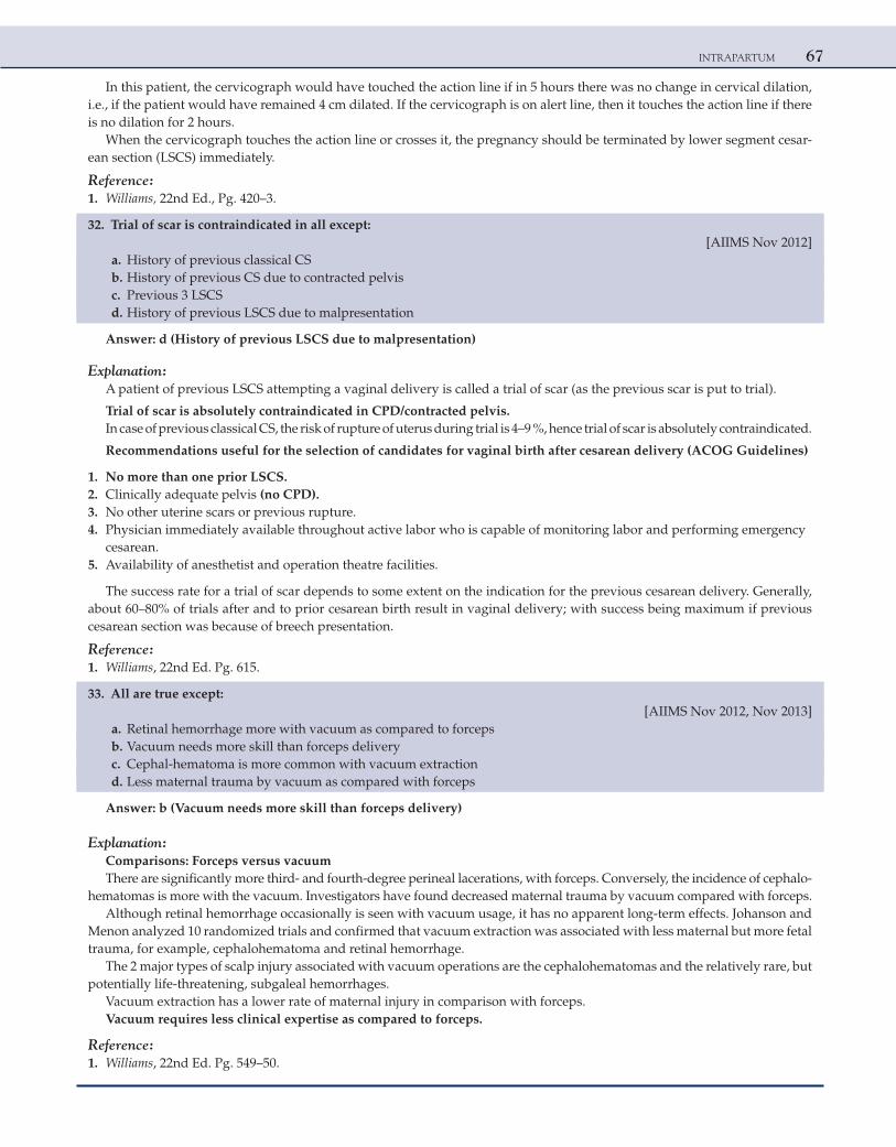
INTRAPARTUM 67
In this patient, the cervicograph would have touched the action line if in 5 hours there was no change in cervical dilation, i.e., if the patient would have remained 4 cm dilated. If the cervicograph is on alert line, then it touches the action line if there is no dilation for 2 hours.
When the cervicograph touches the action line or crosses it, the pregnancy should be terminated by lower segment cesar-ean section (LSCS) immediately.
Reference: 1. Williams, 22nd Ed., Pg. 420–3.
32. Trial of scar is contraindicated in all except: [AIIMS Nov 2012] a. History of previous classical CS b. History of previous CS due to contracted pelvis c. Previous 3 LSCS d. History of previous LSCS due to malpresentation
Answer: d (History of previous LSCS due to malpresentation)
Explanation:A patient of previous LSCS attempting a vaginal delivery is called a trial of scar (as the previous scar is put to trial).
Trial of scar is absolutely contraindicated in CPD/contracted pelvis.In case of previous classical CS, the risk of rupture of uterus during trial is 4–9 %, hence trial of scar is absolutely contraindicated.
Recommendations useful for the selection of candidates for vaginal birth after cesarean delivery (ACOG Guidelines)
1. No more than one prior LSCS. 2. Clinically adequate pelvis (no CPD). 3. No other uterine scars or previous rupture. 4. Physician immediately available throughout active labor who is capable of monitoring labor and performing emergency
cesarean. 5. Availability of anesthetist and operation theatre facilities.
The success rate for a trial of scar depends to some extent on the indication for the previous cesarean delivery. Generally, about 60–80% of trials after and to prior cesarean birth result in vaginal delivery; with success being maximum if previous cesarean section was because of breech presentation.
Reference: 1. Williams, 22nd Ed. Pg. 615.
33. All are true except: [AIIMS Nov 2012, Nov 2013] a. Retinal hemorrhage more with vacuum as compared to forceps b. Vacuum needs more skill than forceps delivery c. Cephal-hematoma is more common with vacuum extraction d. Less maternal trauma by vacuum as compared with forceps
Answer: b (Vacuum needs more skill than forceps delivery)
Explanation:Comparisons: Forceps versus vacuumThere are significantly more third- and fourth-degree perineal lacerations, with forceps. Conversely, the incidence of cephalo-
hematomas is more with the vacuum. Investigators have found decreased maternal trauma by vacuum compared with forceps.Although retinal hemorrhage occasionally is seen with vacuum usage, it has no apparent long-term effects. Johanson and
Menon analyzed 10 randomized trials and confirmed that vacuum extraction was associated with less maternal but more fetal trauma, for example, cephalohematoma and retinal hemorrhage.
The 2 major types of scalp injury associated with vacuum operations are the cephalohematomas and the relatively rare, but potentially life-threatening, subgaleal hemorrhages.
Vacuum extraction has a lower rate of maternal injury in comparison with forceps.Vacuum requires less clinical expertise as compared to forceps.
Reference: 1. Williams, 22nd Ed. Pg. 549–50.

This page intentionally left blank
https://kat.cr/user/Blink99/
69
3Obstetric Complications
OBSTETRIC HEMORRHAGE
Distinguishing features of placenta previa and abruptio placenta
Clinical Features Placenta Previa Abruptio Placenta
Nature of bleeding a. Painless, profuse b. Bleeding is always revealed c. Periodic
a. Painful b. Revealed, concealed, or usually mixed c. Progressive
General condition and anemia
Proportional to visible blood loss
Out of proportion to the visible blood loss in concealed or mixed variety
Features of preeclampsia Unrelated Very likely to be present
Height of uterus Proportionate height May be disproportionately enlarged in concealed type
Feel of uterus Soft and relaxed Tonically contracted uterus
Malpresentation Malpresentation is common (breech, transverse lie). The head is high floating
Unrelated, the head may be engaged
Placentography Placenta in lower segment Placenta in upper segment
Tocolysis Can be given Never
Wait and watch Can be done Never
Delivery LSCS LSCS or vaginal delivery
DIC Less common More common
Risk Factors for Abruptio Placenta
1. Increased age and parity 2. Preeclampsia and chronic hypertension 3. Preterm ruptured membranes 4. Cigarette smoking and cocaine use 5. Thrombophilia 6. Prior abruption (risk of recurrence is 17% for patients with one abruption and 25% for patients with more than
one abruption) 7. Uterine leiomyoma 8. Multifetal gestation 9. Polyhydramnios 10. External trauma
C H A P T E R
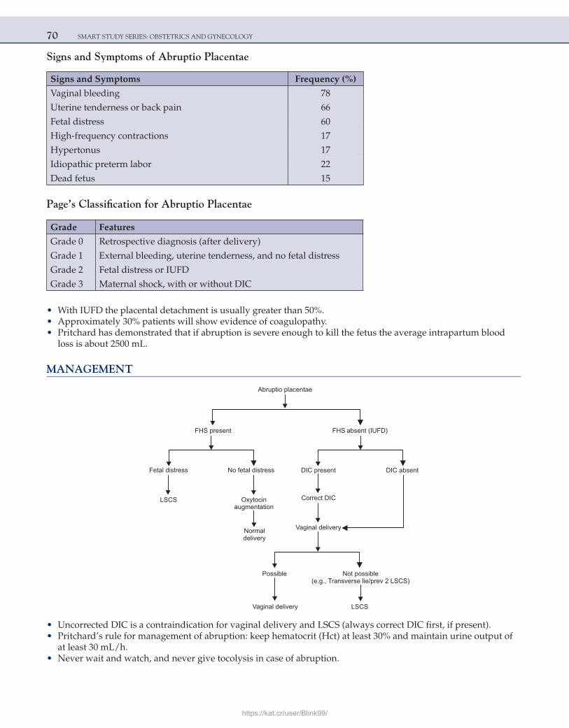
70 SMART STUDY SERIES: OBSTETRICS AND GYNECOLOGY
Signs and Symptoms of Abruptio Placentae
Signs and Symptoms Frequency (%)
Vaginal bleeding 78
Uterine tenderness or back pain 66
Fetal distress 60
High-frequency contractions 17
Hypertonus 17
Idiopathic preterm labor 22
Dead fetus 15
Page’s Classification for Abruptio Placentae
Grade Features
Grade 0 Retrospective diagnosis (after delivery)
Grade 1 External bleeding, uterine tenderness, and no fetal distress
Grade 2 Fetal distress or IUFD
Grade 3 Maternal shock, with or without DIC
• With IUFD the placental detachment is usually greater than 50%. • Approximately 30% patients will show evidence of coagulopathy. • Pritchard has demonstrated that if abruption is severe enough to kill the fetus the average intrapartum blood
loss is about 2500 mL.
MANAGEMENT
Abruptio placentae
DIC present DIC absent
Correct DIC
Possible Not possible(e.g., Transverse lie/prev 2 )LSCS
Vaginal delivery LSCS
FHS present
Normaldelivery
Oxytocinaugmentation
LSCS
Fetal distress No fetal distress
FHS absent (IUFD)
Vaginal delivery
• Uncorrected DIC is a contraindication for vaginal delivery and LSCS (always correct DIC first, if present). • Pritchard’s rule for management of abruption: keep hematocrit (Hct) at least 30% and maintain urine output of
at least 30 mL/h. • Never wait and watch, and never give tocolysis in case of abruption.
https://kat.cr/user/Blink99/
OBSTETRIC COMPLICATIONS 71
Consumptive Coagulopathy
• One of the most common causes of clinically significant consumptive coagulopathy in obstetrics is placental abruption.
• Overt hypofibrinogenemia (less than 150 mg/dL of plasma) along with elevated levels of fibrin degradation products, D-dimers, and variable decreases in other coagulation factors are found in about 30% of women with placental abruption severe enough to kill the fetus. Such severe coagulation defects are seen less commonly in those cases in which the fetus survives.
Couvelaire Uterus
• There may be widespread extravasation of blood into the uterine musculature and beneath the uterine serosa. This so-called uteroplacental apoplexy, first described by Couvelaire in the early 1900s, is now frequently called Couvelaire uterus. It is seen in cases of severe concealed abruption.
• Such effusions of blood are also occasionally seen beneath the tubal serosa, in the connective tissue of the broad ligaments, and in the substance of the ovaries, as well as free in the peritoneal cavity.
• These myometrial hemorrhages seldom interfere with uterine contractions sufficiently to produce severe postpartum hemorrhage and are not an indication for hysterectomy.
PLACENTA PREVIA
Definition: In placenta previa, the placenta is located over or very near the internal os. Four degrees of this abnor-mality have been recognized:
• Total placenta previa: The internal cervical os is covered completely by placenta. • Partial placenta previa: The internal os is partially covered by placenta. • Marginal placenta previa: The edge of the placenta is at the margin of the internal os. • Low-lying placenta: The placenta is implanted in the lower uterine segment such that the placenta edge actually
does not reach the internal os but is in close proximity to it.
Risk Factors
1. Increasing age and increasing parity 2. Past history (12 times risk of another placenta previa) 3. Previous LSCS (probability of previa is four times greater than in patients without any uterine scar) 4. Multiple pregnancy 5. Prematurity 6. Smoking
• In a case of placenta previa, one-third patients bleed before 30 weeks, one-third from 30 to 36 weeks, and one-third bleed after 36 weeks.
• In a case of placenta previa with previous one LSCS, the incidence rate of placenta accreta is 25%, which increases to 67% with previous four LSCS.
• Frederiksen and coworkers reported a 25% hysterectomy rate in women undergoing repeat cesarean for a previa compared with only 6% in those undergoing primary cesarean for placenta previa.
• The simplest, most precise, and safest method of placental localization is provided by transabdominal sonography, which is used to locate the placenta with considerable accuracy. False-positive results are often a result of bladder distention. Therefore, ultrasonic scans in apparently positive cases should be repeated after emptying the bladder.
• Type 2 b = dangerous placenta previa. • Stallworthy’s sign (slowing of FHR on pressing the head down into the pelvis) is seen in placenta previa. • Cesarean delivery is necessary in practically all women with placenta previa (even if the fetus is dead).
72 SMART STUDY SERIES: OBSTETRICS AND GYNECOLOGY
McAfee and Johnson Regimen (Conservative Management in Placenta Previa)
This consists of complete bed rest, tocolysis, and close observation of patient.Steroids are generally given to enhance lung maturity.To undertake this regimen (to wait and watch), all the three criteria should be fulfilled:
1. Mother should be hemodynamically stable. 2. There should be no fetal distress. 3. Pregnancy should be less than 36 weeks of gestation.
If any one of these criteria is not met, then the patient should be delivered by LSCS.
POSTPARTUM HEMORRHAGE
Definition
1. Blood loss of 500 mL or more after completion of third stage of labor. 2. ACOG definition: Bleeding which causes Hct to decrease by 10% or the need of blood transfusion after delivery.
Predisposing Factors and Causes of Immediate Postpartum Hemorrhage
Bleeding from placental implantation site Hypotonic myometrium—uterine atony (MC)
Hypertensive disordersAntepartum hemorrhageOverdistended uterus—large fetus, twins, and hydramniosFollowing prolonged laborFollowing very rapid laborFollowing oxytocin-induced or augmented laborHigh parityUterine atony in previous pregnancyChorioamnionitisDrugs-tocolytic agents, halothaneRetained placental tissueAvulsed cotyledon and succenturiate lobeAbnormally adherent—accreta, increta, and percreta
Trauma to the genital tractLarge episiotomy, including extensions Lacerations of perineum, vagina, or cervix Ruptured uterus
Coagulation defectsIntensify all of the above
Important points in management of atonic PPH: • Rapid, continuous infusion of dilute IV oxytocin (40-80 U) in 1L NS to be started. • Methergine: 0.2 mg IM repeat every 5 mins as needed up to 5 doses. • The 15-methyl derivative of prostaglandin F2α (carboprost tromethamine) is used for uterine atony. The initial
recommended dose is 250 μg (0.25 mg) given intramuscularly, and this is repeated if necessary at 15–90 min intervals up to a maximum of eight doses.
• Misoprostol, a synthetic prostaglandin E1 analog, is also effective for the treatment of uterine atony. WHO recommends that misoprostol (800 μg) be given rectally.
• An intravenous bolus of 10 units of oxytocin causes a transient but marked fall in arterial blood pressure that is followed by an abrupt increase in cardiac output.
• Oxytocin should not be given intravenously as a large bolus, but rather as a much more dilute solution by continuous intravenous infusion or as an intramuscular injection.
https://kat.cr/user/Blink99/
OBSTETRIC COMPLICATIONS 73
• The half-life of intravenously infused oxytocin is approximately 3 minutes. Prolonged oxytocin administration can cause water intoxication due to its antidiuretic action • Shivkar’s pack: Condom inflated with saline acts as tamponade. • Internal iliac artery ligation:
Ligation of the internal iliac arteries (anterior division) at times reduces the hemorrhage appreciably.The most important mechanism of action with internal iliac artery ligation is an 85-percent reduction in pulse
pressure in those arteries distal to the ligation.This converts an arterial pressure system into one with pressures approaching those in the venous circulation
and more amenable to hemostasis via simple clot formation. Bilateral ligation of these arteries does not appear to interfere with subsequent reproduction.
• Uterine compression sutures: a. In 1997, B-Lynch described a surgical technique for severe postpartum hemorrhage in which a pair of vertical
brace chromic sutures were secured around the uterus, giving the appearance of suspenders, to compress together the anterior and posterior walls.
b. Hayman sutures c. Cho square sutures d. Gunshella sutures • Uterine artery embolization. • Recombinant activated factor VII: This vitamin K-dependent protein has been licensed by the Food and Drug
Administration for the treatment of bleeding in individuals with hemophilia, acquired antibodies to components of the intrinsic pathway, and congenital factor VII deficiency. Other clinicians have explored its usefulness for the control of hemorrhage due to other causes, including traumatic and surgical bleeding.
• Obstetric hysterectomy is used as the last resort.
Hysterectomy performed at or following delivery may be lifesaving if there is severe obstetrical hemorrhage. It can be carried out in conjunction with cesarean delivery or following vaginal delivery.
The majority of procedures are performed to arrest hemorrhage from intractable uterine atony, lower-segment bleeding associated with the uterine incision or placental implantation, or a laceration of major uterine vessels. Pla-centa accreta, often in association with repeat cesarean delivery, and uterine atony are the most common indications today for cesarean or postpartum hysterectomy.
ADHERENT PLACENTA
The term placenta accreta is used to describe any placental implantation in which there is abnormally firm adher-ence to the uterine wall. As a consequence of partial or total absence of the decidua basalis and imperfect develop-ment of the fibrinoid layer (Nitabuch layer), placental villi are attached to the myometrium in placenta accreta; these actually invade the myometrium in placenta increta, or penetrate through the myometrium in placenta percreta. The abnormal adherence may involve all of the cotyledons (total placenta accreta) or a single cotyledon (focal placenta accreta).
• The incidence of placenta accreta, increta, and percreta has increased, most likely because of the increased cesarean delivery rate.
• Abnormal placental adherence is found when decidual formation is defective. Associated conditions include implantation in the lower uterine segment over a previous surgical scar or after uterine curettage.
• Ultrasound Doppler color flow mapping: Two factors are highly predictive of myometrial invasion (sensitivity of 100% and positive predictive value of 78%): (1) a distance less than 1 mm between the uterine serosal bladder interface and the retroplacental vessels and (2) the presence of large intraplacental lakes.
• With more extensive involvement, hemorrhage becomes profuse as delivery of the placenta is attempted. Successful treatment depends on immediate blood replacement therapy and prompt hysterectomy. Alternative measures include uterine or internal iliac artery ligation or angiographic embolization.
• Another possible option for women who are not bleeding significantly is to leave the entire placenta in place and giving postoperative methotrexate.
74 SMART STUDY SERIES: OBSTETRICS AND GYNECOLOGY
Blood Products Commonly Transfused in Obstetrical Hemorrhage
One Unit Volume per Unit Contents per UnitEffect(s) in Obstetrical Hemorrhage
Whole blood About 500 mL, Hct ∼40% RBCs, plasma, 600–700 mg of fibrinogen, no platelets
Restores TBV and fibrinogen, increases Hct 3–4 volume% per unit
Packed RBCs (“packed cells”)
About 250 mL plus additive solutions, Hct ∼55–80%
RBCs only, no fibrinogen, and no platelets
Increases Hct 3–4 volume% per unit
Fresh frozen plasma
About 250 mL, 30 min thaw needed before use
Colloid plus about 600–700 mg fibrinogen, no platelets
Restores TBV and fibrinogen
Cryoprecipitate About 15 mL, frozen About 200 mg fibrinogen plus other clotting factors, no platelets
About 3000–4000 mg total is needed to restore maternal fibrinogen to >150 mg/dL
Platelets About 50 mL, stored at room temperature
One unit has 5.5 × 10¹⁰ platelets in 50 mL plasma
6–10 units usually transfused, each increases platelets 5000/micro L
A fibrinogen level of less than 100 mg/dL or sufficiently prolonged prothrombin or partial thromboplastin times in a woman with surgical bleeding is an indication for fresh frozen plasma administration in doses of 10–15 mL/kg.
Inversion of the Uterus
• Complete uterine inversion after delivery almost always the consequence of strong traction on an umbilical cord attached to a placenta implanted in the fundus.
• Contributing to uterine inversion is a tough cord that does not readily break away from the placenta, combined with fundal pressure and a relaxed uterus.
• Placenta accreta may be implicated, although uterine inversion can occur without the placenta being so firmly adherent.
Treatment
Delay in treatment increases the mortality rate. It is necessary that a number of steps be taken immediately and simultaneously:
• Call for help, including an anesthesiologist immediately. • Immediately push up on the fundus with the palm of the hand and fingers in the direction of the long axis of the
vagina to replace the freshly inverted uterus. • If placenta is attached, do not remove the placenta until fluids are being given, and anesthesia, preferably
halothane or enflurane, has been administered. Terbutaline, ritodrine, or magnesium sulfate have been used successfully for uterine relaxation and repositioning.
• After removing the placenta, the palm of the hand is placed on the center of the fundus, with the fingers extended to identify the margins of the cervix. Pressure is then applied with the hand so as to push the fundus upward through the cervix.
• As soon as the uterus is restored to its normal configuration, oxytocin drip starts to contract the uterus while the operator maintains the fundus in normal position.
Various Surgeries for Inversion of Uterus
Hydrostatic Technique Abdominal Vaginal
O’Sullivan Haultain Kustner
Ogueh Huntington Spinelli
Ocejo
https://kat.cr/user/Blink99/
OBSTETRIC COMPLICATIONS 75
HYPERTENSIVE DISORDERS COMPLICATING PREGNANCY
Diagnosis of Hypertensive Disorders Complicating Pregnancy
Gestational HypertensionBP ≥ 140/90 mmHg for first time during pregnancyNo proteinuriaBP returns to normal within 12 weeks postpartumFinal diagnosis made only postpartum.
PreeclampsiaMinimum criteria:
1. BP ≥ 140/90 mmHg after 20 weeks of gestation 2. Proteinuria ≥ 300 mg per 24 h or ≥ 1 + dipstick
Increased certainty of preeclampsia:
BP ≥ 160/100 mmHgProteinuria 2.0 g per 24 h or ≥ 2 + dipstickSerum creatinine > 1.2 mg/dL unless known to be previously elevatedPlatelets <100,000/mm3
Microangiopathic hemolysis (increased LDH)Elevated SGOT or SGPTPersistent headache or other cerebral or visual disturbancesPersistent epigastric pain.
EclampsiaSeizures that cannot be attributed to other causes in a woman with preeclampsia.
Superimposed preeclampsia (on chronic hypertension)New-onset proteinuria ≥ 300 mg per 24 h in hypertensive women but no proteinuria before 20 weeks of gestationORA sudden increase in proteinuria or blood pressure or platelet count <100,000/mm3 in women with hypertension and proteinuria before 20 weeks of gestation.
Chronic hypertensionBP ≥ 140/90 mmHg before pregnancy or diagnosed before 20 weeks of gestationOrHypertension first diagnosed after 20 weeks of gestation and persistent after 12 weeks postpartum
• Hypertension is diagnosed when the resting blood pressure is 140/90 mmHg or greater; Korotkoff phase V is used to define diastolic pressure. In the past, it had been recommended that an incremental increase of 30 mmHg systolic or 15 mmHg diastolic pressure be used as diagnostic criteria, even when absolute values were below 140/90 mmHg. These criteria are no longer recommended because evidence shows that these women are not likely to suffer increased adverse pregnancy outcomes.
• Edema has been abandoned as a diagnostic criterion because it occurs in too many normal pregnant women.
Risk Factors for Preeclampsia • Patient younger than 20 or older than 35 years of age • Young primigravida (exposed to chorionic villi for the first time) • Vesicular mole, multiple pregnancy (exposed to a superabundance of chorionic villi) • Maternal obesity, preexisting DM, and preexisting hypertension/renal disease • Past history/family history of preeclampsia • Autoimmune disease (APLA syndrome) • Fetal hydrops • Smoking is protective for preeclampsia
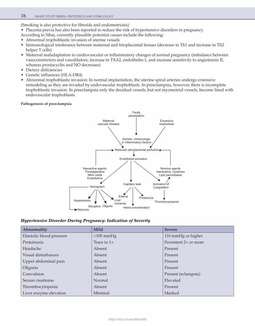
76 SMART STUDY SERIES: OBSTETRICS AND GYNECOLOGY
(Smoking is also protective for fibroids and endometriosis) • Placenta previa has also been reported to reduce the risk of hypertensive disorders in pregnancy. According to Sibai, currently plausible potential causes include the following: • Abnormal trophoblastic invasion of uterine vessels • Immunological intolerance between maternal and fetoplacental tissues (decrease in Th1 and increase in Th2
helper T cells) • Maternal maladaptation to cardiovascular or inflammatory changes of normal pregnancy (imbalance between
vasoconstrictors and vasodilators, increase in TXA2, endothelin-1, and increase sensitivity to angiotensin II, whereas prostacyclin and NO decreases)
• Dietary deficiencies • Genetic influences (HLA-DR4) • Abnormal trophoblastic invasion: In normal implantation, the uterine spiral arteries undergo extensive
remodeling as they are invaded by endovascular trophoblasts. In preeclampsia, however, there is incomplete trophoblastic invasion. In preeclampsia only the decidual vessels, but not myometrial vessels, become lined with endovascular trophoblasts.
Pathogenesis of preeclampsia
Maternalvascular disease
Faultyplacentation
Excessivetrophoblast
Genetic, immunologicor inflammatory factors
Reduced uteroplacental perfusion
Endothelial activation
Vasoactive agents:Prostaglandins
Nitric oxideEndothelins
Noxious agentsinterleukins, cytokines
Lipid peroxidases
VasospasmCapillary leak Activation of
Coagulation
Hypertension
AbruptionSeizures
Oliguria
Liverischemia
Proteinuria
Hemo-concentration
Thrombocytopenia
Edema
Hypertensive Disorder During Pregnancy: Indication of Severity
Abnormality Mild Severe
Diastolic blood pressure <100 mmHg 110 mmHg or higher
Proteinuria Trace to 1+ Persistent 2+ or more
Headache Absent Present
Visual disturbances Absent Present
Upper abdominal pain Absent Present
Oliguria Absent Present
Convulsion Absent Present (eclampsia)
Serum creatinine Normal Elevated
Thrombocytopenia Absent Present
Liver enzyme elevation Minimal Marked
https://kat.cr/user/Blink99/
OBSTETRIC COMPLICATIONS 77
Abnormality Mild Severe
Fetal growth restriction Absent Obvious
Pulmonary edema Absent Present
Complications
Maternal Fetal
Eclampsia IUFD
Abruption IUGR
Preterm labor Oligohydramnios
PPH Prematurity
HELLP Syndrome, DIC
Blindness
Antihypertensives in pregnancy:
1. Alpha methyldopa 2. Nifedipine 3. Hydralazine 4. Labetalol
ACE inhibitors are contraindicated.NOTE: As per the latest guidelines DOC for hypertension in pregnancy = Labetalol followed by alpha methyldopaDOC for hypertensive crisis in pregnancy = Labetalol followed by hydralazine
Prediction of Preeclampsia • Roll-over test: A positive test is an elevation of >20 mmHg when patient rolls over from lateral to supine position. • Urinary calcium ≥ 12 mg% in 24 h has good positive and negative predictive values for diagnosis of
preeclampsia. • Elevated levels of serum uric acid. • Persistence of diastolic notch on uterine artery waveforms on color Doppler at 18–20 weeks of gestation.
Measurement of uteroplacental vascular resistance during Doppler ultrasound evaluation of uterine artery impedance in the second trimester has been used as an early screening test for preeclampsia. The rationale for this is based on the presumption that the pathophysiology of preeclampsia includes impaired trophoblastic invasion of the spiral arteries leading to reduction in uteroplacental blood flow.
Management • On antihypertensives, if the BP is under control and there are no premonitory symptoms, then the pregnancy is
allowed to continue till 37 weeks (keeping a close watch on maternal and fetal well-being) • ·Thereafter, the patient should be delivered even if the BP is under control, as the risks of continuation of
pregnancy far outweigh the benefits (as this is a pregnancy-induced condition and delivery is the ultimate or definitive treatment for pregnancy-induced hypertension).
• It is not advisable to wait beyond 37 weeks because the BP can rise and there can be complications (eclampsia, HELLP syndrome, IUFD, abruption, DIC, etc) and there are no added benefits of continuing pregnancy beyond 37 weeks.
Impending EclampsiaThe dangerous symptoms (premonitory symptoms) that indicate impending eclampsia in case of preeclampsia are:
1. Headache 2. Oliguria 3. Epigastric pain 4. Nausea, vomiting 5. Blurring of vision
78 SMART STUDY SERIES: OBSTETRICS AND GYNECOLOGY
Whenever the above symptoms develop in a case of severe preeclampsia the patient is at a risk of eclampsia; the patient should be given anticonvulsant (MgSO4) and antihypertensive medication, and the patient to be delivered irrespective of the weeks of gestation.
Magnesium sulfate is the drug of choice for eclampsia and also for impending eclampsia. Prophylactic magne-sium sulfate decreases the risk of convulsion, abruption, and maternal mortality in this scenario.
The indications for termination of pregnancy irrespective of the weeks of gestation in a case of preeclampsia are:
1. Severe preeclampsia, with impending eclampsia 2. Eclampsia (give MgSO4 first, followed by induction of labor) 3. HELLP syndrome.
Prevention of hypertension in pregnancy:
1. Low-dose aspirin 2. Antioxidants (vitamin E, vitamin A, vitamin C, and lycopene) 3. Calcium (2 g/day) 4. Omega 3 fatty acids
Criteria for the diagnosis of HELLP syndrome
Hemolysis (H)
Schistocytes in the blood smearBilirubin >1.2 mg/dLAbsent plasma haptoglobin
Elevated liver enzymes (EL)SGOT >72 IU/LLDH >600 IU/L
Low platelet count (LP)Platelets <100 × 103/mm3
EclampsiaIncidenceAntepartum (50%)Intrapartum (30%)Postpartum (20%)
Mechanisms implicated in the etiology of eclamptic convulsion
• Cerebral edema and hemorrhage • Cerebral infarction • Cerebral vasospasm • Metabolic abnormality • Hypertensive encephalopathy
Prevention and treatment of convulsions with magnesium sulfate
Magnesium sulfate is the DOC for eclampsia and it is also the DOC for severe preeclampsia with impending eclampsia (prophylactic magnesium sulfate can prevent convulsions and it also decreases the risk of abruption— MAGPIE trial).
It can be given by various protocols:
1. Pritchard 2. Sibai 3. Zuspan 4. Sardesai
https://kat.cr/user/Blink99/

OBSTETRIC COMPLICATIONS 79
Pritchard Protocol
Loading dose:
4 g (20 mL of 20%) IV over 4 min (only in severe preeclampsia-eclampsia) immediately followed by 10 g (20 mL of 50%) IM—5 g in each buttockIf convulsions persist after 15 min: IV 2 g (10 mL of 20%) over 2 min (if the woman is large—4 g)
Maintenance:
5 g (10 mL of 50%) IM every 4 h—alternate sidesOR
Sibai Protocol
Loading dose:
6 g IV over 20 min
Maintenance:
2–3 g/h IV
Monitoring of MgSO4 therapy
1. Patellar reflexes 2. Respiratory rate (>14/min) 3. Urine output (100 cc in 4 h or 30 cc/h) • Therapeutic range of magnesium is 4–7 mEq/L. • Uterus stops contracting at 8–10 mEq/L • Patellar reflexes disappear when the plasma magnesium level reaches 10 mEq/L, presumably because of a
curariform action. This sign serves to warn of impending magnesium toxicity, because a further increase leads to respiratory depression.
• When plasma levels rise above 10 mEq/L, respiratory depression develops, and at 12 mEq/L or more, respiratory paralysis and arrest follow.
• Treatment with calcium gluconate, 1 g intravenously, along with withholding further magnesium sulfate usually reverses mild-to-moderate respiratory depression.
• Because magnesium is cleared almost exclusively by renal excretion, plasma magnesium concentration, using the doses described previously, is excessive if glomerular filtration is decreased substantively. The initial standard dose of magnesium sulfate can be safely administered without knowledge of renal function. Renal function is thereafter estimated by measuring plasma creatinine, and whenever it is 1.3 mg/dL or higher, only half of the maintenance intramuscular magnesium sulfate dose should be administered.
MULTIFETAL GESTATION
Etiology
1. Increasing age and increasing parity: The rate of natural twinning rises from 0 at puberty, a time of minimal ovarian activity, to a peak at 37 years of age, when maximal hormonal stimulation increases the rate of double ovulation. This is in accordance with the first consistently observed sign of reproductive aging, an isolated rise in serum FSH. The fall in incidence after 37 years of age probably reflects depletion of the Graafian follicles.
2. Personal/family history of twinning. 3. Treatment for infertility (ovulation induction agents/IVF). 4. Negroes have the highest risk and Mongols have the least risk.
Twins can be of two varieties: dizygotic and monozygotic.
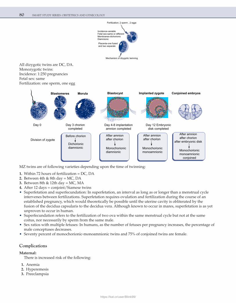
80 SMART STUDY SERIES: OBSTETRICS AND GYNECOLOGY
Fertilization; 2 sperm , 2 eggs
Incidence-variableFetal sex-same or differentMembranes-dichorionicDiamnionic
Placenta-one fusedand two separate
Mechanism of dizygotic twinning
All dizygotic twins are DC, DA.Monozygotic twins:Incidence: 1:250 pregnanciesFetal sex: sameFertilization: one sperm, one egg
Blastomeres Morula Blastocyst Implanted zygote Conjoined embryos
Day 0 Day 3 chorioncompleted
Division of zygoteBefore chorion
Dichorionicdiamnionic
After amnionafter chorion
Monochorionicdiamnionic
After amnionafter chorion
Monochorionicmonoamnionic
After amnionafter chorion
after embryonic disk
Monochorionicmonoamnionic
conjoined
Day 4-8 implantationamnion completed
Day 12 Embryonicdisk completed
MZ twins are of following varieties depending upon the time of twinning:
1. Within 72 hours of fertilization = DC, DA 2. Between 4th & 8th day = MC, DA 3. Between 8th & 12th day = MC, MA 4. After 12 days = conjoint/Siamese twins • Superfetation and superfecundation: In superfetation, an interval as long as or longer than a menstrual cycle
intervenes between fertilizations. Superfetation requires ovulation and fertilization during the course of an established pregnancy, which would theoretically be possible until the uterine cavity is obliterated by the fusion of the decidua capsularis to the decidua vera. Although known to occur in mares, superfetation is as yet unproven to occur in human.
• Superfecundation refers to the fertilization of two ova within the same menstrual cycle but not at the same coitus, nor necessarily by sperm from the same male.
• Sex ratios with multiple fetuses: In humans, as the number of fetuses per pregnancy increases, the percentage of male conceptuses decreases.
• Seventy percent of monochorionic-monoamnionic twins and 75% of conjoined twins are female.
Complications
Maternal:There is increased risk of the following:
1. Anemia 2. Hyperemesis 3. Preeclampsia
https://kat.cr/user/Blink99/
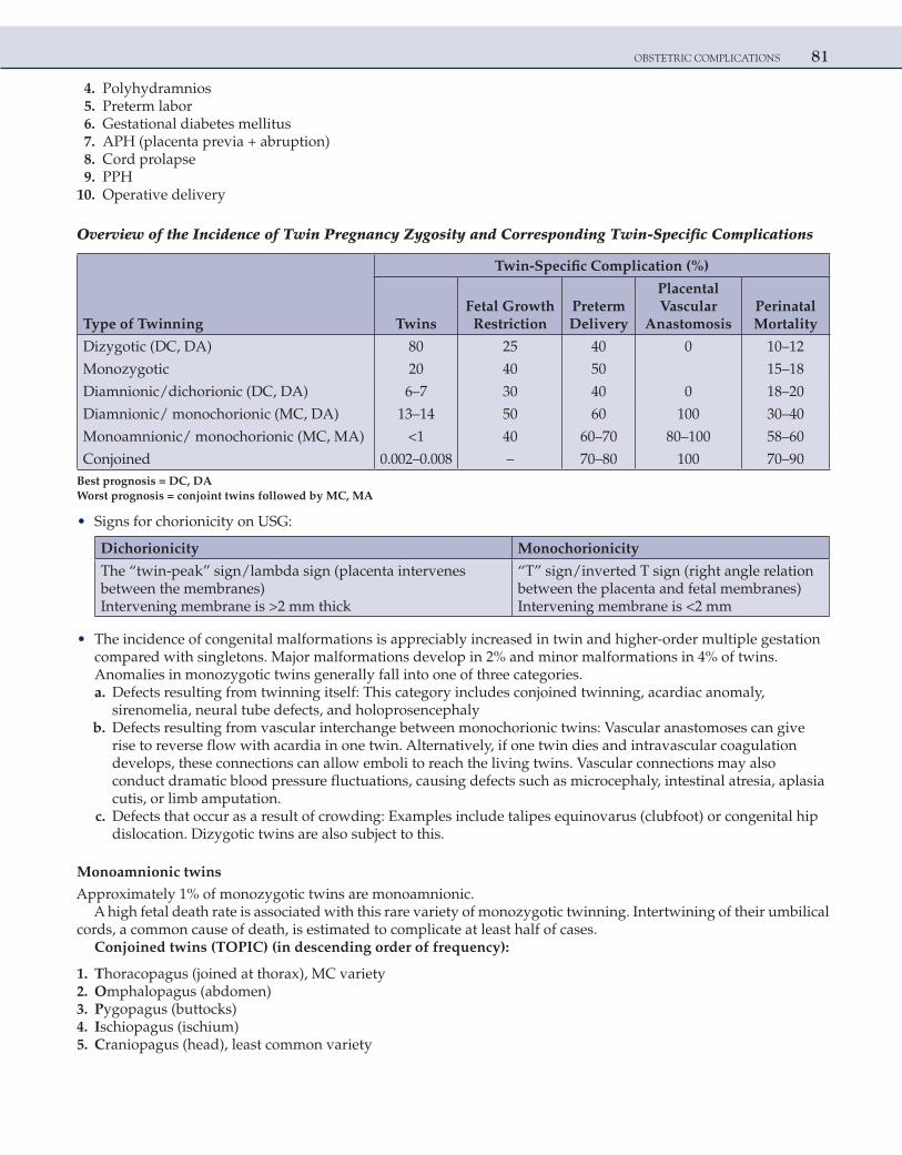
OBSTETRIC COMPLICATIONS 81
4. Polyhydramnios 5. Preterm labor 6. Gestational diabetes mellitus 7. APH (placenta previa + abruption) 8. Cord prolapse 9. PPH 10. Operative delivery
Overview of the Incidence of Twin Pregnancy Zygosity and Corresponding Twin-Specific Complications
Twin-Specific Complication (%)
Type of Twinning TwinsFetal Growth
RestrictionPreterm Delivery
Placental Vascular
AnastomosisPerinatal Mortality
Dizygotic (DC, DA) 80 25 40 0 10–12
Monozygotic 20 40 50 15–18
Diamnionic/dichorionic (DC, DA) 6–7 30 40 0 18–20
Diamnionic/ monochorionic (MC, DA) 13–14 50 60 100 30–40
Monoamnionic/ monochorionic (MC, MA) <1 40 60–70 80–100 58–60
Conjoined 0.002–0.008 – 70–80 100 70–90Best prognosis = DC, DAWorst prognosis = conjoint twins followed by MC, MA
• Signs for chorionicity on USG:
Dichorionicity Monochorionicity
The “twin-peak” sign/lambda sign (placenta intervenes between the membranes)Intervening membrane is >2 mm thick
“T” sign/inverted T sign (right angle relation between the placenta and fetal membranes)Intervening membrane is <2 mm
• The incidence of congenital malformations is appreciably increased in twin and higher-order multiple gestation compared with singletons. Major malformations develop in 2% and minor malformations in 4% of twins. Anomalies in monozygotic twins generally fall into one of three categories.
a. Defects resulting from twinning itself: This category includes conjoined twinning, acardiac anomaly, sirenomelia, neural tube defects, and holoprosencephaly
b. Defects resulting from vascular interchange between monochorionic twins: Vascular anastomoses can give rise to reverse flow with acardia in one twin. Alternatively, if one twin dies and intravascular coagulation develops, these connections can allow emboli to reach the living twins. Vascular connections may also conduct dramatic blood pressure fluctuations, causing defects such as microcephaly, intestinal atresia, aplasia cutis, or limb amputation.
c. Defects that occur as a result of crowding: Examples include talipes equinovarus (clubfoot) or congenital hip dislocation. Dizygotic twins are also subject to this.
Monoamnionic twins
Approximately 1% of monozygotic twins are monoamnionic.A high fetal death rate is associated with this rare variety of monozygotic twinning. Intertwining of their umbilical
cords, a common cause of death, is estimated to complicate at least half of cases.Conjoined twins (TOPIC) (in descending order of frequency):
1. Thoracopagus (joined at thorax), MC variety 2. Omphalopagus (abdomen) 3. Pygopagus (buttocks) 4. Ischiopagus (ischium) 5. Craniopagus (head), least common variety
82 SMART STUDY SERIES: OBSTETRICS AND GYNECOLOGY
Special Complications 1. Twin-to-twin transfusion syndrome (occurs in monochorionic twins only): a. In this syndrome, blood is transfused from a donor twin to its recipient sibling such that the donor becomes
anemic and oligohydramniotic, and its growth may be restricted, whereas the recipient has polyhydramnios and becomes polycythemic and may develop circulatory overload manifest as hydrops. Similarly, one portion of the placenta often appears pale compared with the rest of the placenta.
b. This is due to deep arteriovenous anastomosis. c. Antenatal criteria recommended for defining the twin-to-twin transfusion syndrome include the following:
same sex fetuses, monochorionicity with placental vascular anastomoses, weight difference between twins greater than 20%, polyhydramnios in the larger twin, oligohydramnios in donor twin, and hemoglobin difference greater than 5 g/dL.
d. The donor twin has better prognosis. 2. Acardiac twin: Twin reversed arterial perfusion (TRAP) sequence is a rare (1 in 35,000 births) but serious
complication of monochorionic, monozygotic multiple gestation. In the TRAP sequence, there is usually a normally formed donor twin that has features of heart failure as well as a recipient twin that lacks a heart (acardius) and various other structures.
3. Discordant growth (can occur in DZ and MZ twins): There is difference in weights of twins and is expressed as % of larger twin’s weight:
Grade 1= difference of 15–25%Grade 2 = difference >25%
Delivery
• Route of delivery is decided by the position of first baby. • Only if the first fetus is in vertex position, then normal vaginal delivery is possible. • Twins with first fetus in nonvertex position (breech, transverse, oblique,…) are to be delivered by LSCS. • MC, MA twins are always to be delivered by LSCS (even if the first fetus is in vertex position) because of very
high risk of cord prolapse and cord entanglement.
ABORTIONS
Abortion
Spontaneous Induced
Isolated (sporadic) Reccurrent
Threatened Inevitable Complete Incomplete Missed
Legal (MTP) Illegal
Septic
Common Causes of Abortion
First trimester:
1. Genetic factors (50%) 2. Endocrine disorders (luteal phase defect, thyroid abnormalities, and diabetes) 3. Immunological disorders (autoimmune and alloimmune) 4. Infection 5. Unexplained
https://kat.cr/user/Blink99/
OBSTETRIC COMPLICATIONS 83
Second trimester:
1. Anatomic abnormalities: a. Cervical incompetence (congenital or acquired) b. Mullerian fusion defects (bicornuate uterus and unicornuate) c. Uterine synechiae d. Uterine fibroid 2. Maternal medical illness 3. Unexplained • Abortion occurring without medical or mechanical means to empty the uterus is referred to as spontaneous. • More than 80% of abortions occur in the first 12 weeks of pregnancy, and at least half result from chromosomal
anomalies. After the first trimester, both the abortion rate and the incidence of chromosomal anomalies decrease. • Trisomy 16 is the most common abnormal karyotype found in the abortus. • Monosomy X (45 X), the second most frequent chromosomal abnormality (after trisomy), usually results in
abortion and much less frequently in live born female infants (Turner syndrome). • Advanced maternal and paternal ages do not increase the incidence of triploidy. • Euploid abortion: Euploid fetuses tend to abort later in gestation than aneuploid ones. Three-fourths of
aneuploid abortions occur before 8 weeks; euploid abortions peak at about 13 weeks. The incidence of euploid abortions increase dramatically after maternal age exceeds 35 years.
• Autoimmune factors: Antiphospholipid antibodies are a family of autoantibodies that bind to negatively charged phospholipids, phospholipids-binding proteins, or a combination of the two. Two of these, lupus anticoagulant and anticardiolipin antibody, have been implicated in spontaneous abortion.The mechanism of pregnancy loss in women with these antibodies involves placental thrombosis and infarction. In one postulated mechanism, antibodies may inhibit the release of prostacyclin, a potent vasodilator and inhibitor of platelet aggregation. In contrast, platelets produce thromboxane A2, a vasoconstrictor and platelet aggregator. They have also been shown to inhibit protein C activation, resulting in coagulation and fibrin formation. Treatment with a combination of heparin and low-dose aspirin improves the chance of live birth in a subsequent pregnancy in women with this syndrome.
• Asherman syndrome, characterized by uterine synechiae, usually results from destruction of large areas of endometrium by overzealous curettage. The risk is maximum if curettage is done in the postpartum period.If pregnancy follows, the amount of remaining endometrium may be insufficient to support the pregnancy, and abortion may ensue. A hysterosalpingogram that shows characteristic multiple filling defects may indicate Asherman syndrome, but hysteroscopy most accurately and directly identifies this condition. Recommended treatment consists of lysis of the adhesions via hysteroscopy and placement of an intra-uterine contraceptive device to prevent recurrence. Some practitioners also recommend continuous high-dose estrogen therapy for 60–90 days following adhesiolysis.
CERVICAL INCOMPETENCE
• Classically, it is characterized by painless cervical dilatation in the second trimester, with prolapse and ballooning of membranes into the vagina, preterm premature rupture of membranes (PPROM), followed by expulsion of an immature fetus. Unless effectively treated, this sequence may repeat in future pregnancies.
• Multiple studies have demonstrated that certain features of the cervix, primarily cervical length, when measured in the mid-second trimester, may predict preterm delivery. Cervical length less than 2.5–3 cm is considered as short cervix.
• Another feature termed funneling—ballooning of the membranes into a dilated internal os, but with a closed external os—has also been assessed.
• Etiology: Although the cause of cervical incompetence is obscure, previous trauma to the cervix—especially in the course of dilatation and curettage, conization, cauterization, or amputation—appears to be a factor in some cases. In other instances, abnormal cervical development, including that following exposure to diethylstilbestrol in utero, may play a role.
• The treatment of classical cervical incompetence is cerclage (os tightening). The operation is performed to surgically reinforce the weak cervix by some type of purse-string suturing. Bleeding, uterine contraction, or ruptured membranes are usually contraindications to cerclage.
• Cerclage procedure: Two types of vaginal operations are commonly used during pregnancy. One is McDonald and the other is Shirodkar.
• Complications: PROM, uterine contractions and abortion may occur.
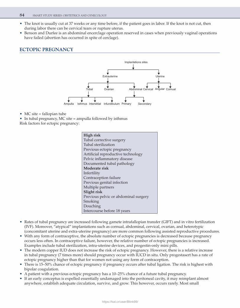
84 SMART STUDY SERIES: OBSTETRICS AND GYNECOLOGY
• The knot is usually cut at 37 weeks or any time before, if the patient goes in labor. If the knot is not cut, then during labor there can be cervical tears or rupture uterus.
• Benson and Durfee is an abdominal encerclage operation reserved in cases when previously vaginal operations have failed (abortion has occurred in spite of cerclage).
ECTOPIC PREGNANCY
Implantations sites
Extrauterine
Tubal Ovarian Abdominal
Ampulla Isthmus Interstitial Infundibulum
Cervical Angular Cornual
Uterine
Primary Secondary
• MC site = fallopian tube • In tubal pregnancy, MC site = ampulla followed by isthmusRisk factors for ectopic pregnancy:
High riskTubal corrective surgeryTubal sterilizationPrevious ectopic pregnancyArtificial reproductive technologyPelvic inflammatory diseaseDocumented tubal pathologyModerate riskInfertilityContraception failurePrevious genital infectionMultiple partnersSlight riskPrevious pelvic or abdominal surgerySmokingDouchingIntercourse before 18 years
• Rates of tubal pregnancy are increased following gamete intrafallopian transfer (GIFT) and in vitro fertilization (IVF). Moreover, “atypical” implantations such as cornual, abdominal, cervical, ovarian, and heterotypic (concomitant uterine and extra-uterine pregnancy) are more common following assisted reproductive procedures.
• With any form of contraceptive, the absolute number of ectopic pregnancies is decreased because pregnancy occurs less often. In contraceptive failure, however, the relative number of ectopic pregnancies is increased. Examples include tubal sterilization, intra-uterine devices, and progestin-only mini pills.
• The modern copper IUD does not increase the risk of ectopic pregnancy. However, there is a relative increase in tubal pregnancy (7 times more) should pregnancy occur with IUCD in situ. Only progestasert has a rate of ectopic pregnancy higher than that for women not using any form of contraception.
• There is 15–50% chance of ectopic pregnancy if pregnancy occurs after tubal ligation. The risk is highest with bipolar coagulation.
• A patient with a previous ectopic pregnancy has a 10–25% chance of a future tubal pregnancy. • If an early conceptus is expelled essentially undamaged into the peritoneal cavity, it may reimplant almost
anywhere, establish adequate circulation, survive, and grow. This however, occurs rarely. Most small
https://kat.cr/user/Blink99/
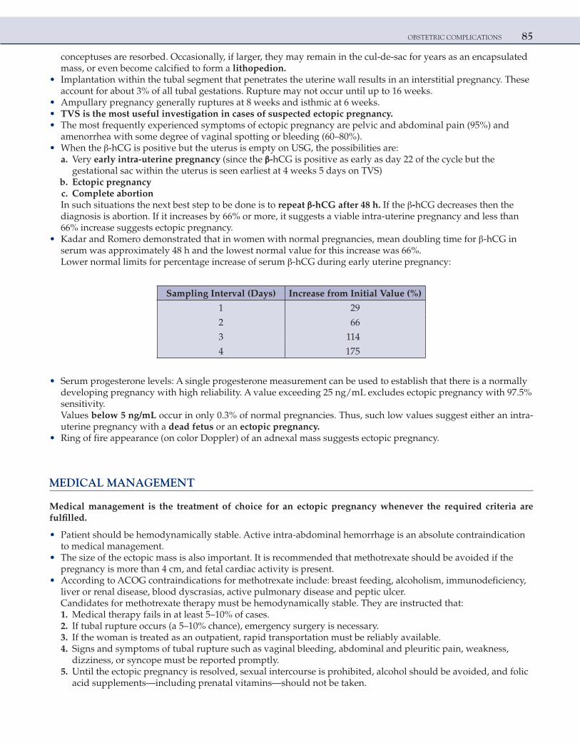
OBSTETRIC COMPLICATIONS 85
conceptuses are resorbed. Occasionally, if larger, they may remain in the cul-de-sac for years as an encapsulated mass, or even become calcified to form a lithopedion.
• Implantation within the tubal segment that penetrates the uterine wall results in an interstitial pregnancy. These account for about 3% of all tubal gestations. Rupture may not occur until up to 16 weeks.
• Ampullary pregnancy generally ruptures at 8 weeks and isthmic at 6 weeks. • TVS is the most useful investigation in cases of suspected ectopic pregnancy. • The most frequently experienced symptoms of ectopic pregnancy are pelvic and abdominal pain (95%) and
amenorrhea with some degree of vaginal spotting or bleeding (60–80%). • When the β-hCG is positive but the uterus is empty on USG, the possibilities are: a. Very early intra-uterine pregnancy (since the β-hCG is positive as early as day 22 of the cycle but the
gestational sac within the uterus is seen earliest at 4 weeks 5 days on TVS) b. Ectopic pregnancy c. Complete abortion In such situations the next best step to be done is to repeat β-hCG after 48 h. If the β-hCG decreases then the
diagnosis is abortion. If it increases by 66% or more, it suggests a viable intra-uterine pregnancy and less than 66% increase suggests ectopic pregnancy.
• Kadar and Romero demonstrated that in women with normal pregnancies, mean doubling time for β-hCG in serum was approximately 48 h and the lowest normal value for this increase was 66%.
Lower normal limits for percentage increase of serum β-hCG during early uterine pregnancy:
Sampling Interval (Days) Increase from Initial Value (%)
1 29
2 66
3 114
4 175
• Serum progesterone levels: A single progesterone measurement can be used to establish that there is a normally developing pregnancy with high reliability. A value exceeding 25 ng/mL excludes ectopic pregnancy with 97.5% sensitivity. Values below 5 ng/mL occur in only 0.3% of normal pregnancies. Thus, such low values suggest either an intra-uterine pregnancy with a dead fetus or an ectopic pregnancy.
• Ring of fire appearance (on color Doppler) of an adnexal mass suggests ectopic pregnancy.
MEDICAL MANAGEMENT
Medical management is the treatment of choice for an ectopic pregnancy whenever the required criteria are fulfilled.
• Patient should be hemodynamically stable. Active intra-abdominal hemorrhage is an absolute contraindication to medical management.
• The size of the ectopic mass is also important. It is recommended that methotrexate should be avoided if the pregnancy is more than 4 cm, and fetal cardiac activity is present.
• According to ACOG contraindications for methotrexate include: breast feeding, alcoholism, immunodeficiency, liver or renal disease, blood dyscrasias, active pulmonary disease and peptic ulcer.
Candidates for methotrexate therapy must be hemodynamically stable. They are instructed that: 1. Medical therapy fails in at least 5–10% of cases. 2. If tubal rupture occurs (a 5–10% chance), emergency surgery is necessary. 3. If the woman is treated as an outpatient, rapid transportation must be reliably available. 4. Signs and symptoms of tubal rupture such as vaginal bleeding, abdominal and pleuritic pain, weakness,
dizziness, or syncope must be reported promptly. 5. Until the ectopic pregnancy is resolved, sexual intercourse is prohibited, alcohol should be avoided, and folic
acid supplements—including prenatal vitamins—should not be taken.
86 SMART STUDY SERIES: OBSTETRICS AND GYNECOLOGY
Methotrexate Therapy for Primary Treatment of Ectopic Pregnancy
Regimen Follow-up
Single dose: methotrexate, 50 mg/m2 Measure β-hCG at days 4 and 7
If difference is ≥15%, repeat weekly until undetectable
If difference <15%, repeat methotrexate dose and begin new day 1
If fetal cardiac activity present at day 7, repeat methotrexate dose, begin new day
Surgical treatment if β-hCG levels not decreasing or fetal cardiac activity persists after three doses of methotrexate
Variable dose
Methotrexate, 1 mg/kg i.m., days 1, 3, 5, and 7 plus leukovorin, 0.1 mg/kg i.m., days 2, 4, 6, and 8
Continue alternate day injection until β-hCG levels decreases to >15% in 48 h, or four does methotrexate givenThen, weekly β-hCG until undetectable
i.m. = intramuscular
Surgical Management
• In cases of ruptured ectopic pregnancy (shock and hemodynamic instability), blood transfusion and i.v fluids are to be given and simultaneously exploratory laparotomy with salpingectomy should be performed.
• Laparoscopic salpingectomy can be performed in cases of unruptured ectopic, chronic ectopic pregnancies, or in cases of early rupture (stable patient).
Spielberg’s Criteria for Diagnosis of Primary Ovarian Pregnancy 1. The fallopian tube on the affected side must be intact. 2. The fetal sac must occupy the position of the ovary. 3. The ovary must be connected to the uterus by the ovarian ligament. 4. Ovarian tissue must be located in the sac wall.
Studdiford’s Criteria for Diagnosis of Primary Abdominal Pregnancy 1. Presence of normal tubes and ovaries with no evidence of recent or past pregnancy 2. No evidence of uteroperitoneal fistula 3. Presence of a pregnancy related exclusively to the peritoneal surface and early enough to eliminate the
possibility of secondary implantation after primary tubal nidation.
Ultrasound Criteria for Cervical Pregnancy (Paalman’s) 1. Echo-free uterine cavity or the presence of a false gestational sac only 2. Hourglass uterine shape 3. Ballooned cervical canal 4. Gestational sac in the endocervix 5. Placental tissue in the cervical canal 6. Closed internal os
Heterotypic Ectopic Pregnancy
Tubal pregnancy may be accompanied by a coexisting uterine gestation. Until recently, such heterotypic pregnancies were rare, with an incidence of 1 per 30,000 pregnancies. Currently, because of assisted reproduction, the incidence is likely 1 in 7000 overall, and following ovulation induction it may be as high as 1 in 900.
A heterotypic pregnancy is more likely, and should be considered:
1. After assisted reproductive techniques. 2. With persistent or rising chorionic gonadotropin levels after dilatation and curettage for an induced or
spontaneous abortion.
https://kat.cr/user/Blink99/
OBSTETRIC COMPLICATIONS 87
3. When the uterine fundus is larger than menstrual dates. 4. With more than one corpus luteum. 5. With absence of vaginal bleeding in the presence of signs and symptoms of an ectopic pregnancy.
TROPHOBLASTIC DISEASE
Hydatidiform mole (molar pregnancy): Molar pregnancy is characterized histologically by abnormalities of the cho-rionic villi that consist of trophoblastic proliferation and edema of villus stroma (hydropic degeneration).
Features of Partial and Complete Hydatidiform Moles
Feature Partial Mole Complete Mole
Karyotype Usually 69,XXX or 69,XXY 46,XX or 46,XY
Embryo-fetus Often present Absent
Amnion, fetal red blood cells Often present Absent
Villus edema Variable, focal Diffuse
Uterine size Small for dates 50% large for dates
Theca-lutein cysts Rare 25–30%
Medical complications Rare Frequent
Gestational trophoblastic neoplasia <5–10% 20%
• Past history of hydatidiform mole increases the chance of recurrence (1–4%). • Familial repetitive hydatidiform mole has been linked to a missense mutation in the NLRP7 locus
on chromosome 19; in one report, this mutation was present in 60% of patients who had two molar pregnancies.
• Vitamin A deficiency and diet deficient in proteins and animal fat is also associated with molar pregnancy. • The chromosomal composition in 85% of complete molar pregnancies is 46 XX with both chromosomes being
of paternal origin. This phenomenon is termed androgenesis. • Theca-lutein cysts: In many cases of hydatidiform mole, the ovaries contain multiple theca-lutein cysts. Theses
may vary from microscopic size to 10 cm or more diameters. Their surfaces are smooth, often yellowish and lined with lutein cells. The incidence of obvious cysts in association with a mole is reported to be from 25% to 60%. They are thought to result from overstimulation of lutein elements by large amounts of hCG secreted by proliferating trophoblastic cells.
• Incidence of molar pregnancy is highest in women aged 15 years or younger and those aged 45 years or older. In the latter group, the relative frequency of the lesion is at least 10 times greater than that at ages 20–40 years.
• Uterine bleeding is almost universal and may vary from spotting to profuse hemorrhage. It is the MC presenting feature. The discharge has “white currant in red currant juice” appearance.
• Uterine size: The growing uterus often enlarges more rapidly than usual, exceeding in about half of cases that expected from the gestational age.
• Gestational hypertension: Because hypertension caused by pregnancy is rarely seen before 24 weeks, preeclampsia that develops before this gestational age may be from hydatidiform mole or extensive molar degeneration. Thyrotoxicosis (alpha component of hCG is similar to TSH): Plasma thyroxine levels in women with molar pregnancy are often elevated.
Management
• Vacuum aspiration: Suction evacuation is the treatment of choice for hydatidiform mole, regardless of uterine size. After most of the molar tissue has been removed by aspiration, oxytocin is given. After the myometrium has contracted, thorough but gentle curettage with a large sharp curette usually is performed. Intraoperative ultrasonographic examination may help document that the uterine cavity has been emptied.
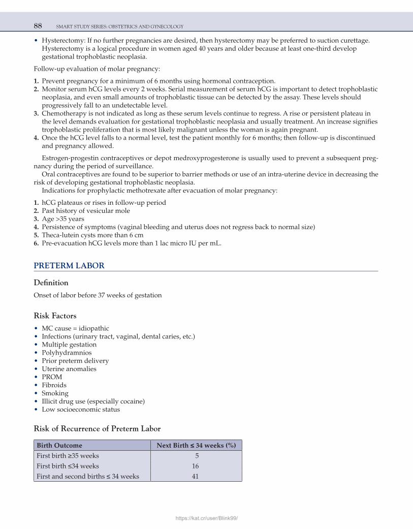
88 SMART STUDY SERIES: OBSTETRICS AND GYNECOLOGY
• Hysterectomy: If no further pregnancies are desired, then hysterectomy may be preferred to suction curettage. Hysterectomy is a logical procedure in women aged 40 years and older because at least one-third develop gestational trophoblastic neoplasia.
Follow-up evaluation of molar pregnancy:
1. Prevent pregnancy for a minimum of 6 months using hormonal contraception. 2. Monitor serum hCG levels every 2 weeks. Serial measurement of serum hCG is important to detect trophoblastic
neoplasia, and even small amounts of trophoblastic tissue can be detected by the assay. These levels should progressively fall to an undetectable level.
3. Chemotherapy is not indicated as long as these serum levels continue to regress. A rise or persistent plateau in the level demands evaluation for gestational trophoblastic neoplasia and usually treatment. An increase signifies trophoblastic proliferation that is most likely malignant unless the woman is again pregnant.
4. Once the hCG level falls to a normal level, test the patient monthly for 6 months; then follow-up is discontinued and pregnancy allowed.
Estrogen-progestin contraceptives or depot medroxyprogesterone is usually used to prevent a subsequent preg-nancy during the period of surveillance.
Oral contraceptives are found to be superior to barrier methods or use of an intra-uterine device in decreasing the risk of developing gestational trophoblastic neoplasia.
Indications for prophylactic methotrexate after evacuation of molar pregnancy:
1. hCG plateaus or rises in follow-up period 2. Past history of vesicular mole 3. Age >35 years 4. Persistence of symptoms (vaginal bleeding and uterus does not regress back to normal size) 5. Theca-lutein cysts more than 6 cm 6. Pre-evacuation hCG levels more than 1 lac micro IU per mL.
PRETERM LABOR
Definition
Onset of labor before 37 weeks of gestation
Risk Factors
• MC cause = idiopathic • Infections (urinary tract, vaginal, dental caries, etc.) • Multiple gestation • Polyhydramnios • Prior preterm delivery • Uterine anomalies • PROM • Fibroids • Smoking • Illicit drug use (especially cocaine) • Low socioeconomic status
Risk of Recurrence of Preterm Labor
Birth Outcome Next Birth ≤ 34 weeks (%)
First birth ≥35 weeks 5
First birth ≤34 weeks 16
First and second births ≤ 34 weeks 41
https://kat.cr/user/Blink99/

OBSTETRIC COMPLICATIONS 89
Fetal fibronectin (FFN) in cervical/vaginal secretions is a predictor of preterm labor. Increase in maternal salivary estriol is also a predictor.
• Steroids (dexamethasone or betamethasone) are given to enhance fetal lung maturity and they also decrease the incidence of intraventricular hemorrhage.
• Betamethasone is preferred over dexamethasone, as it also prevents periventricular leukomalacia. • Chorioamnionitis and active infection in mother (e.g., open pulmonary koch) are the only contraindications for
the use of steroids. They can be given to patients of hypertension and diabetes mellitus. • Repeated doses of steroids (weekly) are to be avoided as they are a/w risk of necrotizing enterocolitis,
intrauterine growth restriction (IUGR), pulmonary edema, and PIH.
Tocolytic Agents
1. Beta 2 agonist (e.g., isoxsuprine, ritodrine, terbutaline, etc.) 2. Calcium channel blocker (nifedipine) 3. Indomethacin 4. Magnesium sulfate 5. Atosiban (oxytocin antagonist) 6. Progesterone
Ritodrine is the only tocolytic agent approved by US FDA.
• Most commonly used = beta 2 agonist • Maximum side effects = beta 2 agonist • Least side effects = progesterone followed by atosiban • Tocolytic of choice in heart disease patients = atosiban
Potential Complications of Tocolytic Agents • Beta-adrenergic agents a. Hyperglycemia b. Hypokalemia c. Hypotension d. Pulmonary edema e. Cardiac insufficiency f. Arrhythmias g. Myocardial ischemia h. Maternal death • Magnesium sulfate (toxicity) a. Pulmonary edema b. Respiratory depression c. Cardiac arrest d. Maternal tetany e. Muscular paralysis • Indomethacin a. Oligohydramnios b. Premature closure of DA c. Renal failure d. Gastrointestinal bleeding • Nifedipine a. Transient hypotension
Contraindications to Tocolysis 1. Chorioamnionitis 2. Preeclampsia/eclampsia 3. Advanced labor 4. Fetal distress
90 SMART STUDY SERIES: OBSTETRICS AND GYNECOLOGY
5. Abruption 6. IUFD 7. Congenital anomalies not compatible with life 8. Pregnancy >34 weeks
PREMATURE RUPTURE OF MEMBRANES
• It is defined as rupture of membranes at least 1 hour prior to onset of labor. • If this happens before 37 weeks, it is known as PPROM.
Risk Factors for PROM • Increase friability/decrease in tensile strength of membranes (mainly due to infections with Chlamydia or
bacterial vaginosis, etc.) • Polyhydramnios • Multiple pregnancy • Cervical incompetence • Previous history of PROM
DiagnosisA history of a gush of fluid or trickle causing a woman to be constantly wet may suggest the diagnosis. The following can be done:
1. Sterile speculum examination 2. Fluid turns yellow, nitrazine paper blue (pH of amniotic fluid is 7.0–7.7 compared with vaginal pH of 4.5) 3. Red litmus paper turns blue 4. Microscopic ‘ferning’ of vaginal fluid (refers to crystallization of amniotic fluid on drying) 5. Alpha-fetoprotein levels in the fluid (amniotic fluid contains AFP) 6. FFN in the vaginal fluid will indicate that the fluid is liquor 7. 0.1% Nile blue sulfate test (orange colored cells seen) 8. USG will show oligohydramnios
Complications 1. Preterm labor 2. Chorioamnionitis 3. Abruption 4. Fetal pulmonary hypoplasia especially in PPROM
Chorioamnionitis
• Inflammation of the fetal membranes usually is a manifestation of intra-uterine infection. It frequently is associated with prolonged membrane rupture and long labor. Grossly, infection is characterized by clouding of the membranes.
• The diagnosis is clinical. There is presence of fever and at least two of the following: maternal tachycardia, fetal tachycardia, uterine tenderness, foul odor of amniotic fluid, or maternal leukocytosis.
• When mono- and polymorphonuclear leukocytes infiltrate the chorion, the resulting microscopical finding is designated chorioamnionitis. These cells are maternal in origin. Conversely, if leukocytes are found in amniotic fluid (amnionitis), or the umbilical cord (funisitis), the cells are fetal in origin.
• Management of overt clinical chorioamnionitis is antimicrobial administration and delivery.
POSTDATISM AND POSTTERM PREGNANCY
Postdatism = pregnancy continuing beyond EDD or 40 weeksPostterm = pregnancy continuing more than 42 weeks
https://kat.cr/user/Blink99/
OBSTETRIC COMPLICATIONS 91
• Etiology a. Idiopathic b. Past history c. Anencephaly d. Fetal adrenal hypoplasia e. X-linked placental sulfatase deficiency • Complications a. Oligohydramnios b. MSAF c. Shoulder dystocia d. Sudden IUFD e. Uterine dysfunction f. Increased risk of operative delivery
To confirm postdatism USG in first trimester (dating scan) is most useful.
Evaluation and Management of Postterm Pregnancy (ACOG Guidelines)
1. Women with a postterm gestation who have an unfavorable cervix can either undergo labor induction or be managed expectantly.
2. Prostaglandin can be used for cervical ripening and labor induction. 3. Delivery should be effected if there is evidence of fetal compromise or oligohydramnios. 4. It is reasonable to initiate antenatal surveillance between 40 and 42 weeks. 5. A nonstress test (biweekly) and amniotic fluid volume assessment should be adequate. 6. Many recommend prompt delivery in a woman with a postterm pregnancy, a favorable cervix, and no other
complications.
NOTE: Normally the validity of NST is 7 days (i.e. if the NST is reactive it can be repeated after 7 days). But in cases of diabetes mellitus and postdatism the validity of NST is only 48 hours (it should be repeated every third day).
AMNIOTIC FLUID VOLUME DISORDERS (POLY/OLIGOHYDRAMNIOS)
Polyhydramnios
Definitions 1. More than 2 litre of amniotic fluid is termed as polyhydramnios, or 2. AFI ≥ 25 cm, or 3. Single largest vertical pocket of liquor >8 cm (normal = 2–8 cm)
Classification
Single Largest Vertical Pocket (cm)
Mild >8–11
Moderate 12–15
Severe >15
Etiology 1. MC cause = idiopathic 2. Fetal anomalies: ○ Obstruction of fluid transit through the gastrointestinal tract: esophageal/duodenal atresia and diaphragmatic
hernia ○ Anencephaly ○ Open spina bifida 3. Multiple pregnancy 4. Hydrops fetalis (immune and nonimmune)
92 SMART STUDY SERIES: OBSTETRICS AND GYNECOLOGY
5. Chromosomal abnormalities (e.g., trisomy 18) 6. Twin-twin transfusion syndrome 7. Diabetes insipidus/Bartter syndrome 8. Maternal: diabetes mellitus and cardiac disease 9. Placental: chorioangioma (a/w acute polyhydramnios)
Complications 1. Preterm labor 2. PROM 3. Malpresentation 4. Cord prolapse 5. Abruption 6. PPH 7. Subinvolution of uterus • Polyhydramnios associated with fetal hydrops may cause the MIRROR SYNDROME, whereby the maternal
condition mimics the fetus and mother develops edema, proteinuria, and PIH. • Indomethacin and sulindac are NSAIDs that decrease fetal urine production and are used in medical manage-
ment of polyhydramnios in symptomatic patients. • A major concern for the use of indomethacin/sulindac is the risk of premature closure of the fetal ductus arte-
riosus. Hence, these drugs should not be used beyond 34 weeks of gestation.
Chorioangioma (Hemangioma) • These are the only benign tumors of the placenta. • They most likely are hamartomas of primitive chorionic mesenchyme and have an incidence of about 1%. • Small growths are usually asymptomatic, but large tumors may be associated with polyhydramnios or
antepartum hemorrhage.
Oligohydramnios
1. AFI <5 cmor 2. Amniotic fluid less than 100 mL
Etiology
Chromosomal Abnormalities Uteroplacental Insufficiency
Congenital anomalies (e.g., renal agenesis and posterior urethral valves)IUGRPostdatism/postterm pregnancyPROMTwin-to-twin transfusion
HypertensionPreeclampsiaNSAIDs, angiotensin-converting enzyme inhibitorsIdiopathic
• Amnion nodosum are tiny, light tan, creamy nodules in the amnion made up of vernix caseosa with hair, degenerated squames, and sebum. They result from oligohydramnios and are most commonly found in fetuses with renal agenesis and prolonged preterm ruptured membranes, or in the placenta of the donor fetus with twin-to-twin transfusion syndrome.
• Amniotic bands are caused when disruption of the amnion leads to formation of bands or strings that entrap the fetus and impair growth and development of the involved structure. Fetal conditions that appear to be the consequence of this phenomenon include intra-uterine amputations.
• Tetrad of early-onset oligohydramnios: a. Facial clefts (cleft lip/palate) b. IUGR c. Limb reduction defects d. Pulmonary hypoplasia
https://kat.cr/user/Blink99/
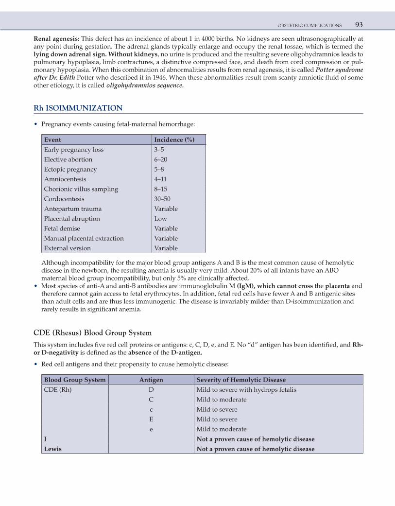
OBSTETRIC COMPLICATIONS 93
Renal agenesis: This defect has an incidence of about 1 in 4000 births. No kidneys are seen ultrasonographically at any point during gestation. The adrenal glands typically enlarge and occupy the renal fossae, which is termed the lying down adrenal sign. Without kidneys, no urine is produced and the resulting severe oligohydramnios leads to pulmonary hypoplasia, limb contractures, a distinctive compressed face, and death from cord compression or pul-monary hypoplasia. When this combination of abnormalities results from renal agenesis, it is called Potter syndrome after Dr. Edith Potter who described it in 1946. When these abnormalities result from scanty amniotic fluid of some other etiology, it is called oligohydramnios sequence.
Rh ISOIMMUNIZATION
• Pregnancy events causing fetal-maternal hemorrhage:
Event Incidence (%)
Early pregnancy loss 3–5
Elective abortion 6–20
Ectopic pregnancy 5–8
Amniocentesis 4–11
Chorionic villus sampling 8–15
Cordocentesis 30–50
Antepartum trauma Variable
Placental abruption Low
Fetal demise Variable
Manual placental extraction Variable
External version Variable
Although incompatibility for the major blood group antigens A and B is the most common cause of hemolytic disease in the newborn, the resulting anemia is usually very mild. About 20% of all infants have an ABO maternal blood group incompatibility, but only 5% are clinically affected.
• Most species of anti-A and anti-B antibodies are immunoglobulin M (IgM), which cannot cross the placenta and therefore cannot gain access to fetal erythrocytes. In addition, fetal red cells have fewer A and B antigenic sites than adult cells and are thus less immunogenic. The disease is invariably milder than D-isoimmunization and rarely results in significant anemia.
CDE (Rhesus) Blood Group System
This system includes five red cell proteins or antigens: c, C, D, e, and E. No “d” antigen has been identified, and Rh- or D-negativity is defined as the absence of the D-antigen.
• Red cell antigens and their propensity to cause hemolytic disease:
Blood Group System Antigen Severity of Hemolytic Disease
CDE (Rh) D Mild to severe with hydrops fetalis
C Mild to moderate
c Mild to severe
E Mild to severe
e Mild to moderate
I Not a proven cause of hemolytic disease
Lewis Not a proven cause of hemolytic disease
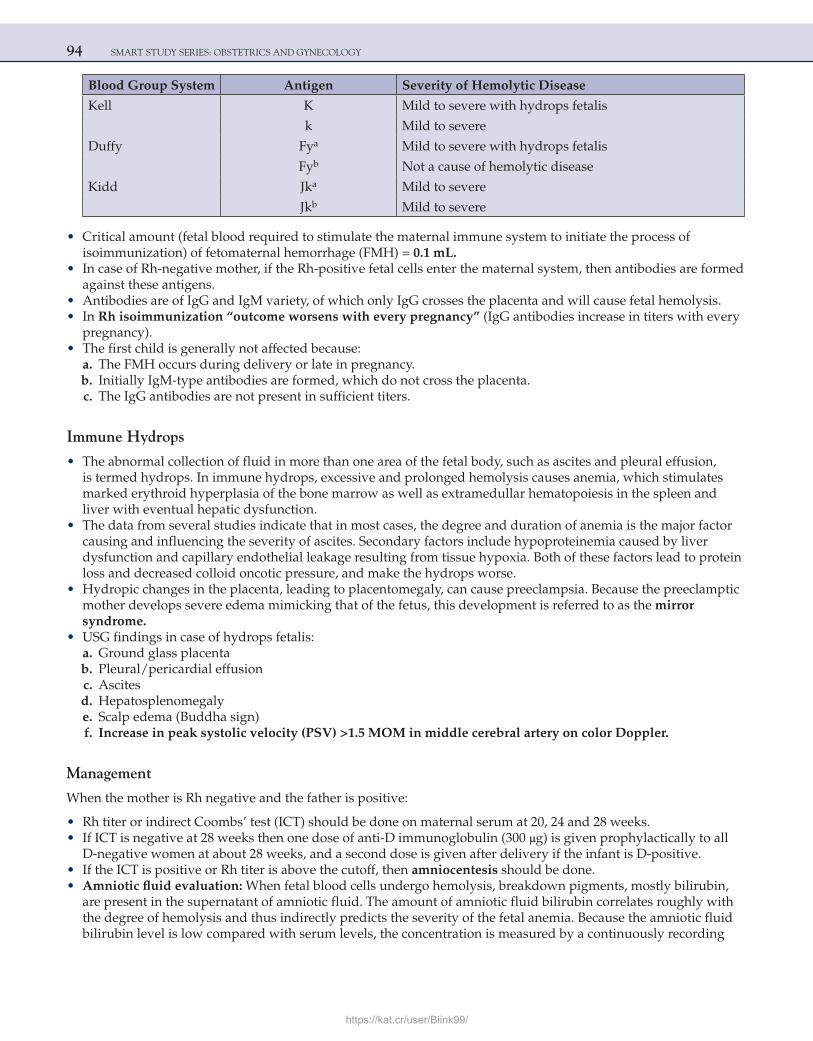
94 SMART STUDY SERIES: OBSTETRICS AND GYNECOLOGY
Blood Group System Antigen Severity of Hemolytic Disease
Kell K Mild to severe with hydrops fetalis
k Mild to severe
Duffy Fya Mild to severe with hydrops fetalis
Fyb Not a cause of hemolytic disease
Kidd Jka Mild to severe
Jkb Mild to severe
• Critical amount (fetal blood required to stimulate the maternal immune system to initiate the process of isoimmunization) of fetomaternal hemorrhage (FMH) = 0.1 mL.
• In case of Rh-negative mother, if the Rh-positive fetal cells enter the maternal system, then antibodies are formed against these antigens.
• Antibodies are of IgG and IgM variety, of which only IgG crosses the placenta and will cause fetal hemolysis. • In Rh isoimmunization “outcome worsens with every pregnancy” (IgG antibodies increase in titers with every
pregnancy). • The first child is generally not affected because: a. The FMH occurs during delivery or late in pregnancy. b. Initially IgM-type antibodies are formed, which do not cross the placenta. c. The IgG antibodies are not present in sufficient titers.
Immune Hydrops
• The abnormal collection of fluid in more than one area of the fetal body, such as ascites and pleural effusion, is termed hydrops. In immune hydrops, excessive and prolonged hemolysis causes anemia, which stimulates marked erythroid hyperplasia of the bone marrow as well as extramedullar hematopoiesis in the spleen and liver with eventual hepatic dysfunction.
• The data from several studies indicate that in most cases, the degree and duration of anemia is the major factor causing and influencing the severity of ascites. Secondary factors include hypoproteinemia caused by liver dysfunction and capillary endothelial leakage resulting from tissue hypoxia. Both of these factors lead to protein loss and decreased colloid oncotic pressure, and make the hydrops worse.
• Hydropic changes in the placenta, leading to placentomegaly, can cause preeclampsia. Because the preeclamptic mother develops severe edema mimicking that of the fetus, this development is referred to as the mirror syndrome.
• USG findings in case of hydrops fetalis: a. Ground glass placenta b. Pleural/pericardial effusion c. Ascites d. Hepatosplenomegaly e. Scalp edema (Buddha sign) f. Increase in peak systolic velocity (PSV) >1.5 MOM in middle cerebral artery on color Doppler.
Management
When the mother is Rh negative and the father is positive:
• Rh titer or indirect Coombs’ test (ICT) should be done on maternal serum at 20, 24 and 28 weeks. • If ICT is negative at 28 weeks then one dose of anti-D immunoglobulin (300 μg) is given prophylactically to all
D-negative women at about 28 weeks, and a second dose is given after delivery if the infant is D-positive. • If the ICT is positive or Rh titer is above the cutoff, then amniocentesis should be done. • Amniotic fluid evaluation: When fetal blood cells undergo hemolysis, breakdown pigments, mostly bilirubin,
are present in the supernatant of amniotic fluid. The amount of amniotic fluid bilirubin correlates roughly with the degree of hemolysis and thus indirectly predicts the severity of the fetal anemia. Because the amniotic fluid bilirubin level is low compared with serum levels, the concentration is measured by a continuously recording
https://kat.cr/user/Blink99/
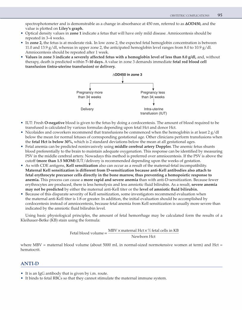
OBSTETRIC COMPLICATIONS 95
spectrophotometer and is demonstrable as a change in absorbance at 450 nm, referred to as ΔOD450, and the value is plotted on Liley’s graph.
• Optical density values in zone 1 indicate a fetus that will have only mild disease. Amniocentesis should be repeated in 3–4 weeks.
• In zone 2, the fetus is at moderate risk. In low zone 2, the expected fetal hemoglobin concentration is between 11.0 and 13.9 g/dl, whereas in upper zone 2, the anticipated hemoglobin level ranges from 8.0 to 10.9 g/dl. Amniocentesis should be repeated after 1 week.
• Values in zone 3 indicate a severely affected fetus with a hemoglobin level of less than 8.0 g/dl, and, without therapy, death is predicted within 7–10 days. A value in zone 3 demands immediate fetal red blood cell transfusion (intra-uterine transfusion) or delivery.
∆OD450 in zone 3
Pregnancy more Pregnancy less than 34 weeks than 34 weeks
Delivery Intra-uterinetransfusion (IUT)
• IUT: Fresh O-negative blood is given to the fetus by doing a cordocentesis. The amount of blood required to be transfused is calculated by various formulas depending upon fetal Hct and donor Hct.
• Nicolaides and coworkers recommend that transfusions be commenced when the hemoglobin is at least 2 g/dl below the mean for normal fetuses of corresponding gestational age. Other clinicians perform transfusions when the fetal Hct is below 30%, which is 2 standard deviations below the mean at all gestational ages.
• Fetal anemia can be predicted noninvasively using middle cerebral artery Doppler. The anemic fetus shunts blood preferentially to the brain to maintain adequate oxygenation. This response can be identified by measuring PSV in the middle cerebral artery. Nowadays this method is preferred over amniocentesis. If the PSV is above the cutoff (more than 1.5 MOM) IUT/delivery is recommended depending upon the weeks of gestation.
• As with CDE antigens, Kell sensitization also can occur as a result of the maternal-fetal incompatibility. Maternal Kell sensitization is different from D-sensitization because anti-Kell antibodies also attach to fetal erythrocyte precursor cells directly in the bone marrow, thus preventing a hemopoietic response to anemia. This process can cause a more rapid and severe anemia than with anti-D-sensitization. Because fewer erythrocytes are produced, there is less hemolysis and less amniotic fluid bilirubin. As a result, severe anemia may not be predicted by either the maternal anti-Kell titer or the level of amniotic fluid bilirubin.
• Because of this disparate severity of Kell sensitization, some investigators recommend evaluation when the maternal anti-Kell titer is 1:8 or greater. In addition, the initial evaluation should be accomplished by cordocentesis instead of amniocentesis, because fetal anemia from Kell sensitization is usually more severe than indicated by the amniotic fluid bilirubin level.
Using basic physiological principles, the amount of fetal hemorrhage may be calculated form the results of a Kleihauer-Betke (KB) stain using the formula:
Fetal blood volume =MBV × maternal Hct ×% fetal cells in KB
Newborn Hct
where MBV = maternal blood volume (about 5000 mL in normal-sized normotensive women at term) and Hct = hematocrit.
ANTI-D
• It is an IgG antibody that is given by i.m. route. • It binds to fetal RBCs so that they cannot stimulate the maternal immune system.
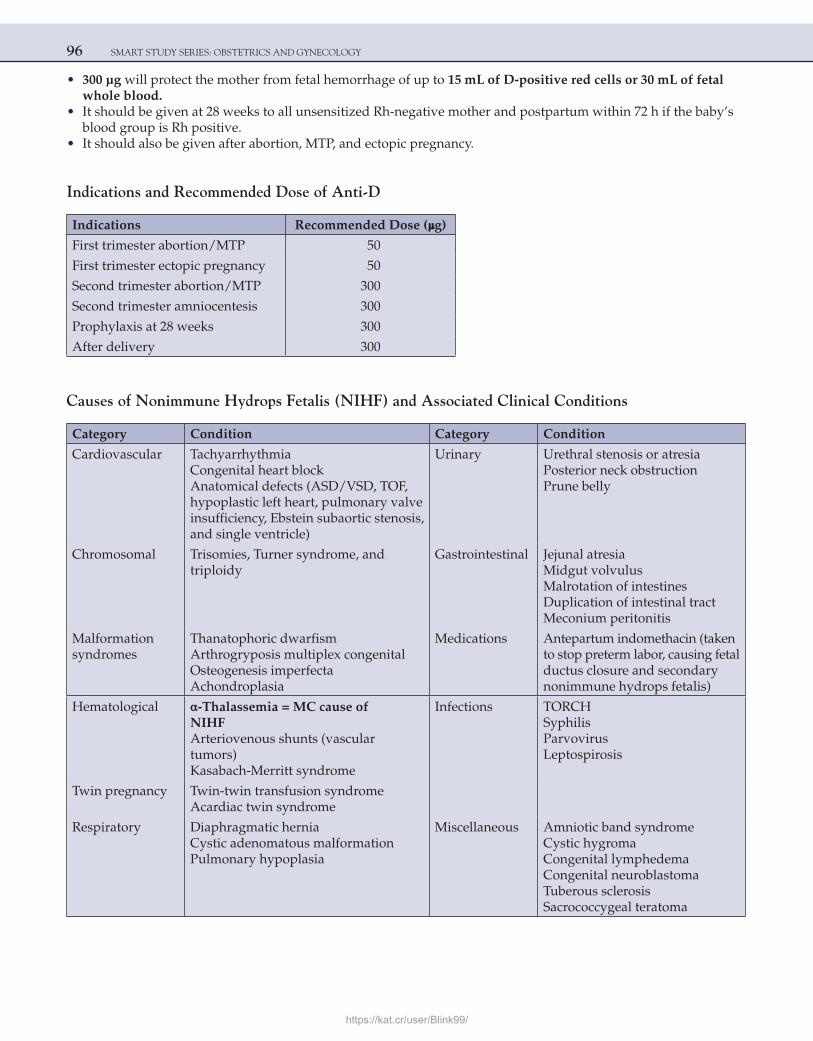
96 SMART STUDY SERIES: OBSTETRICS AND GYNECOLOGY
• 300 μg will protect the mother from fetal hemorrhage of up to 15 mL of D-positive red cells or 30 mL of fetal whole blood.
• It should be given at 28 weeks to all unsensitized Rh-negative mother and postpartum within 72 h if the baby’s blood group is Rh positive.
• It should also be given after abortion, MTP, and ectopic pregnancy.
Indications and Recommended Dose of Anti-D
Indications Recommended Dose (μg)
First trimester abortion/MTP 50
First trimester ectopic pregnancy 50
Second trimester abortion/MTP 300
Second trimester amniocentesis 300
Prophylaxis at 28 weeks 300
After delivery 300
Causes of Nonimmune Hydrops Fetalis (NIHF) and Associated Clinical Conditions
Category Condition Category Condition
Cardiovascular TachyarrhythmiaCongenital heart blockAnatomical defects (ASD/VSD, TOF, hypoplastic left heart, pulmonary valve insufficiency, Ebstein subaortic stenosis, and single ventricle)
Urinary Urethral stenosis or atresiaPosterior neck obstructionPrune belly
Chromosomal Trisomies, Turner syndrome, andtriploidy
Gastrointestinal Jejunal atresiaMidgut volvulusMalrotation of intestinesDuplication of intestinal tractMeconium peritonitis
Malformationsyndromes
Thanatophoric dwarfismArthrogryposis multiplex congenitalOsteogenesis imperfectaAchondroplasia
Medications Antepartum indomethacin (takento stop preterm labor, causing fetalductus closure and secondarynonimmune hydrops fetalis)
Hematological α-Thalassemia = MC cause ofNIHFArteriovenous shunts (vasculartumors)Kasabach-Merritt syndrome
Infections TORCHSyphilisParvovirusLeptospirosis
Twin pregnancy Twin-twin transfusion syndromeAcardiac twin syndrome
Respiratory Diaphragmatic herniaCystic adenomatous malformationPulmonary hypoplasia
Miscellaneous Amniotic band syndromeCystic hygromaCongenital lymphedemaCongenital neuroblastomaTuberous sclerosisSacrococcygeal teratoma
https://kat.cr/user/Blink99/
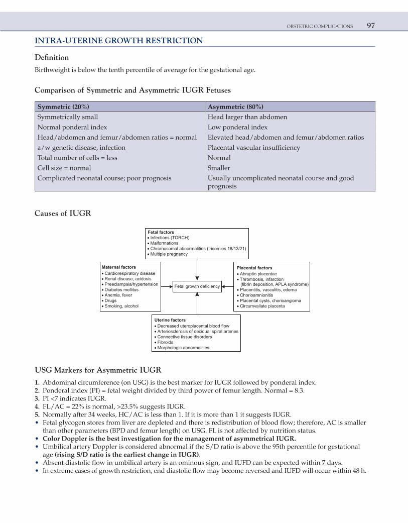
OBSTETRIC COMPLICATIONS 97
INTRA-UTERINE GROWTH RESTRICTION
Definition
Birthweight is below the tenth percentile of average for the gestational age.
Comparison of Symmetric and Asymmetric IUGR Fetuses
Symmetric (20%) Asymmetric (80%)
Symmetrically small Head larger than abdomen
Normal ponderal index Low ponderal index
Head/abdomen and femur/abdomen ratios = normal Elevated head/abdomen and femur/abdomen ratios
a/w genetic disease, infection Placental vascular insufficiency
Total number of cells = less Normal
Cell size = normal Smaller
Complicated neonatal course; poor prognosis Usually uncomplicated neonatal course and good prognosis
Causes of IUGR
Fetal factorsInfections (TORCH)MalformationsChromosomal abnormalities (trisomies 18/13/21)Multiple pregnancy
Cardiorespiratory diseaseRenal disease, acidosisPreeclampsia/hypertensionDiabetes mellitusAnemia, feverDrugsSmoking, alcohol
Maternal factorsAbruptio placentaeThrombosis, infarction(fibrin deposition, APLA syndrome)Placentitis, vasculitis, edemaChorioamnionitisPlacental cysts, chorioangiomaCircumvallate placenta
Placental factors
Decreased uteroplacental blood flowArteriosclerosis of decidual spiral arteriesConnective tissue disordersFibroidsMorphologic abnormalities
Uterine factors
Fetal growth deficiency
USG Markers for Asymmetric IUGR
1. Abdominal circumference (on USG) is the best marker for IUGR followed by ponderal index. 2. Ponderal index (PI) = fetal weight divided by third power of femur length. Normal = 8.3. 3. PI <7 indicates IUGR. 4. FL/AC = 22% is normal, >23.5% suggests IUGR. 5. Normally after 34 weeks, HC/AC is less than 1. If it is more than 1 it suggests IUGR. • Fetal glycogen stores from liver are depleted and there is redistribution of blood flow; therefore, AC is smaller
than other parameters (BPD and femur length) on USG. FL is not affected by nutrition status. • Color Doppler is the best investigation for the management of asymmetrical IUGR. • Umbilical artery Doppler is considered abnormal if the S/D ratio is above the 95th percentile for gestational
age (rising S/D ratio is the earliest change in IUGR). • Absent diastolic flow in umbilical artery is an ominous sign, and IUFD can be expected within 7 days. • In extreme cases of growth restriction, end diastolic flow may become reversed and IUFD will occur within 48 h.
98 SMART STUDY SERIES: OBSTETRICS AND GYNECOLOGY
• As the S/D ratio begins to rise in fetus with asymmetric IUGR, the blood flow in MCA increases. There is redistribution of blood flow, and vital organs like brain continue to receive adequate blood at the expense of liver and kidney. This is called as BRAIN-SPARING EFFECT.
• Absent and reversed diastolic flow in umbilical artery on color Doppler is an indication of immediate LSCS.
• Low-dose aspirin is thought to improve the uteroplacental circulation and can be given to patients of IUGR and is also given in subsequent pregnancy to prevent IUGR.
• Asymmetric IUGR has better prognosis compared to symmetric IUGR. • Nitroglycerin (NTG) patches can be applied to maternal abdomen to increase the uterine blood flow. This is
currently under research/trials.
INTRA-UTERINE FETAL DEATH (IUFD)
Categories and Causes of Fetal Death
Fetal (25–40%)Chromosomal anomaliesNonchromosomal birth defectsNonimmune hydropsInfections
Placental (25–35%)AbruptionCord accidentPlacental insufficiencyIntrapartum asphyxiaPreviaTwin-to-twin transfusionChorioamnionitis
Maternal (5–10%)Antiphospholipid antibodiesDiabetesHypertensive disordersTraumaAbnormal laborSepsisAcidosis/hypoxiaUterine rupturePostterm pregnancyDrugs
Unexplained (25–35%)
• Hypertensive disorders and diabetes are the two most commonly cited maternal diseases, associated with stillbirths.
• Thromboplastin from the dead fetus can enter the maternal system and cause DIC. • This only happens when the dead fetus is retained inside for 3–4 weeks.
Radiological signs of IUFD:
Sign Interval (After Death)
Robert sign (gas in great vessels) 12 h
Spalding sign (overlapping of skull bones) 1 week
Blair-Hartley/Ball sign (hyperflexion/hyperextension of spine with overcrowding of ribs 3–4 weeks
https://kat.cr/user/Blink99/
OBSTETRIC COMPLICATIONS 99
Amniotic Fluid Embolism
• This is a complex disorder classically characterized by the abrupt onset of hypotension, hypoxia, and consumptive coagulopathy. There are variations in its clinical manifestation.
• In obvious cases, the clinical frequency is dramatic. Classically, a woman in the late stages of labor or immediately postpartum begins gasping for air and then rapidly suffers seizures or cardiorespiratory arrest, complicated by consumptive coagulopathy, massive hemorrhage, and death.
• Other features common to amniotic fluid embolism are meconium staining and rapid labor. • Amniotic fluid enters the circulation as a result of breach in the physiological barrier that normally exists
between maternal and fetal compartments. • There may be maternal exposure to various fetal elements during pregnancy termination, following
amniocentesis or trauma or, more commonly, during labor or delivery, as small lacerations develop in the lower uterine segment or cervix. Alternatively, cesarean delivery affords ample opportunity for mixture of maternal blood and fetal tissue.
DiagnosisIn the past, the detection of squamous cells or other debris of fetal origin in the central pulmonary circulation was believed to be pathognomonic for amniotic fluid embolism. Indeed, in fatal cases, histopathological findings may be dramatic, especially in those involving meconium-stained amniotic fluid.
ManagementWomen who survive long enough to receive any treatment other than cardiopulmonary resuscitation should receive therapy directed at oxygenation and support of the failing myocardium. Circulatory support and blood and compo-nent replacement are paramount. There are no data that any type of intervention improves maternal prognosis with amnionic fluid embolism. In undelivered women suffering cardiac arrest, consideration should be given to emer-gency perimortem cesarean delivery in an effort to improve newborn outcome.
MULTIPLE CHOICE QUESTIONS
1. A 32-year-old primigravida at 39 weeks of gestational age has a blood pressure reading of 150/100 mm Hg obtained during a routine visit. Her baseline blood pressure during the pregnancy was 120/70 mmHg. The patient denies any headache, visual changes, nausea, vomiting, or abdominal pain. Her repeat BP is 160/90 mmHg, and urinalysis is negative for protein. Which of the following is the most likely diagnosis?
a. Preeclampsia b. Chronic hypertension with superimposed preeclampsia c. Eclampsia d. Gestational hypertension
Answer: d (Gestational hypertension)
Explanation:Hypertension in pregnancy is defined as blood pressure of 140/90 mmHg or greater on at least two separate occasions that
are 6 h or more apart. The presence of edema is no longer used as a diagnostic criterion because it is so prevalent in normal pregnant women. A rise in systolic blood pressure of 30 mmHg and a rise in diastolic blood pressure of 15 mmHg are also no longer used.
In gestational hypertension, maternal blood pressure reaches 140/90 or greater for the first time during pregnancy, and proteinuria is not present. In preeclampsia, blood pressure increases to 140/90 after 20 weeks of gestation and proteinuria is present (300 mg in 24 h or 1+ protein or greater on dipstick.)
Eclampsia is present when women with preeclampsia develop seizures.Chronic hypertension is defined as BP >140/90 mmHg before pregnancy or diagnosed before 20 weeks of gestation, or
hypertension first diagnosed after 20 weeks of gestation and persistent after 12 weeks postpartum.A woman with hypertension who develops preeclampsia is described as having chronic hypertension with superimposed
preeclampsia.
Reference:
1. Williams, 22nd Ed., Pg. 762.
100 SMART STUDY SERIES: OBSTETRICS AND GYNECOLOGY
2. A 27-year primigravida presents with pregnancy-induced hypertension with blood pressure of 150/100 mmHg at 32 weeks of gestation with no other complications. Subsequently, her blood pressure is controlled on treatment. If there are no complications, the pregnancy should be best terminated by induction at:
[AIIMS May 2006] a. 40 completed weeks b. 37 completed weeks c. 36 completed weeks d. Await spontaneous onset of labor
Answer: b (37 completed weeks)
Explanation:If the preeclamptic features of a patient completely subside on treatment and blood pressure is controlled, then further
management is as follows:If the duration of pregnancy is far from term, then discharge patient while advising regular follow-ups.If patient is near term, then she should be kept in hospital till completion of 37 weeks. Thereafter the labor should
be induced even if the BP is under control, as the risks of continuation of pregnancy far outweigh the benefits (as delivery is the ultimate treatment for pregnancy-induced hypertension, and it is not advisable to wait further because the BP can rise and there can be complications, and there are no added benefits of continuing pregnancy beyond 37 weeks).
The woman in question has controlled BP and is at 32 weeks of gestation.So the best management would be to terminate pregnancy at 37 completed weeks.
Reference:
1. Williams, 22nd Ed., Pg. 780–3.
3. There is loss of knee jerks when magnesium sulfate concentration reaches: a. 5–7 mEq/L b. 10–11 mEq/L c. 8–10 mEq/L d. >12 mEq/L
Answer: b (10–11 mEq/L)
Explanation:Magnesium sulfate is the treatment of choice for the prevention and treatment of eclamptic seizures. It reduces motor end
plate sensitivity to acetyl choline. It induces cerebral vasodilatation, dilates uterine arteries, increases production of endo-thelial prostacyclin, and inhibits platelet activation. Magnesium may also prevent seizures by interacting with N-methyl-D-aspartate (NMDA) receptors in the central nervous system.
After the initial dose, repeat injections are given only if knee jerks are present, urine output exceeds 30 mL/h, and respira-tion rate is more than 12/min.
• The therapeutic level of serum magnesium is 4–7 mEq/L. • 8–10 mEq/L = uterus stops contracting. • Patellar reflux disappears when magnesium level reaches above 10 mEq/L (12 mg/dL), presumably because of
curariform action. • This sign serves to warn of impending magnesium toxicity, because a further increase leads to respiratory
depression. • When magnesium levels reach >12 mEq/L, respiratory depression develops, and respiratory paralysis and arrest
follow. • Treatment is with calcium gluconate 1 g IV and withholding magnesium sulfate reverse mild-to-moderate respiratory
depression.
Reference:
1. Williams, 22nd Ed., Pg. 789–90.
https://kat.cr/user/Blink99/
OBSTETRIC COMPLICATIONS 101
4. In a case of recurrent spontaneous abortion the following investigation is unwanted: [AIIMS Nov 2002; All India 2008] a. Hysteroscopy b. Testing for antiphospholipid antibodies c. Testing for TORCH infections d. Thyroid function tests
Answer: c (Testing for TORCH infections)
Explanation:Patients with recurrent spontaneous abortions may have significant thyroid disease (viz. hypothyroidism). Hence, to rule
out thyroid disease TSH level may be estimated.Uterine abnormalities can lead to impaired vascularization due to a distorted uterine cavity. In all, 12–15% of women with
recurrent abortions have a uterine malformation (e.g., septate uterus/T-shaped uterus). In addition, pathological causes such as fibroids and intra-uterine synechiae (Asherman syndrome) may also lead to recurrent spontaneous abortion. Hysteroscopy is a very useful tool in both diagnosis and correction of these factors.
Anticardiolipin antibodies and lupus anticoagulant are antiphospholipid antibodies. They cause thrombosis, spontaneous abortion, and fetal wastage. A total of 10–15% of women with recurrent abortions have these antibodies.
Despite periodic reports that have implicated specific infectious agents as etiological factors in recurrent spontaneous abortions, there is currently no evidence implicating bacterial or viral agents as etiological factors for recurrent abortions. Hence, testing for TORCH infections is now thought to be unwarranted.
TORCH infection gives lifelong immunity; hence, TORCH infection can cause an abortion but not recurrent abortions.
Reference:
1. Williams, 22nd Ed., Pg. 241.
5. In which of the following conditions the medical treatment of ectopic pregnancy is contraindicated? [AIIMS May 2004] a. Sac size is 3 cm b. 50 mL blood in pelvis c. Presence of fetal heart activity d. Previous ectopic pregnancy
Answer: c (Presence of fetal heart activity)
Explanation:Medical management (methotrexate) is the treatment of choice for an ectopic pregnancy whenever the required criteria
are fulfilled.The following criteria should be fulfilled for medical management of ectopic pregnancy:
1. Patient should be hemodynamically stable (unruptured tubal ectopic pregnancy) 2. Fetal cardiac activity absent (presence of cardiac activity is a relative contraindication) 3. β-hCG levels <5,000 μIU/mL (levels > 5,000 micro IU/mL is a relative contraindication) 4. Gestational sac diameter <4 cm 5. Free fluid in POD <100 mL
Reference:
1. Williams, 22nd Ed., Pg. 262–3.
6. Which of the following statements concerning abdominal pregnancy is correct? a. Gastrointestinal symptoms are quite often very severe b. Fetal survival is approximately 80% c. Aggressive attempts should be made to remove the placenta at the time of initial surgery d. Placenta can be left in situ at the time of surgery
Answer: d (Placenta can be left in situ at the time of surgery)
102 SMART STUDY SERIES: OBSTETRICS AND GYNECOLOGY
Explanation:Secondary abdominal pregnancy usually follows a tubal pregnancy with either tubal rupture or spontaneous passage
through the fimbriated end. Primary cases are extremely rare.Although women with abdominal pregnancy usually report an increase in gastrointestinal symptoms, these are rarely
severe enough to lead to investigation. Fetal death rates are reported to be above 90% with abdominal pregnancies. It is almost impossible and dangerous to salvage the fetus.
Infections of the gestational products can occur especially when the placenta adheres to the intestines. This can lead to abscess formation and the possibility of rupture.
Although leaving the placenta in the abdomen following surgical delivery predisposes to risks of postoperative infections, the risk is much less severe than the hemorrhage associated with attempts at removal of placenta at the time of primary surgery.
If the placenta cannot easily be removed, recommendations are to leave it in place at the time of the first surgery. Metho-trexate should be given postoperatively to take care of the placenta in situ.
Reference:
1. Williams, 22nd Ed., Pg. 265.
7. A 26-year-old primigravida with a twin gestation at 30 weeks presents for an USG. The sonogram indicates that the fetuses are both male, and the placenta appears to be diamniotic and monochorionic. Twin B is noted to have oligohydramnios and to be much smaller than twin A. In this clinical picture, all of the following are concerns for twin A, except:
a. Congestive heart failure b. Anemia c. Hypervolemia d. Hydramnios
Answer: b (Anemia)
Explanation:This is a case of twin-to-twin transfusion syndrome.In twin gestations where monochorionic placentas exist, twin-to-twin transfusion syndrome can occur. In this syndrome,
there are vascular communications or anastomoses between the twins. There is blood flow or transfusion from one twin to another. The donor twin becomes anemic and may suffer growth retardation and oligohydramnios. The recipient twin may develop hydramnios, hypervolemia, hypertension, polycythemia, and congestive heart failure.
Reference:
1. Williams, 22nd Ed., Pg. 929–30.
8. A 24-year-old presents at 35 weeks with an AFI of 30 cm. Which of the following statements is true? [AIIMS Nov 2003] a. The incidence of associated malformations is approximately 2% b. Maternal edema, especially of the lower extremities, is rare c. Esophageal atresia is accompanied by polyhydramnios in nearly 10% of cases d. Complications include placental abruption, uterine dysfunction, and postpartum hemorrhage
Answer: d (Complications include placental abruption, uterine dysfunction, and postpartum hemorrhage)
Explanation:Polyhydramnios is an excessive quantity of amniotic fluid (AFI >25 cm).The incidence of associated malformations is about 20%, with CNS and GI abnormalities being particularly common.
For example, polyhydramnios accompanies about half of the cases of anencephaly and nearly all cases of esophageal atre-sia. Edema of the lower extremities, vulva, and abdominal wall results from compression of major venous systems. Acute hydramnios tends to occur early in pregnancy and, as a rule, leads to preterm labor. The most frequent maternal complica-tions are placental abruption, uterine dysfunction, and atonic postpartum hemorrhage.
Reference:
1. Williams, 22nd Ed., Pg. 527–8.
https://kat.cr/user/Blink99/
OBSTETRIC COMPLICATIONS 103
9. The placenta of twins can be: a. Dichorionic and monoamnionic in dizygotic (DZ) twins b. Dichorionic and monoamnionic in monozygotic (MZ) twins c. Monochorionic and monoamnionic in DZ twins d. Dichorionic and diamniotic in MZ twins
Answer: d (Dichorionic and diamniotic in MZ twins)
Explanation:Dizygotic twins always have dichorionic and diamniotic placenta. The dichorionic placentas of dizygotic twins may be
totally separated or intimately fused.In monozygotic twins, if the twinning occurs in less than 3 days the result is dichorionic, diamniotic placentation. The
majority of monozygotic twins have a diamniotic and monochorionic placenta (twinning between 4 and 8 days). The least common type of placentation in monozygotic twins is the monochorionic and monoamniotic placenta, which happens if the twinning happens after 8 days.
Reference:
1. Williams, 22nd Ed., Pg. 914–5.
10. Embryo reduction of multiple pregnancy is done at: [All India 2013] a. 8–10 weeks b. 11–13 weeks c. 13–15 weeks d. 16–18 weeks
Answer: b (11–13 weeks)
Explanation:Multifetal pregnancies of higher order (triplets, quadruplets, etc.) can be reduced to twin pregnancy or twin pregnancy can
be reduced to singleton pregnancy by selective embryo reduction, done between 10 and 13 weeks.It is done by injection of potassium chloride in fetal heart through transabdominal route under USG guidance.
Reference:
1. Williams, 22nd Ed., Pg. 941–2.
11. A 28-year-old primigravida presents, at 18 weeks of gestational age, with right-sided groin pain. She describes the pain as sharp and occurring with movement. She denies any change in urinary or bowel habits. She also denies any fever or chills. The application of a heating pad helps to relieve the discomfort. The most likely etiology of this pain is:
a. Round ligament pain b. Preterm labor c. Kidney stone d. Urinary tract infection
Answer: a (Round ligament pain)
Explanation:The patient is giving a classic description of round ligament pain. Each round ligament extends from the lateral portion
of the uterus below the tube and travels in a fold of peritoneum downward to the inguinal canal and inserts in the upper portion of the labium majus. During pregnancy, these ligaments stretch as the gravid uterus grows further out of the pelvis and can thereby cause sharp pains, particularly with sudden movements. Round ligament pain is usually more frequently experienced on the right side, due to the dextrorotation of the uterus that commonly occurs in pregnancy. Usually, this pain is greatly improved by avoiding sudden movements and by rising and sitting down gradually. Local heat and analgesics may also help with pain control.
104 SMART STUDY SERIES: OBSTETRICS AND GYNECOLOGY
The diagnosis of preterm labor is unlikely because the pain is localized to the groin area on one side and is alleviated with a heating pad, which would not be the case with labor contractions. In addition, when labor occurs, the pain would persist at rest, not just with movement.
A urinary tract infection is unlikely because the patient has no urinary symptoms. A kidney stone is unlikely because, usu-ally, the patient would complain of pain in the back, not low in the groin. In addition, with a kidney stone the pain would not only occur with movement, but would persist at rest as well.
Reference:
1. Williams, 22nd Ed., Pg. 224.
12. The shape of cervix (on USG) which indicates a competent os is: [All India 2007] a. T-shaped b. Y-shaped c. V-shaped d. U-shaped
Answer: a (T-shaped)
Explanation:Closed cervix (competent os) on USG appears like the letter T.Incompetent os on USG shows the following features: Before opening, the cervix shortens and then funneling can take
place, which on USG looks like the letter Y (indicating incompetent os) that can progress to look like the letter V (cervix is just about to open).
When the os is open the membranes can herniate, giving the appearance of letter U.
Reference:
1. Callen. USG in Obstetrics and Gynecology, 4th Ed., Pg. 582.
13. A 32-year-old G2P1L1 presents at 35 weeks of gestation, complaining of leaking PV. A sample of pooled fluid seen in the vaginal vault turned red litmus to blue and showed a fern pattern on microscopy. The patient has a temperature of 102°F and P = 102, and her fundus is tender to deep palpation. What is the next appropriate step in the management of this patient?
a. Administer betamethasone b. Administer tocolytics c. Place a cervical cerclage d. Administer antibiotics
Answer: d (Administer antibiotics)
Explanation:The fluid in the vagina is amniotic fluid, as it showed a fern pattern on microscopy (presence of sodium chloride in liquor)
and the red litmus turned blue (vaginal pH is acidic; amniotic fluid is alkaline).This patient with premature rupture of membranes (PROM) has a physical examination consistent with an intra-
uterine infection or chorioamnionitis. Chorioamnionitis can be diagnosed clinically by the presence of maternal fever, tachycardia, and uterine tenderness. Leukocyte counts are a nonspecific indicator of infection because they can be ele-vated with labor.
When chorioamnionitis is diagnosed, fetal and maternal morbidities increase and delivery is indicated, regardless of the fetus’s gestational age. In the case described, antibiotics need to be administered to avoid neonatal sepsis. Ampicillin is the drug of choice to treat group B streptococcal infection. Labor should be induced.
There is no role for tocolysis in the setting of chorioamnionitis, since delivery is the goal. There is also no role for the admin-istration of steroids as it is contraindicated in chorioamnionitis.
Reference:
1. Williams, 22nd Ed., Pg. 864–5.
https://kat.cr/user/Blink99/
OBSTETRIC COMPLICATIONS 105
14. The most important indication for surgical repair of a double uterus, such as a septate or bicornuate uterus, is: [All India 2013] a. Habitual abortion b. Dysmenorrhea c. Dyspareunia d. Premature delivery
Answer: a (Habitual abortion)
Explanation:The most important indication for surgical treatment of women who have a double uterus is habitual/repeated abor-
tions. The abortion rate in women who have a double uterus is two to three times greater than that of the general population. Therefore, women who present with habitual abortion should be evaluated to detect a possible double uterus. USG (especially 3D), HSG, and hysteroscopy are all useful imaging modalities in this investigation. Dysmenorrhea, premature delivery, and menometrorrhagia are other less important indicators for surgical intervention.
Reference:
1. Williams, 22nd Ed., Pgs. 236, 237.
15. All are the causes of intra-uterine growth retardation, except: [All India 2005] a. Anemia b. Pregnancy-induced hypertension c. Maternal heart disease d. Gestational diabetes
Answer: d (Gestational diabetes)
Explanation:Intra-uterine growth restriction is of two types. Type I is associated with early-onset IUGR with congenital infections/
chromosomal detects with fewer number of fetal cells. Type II is late onset, that is, generally after 24–28 weeks, which is associated with decreased availability of nutrients and/or oxygen for cell growth. Anemia, PIH, and heart disease, all lead to lower quality and quantity of placental perfusion and hence can cause IUGR.
Gestational diabetes is associated with maternal and hence fetal hyperglycemia, which, in turn, leads to excessive deposi-tion of adipose tissue in the fetus causing macrosomia.
Reference:
1. James, 3rd Ed., Pg. 241–2.
16. A 38-year-old G2P1L1 comes to see you for her first prenatal visit at 14 weeks of gestational age with the following reports: Her blood type is A negative and an anti-D antibody titer of 1:4. What is the most appropriate next step in the management of this patient?
a. Perform an amniocentesis for amniotic fluid spectrophotometric analysis b. Repeat the titer in 4 weeks c. Give her injection anti-D d. Do cordocentesis to determine fetal hematocrit and perform intra-uterine transfusion (IUT)
Answer: b (Repeat the titer in 4 weeks)
Explanation:During the first prenatal visit, all pregnant women are screened for the ABO blood group and the Rh group, which
includes the D antigen. If the woman is Rh negative, antibody screening is performed. If the antibody D titer is positive, the woman is considered sensitized because she has produced antibodies against the D antigen. Sensitization gener-ally occurs as a result of exposure to blood from an Rh+ fetus in a prior pregnancy. A fetus that is Rh+ has red blood cells that express the D antigen. Therefore, the maternal anti-D antibodies can cross the placenta and cause fetal hemolysis.
Once the antibody screen is positive for isoimmunization, the titer should be followed at regular intervals (about every 4 weeks).
106 SMART STUDY SERIES: OBSTETRICS AND GYNECOLOGY
A titer of 1:16 or greater is usually indicative of the possibility of severe hemolytic disease of the fetus. Once the critical titer is reached, further evaluation is done by amniotic fluid. In the presence of fetal hemolysis, the amniotic fluid contains elevated levels of bilirubin that can be determined via spectrophotometric analysis. Cordocentesis is done mainly to perform IUT in cases where fetal anemia is causing hydrops and the fetus is still premature (<34 weeks).
Cordocentesis, or percutaneous umbilical blood sampling, involves obtaining a blood sample from the umbilical cord under ultrasound guidance. The fetal blood sample can then be analyzed for Hct, and cordocentesis allows the fetus with hydrops to undergo a blood transfusion (IUT). This is only required when the delta 450 value obtained by amniocentesis and spectrophotometric analysis falls in Liley’s zone 3 and the fetus is still premature (<34 weeks).
Injection anti-D is given to prevent isoimmunization and has no role when the mother is already sensitized (as indicated by positive anti-D titer).
Reference:
1. Williams, 22nd Ed., Pg. 666–7.
17. All of the following are scenarios in which it is necessary to administer anti-D, except: [All India 2004, 2013] a. After a spontaneous first-trimester abortion b. After an ectopic pregnancy c. After cordocentesis for IUT d. After manual removal of placenta
Answer: c (After cordocentesis for IUT)
Explanation:To prevent maternal Rh sensitization, pregnant women who are Rh negative should receive Rh immune globulin (antibody
to the D antigen) in the following situations:
1. At 28 weeks to all unsensitized Rh-negative mothers and postpartum within 72 h if the baby’s blood group is Rh positive 2. After abortion, MTP, ectopic pregnancy 3. After amniocentesis, CVS, cordocentesis 4. After ECV 5. After manual removal of placenta (in any situation where fetomaternal hemorrhage is expected)
The anti-D binds to fetal RBCs and prevents them from stimulating the maternal immune system.When the mother is already sensitized (positive indirect Coombs’ test or positive Rh titer), there is no role of anti-D.When cordocentesis/PUBS is being done for IUT, it means that the fetus is having severe anemia and hydrops due to Rh
isoimmunization and maternal antibodies are already present and hence Anti-D has no role.
Reference:
1. Williams, 22nd Ed., Pg. 665–8.
18. The consequences of Rh incompatibility are not serious during first pregnancy because: [All India 2004] a. In first pregnancy only IgM antibody is formed b. Antibody titer is very low during primary immune response c. IgG generated is ineffective against fetal red cells d. Massive hemolysis is compensated by increased erythropoiesis
Answer: b (Antibody titer is very low during primary immune response)
Explanation:If the ABO-compatible Rh-positive fetal red cells enter the mother’s blood, they remain in the circulation for their remain-
ing life span. Thereafter, they are removed from the circulation by the reticuloendothelial tissues and are broken down with liberation of the antigen. The antibody production is related not only to the responsiveness of the reticuloendothelial system but also to the amount of Rh antigen liberated (the number of red cells that have entered the maternal blood).
Because this takes a long time, immunization in a first pregnancy is unlikely. Detectable antibodies usually develop after 6 months following a large volume of fetomaternal bleed.
Initially, IgM antibodies are formed followed by IgG. IgG antibodies can be present even in first pregnancy. But both the antibodies are in very low titers.
Antibodies once formed remain throughout life.
https://kat.cr/user/Blink99/
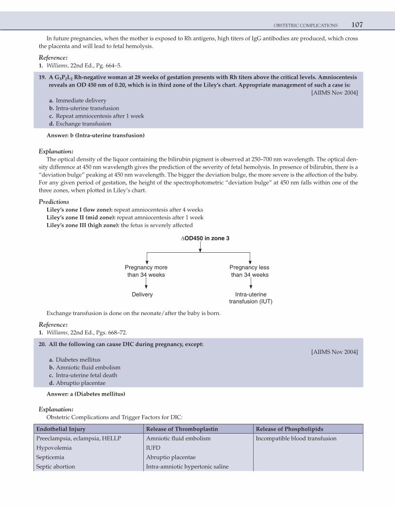
OBSTETRIC COMPLICATIONS 107
In future pregnancies, when the mother is exposed to Rh antigens, high titers of IgG antibodies are produced, which cross the placenta and will lead to fetal hemolysis.
Reference:
1. Williams, 22nd Ed., Pg. 664–5.
19. A G3P2L2 Rh-negative woman at 28 weeks of gestation presents with Rh titers above the critical levels. Amniocentesis reveals an OD 450 nm of 0.20, which is in third zone of the Liley’s chart. Appropriate management of such a case is:
[AIIMS Nov 2004] a. Immediate delivery b. Intra-uterine transfusion c. Repeat amniocentesis after 1 week d. Exchange transfusion
Answer: b (Intra-uterine transfusion)
Explanation:The optical density of the liquor containing the bilirubin pigment is observed at 250–700 nm wavelength. The optical den-
sity difference at 450 nm wavelength gives the prediction of the severity of fetal hemolysis. In presence of bilirubin, there is a “deviation bulge” peaking at 450 nm wavelength. The bigger the deviation bulge, the more severe is the affection of the baby. For any given period of gestation, the height of the spectrophotometric “deviation bulge” at 450 nm falls within one of the three zones, when plotted in Liley’s chart.
PredictionsLiley’s zone I (low zone): repeat amniocentesis after 4 weeksLiley’s zone II (mid zone): repeat amniocentesis after 1 weekLiley’s zone III (high zone): the fetus is severely affected
∆OD450 in zone 3
Pregnancy more Pregnancy less than 34 weeks than 34 weeks
Delivery Intra-uterinetransfusion (IUT)
Exchange transfusion is done on the neonate/after the baby is born.
Reference:
1. Williams, 22nd Ed., Pgs. 668–72.
20. All the following can cause DIC during pregnancy, except: [AIIMS Nov 2004] a. Diabetes mellitus b. Amniotic fluid embolism c. Intra-uterine fetal death d. Abruptio placentae
Answer: a (Diabetes mellitus)
Explanation:Obstetric Complications and Trigger Factors for DIC:
Endothelial Injury Release of Thromboplastin Release of Phospholipids
Preeclampsia, eclampsia, HELLP Amniotic fluid embolism Incompatible blood transfusion
Hypovolemia IUFD
Septicemia Abruptio placentae
Septic abortion Intra-amniotic hypertonic saline
108 SMART STUDY SERIES: OBSTETRICS AND GYNECOLOGY
Endothelial Injury Release of Thromboplastin Release of Phospholipids
Chorioamnionitis
Reference:
1. Williams, 22nd Ed., Pgs. 843–4.
21. A 28-year-old primigravida was diagnosed as a case of gestational hypertension at 28 weeks of gestation. She presents at 32 weeks with pain in abdomen. On examination: P = 98/m, BP = 100/60 mmHg, and Hb 6 g%. P/A—uterus is 32–34 weeks tonically contracted with fetal heart absent. P/V—no bleeding seen. The diagnosis is:
[All India 2003] a. Concealed placenta previa b. Revealed placenta previa c. Concealed abruptio placentae d. Revealed abruptio placentae
Answer: c (Concealed abruptio placentae)
Explanation:Hypertensive disorder is one of the most important causes for placental abruption.Tonically contracted uterus is classically seen in abruptio placentae.Besides tachycardia, hypotension and decrease in Hb suggest that the patient is losing blood.Abruption can be of two varieties: concealed and revealed.In this case there is no externally visible bleeding, and uterus is more than the number of weeks of gestation, which points
to the diagnosis of concealed abruptio placentae.In placenta previa the bleeding is painless, uterus is not tonically contracted.In placenta previa, the bleeding is always revealed. There is nothing like concealed placenta previa.
Reference:
1. Williams, 22nd Ed., Pgs. 811–3.
22. A primigravida with 36 weeks of pregnancy is in labor with 3 cm dilatation and minimal uterine contraction. On rupture of membranes, fresh bleeding is noted with late fetal deceleration up to 50 beats/min. The patient was taken for LSCS but fetus could no be saved. No abruptio or placenta previa was seen. The likely diagnosis is:
a. Placenta previa b. Revealed abruptio c. Circumvallate placenta d. Vasa previa
Answer: d (Vasa previa)
Explanation:This is a case of vasa previa.Two main causes of bleeding in third trimester include placenta previa and abruption. But in both these conditions, the
blood loss is mixed maternal and fetal.In vasa previa there is exclusively fetal blood loss and even 40–50 mL blood loss is fatal for the fetus. Fresh bleeding and
severe fetal distress (late fetal deceleration up to 50 b/m) and the fact that in spite of LSCS the fetus could no be saved all point to the diagnosis of vasa previa.
Besides “ no abruptio or placenta previa seen” is also mentioned in the MCQ.Vasa previa (1:2500) is a rare condition in which fetal blood vessels are in front of the presenting part and cross the cervix.
The condition has a high fetal mortality rate (50–95%). This is attributed to rapid fetal exsanguination, resulting from the ves-sels tearing when the cervix dilates, membrane rupture.
Vasa previa might be present if any of the following conditions exist:
• Velamentous cord insertion • Bilobed placenta • Succenturiate-lobed placenta • Low-lying placenta or placenta previa • IVF pregnancies
https://kat.cr/user/Blink99/
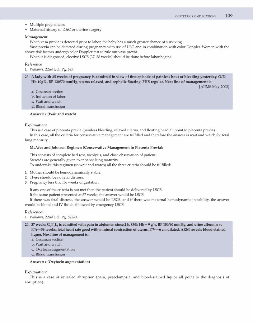
OBSTETRIC COMPLICATIONS 109
• Multiple pregnancies • Maternal history of D&C or uterine surgery
ManagementWhen vasa previa is detected prior to labor, the baby has a much greater chance of surviving.Vasa previa can be detected during pregnancy with use of USG and in combination with color Doppler. Women with the
above risk factors undergo color Doppler test to rule out vasa previa.When it is diagnosed, elective LSCS (37–38 weeks) should be done before labor begins.
Reference
1. Williams, 22nd Ed., Pg. 627.
23. A lady with 35 weeks of pregnancy is admitted in view of first episode of painless bout of bleeding yesterday. O/E: Hb 10g%, BP 120/70 mmHg, uterus relaxed, and cephalic floating. FHS regular. Next line of management is:
[AIIMS May 2003] a. Cesarean section b. Induction of labor c. Wait and watch d. Blood transfusion
Answer: c (Wait and watch)
Explanation:This is a case of placenta previa (painless bleeding, relaxed uterus, and floating head all point to placenta previa).In this case, all the criteria for conservative management are fulfilled and therefore the answer is wait and watch for fetal
lung maturity.
McAfee and Johnson Regimen (Conservative Management in Placenta Previa):
This consists of complete bed rest, tocolysis, and close observation of patient.Steroids are generally given to enhance lung maturity.To undertake this regimen (to wait and watch) all the three criteria should be fulfilled:
1. Mother should be hemodynamically stable. 2. There should be no fetal distress. 3. Pregnancy less than 36 weeks of gestation.
If any one of the criteria is not met then the patient should be delivered by LSCS.If the same patient presented at 37 weeks, the answer would be LSCS.If there was fetal distress, the answer would be LSCS, and if there was maternal hemodynamic instability, the answer
would be blood and IV fluids, followed by emergency LSCS.
Reference:
1. Williams, 22nd Ed., Pg. 822–3.
24. 37 weeks G2P1L1 is admitted with pain in abdomen since 2 h. O/E: Hb = 9 g%, BP 150/90 mmHg, and urine albumin +. P/A—36 weeks, fetal heart rate good with minimal contraction of uterus. P/V—6 cm dilated. ARM reveals blood-stained liquor. Next line of management is:
a. Cesarean section b. Wait and watch c. Oxytocin augmentation d. Blood transfusion
Answer: c (Oxytocin augmentation)
Explanation:This is a case of revealed abruption (pain, preeclampsia, and blood-stained liquor all point to the diagnosis of
abruption).
110 SMART STUDY SERIES: OBSTETRICS AND GYNECOLOGY
Abruptio placentae
DIC present DIC absent
Correct DIC
Possible Not possible(e.g., Transverse lie/prev 2 )LSCS
Vaginal delivery LSCS
FHS present
Normaldelivery
Oxytocinaugmentation
LSCS
Fetal distress No fetal distress
FHS absent(IUFD)
Vaginal delivery
In abruption the aim should be to deliver the patient soon, as the bleeding stops only after the delivery of the placenta. If there is fetal distress immediate LSCS should be done.
If the fetus is alive and there is no fetal distress and there is a possibility that delivery can happen soon (as in this case where the FHR is good and patient is already 6 cm dilated), then labor should be augmented by ARM and oxytocin drip, keeping a close watch on FHR.
So remember that abruption is not an absolute indication for LSCS, it is a relative indication.
Reference:
1. Williams, 22nd Ed., Pg. 817–8.
25. A 24-year-old patient comes with 4½ weeks of amenorrhea and PV spotting 15 days back. O/E: P 96 b/m, BP 120/80 mmHg. β-hCG is positive but USG reveals empty uterine cavity. Likely diagnosis is:
a. Ectopic pregnancy b. Abortion c. Early intra-uterine pregnancy d. All of the above
Answer: d (All of the above)
Explanation:Radio Immunoassay can detect hCG on the 25th day of the menstrual cycle and radio receptor assay (RRA) can detect hCG
on the 22nd day of the cycle.When the β-hCG is positive but the uterus is empty on USG, the possibilities are:
1. Very early intra-uterine pregnancy (since the β-hCG is positive as early as day 22 of the cycle, but the gestational sac within the uterus is seen earliest at 4 weeks 5 days or 33 days on TVS and after 40 days on TAS)
2. Ectopic pregnancy 3. Complete abortion (uterus will be empty but hCG does not immediately disappear from circulation after an abortion. It
decreases but can be detected for 1–2 weeks following an abortion).
In such situations the next best step to be done is to repeat β-hCG after 48 h.NOTE: Investigation of choice in a case of suspected ectopic pregnancy is transvaginal sonography. (AIIMS Nov 2009)
Reference:
1. Williams, 22nd Ed., Pg. 258–9.
https://kat.cr/user/Blink99/

OBSTETRIC COMPLICATIONS 111
26. A 21-year-old female presents to emergency ward with 2 months of amenorrhea with pain in abdomen and shock. BP 90/60 mmHg and Hb 6 g%. Urine pregnancy test is found positive. Next immediate line of treatment is:
[AIIMS Nov 2002] a. Laparotomy b. Blood transfusion c. Medical management d. Laparoscopy
Answer: b (Blood transfusion)
Explanation:This is a case of ruptured ectopic pregnancy. Positive Urine Pregnancy Test indicates that the amenorrhea is due to preg-
nancy. Pain and shock in early pregnancy are mostly always due to ruptured ectopic.When the patient is in shock, the next immediate line of treatment is to resuscitate the patient and correct the shock with
blood and IV fluids, and start preparations for surgery simultaneously.This should be followed by immediate exploratory laparotomy.When the patient is in shock, the next immediate treatment should always be measures to correct the shock first (blood
and IV fluids).By the time you prepare the patient for exploration (alert the OT and shift the patient to OT), and start blood
transfusion.Medical management and laparoscopy are contraindicated in shock.
Reference:
1. Williams, 22nd Ed., Pg. 258–60.
27. A 30-year-old G3P2 is 14 weeks pregnant. She had two painless deliveries at 16 weeks earlier. Next line of management is:
a. Cervical encerclage b. Evaluation for diabetes mellitus and thyroid disorders c. Cervical length assessment d. Tocolytics
Answer: c (Cervical length assessment)
Explanation:The patient had two painless abortions at 16 weeks in the past, so mostly it is a case of incompetent os.Next line of management in these patients is frequent cervical length assessment: clinically or by USG.The patient is evaluated more frequently and if the cervix is short (less than 2.5–3 cm) than cervical encerclage has to be
done.Cervical encerclage is the surgery of choice for incompetent os, but the surgery itself can lead to complications such as
uterine contractions, abortions, and PROM.So the surgery is only to be done if it is indicated.Surgery is not required if the cervical length (on USG or digital examination) is adequate (>3 cm).Diabetes mellitus and thyroid disorders are causes of first trimester abortions and not second trimesters.Tocolytics are indicated in preterm labor.
Reference:
1. Williams, 22nd Ed., Pg. 236–7.
28. Shilu with 18 weeks of pregnancy is diagnosed as severe oligohydramnios. The most likely fetal consequence expected is:
a. Cord compression b. PPROM c. Fetal limb deformities d. Fetal cardiac anomalies
Answer: c (Fetal limb deformities)
112 SMART STUDY SERIES: OBSTETRICS AND GYNECOLOGY
Explanation:When the patient has severe oligohydramnios from early weeks of gestation, there can be permanent contractures and
even amniotic bands can get formed, which can give rise to amputations.Tetrad of early-onset oligohydramnios:
1. Facial clefts (cleft lip/palate) 2. IUGR (no space for the fetus to grow) 3. Limb reduction defects 4. pulmonary hypoplasia
This tetrad is not seen if the patient develops oligohydramnios in third trimester.Cord compression in case of oligohydramnios only happens during labor; the cord gets compressed between the fetus and
uterus.
Reference:
1. Williams, 22nd Ed., Pg. 530–1.
29. A 28-year-old primigravida with 33 weeks of pregnancy suddenly complains of headache, oliguria, and blurred vision. Her BP is 180/110 and urine albumin is +3. The line of further management is:
[AIIMS Nov 2012] a. Wait and watch b. LSCS c. Induction of labor d. Anticonvulsant + antihypertensive therapy
Answer: d (Anticonvulsant + antihypertensive therapy)
Explanation:The patient is a case of severe preeclampsia, with impending eclampsia.The dangerous symptoms that indicate impending eclampsia in case of preeclampsia are:
1. Headache 2. Oliguria 3. Epigastric pain 4. Nausea, vomiting 5. Blurring of vision
Whenever the above symptoms develop in a case of severe preeclampsia the patient is at a risk of eclampsia; the patient should be given anticonvulsant (MgSO4) and antihypertensive medication, and the pregnancy should be terminated by induction of labor irrespective of the weeks of gestation.
Magnesium sulfate is the drug of choice for eclampsia and also for impending eclampsia.Prophylactic magnesium sulfate decreases the risk of convulsion, abruption, and maternal mortality in this scenario.Labetalol is the DOC for hypertensive crisis followed by hydralazine.Never wait and watch in case of impending eclampsia and never directly proceed for LSCS as it can be fatal for the mother.
Vaginal delivery is safest for mother, and hence labor should be induced after stabilization of mother (after MgSO4 and anti-hypertensive medications).
If after induction of labor there is fetal distress or failure of induction, then LSCS can be done. The indications for termina-tion of pregnancy (irrespective of the weeks of gestation) in a case of preeclampsia are:
1. Severe preeclampsia, with impending eclampsia 2. Eclampsia (give MgSO4 first, followed by induction of labor) 3. HELLP syndrome.
Reference:
1. Williams, 22nd Ed., Pg. 780–1.
https://kat.cr/user/Blink99/
OBSTETRIC COMPLICATIONS 113
30. Which of the following is the most common predisposing factor for placenta accreta? [All India 2008] a. Tubal surgery b. Recent curettage c. Previous cesarean section d. Placenta previa
Answer: d (Placenta previa)
Explanation:One significant and dangerous complication of placenta previa is placenta accreta, increta, and percreta.This is usually seen when a placenta previa gets implanted over previously injured sites such as scar of cesarean section,
myomectomy, dilatation and curettage.Placenta accreta refers to the placenta being attached to the myometrium but does not invade the muscle; increta is seen
with the villi invades the myometrium; and percreta is seen when the villi penetrates through the entire uterine wall and into the bladder or rectum.
Nitabuch’s fibrinoid layer is absent.The presence of placenta previa in a patient with a prior cesarean section is associated with accreta in 10–35% of cases. With
multiple cesarean sections, the risk may be as high as 60–65%.USG and color Doppler assessment are very helpful in demonstrating marked or turbulent blood flow within the placenta
and extending into the surrounding tissues, which is also described as lacunar flow. MRI can demonstrate placental tissue extension through the uterus.
ManagementCareful attention should be paid to the lower uterine segment after delivery of the placenta. If bleeding persists despite
the usual postpartum uterotonic agents and uterine or hypogastric artery ligation, hysterectomy must be considered. It is considered a definitive and the safest treatment in these cases.
It may be a worthwhile exercise to attempt other methods to control the bleeding before hysterectomy such as oversewing the lower uterine segment, uterine artery ligation, ligation of internal iliac and uterine packing.
If definitive hysterectomy is not performed, bilateral arterial embolization of the uterine arteries should be the next option although more experience is needed to determine the success rate of such a procedure for placenta percreta. Precautions to control hemorrhage in general should include intravenous access, blood products, and anesthesiology assistance.
If these conditions are suspected by imaging studies prior to delivery, a planned cesarean section after uterine artery cath-eters are placed for possible embolization, may be useful to avoid a hysterectomy.
Other option for a woman, who has no active bleeding is to leave the entire placenta in place. Postoperative methotrexate is given for placenta accreta tissue left in situ.
Reference:
1. Williams, 22nd Ed., Pg. 832–3.
31. False about partial mole is: [All India 2010] a. Caused by triploidy b. Can be diagnosed very early by USG c. Can present as missed abortion d. Rarely causes persistent GTD
Answer: d (Rarely causes persistent GTD)
Explanation:Partial mole can cause GTD in 5 to 10 % cases
Feature Partial Mole
Karyotype Usually 69.XXX or 69.XXY
Embryo-fetus Often present
Amnion, fetal red blood cells Often present
Villus edema Variable, focal
114 SMART STUDY SERIES: OBSTETRICS AND GYNECOLOGY
Feature Partial Mole
Uterine size Small for dates
Theca-lutein cysts Rare
Medical complications Rare
Gestational trophoblastic neoplasia <5–10%
Patients with partial mole do not have dramatic clinical features of complete molar pregnancy. In general these patients have signs and symptoms of incomplete or missed abortion.
USG is a reliable and sensitive technique for the diagnosis of molar pregnancy.Presence of focal cystic spaces in the placental tissue and increase in the transverse diameter of the gestational sac has a
positive predictive value of 90% for the diagnosis of partial mole.
References:
1. Williams, 22nd Ed., Pg. 274. 2. Novak’s, 14th Ed., Pg. 1587–88.
32. In case of a transformation of a molar pregnancy to choriocarcinoma all of the following are associated except: [All India 2010] a. Enlarged uterus b. Persistence of lutein cyst in ovaries c. Plateau of HCG d. Sub urethral nodule
Answer: c (Plateau of HCG)
Explanation:Vesicular mole is a pre-malignant condition and can develop into choriocarcinoma.In case of transformation of a molar pregnancy to choriocarcinoma the following features are seen:
1) Irregular bleeding and subinvolution of the uterus. The uterus remains enlarged and does not return back to normal size. 2) Rising levels of HCG (not plateau). 3) Theca lutein cysts will persist. 4) Depending on the sites of metastasis following features are seen: a) Lungs: dyspnea, hemoptysis, chest pain,cough b) Vagina: bluish nodule in the sub urethra region,irregular bleeding, purulent discharge c) Liver: right upper quadrant or epigastric pain, jaundice d) Brain: convulsions, neurological deficits.
Reference:
1. Novak’s, 14th Ed., Pg. 1591–2.
33. A primigravida at 37 week of gestation reported to labor room with central placenta previa with heavy bleeding per vaginam. The fetal heart rate was normal at the time of examination. The best management option for her is:
[All India 2003] a. Expectant management b. Cesarean section c. Induction and vaginal delivery d. Induction and forceps delivery
Answer: b (Cesarean section)
Explanation:
• In a case of central placenta previa, the delivery is always by LSCS (even if the fetus is dead). • Vaginal delivery is not possible as it leads to severe hemorrhage and can lead to maternal mortality. • The patient is already 37 weeks and has come with heavy bleeding; so, the best management is immediate LSCS.
https://kat.cr/user/Blink99/
OBSTETRIC COMPLICATIONS 115
• There is no need to wait and watch. • Expectant management is done if the pregnancy is <36 weeks, provided the mother is stable and there is no fetal
distress.
Reference:
1. Williams, 22nd Ed., Pg. 822–3.
34. A hemodynamically stable nulliparous patient with ectopic pregnancy has adnexal mass of 2.5 × 3 cm and β-hCG titer of 1500 mIU/mL. What modality of treatment is suitable for her?
[All India 2003] a. Conservative management b. Medical management c. Laparoscopic surgery d. Laparotomy
Answer: b (Medical management)
Explanation:This is a case of unruptured ectopic pregnancy.Medical management is the treatment of choice for an ectopic pregnancy whenever the required criteria are fulfilled.
1. Patient should be hemodynamically stable. Active intra-abdominal hemorrhage is a contraindication to medical management.
2. The size of the ectopic mass is also important. It is recommended that methotrexate should be avoided if the pregnancy is >4 cm and fetal cardiac activity is present.
Candidates for methotrexate therapy must be hemodynamically stable. They are instructed that:
1. Medical therapy fails in at least 5–10% of cases. 2. If tubal rupture occurs (a 5–10% chance), emergency surgery is necessary. 3. If the woman is treated as an outpatient, rapid transportation must be reliably available. 4. Signs and symptoms of tubal rupture such as vaginal bleeding, abdominal and pleuritic pain, weakness, dizziness, or
syncope must be reported promptly.
Surgical management:
• In cases of ruptured ectopic pregnancy (shock and hemodynamic instability), blood transfusion and i.v. fluids are to be given, and simultaneously, exploratory laparotomy with salpingectomy should be performed.
• Laparoscopic salpingectomy can be performed in cases of unruptured ectopic, chronic ectopic pregnancies, or in cases of early rupture (stable patient).
Reference:
1. Williams, 22nd Ed., Pg. 262–3.
35. All of the following drugs are used for the management of postpartum hemorrhage, EXCEPT: [All India 2003, 2013] a. Misoprostol b. Oxytocin c. Prostaglandin d. Mifepristone (RU-486)
Answer: d (Mifepristone [RU-486])
Explanation:The various drugs used in the management of PPH are as follows:
1. The 15-methyl derivative of prostaglandin F2α (carboprost tromethamine) is used for uterine atony. 2. Misoprostol, a synthetic prostaglandin E1 analog, is also effective for the treatment of atonic PPH. WHO recommends
that misoprostol (800 μg) be given rectally
116 SMART STUDY SERIES: OBSTETRICS AND GYNECOLOGY
3. Oxytocin should not be given i.v. as a large bolus, but rather as a much more dilute solution by continuous i.v. infusion or as an i.m. injection.
4. Methergin (methylergometrine ) injection i.m. or i.v.
RU-486 is antiprogesterone used in medical abortion, medical management of fibroids, and induction of labor.
Reference:
1. Williams, 22nd Ed., Pg. 826–7.
36. B-Lynch suture is applied on: [All India 2003] a. Cervix b. Uterus c. Fallopian tubes d. Ovaries
Answer: b (Uterus)
Explanation:Described first by Christopher, B-Lynch is a compression suture placed on uterus in the management of atonic PPH when
the medical methods fail.This technique involves opening the lower segment and passing a suture (delayed absorbable suture) through the poste-
rior uterine wall and then over the fundus to be tied anteriorly. It can also be performed without opening the uterus. A long, straight needle is passed anterior to posterior through the lower uterine segment; the suture is passed over the fundus and then tied anteriorly. Both techniques use bilateral stitches. Basically these procedures work on the principal of tamponade by compressing together the anterior and posterior walls.
Future fertility is not affected. The technique has the advantage of being very simple to perform and a hysterectomy can be avoided.
Reference:
1. Williams, 22nd Ed., Pg. 830.
37. Cut-off value of cervical length at 24 weeks of gestation for prediction of preterm delivery is: [All India 2003] a. 0.5 cm b. 1.5 cm c. 2.5 cm d. 3.5 cm
Answer: c (2.5 cm)
Explanation:The most sensitive prenatal predictor of preterm birth is cervical length assessment, particularly at 24–28 weeks’ gestation.
TVS measured cervical length at 24 weeks highly correlates with the risk of preterm delivery before 35 weeks. The risk of preterm delivery among women with a cervix 25 mm or shorter at 24 weeks is very high.
At 28 weeks also, a short cervix (≤25 mm) is associated with a high relative risk of preterm delivery. Cervical length 25 mm or shorter at 28 weeks had a greater sensitivity for prediction of preterm delivery than that of cervical funneling.
Among high-risk women with a history of 1 or more preterm births around 20% of patients demonstrated a cervical length shorter than 25 mm by TVS. Among these patients more than one-third of the patients delivered at <35 weeks. Cervical length has similarly been demonstrated as the optimal predictor of preterm delivery in low-risk women. As compared with fetal fibronectin or Bishop score, cervical length demonstrated the greatest sensitivity (39%), with a specificity of 92.5% and a nega-tive predictive value of 98%.
Reference:
1. Williams, 22nd Ed., Pg. 861.
https://kat.cr/user/Blink99/
OBSTETRIC COMPLICATIONS 117
38. A case of 34-week pregnancy with hydramnios and marked respiratory distress is best treated by: [All India 2004] a. Intravenous furosemide b. Saline infusion c. Amniocentesis d. Artificial rupture of membranes
Answer: c (Amniocentesis)
Explanation: PolyhydramniosDefinitions:
1. More than 2 L of amniotic fluid is termed as polyhydramnios, or 2. AFI ≥ 25 cm
Indomethacin and sulindac are NSAIDs that decrease fetal urine production and are used in medical management of poly-hydramnios in symptomatic patients. A major concern for the use of indomethacin/sulindac is the risk of premature closure of the fetal ductus arteriosus. Hence, these drugs should not be used beyond 34 weeks of gestation. Intravenous furosemide and saline have no role, and they do not decrease the amniotic fluid.
Artificial rupture of membranes (ARM) will lead to labor and hence should not be done as the patient is preterm (34 weeks).Amniocentesis is the best treatment. It will provide symptomatic relief to the patient. It is generally done with an 18-gauge
needle. About 1.5–2 L can be removed at a time at the rate of around 500 mL/h. This provides dramatic maternal relief.
Reference:
1. Williams, 22nd Ed., Pg. 530.
39. All of the following may be used in pregnancy-associated hypertension, EXCEPT: [All India 2004] a. Nifedipine b. Captopril c. Methyldopa d. Hydralazine
Answer: b (Captopril)
Explanation:Antihypertensives in pregnancy:
1. Alpha methyldopa 2. Nifedipine 3. Hydralazine 4. Labetalol
Angiotensin-converting enzyme (ACE) inhibitors are contraindicated as they are a/w: Oligohydramnios, renal anomalies, neonatal renal failure, pulmonary hypoplasia, hypocalvaria, growth restriction, and death.
Reference:
1. Williams, 22nd Ed., Pg. 782.
40. Conservative management is contraindicated in a case of placenta previa under the following situations, EXCEPT: [AIIMS May 2004] a. Evidence of fetal distress b. Fetal malformations c. Mother in a hemodynamically unstable condition d. Women in labor
Answer: d (Women in labor)
Explanation:McAfee and Johnson Regimen (conservative management in placenta previa)This consists of complete bed rest, tocolysis, and close observation of patient. Steroids are generally given to enhance lung
maturity.
118 SMART STUDY SERIES: OBSTETRICS AND GYNECOLOGY
To undertake this regimen (to wait and watch), all the 3 criteria should be fulfilled:
1. Mother should be hemodynamically stable, 2. There should be no fetal distress, and 3. Pregnancy should be <36 weeks of gestation.
If any one of these criteria is not met, then the patient should be delivered by LSCS.If the women is in labor and 36 weeks are not over, tocolysis can be given and conservative management can be done pro-
vided the mother is stable and there is no fetal distress.In cases of congenital malformations (not compatible with life), there is no need for conservative management and preg-
nancy can be terminated.
Reference:
1. Williams, 22nd Ed., Pg. 822–3.
41. Which of the following drug is NOT used for the medical management of ectopic pregnancy? [AIIMS Nov 2003] a. Potassium chloride b. Methotrexate c. Actinomycin D d. Misoprostol
Answer: d (Misoprostol)
Explanation:Medical management (methotrexate) is the treatment of choice for an ectopic pregnancy whenever the required criteria
are fulfilled.The following criteria should be fulfilled for medical management of ectopic pregnancy:
1. Patient should be hemodynamically stable (unruptured tubal ectopic pregnancy) 2. Fetal cardiac activity absent. (Presence of cardiac activity is a relative contraindication). 3. β-hCG levels <5,000 μIU/mL (levels > 5,000 micro IU/mL is a relative contraindication). 4. Gestational sac diameter <4 cm 5. Free fluid in POD <100 mL
Actinomycin D can be used instead of methotrexate.Potassium chloride injection directly into the ectopic sac under sonography guidance was used in the past.Misoprostol has no role in management of ectopic pregnancy.
Reference:
1. Williams, 22nd Ed., Pg. 262–3.
42. Vaginal delivery is allowed in all, EXCEPT: [All lndia 2009; AIIMS May 2011] a. Monochorionic monoamnionic twins b. Extended breech c. Dichorionic twins with first cephalic and second breech presentation d. Mentoanterior face
Answer: a (Monochorionic monoamnionic twins)
Explanation:In twins, route of delivery is decided by the position of first baby. Only if the first fetus is in vertex position, then normal vagi-
nal delivery is possible. Twins with first fetus in non-vertex position (breech, transverse, oblique, etc) are to be delivered by LSCS.MC, MA twins are always to be delivered by LSCS (even if the first fetus is in vertex position) because of very high risk of
cord prolapsed and cord entanglement during labor.In mentoanterior face also, vaginal delivery is possible.In breech with extended limbs (frank breech) also, vaginal delivery is possible.
Reference:
1. Williams, 22nd Ed., Pg. 914–5.
https://kat.cr/user/Blink99/
OBSTETRIC COMPLICATIONS 119
43. Regimen used for expectant management of placenta previa is: [AIIMS Nov 2010] a. McAfee and Johnson regimen b. Brandt-Andrews method c. Crede’s method d. Liley’s method
Answer: a (McAfee and Johnson regimen)
Explanation:Risk factors for placenta previa:
1. Increasing age and increasing parity 2. Past history (12 times risk of another placenta previa) 3. Previous LSCS (probability of previa is 4 times greater than in patients without any uterine scar) 4. Multiple pregnancy 5. Prematurity 6. Smoking
McAfee and Johnson regimen (conservative management in placenta previa): This consists of complete bed rest, tocolysis, and close observation of patient. Steroids are generally given to enhance lung maturity. To undertake this regimen (to wait and watch), all the 3 criteria should be fulfilled:
1. Mother should be hemodynamically stable, 2. There should be no fetal distress, and 3. Pregnancy should be <36 weeks of gestation.
If any one of these criteria is not met, then the patient should be delivered by LSCS.Brandt-Andrews method: Controlled cord traction to deliver the placenta.Crede’s method: Simultaneous fundal pressure and pulling of cord to deliver the placenta. This technique is no longer
done as it is a/w increased risk of inversion of uterus.Liley’s chart: After amniocentesis in cases of Rh-sensitized mother.Page’s classification: Severity of abruption (AIIMS Nov 2010).
Reference:
1. Willams, 22nd Ed., Pg. 822–3.
44. Patient presenting with shock after normal labor. The most likely cause is: [AIIMS Nov 2010, AIIMS May 2012] a. Uterine inversion b. PPH c. Amniotic fluid embolization d. Eclampsia
Answer: b (PPH)
Explanation:This is an incomplete question as the type of shock is not mentioned.Hypovolumic shock is the most common shock in obstetrics and obstetric hemorrhage (APH, PPH) is its MC cause.Therefore, the MC cause of shock following a delivery would be PPH.Hemorrhage is the most common cause of maternal mortality in developing countries.Options (a) and (c) are very rare.In uterine inversion, there is neurogenic shock.
Reference:
1. Williams, 22nd Ed., Pg. 824–6.
120 SMART STUDY SERIES: OBSTETRICS AND GYNECOLOGY
45. A lady with previous LSCS presents with BP of 150/100 mmHG at 37-week gestation. On PV examination OS closed, cervix soft, posterior 50% effaced, station minus 3. Her pelvis is adequate. What is the best treatment?
[AIIMS Nov 2010] a. Induction of labor b. Cesarean section c. Vaginal delivery d. Appropriate rest, antihypertensive therapy, and wait for normal labor
Answer: b (Cesarian section)
Explanation:In gestational hypertension, maternal BP reaches 140/90 or greater for the first time during pregnancy, and proteinuria
is not present. In preeclampsia, BP increases to 140/90 after 20 weeks of gestation and proteinuria is present (300 mg in 24 h or 1+ protein or greater on dipstick).
In a case of PIH, the pregnancy can be allowed to continue till 37 weeks (unless there is eclampsia or severe uncontrolled hyper-tension with impending eclampsia or HELLP syndrome where pregnancy has to be terminated irrespective of weeks of gestation).
Thereafter, the pregnancy should be terminated even if the BP is under control, as the risks of continuation of pregnancy far outweigh the benefits (as delivery is the ultimate treatment for pregnancy-induced hypertension, and it is not advis-able to wait further because the BP can rise and there can be complications, and there are no added benefits of continuing pregnancy beyond 37 weeks).
So the best management would be to terminate pregnancy at 37 completed weeks. The patient is a case of previous LSCS.Induction of labor in a case of previous LSCS is absolutely contraindicated.Hence, in this patient antihypertensives should be given and LSCS should be performed at 37 weeks.If the patient did not have a previous LSCS, then labor should be induced at 37 weeks.
Reference:
1. Willams, 22nd Ed., Pg. 780–3.
46. Blood ‘chimerism’ is most frequently seen in: [All India 2011] a. Monochorionic and dizygotic twins b. Dichorionic and diamniotic twins c. Vanishing twin d. Single twin
Answer: a (Monochorionic and dizygotic twins)
Explanation:A chimera is an animal that has 2 or more different populations of genetically distinct cells that originated in different zygotes
involved with sexual reproduction. Chimeras are formed from 4 parent cells (2 fertilized eggs or early embryos fused together). Each population of cells keeps its own character and the resulting animal is a mixture of tissues. Chimeras in human are very rare.
A person composed of 2 genetically distinct types of cells is called chimera. With the advent of blood typing it was found that some people had more than 1 blood type. This is how human chimeras were first discovered Most of them proved to be ‘blood chimeras’—non-identical twins who shared a blood supply in the uterus. Twin embryos often share a blood supply in the placenta, allowing blood stem cells to pass from one and settle in the bone marrow of the other. About 8% of non-identical twin pairs are chimeras.
Human chimeras are increasing due to IVF. To improve success rates, 2 or more embryos are transferred, so women who have IVF have more twin pregnancies than usual and more risk of chimeras.
Monochorionic dizygous twinning is theoretically possible after assisted reproduction. It is presumed that outer cell mass fusion may occur when 2 embryos are replaced in close proximity. These pregnancies have the potential to be heterokaryot-ypic and postnatally, the twins may have long-term blood chimerism.
References:
1. Williams, 22nd Ed. 2. www.emedicine.com
https://kat.cr/user/Blink99/
OBSTETRIC COMPLICATIONS 121
47. In expectant management of placenta previa, all are done, EXCEPT: [All India 2011] a. Blood transfusion b. Steroids c. Cervical encerclage d. Anti-D
Answer: a (Blood transfusion)
Explanation:McAfee and Johnson RegimenConservative management in placenta previa:This consists of complete bed rest, tocolysis, and close observation of patient. Steroids are given to enhance lung maturity.Role of cervical encerclage has some beneficial effect in patients of placenta previa.The rationale behind this approach is that the cerclage limits the development of the lower uterine segment and thus
avoids the partial detachment of placenta from the lower uterine segment, which most of the times is the cause of bleeding in these patients.
If there has been a bleeding episode, anti-D should be given if the mother is Rh negative and the father is Rh positive.To undertake this regimen (to wait and watch), all the three criteria should be fulfilled:
1. Mother should be hemodynamically stable. 2. There should be no fetal distress. 3. Pregnancy should be <36 weeks of gestation.
If any one of these criteria is not met, then the patient should be delivered by LSCS.Need for blood transfusion means patient is not hemodynamically stable and is in shock, and therefore, after transfusion,
the pregnancy has to be terminated by LSCS (irrespective of weeks of gestation). Hence, if there is need for blood transfusion, the conservative management is not possible.
Reference:
1. Williams, 22nd Ed., Pg. 822–3.
48. In case of preterm labor, true about twin delivery is: [All India 2012] a. First has more chance of asphyxia b. Second has more chance of polycythemia c. Second is more likely to develop hyaline membrane disease d. Increased mortality in first twin
Answer: b (Second has more chance of polycythemia)
Explanation:In case of vaginal twin delivery (whether at term or preterm), always the second fetus is at a greater risk of hypoxia,
asphyxia, and mortality. This is due to various reasons, the important being: increased risk of cord prolapse, abruption due to decompression, and need for IPV/ECV (change in lie of the second twin after delivery of the first twin).
The gestation age of the twins is same, and therefore, both have the same risk of developing hyaline membrane disease (HMD) in case of preterm delivery.
Fetal hypoxia (both acute and chronic) is an important cause of neonatal polycythemia.As the second twin is more likely to have asphyxia and intrapartum hypoxia, it has more chance of developing
polycythemia.Some important causes of polycythemia in the newborn include:
• Fetal hypoxia (which causes increase in fetal erythropoiesis). This could be due to various causes like placental insufficiency secondary to preeclampsia, primary renovascular disease, abruption, maternal cyanotic congenital heart disease, postdatism and smoking.
• Congenital thyrotoxicosis and Beckwith–Wiedemann syndrome or infants of a diabetic mother. These endocrine abnormalities are associated with increased fetal oxygen consumption resulting in fetal hypoxia.
• Genetic disorders (e.g. trisomy).
122 SMART STUDY SERIES: OBSTETRICS AND GYNECOLOGY
• Hypertransfusion: When cord clamping is delayed >3 minutes after birth, blood volume increases by 30% or in cases of recipient baby in TTTS.
• In intrapartum asphyxia, blood volume is shifted from the placenta to the fetus.
Reference:
1. Williams, 22nd Ed., Pg. 924, 939–40.
49. Test used to differentiate maternal and fetal blood is: [AIIMS May 2012] a. APT test b. Osmotic fragility test c. Bubblin test d. Kleihauer–Betke test
Answer: a (APT test)
Explanation:The Apt test is a medical test used to differentiate maternal and fetal blood.Leonard Apt, an American pediatric ophthalmologist, first developed this. It was used by him to identify the source of
blood in stools in newborn infants. It was then modified to distinguish fetal hemoglobin from maternal hemoglobin in blood samples from any source.
The Apt test may be used in cases of vaginal bleeding late during pregnancy (in cases of APH) to determine if the bleeding is from the mother or the fetus.
A positive test would indicate that blood is of fetal origin, and could be due to vasa previa. A negative test indicates that the blood is of maternal origin. In practice, the Apt test is almost never done when there is bleeding PV and a suspicion of vasa previa, because the time to fetal collapse with bleeding from vasa previa is often very short.
The test is based on the differences in the chemical properties between maternal and fetal hemoglobin. Adult hemoglo-bin is susceptible to alkaline denaturation whereas the fetal hemoglobin is resistant to it. The blood specimen is exposed to sodium hydroxide (NaOH) which will denature the adult hemoglobin but not the fetal hemoglobin. Under the microscope, the fetal hemoglobin will appear as a pinkish color while the adult hemoglobin will appear as a yellow-brownish color.
Other uses of APT test: If the newborn has blood in vomiting, or stools, or active bleeding from the nasogastric tube; a positive Apt test would mean that the bleeding is from the neonate. A negative Apt test would indicate that the blood is of maternal origin, suggesting that the neonate swallowed or aspirated maternal blood, either during delivery or during breast-feeding (e.g. from breast fissures).
The Kleihauer–Betke (KB) test, is a blood test used to measure the amount of fetal hemoglobin transferred from a fetus to a mother’s bloodstream (to quantify the fetomaternal hemorrhage). It is performed on Rh negative mothers to help calculate the dose of ANTI-D injection.
NOTE: Apt test is NOT the investigation of choice for vasa previa.Colour Doppler is the investigation of choice for vasa previa.
References:
1. www.emedicine.com. 2. Williams, 22nd Ed. Pg. 627,663.
50. A female presents with 8 week amenorrhea with pain in left lower abdomen. On USG there is thick endometrium with mass in left adnexa. Diagnosis is:
[AIIMS Nov 2012] a. Ectopic pregnancy b. Torsion of dermoid cyst c. Tubo ovarian mass d. Hydrosalpinx
Answer: a (Ectopic pregnancy)
Explanation:The classic clinical triad of ectopic pregnancy is pain, amenorrhea, and vaginal bleeding.
https://kat.cr/user/Blink99/
OBSTETRIC COMPLICATIONS 123
USG (especially TVS) is probably the most important tool for diagnosing an extrauterine pregnancy, although it is more frequently used to confirm an intrauterine pregnancy.
Presumed ectopic pregnancy:An empty uterus on TVS in patients with a serum β-HCG levels greater than the discriminatory cut-off value is an ectopic
pregnancy until proven otherwise. The endometrium may be thick and/or there could be presence of a pseudo sac.Definite ectopic pregnancy:Presence of a thick, brightly echogenic, ring-like structure is located outside the uterus, with a gestational sac containing
an obvious fetal pole, a yolk sac, or both. The endometrium could be thick or shows pseudo sac.The presence of a tender adnexal mass on USG suggests an ectopic pregnancy.Amenorrhea will not be present in options b), c) & d).
Reference:
1. Williams, 22nd Ed. Pg. 258–262.
51. Test not useful in case of tubal pregnancy is: [AIIMS Nov 2012] a. Pelvic examination b. USG c. HCG levels d. Hysterosalpingography (HSG)
Answer: d (Hysterosalpingography [HSG])
Explanation:Clinical findings that may be suggestive of ectopic pregnancy include the following:
• Presence of peritoneal signs • Cervical motion tenderness • Unilateral or bilateral abdominal or pelvic tenderness – usually much more on the affected side.
The uterus may be soft and slightly enlarged, and uterine or cervical motion tenderness may suggest peritoneal inflammation. An adnexal mass may be palpated although it is usually difficult to differentiate it from the ipsilateral ovary.
Serum β-HCG levels correlate with the size and gestational age in normal embryonic growth. In a normal pregnancy, the β-HCG level doubles every 48 hours until it reaches 10,000–20,000 mIU/mL.
An increase in serum β-HCG of less than 66% is suggestive of an ectopic pregnancy.USG (especially TVS) is probably the most important tool for diagnosing an extrauterine pregnancy, although it is more
frequently used to confirm an intrauterine pregnancy.The presence of an intrauterine gestation almost rules out ectopic pregnancy (the only being heterotypic pregnancy which
is very rare).Presumed ectopic pregnancy: An empty uterus on TVS in patients with a serum β-HCG levels greater than the discrimi-
natory cut-off value is an ectopic pregnancy until proven otherwise. The endometrium may be thick and/or there could be presence of a pseudo sac.
Definite ectopic pregnancy: Presence of thick, brightly echogenic, ring-like structure is located outside the uterus, with a gestational sac containing an obvious fetal pole, a yolk sac, or both on TVS .
The endometrium could be thick or show pseudo sac.The presence of a tender adnexal mass on USG suggests an ectopic pregnancy. HSG is a test for tubal patency done in
infertile patients.
Reference:
1. Williams, 22nd Ed. Pg. 258–262.
52. At 34 weeks, a multigravida with previous two normal deliveries has unstable lie. This is most likely due to: [AIIMS Nov 2012] a. Oligohydramnios b. Placenta previa c. Pelvic tumor d. Uterine anomalies
Answer: b (Placenta previa)
124 SMART STUDY SERIES: OBSTETRICS AND GYNECOLOGY
Explanation:Unstable lie means changing lie. Unstable lie refers to the frequent changing of fetal lie and presentation in late pregnancyFactors contributing to it include:
• High parity • Placenta previa • Polyhydramnios • Macrosomy and/or pelvic inlet contracture • Pendulous abdomen • Uterine anomalies • Uterine fibroids • Fetal anomaly (e.g. tumors of the neck or sacrum, hydrocephaly, abdominal distension)
In oligohydramnios there may be malpresentations but lie would not be changing due to less liquor.Uterine anomalies would be congenital and should have manifested in previous two pregnancies.Placenta previa prevents the head from getting engaged and is also associated with malpresentation and hence there is
a possibility of unstable lie in these cases.Risk factors for placenta previa are:
1. Increasing age and increasing parity (present in this case) 2. Past history (12 times risk of another placenta previa) 3. Previous LSCS 4. Multiple pregnancies 5. Prematurity (present in this case) 6. Smoking
Reference:
1. Williams, 22nd Ed. Pg. 822–3.
53. Beyond which critical values shock index (heart rate/BP) in pregnancy is considered abnormal? [AIIMS Nov 2012] a. 0.9–1.1 b. 0.5–0.7 c. 0.3–0.5 d. 0.7–0.9
Answer: a (0.9–1.1)
Explanation:Hypovolemic shock is the most common shock in obstetrics and obstetric hemorrhage (APH, PPH) is its MC cause.
Hemorrhage is the most common cause of maternal mortality in developing countries like India.Shock Index = HR/Systolic BPNormal = 0.5–0.7Shock index > 0.9 indicates state of shock that needs urgent resuscitation.
Reference:
1. www.emedicine.com
54. A 25-year-old female presents with history of recurrent abortions. The most relevant test for identifying the cause is:
[AIIMS May 2012] a. Prothrombin time b. Bleeding time c. Dilute Russell’s viper venom time d. Clot retraction time
Answer: c (Dilute Russell’s viper venom time)
https://kat.cr/user/Blink99/
OBSTETRIC COMPLICATIONS 125
Explanation:Antiphospholipid antibodies including lupus anticoagulant (LA) and anticardiolipin antibodies (ACL).The APLA syndrome is characterized by recurrent arterial and/or venous thrombosis.Indications to identify lupus anticoagulant and ACL:
1. Recurrent pregnancy loss (first trimester abortions) 2. Unexplained second-or third-trimester loss 3. Early-onset severe preeclampsia and/or IUGR 4. Venous or arterial thrombosis 5. Autoimmune or connective-tissue disease 6. False-positive serological test for syphilis.
For detection of lupus anticoagulant (LA) dilute Russell’s viper venom time (dRVVT) test is often used.This laboratory test is based on the ability of the venom of the Russell’s viper to induce thrombosis. The coagulant in the
venom directly activates factor X. In the presence of factor V and phospholipid, factorX turns prothrombin to thrombin. In the dRVVT assay, low rate-limiting concentrations of both Russell’s viper venom and phospholipid are used to give a standard clotting time of 23–27 seconds. This makes the test sensitive to the presence of lupus anticoagulants, because these antibodies interfere with the clot-promoting role of phospholipid in vitro and their presence results in a prolonged clotting time.
aPTT and dilute Russell viper venom time (dRVVT) both can be done to identify LA. Out of the two dRVVT is better as the dRVVT test has a higher specificity than the aPTT test for the detection of lupus anticoagulant, as it is not influenced by deficiencies or inhibitors of clotting factors VIII, IX or XI.
Reference:
1. Williams Obstetrics, 22nd Ed. Pg. 1217.
55. A 32-year-old female with mild hypertension. Two days after normal delivery, she develop seizures, headache. No proteinuria was there. On imaging she was found to have parasagittal infarction and hematoma 3x2cm.The most probable cause is:
[AIIMS Nov 2013] a. Eclampsia b. Superior sagittal sinus thrombosis c. Pituitary apoplexy d. Subarachnoid hemorrhage
Answer: b (Superior sagittal sinus thrombosis)
Explanation:The various etiologies for dural sinus thrombosis are:
• Thrombophilia (factor V Leiden mutation, prothrombin gene mutation 20210, deficiencies of antithrombin, protein C, and protein S, APLA syndrome, hyperhomocysteinemia)
• Pregnancy • Postpartum state • Hormonal contraceptive or replacement therapy • Infection (localized infections such as otitis, mastoiditis, sinusitis, meningitis) • Chronic inflammatory diseases • Vasculitides • Inflammatory bowel disease • Cancer • Hematologic disorders (polycythemia, essential thrombocytosis, PNH) • Trauma • Nephrotic syndrome • Dehydration.
Headache is the most common presenting feature (75%). Seizures occur in 10–37% patients.
126 SMART STUDY SERIES: OBSTETRICS AND GYNECOLOGY
Diagnostic findings on CT scan include the following:
• On noncontrast CT scan, the classic finding is the delta sign, which is a dense triangle due to hyperdense thrombus within the superior sagittal sinus (SSS).
• On contrast-enhanced CT scan, the reverse delta sign (empty triangle) can be observed in the SSS from enhancement of the dural leaves surrounding the comparatively less dense thrombosed sinus. The presence of both these signs (delta and reverse delta) increases the likelihood of the diagnosis.
• Other CT scan findings include, CT brain scan, infarctions in a nonarterial distribution in the white matter and/or cortical white matter junction, often associated with hemorrhage.
Indirect CT signs include focal cerebral cortical ischemia with gyral enhancement, small ventricles due to compression by cerebral edema, and intense tentorial enhancement.
Absence of proteinuria, rules out eclampsia and besides postpartum eclampsia generally occurs in the first 48 hours.Pituitary apoplexy or Sheehan syndrome, also known as postpartum hypopituitarism or postpartum pituitary necrosis, is
hypopituitarism caused by necrosis due to blood loss and hypovolemic shock during and after childbirth.Subarachnoid hemorrhage (SAH) is more likely caused by underlying cerebrovascular malformation. Ruptured aneu-
rysms cause 80% of all SAH. There will not be a parasagittal infarction and hematoma in these cases.
Reference:
1. Williams, 22nd Ed. Pg. 1234–5.
https://kat.cr/user/Blink99/
127
4Medical and Surgical Complications
in PregnancyANEMIA
As per WHO, anemia is defined as hemoglobin less than 11 g%.Causes of anemia in pregnancy:
1. Physiological (hemodilution) 2. Pathological: (a) Iron deficiency anemia (IDA) (hypochromic and microcytic) (b) Megaloblastic anemia (macrocytes, hypersegmented neutrophils, and Howell-Jolly bodies) (c) Dimorphic/nutritional anemia (d) Hemorrhagic anemia (e) Hemolytic anemia (f) Hemoglobinopathies
Criteria of physiological anemia:
1. Hb: 10 g% 2. RBC: 3.2 million/mm3
3. PCV: 30% 4. Peripheral smear: normocytic and normochromic 5. Mean Corpuscular Hemoglobin Concentration remains unchanged in pregnancy.
Effects of Anemia
Maternal Fetal
Easy fatigability IUGR
Palpitations/tachycardia/cardiac failure Prematurity
Increased susceptibility to infection Increased perinatal mortality
Preterm labor Oligohydramnios
Maternal death
• MC cause of anemia in pregnancy is dimorphic anemia (iron, folic acid, and vitamin B12 deficiency). • Anemia is the most common indirect cause of maternal mortality. • As per CDC, Sr. ferritin less than 15 μg/L confirms iron deficiency anemia. • Indications for parenteral iron: a. Noncompliant patient b. Nontolerance to oral iron c. Malabsorption syndrome • Parenteral iron is not given for rapid rise of Hb as the rise in Hb is the same with oral, i.m., and i.v iron. • It is about 0.7–1 g/dL per week. • Fastest rise of Hb is with blood transfusion.
C H A P T E R
128 SMART STUDY SERIES: OBSTETRICS AND GYNECOLOGY
For i.v. iron test dose is to be given by i.v. route and for i.m. iron test dose is to be given i.m.Different formulae for calculations of dose of parenteral iron:
– Formula 1 (Normal Hb in g - patient’s Hb in g) × Weight (in kg) × 2.21 + 1000 (for stores) = mg of iron needed – Formula 2 250 mg of iron is required for each gram of Hb below normal – Formula 3 0.3 × weight (in pounds) × (100 - Hb%) = mg of iron needed. Add 50% of this for stores Government of
India distributes tablets at Primary health care centers. The iron content of each tablet is 100 mg and the folic acid content is 500 μg.
SICKLE CELL DISEASE
Pregnancy can precipitate sickle cell crisis. Clinical features of sickle cell crisis:
1. Anemia and infections 2. Acute chest syndrome 3. Retinopathy 4. Leg ulcers 5. Stroke 6. Avascular necrosis of bone, renal papillary necrosis, and splenic sequestration
Effect of Pregnancy on Disease Effect of Disease on Pregnancy
Acute chest syndrome Abortions
Sickle cell crisis IUGR
UTI Preterm labor
Puerperal sepsis IUFD
Pneumonia Fetal distress
Pulmonary embolism
Management of sickle cell crisis
1. IV hydration 2. Antibiotics 3. Sodium bicarbonate (to avoid acidosis) 4. Oxygenation (avoid hypoxia) 5. Warmth 6. Maintaining Hb “S” below 50% by exchange transfusion
DIABETES
Pregnancy is a diabetogenic state because of:
1. Insulin resistance • Production of HPL • Increased production of cortisol, estrogen, and progesterone • Increased destruction of insulin by kidneys and placenta 2. Increased lipolysis 3. Altered gluconeogenesis
Effects of Pregnancy on Diabetes
1. Increased insulin requirement 2. Progression of diabetic retinopathy 3. Worsening of diabetic nephropathy
https://kat.cr/user/Blink99/
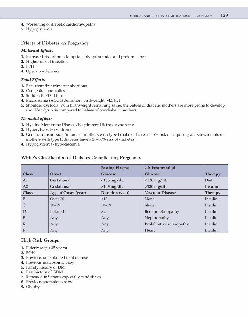
MEDICAL AND SURGICAL COMPLICATIONS IN PREGNANCY 129
4. Worsening of diabetic cardiomyopathy 5. Hypoglycemia
Effects of Diabetes on Pregnancy
Maternal Effects 1. Increased risk of preeclampsia, polyhydramnios and preterm labor 2. Higher risk of infection 3. PPH 4. Operative delivery
Fetal Effects 1. Recurrent first trimester abortions 2. Congenital anomalies 3. Sudden IUFD at term 4. Macrosomia (ACOG definition: birthweight >4.5 kg) 5. Shoulder dystocia. With birthweight remaining same, the babies of diabetic mothers are more prone to develop
shoulder dystocia compared to babies of nondiabetic mothers
Neonatal effects 1. Hyaline Membrane Disease/Respiratory Distress Syndrome 2. Hyperviscosity syndrome 3. Genetic transmission (infants of mothers with type I diabetes have a 4–5% risk of acquiring diabetes; infants of
mothers with type II diabetes have a 25–50% risk of diabetes) 4. Hypoglycemia/hypocalcemia
White’s Classification of Diabetes Complicating Pregnancy
Fasting Plasma 2-h Postprandial
Class Onset Glucose Glucose Therapy
A1
A2
Gestational
Gestational
<105 mg/dL
>105 mg/dL
<120 mg/dL
>120 mg/dL
Diet
Insulin
Class Age of Onset (year) Duration (year) Vascular Disease Therapy
B
C
D
F
R
F
Over 20
10–19
Before 10
Any
Any
Any
<10
10–19
>20
Any
Any
Any
None
None
Benign retinopathy
Nephropathy
Proliferative retinopathy
Heart
Insulin
Insulin
Insulin
Insulin
Insulin
Insulin
High-Risk Groups
1. Elderly (age >35 years) 2. BOH 3. Previous unexplained fetal demise 4. Previous macrosomic baby 5. Family history of DM 6. Past history of GDM 7. Repeated infections especially candidiasis 8. Previous anomalous baby 9. Obesity

130 SMART STUDY SERIES: OBSTETRICS AND GYNECOLOGY
O’Sullivan Blood Sugar Screening Test (Glucose Challenge Test)
• The ideal time to do this test is 24–28 weeks of gestation (as insulin resistance in pregnancy is maximum at 28 weeks of gestation).
• 50 g glucose is given irrespective of the period of fasting and plasma glucose is measured after 1 h. If it is >140 mg/dL it is an indication for further testing.
• RBS >200 mg/dL or FBS >125 mg/dL indicates overt DM and there is no need to do GTT • Glycosylated Hb (HbA1C): <8% = minimal risk of anomalies/abortions and >9% = poor glycemic control and
increased risk of anomalies/abortions
Confirmatory Tests
Glucose Tolerance Test (GTT) (upper limit of normal during pregnancy)
Glucose Load (g) FBS (mg/dL) 1 h (mg/dL) 2 h (mg/dL) 3 h (mg/dL)
WHOCarpenter/CoustanNational Diabetes DataGroup
75100100
>14095105
180190
>200155165
140145
NOTE: ACOG also recommends 100 g glucose load for GTT. If 2 or more values are abnormal, patient has gestational diabetes.
Congenital Malformations in Infants of Diabetic Mothers
MC anomaly = neural tube defects (anencephaly and spina bifida) followed by cardiac anomalies Most specific anomaly = caudal regression syndrome/sacral agenesis
1. Central Nervous System • Anencephaly and spina bifida • Encephalocele • Meningomyelocele and holoprosencephaly • Microcephaly
2. Cardiovascular • Transposition of the great vessels • Ventricular septal defect and atrial septal defect • Hypoplastic left ventricle • HOCM
NOTE: VSD is the MC cardiac anomaly, TGV is the most specific cardiac anomaly in infants of diabetic mothers.
3. Skeletal • Caudal regression syndrome (sacral agenesis)
4. Genitourinary • Absent kidneys • Polycystic kidneys • Double ureter
5. Gastrointestinal • Tracheoesophageal fistula • Bowel atresia • Imperforate anus
https://kat.cr/user/Blink99/
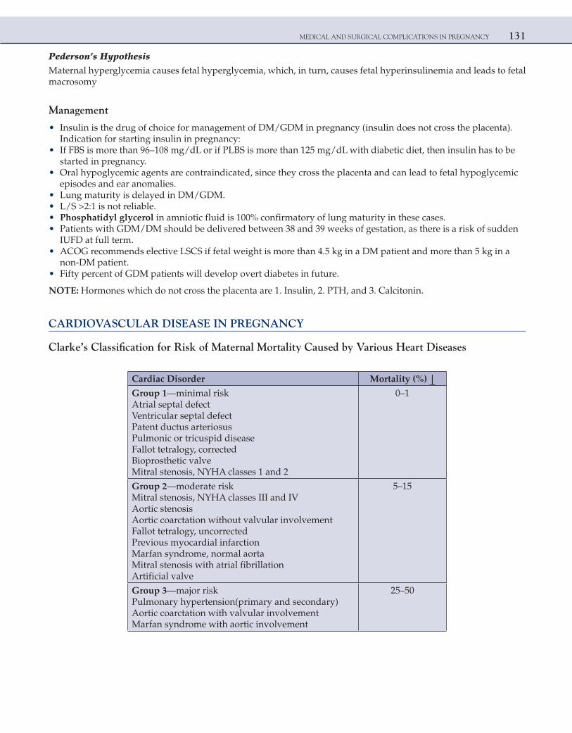
MEDICAL AND SURGICAL COMPLICATIONS IN PREGNANCY 131
Pederson’s HypothesisMaternal hyperglycemia causes fetal hyperglycemia, which, in turn, causes fetal hyperinsulinemia and leads to fetal macrosomy
Management
• Insulin is the drug of choice for management of DM/GDM in pregnancy (insulin does not cross the placenta). Indication for starting insulin in pregnancy:
• If FBS is more than 96–108 mg/dL or if PLBS is more than 125 mg/dL with diabetic diet, then insulin has to be started in pregnancy.
• Oral hypoglycemic agents are contraindicated, since they cross the placenta and can lead to fetal hypoglycemic episodes and ear anomalies.
• Lung maturity is delayed in DM/GDM. • L/S >2:1 is not reliable. • Phosphatidyl glycerol in amniotic fluid is 100% confirmatory of lung maturity in these cases. • Patients with GDM/DM should be delivered between 38 and 39 weeks of gestation, as there is a risk of sudden
IUFD at full term. • ACOG recommends elective LSCS if fetal weight is more than 4.5 kg in a DM patient and more than 5 kg in a
non-DM patient. • Fifty percent of GDM patients will develop overt diabetes in future.
NOTE: Hormones which do not cross the placenta are 1. Insulin, 2. PTH, and 3. Calcitonin.
CARDIOVASCULAR DISEASE IN PREGNANCY
Clarke’s Classification for Risk of Maternal Mortality Caused by Various Heart Diseases
Cardiac Disorder Mortality (%) |
Group 1—minimal riskAtrial septal defectVentricular septal defectPatent ductus arteriosusPulmonic or tricuspid diseaseFallot tetralogy, correctedBioprosthetic valveMitral stenosis, NYHA classes 1 and 2
0–1
Group 2—moderate riskMitral stenosis, NYHA classes III and IVAortic stenosisAortic coarctation without valvular involvementFallot tetralogy, uncorrectedPrevious myocardial infarctionMarfan syndrome, normal aortaMitral stenosis with atrial fibrillationArtificial valve
5–15
Group 3—major riskPulmonary hypertension(primary and secondary)Aortic coarctation with valvular involvementMarfan syndrome with aortic involvement
25–50
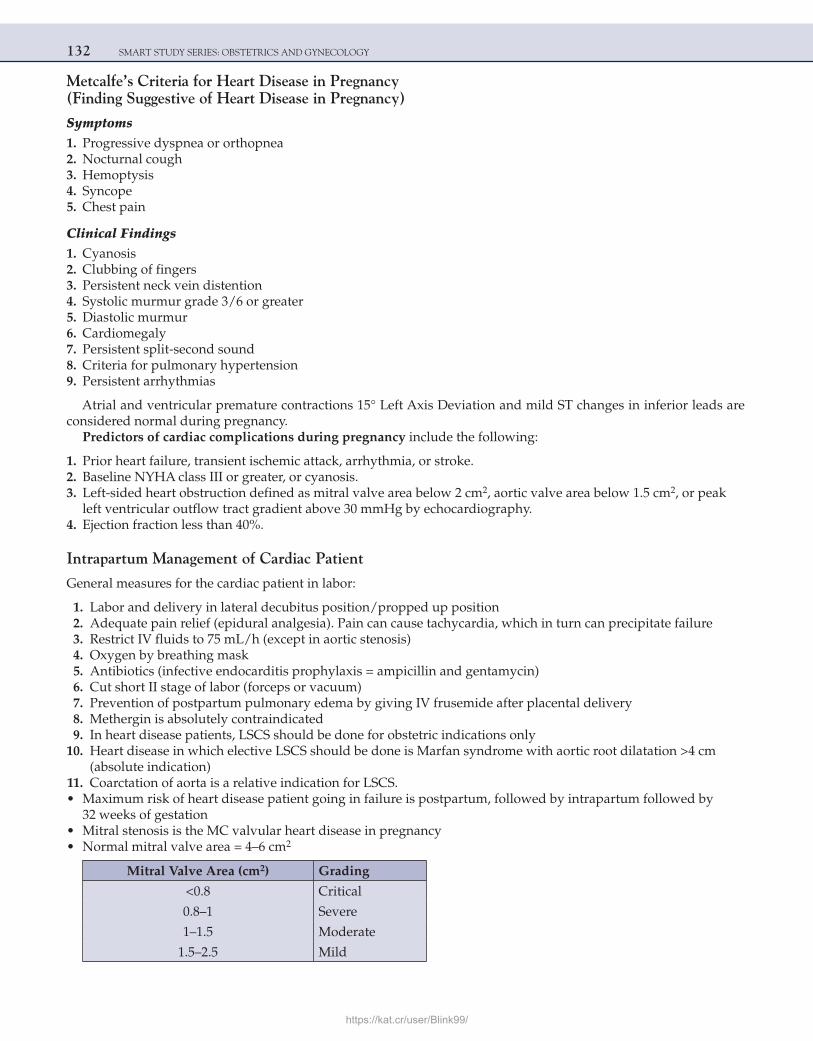
132 SMART STUDY SERIES: OBSTETRICS AND GYNECOLOGY
Metcalfe’s Criteria for Heart Disease in Pregnancy (Finding Suggestive of Heart Disease in Pregnancy)
Symptoms 1. Progressive dyspnea or orthopnea 2. Nocturnal cough 3. Hemoptysis 4. Syncope 5. Chest pain
Clinical Findings 1. Cyanosis 2. Clubbing of fingers 3. Persistent neck vein distention 4. Systolic murmur grade 3/6 or greater 5. Diastolic murmur 6. Cardiomegaly 7. Persistent split-second sound 8. Criteria for pulmonary hypertension 9. Persistent arrhythmias
Atrial and ventricular premature contractions 15° Left Axis Deviation and mild ST changes in inferior leads are considered normal during pregnancy.
Predictors of cardiac complications during pregnancy include the following:
1. Prior heart failure, transient ischemic attack, arrhythmia, or stroke. 2. Baseline NYHA class III or greater, or cyanosis. 3. Left-sided heart obstruction defined as mitral valve area below 2 cm2, aortic valve area below 1.5 cm2, or peak
left ventricular outflow tract gradient above 30 mmHg by echocardiography. 4. Ejection fraction less than 40%.
Intrapartum Management of Cardiac Patient
General measures for the cardiac patient in labor:
1. Labor and delivery in lateral decubitus position/propped up position 2. Adequate pain relief (epidural analgesia). Pain can cause tachycardia, which in turn can precipitate failure 3. Restrict IV fluids to 75 mL/h (except in aortic stenosis) 4. Oxygen by breathing mask 5. Antibiotics (infective endocarditis prophylaxis = ampicillin and gentamycin) 6. Cut short II stage of labor (forceps or vacuum) 7. Prevention of postpartum pulmonary edema by giving IV frusemide after placental delivery 8. Methergin is absolutely contraindicated 9. In heart disease patients, LSCS should be done for obstetric indications only 10. Heart disease in which elective LSCS should be done is Marfan syndrome with aortic root dilatation >4 cm
(absolute indication) 11. Coarctation of aorta is a relative indication for LSCS. • Maximum risk of heart disease patient going in failure is postpartum, followed by intrapartum followed by
32 weeks of gestation • Mitral stenosis is the MC valvular heart disease in pregnancy • Normal mitral valve area = 4–6 cm2
Mitral Valve Area (cm2) Grading
<0.8 Critical
0.8–1 Severe
1–1.5 Moderate
1.5–2.5 Mild
https://kat.cr/user/Blink99/
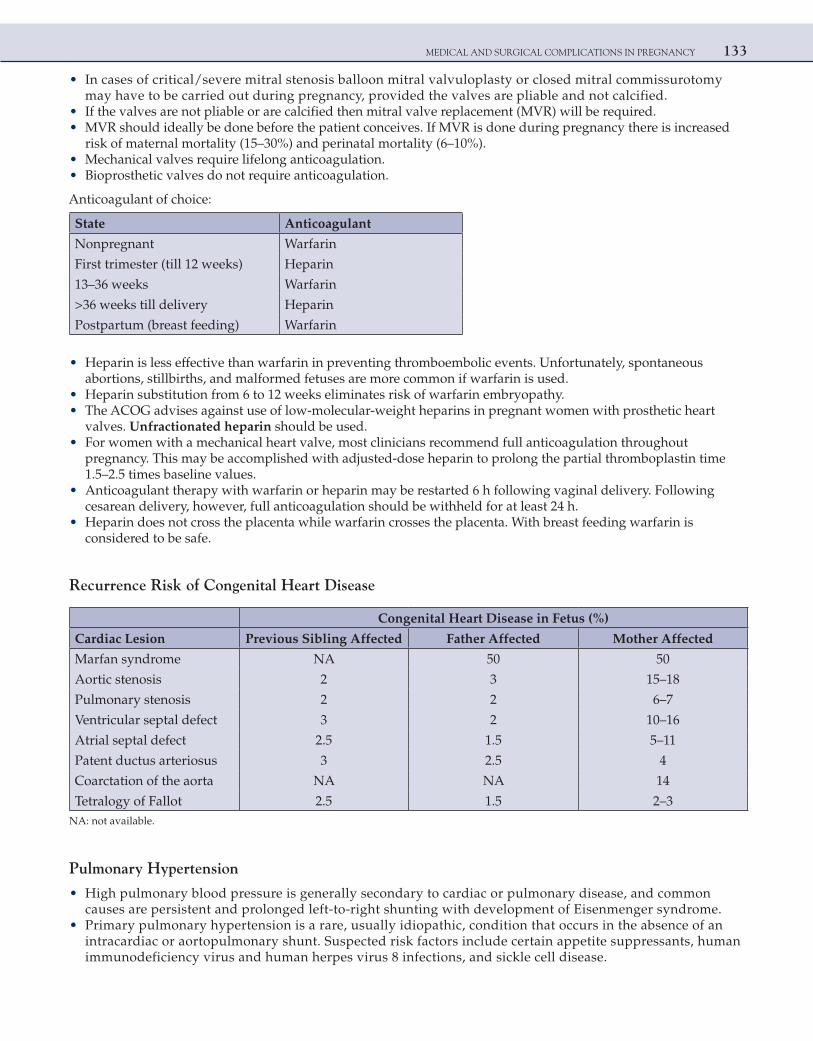
MEDICAL AND SURGICAL COMPLICATIONS IN PREGNANCY 133
• In cases of critical/severe mitral stenosis balloon mitral valvuloplasty or closed mitral commissurotomy may have to be carried out during pregnancy, provided the valves are pliable and not calcified.
• If the valves are not pliable or are calcified then mitral valve replacement (MVR) will be required. • MVR should ideally be done before the patient conceives. If MVR is done during pregnancy there is increased
risk of maternal mortality (15–30%) and perinatal mortality (6–10%). • Mechanical valves require lifelong anticoagulation. • Bioprosthetic valves do not require anticoagulation.
Anticoagulant of choice:
State Anticoagulant
Nonpregnant Warfarin
First trimester (till 12 weeks) Heparin
13–36 weeks Warfarin
>36 weeks till delivery Heparin
Postpartum (breast feeding) Warfarin
• Heparin is less effective than warfarin in preventing thromboembolic events. Unfortunately, spontaneous abortions, stillbirths, and malformed fetuses are more common if warfarin is used.
• Heparin substitution from 6 to 12 weeks eliminates risk of warfarin embryopathy. • The ACOG advises against use of low-molecular-weight heparins in pregnant women with prosthetic heart
valves. Unfractionated heparin should be used. • For women with a mechanical heart valve, most clinicians recommend full anticoagulation throughout
pregnancy. This may be accomplished with adjusted-dose heparin to prolong the partial thromboplastin time 1.5–2.5 times baseline values.
• Anticoagulant therapy with warfarin or heparin may be restarted 6 h following vaginal delivery. Following cesarean delivery, however, full anticoagulation should be withheld for at least 24 h.
• Heparin does not cross the placenta while warfarin crosses the placenta. With breast feeding warfarin is considered to be safe.
Recurrence Risk of Congenital Heart Disease
Congenital Heart Disease in Fetus (%)
Cardiac Lesion Previous Sibling Affected Father Affected Mother Affected
Marfan syndrome NA 50 50
Aortic stenosis 2 3 15–18
Pulmonary stenosis 2 2 6–7
Ventricular septal defect 3 2 10–16
Atrial septal defect 2.5 1.5 5–11
Patent ductus arteriosus 3 2.5 4
Coarctation of the aorta NA NA 14
Tetralogy of Fallot 2.5 1.5 2–3NA: not available.
Pulmonary Hypertension
• High pulmonary blood pressure is generally secondary to cardiac or pulmonary disease, and common causes are persistent and prolonged left-to-right shunting with development of Eisenmenger syndrome.
• Primary pulmonary hypertension is a rare, usually idiopathic, condition that occurs in the absence of an intracardiac or aortopulmonary shunt. Suspected risk factors include certain appetite suppressants, human immunodeficiency virus and human herpes virus 8 infections, and sickle cell disease.
134 SMART STUDY SERIES: OBSTETRICS AND GYNECOLOGY
• Some previously unexplained cases are now thought to be due to antiphospholipid antibodies. • The criteria for diagnosis established by National Institutes of Health Registry included a mean pulmonary
artery pressure of more than 25 mmHg at rest, or 30 mmHg with exertion, in the absence of heart disease, chronic thromboembolic disease, underlying pulmonary disorder, or other secondary causes.
• The prognosis is poor, and the mean survival from diagnosis is about 2 years. Long-term therapy with intravenous epoprostenol (prostacyclin) or with subcutaneous treprostinil, a prostacyclin analog, significantly lowers pulmonary vascular resistance.
UTI IN PREGNANCY
• The most common infecting organism is Escherichia coli (90%). • Asymptomatic bacteriuria: This refers to persistent, actively multiplying bacteria within the urinary tract in
women who have no symptoms. • A clean-voided specimen containing more than 100,000 organisms per milliliter is diagnostic. It may be prudent
to treat when lower concentrations are identified, because pyelonephritis develops in some women with colony counts of 20,000–50,000 organisms/mL.
• A single episode of asymptomatic bacteriuria can cause acute pyelonephritis in 25–40% cases. • Acute pyelonephritis can cause: a. IUGR b. Preterm labor c. Anemia d. Increased risk of PIH • Cranberry fruit juice is known to prevent recurrences of UTI. It prevents the adhesions of the pilins of E. coli to
uroepithelium. • Nitrofurantoin is the drug of choice for prophylaxis of recurrent UTI in pregnancy.
LIVER DISORDERS
Intrahepatic Cholestasis of Pregnancy (IHCP) = Icterus Gravidarum = Obstetric Cholestasis = Cholestatic Jaundice of Pregnancy = Obstetric Hepatosis
• 10–100-fold increase in bile acids (cholic/deoxycholic acids). • Pruritus is the most common presenting feature. • Onset is generally after 30 weeks of pregnancy. • Serum bilirubin rarely exceeds 5 mg/dL. • Serum transaminases are normal to moderately elevated (seldom exceeds 250 U/L). • Biopsy—centrilobular bile staining with bile plugs in canaliculi. • Complications: preterm labor, PPH, IUFD, and MSAF (meconium stained amniotic fluid). • Recurrences in future pregnancies is very common.
Treatment 1. Antihistamines and emollients 2. Cholestyramine 3. Vitamin K 4. Ursodeoxycholic acid is the drug of choice
Acute Fatty Liver of Pregnancy = Acute Metamorphosis = Acute Yellow Atrophy
• Abnormal fatty acid oxidation • LCHAD deficiency (long-chain hydroxyl acyl coenzyme A dehydrogenase)
https://kat.cr/user/Blink99/
MEDICAL AND SURGICAL COMPLICATIONS IN PREGNANCY 135
• Microvascular steatosis with periportal sparing • Greasy soft yellow liver • Hyperbilirubinemia is less than 10 mg/dL • Complications—hypoglycemia, hepatic encephalopathy, coagulopathy, renal failure, mortality (10–75%), and
increased risk of PIH • Decrease in fibrinogen and increase in ammonia and SGOT • Treatment—fresh frozen plasma, cryoprecipitate, platelets, and blood. Treat hepatic encephalopathy, deliver the
patient • Transient diabetes insipidus occurs during the period of recovery (due to elevated vasopressinase
concentration).
Viral Hepatitis
• Maximum risk of maternal mortality is with hepatitis E. • Maximum risk of hepatic encephalopathy is with hepatitis E. • Maximum risk of perinatal transmission is with hepatitis B. • Active and passive immunization, both are required for the newborn if the mother is HBsAg positive.
THYROID DISORDERS
• Moderate thyroid enlargement occurs in pregnancy due to glandular hyperplasia, and thyroid volume determined ultrasonographically increases, although its echostructure and echogenicity remain unchanged. Thyrotropin, or thyroid-stimulating hormone (TSH), currently plays a central role in screening and diagnosis of many thyroid disorders. In early pregnancy, thyrotropin activity decreases because of thyroid stimulation from the weak crossover activity of chorionic gonadotropin. The hormone does not cross the placenta. In the first 12 weeks, when chorionic gonadotropin levels are maximal, free thyroxine levels increase, and this suppresses thyrotropin levels.
• Thyroid-stimulating autoantibodies, also called thyroid-stimulating immunoglobulins, attach to the thyrotropin receptor and activate it, causing thyroid hyperfunction and growth. These antibodies are identified in the majority of patients with classic Graves’ disease.
• Thyroid peroxidase antibodies, previously called thyroid microsomal autoantibodies, have been identified in 10–20% of pregnant women. Up to half develop autoimmune thyroiditis that may be transient, but thyroid failure occurs in a significant number of women. These antibodies are also associated with miscarriage and Down syndrome.
• Graves’ disease is the MC cause of hyperthyroidism in pregnancy. • Hashimoto’s thyroiditis is the MC cause of hypothyroidism in pregnancy.
Complications associated with both hypo- and hyperthyroidism in pregnancy:
1. Recurrent first trimester abortions 2. IUGR 3. Increased risk of preeclampsia 4. Abruption 5. Stillbirths, hydrop fetalis 6. Preterm labor • Hypothyroidism is also associated with cretinism. • Propylthiouracil is the DOC for hyperthyroidism in pregnancy. • Methimazole and carbimazole used in early pregnancy have been associated with esophageal and choanal
atresia, aplasia cutis, and fetal agranulocytosis. • Labor and LSCS can precipitate thyroid storm. • Cord blood should be collected at the time of delivery for estimation of TSH, T3, T4 to detect neonatal thyroid
disorders.
NOTE: Fetal thyroid gland is able to synthesize hormones by 10–12 weeks of gestation.
136 SMART STUDY SERIES: OBSTETRICS AND GYNECOLOGY
Clinical Phases of Postpartum Thyroiditis
Postpartum Thyroiditis
Characteristics Thyrotoxicosis Hypothyroidism
Onset 1–4 months postpartum 4–8 months postpartum
Incidence 4% 2–5%
Mechanism Destruction-induced hormone release Thyroid insufficiency
Symptoms Small, painless goiter, fatigue, palpitations Goiter, fatigue, inability to concentrate
Treatment (3-Blockers for symptoms Thyroxine for 6–12 months
Sequelae Two-thirds become euthyroid One-third develop hypothyroidism
One-third permanent hypothyroidism
EPILEPSY IN PREGNANCY
• Epilepsy is the most common neurological disorder encountered in pregnancy. • The most common cause for epilepsy in pregnant women is idiopathic. • D/D’s of convulsions in pregnancy Eclampsia
Cerebral vein thrombosisCerebral infarctionHypoglycemia/hyponatremia/hypocalcemia
• Risk of congenital anomalies is about 4% in epileptic patients and there is 4–5% risk of epilepsy in the child if parents are affected.
• All anticonvulsant drugs are associated with congenital anomalies (6%—one drug; 15%—two or more drugs). Phenobarbitone is considered the safest in pregnancy.
• All women on anticonvulsants should take folic acid: 4 mg/day for 12 weeks preconception and throughout pregnancy.
• Prenatal screening: MSAFP + level II USG (watch for neural tube defects). • Therapeutic drug monitoring. • Vitamin K 10 mg/day orally from 36 weeks onward to prevent hemorrhagic disease of newborn. • Higher dose of estrogen required in OCPs if patient is on phenytoin, phenobarbitone and carbamazepine.
THROMBOEMBOLIC DISORDERS
Deep Vein Thrombosis
• In 1856, Virchow postulated the conditions that predispose to the development of venous thrombosis: (1) stasis, (2) local trauma to the vessel wall, and (3) hypercoagulability. The risk for each increases during normal pregnancy.
• The incidence of deep vein thrombosis is 1/1000 pregnancies. Fifty percent occur in the antepartum period and 50% in the postpartum period.
• Several independent risk factors are associated with the development of thromboembolism during pregnancy: a. Severe preeclampsia b. Cesarean delivery c. Diabetes d. Multifetal gestation e. Age 35 years or more f Obesity g. Smoking h. Dehydration i. Prolonged bed rest j. Prior thromboembolism
https://kat.cr/user/Blink99/
MEDICAL AND SURGICAL COMPLICATIONS IN PREGNANCY 137
Treatment of Deep Vein Thrombosis
1. IV unfractionated heparin for 5–7 days followed by subcutaneous heparin for the rest of pregnancy to maintain APTT 1.5–2.5 times control
2. Warfarin for 6–18 weeks in the postpartum period.
THROMBOPHILIAS
Thrombophilias
Congenital Acquired (antiphospholipid )emordnysydobitna
1. Factor V Leiden mutation 1. Anti-cardiolipin antibody
2. Prothrombin G20210A mutation 2. Lupus anticoagulant
3. Antithrombin deficiency
4. Protein C deficiency
5. Protein S deficiency
6. Hyperhomocysteinemia
Obstetric Complications of Thrombophilias (Congenital and Acquired)
1. Recurrent abortions 2. Severe preeclampsia 3. IUGR 4. Sudden unexplained IUFD 5. Abruption
Some Aspects of the More Common Congenital Thrombophilias
Thrombophilia Genetics Prevalence (%)Increased Relative Risk of Venous Thromboembolism
Factor V Leiden mutation (most common) AD 2–15 3–8-fold
Prothrombin G20210A mutation AD 2–3 3-fold
Antithrombin deficiency (most thrombogenic) AD 0.02 25–50-fold
Protein C deficiency AD 0.2–0.3 10–15-fold
Protein S deficiency AD 0.1–2.1 2-fold
Hyperhomocysteinemia AR 11 2.5-fold (if >18.5 μmol/L) 3–4-fold (if >20 μmol/L)
The anticardiolipin antibodies and the lupus anticoagulant bind to “annexin V” and “beta 2 microglobulin,” which are naturally occurring anticoagulants present in our body. This leads to decrease in levels of free “annexin V” and “beta 2 microglobulin,” leading to thrombosis.
Treatment of Thrombophilias (Acquired and Congenital)
Treatment includes low-dose aspirin as soon as the pregnancy is confirmed and injection heparin when the cardiac activity is confirmed on USG. Aspirin is to be omitted at 34 weeks and heparin is stopped 24 h before planned deliv-ery (induction) or LSCS.
138 SMART STUDY SERIES: OBSTETRICS AND GYNECOLOGY
PULMONARY DISORDERS
Pneumonia
• Many bacteria that cause community-acquired pneumonia, such as Streptococcus pneumoniae, are part of the normal resident flora.
• A number of factors can upset the symbiotic relationship between colonizing bacteria and mucosal phagocytic defenses. Examples include acquisition of a virulent and invasive strain or bacterial infection following a viral infection.
• Cigarette smoking and chronic bronchitis favor colonization with S. pneumoniae, Haemophilus influenzae, and Legionella.
• Other risk factors include asthma, binge drinking, smoking, and HIV infection.
Factors That Increase the Risk of Death or Complications with Community-Acquired Pneumonia
1. Coexisting chronic conditions 2. Clinical findings: Respiratory rate ≥30/min, hypotension, pulse ≥125 bpm, hypothermia (<35°C), temperature
>40°C, altered mental status, and extrapulmonary disease 3. Laboratory findings: Leukopenia (<4000/μL) or leukocytosis >30,000/μL; Po2 ≤60 mmHg or CO2 retention
while breathing room air; elevated serum creatinine; anemia; or evidence of sepsis or organ dysfunction such as acidosis or coagulopathy
4. Radiological findings: More than one-lobe involvement, cavitation, or pleural effusion • Antimicrobial treatment is empirical. Because the majority of adult pneumonias are caused by pneumococci,
mycoplasma, or chlamydia; therapy with erythromycin or one of its newer analogs is given. • In whom staphylococcal or Hemophilus pneumonia is suspected, cefotaxime or ceftriaxone is given in addition to
erythromycin therapy.
ASTHMA
Clinical Stages of Asthma
Stage Po2 Pco2 pH FEV1 (%) predicted
Mild respiratory alkalosis Normal ↓ ↑ 65–80
Respiratory alkalosis ↓ ↓ ↑ 50–64
Danger zone ↓ Normal Normal 35–49
Respiratory acidosis ↓ ↑ ↓ <35
Step Therapy of Chronic Asthma During Pregnancy
Severity Step Therapy
Mild intermittent Inhaled β-agonists—salbutamol, metaproterenol, isoproterenol, salmeterol
Mild persistent Inhaled β-agonists—as aboveInhaled cromolyn—continue if taking prior to pregnancy with good responseSubstitute inhaled corticosteroids if no response—beclomethasone, budesonide, triamcinolone
Moderate persistent
Inhaled β-agonists as aboveInhaled corticosteroids as above, add oral theophylline and/or inhaled salmeterol if inhaledmedium-dose steroids inadequate
Severe persistent For moderate, as above plus oral corticosteroids—burst for active symptoms, alternate day ordaily if necessary
PGF2 alpha is absolutely contraindicated, PGE1 or PGE2 can be used with caution
https://kat.cr/user/Blink99/
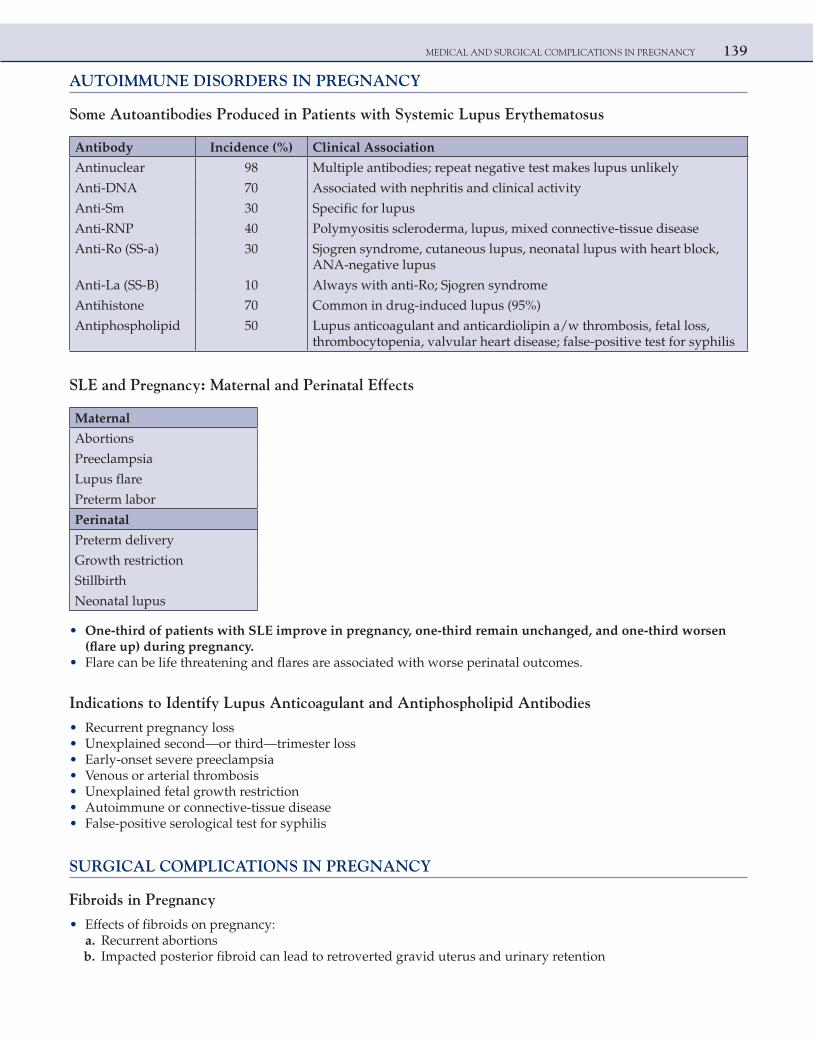
MEDICAL AND SURGICAL COMPLICATIONS IN PREGNANCY 139
AUTOIMMUNE DISORDERS IN PREGNANCY
Some Autoantibodies Produced in Patients with Systemic Lupus Erythematosus
Antibody Incidence (%) Clinical Association
Antinuclear 98 Multiple antibodies; repeat negative test makes lupus unlikely
Anti-DNA 70 Associated with nephritis and clinical activity
Anti-Sm 30 Specific for lupus
Anti-RNP 40 Polymyositis scleroderma, lupus, mixed connective-tissue disease
Anti-Ro (SS-a) 30 Sjogren syndrome, cutaneous lupus, neonatal lupus with heart block, ANA-negative lupus
Anti-La (SS-B) 10 Always with anti-Ro; Sjogren syndrome
Antihistone 70 Common in drug-induced lupus (95%)
Antiphospholipid 50 Lupus anticoagulant and anticardiolipin a/w thrombosis, fetal loss, thrombocytopenia, valvular heart disease; false-positive test for syphilis
SLE and Pregnancy: Maternal and Perinatal Effects
Maternal
Abortions
Preeclampsia
Lupus flare
Preterm labor
Perinatal
Preterm delivery
Growth restriction
Stillbirth
Neonatal lupus
• One-third of patients with SLE improve in pregnancy, one-third remain unchanged, and one-third worsen (flare up) during pregnancy.
• Flare can be life threatening and flares are associated with worse perinatal outcomes.
Indications to Identify Lupus Anticoagulant and Antiphospholipid Antibodies
• Recurrent pregnancy loss • Unexplained second—or third—trimester loss • Early-onset severe preeclampsia • Venous or arterial thrombosis • Unexplained fetal growth restriction • Autoimmune or connective-tissue disease • False-positive serological test for syphilis
SURGICAL COMPLICATIONS IN PREGNANCY
Fibroids in Pregnancy
• Effects of fibroids on pregnancy: a. Recurrent abortions b. Impacted posterior fibroid can lead to retroverted gravid uterus and urinary retention
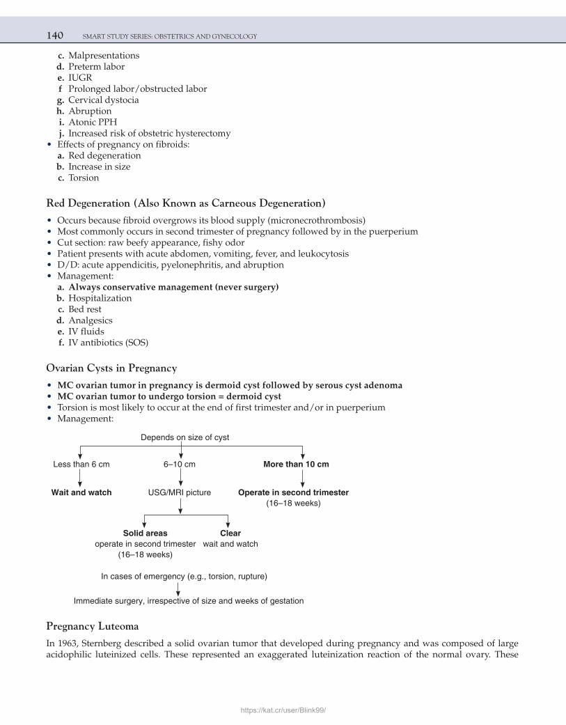
140 SMART STUDY SERIES: OBSTETRICS AND GYNECOLOGY
c. Malpresentations d. Preterm labor e. IUGR f Prolonged labor/obstructed labor g. Cervical dystocia h. Abruption i. Atonic PPH j. Increased risk of obstetric hysterectomy • Effects of pregnancy on fibroids: a. Red degeneration b. Increase in size c. Torsion
Red Degeneration (Also Known as Carneous Degeneration)
• Occurs because fibroid overgrows its blood supply (micronecrothrombosis) • Most commonly occurs in second trimester of pregnancy followed by in the puerperium • Cut section: raw beefy appearance, fishy odor • Patient presents with acute abdomen, vomiting, fever, and leukocytosis • D/D: acute appendicitis, pyelonephritis, and abruption • Management: a. Always conservative management (never surgery) b. Hospitalization c. Bed rest d. Analgesics e. IV fluids f. IV antibiotics (SOS)
Ovarian Cysts in Pregnancy
• MC ovarian tumor in pregnancy is dermoid cyst followed by serous cyst adenoma • MC ovarian tumor to undergo torsion = dermoid cyst • Torsion is most likely to occur at the end of first trimester and/or in puerperium • Management:
Depends on size of cyst
Less than 6 cm 6–10 cm More than 10 cm
Wait and watch USG/MRI picture Operate in second trimester(16–18 weeks)
Solid areas Clear operate in second trimester wait and watch (16–18 weeks)
In cases of emergency (e.g., torsion, rupture)
Immediate surgery, irrespective of size and weeks of gestation
Pregnancy Luteoma
In 1963, Sternberg described a solid ovarian tumor that developed during pregnancy and was composed of large acidophilic luteinized cells. These represented an exaggerated luteinization reaction of the normal ovary. These
https://kat.cr/user/Blink99/
MEDICAL AND SURGICAL COMPLICATIONS IN PREGNANCY 141
so-called luteomas of pregnancy are variable in size, ranging from microscopic to over 20 cm in diameter. Although luteomas regress after delivery, they may recur in subsequent pregnancies. Pregnancy luteomas may result in mater-nal virilization, but usually the female fetus is not affected.
Theca-Lutein Cysts
These benign ovarian lesions result from exaggeration by physiological follicle stimulation, which is termed hyper-reactio luteinalis. The reaction is associated with markedly elevated serum levels of hCG.
Acute Appendicitis
• There is no increase in incidence in pregnancy, but mortality is higher. • During pregnancy the cecum and appendix are displaced upward and to the right. Therefore, classical right iliac
fossa pain may not be present. • Rebound tenderness is less obvious. • Therefore, diagnosis is delayed and perforation rates are higher. • Treatment of acute appendicitis in pregnancy = emergency surgery (appendectomy irrespective of weeks of
gestation).
MULTIPLE CHOICE QUESTIONS
1. In pregnant female with prosthetic valves, which of the following is given for the prevention of thrombosis? a. LMW heparin b. Unfractionated heparin c. Aspirin d. No anticoagulants are required in women with metallic valves
Answer: b (Unfractionated heparin)
Explanation:Unfractionated heparin is a group of large (molecular weight 4000–30,000), highly polar molecules that do not cross the
placenta and are not associated with congenital anomalies. It may be given intravenously, either continuously or intermit-tently, or by subcutaneous injection.
ACOG recommends the use unfractionated heparin for thromboprophylaxis in patients with artificial metallic valves.
Its protracted use may cause maternal osteopenia, osteoporosis, and thrombocytopenia.The safety and efficacy of low-molecular-weight (LMW) heparin use during pregnancy have not been adequately evalu-
ated. ACOG does not recommend the use of LMW heparin in pregnancy.No anticoagulants are required in women with bioprosthetic valves.
Reference: 1. Williams, 22nd Ed., Pg. 1022–3.
2. Tablets supplied by Government of India contain the following amount of iron and folic acid (FA): a. 60 mg elemental iron + 500 μg FA b. 100 mg elemental iron + 500 μg FA c. 200 mg elemental iron + 1 mg FA d. 100 mg elemental iron + 5 mg FA
Answer: b (100 mg elemental iron + 500 μg FA)
Explanation:The National Nutritional Anemia Prevention Program (NNAPP) was launched by the Government of India in 1970 to con-
trol iron deficiency anemia in vulnerable groups (such as pregnant women) through daily supplements of iron and folic acid tablets. The suggested prophylactic doses were 60 mg of elemental iron and 500 μg of folic acid for pregnant women. These
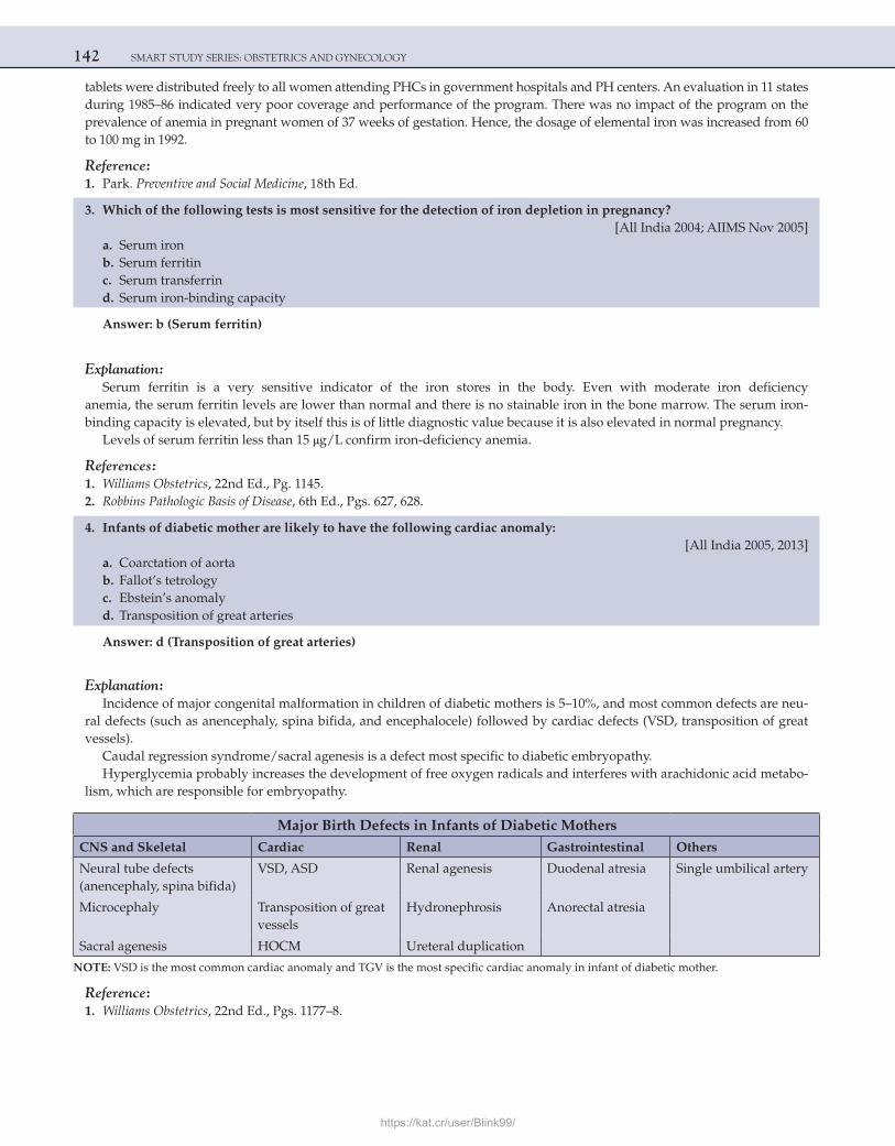
142 SMART STUDY SERIES: OBSTETRICS AND GYNECOLOGY
tablets were distributed freely to all women attending PHCs in government hospitals and PH centers. An evaluation in 11 states during 1985–86 indicated very poor coverage and performance of the program. There was no impact of the program on the prevalence of anemia in pregnant women of 37 weeks of gestation. Hence, the dosage of elemental iron was increased from 60 to 100 mg in 1992.
Reference: 1. Park. Preventive and Social Medicine, 18th Ed.
3. Which of the following tests is most sensitive for the detection of iron depletion in pregnancy? [All India 2004; AIIMS Nov 2005] a. Serum iron b. Serum ferritin c. Serum transferrin d. Serum iron-binding capacity
Answer: b (Serum ferritin)
Explanation:Serum ferritin is a very sensitive indicator of the iron stores in the body. Even with moderate iron deficiency
anemia, the serum ferritin levels are lower than normal and there is no stainable iron in the bone marrow. The serum iron-binding capacity is elevated, but by itself this is of little diagnostic value because it is also elevated in normal pregnancy.
Levels of serum ferritin less than 15 μg/L confirm iron-deficiency anemia.
References: 1. Williams Obstetrics, 22nd Ed., Pg. 1145. 2. Robbins Pathologic Basis of Disease, 6th Ed., Pgs. 627, 628.
4. Infants of diabetic mother are likely to have the following cardiac anomaly: [All India 2005, 2013] a. Coarctation of aorta b. Fallot’s tetrology c. Ebstein’s anomaly d. Transposition of great arteries
Answer: d (Transposition of great arteries)
Explanation:Incidence of major congenital malformation in children of diabetic mothers is 5–10%, and most common defects are neu-
ral defects (such as anencephaly, spina bifida, and encephalocele) followed by cardiac defects (VSD, transposition of great vessels).
Caudal regression syndrome/sacral agenesis is a defect most specific to diabetic embryopathy.Hyperglycemia probably increases the development of free oxygen radicals and interferes with arachidonic acid metabo-
lism, which are responsible for embryopathy.
Major Birth Defects in Infants of Diabetic MothersCNS and Skeletal Cardiac Renal Gastrointestinal Others
Neural tube defects (anencephaly, spina bifida)
VSD, ASD Renal agenesis Duodenal atresia Single umbilical artery
Microcephaly Transposition of great vessels
Hydronephrosis Anorectal atresia
Sacral agenesis HOCM Ureteral duplication
NOTE: VSD is the most common cardiac anomaly and TGV is the most specific cardiac anomaly in infant of diabetic mother.
Reference: 1. Williams Obstetrics, 22nd Ed., Pgs. 1177–8.
https://kat.cr/user/Blink99/
MEDICAL AND SURGICAL COMPLICATIONS IN PREGNANCY 143
5. A 30-year-old class D diabetic is concerned about pregnancy. She can be assured that one of the following risks is the same for her as for the general population. Which one is that risk?
a. Preeclampsia b. Infection c. Fetal cystic fibrosis d. Postpartum hemorrhage
Answer: c (Fetal cystic fibrosis)
Explanation:Maternal diabetes mellitus can affect a pregnant woman and her fetus in many ways. The development of preeclampsia or
eclampsia is about four times as likely as among nondiabetic women.Infection is also more likely not only to occur but also to be severe.The incidences of fetal macrosomia or death and of dystocia are increased, and hydramnios is common. The likelihood of
postpartum hemorrhage after vaginal delivery and the frequency of cesarean section are both increased in diabetic women.The incidence of fetal genetic disorders such as cystic fibrosis is unaffected by diabetes.
Reference:
1. Williams, 22nd Ed., Pg. 1180.
6. A 35-year-old primigravida presents at 8 weeks of gestation. She has a history of type I diabetes and is very concerned regarding the possible risks this may have on her fetus. You recommend that the patient undergo all of the following tests because of her diabetes, except:
a. Maternal serum AFP test at about 16–18 weeks b. Fetal echocardiography at 20 weeks c. Twenty-four-hour urine study d. Fetal surveillance with contraction stress tests starting at 28 weeks
Answer: d (Fetal surveillance with contraction stress tests starting at 28 weeks)
Explanation:Fetuses of women with overt diabetes are at increased risk of having spina bifida; therefore, patients should be counseled
appropriately regarding obtaining a test for maternal serum α-fetoprotein to screen for neural tube defects. Fetal echocardiog-raphy is recommended because infants of diabetic mothers have an increased risk of heart anomalies, including transposition of the great vessels, ventricular septal defects, and atrial septal defects. Performance of serial 24-h urine samples will docu-ment absence of nephropathy by measuring protein and creatinine clearance.
In the third trimester, ultrasounds should be performed to evaluate both excessive and insufficient fetal growth as well as amniotic fluid levels.
Beginning at 32–34 weeks of gestation, a program of weekly or twice-weekly fetal surveillance is usually commenced to document fetal well-being. Testing protocols utilize NST and biophysical profiles.
Since contraction stress testing involves using oxytocin to cause uterine contractions, this is not usually used as a first-line surveillance test. It is almost outdated in modern-day obstetrics.
Reference:
1. Willams, 22nd Ed., Pg. 1180–2.
7. The drug of choice for the treatment of thyrotoxicosis during pregnancy is: [All India 2009] a. Carbimazole b. Iodine therapy c. Propylthiouracil d. Methimazole
Answer: c (Propylthiouracil)
Explanation:In a case of thyrotoxicosis during pregnancy, l131 is contraindicated.
144 SMART STUDY SERIES: OBSTETRICS AND GYNECOLOGY
Propylthiouracil is the drug of choice for thyrotoxicosis in pregnancy. It is highly protein bound and therefore less amount of drug is transferred across placenta and in milk.
With carbimazole and methimazole there is risk of fetal hypothyroidism, aplasia cutis, and fetal agranulocytosis.
References:
1. Tripathi KD. Pharmacology, 4th Ed., Pg. 260. 2. Williams, 22nd Ed. Pg. 1192.
8. Which of the following statements is incorrect in relation to pregnant women with epilepsy? [All India 2005] a. The rate of congenital malformation is increased in the offspring of women with epilepsy b. Seizure frequency increases in approximately 70% of women c. Breast feeding is safe with most anticonvulsants d. Folic acid supplementation may reduce the risk of neural tube defect
Answer: b (Seizure frequency increases in approximately 70% of women)
Explanation:Epilepsy is the most common neurological disorder in pregnancy. There is no change in seizure frequency in 60–85%
patients.Women with epilepsy have an increased risk of congenital anomalies in the fetus over and above the risk that is due to the
anticonvulsant medications (epilepsy itself increases the risk even if the patient is not on any anticonvulsant medications).There is a two-fold increased risk of malformed fetus in an epileptic patient on anticonvulsant medication; the risk is primi-
done > valproate > phenytoin > carbamazepine > phenobarbitone.The embryotoxicity of these medications is due to their intermediate metabolites and is genetically mediated. There is defect
in detoxification. All these drugs commonly alter CNS of the fetus leading to NTDS, and folic acid is known to be protective.The drug concentration in breast milk is minimal, and hence breast feeding is quite safe.
Reference:
1. Williams, 22nd Ed., Pg. 1232–3.
9. A 25-year-old primigravida with 20 weeks of pregnancy has a first episode of asymptomatic bacteriuria. The risk of having pyelonephritis is:
a. No risk with first episode b. 5% c. 15% d. 25%
Answer: d (25%)
Explanation:Asymptomatic bacteriuria is when bacterial count of the same species over 105/mL in midstream clean catch specimen of
urine is detected without symptoms of urinary infection.Twenty-five percent of pregnant women with asymptomatic bacteriuria are likely to develop acute pyelonephritis if left
untreated.
Reference:
1. Williams, 22nd Ed., Pg. 1095.
10. Which of the following is not an indication for antiphospholipid antibody testing? [All India 2004] a. Three or more consecutive first trimester pregnancy losses b. Unexplained cerebrovascular accidents c. Early onset severe preeclampsia d. Gestational diabetes
Answer: d (Gestational diabetes)
Explanation:Antiphospholipid antibodies including lupus anticoagulant (LA) and anticardiolipin antibodies (ACL).
https://kat.cr/user/Blink99/

MEDICAL AND SURGICAL COMPLICATIONS IN PREGNANCY 145
The antiphospholipid antibody syndrome is characterized by recurrent arterial and/or venous thrombosis, thrombocyto-penia, and fetal loss—especially stillbirths, during the second half of pregnancy.
Pathological changes seen are placental vascular atherosis, intervillus thrombosis, and decidual vasculopathy with fibri-noid necrosis leading to inadequate blood supply to fetus.
Indications to identify lupus anticoagulant and ACL:
1. Recurrent pregnancy loss (first trimester abortions) 2. Unexplained second—or third—trimester loss 3. Early-onset severe preeclampsia 4. Venous or arterial thrombosis 5. Unexplained fetal growth restriction 6. Autoimmune or connective-tissue disease 7. False-positive serological test for syphilis
aPTT and diluted Russel viper venom test (dRVVT) are done to identify LA (both are prolonged). Out of the two dRVVT is the best.NOTE: Treatment of APLA syndrome is low dose aspirin and heparin. (All India 2010)
Reference:
1. Williams Obstetrics, 22nd Ed., Pg. 1217.
11. A 32-year-old primigravida reports for a routine visit at 14 weeks of gestational age. Blood drawn at her first prenatal visit 4 weeks ago reveal a platelet count of 60,000. During the present visit, the patient has a blood pressure of 120/70. Her urine reveals absence of proteins. The patient denies any complaints. On taking a more in-depth history you learn that, prior to pregnancy, your patient had a history of occasional nose and gum bleeds, but no serious bleeding episodes. Most likely diagnosis is:
a. Gestational thrombocytopenia b. Immune thrombocytopenic purpura c. HELLP syndrome d. Any of the above
Answer: b (Immune thrombocytopenic purpura)
Explanation:Immune thrombocytopenic purpura (ITP) typically occurs in the second or third decade of life and is more common in
women than in men. The diagnosis of ITP is one of exclusion, because there are no pathognomonic signs, symptoms, or diag-nostic tests. Traditionally, ITP is associated with a persistent platelet count of less than 100,000 in the absence of splenomegaly. Most women have a history of easy bruising and nose and gum bleeds that precede pregnancy. If the platelet count is main-tained above 20,000, hemorrhagic episodes rarely occur. In cases of ITP, the patient produces IgG antiplatelet antibodies that increase platelet consumption in the spleen and in other sites.
Gestational thrombocytopenia occurs in up to 8% of pregnancies. Affected women are usually asymptomatic, have no prior history of bleeding, and usually maintain platelet counts above 70,000. The cause of gestational thrombocytopenia has not been clearly elucidated. Antiplatelet antibodies are often detected in women with gestational thrombocytopenia.
HELLP syndrome of severe preeclampsia is associated with thrombocytopenia, but this condition generally occurs in the third trimester and is associated with hypertension and proteinuria.
Reference:
1. Williams, 22nd Ed., Pg. 1157.
12. Which of the following statements concerning hepatitis infection in pregnancy is true? [AIIMS Nov 2001] a. Hepatitis B core antigen status is the most sensitive indicator of positive vertical transmission of disease b. Hepatitis B is the most common form of hepatitis after blood transfusion c. The proper treatment of infants born to infected mothers includes the administration of hepatitis B immune globulin
as well as vaccine d. Patients who develop chronic active hepatitis should undergo MTP
Answer: c (The proper treatment of infants born to infected mothers includes the administration of hepatitis B immune globulin as well as vaccine)
146 SMART STUDY SERIES: OBSTETRICS AND GYNECOLOGY
Explanation:Persons at increased risk of hepatitis B infection include homosexuals, abusers of intravenous drugs, health-care person-
nel, and people who have received blood or blood products.However, because of intensive screening of blood for type B hepatitis, hepatitis C has become the major form of hepatitis
after blood transfusion. Venereal transmission and the sharing of needles in persons who abuse intravenous drugs have played major roles in the transmission of hepatitis B. A variety of immunologic markers exist to identify patients who have active disease, are chronic carriers of disease, or have antibody protection.
Among the markers, the e antigen is very similar to the virus and is an indicator of the infectious state. Mothers who are e antigen-positive are more likely to transmit the disease to their infants, whereas the absence of the e antigen in the presence of anti-E antibody appears to be protective. The proper treatment of infants born to infected mothers includes the administra-tion of hepatitis B immune globulin as well as vaccine.
Chronic acute hepatitis does not necessarily warrant therapeutic abortion. Fertility is decreased, but pregnancy may pro-ceed on a normal course as long as steroid therapy is continued. Prematurity and fetal loss are increased, but there is no increase in malformations.
NOTE:Maximum risk of maternal mortality is with hepatitis E. Maximum risk of hepatic encephalopathy is with hepatitis E.
Maximum risk of perinatal transmission is with hepatitis B.
Reference:
1. Williams, 22nd Ed., Pgs. 1130–1.
13. Which one of the following perinatal infections has the highest risk of fetal infection in the first trimester? [All India 2004] a. Hepatitis B virus b. Syphilis c. Toxoplasmosis d. Rubella
Answer: d (Rubella)
Explanation:Rubella is one of the most teratogenic agents known. Eighty percent of women with rubella infection and a history of rash dur-
ing the first 12 weeks have a fetus with congenital infection. At 13–14 weeks, the incidence is about 54%, and it is 25% by the end of the second trimester. As the duration of pregnancy increases, fetal infections are less likely to cause congenital malformations.
In order to prevent rubella during pregnancy and congenital rubella syndrome, ACOG recommends that the MMR vaccine should be offered to women of childbearing age who do not have evidence of immunity whenever they make contact with the health-care system. Vaccination of susceptible women should
1. Be part of routine general medical and gynecological care, including college health services. 2. Take place in all family planning settings. 3. Be provided routinely to unimmunized women immediately after hospitalization, childbirth, or abortion, unless there
are specific contraindications.
Vaccination of all susceptible hospital personnel who might be exposed to patients with rubella or who might have contact with pregnant women is recommended. Rubella vaccination should be avoided 1 month before or during pregnancy because the vaccine contains attenuated live virus.
In toxoplasmosis, the incidence and severity of congenital infection depend on the gestational age of the fetus at the time of maternal primary infection. Infection increases with duration of pregnancy, with the risk the fetal infection rising from 6% at 13 weeks to 72% at 36 weeks. Fetal infection is more virulent the earlier the infection is acquired.
Treatment of pregnant women is thought to prevent and reduce, but not eliminate, the risk of congenital infection. Spira-mycin, used alone, is thought to reduce the risk of congenital infection but not to treat established fetal infection.
When fetal infection in diagnosed by prenatal testing, pyrimethamine, sulfonamides and folinic acid are added to spira-mycin to eradicate parasites in the placenta and fetus.
Transmission of syphilis from a syphilitic mother to her fetus across the placenta may occur at any stage of pregnancy, but the lesions of congenital syphilis generally develop after the fourth month of gestation, when fetal immunologic competence begins to develop.
https://kat.cr/user/Blink99/
MEDICAL AND SURGICAL COMPLICATIONS IN PREGNANCY 147
Perinatal transmission of hepatitis B occurs primarily in infants born to HbsAg carrier mothers or mothers with acute hep-atitis B during the third trimester of pregnancy or during the early postpartum period. Most infections occur approximately at the time of delivery and are not related to breast feeding.
Reference:
1. Williams, 22nd Ed., Pg. 1281–3.
14. In HIV with pregnancy, which drug is given to mother during labor to prevent HIV transmission to the new born?
[AIIMS Nov 2011] a. Lamivudine b. Stavudine c. Nevirapine d. Efavirenz
Answer: c (Nevirapine)
Explanation:Single-dose nevirapineThe simplest of all prevention of parent-to-child transmission (PPTCT) drug regimens was tested in the HIVNET 012
trial, which was conducted in Uganda between 1997 and 1999. This study found that a single dose of nevirapine given to the mother at the onset of labor and to the new born baby after delivery roughly halved the rate of transmission of HIV.
Other treatments require women to take drugs during pregnancy as well as during labor and delivery. So they are much more expensive and more difficult to implement than nevirapine. So, single-dose nevirapine remains the practical choice for PPTCT of HIV in areas with minimal medical resources.
Reference:
1. Williams, 22nd Ed., Pgs. 1314–5.
15. As per CDC, screening for HIV in pregnancy should be: [AIIMS May 2009] a. Opt-out testing b. Opt-in testing c. Universal testing d. Symptomatic
Answer: a (Opt-out testing)
Explanation:Controversial options such as the CDC guidelines recommend universal opt-out screening, but opt-out screening is a bet-
ter option to mark as explained below.Definitions:Diagnostic testing: Performing an HIV test for persons with clinical signs or symptoms consistent with HIV infection.Screening: Performing an HIV test for all persons in a defined population.Targeted testing: Performing an HIV test for subpopulations of persons at higher risk, typically defined on the basis of
behavior, clinical, or demographic characteristics.Informed consent: A process of communication between patient and provider through which an informed patient can
choose whether to undergo HIV testing or decline to do so. Elements of informed consent typically include providing oral or written information regarding HIV, the risks and benefits of testing, the implications of HIV test results, how test results will be communicated, and the opportunity to ask questions.
Opt-out screening: Performing HIV screening after notifying the patient that (1) the test will be performed and (2) the patient may elect to decline or defer testing. Assent is inferred unless the patient declines testing.
HIV-prevention counseling: An interactive process of assessing risk, recognizing specific behaviors that increase the risk for acquiring or transmitting HIV, and developing a plan to take specific steps to reduce risks.
148 SMART STUDY SERIES: OBSTETRICS AND GYNECOLOGY
CDC guidelines are as follows:HIV screening for pregnant women and their infants Universal opt-out screening:
• All pregnant women should be screened for HIV infection. • Screening should occur after a woman is notified that HIV screening is recommended for all pregnant patients
and that she will receive an HIV test as part of the routine panel of prenatal tests unless she declines (opt-out screening).
• HIV testing must be voluntary and free from coercion. No woman should be tested without her knowledge. • Pregnant women should receive oral or written information that includes an explanation of HIV infection, a description
of interventions that can reduce HIV transmission from mother to infant, and the meanings of positive and negative test results, and should be offered an opportunity to ask questions and to decline testing.
• No additional process or written documentation of informed consent beyond what is required for other routine prenatal tests should be required for HIV testing.
• If a patient declines an HIV test, this decision should be documented in the medical record.
Addressing reasons for declining testing
• Providers should discuss and address reasons for declining an HIV test (e.g., lack of perceived risk, fear of the disease, and concerns regarding partner violence or potential stigma or discrimination).
• Women who decline an HIV test because they have had a previous negative test result should be informed of the importance of retesting during each pregnancy.
• Logistical reasons for not testing (e.g., scheduling) should be resolved. • Certain women who initially decline an HIV test might accept it at a later date, especially if their concerns are discussed.
Certain women will continue to decline testing, and their decisions should be respected and documented in the medical record.
Timing of HIV testing
• To promote informed and timely therapeutic decisions, health-care providers should test women for HIV as early as possible during each pregnancy. Women who decline the test early in prenatal care should be encouraged to be tested at a subsequent visit.
Rapid testing during labor
• Any woman with undocumented HIV status at the time of labor should be screened with a rapid HIV test unless she declines (opt-out screening).
• Reasons for declining a rapid test should be explored. • Immediate initiation of appropriate antiretroviral prophylaxis should be recommended to women on the basis of a
reactive rapid test result without waiting for the result of a confirmatory test.
Reference:
1. CDC Guidelines for HIV.
16. Misoprostol has been found to be effective in all of the following, except: [All India 2005] a. Medical method of abortion b. Induction of labor c. Menorrhagia d. Prevention of postpartum hemorrhage (PPH)
Answer: c (Menorrhagia)
Explanation:Misoprostol is a prostaglandin E1 analog having positive effect on myometrial contractility irrespective of duration of
gestation. Hence, it helps in expulsion of fetus in abortion, as well as to control hemorrhage from opened sinuses in an atonic postpartum uterus (800 micrograms per rectally).
It effects cervical ripening and is used for induction of labor. ACOG recommends 25 microgram for this purpose.Menorrhagia is characterized by increased menstrual blood loss (>80 mL/cycle) and is postulated to be due to detects in
endometrial prostacyclin-thromboxane system rather than myometrial contractility.
https://kat.cr/user/Blink99/
MEDICAL AND SURGICAL COMPLICATIONS IN PREGNANCY 149
Hence, misoprostol does not hold any value for menorrhagia.
Reference:
1. Williams, 22nd Ed., Pgs. 246, 538.
17. All of the following strategies are effective in preventing mother to child transmission of HIV, except: [AIIMS Nov 2008] a. Zidovudine to mother and baby b. Vaginal cleansing before delivery c. Stopping breast feeding d. Elective cesarean section
Answer: b (Vaginal cleansing before delivery)
Explanation:In most cases the virus is transmitted in the peripartum period, and 15–40% of neonates born to non-breast-feeding,
untreated, HIV-infected mothers are infected.ACOG guidelines for management of HIV in pregnancy are as follows:
1. If maternal HIV RNA level is more than 1000 copies/mL, the combination antiretroviral (Highly Active Antiretroviral Treatment) therapy is indicated.
2. When there is less than 1000 copies/mL, either zidovudine monotherapy or combination antiretroviral therapy can be given. 3. Intrapartum prophylaxis with zidovudine, zidovudine with lamivudine, zidovudine with nevirapine, or nevirapine
alone is recommended for women who are not on any treatment prior to labor. 4. If child birth occurs before treatment is given to the mother then the newborn can receive prophylaxis for 6 weeks with
zidovudine, or, in some cases, combination antiretroviral treatment.
LSCS is recommended if HIV-1 RNA load exceeds 1000 copies/mL. LSCS is recommended as early as 38 weeks. There is insufficient data to estimate any benefits of cesarean delivery for women whose HIV RNA levels are below 1000 copies/mL.
Breast milk increases the risk of neonatal transmission and in general is not recommended in HIV-positive women.One- to two-thirds transmission is noted in infants who are breast-fed.These practices have resulted in dramatic reduction in perinatal transmission to current levels of 1–2%.Cleansing the vagina prior to delivery was used in the past but was not found to be useful.
NOTE: There is NO need to avoid ergometrine. It can be given as it doesnot increase the risk of transmission of HIV from mother to child. (All India 2010)
Reference:
1. Williams, 22nd Ed., Pg. 1316–7.
18. G2P1L1, diabetic pregnant lady with 32 weeks of pregnancy comes with a USG showing fetal weight of 3.11 Kg. The previous pregnancy was terminated by emergency LSCS for fetal distress. The best plan of action is:
[AIIMS Nov 2001] a. Induction at 38 weeks b. Elective LSCS at 36 weeks c. Elective LSCS at 38 weeks d. Elective LSCS at 40 weeks
Answer: c (Elective LSCS at 38 weeks)
Explanation:The patient is a case of previous LSCS with big baby (already the weight at 32 weeks is 3.1 Kg; the normal weight at 32
weeks is 1.5–1.8 Kg).Induction of labor in a case of previous LSCS is contraindicated and trial of scar in case of big baby increases the risk of
scar rupture.So LSCS is the preferred modality of delivery in a case of previous LSCS with a big baby and patient not in labor.Now the question is when to do the LSCS?In babies of diabetic mother, the lung maturity is delayed and so LSCS cannot be done at 36 weeks.But babies of diabetic mothers have one more risk, which is that of sudden IUFD at term (40 weeks).
150 SMART STUDY SERIES: OBSTETRICS AND GYNECOLOGY
So the babies of diabetic mothers should be delivered (by LSCS in this case) between 38 and 39 weeks of gestation.If she is a patient without any previous scar on uterus and if the pelvis is adequate, then labor should be induced at 38–39
weeks.
Reference:
1. Williams, 22nd Ed., Pg. 1117–83.
19. A primigravida had developed varicella infection 3 days before her delivery. Which of the following statement is true?
[AIIMS Nov 2008; AIIMS May 2011] a. The baby will develop congenital varicella syndrome b. There is no risk of infection to the baby c. Mother should be given the vaccine and immunoglobulin before delivery d. Immunoglobulin should be given to the neonate
Answer: d (Immunoglobulin should be given to the neonate)
Explanation:Administration of varicella-zoster immunoglobulin (VZIG) prevents or attenuates varicella infection in exposed suscep-
tible individuals if given within 96 hours of viral exposure.The center for Disease Control and Prevention recommends VZIG for immunocompromised susceptible adults who are
exposed to varicella, and it should be strongly considered for all susceptible pregnant women (not when the women has already developed the infection).
An attenuated live-virus vaccine (Varivax) was approved for use in 1995.The vaccine is not recommended for pregnant women.Maternal varicella during the first half of pregnancy may cause congenital malformations which include chorioretinitis,
cerebral cortical atrophy, hydronephrosis, and cutaneous and bony leg defects.There is no clinical evidence of congenital varicella infection after 20 weeks of gestation.Fetal exposure to the virus just before or during delivery, and therefore before maternal antibody has been formed, poses
a serious threat to newborns.The incubation period for varicella infection is short, usually less than 2 weeks. In some instances, neonates develop dis-
seminated visceral and central nervous system disease, which is commonly fatal.For this reason, VZIG should be administered to neonates whenever the onset of maternal disease is within about 5 days
before or after delivery.
Reference:
1. Williams, 22nd Ed., Pg. 1277.
20. A 6-week pregnant lady is diagnosed with sputum positive TB. Best management is: [AIIMS May 2009; All India 2011] a. Wait for 2nd trimester to start ATT b. Start Category I ATT in first trimester c. Start Category II ATT in first trimester d. Start Category III ATT in second trimester
Answer: b (Start Category I ATT in first trimester)
Explanation:Tuberculosis during pregnancy should be diagnosed promptly and as early as possible. Late diagnosis and care is associ-
ated with 4-fold increase in obstetric morbidity and 9-fold increase in preterm labor.Poor nutritional states, hypoproteinemia, anemia and associated medical conditions add to maternal morbidity and mortality.True congenital TB is believed to be rare. A fetus can get TB infection either by hematogenous spread through umbilical vein
or by ingestion or aspiration of infected amniotic fluid. The risk to neonate of getting TB infection shortly after birth is greater.ATT should be started as soon as possible, as untreated disease is a hazard to the mother and fetus.The regimens recommended for use in pregnancy are same as for the nonpregnant state except for withholding of strep-
tomycin. Currently, an intermittent regimen (thrice weekly on alternate days) under the DOTS strategy of RNTCP is being increasingly used worldwide for pregnant women having TB.
None of the AKT drugs are teratogenic and AKT should be started as soon as the diagnosis is made. Sputum positive tuberculosis is category 1.
https://kat.cr/user/Blink99/
MEDICAL AND SURGICAL COMPLICATIONS IN PREGNANCY 151
NOTE: TB flares in the postpartum period [All India 2006].
Reference:
1. Indian journal of tuberculosis.
21. Absolute contraindication for cesarean section in pregnancy is: [AIIMS May 2009] a. Eisenmenger syndrome b. AS c. Aortic regurgitation d. Aortic aneurysm
Answer: a (Eisenmenger syndrome)
Explanation:Eisenmenger syndrome carries a very high mortality in pregnancy (up to 50%) and hence pregnancy and cesarean section
are both contraindicated in this condition.Per se, in heart disease patients cesarean section is done only for obstetric indications.Heart disease in which elective cesarean section should be done is:Marfan syndrome with aortic root dilatation > 4 cm (due to risk of aortic dissection during labor)Pregnant women with coarctation of aorta and aortic aneurysm should also be preferably delivered by cesarean section
due to risk of rupture during labor.
Reference:
1. Williams, 22nd Ed., Pgs. 1029, 1034–35.
22. A pregnant lady presents with jaundice and distension and pedal edema after delivering normal baby. Her clinical condition deteriorates with increasing abdominal distension and severe ascites. Her bilirubin is 5 mg/dL, S. alkaline phosphatase was 450 u/L and ALT 345 IU/L. There is tender hepatomegaly 6 cm below costal margin and ascitic fluid shows protein 3 mg%. Diagnosis is:
[AIIMS May 2007] a. Preeclampsia b. Acute fatty liver of pregnancy c. HELLP syndrome d. Budd-Chiari syndrome
Answer: d (Budd-Chiari syndrome)
Explanation:Budd-Chiari SyndromeBudd-Chiari syndrome, a disorder characterized by thrombotic occlusion of the hepatic veins, is a rare complication of
pregnancy. Most reported cases presented within a few weeks of delivery, but in several cases onset occurred during preg-nancy. The increased synthesis of factors II, VII, and X, as well as of fibrinogen observed in normal pregnancy, may be a predisposing factor.
The onset may be acute, with the rapid development of abdominal pain and distension and sometimes jaundice. There is tender hepatomegaly, and ascites of high protein content is almost always present. Aminotransferases are often markedly raised when the onset is rapid, but jaundice is present in only half the cases.
Treatment is often unsatisfactory, and the prognosis guarded.Preeclampsia and HELLP Syndrome
Preeclampsia is the most common cause of abnormal liver chemistry tests in pregnant women. When this disease affects the liver, the patients often develop right upper quadrant or epigastric pain, but only rarely manifest clinical jaundice. In very severe cases, however, jaundice occurs due to intravascular hemolysis (HELLP syndrome).
Resolution of liver injury, along with the features of preeclampsia, usually occurs within the first 2 days after delivery, but recovery may take up to 1 week.
Delivery of the fetus is followed by rapid normalization of the hepatic abnormalities.Acute Fatty Liver of PregnancyThe onset of this disease is usually after the 34th week of pregnancy.The symptoms invariably progress if delivery does not occur, and vomiting and abdominal pain usually develop. Abdomi-
nal pain is often localized to the right upper quadrant, but it may be diffuse.
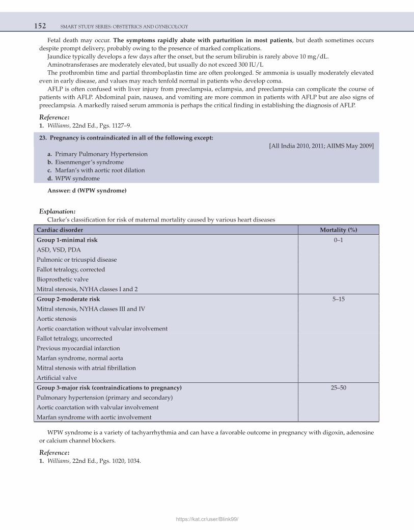
152 SMART STUDY SERIES: OBSTETRICS AND GYNECOLOGY
Fetal death may occur. The symptoms rapidly abate with parturition in most patients, but death sometimes occurs despite prompt delivery, probably owing to the presence of marked complications.
Jaundice typically develops a few days after the onset, but the serum bilirubin is rarely above 10 mg/dL.Aminotransferases are moderately elevated, but usually do not exceed 300 IU/LThe prothrombin time and partial thromboplastin time are often prolonged. Sr ammonia is usually moderately elevated
even in early disease, and values may reach tenfold normal in patients who develop coma.AFLP is often confused with liver injury from preeclampsia, eclampsia, and preeclampsia can complicate the course of
patients with AFLP. Abdominal pain, nausea, and vomiting are more common in patients with AFLP but are also signs of preeclampsia. A markedly raised serum ammonia is perhaps the critical finding in establishing the diagnosis of AFLP.
Reference:
1. Williams, 22nd Ed., Pgs. 1127–9.
23. Pregnancy is contraindicated in all of the following except: [All India 2010, 2011; AIIMS May 2009] a. Primary Pulmonary Hypertension b. Eisenmenger’s syndrome c. Marfan’s with aortic root dilation d. WPW syndrome
Answer: d (WPW syndrome)
Explanation:Clarke’s classification for risk of maternal mortality caused by various heart diseases
Cardiac disorder Mortality (%)
Group 1-minimal risk 0–1
ASD, VSD, PDA
Pulmonic or tricuspid disease
Fallot tetralogy, corrected
Bioprosthetic valve
Mitral stenosis, NYHA classes I and 2
Group 2-moderate risk 5–15
Mitral stenosis, NYHA classes III and IV
Aortic stenosis
Aortic coarctation without valvular involvement
Fallot tetralogy, uncorrected
Previous myocardial infarction
Marfan syndrome, normal aorta
Mitral stenosis with atrial fibrillation
Artificial valve
Group 3-major risk (contraindications to pregnancy) 25–50
Pulmonary hypertension (primary and secondary)
Aortic coarctation with valvular involvement
Marfan syndrome with aortic involvement
WPW syndrome is a variety of tachyarrhythmia and can have a favorable outcome in pregnancy with digoxin, adenosine or calcium channel blockers.
Reference:
1. Williams, 22nd Ed., Pgs. 1020, 1034.
https://kat.cr/user/Blink99/
MEDICAL AND SURGICAL COMPLICATIONS IN PREGNANCY 153
24. A female presents with leaking and meconium stained liquor at 32 weeks of gestation. Which of the following organism would be responsible:
[All India 2010] a. CMV b. Listeria c. Toxoplasma d. Rubella
Answer: b (Listeria)
Explanation:Listerial infections are more common in very old or young, pregnant or immunocompromised patients. Listeriosis during
pregnancy can have the following effects:Maternal
1) Fever 2) Pyelonephritis 3) Meningitis 4) Preterm labor
Fetal
1) Discolored, brownish or Meconium Stained Amniotic Fluid even with preterm gestations. (Generally MSAF is seen with postdatism and is very rarely seen with preterm labor)
2) Chorioamnionitis 3) Fetal infection: disseminated granulomatous lesions with microabscesses 4) IUFD 5) Neonatal sepsis and mortality
CMV, Rubella and Toxoplasma infections have a teratogenic effect and are unlikely to cause preterm labor and MSAF.
Reference:
1. Williams, 22nd Ed., Pg. 1287–8.
25. A 27-year-old multipara construction laborer has a blood picture showing hypochromic anisocytosis. This is most likely indicative of:
[All India 2004] a. Iron deficiency b. Folic acid deficiency c. Malnutrition d. Combined iron and folic acid deficiency
Answer: d (Combined iron and folic acid deficiency)
Explanation:As per WHO, anemia is defined as hemoglobin less than 11 g%.
Causes of anemia in pregnancy:
1. Physiological (hemodilution) 2. Pathological: (a) Iron deficiency anemia (IDA) (hypochromic and microcytic) (b) Megaloblastic anemia (macrocytes, hypersegmented neutrophils, and Howell-Jolly bodies) (c) Dimorphic/nutritional anemia (d) Hemorrhagic anemia (e) Hemolytic anemia (f) Hemoglobinopathies
154 SMART STUDY SERIES: OBSTETRICS AND GYNECOLOGY
MC cause of anemia in pregnancy is a dimorphic anemia, i.e., combination of iron and vitamin B12 and folic acid deficiencyAnemia is the most common indirect cause of maternal mortality.
Reference:
1. Williams, 22nd Ed., Pg. 1144–5.
26. All of the following conditions are the risk factors for urinary tract infections in pregnancy, EXCEPT: [All India 2004] a. Diabetes b. Hypertension c. Sickle cell anemia d. Vesicoureteral reflux
Answer: b (Hypertension)
Explanation:Diabetes is a definite risk factor for recurrent vaginal tract infection and urinary tract infection (UTI) during pregnancy.In patients with sickle cell anemia, there is 2-fold increase in risk of asymptomatic bacteriuria and UTI.Pregnancy causes numerous changes in the woman’s body. Hormonal and mechanical changes during pregnancy increase
the risk of urinary stasis and vesicoureteral reflux. These changes and difficulty with hygiene due to a distended pregnant belly and presence of short urethra in females all increase the frequency of UTIs in pregnant women. UTIs are one of the most common bacterial infections during pregnancy.
In up to 40% of these cases, bacteriuria may progress to symptomatic upper tract UTI or pyelonephritis.Various risk factors are associated with an increased frequency of bacteriuria during pregnancy. The risk is doubled in
women with sickle cell trait. Other risk factors include diabetes mellitus, neurogenic bladder retention, history of vesicoure-teral reflux (treated or untreated), previous renal transplantation, and a history of previous UTIs.
The most common uropathogen in the pregnant patient is E. coli (80–90%).Other pathogens include the following:
• Klebsiella pneumoniae (5%) • Proteus mirabilis (5%) • Enterobacter species (3%) • Staphylococcus saprophyticus (2%) • Group B P-hemolytic Streptococcus (1%) • Proteus species (2%)
Reference:
1. Williams, 22nd Ed., Pg. 1095–9.
27. All of the following are associated with gestational diabetes mellitus (GDM), EXCEPT: [AIIMS May 2010, AIIMS May 2011] a. Previous H/O macrosomic baby b. Obesity c. Malformations d. Polyhydramnios
Answer: c (Malformations)
Explanation:
• Students to take note: The question is about ‘gestational diabetes’ and not ‘overt diabetes’. • Congenital malformations (NTDs, CVS, etc) are seen with overt diabetes.
Effects of overt diabetes on pregnancy:Mother:
1. Increased risk of preeclampsia and polyhydramnios 2. Higher risk of infection 3. PPH 4. Operative delivery
https://kat.cr/user/Blink99/
MEDICAL AND SURGICAL COMPLICATIONS IN PREGNANCY 155
Fetal effects:
1. Recurrent first trimester abortions 2. Congenital anomalies 3. Sudden IUFD at term 4. Macrosomia 5. Shoulder dystocia • The complications of gestational diabetes are the same as above, except abortions and congenital anomalies. • Gestational diabetes mostly develops at around 24–28 weeks, and hence, there is no risk of first trimester abortions
and congenital anomalies in the fetus as sugars would be normal in the first trimester. • So remember that the 2 ‘A’s: ‘Anomalies’ and ‘Abortions’ are seen in overt diabetes and not in GDM.
Reference:
1. Williams, 22nd Ed., Pg. 1177.
28. In cases of intrahepatic cholestasis of pregnancy, ideal time for termination of pregnancy is: [AIIMS May 2010] a. 34 weeks b. 36 weeks c. 38 weeks d. 40 weeks
Answer: c (38 weeks)
Explanation:Intrahepatic cholestasis of pregnancy (IHCP) or obstetric cholestasis is an uncommon liver disorder. There is generalized
pruritus, often commencing in the palms of the hands and soles of the feet. It most often presents in the late second or early third trimester of pregnancy. Maternal outcomes for patients diagnosed with IHCP are generally good; however, fetal out-comes can be devastating. Thus, early recognition, treatment, and timely delivery are imperative.
There is 10- to 100-fold increase in bile acids (cholic/deoxycholic acids) probably due to excess circulating estrogen.Pruritus is the most common presenting feature.Complications include: Preterm labor, PPH, sudden IUFD (at 39–40 weeks) and MSAF (meconium-stained amniotic fluid).Delivery should be induced at 37–38 weeks due to increased risk of fetal mortality (at 39–40 weeks). If the fetal monitoring
is non-reassuring, delivery would even be needed earlier.
NOTE:
• UDCA is the DOC for this condition [All India 2010] • Best marker for IHCP/investigation of choice = Bile acids [All India 2011]
Reference:
1. Williams, 22nd Ed., Pg. 1126.
29. In a pregnant woman, all can be given for SLE, EXCEPT: [All lndia 2009; AIIMS May 2010] a. Methotrexate b. Sulfasalazine c. Hydroxychloroquine d. Prednisone
Answer: a (Methotrexate)
Explanation:Methotrexate is category X drug (FDA) and is highly teratogenic. Women must not take the drug during pregnancy, if there
is a risk of becoming pregnant, or if they are breastfeeding.Methotrexate can be toxic to the embryo and can cause fetal defects and spontaneous abortion. It should be discontinued
prior to conception if used in either partner. Females should discontinue use for at least 1 ovulatory cycle before conception and the male patients should stop taking methotrexate at least 3 months prior to a planned conception.
• Hydroxychloroquine: Pregnancy category C • Prednisolone: Pregnancy category B
156 SMART STUDY SERIES: OBSTETRICS AND GYNECOLOGY
Reference:
1. Williams, 22nd Ed., Pg. 1214.
30. The measures to prevent vertical transmission of HIV are all, EXCEPT: [All India 2011, 2013] a. Vaginal delivery b. Administration of vitamin A c. Stop breast feeding d. Treatment with zidovudine
Answer: a (Vaginal delivery)
Explanation:For prevention of mother-to-child transmission of HIV infection, the following is advocated:
1) Antenatal antiretroviral therapy (HAART) 2) Nevirapine during labor (if the patient is not on HAART) 3) ARM during labor is avoided 4) Avoid breast feeding 5) Vitamin A supplementation to mother has been found to decrease the vertical transmission in few studies 6) If viral copies is >1000/mL, then elective cesarean should be done.
Vaginal delivery increases the risk of vertical transmission.
Reference:
1. Williams, 22 Ed., Pg. 1316–7.
31. Regarding prolactinoma in pregnancy, all are true, EXCEPT: [AIIMS May 2011] a. Most common pituitary tumor but rarely symptomatic b. Increase in prolactin levels a/w worse prognosis c. Macroadenoma may increase in size d. Regular visual check-up needed
Answer: b (Increase in prolactin levels a/w worse prognosis)
Explanation:During pregnancy the levels of circulating estrogen is very high. This results in a parallel increase in the circulating levels
of prolactin. Prolactin levels begin to rise at 5–8 weeks of gestation period and it parallels the increase in the size and number of lactotrophs. At the end of the first trimester, serum prolactin levels are approximately 20–40 ng/mL. It further increases to 50–150 ng/mL and are 100–400 ng/mL at the end of the second and third trimesters, respectively.
Given the stimulatory effects of pregnancy on the normal lactotrophs, enlargement of the normal pituitary can be expected. Prolactinomas that symptomatically enlarge during pregnancy are uncommon. Symptoms suggestive of growth are head-ache, visual field changes, and diabetes insipidus.
Women with prolactin-secreting tumors may experience further pituitary enlargement and must be closely monitored during pregnancy. However, damage to the pituitary or optic nerves occurs in <1% of pregnant women with prolactinoma. If a woman has completed a successful pregnancy, the chances of her completing additional successful pregnancies are extremely high.
So per se increase in prolactin levels does not indicate poor prognosis, as during pregnancy, there is going to be increase in prolactin levels.
Reference:
1. Speroff, 7th Ed., Pg. 450–60.
32. A 36 weeks’ pregnant diabetic female has a non-reactive NST. What should be done next? [AIIMS May 2011] a. Induction of labor b. LSCS c. Do NST after 1 hour d. Proceed to biophysical profile
Answer: d (Proceed to biophysical profile)
https://kat.cr/user/Blink99/
MEDICAL AND SURGICAL COMPLICATIONS IN PREGNANCY 157
Explanation:A non-reactive NST is an indication for doing a biophysical profile (BPP). It is not an indication for directly doing an LSCS or inducing labor.In babies of diabetic mother, the lung maturity is delayed and so pregnancy should not be terminated at 36 weeks (unless there is fetal distress).But babies of diabetic mothers have one more risk, which is that of sudden IUFD at term (40 weeks).So the babies of diabetic mothers should preferably be delivered between 38 weeks and 39 weeks.If the BPP score is poor, then it is an indication of immediate termination of pregnancy.
Reference:
1. Williams, 22nd Ed., Pg. 1117–83.
33. Glucose tolerance test is indicated in all, EXCEPT: [AIIMS Nov 2011] a. Previous congenital anomaly b. Previous eclampsia c. Polyhydramnios d. Previous unexpected fetal death
Answer: b (Previous eclampsia)
Explanation:
• Glucose challenge test (GCT) is a screening test and glucose tolerance test (GTT) is a confirmatory test for gestational diabetes mellitus (GDM).
• It should be done for patients who are at high risk of development of GDM or if GDM is suspected. • Previous eclampsia does not predispose the patient to GDM. • Indications:
1. Age >25years 2. BMI >25 3. Previous GDM 4. Family h/o DM in first-degree relative 5. Previous baby with macrosomy/congenital anomalies 6. H/o abnormal glucose tolerance/IR (PCOS) 7. Macrosomy/polyhydramnios in current pregnancy 8. Previous unexplained stillbirth
References:
1. Williams, 22nd Ed., Pg. 1172. 2. American Diabetes Association. Standards of medical care in diabetes—2007. Diabetes Care. Jan 2007.
34. A 26-year-old primigravida with juvenile myoclonic epilepsy comes to you at 4 months with concern regarding continuing sodium valproate treatment. Your advice is:
[AIIMS Nov 2011] a. Add lamotrigine to sodium valproate b. Taper sodium valproate and add lamotrigine c. Switch on to carbamazepine d. Continue sodium valproate with regular monitoring of serum levels
Answer: d (Continue sodium valproate with regular monitoring of serum levels)
Explanation:Epilepsy is the MC neurological disorder encountered in pregnancy.The use of any anti-epileptic drugs (AEDs) is associated with a greater baseline risk of fetal malformations during preg-
nancy. When treating pregnant women who have epilepsy, the risks of increased seizure frequency vs the risks of AED use must be weighed carefully.
As per ACOG and RCOG guidelines, there is no particular drug of choice for epilepsy in pregnancy.
158 SMART STUDY SERIES: OBSTETRICS AND GYNECOLOGY
All have teratogenic effects. So, the guidelines recommend that whichever drug the patient is on before pregnancy should be continued during pregnancy.
The choice of drug depends on the type of epilepsy.There is a risk of increase in seizure frequency if the patient is shifted to a relatively less teratogenic newer drug.Patients are advised to switch to a single AED prior to conception and to taper to the lowest possible dose as exposure
to multiple AEDs is more teratogenic than monotherapy.Patients who have not had a seizure for 2–5 years may wish to attempt complete withdrawal from AEDs prior to conception.So, monotherapy at the least possible dose is the best (so, first 2 options are ruled out).As the patient has already finished the first trimester (teratogenic period), it is best to continue with the same AED.However, during pregnancy, because of hemodilution, the dose of AED needs to be generally increased and hence thera-
peutic drug monitoring (TDM) should be done for all AEDs.
References:
1. Williams, 22nd Ed. 2. ACOG and RCOG Guidelines.
35. G3P2L2 at 8 weeks of gestation is VDRL positive. The drug of choice is: [All India 2012] a. Erythromycin b. Penicillin c. Probenecid d. Azithromycin
Answer: b (Penicillin)
Explanation:Parenteral penicillin G is the preferred drug for treating all stages of syphilis. The preparation used, the dosage, and the
length of treatment depend on the stage and clinical manifestations of the disease. Selection of the appropriate penicillin preparation is important, because Treponema pallidum can reside in sequestered sites (e.g., the CNS and aqueous humor) that are poorly accessed by some forms of penicillin. Early syphilis:
• Benzathine penicillin G 2.4 million units IM in a single dose. • Some recommend a second dose 1 week later. Tertiary syphilis: • Latent syphilis more than 1-year duration: • Benzathine penicillin G 7.2 million units total, administered as 3 doses of 2.4 million units IM weekly.Neurosyphilis: • Aqueous crystalline penicillin G 18–24 million units/day, administered as 3–4 million units IV every 4 hours or
continuous infusion, for 10–14 days. • If compliance with therapy can be ensured, the following alternative regimen might be considered.Alternative regimen: • Procaine penicillin 2.4 million units IM once daily PLUS • Probenecid 500 mg orally 4 times a day, both for 10–14 days
Reference:
1. CDC Guidelines. Williams, 22nd Ed., Pg. 1304.
36. In an HBsAg-positive female, which of the following statements is true? [All India 2012] a. Transmission is mainly transplacental b. Immunoglobulin should be given to baby within 12 hours c. Active immunization should be done within 48 hours d. Immunization can be delayed up to 96 hours
Answer: b (Immunoglobulin should be given to baby within 12 hours)
Explanation:Hepatitis B virus (HBV) does not cross placenta because of its size, and it cannot infect the fetus unless there have been
breaks in the maternal-fetal barrier, such as those that occur during amniocentesis. Women who are infected can transmit HBV
https://kat.cr/user/Blink99/
MEDICAL AND SURGICAL COMPLICATIONS IN PREGNANCY 159
to the infant during delivery. Consequently, unless adequate prophylaxis is provided, the newborn is at high risk to develop a chronic HBV infection, with its known long-term complications.
Perinatal transmission from the mother to her newborn baby is the most important mode of infection. If a pregnant woman is an HBV carrier and is also positive for hepatitis B ‘e’ antigen (HBeAg), her newborn baby has a 90% likelihood of becoming infected. Approximately 25% of infected infants will become chronic carriers. Most HbsAg carriers are asymptomatic, poten-tially infectious, and a constant source of new infections.
Immunization with hepatitis B immunoglobulin (HBIg) should especially be considered for neonates born of mothers positive for HBsAg. Such infants often acquire chronic infection, especially when mothers are HBeAg positive, in whom the risk of becoming chronic carriers is extremely high (90%). When HBIg is given within the first hours, up to 24 hours after birth, the risk of HBV infection can be reduced to 20%.
The vertical transmission rate is dramatically decreased when HBIg is given with the first dose of HBV vaccine very soon after birth.
When administered within 24 hours after birth, HBIg and vaccination are 85–95% effective in preventing HBV infection and the chronic carrier state. In contrast, administration of the HBV vaccine alone beginning within 24 hours after birth is 70–95% effective in preventing perinatal HBV infection.
Reference:
1. Williams, 22nd Ed., Pg. 1130–1.
37. Lady with MS + MR with full term gestation, obstetrician planning to conduct normal delivery, what would be anesthesia of choice?
[AIIMS May 2012] a. Parenteral opioids b. Spinal anesthesia c. Inhalational analgesia d. Neuraxial analgesia
Answer: d (Neuraxial analgesia)
Explanation:Pain relief is important for heart disease patients as pain can cause tachycardia, which in turn can cause cardiac failure.
Epidural and spinal techniques are the most effective means of providing pain relief for labor. These are also known as regional techniques because pain relief is limited to a specific anatomical region. These modalities are also known as neur-axial techniques, since both the approaches involve administration of drugs that exert their effects in the axial portion of the CNS.
The term ‘regional analgesia’ refers to the inhibition of labor pain, whereas the term ‘regional anesthesia’ implies the use of a higher concentration of anesthetic agent which results in more intense ablation of all sensation and is used for LSCS.
Reference:
1. Williams, 22nd Ed. Pg. 483.
38. Which of the following is seen during heart disease in pregnancy but not during normal pregnancy? [AIIMS Nov 2012, AIIMS Nov 2013] a. Pedal edema b. Engorged neck veins c. Dyspnea d. Hypotension
Answer: b (Engorged neck veins)
Explanation:Finding suggestive of heart disease in pregnancy (METCALFE’S criteria):
1. Cyanosis 2. Clubbing of fingers 3. Persistent neck vein distention 4. Systolic murmur grade 3/6 or greater

160 SMART STUDY SERIES: OBSTETRICS AND GYNECOLOGY
5. Diastolic murmur 6. Cardiomegaly 7. Persistent split-second sound 8. Criteria for pulmonary hypertension 9. Persistent arrhythmias
Pedal edema. dyspnea and hypotension can also be physiological during pregnancy.
Reference: 1. Williams, 22nd Ed. Pg. 1029, 1034–35.
https://kat.cr/user/Blink99/
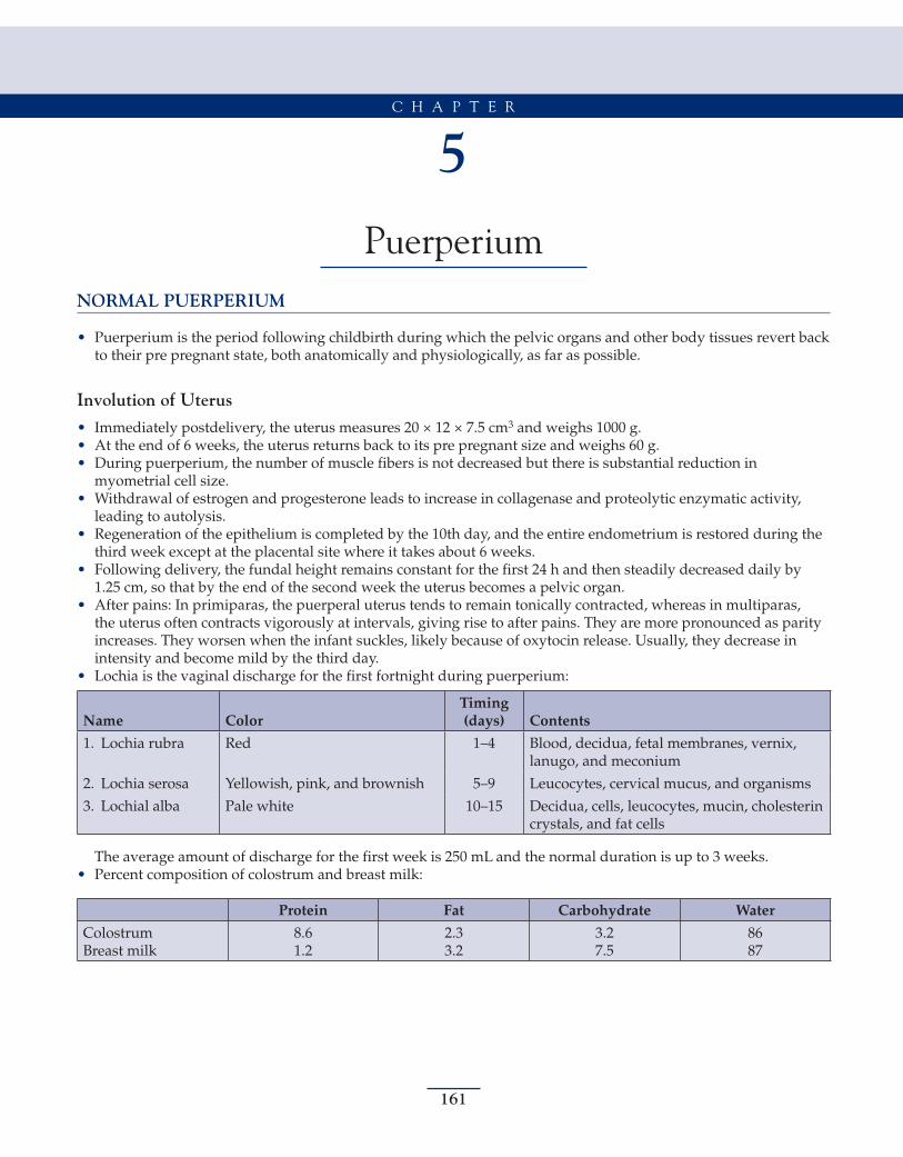
161
5Puerperium
NORMAL PUERPERIUM
• Puerperium is the period following childbirth during which the pelvic organs and other body tissues revert back to their pre pregnant state, both anatomically and physiologically, as far as possible.
Involution of Uterus
• Immediately postdelivery, the uterus measures 20 × 12 × 7.5 cm3 and weighs 1000 g. • At the end of 6 weeks, the uterus returns back to its pre pregnant size and weighs 60 g. • During puerperium, the number of muscle fibers is not decreased but there is substantial reduction in
myometrial cell size. • Withdrawal of estrogen and progesterone leads to increase in collagenase and proteolytic enzymatic activity,
leading to autolysis. • Regeneration of the epithelium is completed by the 10th day, and the entire endometrium is restored during the
third week except at the placental site where it takes about 6 weeks. • Following delivery, the fundal height remains constant for the first 24 h and then steadily decreased daily by
1.25 cm, so that by the end of the second week the uterus becomes a pelvic organ. • After pains: In primiparas, the puerperal uterus tends to remain tonically contracted, whereas in multiparas,
the uterus often contracts vigorously at intervals, giving rise to after pains. They are more pronounced as parity increases. They worsen when the infant suckles, likely because of oxytocin release. Usually, they decrease in intensity and become mild by the third day.
• Lochia is the vaginal discharge for the first fortnight during puerperium:
Name ColorTiming (days) Contents
1. Lochia rubra Red 1–4 Blood, decidua, fetal membranes, vernix, lanugo, and meconium
2. Lochia serosa Yellowish, pink, and brownish 5–9 Leucocytes, cervical mucus, and organisms
3. Lochial alba Pale white 10–15 Decidua, cells, leucocytes, mucin, cholesterin crystals, and fat cells
The average amount of discharge for the first week is 250 mL and the normal duration is up to 3 weeks. • Percent composition of colostrum and breast milk:
Protein Fat Carbohydrate Water
ColostrumBreast milk
8.61.2
2.33.2
3.27.5
8687
C H A P T E R
162 SMART STUDY SERIES: OBSTETRICS AND GYNECOLOGY
• Lactation suppressors and galactagogues
Lactation suppressors Galactagogues
1. Bromocriptine/cabergoline 1. Nipple stimulation
2. Testosterone 2. Breast pump
3. Ethinyl estradiol 3. Metoclopramide
4. Pyridoxine
ABNORMAL PUERPERIUM
Puerperal Pyrexia
A rise in temperature reaching 100.4°F (38°C) or more (measured orally) on two separate occasions at 24 h apart (excluding the first 24 h) within the first 10 days following delivery is called puerperal pyrexia.
Causes:
1. Puerperal sepsis 2. Acute pyelonephritis 3. Breast engorgement 4. Wound infection 5. Thrombophlebitis 6. Atelectasis and pneumonia
Puerperal Sepsis
• An infection of the genital tract which occurs as a complication of delivery is called puerperal sepsis. • Postpartum uterine infection has been called variously endometritis, endomyometritis, and endoparametritis.
Because infection actually involves not only the decidua but also the myometrium and parametrial tissues, the preferred term is metritis with pelvic cellulitis.
• The route of delivery is the single most significant risk factor for the development of uterine infection. • Compared with cesarean delivery, metritis following vaginal delivery is relatively uncommon. • Most female pelvic infections are caused by bacteria indigenous to the female genital tract.
Predisposing Factors of Puerperal Sepsis
Antepartum Intrapartum
1. Malnutrition 1. Multiple cervical examinations
2. Anemia 2. Internal fetal monitoring
3. Preeclampsia 3. Chorioamnionitis
4. PROM 4. Retained placenta
5. Immunocompromised status (HIV) 5. PPH
6. Diabetes mellitus 6. Prolonged labor
7. Obesity 7. Operative delivery (LSCS)
8. MSAF
Bacteria Commonly Responsible for Female Genital InfectionsAerobes
• Group A, B, D streptococci • Enterococcus • Gram-negative bacteria—Escherichia coli, Klebsiella, and Proteus species • Staphylococcus aureus • Gardnerella vaginalis
https://kat.cr/user/Blink99/
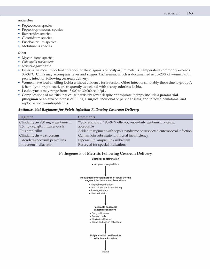
PUERPERIUM 163
Anaerobes
• Peptococcus species • Peptostreptococcus species • Bacteroides species • Clostridium species • Fusobacterium species • Mobiluncus species
Other
• Mycoplasma species • Chlamydia trachomatis • Neisseria gonorrheae • Fever is the most important criterion for the diagnosis of postpartum metritis. Temperature commonly exceeds
38–39°C. Chills may accompany fever and suggest bacteremia, which is documented in 10–20% of women with pelvic infection following cesarean delivery.
• Women have foul-smelling lochia without evidence for infection. Other infections, notably those due to group A β-hemolytic streptococci, are frequently associated with scanty, odorless lochia.
• Leukocytosis may range from 15,000 to 30,000 cells/μL. • Complications of metritis that cause persistent fever despite appropriate therapy include a parametrial
phlegmon or an area of intense cellulitis, a surgical incisional or pelvic abscess, and infected hematoma, and septic pelvic thrombophlebitis.
Antimicrobial Regimens for Pelvic Infection Following Cesarean Delivery
Regimen Comments
Clindamycin 900 mg + gentamicin 1.5 mg/kg, q8h intravenously
“Gold standard,” 90–97% efficacy, once-daily gentamicin dosing acceptable
Plus ampicillin Added to regimen with sepsis syndrome or suspected enterococcal infectionClindamycin + aztreonam Gentamicin substitute with renal insufficiencyExtended-spectrum penicillins Piperacillin, ampicillin/sulbactamImipenem + cilastatin Reserved for special indications
Pathogenesis of Metritis Following Cesarean DeliveryBacterial contamination
Indigenous vaginal flora
Inoculation and colonization of lower uterinesegment, incisions, and lacerations
Vaginal examinationsInternal electronic monitoringProlonged laborUterine incision
Favorable anaerobicbacterial conditions
Surgical traumaForeign bodyDevitalized tissueBlood and serum collection
Polymicrobial proliferationwith tissue invasion
Metritis
164 SMART STUDY SERIES: OBSTETRICS AND GYNECOLOGY
Parametrial Phlegmon • In some women in whom metritis develops following cesarean delivery, parametrial cellulitis is intensive and
forms an area of induration, termed a phlegmon, within the leaves of the broad ligament. These infections should be considered when fever persists longer than 72 h despite intravenous antimicrobial therapy.
• Areas of parametrial cellulitis are more often unilateral, and they frequently may remain limited to the base of the broad ligament.
• Severe cellulitis of the uterine incision may cause necrosis and separation. Extrusion of purulent material commonly leads to peritonitis. Because puerperal metritis with cellulitis is typically a retroperitoneal infection, evidence of peritonitis suggests the possibility of uterine incisional necrosis, or, less commonly, a bowel injury or other lesion.
• In most women with a phlegmon, clinical improvement follows continued treatment with a broad-spectrum antimicrobial regimen.
Septic Pelvic Thrombophlebitis • Puerperal infection may extend along venous routes and cause thrombosis. Lymphangitis often coexists. • The ovarian veins may then become involved because they drain the upper uterus, which most often includes
veins draining the placental site. Puerperal septic thrombophlebitis is likely to involve one or both ovarian venous plexuses.
• In a fourth of women, the clot extends into the inferior vena cava, and occasionally extends to the renal vein. • Women with septic pelvic thrombophlebitis usually display some clinical improvement of their pelvic infection
following antimicrobial treatment. • When imaging modalities were not available to confirm venous involvement, “heparin challenge test” was used.
If the temperature decreases on giving IV heparin, then it is diagnostic of septic pelvic thrombophlebitis. • However, recent studies show that there is no role of heparin in the management of this condition.
SUBINVOLUTION
• This term describes an arrest or retardation of involution. It is accompanied by prolongation of lochial discharge and irregular or excessive uterine bleeding, which sometimes may be profuse.
• On bimanual examination, the uterus is larger and softer than would be expected. Some causes of subinvolution are retention of placental fragments and pelvic infection.
• Because most cases of subinvolution result from local causes, they are usually amenable to early diagnosis and treatment.
• Methylergometrine (methergin), 0.2 mg every 3–4 h for 24–48 h, is recommended. On the other hand, metritis responds to oral antimicrobial therapy. Almost a third of cases of late postpartum uterine infection are caused by Chlamydia trachomatis; thus, azithromycin or doxycycline therapy may be appropriate.
MASTITIS
• Parenchymatous infection of the mammary glands is a rare complication occasionally observed during the puerperium and lactation.
• It is estimated to occur in anywhere from 2% to 33% of breast-feeding women. The first sign of inflammation is chills or actual rigor, soon followed by fever and tachycardia. The breast becomes hard and reddened, and the woman complains of severe pain. About 10% of women with mastitis develop an abscess.
• Constitutional symptoms attending a mammary abscess are generally severe, but in some cases the first indication of the true diagnosis often is afforded by the detection of fluctuation. Ultrasonography may be helpful to detect an abscess.
• The most commonly isolated organism is Staphylococcus aureus. Other commonly isolated organisms are coagulase-negative staphylococci and viridans streptococci. The immediate source of organisms that cause mastitis is almost always the infant’s nose and throat.
• Mastitis requires antibiotics (penicillin/cephalosporins/erythromycin). • Abscess requires incision and drainage under general anesthesia.
https://kat.cr/user/Blink99/
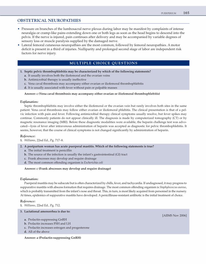
PUERPERIUM 165
OBSTETRICAL NEUROPATHIES
• Pressure on branches of the lumbosacral nerve plexus during labor may be manifest by complaints of intense neuralgia or cramp-like pains extending down one or both legs as soon as the head begins to descend into the pelvis. If the nerve is injured, pain continues after delivery and may be accompanied by variable degrees of sensory loss or muscle paralysis supplied by the damaged nerve.
• Lateral femoral cutaneous neuropathies are the most common, followed by femoral neuropathies. A motor deficit is present in a third of injuries. Nulliparity and prolonged second stage of labor are independent risk factors for nerve injury.
MULTIPLE CHOICE QUESTIONS
1. Septic pelvic thrombophlebitis may be characterized by which of the following statements? a. It usually involves both the iliofemoral and the ovarian veins b. Antimicrobial therapy is usually ineffective c. Vena caval thrombosis may accompany either ovarian or iliofemoral thrombophlebitis d. It is usually associated with fever without pain or palpable masses
Answer: c (Vena caval thrombosis may accompany either ovarian or iliofemoral thrombophlebitis)
Explanation:Septic thrombophlebitis may involve either the iliofemoral or the ovarian vein but rarely involves both sites in the same
patient. Vena caval thrombosis may follow either ovarian or iliofemoral phlebitis. The clinical presentation is that of a pel-vic infection with pain and fever. Following antimicrobial therapy clinical symptoms usually resolve, but fever spikes may continue. Commonly patients do not appear clinically ill. The diagnosis is made by computerized tomography (CT) or by magnetic resonance imaging (MRI). Before these diagnostic modalities were available, the heparin challenge test was advo-cated—lysis of fever after intravenous administration of heparin was accepted as diagnostic for pelvic thrombophlebitis. It seems, however, that the course of clinical symptoms is not changed significantly by administration of heparin.
Reference: 1. Williams, 22nd Ed., Pg. 717–8.
2. A postpartum woman has acute puerperal mastitis. Which of the following statements is true? a. The initial treatment is penicillin b. The source of the infection is usually the infant’s gastrointestinal (GI) tract c. Frank abscesses may develop and require drainage d. The most common offending organism is Escherichia coli
Answer: c (Frank abscesses may develop and require drainage)
Explanation:Puerperal mastitis may be subacute but is often characterized by chills, fever, and tachycardia. If undiagnosed, it may progress to
suppurative mastitis with abscess formation that requires drainage. The most common offending organism is Staphylococcus aureus, which is probably transmitted from the infant’s nose and throat. This, in turn, is most likely acquired from personnel in the nursery. At times, epidemics of suppurative mastitis have developed. A penicillinase-resistant antibiotic is the initial treatment of choice.
Reference: 1. Williams, 22nd Ed., Pg. 712.
3. Lactational amenorrhea is due to: [AIIMS Nov 2006] a. Prolactin-suppressing GnRH b. Prolactin increases FSH and LH c. Prolactin increases estrogen and progesterone d. All of the above
Answer: a (Prolactin-suppressing GnRH)
166 SMART STUDY SERIES: OBSTETRICS AND GYNECOLOGY
Explanation:Scheme of Mechanism of Amenorrhea and Anovulation in Lactating Mothers
Increasedprolactin level
Inhibits ovarianresponse to FSH
Suppresses therelease of LH
GnRHsecretion
FSH and LHLessfollicular growth
No LH surge
Hypoestrogenicstate
No menstruation
Anovulation
↓
↓
Increased frequency, intensity, and duration of suckling are associated with high prolactin level, prolonged ovarian suppression, and lactational amenorrhea.
References: 1. Dutta DC, 6th Ed., Pg. 148. 2. Chaudhari SK, 6th Ed., Pg. 65–6.
4. From which of the following layers the regeneration of endometrium takes place? [AIIMS May 2004] a. Zona basalis b. Zona pellucidum c. Zona compacta d. Zona spongiosa
Answer: a (Zona basalis)
Explanation:Puerperium is the period following childbirth during which the pelvic organs and other body tissues revert back to their
pre pregnant state, both anatomically and physiologically, as far as possible.At the end of 6 weeks, the uterus returns back to its pre pregnant size.During puerperium, the number of muscle fibers is not decreased but there is substantial reduction in myometrial cell size.Withdrawal of estrogen and progesterone leads to increase in collagenase and proteolytic enzymatic activity, leading to
autolysis.Regeneration of the epithelium is completed by the 10th day, and the entire endometrium is restored during the third
week, except at the placental site where it takes about 6 weeks. Regeneration takes place from zona basalis.
Reference: 1. Williams, 22nd Ed., Pg. 698–9.
https://kat.cr/user/Blink99/
167
6Contraception
Pearl rate: The Pearl index of contraceptive failure was first introduced by Raymond Pearl. This is expressed as pregnancy rate per 100 woman-years or women-year (HWY) and is calculated according to the following formula:
Pregnancy rate per HWY =
Total accidental pregnancies × 1200
Total months of exposure to unintended pregnancy
WHO ELIGIBILITY CRITERIA FOR REVERSIBLE CONTRACEPTIVES
The contraceptive methods are organized into four categories:Category 1: No restrictions of use.Category 2: Advantages of using the method outweigh the theoretical or proven risks.Category 3: Theoretical or proven risks outweigh the advantages of using the method.Category 4: Use of the method presents unacceptable health risks. For most of the cases, eligibility can be determined by taking client’s medical history only. Physical examination and laboratory tests are not generally necessary.
Categories 1 and 4 are self-explanatory (category 1 denotes indications and category 4 indicates absolute contra-indications; categories 2 and 3 indicate relative contraindications).
Chances of Women Dying from Complications of Pregnancy, Childbirth, or Unsafe Abortion During Her Lifetime: Lifetime Risk
Ethiopia 1 in 7 Africa 1 in 15
India 1 in 57 Asia 1 in 105
Brazil 1 in 128 Europe 1 in 1895
United States of America 1 in 3418
Italy 1 in 6261
Developed countries 1 in 2125
Developing countries 1 in 65
World 1 in 70
NATURAL FAMILY PLANNING METHODS
The most fertile period of a woman is from the 10th (rarely 9th) to the 18th day, provided the cycle is of 28 days. Natural family planning (NFP) methods are based on the premise that coitus should be avoided during this fertile period of the woman, as determined by timing or calculating the time of ovulation.
C H A P T E R
168 SMART STUDY SERIES: OBSTETRICS AND GYNECOLOGY
1. Rhythm Method
In a woman having a regular 28-day cycle, the unsafe period is from day 7 to day 21. The chance of pregnancy is at its minimum (10 per 100 women-year) when coitus is avoided between day 7 and day 21, whereas the failure rate rises to 25–35 per 100 women-year if coitus is avoided only between day 8 and day 18.
2. Basal Body Temperature Method
This method is based on the fact that after ovulation, the progesterone level in the blood rises, increasing the basal metabolic rate and causing rise of temperature by 0.5–0.8°F or 0.2–0.4°C in the luteal phase. Sometimes there is a slight drop of 0.2°F just before the rise.
3. Cervical Mucus Method
The cervical mucus method is also called “ovulation method” or more commonly “Billings’ ovulation method.” This method is based on recognizing the changes that occur in cervical mucus due to the effect of estrogen and progester-one at different times of the menstrual cycle.
Just before that, at the time of ovulation, the mucus becomes more copious, clear, and slippery, resembling the white of a raw egg, and can be stretched slowly between two fingers. The vagina and vulva feel moist or wet. This persists for about 3 days. These are called “wet days”; the last day of wet mucus is called the “peak day.” These days mark the peak of fertility.
4. Symptothermal Method
The symptothermal method pinpoints the fertile period with greater precision and reliability. It is based on observa-tion of basal body thermal change (by basal body temperature—BBT—method), cervical mucus change (by Billings’ method), and other manifestations of the fertile period such as mid-cycle pain, mid-cycle light spotting, or bleeding and breast tenderness.
Typical failure rates of NFP methods as commonly used is 20% (20 per 100 women) in the first year of use. However, it can be reduced to 1–9% in the first year of use, when used consistently and correctly.
ContraindicationsNFP methods are not suitable for women:
1. With irregular cycles, cycles shorter than 21 days 2. During adolescence, lactation, and premenopause 3. Who have had cervical surgery (cautery and conization) 4. With vaginal infection (until cure) 5. Who have sexually transmitted disease (STD) or pelvic inflammatory disease (PID) in the last 3 months 6. Who had abortion recently 7. Noncooperative husbands and couples who have casual sex.
Withdrawal Method
Withdrawal method or coitus interruptus means discharge of semen outside the female genitalia at the end of intercourse.
Typical average failure rate per 100 users in the first year is 18.
ContraindicationPremature ejaculation is the only contraindication.
Comments
The advantages of the withdrawal method are that it (a) involves no expense, (b) needs no medical supervision, (c) requires no prior preparation, and (d) causes no definite harm. The main drawbacks are the lack of full sexual satisfaction and the relatively higher failure rate.
https://kat.cr/user/Blink99/
CONTRACEPTION 169
Persona: It is a device that detects urinary estrone-3-glucuronide, which indicates the beginning of fertile period, and LH, which indicates ovulation.
LACTATIONAL AMENORRHEA METHOD
Excessive secretion of prolactin, which controls lactation, inhibits the pituitary. Prolactin inhibits LH but has no effect on FSH. However, it partially inhibits ovarian response to both of these gonadotropins. As a result, while the prolac-tin level remains high, the ovary produces little estrogen and no progesterone. Hence, ovulation and menstruation are affected.
Failure rate of lactational amenorrhea method (LAM) (for 6 months only) is less than 2% when correctly and consistently used, but it is more otherwise.
The breast-feeding practices required by LAM have other health benefits for mother and baby:
1. It provides the healthiest food for the baby. 2. It protects the baby from life-threatening diarrhea. 3. It protects the baby from diseases such as measles and pneumonia by passing on the mother’s immunities to
the baby. 4. It helps to develop a close relationship between mother and baby. 5. It protects the mother from diseases such as subinvolution, fibroadenosis, and fibroadenoma of the uterus.
Breast feeding reduces risks of breast cancer and epithelial ovarian cancer.
BARRIER CONTRACEPTIVES
1. Condoms
• Condoms are contraceptive sheaths meant to cover the penis during coitus to prevent pregnancy. They are also known as French letters.
• The condom is the oldest and most widely used birth control device in the world. In the folklore of contraception, its invention is attributed to a physician named Dr Condom, who recommended it to Charles II.
• Condoms are mostly made of fine latex rubber and are available in various shapes and colors. They are circular cylinders, 15–20 cm in length, 3–3.5 cm in diameter and 0.003–0.007 cm in thickness; they are closed at one end and open at the other with an integral rim.
• Nonlatex forms of male condoms are now commercially made of polyurethane. Polyurethane condoms have a longer shelf life and can be used with oil-based lubricants, which can damage latex condoms.
• It is most harmless method of contraception. • When used properly, the condoms give very good protection against STDs. These include not only traditional
syphilis and gonorrhea but also trichomoniasis, moniliasis, nongonococcal urethritis, and infection with chlamydia and herpes virus.
• The condom seems to give best protection against sexually transmitted AIDS. Condoms also give protection against sexually transmitted hepatitis B virus. Protection against STD benefits male and female partners as well as their children.
• When used for more than 5 years, barrier methods, particularly the condom, reduce the chance of developing severe cervical dysplasia and cervical cancer as compared to the use of oral pills or to nonuse of contraceptives.
• Storage and disposal problems affect village people and reduce use of condoms. They should be wrapped in a piece of paper and thrown in dustbins or buried underneath the soil but should never be left in commodes or flushing-type latrine pans.
• Typical average failure rate of condom as commonly used is 12%.Total condom failure rates (breakage and slippage rate combined) range from 4% to 13%.
Non-contraceptive uses of condom include.
a. Prevention of STDs b. Condom catheter in males c. To cover the TVS probe
170 SMART STUDY SERIES: OBSTETRICS AND GYNECOLOGY
d. After vaginoplasty e. Shivkar’s pack (condom tamponade) for atonic PPH. f. In cases of antisperm antibodies present in cervical mucus.
2. Occlusive Caps (Vaginal Diaphragm and Cervical Cap)
• Occlusive caps do not act as sperm-proof mechanical barriers like condoms but are used as a means to retain spermicides in contact with the cervical os.
• Spermicides must be used along with these devices. • After intercourse, the vaginal diaphragm and vault cap should not be removed before 6–8 h of the last act and
should not be kept for more than 24 h. The best time to introduce it is from a few minutes to 2 h before the sexual act, mostly at bedtime, and it should be removed next morning.
• Like condoms, diaphragms and cervical caps prevent spread of STDs, although less effectively. • However, AIDS is not prevented by these contraceptives.
Disadvantages 1. Infection may set in if caps are not removed for a long time. 2. The chance of erosion may increase. 3. Diaphragms increase the chance of urinary infection. 4. Occlusive caps do not prevent spread of AIDS. 5. Very rarely, diaphragms and occlusive caps may produce toxic shock syndrome (TSS).
Contraindications 1. Prolapse uterus, cystocele 2. Badly lacerated or eroded cervix 3. VVF (vesico vaginal fistula) 4. RVF (rectovaginal fistula)
Failure RateVaginal diaphragms and cervical caps have typical average failure rates, as commonly used, of 18–28%. Diaphragms should be replaced anytime between 6 months and 2 years (depending on its care), for the rubber may perish. Caps need less frequent replacement.
3. Vaginal Sponge
• “Today” is a soft, disposable foam sponge made of polyurethane. It is round shaped, with a depression at the center of the upper surface designed to fit over the cervix, and is saturated with nonoxynol-9, the most powerful spermicide: it has an attached nylon loop that helps in its removal. It is moistened with water, squeezed gently to remove excess water and inserted high up in the vagina to cover the cervix.
• It acts for 24 h, and intercourse may be repeated as often as desired during this period. Like the cervical cap, it can be introduced long before the sex act. The failure rate varies between 9 and 27 per 100 users in the first year.
• It must be removed and thrown away after 8–24 h but not before 6 h of the last act. The real danger of the sponge is development of TSS, although it happens very rarely.
4. Spermicides
• Spermicides are contraceptive chemical agents. They comprise a chemical capable of destroying sperms incorporated into an inert base. The commonly used spermicidal agents contain nonionic surfactants that alter sperm surface membrane permeability, causing osmotic changes resulting in the killing of sperms. Most spermicides contain nonoxynol-9, which is best for the purpose.
• Their main role is to improve the contraceptive effect of other barrier methods. They are mostly used along with diaphragms, cervical caps, and condoms.
• Spermicidal agents nowadays contain nonoxynol-9. A few products contain octoxynol-9 and menfegol.
https://kat.cr/user/Blink99/
CONTRACEPTION 171
• There is no evidence that spermicides including nonoxynol-9 offer any protection against HIV and other STIs. Furthermore, there is some evidence that frequent use of nonoxynol-9 (twice a day or more) increases, rather than reduces, the chance of HIV transmission, perhaps by irritating the vaginal and cervical mucosa.
• Typical average failure rate, as commonly used, is 21%.
5. Female Condom
• A female condom, by the trade names of “Femidom” or “Reality,” is a new disposable barrier contraceptive for women. It consists of soft, loose-fitting polyurethane sac about 15 cm long and 7 cm in diameter.
• Sexual intercourse takes place within the cavity of the device. • It is a women-controlled method and can even be used without the partner’s cooperation. It prevents STDs
including HIV/AIDS.
Disadvantages 1. Intercourse is noisy, and slippage occurs in about one in 5–10 uses; however, female condom rarely breaks. 2. Occasionally the penis is introduced, by mistake, outside the female condom, which may lead to pregnancy and
STDs including HIV. 3. It is an expensive method.
Use effectiveness is similar to that of a diaphragm with spermicide.Typical failure rate, as commonly used, is 21%.
INTRA-UTERINE CONTRACEPTIVE DEVICE
The intra-uterine device (IUD) is the second most commonly used family planning method, after voluntary female sterilization.
The IUD is one of the best methods of contraception during lactation because of its high efficacy and its lack of effect on breast milk or infant growth.Generations of IUD:
• First: inert devices e.g., Lippes loop • Second: all the copper-containing devices • Third: hormonal devices e.g., Progestasert and Mirena
Mechanism of Action
The precise mechanism of action of the IUD is still unknown.
1. New studies prove that the IUDs act mostly by preventing sperms from fertilizing ova. The primary mechanisms of action of copper-releasing IUD are by impeding sperm transport and inhibiting their capacity to fertilize ova.
2. All unmedicated and copper devices produce an inflammatory or foreign body reaction, which in turn causes cellular and biochemical changes in the endometrium and in uterine and tubal fluids. Prostaglandin level increase and the fibrinolytic mechanism needed for hemostasis are affected. Numerous polymorphs, giant cells, mononuclear cells, plasma cell, and macrophages appear in the endometrium as well as in the uterine and tubal fluids. These cells engulf or consume sperms and ova by the process of phagocytosis and thus prevent fertilization. Besides, normal cyclical changes in the endometrium may be delayed or deranged by the inflammatory reaction and liberation of prostaglandins, making it inhospitable for implantation of the blastocyst.
3. When inserted postcoitally, IUDs can prevent implantation of the fertilized ovum. 4. Copper causes more intense inflammatory reaction and interferes with enzymes in the uterus, the amount of
DNA in endometrial cells, glycogen metabolism, and estrogen uptake by the uterine mucosa. 5. Sperm motility, capacitation, and survival are also affected by the biochemical changes in the cervical mucus
produced by copper.
172 SMART STUDY SERIES: OBSTETRICS AND GYNECOLOGY
6. IUDs containing progesterone prevent sperm passing through the cervical mucus and maintain high progesterone level and, in consequence, relatively low estrogen levels locally. They, thereby, keep the endometrium in a state in which implantation is hindered.
• In Cu T 200 the copper portion has an exposed surface area of 200 mm2. • The Multiload Cu 250 has a recommended life span of 3 years, and the Multiload Cu 375 of 5 years.
Copper T 380A (Ca T 380A), Ca T 380 Ag, and Cu T 380S (Slimline)
They are T-shaped, look almost alike, and are made of polyethylene impregnated with barium sulfate. They have 314 mm2 copper wire on the vertical stem and two 33 mm2 copper sleeves on each of the two transverse arms. The wire in the 380 Ag has a sliver core. The approved life span of the Cu T 380A is 10 years.
Progesterone IUD (Progestasert)
The vertical shaft is fitted with a capsule containing 38 mg of progesterone dispensed in silicone oil. It delivers pro-gesterone to the uterus at the rate of 65 μg/day.
The US Food and Drug Administration (USFDA)-approved effective life is only 1 year.The contraceptive effectiveness of the progestasert is similar to that of Cu IUDs; it reduces menstrual loss, but has
to be replaced every year, and possibly increases the risk of ectopic pregnancy (as it decreases tubal motility).
Mirena/LNG IUD/LNG 20/Levonova/LNG IUS
Mirena contains a total of 52 mg levonorgestrel (LNG). LNG is released into the uterine cavity at a rate of approxi-mately 20 μg/day. The LNG IUD is about as effective as sterilization, but, unlike sterilization, it is easily reversible. These devices act mainly by local progestogenic effects and act for up to 5 years. Pearl index after 5 years is 0.09/100 women-years (most effective reversible contraception available today). The ovarian functions are not disturbed by LNG 20.
Advantages and Noncontraceptive BenefitsHealth benefits of Mirena include:
1. Reduction of blood loss, which benefits patients with anemia and dysfunctional uterine bleeding 2. Reduction of pain and dysmenorrhea in endometriosis and adenomyosis 3. Beneficial effect on fibroids 4. The advantage that IUDs introduced 6 weeks after delivery do not influence lactation or affect infant growth and
development. 5. Can be used in prevention and treatment of endometrial hyperplasia. 6. Decreases the risk of endometrial cancer. 7. Decreases the risk of PID and hence protects against ectopic pregnancy.
Drawbacks 1. Irregular bleeding and oligomenorrhea, which happen quite commonly in the first 3–4 months 2. Amenorrhea, which affects up to 20–50% cases by 1 year. But this is not at all harmful as it is a progesterone-
induced amenorrhea. 3. Difficulty of introduction, needing local anesthesia in many cases 4. Slightly higher rates of minor side effects such as acne, dizziness, headaches, breast tenderness, nausea and
vomiting, and weight gain
Pearl Index of IUDIUDs can be divided into three groups according to the pregnancy rate, indicating their contraceptive efficacy:
1. Group I (pregnancy rates greater than 2.0 per 100 women-year): Lippes loop, Cu 7 T 200 2. Group II (pregnancy rates less than 2.0 but more than 1 per 100 women-year): Nova T, ML Cu 250, and Cu T
220C 3. Group III (pregnancy rates less than 1 (mostly less than 0.5) per 100 women-year): Cu T 380A, Cu T 380S, ML Cu
375, and LNG 20
https://kat.cr/user/Blink99/
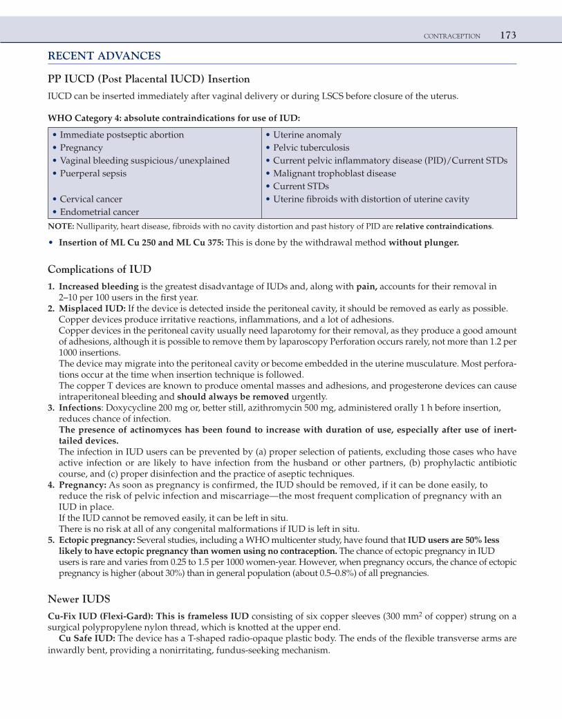
CONTRACEPTION 173
RECENT ADVANCES
PP IUCD (Post Placental IUCD) Insertion
IUCD can be inserted immediately after vaginal delivery or during LSCS before closure of the uterus.
WHO Category 4: absolute contraindications for use of IUD:
• Immediate postseptic abortion • Pregnancy • Vaginal bleeding suspicious/unexplained • Puerperal sepsis
• Cervical cancer • Endometrial cancer
• Uterine anomaly • Pelvic tuberculosis • Current pelvic inflammatory disease (PID)/Current STDs • Malignant trophoblast disease • Current STDs • Uterine fibroids with distortion of uterine cavity
NOTE: Nulliparity, heart disease, fibroids with no cavity distortion and past history of PID are relative contraindications.
• Insertion of ML Cu 250 and ML Cu 375: This is done by the withdrawal method without plunger.
Complications of IUD
1. Increased bleeding is the greatest disadvantage of IUDs and, along with pain, accounts for their removal in 2–10 per 100 users in the first year.
2. Misplaced IUD: If the device is detected inside the peritoneal cavity, it should be removed as early as possible. Copper devices produce irritative reactions, inflammations, and a lot of adhesions.Copper devices in the peritoneal cavity usually need laparotomy for their removal, as they produce a good amount of adhesions, although it is possible to remove them by laparoscopy Perforation occurs rarely, not more than 1.2 per 1000 insertions.The device may migrate into the peritoneal cavity or become embedded in the uterine musculature. Most perfora-tions occur at the time when insertion technique is followed.The copper T devices are known to produce omental masses and adhesions, and progesterone devices can cause intraperitoneal bleeding and should always be removed urgently.
3. Infections: Doxycycline 200 mg or, better still, azithromycin 500 mg, administered orally 1 h before insertion, reduces chance of infection.
The presence of actinomyces has been found to increase with duration of use, especially after use of inert-tailed devices.The infection in IUD users can be prevented by (a) proper selection of patients, excluding those cases who have active infection or are likely to have infection from the husband or other partners, (b) prophylactic antibiotic course, and (c) proper disinfection and the practice of aseptic techniques.
4. Pregnancy: As soon as pregnancy is confirmed, the IUD should be removed, if it can be done easily, to reduce the risk of pelvic infection and miscarriage—the most frequent complication of pregnancy with an IUD in place.
If the IUD cannot be removed easily, it can be left in situ.There is no risk at all of any congenital malformations if IUD is left in situ.
5. Ectopic pregnancy: Several studies, including a WHO multicenter study, have found that IUD users are 50% less likely to have ectopic pregnancy than women using no contraception. The chance of ectopic pregnancy in IUD users is rare and varies from 0.25 to 1.5 per 1000 women-year. However, when pregnancy occurs, the chance of ectopic pregnancy is higher (about 30%) than in general population (about 0.5–0.8%) of all pregnancies.
Newer IUDS
Cu-Fix IUD (Flexi-Gard): This is frameless IUD consisting of six copper sleeves (300 mm2 of copper) strung on a surgical polypropylene nylon thread, which is knotted at the upper end.
Cu Safe IUD: The device has a T-shaped radio-opaque plastic body. The ends of the flexible transverse arms are inwardly bent, providing a nonirritating, fundus-seeking mechanism.
174 SMART STUDY SERIES: OBSTETRICS AND GYNECOLOGY
ORAL CONTRACEPTIVE PILLS
Combined Pills
These are of two types: monophasic pills and multiphasic pills.
Monophasic PillsThese pills contain estrogen and progestogen in the same amount in each pill.They are divided into three subgroups:
1. Standard dose containing ethinyl estradiol (EE) 0.05 mg/day (50 μg/day). 2. Low-dose pills containing EE 0.03–0.035 mg in each pill 3. Very low-dose containing 0.020 mg EE in each pill
Each pill contains a progestogen such as levonorgestrel 0.015 mg or other newer varieties such as desogestrel, ges-todene, norgestimate, norethisterone, and drospirenone (DRSP).
Multiphasic PillsThese phasic formulations employ low doses and variable amounts of estrogen and progestogen in two (biphasic) or three (triphasic) periods within the menstrual cycle. The dose of progestogen is low at the beginning and higher at the end, while the estrogen remains either constant or rises slightly in mid-cycle. The total doses of steroids in a whole cycle are less in these pills.Very rarely used today.Four groups of progestogens are used nowadays in oral contraceptives (OCs):
1. Norethisterone group
Pills containing these drugs are called first-generation pills.
2. Norgestrel
Pills containing norgestrel are called second-generation pills.
3. 19-Nortestosterone Derivatives
Three progestogens, namely desogestrel, gestodene, and norgestimate—have been developed for contraceptive use.They have minimal androgenic and anabolic effects, indeed virtually none. The decreased androgenicity of the
new products is reflected in increased sex-hormone-binding globulin and decreased free testosterone concentration. This effect has the potential to decrease acne, hirsutism and promote favorable lipid changes.
OCs containing desogestrel or gestodene produce less break through bleeding (BTB) and do not increase body weight in most cases. On the other hand, OCs containing desogestrel or gestodene probably carry a small extra risk of venous thromboembolism (VTE) beyond that attributable to OCs containing LNG.
4. A new progestin called drospirenone (DRSP) is derived from 17-alpha spironolactone, an analog of spironolactone. It has antiandrogenic and antimineralocorticoid activities. The USFDA in May 2001 approved “Yasmin,” containing 0.03 mg of EE and 3 mg of DRSP, as a monophasic birth control pill for women. OCs containing DRSP/EE have been found to be highly effective and provide a safety level equivalent to that of other pills. These OCs lessen acne, hirsutism, seborrhea, and premenstrual syndrome.
OCs containing desogestrel, gestodene, and DRSP are called third-generation pills.
• Mala-N (30 μg EE + 0.30 mg norgestrel per pill) is supplied free in India through family planning (welfare) clinics. Mala-D (30 μg EE + 0.15 mg levonorgestrel per pill) is sold at a subsidized rate (1/10th or 1/30th the price of other preparations).
• One OC pill is to be taken during the first cycle from the first day or any of the next 4 days and should be continued daily for 21 days, stopped and restarted after a gap of 7 days, irrespective of onset or stoppage of menstruation during these pill-free days.
In lactating women, it is preferable to use progestogen-only pills, if they are available, and the women choose to use them. Otherwise, combined oral contraceptives (COCs) should be used after breast feeding is stopped fully or
https://kat.cr/user/Blink99/
CONTRACEPTION 175
nearly fully, or 6 months after childbirth, whichever comes first. In nonlactating women, COCs should be started 3–6 weeks after childbirth or if menstruation starts, whichever is earlier.
Mechanism of Action 1. Inhibition of ovulation: The combined pills inhibit ovulation by suppressing hypothalamic-releasing factors,
which in turn leads to inappropriate secretion of FSH and LH: these hormones are maintained at constant low levels similar to those seen in the proliferative phase of the cycle. As a result, no LH surge occurs and ovulation is suppressed.
2. Alteration of endometrium: OCs alter maturation of the endometrium, rendering it unsuitable for implantation of the fertilized ovum.
3. Changes in cervical mucus: Cervical mucus becomes scanty, viscous, and cellular with low spinnbarkeit and no ferning; these changes impair sperm transport and penetration.
Pearl index: Combined pills are very effective. The failure rate when correctly and consistently used is only 0.1% or 1 per 1000 in the first year of use, but the typical failure rate, as is commonly used, is 1.8%.
The failures are mostly due to missed pills, delay in starting the next course, and stoppage of the drug due to side effect or fear complex without taking other contraceptive measures.
Advantages 1. Cure of menstrual disorders: OCs cure dysmenorrhea and ovulation pain. Menorrhagia and metrorrhagia can
always be controlled by the use of COCs. OCs also lessen premenstrual tensions such as nervousness, irritability, depression, etc., during 7–10 days before menses. Also the cycles become regular.
2. Protection against cancer: It has been conclusively proved that OCs directly prevent two common types of genital cancer: endometrial cancer and ovarian cancer; it also indirectly prevents choriocarcinoma by preventing pregnancy.
COCs decrease the ovarian cancer by about 40% and the effect persists for at least 10 years. COCs also lower the risk of endometrial cancer by about 50%; the effect lasts for up to 15 years.They also decrease the risk of colon cancer.
3. Protection against benign tumors and related diseases: a. Benign breast diseases (BBDs): It is well documented that BBDs, such as fibrocystic and fibroadenomatosis
diseases, are reduced by 50–70% in pill users. b. Ovarian functional cysts: Various studies have shown that low-dose OCs lower the risk of developing
functional ovarian cysts. The risk of follicular cysts goes down by 50% and that of corpus luteum cysts by about 80%.
c. Fibromyoma of the uterus: The risk of uterine fibroid is reduced by about 30% in women who have used OCs for 10 years. Low-dose OCs help reduce fibroids and lessen menstrual flow.
4. Protection against diseases: a. Ectopic pregnancy: Chance of ectopic pregnancy with its grave consequences is lowered by 50% in low- dose
OC users. b. Pelvic inflammatory diseases: Several studies have shown that regular pill users are protected from PIDs to
the extent of 50%. OCs reduce PIDs by hindering the ascent of STD bacteria (including chlamydia) from the vagina upward by thickening the cervical mucus and lessening uterine motility, as well as by obviating illegal abortions and delivery of unwanted children.However, barrier contraceptives protect women better against STDs and HIV/AIDS than OCs do.
c. Anemia and malnutrition: Pills reduce iron deficiency anemia by reducing menstrual flow in 60–80% of pill users; they improve nutrition of women by preventing repeated and frequent pregnancies.
d. Endometriosis: Combined high-dose pills control endometriosis to a good extent when used continuously with increasing doses to produce pseudopregnancy.
e. Acne and hirsutism: OCs are effective in treating acne and hirsutism by increasing sex-hormone-binding globulin and significantly decreasing free testosterone levels. Formulations with desogestrel, DRSP and cyproterone are specially effective in this respect.
5. Premenstrual syndrome: OCs and pills containing DRSP reduce premenstrual syndrome.
176 SMART STUDY SERIES: OBSTETRICS AND GYNECOLOGY
Side Effects and Risks 1. Breakthrough bleeding: This is slightly more common with the lower-dose pills. The women should have two pills a day for 2 or 3 days, which usually controls BTB; if not, EE 0.02 mg may be
taken for 7 days along with the pills. 2. Oligomenorrhea happens sometimes with low-dose pills. The women should be reassured that oligomenorrhea
is not harmful but rather good for health. But if they are not convinced, EE 0.02 mg may be added in the last 7 days for a few cycles.
Amenorrhea is usually temporary and not harmful. Change to triphasic pills or supplementation with EE for two to three cycles usually cures amenorrhea. 3. Stroke and myocardial infarction: Women who do not smoke, have their blood pressure checked, and do not
have hypertension or diabetes are at no increased risk of myocardial infarction if they use low-dose COCs, irrespective of their age and duration of OC use.
The risk of hemorrhagic stroke does not increase in women below 35 years of age who do not smoke and are not hypertensive.
Current users of low-dose COCs have a low absolute risk of VTE mainly because incidence of VTE is very low in nonpregnant women. Nevertheless, this risk is three to six times more than nonusers. The absolute risk of VTE attributable to OC use rises with increasing age, recent surgery, and some forms of thrombophilia. Progestogens are associated with the increase of low-density lipoprotein cholesterol and a decrease of high-density cholesterol, which enhance the risk of atherosclerosis, coronary heart disease and cerebral thrombosis; but estrogens have the opposite effect, and these actions seem relatively balanced in low-dose COCs.
4. Breast and cervical cancer: There is a small increase in risk of current users of the pill (relative risk 1.24), and the risk reduces gradually over the 10 years after discontinuing use.
Breast cancer in current or past OC users is largely localized in the breast—a condition that usually has a better prognosis.
The risk of breast cancer is due to the progestogen component of the pills, as the risk is same among users of progestogen-only methods.
Studies in developed and developing countries have shown a modest increase in the risk of cervical cancer (1.3–1.8-fold) among women who have used COCs for more than 5 years. However, it is not clear whether the increased risk is due to direct effect of the pill or some characteristics of the pills’ users such as age at first intercourse, number of sexual partners, parity, and smoking status.
5. Liver tumor: OCs increase the incidence of a rare benign liver tumor, namely, primary hepatocellular adenoma. • Vitamin B6 (pyridoxine) may help curing depression after OC use.
Drug InteractionsBarbiturates, sulfonamides, rifampicin, and anticonvulsant drugs interfere with the effect of OCs and increase fail-ure rates. As such, it would be prudent to use high-dose formulations if other contraceptives prove unsuitable for patients taking those drugs.
Medical Eligibility Criteria for Initiation and Continuation of Combined OCs/Combined Injections/Transdermal Patches and Vaginal Rings.
WHO Category 4: Absolute contraindications for combined OC pills/combined injections/combined vaginal rings and patches
• Active liver disease (hepatitis/tumor) • Postpartum: breast-feeding women <6 week
postpartum • Thrombophilias • Ischemic heart disease • Complicated migraine • Pregnancy • Complicated valvular heart disease
• Breast cancer (current or past history) • Severe hypertension (systolic> 160 or diastolic >100) • DM with vascular complications • Current history of thromboembolism/stroke/deep
vein thrombosis
NOTE: Smoking, age more than 35 years, mild hypertension and uncomplicated DM are relative contraindications.
https://kat.cr/user/Blink99/
CONTRACEPTION 177
Progesterone-Only Pills/Mini Pills
Women who can use (indications) progestogen-only contraceptives safely and effectively include:
• Age: menarche to menopause • Hypertension adequately controlled • Thrombotic disorders • Obesity • Breast feeding >6 weeks onward • DVT/PTE • Non-breast-feeding before or after 21 days • Valvular heart disease • Smoking: any age
Category 4 for progesterone-only pills:
1. Pregnancy 2. Breast cancer 3. Unexplained vaginal bleeding
Mechanism of action: same as COC.They should be taken at the same time every day.Pearl index for progesterone-only pills = 3%
CentchromanTo avoid bad effects of OCs, centchroman has been produced by the researchers of Central Drug Research Institute, Lucknow, India. It is a nonhormonal, chemical-synthetic, once-a-week OC. “Centron” and “Saheli,” contain 30 mg of centchroman.
Centchroman has a weak estrogenic and potent antiestrogenic effect—acting mostly on the endometrial target organs to suppress proliferation of the endometrium, thereby interfering with nidation of the embryo; it has no pro-gestational, androgenic, or antiandrogenic properties.
Antiprogesterone RU-486 (Mifepristone)This antiprogesterone compound, which prevents hormone action at the receptor level, produces a contraceptive effect at several points in the menstrual cycle. Given orally in mid-cycle, it can delay or inhibit the mid-cycle LH surge; administered in the late luteal phase, it induces menstruation or, when menses is delayed, very early abortion.
It has potential in future for use as a once-a-month pill during the luteal phase, however as of now it is not used for contraception.
INJECTABLE CONTRACEPTIVES
Progestogen-only injectable contraceptives:
1. Depot-medroxyprogesterone acetate (DMPA) 2. Norethisterone enanthate (NET EN or Noristerat)
One injection of Depo-Provera remains effective for 3 months. It is administered in the form of a 150 mg injection once every 3 months plus or minus 14 days.
One 200 mg NET EN injection is to be taken every 2 monthsBoth DMPA and NET EN are highly effective methods of contraception.Pearl index: Typical failure rate of progestogen-only injectables, as commonly used, is 0.1–0.4%.
Mechanism of Action
The injectable contraceptives act by inhibiting ovulation in most women. They also work by making cervical mucus thick and scanty, thus creating a barrier to sperm penetration, and making the endometrium less suitable for implantation.
178 SMART STUDY SERIES: OBSTETRICS AND GYNECOLOGY
Noncontraceptive Benefits
1. It cures menstrual troubles like menorrhagia and dysmenorrhea 2. Medical management of endometriosis (pseudo pregnancy regimen) 3. Prevention and treatment of endometrial hyperplasia. 4. DMPA prevents sickling and the development of abnormal-shaped red blood cells, and lessens episodic bone
pain in women suffering from sickle cell diseases; it is thought to be the best contraceptive for patients of sickle cell anemia.
5. DMPA reduces the risk of pelvic inflammatory disease and ectopic pregnancy. 6. DMPA use protects against the risk of endometrial and ovarian cancer. 7. Injectables are suitable in cases with myoma and endometriosis, as contraception is provided without estrogen
effect.
Side Effects
1. Irregular menstrual bleeding and spotting, as well as temporary amenorrhea, are the most common side effects in DMPA and NET EN users.
2. Weight gain: The average weight gain is 1–3 kg in most cases. 3. There is a delay of few months in becoming pregnant following discontinuation of the injection. 4. Bone density changes: There is a risk of bone loss among long-term DMPA users leading to osteoporosis;
however, this bone loss is reversible on cessation of the contraception.
Combined (estrogen + progesterone) monthly injectable contraceptives:
1. DMPA 25 mg plus estradiol cypionate 5 mg marketed as “Cyclofem” 2. NET EN 50 mg plus estradiol valerate 5 mg marketed as “Mesigna”
CONTRACEPTIVE IMPLANTS
The Norplant system consists of six silastic capsules each containing 36 mg of LNG. These are inserted under the skin in the inside of the upper arm or forearm in most cases, in a fan-shaped manner under local anesthesia. It is effective for 5 years.
Norplant II or Jadelle has two rods, and remains effective for 5 years.Norplant prevents pregnancy in three ways: (1) it makes cervical mucus thicker and scantier, preventing sperm
penetration; (2) LNG suppresses ovulation and (3) it depresses the endometrial growth, necessary for implantation of the ovum.
Both Norplant and LNG rod (Norplant II or Jadelle) have a failure rate of 0.4–0.8%.Implanon is a new contraceptive implant. It is a single-rod device containing 67 mg of the progestogen 3-keto-
desogestrel, also called etonogestrel (ENG).It is placed subcutaneously on the inner side of the upper arm under local anesthesia. Implanon acts primarily by
inhibiting ovulation, supplemented by the usual mucus and endometrial effects (similar to Norplant).NO PREGNANCIES have been reported so far with the use of Implanon. Trials in India are being conducted by
ICMR (Indian Council of Medical Research). It is likely to be launched in India by the year 2011.
CONTRACEPTIVE RINGS
1. Nuva Ring: It is a soft vaginal ring that releases 15 μg EE and 120 μg ENG, the active metabolite of desogestrel, per day as a controlled delivery system.
Women keep the NuvaRing in the vagina for 3 weeks and then remove it for 1 week, during which they have withdrawal bleeding.
A new vaginal ring is needed for each 4-week cycle. Increased patient compliance is the advantage over OC pills. It has been launched in India in November 2009. The efficacy rate of NuvaRing is like that of COCs—the failure rate after perfect use is 0.3%.
https://kat.cr/user/Blink99/
CONTRACEPTION 179
2. A vaginal progesterone-only ring called “Progering” has been developed and has been undergoing clinical trials.
It contains natural hormone progesterone. These rings are slightly less effective than combined vaginal rings; however, they are very effective in lactating women because breast feeding itself provides some protection against pregnancy. They do not contain estrogen, which can reduce milk production. Each ring releases 10 mg of progesterone daily and lasts for 3 months.
3. LNG ring: It contains 5 mg LNG, 20 μg/day, is released; left inside vagina for 3 months continuously.
TRANSDERMAL CONTRACEPTIVE PATCH
The combined patch delivers 150 μg of the proestrogen (norelgestromin) and 20 μg of EE per day. A woman wears a patch for 1 week and then replaces it by another one placed at a different site for a total of 3 weeks, followed by 1 week with no patch.
The patches work by preventing ovulation, thickening the cervical mucus, and suppressing endometrial growth.It provides effectiveness and cycle control like those of OCs when used. The failure rate with typical use within
the first year is 2 per 100 women and with perfect use 0.3 per 100 women.
EMERGENCY CONTRACEPTION (INTERCEPTIVES)
Agents that do not interfere with fertilization but act on the endometrium to prevent implantation are called “inter-ceptive agents,” and those that interfere with early gestation causing an abortion are called “contragestives.”Indications:
1. Unplanned , unprotected intercourse 2. After rape 3. Rupture or tear in the condom at the time of intercourse.
Two methods of emergency contraception are available now: (1) hormonal and (2) mechanical (IUD). There are two types of hormonal emergency contraception (emergency window = 72 hours)
1. LNG-only pills (most commonly used)
One tablet of 0.75 mg LNG pill should be taken as soon as possible after unprotected intercourse, followed by a same dose taken 12 h later; both doses must be taken within 72 h of intercourse.
Single 1.5 mg dose of LNG is as effective for emergency contraception as two 0.75 mg doses of LNG taken 12 h apart.
Failure rate (pregnancy rate) = 0–1%
2. Combined Estrogen and Progestogen Pills (Also Known as the Yuzpe Regimen)
High-dose pills contain 50 μg of EE and 250 μg LNG (or 500 μg norgestrel). Two pills should be taken as soon as pos-sible, but not later than 72 h of unprotected coitus; this must be followed by two other pills 12 h later.
When only low-dose pills containing 30 μg of EE and 150 μg of LNG (300 μg of norgestrel) are available, four pills should be taken as the first dose within 72 h of unprotected intercourse, followed by four more pills after 12 h.
Main side effect is nausea and vomitingFailure rate = 0–2%The mechanism of action of emergency contraceptive pills has not been clearly established. They may act through
(1) inhibition or delay of ovulation, (2) prevention of implantation in the altered endometrium (interception = main action), and (3) prevention of fertilization due to quick transport of sperms or ova. They cannot interrupt already established pregnancy.
IUDs introduced postcoitally can prevent pregnancy very successfully. (Failure rate = 0.1%).IUDs can be used postcoitally up to 5 days following sexual exposure. Thus, this method can be used even after
48 h more delay than the hormonal methods allow.
180 SMART STUDY SERIES: OBSTETRICS AND GYNECOLOGY
Antiprogesterone (Mifepristone)
Latest WHO randomized trial has noted that a single dose of 10 mg mifepristone is as effective as LNG for emergency contraception, with no difference in side effects; periods start after 7 days—a bit delayed than after LNG regimen. However, as of date low-dose mifepristone for emergency contraception has not been registered in any country.
MALE STERILIZATION
Two methods of male sterilization are followed nowadays: (1) conventional vasectomy and (2) no-scalpel vasectomy.Sterility does not occur immediately after the procedure. Sperms remain in the semen for 15–20 ejaculations,
requiring continued contraception for about 3 months. Absence of sperms after 3 months must be confirmed with a microscope before confirmation of sterility.
No-Scalpel Vasectomy
This method of vasectomy “without the use of a scalpel” was introduced in China in 1974 by Dr. Li. Contraindica-tions: No permanent contraindications. Failure rate of vasectomy is 0.1 per 100 women partners in first year when performed properly.
Reversal is possible with microsurgery, giving 90% return of sperm and about 70% of pregnancy rate. This declines with time, particularly after 7 years.
FEMALE STERILIZATION
Female sterilization is the most widely used contraceptive method in the world.It can be done by laparotomy or laparoscopy.The following are the laparotomy methods:
1. Pomeroy technique (most commonly done laparotomy method): After bringing out the fallopian tube through the incision, a clamp is placed about 4 cm lateral to the fundus and the tube is pulled up so as to form a loop. The Pomeroy operation is the most simple and safe procedure of tubal ligation. It has got a failure rate of 1 in 300–400 operation.
2. Irving technique: This technique has a very low failure rate, less than 1 in 1000 cases. 3. Uchida technique: Uchida claims no failure in 19,000 cases. 4. Fimbriectomy (Kroener’s technique): This technique has been abandoned at present due to high failure rate (2–3%). 5. Madlener technique: The procedure is very simple but has a high failure rate of 0.3–2% and has been practically
abandoned. 6. Parkland technique: The failure rate of this technique is about 1 in 400 procedures.NOTE: Least failure rate (among laparotomy techniques) = Uchida followed by Irving.
Laparoscopic Tubal Ligation:Female sterilization with the use of an operating laparoscope is getting more and more popular because it has been found to be a safe, simple, and effective procedure that can be performed through one or two very small incisions in the abdomen, mostly under sedation and local anesthesia on an outpatient basis. Verres needle: It is used to intro-duce gas or air for pneumoperitoneum.
Carbon dioxide is the most common gas used for distention. The intra-abdominal pressure during laparoscopy surgery should be kept between 10–15 mmHg and never exceed 20–25 mmHg.
Contraindications of Laparoscopic Tubal Ligation
Absolute Contraindications:
1. Large abdominal mass (uterine or ovarian tumors) needing laparotomy. 2. Decompensated heart disease. 3. Severe respiratory dysfunction. 4. Hiatus hernia. 5. History of abdominal surgery, especially of the bowel.
https://kat.cr/user/Blink99/
CONTRACEPTION 181
Relative contraindications are:
1. Gross obesity with thick abdominal wall and 2. Pelvic adhesion due to previous pelvic infection or operations. Laparoscopic sterilization should not be done
soon after delivery or abortion of more than 12 weeks pregnancy.
Method of Tubal Occlusion
Silastic bands (Yoon) or spring-loaded clip (Hulka-Clemens) are two methods used for occlusion. The electrocoagu-lation methods cause less pain but may produce serious gastrointestinal burns; burns of other organs, vessels, and the abdominal wall may also occur. The incidence of burns after unipolar electrocoagulation method is greater, and as such this method has almost been given up.
1. Falope ring/silastic bands: At present in India, this silastic band technique is most popular and most commonly used for laparoscopic tubal ligation.
2. Clips: Two types of clips are mostly used: the spring-loaded clip (Hulka-Clemens clip) and silicone- titanium clip (Filshie clip). A clip is placed on the isthmus on each tube, 2–3 cm from the uterus, with a special straight-type laparoscope.
The clips cause least damage to the tube (about 1 cm), whereas tubal damage is 3 cm with the Falope ring and 3–5 cm with the Pomeroy technique.
Failure rate of laparoscopic sterilization = 0.2–1.3%. Spring clips have the highest failure rate whereas unipolar coagulation has the least failure.
Hysteroscopic tubal ligation (with silastic plugs, quinacrine, and cautery) is still under research.Essure (available in France, not yet in India): The microcoil Essure is a spring-like device. This is introduced
using a hysteroscope inserter through the vagina into the uterus and then into each fallopian tube. In 3 months’ time, scar tissue grows into the device and plugs the fallopian tube; hence, sperms cannot pass through to fertilize an egg.
MENSTRUAL REGULATION
Menstrual regulation (MR) is the treatment of the delayed menstrual period up to 14 days, to assure a nonpregnant state and normal menstrual cycle next time.
For surgical methods of MR, suction evacuation is usually preferred either by the use of plastic cannulae along with a special plastic syringe (modified Karman syringe) or by using plastic or metal cannulae along with an electri-cally operated suction apparatus.
The modified Karman syringe is a transparent plastic syringe of 50-mL capacity, capable of producing a vacuum of 27 inches or 675 mmHg at sea level.
MEDICAL TERMINATION OF PREGNANCY (MTP)
1. Medical method for first trimester MTP
It is now officially allowed in India up to 9 weeks (63 days) of gestation.Method: combination of RU486 followed by PGE1.
Mifepristone, also known as RU-486, is an antiprogesterone compound
• It acts preferentially on target cells of the endometrium and deciduas, counteracting the effect of progesterone, which is essential for establishment and maintenance of pregnancy.
• It affects the pituitary gonadotropic cells, producing a remarkable decrease of LH secretion, leading to luteolysis. • It causes softening and ripening of the cervix and produces increased contractibility of the myometrium. • It causes a marked increase in sensitivity of the uterus to exogenous PGs.
Misoprostol (PGE1)
It acts by (a) enhancing uterine contraction and thus helping expulsion of the products of contraception and (b) causing cervical ripening or priming. It is used orally as tablets and vaginally as a suppository. Success rate of this combination is 96%.
182 SMART STUDY SERIES: OBSTETRICS AND GYNECOLOGY
Fewer than 5% of women undergoing medical methods of abortion will need surgical intervention (check curet-tage) for incomplete abortion.
For the medical abortion up to 9 completed weeks since last menstrual period, mifepristone plus PGs are used; the dosage regimens recommended by World Health Organization are as follows:
200 mg mifepristone followed after 36–48 h by:
• 800 μg vaginal misoprostol or • 400 μg oral misoprostol
Contraindications (due to medical reasons) for medical method of abortion:
• Smoking > 35 years • Hemoglobin < 8 g% • Confirmed/ suspected ectopic pregnancy/ undiagnosed adnexal mass • Coagulopathy or patient on anticoagulant therapy • Chronic adrenal failure or current use of systemic corticosteroids • Uncontrolled hypertension with BP >160/100mmHg • Certain cardio-vascular diseases • Severe renal, hepatic or respiratory diseases • Glaucoma • Uncontrolled seizure disorder • Allergy or intolerance to mifepristone /misoprostol or other prostaglandins • Lack of access to 24 hours emergency services.
2. Surgical Technique (Suction Evacuation/Manual Vacuum Evacuation)
It is allowed up to 12 weeks of gestation.
Complications of MTP
1. Uterine hemorrhage: It occurs in 1–4% cases. 2. Pelvic infection: It ranges from 0.1% to 1.5%. It is due to incomplete evacuation and improper aseptic technique.
The incidence can be reduced to a great extent by prophylactic use of antibiotic. 3. Cervical injury: This complication occurs in 0.01–1% cases. 4. Uterine perforation: This is the most dangerous complication, but fortunately it happens very rarely in
0.1– 0.28% cases. When perforation occurs or is suspected, the patient should be kept under observation and antibiotic should be
started. Usually she can be discharged in 24 h time. If there is strong suspicion or actual diagnosis of injury to the intestines or omentum, or if hemorrhage occurs, laparotomy should be performed followed by necessary steps.
5. Retained products: Incomplete abortion happens in 24% cases. 6. Continuation of pregnancy: In about 1% cases.
SECOND TRIMESTER MTP (13–20 WEEKS)
1. Misoprostol (PGE1) tablet vaginally is most widely used for second trimester pregnancy termination. 2. Ethacridine lactate extra-amniotically can also be used for second trimester pregnancy termination. A solution
of 10 mL of 0.1% ethacridine is used for each gestational week, up to a maximum 150 mL. Induction abortion interval is about 30 h, even with intravenous oxytocin as an augmenting agent.
NOTE: Intra-amniotic saline/mannitol/urea, etc. are no longer used because of risk of maternal mortality.
TUBAL LIGATION REVERSAL
The remaining length of the tube is one of the most important factors influencing reversal. The more the length, the more successful the results. Minimum length of reconstructed tube should be 4 cm and the ampullary part should be at least 2 cm.
https://kat.cr/user/Blink99/
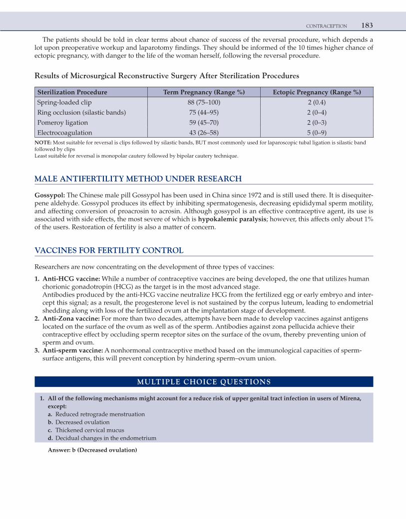
CONTRACEPTION 183
The patients should be told in clear terms about chance of success of the reversal procedure, which depends a lot upon preoperative workup and laparotomy findings. They should be informed of the 10 times higher chance of ectopic pregnancy, with danger to the life of the woman herself, following the reversal procedure.
Results of Microsurgical Reconstructive Surgery After Sterilization Procedures
Sterilization Procedure Term Pregnancy (Range %) Ectopic Pregnancy (Range %)
Spring-loaded clip 88 (75–100) 2 (0.4)
Ring occlusion (silastic bands) 75 (44–95) 2 (0–4)
Pomeroy ligation 59 (45–70) 2 (0–3)
Electrocoagulation 43 (26–58) 5 (0–9)NOTE: Most suitable for reversal is clips followed by silastic bands, BUT most commonly used for laparoscopic tubal ligation is silastic band followed by clipsLeast suitable for reversal is monopolar cautery followed by bipolar cautery technique.
MALE ANTIFERTILITY METHOD UNDER RESEARCH
Gossypol: The Chinese male pill Gossypol has been used in China since 1972 and is still used there. It is disequiter-pene aldehyde. Gossypol produces its effect by inhibiting spermatogenesis, decreasing epididymal sperm motility, and affecting conversion of proacrosin to acrosin. Although gossypol is an effective contraceptive agent, its use is associated with side effects, the most severe of which is hypokalemic paralysis; however, this affects only about 1% of the users. Restoration of fertility is also a matter of concern.
VACCINES FOR FERTILITY CONTROL
Researchers are now concentrating on the development of three types of vaccines:
1. Anti-HCG vaccine: While a number of contraceptive vaccines are being developed, the one that utilizes human chorionic gonadotropin (HCG) as the target is in the most advanced stage.Antibodies produced by the anti-HCG vaccine neutralize HCG from the fertilized egg or early embryo and inter-cept this signal; as a result, the progesterone level is not sustained by the corpus luteum, leading to endometrial shedding along with loss of the fertilized ovum at the implantation stage of development.
2. Anti-Zona vaccine: For more than two decades, attempts have been made to develop vaccines against antigens located on the surface of the ovum as well as of the sperm. Antibodies against zona pellucida achieve their contraceptive effect by occluding sperm receptor sites on the surface of the ovum, thereby preventing union of sperm and ovum.
3. Anti-sperm vaccine: A nonhormonal contraceptive method based on the immunological capacities of sperm-surface antigens, this will prevent conception by hindering sperm–ovum union.
MULTIPLE CHOICE QUESTIONS
1. All of the following mechanisms might account for a reduce risk of upper genital tract infection in users of Mirena, except:
a. Reduced retrograde menstruation b. Decreased ovulation c. Thickened cervical mucus d. Decidual changes in the endometrium
Answer: b (Decreased ovulation)
184 SMART STUDY SERIES: OBSTETRICS AND GYNECOLOGY
Explanation:Mirena/LNG-20 is a third-generation IUCD.The progesterone effect on thickening of the cervical mucus and decidual changes of the endometrium add to prevention
of PID. Hence, it also prevents ectopic pregnancy. The progesterone support of the endometrium decreases the menstrual bleeding and hence the retrograde reflux. Ovulation is not affected by Mirena.
Reference: 1. Chaudhary SK, 7th Ed., Pg. 152.
2. An intra-uterine pregnancy of approximately 10 weeks’ gestation is confirmed in a 30-year-old gravida 5, para 4 woman with an IUD in place. The patient expresses a strong desire for the pregnancy to be continued. On examination, the string of the IUD is noted to be protruding from the cervical os. The most appropriate course of action is to:
a. Leave the IUD in place without any other treatment b. Remove the IUD to decrease the risk of malformations c. Remove the IUD to decrease the risk of infection d. Terminate the pregnancy because of the high risk of malformations
Answer: c (Remove the IUD to decrease the risk of infection)
Explanation:Although there is an increased risk of spontaneous abortion, and a small risk of infection, an intra-uterine pregnancy can
occur and continue successfully to term with an IUD in place. However, if the patient wishes to keep the pregnancy and if the string is visible, the IUD should be removed in an attempt to reduce the risk of infection, abortion, or both. An IUD in situ does not cause any malformations/anomalies in the fetus.
WHO recommends that if the IUD can be removed easily it should be removed to reduce the risk of infection and abortion.
If the IUD cannot be removed easily, it can be kept in situ and it will be expelled after placental delivery.
Reference: 1. Chaudhary SK, 7th Ed., Pg. 110–1.
3. Use of oral contraceptive pills are known to protect against following malignancies, except: [AIIMS Nov 2002] a. Ovarian carcinoma b. Endometrial carcinoma c. Uterine sarcoma d. Carcinoma cervix
Answer: d (Carcinoma cervix)
Explanation:Protection against ovarian carcinoma, one of the most lethal cancers of the female reproductive tract, is one of the benefits of OCPs. The risk of developing epithelial ovarian cancer in OCP users is reduced by 40% compared to that of nonusers. This protective effect increases with duration of use (about 5–10 years) and continues for at least 10–15 years after discontinuation of OCPs. This protection is seen in women who use OCPs for as little as 3–6 months and reaches an 80% reduction in risk with more than 10 years of use.
OCPs also protect against endometrial cancer and uterine sarcomas. Use for at least 12 months reduces the risk by 50, with the greatest protective effect gained by use for more than 3 years. The protection persists for 15 years after discontinuation and is greatest in women at risk: nulliparous and low parity women. OC pills also decrease the risk of colon cancer.
Studies have indicated that there is marginal increase in relative risk for dysplasia of cervix and invasive Ca cervix and breast cancer after prolonged use of OC pills.
https://kat.cr/user/Blink99/
CONTRACEPTION 185
Reference: 1. Chaudhary SK, 7th Ed., Pg. 127–30.
4. The progesterone component of combined oral contraceptive pills acts by: a. Preventing ovulation b. Inhibiting nidation c. Bringing about alterations in the cervical mucus d. All of the above
Answer: d (All of the above)
Explanation:Actions of the progesterone component of combined oral contraceptives:
1. Suppresses ovulation by its inhibitory action on the pituitary and the hypothalamus. This is predominantly achieved by estrogens but even by progesterone.
2. Causes atrophic changes in the endometrium and prevents nidation even if fertilization occurs. 3. Acts on the cervical mucus, making it thick and tenacious and impenetrable by sperms.
The third-generation progestogens have a higher affinity for progesterone receptor and have a role in inhibiting ovulation.The main function of progestogens in combined pills is, however, to counteract the undesirable effects of estrogen such as
endometrial hyperplasia and heavy withdrawal bleeding.
Reference:
1. Chaudhary SK, 7th Ed., Pgs. 125–6, 141.
5. Minimum effective dose of ethinyl estradiol in combined oral pills is — micrograms per pill: [AIIMS May 2004, All India 2013] a. 20 b. 30 c. 50 d. 10
Answer: a (20)
Explanation:The essential constituent of combined oral contraceptives is an estrogen in the form of ethinyl estradiol (EE). They are divided into three subgroups:
1. Standard dose containing EE 0.05 mg/day in each pill 2. Low-dose pills containing EE 0.03–0.035 mg in each pill 3. Very low-dose containing 0.020 mg (20 μg) EE in each pill
For a combined pill to be effective, EE should be minimum 20 μg per pill.
Reference:
1. Chaudhary SK, 7th Ed., Pg. 121–2.
6. Emergency contraception prevents pregnancy by all of the following mechanisms, except: [All India 2003, All India 2006] a. Delaying/inhibiting ovulation b. Inhibiting fertilization c. Preventing implantation of the fertilized egg d. Interrupting an early pregnancy
Answer: d (Interrupting an early pregnancy)
186 SMART STUDY SERIES: OBSTETRICS AND GYNECOLOGY
Explanation:Emergency contraception is used to prevent pregnancy after the act of an unprotected intercourse.It is an interceptive. Its main action is to make the endometrium unsuitable for implantation. It may also prevent or delay
ovulation, and prevent fertilization of the egg by the sperms. It has, however, no role in the interruption of early pregnancy once conceived. They are not abortifacients or contragestives. They cannot interrupt an early pregnancy, and hence a preg-nancy test is recommended if the woman does not bleed within 7 days of the usage.
Reference:
1. Chaudhary SK, 7th Ed., Pg. 170–2.
7. Most commonly removed/resected parts of loop in tubectomy include: [All India 2007] a. Interstitial b. Isthmus c. Ampulla d. Fimbrial end
Answer: b (Isthmus)
Explanation:Tubal ligation and resection (removal) of a portion of the Fallopian tube is the most frequent method of blocking the tubes.
This involves tying a segment of tube and removing it (usually the isthmus and small part of ampulla is removed). There are many variations of this technique. The tubal ligation procedure described by Dr. Ralph Pomeroy a century ago is most com-monly used today.
Steps: With the Pomeroy method of tubal ligation, part of the tube is elevated to create a loop or knuckle. An absorbable ligature is tied around the base of the elevated segment, and the tubal segment is cut out. Within a few days, the peritoneum grows over and covers the cut ends of the tubal segments. The cut ends of the fallopian tube separate as the ligature dissolves. The peritoneal covering and separation of the remaining tubal segments prevent them from reattaching to each other. The Pomeroy method of tubal ligation is good if tubal reversal surgery is to be considered later.
Reference: 1. Chaudhary SK, 7th Ed., Pg. 198–200.
8. The intra-abdominal pressure during laparoscopy should be set between: a. 5–8 mmHg b. 10–15 mmHg c. 20–25 mmHg d. 30–35 mmHg
Answer: b (10–15 mmHg)
Explanation:During laparoscopy, pneumoperitoneum is created with CO2 or nitrous oxide. CO2 is preferred because N2O can cause
explosion in presence of volatile anesthetic drugs. About 2 litres of gas is introduced at 10 mmHg. The intra-abdominal pressure during any laparoscopic surgery should be 10–15 mmHg. This eliminates the risk of hypercarbia or decreased venous return to heart.
Reference:
1. Chaudhary SK, 7th Ed., Pg. 209–11.
9. Mifepristone and misoprostol can be used for induction of abortion for a maximum of up to: [All India 2004, 2008, All India 2013] a. 6 weeks of amenorrhea b. 8 weeks of amenorrhea c. 7 weeks of amenorrhea d. 9 weeks of amenorrhea
Answer: d (9 weeks of amenorrhea)
https://kat.cr/user/Blink99/
CONTRACEPTION 187
Explanation:Mifepristone (Ru-486) is a 19-norsteroid derivative of synthetic progestogen norethindrone. The drug binds to receptors in
the cell nucleus and blocks progesterone action at the target organs.Mifepristone is used in combination with misoprostol for medical induction of abortion up to 9 weeks of amenorrhea
(63 days). Till mid of 2009, this combination was allowed only till 7 weeks, but now it is officially allowed up to 9 weeks of gestation.
Mifepristone is given first followed by misoprostol after 48 h. The success of this combination is 95–96%.USG should be done after about 14 days to see for any retained products of conception, and if they are present then a check
curettage will be required.
Reference:
1. Chaudhary SK, 7th Ed., Pg. 240–4.
10. All are true about intra-uterine contraceptive devices, except: [All India 2007] a. Cu T is a third-generation IUD b. LNG IUD is effective for 5 years c. IUD can be used as an emergency contraception up to 5 days after the unprotected intercourse d. None of the above
Answer: a (Cu T is a third-generation IUD)
Explanation:There are three generations of IUD:
• First-generation: inert or nonmedicated, for example, Lippes loop • Second-generation: all the copper-containing devices, for example, Copper 7, Copper T 200, Multiload Cu 250, Multiload
Cu 375, etc. • Third-generation: which release hormone, for example, Progestasert and Mirena (LNG20) • Mirena is effective for 5 years.
IUD can be used as an emergency contraception. For the hormonal tablets (OC pills or LNG), the emergency window is 72 h, but for IUD it is 5 days.
Reference:
1. Chaudhary SK, 7th Ed., Pg. 95–7.
11. IUCD having the longest life span is: [All India 2007, 2012] a. Progestasert b. Cu T 380A c. Mirena d. Nova T
Answer: b (CuT 380A)
Explanation:The time periods for replacement for various IUDs are:
Copper T 200 3 years
Copper T 380A 10 years
Multiload Cu 250 3 years
Multiload 375 5 years
LNG-IUS/Mirena 5 years
Progestasert 1 year
Nova T 5 years
188 SMART STUDY SERIES: OBSTETRICS AND GYNECOLOGY
Reference: 1. Chaudhary SK, 7th Ed., Pg. 95–7.
12. Success rate of reversal of tubal ligation is maximum in which of the following types of anastomosis? [All India 2008] a. Isthmo-isthmic b. Isthmo-ampullary c. Ampullo-ampullary d. Cornual implantation
Answer: a (Isthmo-isthmic)
Explanation:
• Tubal ligation reversal uses the techniques of microsurgery to open and reconnect the fallopian tube segments that are remaining after a tubal sterilization procedure. Microsurgery minimizes tissue damage and bleeding during surgery.
• Essential elements of microsurgical technique include gentle tissue handling, magnifying the operating field, keeping body tissues in their normal state with warmed irrigation fluids, and using the smallest sutures with the thinnest needles capable of holding the tubal ends together to promote proper healing of the rejoined tubal segments.
• An isthmo-isthmic anastomosis has the best outcome with live birth rates of 60–80%, provided that the reconstructed tube is longer than 4 cm and the ampullary portion is more than 2 cm.
Reference: 1. Chaudhary SK, 7th Ed., Pg. 272.
13. A 30-year-old P1L1 wants contraception for 6 months. She has dysmenorrhea and is a known case of complicated migraine. On USG, uterus has multiple fibroids. Contraception of choice is:
a. Cu T 200 b. OC pills c. Vaginal diaphragm d. Tubal sterilization
Answer: c (Vaginal diaphragm)
Explanation:As the patient wants contraception only for 6 months, tubal ligation cannot be done, as it is a permanent method of
contraception.Complicated migraine is category 4 (absolute contraindication) for OC pills.As the patient has multiple fibroids and dysmenorrhea, Cu T should be avoided.Hence, contraception of choice for her is vaginal diaphragm. It is a barrier method of contraception, which is to be used
along with spermicidal agent.
Reference: 1. Chaudhary SK, 7th Ed., Pg. 70, Pg. 103.
14. A 28-year-old P1L1 had Cu T inserted 2 years back. O/E-Cu-T threads are not seen. USG shows Cu T partly in abdominal cavity. Method of removal is:
a. Hysteroscopy b. No need of removal (wait and watch) c. IUCD hook d. Laparoscopy
Answer: d (Laparoscopy)
https://kat.cr/user/Blink99/
CONTRACEPTION 189
Explanation:Copper can cause inflammatory reaction and can cause intestinal obstruction. Therefore, never wait and watch.When Cu T is embedded within uterine cavity, hysteroscopic removal is the method of choice. It is preferred over IUCD
hook. Hysteroscopy cannot visualize the Cu T that is in the abdominal cavity.However, when IUCD enters the abdominal cavity (partly or completely), laparoscopy is the preferred modality for
retrieval.Sometimes due to dense adhesions around the Cu T, a laparotomy may be required to remove it.
Reference: 1. Chaudhary SK, 7th Ed., Pg. 114.
15. A couple is advised to use barrier methods after vasectomy till: a. 3 months b. No sperms in ejaculate c. Next 15 ejaculations d. None of the above
Answer: b (No sperms in ejaculate)
Explanation:Two methods of male sterilization are followed nowadays: (1) conventional vasectomy and (2) no-scalpel vasectomy.Sterility does not occur immediately after the procedure.Sperms remain in the semen for 15–20 ejaculations, requiring continued contraception for about 3 months. So the couple is
advised to use some form of contraception for the next 3 months or 15 ejaculates, but this can vary from person to person. So the best thing to do is to repeat the semen analysis and confirm that the male partner has become azoospermic.
Absence of sperms after 3 months must be confirmed with a microscope before confirmation of sterility. Once this is con-firmed then there is no need to use any contraceptive method. So option 2 is the single best response.
NOTE: If the fourth option is “all of the above” then it would be the answer.
Reference: 1. Chaudhary SK, 7th Ed., Pg. 189–90.
16. The contraception of choice for a newly married healthy couple is: [AIIMS May 2009, AIIMS May 2011] a. Condoms b. OC pills c. IUCD d. Withdrawal technique
Answer: b (OC pills)
Explanation:Condoms and withdrawal technique have high failure rates, and they decrease the sexual pleasure.IUCD is the best method of contraception for spacing the two pregnancies. Nulliparity is a relative contraindication for
IUCD.OC pills have an extremely low failure rate and a lot of noncontraceptive benefits as well.The clue “healthy” in the question suggests that there would be no contraindication for the use of OC pills.
NOTE:
1. The best method of contraception for a woman with heart disease is vasectomy of male partner if the family is complete or double barrier as a temporary method.
2. The best method of contraception for lactating mother is IUCD [All India 2009].
190 SMART STUDY SERIES: OBSTETRICS AND GYNECOLOGY
Reference: 1. Chaudhary SK, 7th Ed., Pg. 285–7.
17. Norgestimate in OC pills has the following advantage: a. Reduces venous thrombosis b. Is cheaper than standard OC pills c. Reduces acne and hirsutism d. Useful in heart disease
Answer: c (Reduces acne and hirsutism)
Explanation:Three newer progestogens, namely desogestrel, gestodene, and norgestimate are all 19-nortestosterone derivatives. They
are used along with ethinyl estradiol in combined OC pills.They have minimal androgenic and anabolic effects, indeed virtually none. The decreased androgenicity of the new prod-
ucts is reflected in increased sex-hormone-binding globulin and decreased free testosterone concentration, as compared to norethisterone and norgestrel, used in other older OCs.
Therefore, they can decrease the acne and hirsutism as compared to older progesterones, which actually can cause oily skin and acne.
OCs containing desogestrel or gestodene produce less BTB and do not increase body weight in most cases.On the other hand, OCs containing desogestrel or gestodene probably carry a small extra risk of venous
thromboembolism.A new progestin called drospirenone (DRSP) is derived from 17-alpha spironolactone. It has antiandrogenic and antimin-
eralocorticoid activities. It lessens acne, seborrhea, hirsutism, and premenstrual syndrome.Cyproterone acetate is also a newer progesterone with antiandrogenic property.
Reference: 1. Chaudhary SK, 7th Ed., Pg. 122–3.
18. The category 4 for IUCD are all, except: a. Submucous fibroid b. Cervical cancer c. Heart disease d. Acute PID
Answer: c (Heart disease)
Explanation:WHO Category 4: absolute contraindications for IUCD:
• Immediate postseptic abortion • Pregnancy • Vaginal bleeding suspicious/unexplained • Puerperal sepsis • Malignant trophoblast disease • Cervical cancer • Endometrial cancer • Uterine fibroids with distortion of uterine cavity • Distorted uterus (congenital on and after operation) • Current PID • Current STDs • Pelvic tuberculosis
Heart disease, nulliparity and past history of ectopic pregnancy are all relative contraindications for the use of IUCD.
https://kat.cr/user/Blink99/
CONTRACEPTION 191
Reference:
1. Chaudhary SK, 7th Ed., Pg. 102–4.
19. Use of levonorgestrel-releasing, intra-uterine contraceptive device is helpful in all of the following conditions, EXCEPT:
[AIIMS Nov 2002] a. Menorrhagia b. Dysmenorrhea c. Premenstrual symptoms d. Pelvic inflammatory disease
Answer: c (Premenstrual symptoms)
Explanation:Mirena contains a total of 52 mg levonorgestrel (LNG). LNG is released into the uterine cavity at a rate of approximately
20 μg/day. The LNG intra-uterine device (IUD) is about as effective as sterilization; but unlike sterilization, it is easily revers-ible. These devices act mainly by local progestogenic effects (makes uterus unsuitable for implantation and makes cervical mucus thick) and act for up to 5 years.
The ovarian functions are not suppressed by LNG-20. It does not cause anovulation (unlike COC pills and DMPA, which suppress the ovarian function). Hence, it will not be effective for PMS. In patients with PMS, there will be pro-gesterone-induced amenorrhea with Mirena, but the PMS would persist. (The best treatment for PMS is to suppress the ovulation.)Health benefits of Mirena include:
1. Reduction of blood loss, which benefits patients with anemia and dysfunctional uterine bleeding. 2. Reduction of pain and dysmenorrhea in endometriosis and adenomyosis. 3. Beneficial effect on fibroids. 4. The advantage that IUDs introduced 6 weeks after delivery do not influence lactation or affect infant growth and
development. 5. Can be used in prevention and treatment of endometrial hyperplasia. 6. Decreases the risk of endometrial cancer. 7. Decreases the risk of PID and hence protects against ectopic pregnancy.
Reference: 1. Chaudhary SK, 7th Ed., Pg. 152.
20. Use of oral contraceptives (OC) decreases the incidence of all of the following, EXCEPT: [AIIMS Nov 2004] a. Ectopic pregnancy b. Epithelial ovarian malignancy c. Hepatic adenoma d. Pelvic inflammatory disease
Answer: c (Hepatic adenoma)
Explanation:Non-contraceptive benefits/uses of OC pills
Irregular periods Benign breast diseases
Dysmenorrhea Acne and hirsutism (PCOD)
Menorrhagia/DUB Premenstrual syndrome
Decrease in endometrial, ovarian, and colon cancer Ovarian functional cysts
Protection against ectopic pregnancy Fibroid uterus
Endometriosis Decrease in pelvic inflammatory diseases
192 SMART STUDY SERIES: OBSTETRICS AND GYNECOLOGY
Reference:
1. SK Chaudhary, 7th Ed., Pg. 127–30.
21. Ideal contraception for lactating mother is: [All India 2009, AIIMS May 2010, AIIMS May 2011] a. Lactational amenorrhea b. Progestogen-only pills (POPs) c. Combined oral contraceptive (COC) pills d. Barrier method
Answer: b (Progestogen-only pills [POPs])
Explanation:A patient who has delivered and lactating ideally needs a reliable long-term birth control in order to avoid pregnancy for
2–3 years.Barriers have a high failure rate of 4–14% and not reliable for long-term birth control.As estrogens decrease the quality and quantity of milk, COC pills are absolutely contraindicated in lactating mothers.
Lactation Amenorrhea Method (LAM)Excessive secretion of prolactin, which controls lactation, inhibits the pituitary. Prolactin inhibits luteinizing hormone
(LH) but has no effect on follicle-stimulating hormone (FSH). However, it partially inhibits ovarian response to both of these gonadotropins. As a result, while the prolactin level remains high, the ovary produces little estrogen and no progesterone. Hence, ovulation and menstruation are affected.
LAM is effective only till 6 months postpartum. Beyond this, it is not a reliable method.Even for the first 6 months, it is effective only if there is exclusive breastfeeding.If any time in the first 6 months the menses starts, then it cannot be used as birth control.POPs are safe with breastfeeding and very effective. They were mainly designed especially for lactating mothers.Actually the best/ideal contraceptives for lactating mothers are IUCDs as they have very low failure rates (0.5–1.5%)
and would also provide long-term birth control.IUCD is best introduced 6 weeks postpartum but can also be introduced immediate postpartum.But as IUCD is not in the options, the answer is POP.
Reference:
1. Chaudhary SK, 7th Ed., Pg. 285–7.
22. Which of the following is not used as emergency contraceptive: [AIIMS Nov 2010] a. LNG intra-uterine system b. Oral LNG c. Mifepristone d. Cu–T device
Answer: c (Mifepristone)
Explanation:Two methods of emergency contraception are available: (1) Hormonal and (2) mechanical (IUD).The mechanisms of action are:
(1) Inhibition or delay of ovulation (2) Prevention of implantation in the altered endometrium (interception = main action) (3) Prevention of fertilization due to quick transport of sperms or ova
They cannot interrupt already established pregnancy.There are 2 types of hormonal emergency contraception (emergency window = 72 hours)
1. LNG-only pills • One tablet of 0.75 mg LNG pill should be taken as soon as possible after unprotected intercourse, followed by a same
dose taken 12 hours later; both doses must be taken within 72 hours of intercourse. • Single 1.5 mg dose of LNG is as effective for emergency contraception as 2 0.75 mg doses of LNG taken 12 hours apart.
https://kat.cr/user/Blink99/
CONTRACEPTION 193
2. Combined estrogen and progestogen pills (also known as the Yuzpe regimen) • IUDs introduced postcoitally can prevent pregnancy very successfully. • IUDs can be used postcoitally up to 5 days following sexual exposure. • Mirena (LNG IUD) can also act as a emergency contraception as it would prevent implantation, though it is rarely
used. • Antiprogesterone mifepristone (RU486) is used for medical abortion in combination with misoprostol. • As of date, low-dose mifepristone for emergency contraception has not been registered in any country.
Reference:
1. Chaudhary SK, 7th Ed., Pg. 170–2.
23. A lady has history of epilepsy. Which one of the following contraceptives should not be advised? [All India 2011] a. OC pills b. Condoms c. IUCD d. Postcoital pills
Answer: a (OC pills)
Explanation:All commonly used birth control methods, including hormonal contraceptives, barrier devices, IUCD, and timing tech-
niques, can safely be used by women with epilepsy.The choice of contraceptive can be influenced by the type of anti-epileptic drugs (AEDs) used. The effectiveness of hor-
monal contraceptives may be compromised in women with epilepsy who are taking certain AEDs, resulting in unplanned pregnancies.
Hormonal contraceptives do not reduce the efficacy of AEDs, but there is increased risk for women with epilepsy that any hormone-dependent contraceptive system will fail due to enhanced binding and metabolism of the steroid hormones (estro-gen and progesterone).
• Metabolism of contraceptive hormones by the hepatic cytochrome P450 enzyme system (cyP450) is enhanced by some AEDs: Carbamazepine, oxcarbazepine, phenytoin, barbiturates, and topiramate.
• Valproate and felbamate inhibit the cyP450 system, resulting in no change or even increased levels of exogenous steroids. • Gabapentin, lamotrigine, levetiracetam and tiagabine have no effect on this enzyme system and do not interfere with the
effectiveness of hormonal contraception. • Oral contraceptives used by women with epilepsy taking cyP450-inducing AEDs may need to contain higher amounts of
estrogen, although even with higher doses, unplanned pregnancies may occur.
Reference:
1. Chaudhary SK, 7th Ed., Pg. 120–30.
24. Mifepristone is used in: [All India 2011] a. Ectopic pregnancy b. Threatened abortion c. Trophoblastic disease d. Fibroid uterus
Answer: d (Fibroid uterus)
Explanation:Mifepristone is a progesterone receptor antagonist. During early trials, it was known as RU-486, its designation at the
Roussel Uclaf company, which designed the drug.Uses of mifepristone include:
1. Medical termination of intra-uterine pregnancies up to 63 days of gestation in combination with misoprostol. 2. Labor induction.
194 SMART STUDY SERIES: OBSTETRICS AND GYNECOLOGY
3. It has shown to decrease the size of fibroids and hence can be used in medical management of fibroids (mainly prior to surgery to decrease the size and vascularity).
It cannot be given to patients of threatened abortion.Methotrexate is used in the management of ectopic pregnancy and trophoblastic disease.
Reference:
1. Novak’s, 14th Ed., Pg. 469–70,1590.
25. Low-dose oral contraceptive pills contain this progesterone: [All India 2011] a. Levonorgestrel b. Norgestrel c. Desogestrel d. Norethisterone
Answer: c (Desogestrel)
Explanation:Monophasic pills contain estrogen and progestogen in the same amount in each pill.They are divided into 3 subgroups:
1. Standard dose containing ethinyl estradiol (EE) 0.05 mg/day (50 μg/day). 2. Low-dose pills containing EE 0.03–0.035 mg in each pill. 3. Very low-dose containing 0.020 mg EE in each pill.
Each pill contains a progestogen such as levonorgestrel (LNG) or other newer varieties such as desogestrel, gestodene, norgestimate, and drospirenone (DRSP).
Of all the 4 options, most of the low- and very-low-dose OC pills contain desogestrel or DRSP. LNG and norethisterone are being less preferred due to their androgenic side effects.
So, the best option to mark is desogestrel.
Reference:
1. Chaudhary SK, 7th Ed., Pg. 120–30.
26. Ideal contraceptive for a couple living in different cities meeting only occasionally is: [AIIMS May 2011] a. Barrier method b. IUCD c. OCP d. DMPA
Answer: a (Barrier method)
Explanation:
• For the couples staying in different cities and meeting occasionally, barrier method with a backup of emergency contraception is the best.
• Barriers can be used at the time of sex and do not have any side effects. • OCPs and IUCD are very effective but are very good for couples who are regularly sexually active, as in these cases,
condoms have a high failure rate. • Similarly, DMPA is very effective for couples who are regularly sexually active. It can cause weight gain and irregular
bleeding pattern and amenorrhea.
https://kat.cr/user/Blink99/
CONTRACEPTION 195
Reference:
1. Chaudhary SK, 7th Ed.
27. For medical termination of pregnancy (MTP), the consent is to be obtained from: [All India 2012] a. Only husband b. Only wife c. Both husband and wife d. Neither
Answer: b (Only wife)
Explanation:As per MTP Act, MTP can be done if:
1. The continuance of the pregnancy would involve a risk to the life of the pregnant woman or risk of grave injury to her physical or mental health.
2. If the pregnancy is caused by rape. 3. There exists substantial risk that if the child were born, it would suffer from some physical or mental abnormalities so
as to be seriously handicapped. 4. Pregnancy caused as a result of failure of a contraceptive.
Section 3(4) of MTPA clarifies as to whose consent would be necessary for termination of pregnancy. No pregnancy shall be terminated except with the consent of the pregnant woman. It is important to note, in this section, that only the consent of the woman is the essential factor for termination of her pregnancy. The husband’s consent is irrelevant. Therefore, if the woman wants an abortion but her husband’s objects to it, the abortion can still be done. However, if the woman does not want an abortion but her husband wants, it cannot be done.
However, the consent of the guardians is needed in the case of minors or lunatics.
Reference:
1. Chaudhary SK, 7th Ed., Pg. 296.

This page intentionally left blank
https://kat.cr/user/Blink99/
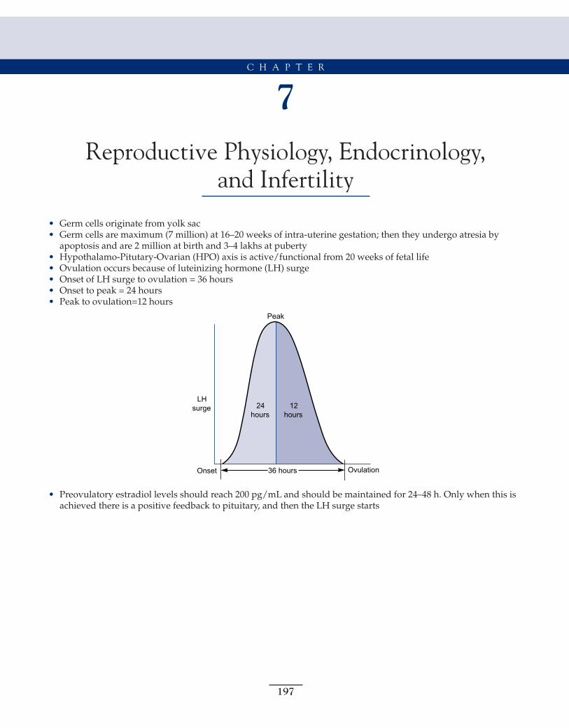
197
7Reproductive Physiology, Endocrinology,
and Infertility
• Germ cells originate from yolk sac • Germ cells are maximum (7 million) at 16–20 weeks of intra-uterine gestation; then they undergo atresia by
apoptosis and are 2 million at birth and 3–4 lakhs at puberty • Hypothalamo-Pitutary-Ovarian (HPO) axis is active/functional from 20 weeks of fetal life • Ovulation occurs because of luteinizing hormone (LH) surge • Onset of LH surge to ovulation = 36 hours • Onset to peak = 24 hours • Peak to ovulation=12 hours
LHsurge
36 hoursOnset Ovulation
Peak
24hours
12hours
• Preovulatory estradiol levels should reach 200 pg/mL and should be maintained for 24–48 h. Only when this is achieved there is a positive feedback to pituitary, and then the LH surge starts
C H A P T E R
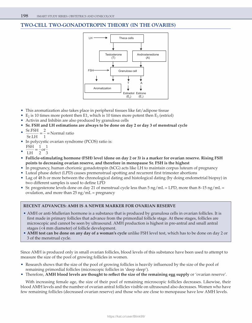
198 SMART STUDY SERIES: OBSTETRICS AND GYNECOLOGY
TWO-CELL TWO-GONADOTROPIN THEORY (IN THE OVARIES)
LH Theca cells
Testosterone(T)
Androstenedione(A)
Granulosa cell
T A
Aromatization
FSH
Estradiol(E2) (E1)
Estrone
• This aromatization also takes place in peripheral tissues like fat/adipose tissue • E2 is 10 times more potent then E1, which is 10 times more potent then E3 (estriol) • Activin and Inhibin are also produced by granulosa cells • Sr. FSH and LH estimations are always to be done on day 2 or day 3 of menstrual cycle
• Sr.FSH
Sr.LH=
2
1= Normal ratio
• In polycystic ovarian syndrome (PCOS) ratio is:
• FSH
LH=
1
2or
1
3 • Follicle-stimulating hormone (FSH) level (done on day 2 or 3) is a marker for ovarian reserve. Rising FSH
points to decreasing ovarian reserve, and therefore in menopause Sr. FSH is the highest • In pregnancy, human chorionic gonadotropin (hCG) acts like LH to maintain corpus luteum of pregnancy • Luteal phase defect (LPD) causes premenstrual spotting and recurrent first trimester abortions • Lag of 48 h or more between the chronological dating and histological dating (by doing endometrial biopsy) in
two different samples is used to define LPD • Sr. progesterone levels done on day 21 of menstrual cycle less than 5 ng/mL = LPD, more than 8–15 ng/mL =
ovulation, and more than 25 ng/mL = pregnancy
Since AMH is produced only in small ovarian follicles, blood levels of this substance have been used to attempt to measure the size of the pool of growing follicles in women.
• Research shows that the size of the pool of growing follicles is heavily influenced by the size of the pool of remaining primordial follicles (microscopic follicles in ‘deep sleep’).
• Therefore, AMH blood levels are thought to reflect the size of the remaining egg supply or ‘ovarian reserve’.
With increasing female age, the size of their pool of remaining microscopic follicles decreases. Likewise, their blood AMH levels and the number of ovarian antral follicles visible on ultrasound also decreases. Women who have few remaining follicles (decreased ovarian reserve) and those who are close to menopause have low AMH levels.
RECENT ADVANCES: AMH IS A NEWER MARKER FOR OVARIAN RESERVE
• AMH or anti-Mullerian hormone is a substance that is produced by granulosa cells in ovarian follicles. It is first made in primary follicles that advance from the primordial follicle stage. At these stages, follicles are microscopic and cannot be seen by ultrasound. AMH production is highest in pre-antral and small antral stages (<4 mm diameter) of follicle development.
• AMH test can be done on any day of a woman’s cycle unlike FSH level test, which has to be done on day 2 or 3 of the menstrual cycle.
https://kat.cr/user/Blink99/
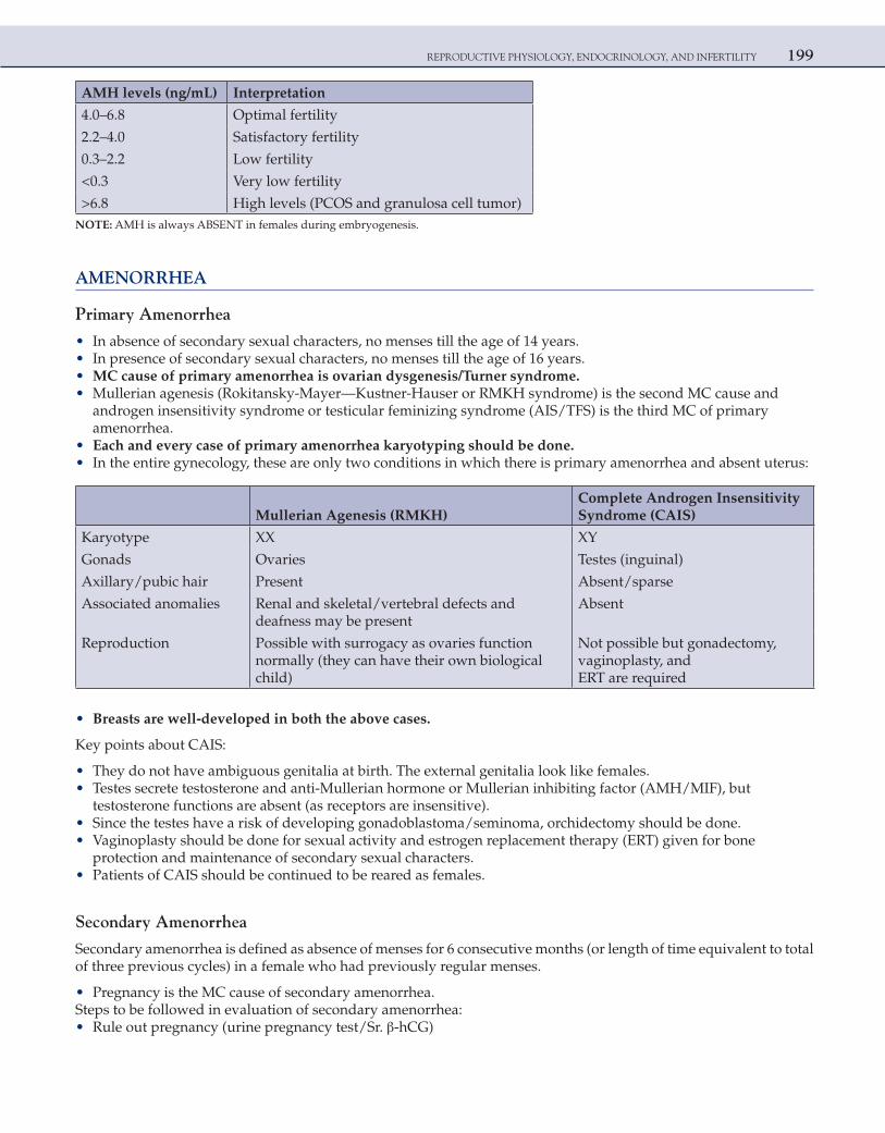
REPRODUCTIVE PHYSIOLOGY, ENDOCRINOLOGY, AND INFERTILITY 199
AMH levels (ng/mL) Interpretation
4.0–6.8 Optimal fertility
2.2–4.0 Satisfactory fertility
0.3–2.2 Low fertility
<0.3 Very low fertility
>6.8 High levels (PCOS and granulosa cell tumor)NOTE: AMH is always ABSENT in females during embryogenesis.
AMENORRHEA
Primary Amenorrhea
• In absence of secondary sexual characters, no menses till the age of 14 years. • In presence of secondary sexual characters, no menses till the age of 16 years. • MC cause of primary amenorrhea is ovarian dysgenesis/Turner syndrome. • Mullerian agenesis (Rokitansky-Mayer—Kustner-Hauser or RMKH syndrome) is the second MC cause and
androgen insensitivity syndrome or testicular feminizing syndrome (AIS/TFS) is the third MC of primary amenorrhea.
• Each and every case of primary amenorrhea karyotyping should be done. • In the entire gynecology, these are only two conditions in which there is primary amenorrhea and absent uterus:
Mullerian Agenesis (RMKH)Complete Androgen Insensitivity Syndrome (CAIS)
Karyotype XX XY
Gonads Ovaries Testes (inguinal)
Axillary/pubic hair Present Absent/sparse
Associated anomalies Renal and skeletal/vertebral defects and deafness may be present
Absent
Reproduction Possible with surrogacy as ovaries function normally (they can have their own biological child)
Not possible but gonadectomy, vaginoplasty, andERT are required
• Breasts are well-developed in both the above cases.
Key points about CAIS:
• They do not have ambiguous genitalia at birth. The external genitalia look like females. • Testes secrete testosterone and anti-Mullerian hormone or Mullerian inhibiting factor (AMH/MIF), but
testosterone functions are absent (as receptors are insensitive). • Since the testes have a risk of developing gonadoblastoma/seminoma, orchidectomy should be done. • Vaginoplasty should be done for sexual activity and estrogen replacement therapy (ERT) given for bone
protection and maintenance of secondary sexual characters. • Patients of CAIS should be continued to be reared as females.
Secondary Amenorrhea
Secondary amenorrhea is defined as absence of menses for 6 consecutive months (or length of time equivalent to total of three previous cycles) in a female who had previously regular menses.
• Pregnancy is the MC cause of secondary amenorrhea. Steps to be followed in evaluation of secondary amenorrhea: • Rule out pregnancy (urine pregnancy test/Sr. β-hCG)
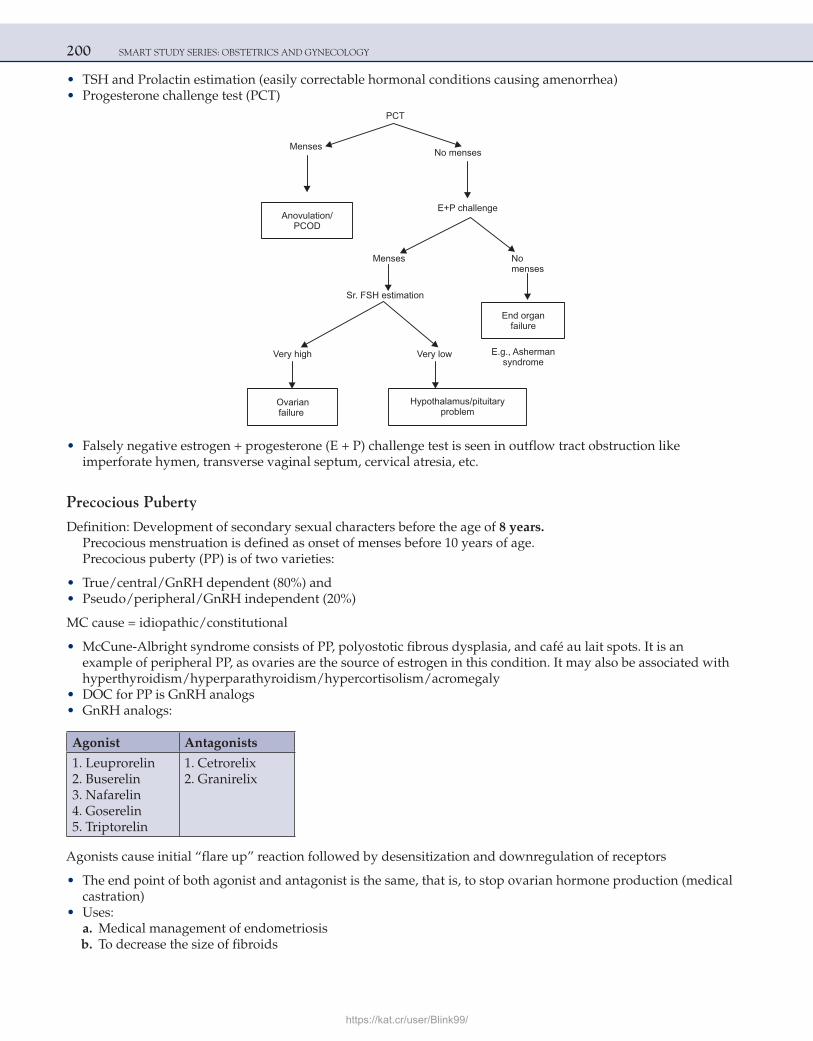
200 SMART STUDY SERIES: OBSTETRICS AND GYNECOLOGY
• TSH and Prolactin estimation (easily correctable hormonal conditions causing amenorrhea) • Progesterone challenge test (PCT)
PCT
MensesNo menses
Anovulation/PCOD
E+P challenge
oNsesneMmenses
Sr. FSH estimation
End organfailure
Hypothalamus/pituitaryproblem
Ovarianfailure
E.g., Ashermansyndrome
lowyreVhighyreV
• Falsely negative estrogen + progesterone (E + P) challenge test is seen in outflow tract obstruction like imperforate hymen, transverse vaginal septum, cervical atresia, etc.
Precocious Puberty
Definition: Development of secondary sexual characters before the age of 8 years. Precocious menstruation is defined as onset of menses before 10 years of age.Precocious puberty (PP) is of two varieties:
• True/central/GnRH dependent (80%) and • Pseudo/peripheral/GnRH independent (20%)
MC cause = idiopathic/constitutional
• McCune-Albright syndrome consists of PP, polyostotic fibrous dysplasia, and café au lait spots. It is an example of peripheral PP, as ovaries are the source of estrogen in this condition. It may also be associated with hyperthyroidism/hyperparathyroidism/hypercortisolism/acromegaly
• DOC for PP is GnRH analogs • GnRH analogs:
Agonist Antagonists
1. Leuprorelin 2. Buserelin 3. Nafarelin 4. Goserelin 5. Triptorelin
1. Cetrorelix 2. Granirelix
Agonists cause initial “flare up” reaction followed by desensitization and downregulation of receptors
• The end point of both agonist and antagonist is the same, that is, to stop ovarian hormone production (medical castration)
• Uses: a. Medical management of endometriosis b. To decrease the size of fibroids
https://kat.cr/user/Blink99/
REPRODUCTIVE PHYSIOLOGY, ENDOCRINOLOGY, AND INFERTILITY 201
c. Precocious Puberty (PP) d. Before in vitro fertilization (IVF) to downregulate the ovaries (so there is better response to subsequent
stimulation of gonadotropins) e. Before endometrial ablation/resection for Dysfunctional Uterine Bleeding (DUB) (to thin out the endometrium) • Craniopharyngioma is the MC neoplasm associated with delayed puberty
NOTE: Kallmann Syndrome (Deficient GnRH Secretion): Hypogonadotropic Hypogonadism Associated with Anosmia
1. Inheritance: X linked/AR/AD 2. KAL gene mutation, failure of production of gene product (Anosmin-1) 3. May be associated with hearing loss, ataxia color blindness, and cleft lip/palate
Perrault Syndrome
Gonadal dysgenesis (46 XX) and sensory neural deafness.
INFERTILITY
• Infertility is defined as an inability to conceive in spite of 1 year of regular unprotected intercourse • Primary = never conceived • Secondary = conceived in the past (irrespective of outcome of that pregnancy) • Fecundity = probability of achieving a live birth within a single cycle • Fecundability = probability of achieving pregnancy within a single menstrual cycle
Causes of Infertility
• Male factor • Female factor • Unexplained infertility • Combined factors
Female FactorsOvarian: 30–40%Tubal: 30–40%Unexplained: 10–15%Miscellaneous (uterine/cervical): 10–15%
WHO Category for Anovulation
• I: Hypothalamic pituitary failure • II: Hypothalamic pituitary disturbance/PCOS • III: Ovarian failure • IV: Hyperprolactinemia
Polycystic Ovarian Syndrome (PCOS)
• Rotterdam 2003 criteria for diagnosis of PCOS/PCOD—at least two out of three should be present: 1. Oligo/anovulation 2. Hyperandrogenism: biochemical or clinical 3. Twelve or more than 12 follicles 2–9 mm in size present within one or both ovaries on USG and/or ovarian
volume >10 mL • Obesity is not required to make the diagnosis and even the ratio of
FSH
LH=
1
2or
1
3 not essential to make the of diagnosis of PCOS
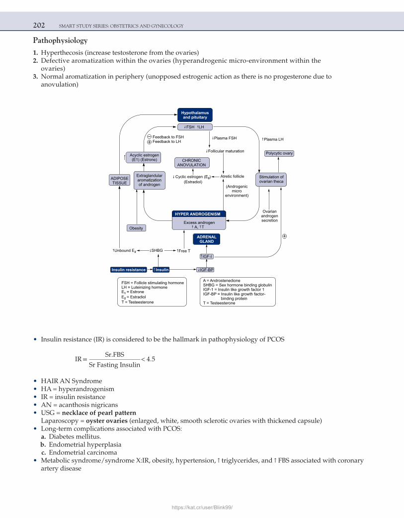
202 SMART STUDY SERIES: OBSTETRICS AND GYNECOLOGY
Pathophysiology
1. Hyperthecosis (increase testosterone from the ovaries) 2. Defective aromatization within the ovaries (hyperandrogenic micro-environment within the
ovaries) 3. Normal aromatization in periphery (unopposed estrogenic action as there is no progesterone due to
anovulation)
Hypothalamusand pituitary
FSH LH
Feedback to FSHFeedback to LH
Plasma FSH
Follicular maturation
Extraglandulararomatizationof androgen
Cyclic estrogen (E )(Estradiol)
2 Atretic follicle
(Androgenicmicro
environment)
Ovarianandrogensecretion
ADRENALGLAND
IGF-IUnbound E2 SHBG Free T
IGF-BPInsulinInsulin resistance
FSH = FollicleLH = Luteinizing hormoneE = EstroneE = EstradiolT = Testeesterone
1
2
stimulating hormone A = AndrostenedioneSHBG = Sex hormone binding globulinIGF-1 = Insulin like growth factor 1IGF-BP = Insulin like growth factor-
bindingT = Testeesterone
protein
CHRONICANOVULATION
Excess androgenA, TObesity
HYPER ANDROGENISM
Stimulation ofovarian theca
Acyclic estrogen(E1) (Estrone)
Plasma LH
ADIPOSETISSUE
Polycytic ovary
• Insulin resistance (IR) is considered to be the hallmark in pathophysiology of PCOS
IR =
Sr.FBS
Sr Fasting Insulin< 4.5
• HAIR AN Syndrome • HA = hyperandrogenism • IR = insulin resistance • AN = acanthosis nigricans • USG = necklace of pearl pattern
Laparoscopy = oyster ovaries (enlarged, white, smooth sclerotic ovaries with thickened capsule) • Long-term complications associated with PCOS: a. Diabetes mellitus. b. Endometrial hyperplasia c. Endometrial carcinoma • Metabolic syndrome/syndrome X:IR, obesity, hypertension, ↑ triglycerides, and ↑ FBS associated with coronary
artery disease
https://kat.cr/user/Blink99/
REPRODUCTIVE PHYSIOLOGY, ENDOCRINOLOGY, AND INFERTILITY 203
Management
Principles of management include:
• Irregular periods/amenorrhea = regularization of menses with OC pills/cyclical progesterone • Hirsutism/acne = suppression of androgens • Infertility = ovulation induction • Amenorrhea because of PCOD is estrogen-induced amenorrhea (unopposed estrogenic action as these is no
ovulation and hence progesterone is absent), and it requires treatment as unopposed estrogenic action is a risk factor for endometrial hyperplasia/cancer
Ovulation Induction Agents 1. Clomiphene citrate (CC) 2. Letrozole, Anastrozole, Tamoxifen 3. Gonadotropins
Clomiphene Citrate: It is a racemic mixture of enclomiphene and zuclomiphene. Enclomiphene is a more potent isomer responsible for its ovulation-inducing action.
• Dose = 50–250 mg. However, the US FDA-approved maximum dose for CC is 100 mg • CC blocks “E” receptors —> increase FSH from pituitary —> growth of follicles • With CC Success rate for ovulation is 80% and success for pregnancy is 40%
Letrozole = aromatase inhibitor blocks conversion of testosterone to estrogen, leading to increased FSH from pituitary.Gonadotropins: HMG (Human Menopausal Gonadotropin) (from the urine of the menopausal women) and recom-binant FSH.
• Menopausal women have high FSH and LH levels in their blood and urine, and HMG is extracted from urine of menopausal females. It mainly contains FSH
• Follicular study is done along with ovulation induction to monitor the growth of follicles and when the dominant follicle is 18–20 mm, ovulation trigger is given to rupture the follicle
• For ovulation trigger, MC drug used is hCG (derived from the urine of pregnant women or by recombinant technology)
• Recombinant LH is can also be used but is very expensive • Ovulation occur 36 hours after injecting hCG
Side Effects of Ovulation Induction 1. Multiple pregnancies: 3–8% with CC, 15–30% with Gonadotropins 2. Ovarian hyperstimulation syndrome (OHSS) ○ Most dangerous complication of ovulation induction ○ Risk factors: PCOS patients and past history of OHSS
Classification of OHSS
Ovary Size (cm) Features
Mild 5–10 Abdominal distention ± GI upset
Moderate > 10 Moderate ascites, normal renal function, hematocrit <0.45
Severe > 12 Marked ascitesHypovolemia ↑ WBCHematocrit >0.45Venous thrombosis↓ Renal function, ± DIC
○ Various factors responsible for development of OHSS include estrogen, prostaglandins, histamine, cytokines, IL-2, IL-6, IL-8, renin, angiotensin II, and Vascular Endothelial Growth Factor (VEGF)
○ VEGF is considered to be the most important
204 SMART STUDY SERIES: OBSTETRICS AND GYNECOLOGY
○ The risk of OHSS is very high when estradiol levels are more than 2500 pg/mL, but OHSS can also happen when it is >1500 pg/mL
○ Treatment: IV fluids, albumin, USG guide taping of ascites and aspiration of follicles ○ Surgery is done only if there is bleeding within ovaries or torsion ovaries 3. Increased risk of epithelial ovarian cancers: Prolonged use of gonadotropins/CC (>6–12 months) increases the
risk of epithelial ovarian cancer.
INSULIN SENSITIZERS
• MC used drug = metformin; others = rosiglitazone/pioglitazone • Metformin will help the patient to lose weight and will either cause spontaneous ovulation or increase the
success of ovulation induction drugs • MC side effects: nausea/vomiting and bloating (GI upset) • Most dangerous side effect: lactic acidosis • Metformin was thought to be teratogenic, but recent consensus is that metformin can be continued throughout
pregnancy and it decreases the risk of spontaneous abortion and development of gestational DM (GDM) • Newer insulin sensitizer myoinositol is now available. It is better tolerated than metformin.
SURGERY FOR PCOS
• Laparoscopic ovarian drilling (LOD) or laparoscopic electrocoagulation of ovarian surface (LEOS) • In this surgery, monopolar current is passed within the ovary to destroy the ovarian theca • This surgery is done only for infertile patients of PCOS who are resistant to ovulation with gonadotropin or
when very high doses of gonadotropins are required for ovulation • Advantages: no risk of OHSS and multiple pregnancy • Disadvantages: surgical procedure, risk of premature ovarian failure if excessive ovarian tissue is damaged, and
adhesion formation postsurgery
ENDOMETRIOSIS
• Definition: Presence of functional endometrium at places other than uterus (ectopic endometrial tissue) • MC sites in order of frequency: a. Ovaries (ovarian endometriosis = endometrioma = chocolate cyst of the ovaries) b. POD c. Uterosacral ligaments • Theories for development of endometriosis: a. Samson’s theory of retrograde menstruation: the most accepted theory b. Ivanoff and Meyer: celomic metaplasia c. Hematogenous spread d. Lymphatic spread (Halban’s theory) e. Direct implantation • Smoking is thought to be protective for endometriosis • Classical PV findings are: fixed RV uterus with nodularity in POD
Clinical Features
• Pain • Dysmenorrhea • Infertility • Dyspareunia (deep)
Reasons for Infertility in Endometriosis
1. Tubal adhesions/blocks or anatomy between the tube and ovary is distorted (main reason) 2. Anovulation
https://kat.cr/user/Blink99/
REPRODUCTIVE PHYSIOLOGY, ENDOCRINOLOGY, AND INFERTILITY 205
3. The uterus is not suitable for implantation 4. ↑ Sperm phagocytosis 5. Dyspareunia decreases coital frequency
Laparoscopy is the Investigation of Choice
Laparoscopy findings are:
• Chocolate cysts • Powder burn spots • Matchstick burnt spots • Blueberry lesion • Red/purple raspberry lesion • White lesion • Red/flame lesion • Subovarian adhesions
Subtle peritoneal defects associated with endometriosis is called “Allen Master” syndrome
Management
• Medical • Surgical
Medical Management • Pseudopregnancy regimen: OC pills, DMPA POP, and Mirena • Pseudomenopause regimen: Danazol (Hardly ever used today because of androgenic side effects) • Medical castration: GnRH analogues (most common drug used for medical management)
Surgical Management 1. Patients with infertility: laparoscopic ovarian cystectomy, adhesiolysis, and electrocoagulation of endometriotic
implants with bipolar current. 2. If the family is complete and the patient has severe pain or menstrual complaints: hysterectomy with bilateral
salpingo-oophorectomy
Generally combined approach is adopted where laparoscopic surgery is followed by GnRHa.
TUBAL FACTORS
(Tubal blocks due to TB/Chlamydia/gonococci/adhesions)Tests for tubal patency:
1. Hysterosalpingography (HSG): cavity of the uterus and fallopian tube patency can be checked: ○ As it does not require anesthesia, it is the first-line investigation for checking tubal patency. ○ Disadvantage: While pushing the dye, there can be cornual spasm and the fallopian tubes appears to be
blocked even if the tubes are healthy. So HSG cannot differentiate between cornual blocks (pathological) and cornual spasm.
2. Sonosalpingography/saline USG: ○ Normal saline is introduced into the uterine cavity, and fallopian tube patency can be checked by seeing free
fluid in POD. ○ It is also very useful to evaluate endometrial polyps. 3. Laparoscopy with chromopertubation with methylene blue dye: ○ Best investigation, as tubal patency can be confirmed under vision and, besides, any pathology can
simultaneously be corrected with operative laparoscopy. ○ As it requires anesthesia and admission, it is never the first-line investigation for tubal patency.
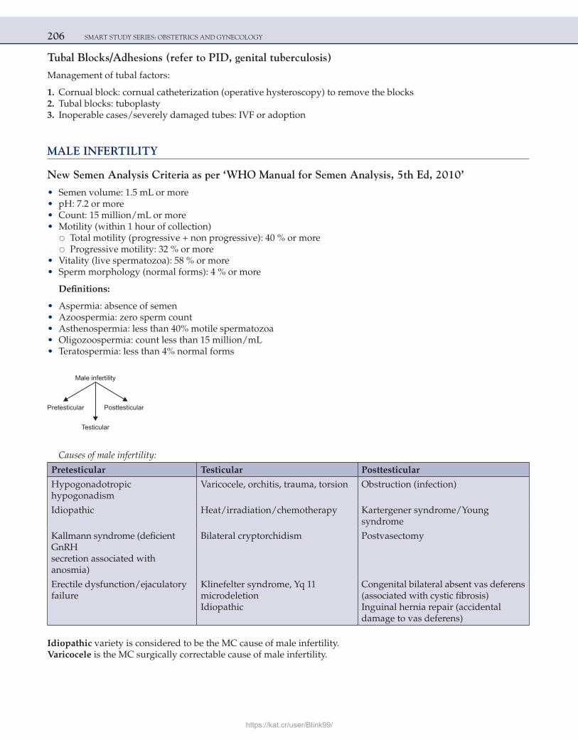
206 SMART STUDY SERIES: OBSTETRICS AND GYNECOLOGY
Tubal Blocks/Adhesions (refer to PID, genital tuberculosis)
Management of tubal factors:
1. Cornual block: cornual catheterization (operative hysteroscopy) to remove the blocks 2. Tubal blocks: tuboplasty 3. Inoperable cases/severely damaged tubes: IVF or adoption
MALE INFERTILITY
New Semen Analysis Criteria as per ‘WHO Manual for Semen Analysis, 5th Ed, 2010’
• Semen volume: 1.5 mL or more • pH: 7.2 or more • Count: 15 million/mL or more • Motility (within 1 hour of collection) ○ Total motility (progressive + non progressive): 40 % or more ○ Progressive motility: 32 % or more • Vitality (live spermatozoa): 58 % or more • Sperm morphology (normal forms): 4 % or more
Definitions:
• Aspermia: absence of semen • Azoospermia: zero sperm count • Asthenospermia: less than 40% motile spermatozoa • Oligozoospermia: count less than 15 million/mL • Teratospermia: less than 4% normal forms
Male infertility
Pretesticular Posttesticular
Testicular
Causes of male infertility:
Pretesticular Testicular Posttesticular
Hypogonadotropic hypogonadism
Varicocele, orchitis, trauma, torsion Obstruction (infection)
Idiopathic Heat/irradiation/chemotherapy Kartergener syndrome/Young syndrome
Kallmann syndrome (deficient GnRHsecretion associated with anosmia)
Bilateral cryptorchidism Postvasectomy
Erectile dysfunction/ejaculatory failure
Klinefelter syndrome, Yq 11microdeletionIdiopathic
Congenital bilateral absent vas deferens (associated with cystic fibrosis)Inguinal hernia repair (accidental damage to vas deferens)
Idiopathic variety is considered to be the MC cause of male infertility.Varicocele is the MC surgically correctable cause of male infertility.
https://kat.cr/user/Blink99/
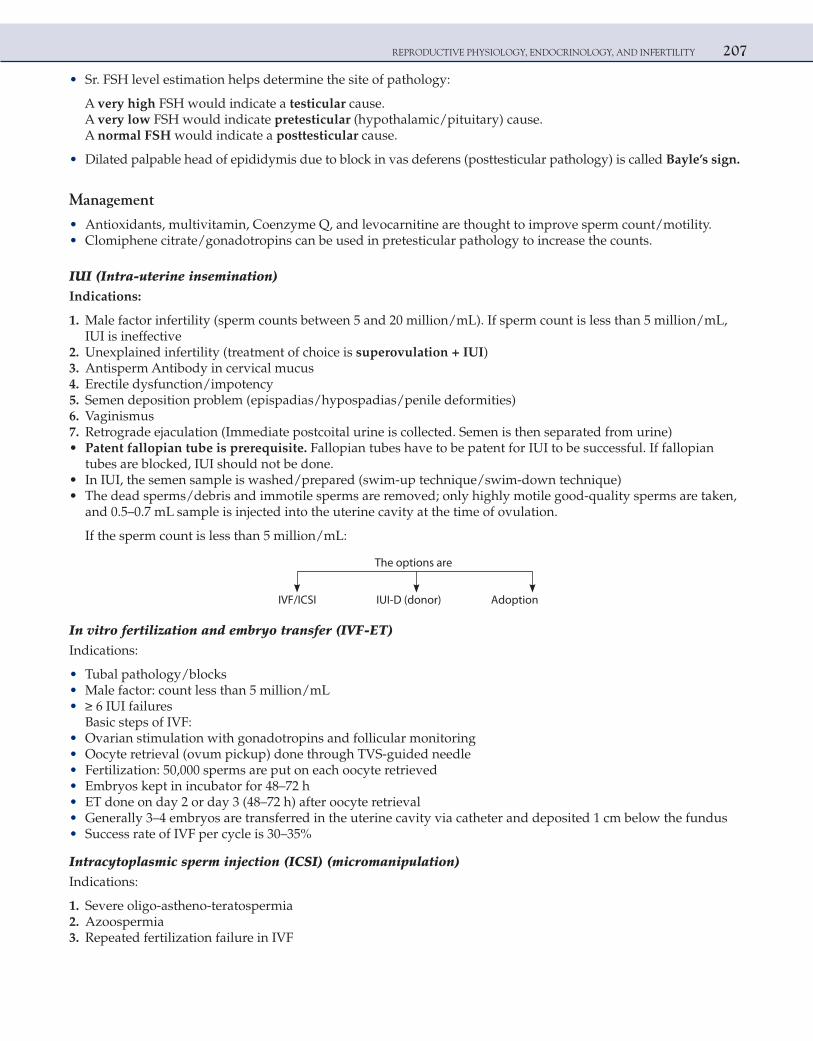
REPRODUCTIVE PHYSIOLOGY, ENDOCRINOLOGY, AND INFERTILITY 207
• Sr. FSH level estimation helps determine the site of pathology:
A very high FSH would indicate a testicular cause.A very low FSH would indicate pretesticular (hypothalamic/pituitary) cause.A normal FSH would indicate a posttesticular cause.
• Dilated palpable head of epididymis due to block in vas deferens (posttesticular pathology) is called Bayle’s sign.
Management
• Antioxidants, multivitamin, Coenzyme Q, and levocarnitine are thought to improve sperm count/motility. • Clomiphene citrate/gonadotropins can be used in pretesticular pathology to increase the counts.
IUI (Intra-uterine insemination)Indications:
1. Male factor infertility (sperm counts between 5 and 20 million/mL). If sperm count is less than 5 million/mL, IUI is ineffective
2. Unexplained infertility (treatment of choice is superovulation + IUI) 3. Antisperm Antibody in cervical mucus 4. Erectile dysfunction/impotency 5. Semen deposition problem (epispadias/hypospadias/penile deformities) 6. Vaginismus 7. Retrograde ejaculation (Immediate postcoital urine is collected. Semen is then separated from urine) • Patent fallopian tube is prerequisite. Fallopian tubes have to be patent for IUI to be successful. If fallopian
tubes are blocked, IUI should not be done. • In IUI, the semen sample is washed/prepared (swim-up technique/swim-down technique) • The dead sperms/debris and immotile sperms are removed; only highly motile good-quality sperms are taken,
and 0.5–0.7 mL sample is injected into the uterine cavity at the time of ovulation.
If the sperm count is less than 5 million/mL:
The options are
IVF/ICSI IUI-D (donor) Adoption
In vitro fertilization and embryo transfer (IVF-ET)Indications:
• Tubal pathology/blocks • Male factor: count less than 5 million/mL • ≥ 6 IUI failures
Basic steps of IVF: • Ovarian stimulation with gonadotropins and follicular monitoring • Oocyte retrieval (ovum pickup) done through TVS-guided needle • Fertilization: 50,000 sperms are put on each oocyte retrieved • Embryos kept in incubator for 48–72 h • ET done on day 2 or day 3 (48–72 h) after oocyte retrieval • Generally 3–4 embryos are transferred in the uterine cavity via catheter and deposited 1 cm below the fundus • Success rate of IVF per cycle is 30–35%
Intracytoplasmic sperm injection (ICSI) (micromanipulation)Indications:
1. Severe oligo-astheno-teratospermia 2. Azoospermia 3. Repeated fertilization failure in IVF
208 SMART STUDY SERIES: OBSTETRICS AND GYNECOLOGY
The steps are identical to IVF (oocyte retrieval and embryo transfer), but for fertilization, one sperm is mechani-cally injected into one oocyte.
Success rate of ICSI per cycle is 30–35%. Sperm retrieval techniques in case of azoospermia before doing ICSI:
• PESA= percutaneous epididymal sperm aspiration • MESA= microscopic epididymal sperm aspiration • TESA= testicular sperm aspiration • TESE= testicular sperm extraction (testicular biopsy)
MULTIPLE CHOICE QUESTIONS
1. Resolution of corpus luteum occurs because of: [AIIMS Nov 2003] a. Increased levels of progesterones b. Increased levels of estrogens c. Decreased levels of LH d. Decreased levels of FSH
Answer: c (Decreased levels of LH)
Explanation:The hormonal changes in the luteal phase of the menstrual cycle are characterized by a series of negative feedback interac-
tions designed to lead to regression of the corpus luteum if pregnancy does not occur. Estradiol and progesterone provide negative central feedback and cause a decrease in FSH and LH production. Continued corpus luteum function depends on continued LH production. In the absence of this stimulation, the corpus luteum will invariably regress after 12–16 days and form scar-like corpora albicans. The exact mechanism of luteolysis is, however, unclear and most likely also involves local paracrine factors. In the absence of pregnancy, the corpus luteum regresses, and estrogen and progesterone levels wane. This, in turn, removes central inhibition on gonadotropin secretion and allows FSH and LH to rise again and recruit another cohort of follicles. If pregnancy occurs, hCG from the placenta will mimic LH action and the corpus luteum continues to secrete progesterone.
Reference: 1. Speroff, 7th Ed., Pg. 226.
2. A 9-year-old girl presents for evaluation of regular vaginal bleeding. History reveals the thelarche at the age of 7 years and adrenarche at the age of 8 years. The most common cause of this condition in girls is:
a. Idiopathic b. Gonadal tumors c. McCune-Albright syndrome d. Hypothyroidism
Answer: a (Idiopathic)
Explanation:Pubertal changes before the age of 8 years in girls and 9 years in boys are regarded as precocious. Although the
most common type of precocious puberty in girls is idiopathic, it is essential to ensure close long-term follow-up of these patients to ascertain that there is no serious underlying pathology, such as tumors of the central nervous system or ovary. Only 1–2% patients with precious puberty have an estrogen-producing ovarian tumor as the causative factor. McCune-Albright syndrome is also relatively rare and consists of fibrous dysplasia and cystic degeneration of the long bones, sexual precocity, and café au lait spots on the skin. Hypothyroidism is a cause of precocious puberty in some children, making thyroid function tests mandatory in these cases. Tumors of the central nervous system as a cause of precious puberty occur more commonly in boys than in girls; they are seen in about 11% of girls with precocious puberty.
Reference: 1. Speroff, 7th Ed., Pg. 392–400.
https://kat.cr/user/Blink99/
REPRODUCTIVE PHYSIOLOGY, ENDOCRINOLOGY, AND INFERTILITY 209
3. Medication used in the treatment of idiopathic central precocious puberty include: a. Exogenous gonadotropins b. Ethinyl estradiol c. GnRH agonists d. Clomiphene citrate
Answer: c (GnRH agonists)
Explanation:Precocious puberty can be treated by agents that reduce gonadotropin levels by exerting negative feedback in the hypotha-
lamic pituitary axis or that directly inhibit gonadotropin secretion from the pituitary gland. Until about 10 years ago, the greatest experience in the treatment of idiopathic central precocious puberty was with medroxyprogesterone acetate (MPA). MPA was usually administered intramuscularly in a dose of 100–200 mg/ week, or orally at 20–40 mg/day. Currently, the most effective treatment for central precocious puberty is the use of a long-acting GnRH agonist, such as leuprolide and others. These drugs act by down regulating pituitary gonadotropes, eventually decreasing the secretion of FSH and LH, which are inappropriately stimulating the ovaries of these patients. As a result of this induced hypogonadotropic state, ovarian steroids (estrogens, proges-tins, and androgens) are suppressed back to prepubertal levels, and precocious pubertal development stops or regresses. During the first 1 or 2 weeks of therapy, there is a flare-up effect of increased gonadotropins and sex steroids—a predicted side effect of these medications. At the time of expected puberty, the GnRH analog is discontinued and the pubertal sequence resumes.
Reference: 1. Speroff, 7th Ed., Pg. 392–400.
4. A 45-year-old woman who had two normal pregnancies 15 and 18 years ago presents with the complaint of amenorrhea for 7 months. She expresses the desire to become pregnant again. After exclusion of pregnancy, which of the following is the next best test indicated in the evaluation of this patient’s amenorrhea?
a. LH and FSH levels b. Endometrial biopsy c. Karyotyping d. HSG
Answer: a (LH and FSH levels)
Explanation:This patient has secondary amenorrhea, which rules out abnormalities associated with primary amenorrhea such as chro-
mosomal abnormalities and congenital Mullerian abnormalities. The most common reason for amenorrhea in a woman of reproductive age is pregnancy, which should be evaluated first. Other possibilities include chronic endometritis or scaring of the endometrium (Asherman syndrome), hypothyroidism, and ovarian failure. The latter is the most likely diagnosis in a woman at this age. In addition, emotional stress, extreme weight loss, and adrenal cortisol insufficiency can bring about secondary amenorrhea. A hysterosalpingogram is part of an infertility workup that may demonstrate Asherman syndrome, but it is not indicated until premature ovarian failure has been excluded. Persistently elevated gonadotropin levels (especially when accompanied by low serum estradiol levels) are diagnostic of ovarian failure.
Reference: 1. Speroff, 7th Ed., Pg. 444–8, 651–6.
5. A 22-year-old woman comes for treatment of hirsutism. She is obese and has facial acne and hirsutism on her face. Serum LH level is 36 mIU/mL and FSH is 9 mIU/mL. Androstenedione and testosterone levels are mildly elevated, but serum DHEAS is normal. The patient does not wish to conceive at this time. Which of the following is the most appropriate treatment of her condition?
[All India 2002] a. Oral contraceptives pills b. Corticosteroids c. GnRH analog d. Wedge resection of ovary
Answer: a (Oral contraceptives pills)
210 SMART STUDY SERIES: OBSTETRICS AND GYNECOLOGY
Explanation:This patient has PCOS, diagnosed by the clinical picture, abnormally high LH-to-FSH ratio, and elevated androgens
but normal DHEAS. DHEAS is a marker of adrenal androgen production; when normal, it essentially excludes adrenal sources of hyperandrogenism. Several medications have been used to treat hirsutism associated with PCOS. OC pills are the most frequently used agents; they can suppress hair growth in up to two-thirds of treated patients. They act by directly suppressing ovarian steroid production and increasing hepatic binding globulin production, which binds circulating hor-mone and lowers the concentrations of metabolically active (free unbound) androgen. However, clinical improvement can take as long as 6 months to manifest. Other medications that can be used include GnRH agonists, which suppress ovarian steroid production. However, GnRH analogs are expensive and have been associated with significant bone demineraliza-tion after only 6 months of therapy in some patients. Surgical wedge resection is no longer considered an appropriate therapy for PCOS, given the success of pharmacologic agents and the ovarian adhesions that were frequently associated with this surgery.
Reference: 1. Speroff, 7th Ed., Pg. 475–80.
6. A 23-year-old woman presents for evaluation of a 7-month history of amenorrhea. Examination discloses bilateral galactorrhea and normal breast and pelvic examinations. Pregnancy test is negative. Which of the following classes of medication is a possible cause of her condition?
a. Antiestrogens b. Gonadotropins c. Phenothiazines d. Prostaglandins
Answer: c (Phenothiazines)
Explanation:Amenorrhea and galactorrhea may be seen when something causes an increase in prolactin secretion. The differential diag-
nosis involves several possible causes. Excessive estrogens, such as with birth control pills, can reduce prolactin-inhibiting factor, thus raising serum prolactin level. Similarly, intensive suckling (during lactation and associated with sexual foreplay) can activate the reflex arc that results in hyperprolactinemia. Many antipsychotic medications, especially the phenothiazines, are also known to have mammotropic properties. Hypothyroidism appears to cause galactorrhea secondary to thyrotropin-releasing hormone (TRH) stimulation of prolactin release. When prolactin levels are persistently elevated without obvious cause (e.g., in breast-feeding), evaluation for pituitary adenoma becomes necessary.
Reference: 1. Speroff, 7th Ed., Pg. 450–60.
7. Which of the following pubertal events in girls is not estrogen dependent? a. Menses b. Vaginal cornification c. Hair growth d. Reaching adult height
Answer: c (Hair growth)
Explanation:The presence of estrogen in a pubertal girl stimulates the formation of secondary sex characteristics, including develop-
ment of breasts, production of cervical mucus, and vaginal cornification. As estrogen levels increase, menses begins and ovulation is maintained for several decades. Ovarian estrogen production late in puberty is at least in part responsible for termination of the pubertal growth spurt, thereby determining adult height. Decreasing levels of estrogen are associated with lower frequency of ovulation, eventually leading to menopause. Hair growth during puberty is caused by androgens from the adrenal gland and, later, the ovary.
Reference: 1. Speroff, 7th Ed., Pg. 320–5.
https://kat.cr/user/Blink99/
REPRODUCTIVE PHYSIOLOGY, ENDOCRINOLOGY, AND INFERTILITY 211
8. A 22-year-old comes with a chief complaint of being too hairy. She reports that her menses started at the age of 13 years and has always been very irregular. She also complains of acne. On physical examination, there is hair around the nipples, chin, and upper lip. No galactorrhea, thyromegaly, or temporal balding is noted. Pelvic examination is normal, and there is no evidence of clitoromegaly. All of the following should be included in the differential diagnosis based on the patient’s history and physical examination, except:
a. Idiopathic or constitutional hirsutism b. Polycystic ovarian syndrome c. Late-onset congenital adrenal hyperplasia d. Sertoli–Leydig cell tumor
Answer: d (Sertoli–Leydig cell tumor)
Explanation:Sertoli–Leydig cell tumors, also known as androblastomas or arrhenoblastomas, are testosterone-secreting ovar-
ian neoplasms. These tumors usually occur in women between the ages of 20 and 40 years and tend to be unilateral and reach a size of 7–10 cm. Women with a Sertoli–Leydig cell tumor tend to have very high levels of testosterone (>200 ng/dL) and rapidly develop virilizing characteristics such as temporal balding, clitoral hypertrophy, voice deep-ening, breast atrophy, and terminal hair between the breasts and on the back. Women with constitutional or idiopathic hirsutism have greater activity of 5-α-reductase than do unaffected women. They have hirsutism with a diagnostic evalu-ation that gives no explanation for the excess hair. Women with attenuated congenital adrenal hyperplasia are hirsute due to an increase in adrenal androgen production caused by a deficiency in 21-hydroxylase. PCOS is the most com-mon cause of androgen excess and hirsutism. Selective insulin resistance is thought to be central to the etiology of this syndrome.
Reference: 1. Speroff, 7th Ed., Pg. 501–5.
9. Normal stature with minimal or absent pubertal development may be seen in: [AIIMS May 2001; All India 2011] a. Testicular feminization b. Kallmann syndrome c. Pure gonadal dysgenesis d. Turner syndrome
Answer: b (Kallmann syndrome)
Explanation:Testicular feminization is a syndrome of androgen insensitivity in genetic males, characterized by a normal 46 XY geno-
type; normal female phenotype during childhood; tall stature; and “normal” breast development with absence of axillary and pubic hair. Breast development (gynecomastia) occurs in these males because high levels of circulating testosterone (which cannot act at its receptor) are aromatized to estrogen, which then acts on the breast. The external genitalia develop as those of a female because testosterone cannot masculinize them, while the Mullerian structures are absent because of testicular secre-tion Mullerian-inhibiting factor in utero.
Gonadal dysgenesis (e.g., 45 X Turner syndrome) is characterized by short stature and absence of pubertal development; in these girls, the ovaries are either absent or steak gonads that are nonfunctional. Kallmann syndrome (hypogonadotropic hypogonadism) should be suspected in patients of normal stature with delayed or absent pubertal development, especially when associated with the classic finding of anosmia. These individuals have a structural defect of the CNS involving the hypothalamus and the olfactory bulbs (located in close proximity to the hypothalamus), such that the hypothalamus does not secrete GnRH in normal pulsatile fashion, if at all. Other causes of minimal or absent pubertal development with normal stature include malnutrition, anorexia nervosa, severe systemic disease, and intensive athletic training, particularly ballet and running.
Reference: 1. Speroff, 7th Ed., Pg. 404–7.
212 SMART STUDY SERIES: OBSTETRICS AND GYNECOLOGY
10. A 19-year-old patient presents to your office with primary amenorrhea. She has normal breast and pubic hair development, but the uterus and vagina are absent. Diagnostic possibility includes:
[AIIMS Nov 2003, AIIMS Nov 2010 , AIIMS Nov 2012, All India 2013] a. Testicular feminization syndrome b. Gonadal dysgenesis c. Mullerian agenesis d. Klinefelter syndrome
Answer: c (Mullerian agenesis)
Explanation:Since this patient has other signs of pubertal development that are sex steroid- dependent, we can conclude that some ovarian
function is present. This excludes conditions such as gonadal dysgenesis and hypothalamic pituitary failure as possible causes of her primary amenorrhea. Mullerian defects are the only plausible cause, and the diagnostic evaluation in this patient would be directed toward both confirmation of this diagnosis and establishment of the exact nature of the Mullerian defect. Mullerian agen-esis, also known as Mayer-Rokitansky-Kuster-Hauser syndrome, presents as amenorrhea with absence of a vagina. The incidence is approximately 1 in 10,000 female births. The karyotype is 46 XX. There is normal development of breasts, sexual hair, ovaries, and external genitalia. There are associated skeletal (12%) and urinary tract (33%) anomalies. Treatment generally consists of pro-gressive vaginal dilation or creation of an artificial vagina with split thickness skin grafts and surrogacy if reproduction is desired.
Testicular feminization, or congenital androgen insensitivity syndrome, is an X-linked recessive disorder with a karyotype of 46 X Y. The patient presents with an absent uterus and blind vaginal canal. However, in these patients the amount of sexual hair is significantly decreased/absent.
Patients with gonadal dysgenesis present with lack of secondary sexual characteristics. Patients with Klinefelter syndrome typically have a karyotype of 47 XXY and a male phenotype.NOTE: In patients of Mullerian agenesis (RMKH syndrome) ovary is present and it functions normally. (AIIMS Nov 2009, AIIMS Nov 2011)
Reference: 1. Speroff, 7th Ed., Pg. 405–10.
11. While evaluating a 30-year-old woman for infertility, you diagnose a bicornuate uterus. You explain that additional testing is necessary for one organ system because of the woman’s increased risk of congenital anomalies. Which is that organ system?
a. Skeletal b. Tracheoesophageal c. Urinary d. Central nervous
Answer: c (Urinary)
Explanation:Failed fusion of the Mullerian ducts can give rise to several types of uterine anomalies, of which bicornuate uterus is repre-
sentative type. This condition is associated with a higher risk of obstetric complications, such as an increase in the rate of second-trimester abortion and premature labor. If these pregnancies go to term, malpresentations such as breech and transverse lie are more frequent. Also, prolonged labor (probably due to inadequate muscle development in the uterus), increased bleeding, and a higher incidence of fetal anomalies caused by defective implantation of the placenta all occur more commonly than in normal pregnancies. An intravenous pyelogram or urinary tract ultrasound is mandatory in patients with Mullerian anomalies, since approximately 30% of patients with Mullerian anomalies have coexisting congenital urinary tract anomalies. In bicornuate uterus (termed uterus bicornis unicollis), there is a double uterine cavity (bicornis) and a single cervix (unicollis) with a normal vagina.
Reference: 1. Speroff, 7th Ed., Pg. 440–2.
12. Artificial insemination with husband’s semen is indicated in all the following situations, except: a. Oligospermia b. Impotency c. Antisperm antibodies in the cervical mucous d. Azoospermia
Answer: d (Azoospermia)
https://kat.cr/user/Blink99/
REPRODUCTIVE PHYSIOLOGY, ENDOCRINOLOGY, AND INFERTILITY 213
Explanation:An artificial insemination with husband’s semen (IUI) is indicated in cases of:
1. Oligospermia 2. Impotency 3. Premature or retrograde ejaculation 4. Hypospadias 5. Antisperm antibodies in the cervical mucous 6. Unexplained infertility 7. X-Y fractionation of sperms for sex selection in genetic and chromosomal abnormalities
Azoospermia will require IUI with donor semen or ICSI, provided the sperms can be obtained by PESA or TESA.
Reference: 1. Speroff, 7th Ed., Pg. 1140.
13. Postcoital test is used to assess: a. Cervical factor b. Uterine factor c. Tubal factor d. Any of the above
Answer: a (Cervical factor)
Explanation:Postcoital test (Sims’ or Huhner’s test).
1. The cervical mucus is examined for its quantity, viscosity, and fern test. The advantage of this test is that the cervical mucus can be simultaneously studied for estrogenic effect and ovulation, its capability to allow sperm penetration, and the presence of any antisperm antibodies.
2. The test is useless in presence of cervical infection, which should be treated before performing the postcoital test. 3. The couple is advised intercourse close to ovulation time, preferably in the early hours of the morning. The woman
presents herself at the clinic within 2 h after the intercourse. The mucus is aspirated from the cervical canal and spread over a glass slide. Another smear made from the posterior fornix serves as a control. Normally 10–50 motile sperms are seen per high-power field in cervical mucus. If there are less than 10 sperms, proper semen analysis should be undertaken. The sperms show progressive, but not rotatory, movements. The presence of antispermal antibodies in the cervical mucus imparts shaky or rotatory movements to the sperms or may totally immobilize them.
4. A test called the Miller–Kurzrok test consists of placing ovulation mucus on a glass side alongside the specimen of the husband’s semen and studying the penetration of sperms under the microscope. A normal cervical mucus permits invasion by motile sperms. Penetration less than 3 cm at 30 min is abnormal.
Reference: 1. Shaw, 13th Ed., Pg. 202–03.
14. The risk of Asherman syndrome is the highest if dilatation and curettage (D&C) is done for the following condition: [AIIMS May 2006] a. Medical termination of pregnancy b. Missed abortion c. Dysfunctional uterine bleeding d. Postpartum hemorrhage
Answer: d (Postpartum hemorrhage)
Explanation:Uterine synechiae (Asherman syndrome) are caused by destruction of large areas of endometrium by curettage. In postpartum
hemorrhage, a greater area of uterine wall is curetted, since the postpartum uterus is larger and bulkier. Hence, the risk of Asherman is greatest if D&C is done in postpartum period.
Reference: 1. William’s Obstetrics, 22nd Ed., Pg. 961.
214 SMART STUDY SERIES: OBSTETRICS AND GYNECOLOGY
15. In an amenorrheic patient who has had pituitary ablation for a craniopharyngioma, which of the following regimens is most likely to result in an ovulatory cycle?
a. Clomiphene citrate b. Letrozole c. Continuous infusion of GnRH d. Human menopausal or recombinant gonadotropin, followed by hCG
Answer: d (Human menopausal or recombinant gonadotropin, followed by hCG)
Explanation:This patient would be unable to produce endogenous gonadotropin, since her pituitary has been ablated. The patient will,
therefore, need to be given exogenous gonadotropin in the form of human menopausal gonadotropin (hMG), which contains an extract of urine from postmenopausal women with FSH and LH in various ratios. Recombinant human FSH (rhFSH) is now also available. Carefully timed administration of hCG, which takes the place of an endogenous LH surge, will be needed to complete oocyte maturation and induce ovulation.
Clomiphene citrate and letrozole block the normal negative feedback of the endogenous estrogens and stimulates release of endogenous GnRH and FSH, but this will not be helpful as the patient’s pituitary has been ablated. Similarly, endogenous or exogenous GnRH cannot stimulate the release of FSH or LH in this woman because the pituitary gland is nonfunctional.
Reference: 1. Speroff, 7th Ed., Pg. 1175–80.
16. Hysterosalpingogram is performed on which day of a normal 28-day menstrual cycle for a woman having menstrual periods for 5 days?
[All India 2013] a. Day 4 b. Day 8 c. Day 14 d. Day 21
Answer: b (Day 8)
Explanation:The diagnostic evaluation of an infertile couple should be thorough and completed as rapidly as possible. The primary
diagnostic steps in the workup of the infertile couple include (1) documentation of ovulation by USG or mid-luteal phase serum progesterone, (2) Semen analysis, and (3) hysterosalpingogram.
Serum progesterone values should be obtained 7 days after ovulation (day 21 of the menstrual cycle) and may also be help-ful in evaluating inadequate luteal phase.
The hysterosalpingogram is performed in the mid-follicular phase (day 8 or 9), in order to evaluate the fallopian tubes and the contour of the uterine cavity.
It should not be done while the patient is menstruating or after ovulation has occurred and in the premenstrual phase as the patient might be pregnant.
Reference: 1. Speroff, 7th Ed., Pg. 1013–37.
17. Semen analysis sample of male partner of an infertile couple shows absence of spermatozoa but presence of fructose. The most probable diagnosis is:
a. Prostatic infection b. Mumps orchitis c. Block in the efferent duct system d. All of the above
Answer: c (Block in the efferent duct system)
https://kat.cr/user/Blink99/
REPRODUCTIVE PHYSIOLOGY, ENDOCRINOLOGY, AND INFERTILITY 215
Explanation:
Condition Semen Analysis
Prostatic infection Sperms will be present. Increased WBC levels—purulent semen
Mumps orchitis Oligo, astheno, and teratospermia
Block in efferent duct system Azoospermia, presence of fructose, and sperms seen on testicular biopsy
Reference: 1. Speroff, 7th Ed., Pg. 1135–40.
18. The most common Mullerian anomaly is: [All India 2013] a. Mullerian agenesis (RMKH) b. Unicornuate uterus c. Bicornuate uterus d. Septate uterus
Answer: d (Septate uterus)
Explanation:Septate uterus is the MC Mullerian anomaly.WHO classification of Mullerian anomalies:
Class I = Mullerian agenesis (RMKH)Class II = Unicornuate uterusClass III = Didelphys uterus (complete duplication: two uteri, two cervices, and longitudinal vaginal septum)Class IV = Bicornuate uterusClass V = Septate uterusClass VI = Arcuate uterusClass VII = DES-related abnormalities/T-shaped uterus
Corrective surgeries:Class III, IV Straussman operation (unification operation) Class V (septate uterus):
a. Hysteroscopic septal resection (most commonly done) b. Jones operation c. Tomkins operation
Reference: 1. Speroff, 7th Ed., Pg. 134.
19. Drug of choice for galactorrhea is: a. Bromocriptine b. Cabergoline c. Metformin d. Dopamine
Answer: b (Cabergoline)
Explanation:Bromocriptine and cabergoline both are dopamine agonist drugs used in the treatment of hyperprolactinemia.
Cabergoline is now the DOC for hyperprolactinemia.Bromocriptine is associated with giddiness, dizziness, postural hypotension, and, rarely, hallucinations. Besides, it needs to be taken daily.
Cabergoline has hardly any side effects (occasional headaches can happen), and it is to be taken once a week.
216 SMART STUDY SERIES: OBSTETRICS AND GYNECOLOGY
Therefore it is preferred over bromocriptine.Metformin is an insulin sensitizer used in patients of PCOS with insulin resistance.Normal level of prolactin is 1–25 ng/mL.Hyperprolactinemia is also called as galactorrhea amenorrhea syndrome.Causes of hyperprolactinemia:
• Stress • Pregnancy • Lactation • Sleep • Pituitary adenomas/prolactinomas (most common cause) • Craniopharyngiomas • Antipsychotic drugs (dopamine antagonists) • Liver failure • Renal failure
Pituitary adenomas can be micro (less than 10 mm) or macro (more than 10 mm).MRI is the investigation of choice for patients of pituitary adenomas.CT scan is the next best investigation of choice after MRI.Macro adenoma can compress the optic chiasma and cause visual field defects, and this may require a surgery.Prolactin levels above 100 ng/mL are mostly due to a macroadenoma.
Reference: 1. Speroff, 7th Ed., Pg. 450–60.
20. BMI of an overweight female would be — kg/m2: a. 19–24 b. 25–29 c. 30–34 d. Less than 19
Answer: b (25–29)
Explanation:BMI is obtained by dividing the weight (kg) by square of height in meters.
BMI (kg/m2) Interpretation
<19 Underweight
19.1–24.9 Normal
25–29.9 Overweight
30–34.9 Obese
>35 Morbidly obese
Waist-to-hip ratio is also helpful in PCOS patients as marker of obesity and hyperinsulinemia.
Waist: Hip Interpretation
Greater than 0.85 Android obesity
Less than 0.75 Gynoid obesity
Hyperinsulinemia is associated with android obesity.The waist measurement is the smallest circumference between ribcage and the iliac crests.The hip measurement is the largest circumference between the waist and the thighs.
Reference: 1. Speroff, 7th Ed., Pg. 470–5, 780.
https://kat.cr/user/Blink99/
REPRODUCTIVE PHYSIOLOGY, ENDOCRINOLOGY, AND INFERTILITY 217
21. A 16-year-old female with primary amenorrhea comes to OPD with bilateral inguinal hernia. She has normal breast development with no pubic hair. USG shows absent uterus. The diagnosis is:
[AIIMS May 2007, AIIMS Nov 2011] a. Androgen insensitivity syndrome b. Turner syndrome c. Mullerian agenesis d. Any of the above
Answer: a (Androgen insensitivity syndrome)
Explanation:Testicular feminization syndrome (androgen insensitivity syndrome):
• X-linked recessive • External genitalia looks normal (like female) • Adequate breast development without axillary and pubic hair • Vagina short and blind
Gonads (testes) are placed in either labia or inguinal canal, or are intra-abdominal • Karyotype XY • Serum testosterone level as in normal maleMullerian agenesis patients have normal axillary and pubic hair and do not present with inguinal hernia. Turner syndrome patients do not have well-developed breast (have shield chest and widely spaced nipples). Uterus is present (smaller than normal due to lack of estrogen) in Turner syndrome.
Reference: 1. Speroff, 7th Ed., Pg. 402–6.
22. The earliest morphological evidence of ovulation is: a. Pseudostratification b. Basal vacuolation c. Decrease in glycogen content d. Predecidual reaction
Answer: b (Basal vacuolation)
Explanation:Endometrial biopsy was used in the past to find out whether the female has ovulated or not. Nowadays USG is used.Subnuclear basal vacuolation is characterized by glandular growth and presence of vacuoles due to secretion of glycogen
between nuclei and basement membrane. It is due to effect of progesterone. Basal vacuolization is the earliest evidence of ovulation (36–48 h after ovulation) and persists until about 21st day of the cycle.
Pseudostratification of nuclei is characteristic of proliferation but persists until active secretion begins. Hence, it is noted until 18th to 19th day of the menstrual cycle. It is not resumed until proliferation begins again with a new cycle.
Predecidual reaction is first evident on day 23 of the menstrual cycle.LPD (luteal phase defect) is a condition in which there is impaired function of corpus luteum, resulting in decrease in pro-
gesterone secretion. It leads to premenstrual spotting and can cause recurrent first trimester abortions.Technically LPD is defined as: lag of 48 h or more between the chronological dating and histological dating (by observing
the endometrium under microscope) in at least two samples.
Reference: 1. Speroff, 7th Ed., Pgs. 120, 190.
23. All the following structures are analogous, except: a. Labia majora and scrotum b. Labia minora and penile urethra c. Epoophoron and caudal end of Wolffian body d. Clitoris and glans penis
Answer: c (Epoophoron and caudal end of Wolffian body)
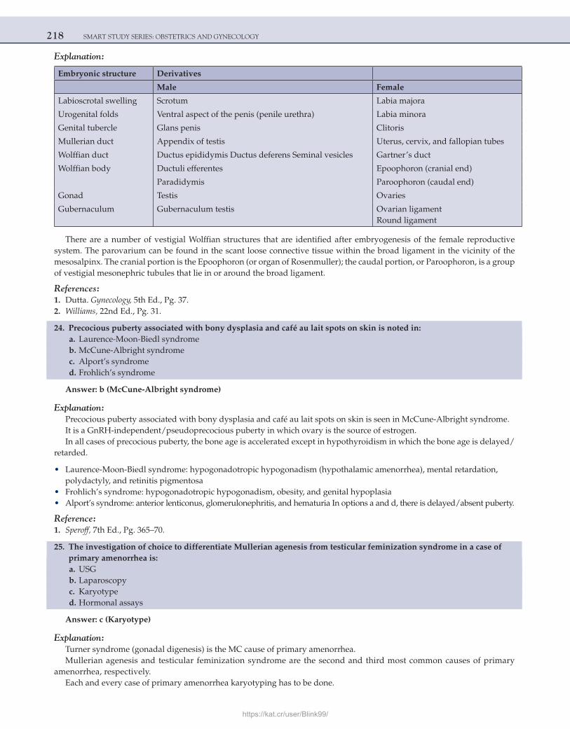
218 SMART STUDY SERIES: OBSTETRICS AND GYNECOLOGY
Explanation:
Embryonic structure Derivatives
Male Female
Labioscrotal swelling Scrotum Labia majora
Urogenital folds Ventral aspect of the penis (penile urethra) Labia minora
Genital tubercle Glans penis Clitoris
Mullerian duct Appendix of testis Uterus, cervix, and fallopian tubes
Wolffian duct Ductus epididymis Ductus deferens Seminal vesicles Gartner’s duct
Wolffian body Ductuli efferentes Epoophoron (cranial end)
Paradidymis Paroophoron (caudal end)
Gonad Testis Ovaries
Gubernaculum Gubernaculum testis Ovarian ligamentRound ligament
There are a number of vestigial Wolffian structures that are identified after embryogenesis of the female reproductive system. The parovarium can be found in the scant loose connective tissue within the broad ligament in the vicinity of the mesosalpinx. The cranial portion is the Epoophoron (or organ of Rosenmuller); the caudal portion, or Paroophoron, is a group of vestigial mesonephric tubules that lie in or around the broad ligament.
References: 1. Dutta. Gynecology, 5th Ed., Pg. 37. 2. Williams, 22nd Ed., Pg. 31.
24. Precocious puberty associated with bony dysplasia and café au lait spots on skin is noted in: a. Laurence-Moon-Biedl syndrome b. McCune-Albright syndrome c. Alport’s syndrome d. Frohlich’s syndrome
Answer: b (McCune-Albright syndrome)
Explanation:Precocious puberty associated with bony dysplasia and café au lait spots on skin is seen in McCune-Albright syndrome.It is a GnRH-independent/pseudoprecocious puberty in which ovary is the source of estrogen.In all cases of precocious puberty, the bone age is accelerated except in hypothyroidism in which the bone age is delayed/
retarded.
• Laurence-Moon-Biedl syndrome: hypogonadotropic hypogonadism (hypothalamic amenorrhea), mental retardation, polydactyly, and retinitis pigmentosa
• Frohlich’s syndrome: hypogonadotropic hypogonadism, obesity, and genital hypoplasia • Alport’s syndrome: anterior lenticonus, glomerulonephritis, and hematuria In options a and d, there is delayed/absent puberty.
Reference: 1. Speroff, 7th Ed., Pg. 365–70.
25. The investigation of choice to differentiate Mullerian agenesis from testicular feminization syndrome in a case of primary amenorrhea is:
a. USG b. Laparoscopy c. Karyotype d. Hormonal assays
Answer: c (Karyotype)
Explanation:Turner syndrome (gonadal digenesis) is the MC cause of primary amenorrhea.Mullerian agenesis and testicular feminization syndrome are the second and third most common causes of primary
amenorrhea, respectively.Each and every case of primary amenorrhea karyotyping has to be done.
https://kat.cr/user/Blink99/
REPRODUCTIVE PHYSIOLOGY, ENDOCRINOLOGY, AND INFERTILITY 219
In patients of Mullerian agenesis the karyotype is 46 XX, whereas in testicular feminization syndrome (androgen insensitivity syndrome) it is 46 XY.
Laparoscopy will reveal absent uterus in both the cases, but in Mullerian agenesis there is presence of ovary and in testicu-lar feminization syndrome there will be testes (in inguinal region).
But just for a diagnosis, laparoscopy is not required.USG would reveal the same findings and help in diagnosis, but the investigation of choice is karyotyping.
Reference: 1. Speroff, 7th Ed., Pg. 340, 421.
26. A 16-year-old girl presents as primary amenorrhea. On examination, breast development is Tanner’s grade 3. USG reveals absence of uterus with normal ovaries. All of following investigations have to be done, except:
a. USG kidneys b. X-ray spine c. Audiogram d. None of the above
Answer: d (None of the above)
Explanation:The diagnosis is RMKH syndrome (Mullerian agenesis) as there is primary amenorrhea with absent uterus and normal ovaries.In these patients, there may be presence of other associated anomalies such as:
1. Renal anomalies 2. Hemi vertebrae and fused vertebrae 3. Sensory neural deafness
Hence, all the three investigations should be done.
Reference: 1. Speroff, 7th Ed., Pg. 410–20.
27. Radha, 35 years old, aborted 5 months back at 17 weeks of a gestation. She has not got her periods yet. Urine pregnancy test is negative. Estrogen progesterone withdrawal test is negative. The likely diagnosis is:
a. Pituitary failure b. Ovarian failure c. Anovulation d. Asherman syndrome
Answer: d (Asherman syndrome)
Explanation:In patients with secondary amenorrhea, after ruling out pregnancy, progesterone challenge test is to be done.Patients with anovulation will get menses with progesterone.If the patient does not get menses with progesterone then E + P challenge test is done.Patients with pituitary failure and ovarian failure will get menses with E + P. Absence of withdrawal by E + P indicates
end organ failure.The patient had a second-trimester abortion, following which a curettage may have been done to remove the retained
products leading to Asherman syndrome.The best diagnostic method is hysteroscopy, and this is treated by adhesiolysis.
References: 1. Speroff, 7th Ed., Pg. 415–25. 2. Williams, 22nd Ed., Pg. 961.
28. During ovulation phase: [AIIMS May 2009, AIIMS May 2011] a. Increase in inhibin A level b. FSH induce steroidogenesis in granulosa cells c. Activin increases d. Stimulation of the arrested meiotic division of the ovum
Answer: c (Activin increases)
220 SMART STUDY SERIES: OBSTETRICS AND GYNECOLOGY
Explanation:The activins and inhibins are glycoproteins that belong to the transforming growth factor-ß superfamily. They are secreted
from granulose cells.FSH induces steroidogenesis (estradiol production) in granulosa cells in the preovulatory phase (follicular phase). Stimulation
of the arrested meiotic division of the ovum occurs after fertilization.Inhibin B has shown increased levels in mid-follicular phase, has a periovular peak, and then declines in luteal phase.
Inhibin A is low in follicular phase, reaches a small peak in mid-follicular phase, and increases to reach a peak in luteal phase.
Activin has biphasic secretion with peak at the time of ovulation and nadir in mid-follicular and mid-luteal phases.
Reference: 1. European Journal of Human Reproduction and Embryology.
29. Fallopian tube dysmotility is associated with this syndrome: [AIIMS 2009; All India 2011] a. Noonan b. Turner c. Kartagener d. Marfan
Answer: c (Kartagener)
Explanation:Primary ciliary dyskinesia (PCD), also known as immotile ciliary syndrome or Kartagener syndrome (KS), is a rare autosomal
recessive genetic disorder which causes a defect in the action of the cilia lining the respiratory tract (lower and upper, sinuses, Eustachian tube, middle ear) and fallopian tube. A poor sense of smell accompanies high mucus production in the sinuses. Infer-tility is common, due to defective ciliary action in the fallopian tube in affected females or diminished sperm motility in males.
Reference: 1. www.emedicine.com.
30. The differentiation of the gonad into male is dependant on: a. Presence of SRY gene b. Lack of SRY gene c. Presence of AMH d. Presence of testosterones
Answer: a (Presence of SRY gene)
5th week
Testosterone(Leydig cells)
AMH(Sertoli cells)
Dihydrotestosterone
Development ofWolffian duct
Development of maleexternal genitalia (AMH = hormone)Anti-Mullerian
Development of Mullerian ductRegression of Wolffian ductDevelopment of femaleexternal genitalia
Cortical regressionMedullary proliferation
Lack of SRYtesticular determinate
6–7 weeks
5 –reductase
Testis Ovary
Absence of testosteroneAbsence ofAMH
Cortical proliferationMedullary regression
SRY gene on theY
autosomal geneschromosome (short-arm)
Undifferentiated(bipotential) gonad
Regression ofMullerian duct
https://kat.cr/user/Blink99/
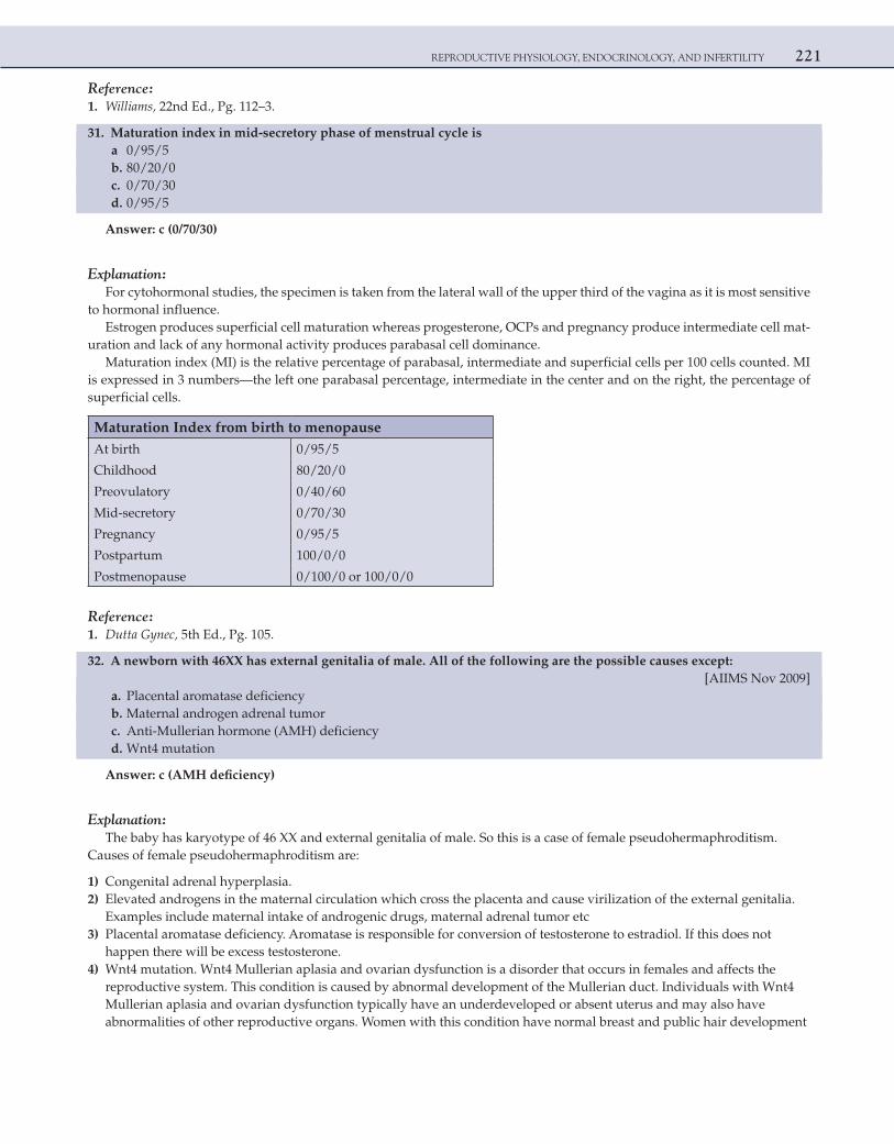
REPRODUCTIVE PHYSIOLOGY, ENDOCRINOLOGY, AND INFERTILITY 221
Reference: 1. Williams, 22nd Ed., Pg. 112–3.
31. Maturation index in mid-secretory phase of menstrual cycle is a 0/95/5 b. 80/20/0 c. 0/70/30 d. 0/95/5
Answer: c (0/70/30)
Explanation:For cytohormonal studies, the specimen is taken from the lateral wall of the upper third of the vagina as it is most sensitive
to hormonal influence.Estrogen produces superficial cell maturation whereas progesterone, OCPs and pregnancy produce intermediate cell mat-
uration and lack of any hormonal activity produces parabasal cell dominance.Maturation index (MI) is the relative percentage of parabasal, intermediate and superficial cells per 100 cells counted. MI
is expressed in 3 numbers—the left one parabasal percentage, intermediate in the center and on the right, the percentage of superficial cells.
Maturation Index from birth to menopauseAt birth 0/95/5
Childhood 80/20/0
Preovulatory 0/40/60
Mid-secretory 0/70/30
Pregnancy 0/95/5
Postpartum 100/0/0
Postmenopause 0/100/0 or 100/0/0
Reference: 1. Dutta Gynec, 5th Ed., Pg. 105.
32. A newborn with 46XX has external genitalia of male. All of the following are the possible causes except: [AIIMS Nov 2009] a. Placental aromatase deficiency b. Maternal androgen adrenal tumor c. Anti-Mullerian hormone (AMH) deficiency d. Wnt4 mutation
Answer: c (AMH deficiency)
Explanation:The baby has karyotype of 46 XX and external genitalia of male. So this is a case of female pseudohermaphroditism.
Causes of female pseudohermaphroditism are:
1) Congenital adrenal hyperplasia. 2) Elevated androgens in the maternal circulation which cross the placenta and cause virilization of the external genitalia.
Examples include maternal intake of androgenic drugs, maternal adrenal tumor etc 3) Placental aromatase deficiency. Aromatase is responsible for conversion of testosterone to estradiol. If this does not
happen there will be excess testosterone. 4) Wnt4 mutation. Wnt4 Mullerian aplasia and ovarian dysfunction is a disorder that occurs in females and affects the
reproductive system. This condition is caused by abnormal development of the Mullerian duct. Individuals with Wnt4 Mullerian aplasia and ovarian dysfunction typically have an underdeveloped or absent uterus and may also have abnormalities of other reproductive organs. Women with this condition have normal breast and public hair development
222 SMART STUDY SERIES: OBSTETRICS AND GYNECOLOGY
and primary amenorrhea. Women with Wnt4 Mullerian aplasia and ovarian dysfunction have higher than normal levels of androgens in their blood. These high levels of androgens cause acne, hirsutism and virilization. Kidney abnormalities may be present in some affected individuals.
AMH DEFICIENCY= PMDS=UTERINE HERNIA SYNDROMEKaryotype = 46 XY and normal male external genitalia.Persistent Mullerian duct syndrome (PMDS) refers to the presence of a uterus and sometimes other Mullerian duct deriva-
tives in a male. In humans, PMDS typically is an autosomal recessive congenital disorder.Typical features include cryptorchidism and the presence of a small, underdeveloped uterus in a male infant or adult. This
condition is usually caused by deficiency of fetal anti-Mullerian hormone (AMH) effect due to mutations of the gene for AMH or the anti-Mullerian hormone receptor.
AMH is produced by the primitive sertoli cells and induces regression of the Mullerian ducts. Mullerian ducts are only sensitive to AMH action around the 8th week of amenorrhea and Mullerian regression is completed by the end of the 9th week. The AMH induced regression of the Mullerian duct occurs in cranio-caudal direction via apoptosis. The Wolffian ducts differentiate into epididymides, vasa deferentia and seminal vesicles under the influence of testosterone, produced by the fetal Leydig cell.
Because both the Wolffian ducts and Mullerian ducts begin to develop, the tissues are often intertwined, resulting in obstruction or nonpatency of the vas deferens or other parts of the male excretory ducts. This can result in infertility, the most serious potential problem caused by this condition.
Other Mullerian derivatives which may be present in at least a rudimentary form are the cervix, upper part of the vagina, and fallopian tubes.
The condition can come to attention because of a bulge in the inguinal canal of a male infant due to herniation of the uterus. The presence of a uterus may be noticed if an ultrasound or MRI of the pelvis is performed to locate the testes or for other reasons.
There is no ambiguity or malformation of the external genitalia. They look like normal male.PMDS type I results from mutations of the gene (AMH) for AMH on chromosome 19p13PMDS type II results from mutations of the gene (AMH-RII) for the AMH receptor on 12q13.
Reference: 1. Speroff, 7th Ed., Pgs. 329, 344.
33. A patient had a spontaneous abortion, then she came with amenorrhea and FSH 6 mIU/mL. What is the most probably diagnosis?
[All India 2010] a. Ovarian failure b. Synechia c. Pregnancy d. Pituitary failure
Answer: b (Synechia)
Explanation:This is a case of secondary amenorrhea with normal FSH values. Normal values range from 3–9 micro IU/mL. Values
higher than this indicate poor ovarian reserve.In cases of ovarian failure and menopause the FSH is above 40 micro IU/mL.In pituitary failure the FSH will be very low.In pregnancy, FSH is suppressed due to high levels of prolactin and inhibin.Normal FSH and amenorrhea point towards uterine pathology. The patient had a spontaneous abortion following which a
curettage is generally required which would be responsible for intra-uterine adhesions (Asherman syndrome)NOTE:High FSH levels are seen in:
1. Premature ovarian failure 2. Poor ovarian reserve 3. Gonadal dysgenesis
https://kat.cr/user/Blink99/
REPRODUCTIVE PHYSIOLOGY, ENDOCRINOLOGY, AND INFERTILITY 223
4. Castration 5. Menopause 6. Testicular failure in males
Low levels of FSH are seen in:
1. Polycystic ovarian syndrome 2. Kallmann syndrome 3. Hypothalamic suppression 4. Hypopituitarism 5. Hyperprolactinemia 6. Gonadal suppression therapy (GnRH analogs)
Reference: 1. Speroff, 7th Ed., Pg. 415–25.
34. Ovarian cycle can be correlated with all except: [All India 2010, 2011] a. Endometrial sampling b. Vaginal cytology c. Blood hormonal levels d. Estrous cycle
Answer: d (Estrous cycle)
Explanation:Ovarian cycles consists of the following : recruitment & growth of the follicles, ovulation, corpus luteum formation and
finally regression of corpus luteum.Because of the changes in the ovary there are simultaneous changes in the uterus (endometrium)-the menstrual cycle.Endometrial sampling and proliferative or secretory endometrium will tell whether the female has ovulated or not.Similarly the hormones FSH, LH, Estradiol, and progesterone will also correlate with the phases of ovarian cycle.Vaginal cytology & calculating the maturation index will tell us the phase of the ovarian & menstrual cycle.Estrous cycle DOES NOT occur in human beings.The estrous cycle comprises of the recurring physiologic changes that are induced by reproductive hormones in most
mammalian placental females. Humans undergo a menstrual cycle instead. Estrous cycles start after puberty in sexually mature females and are interrupted by anestrous phases or pregnancies. Typically estrous cycles continue until death.
Animals that have estrous cycles reabsorb the endometrium if conception does not occur during that cycle. Animals that have menstrual cycles shed the endometrium through menstruation instead. Another difference is sexual activity. In species with estrous cycles, females are generally only sexually active during the estrus phase of their cycle. This is also referred to as being “in heat.” In contrast, females of species with menstrual cycles can be sexually active at any time in their cycle, even when they are not about to ovulate.
Reference: 1. Speroff, 7th Ed., Pgs. 116–20.
35. Presence of both Wolffian and Mullerian ducts are seen in all except: [All India 2010] a. Anti-Mullerian hormone deficiency b. Ovotestis c. FSH receptor mutation d. Mixed Gonadal dysgenesis
Answer: c (FSH receptor mutation)
Explanation:AMH DEFICIENCY= PMDS=UTERINE HERNIA SYNDROME. Karyotype = 46 XY & normal male external genitalia.
224 SMART STUDY SERIES: OBSTETRICS AND GYNECOLOGY
Persistent Mullerian duct syndrome (PMDS) refers to the presence of a uterus and sometimes other Mullerian duct derivatives in a male.
Both the Wolffian ducts and Mullerian ducts develop. The tissues are often intertwined, resulting in obstruction or nonpatency of the vas deferens or other parts of the male excretory ducts.
Ovotestis is seen in true hermaphroditism. Both ovaries & testis are present. There is ambiguity of external genitalia. The internal structures depend on degree of differentiation of the gonads.
Mixed gonadal dysgenesis: 45X/46XY is the MC karyotype seen.A wide variety of phenotypes is seen ranging from ambiguous genitalia to normal fertile males or normal female phenotype
with bilateral streak gonadsThe usual gonadal pattern is streak gonad on one side and a dysgenetic or normal testis on other side. Mullerian and Wolffian
duct development correlates with the character of the ipsilateral gonad.Persons with FSH receptor mutation have either Wolffian duct (males) or Mullerian duct derivatives (females). Never
both.FSH receptor mutation causes infertility or subfertility in males or females.
Reference: 1. Speroff, 7th Ed., Pgs. 344, 348.
36. All of the following are associated with PCOS except: [All India 2010] a. Diabetes mellitus b. Ca endometrium c. Ca ovary d. Osteoporosis
Answer: d (Osteoporosis)
Explanation:PCOS was originally described by Stein and Leventhal in 1935.It is a heterogeneous syndrome complex characterized by chronic anovulation with androgen excess and frequently a/w
insulin resistance, resulting in menstrual irregularity, infertility and hirsutism. It is a state of unopposed estrogenic action (as there is no progesterone due to anovulation)
Insulin resistance is considered to be the hallmark in pathophysiology of PCOS and is present in about 70% cases.Therefore the long term complications a/w PCOS include:
1) Diabetes mellitus. 2) Endometrial hyperplasia 3) Endometrial carcinoma
So now we have to choose b/w options (c) and (d)This was definitely one of the controversial questions in AIPG 2010. But option (d): osteoporosis is a better option to mark.
The main theory for development of epithelial ovarian cancer (which accounts for 85–90% of all ovarian CA) is the “Theory of incessant ovulation” which means “more the ovulation , more the risk”
But in PCOS there is anovulation and hence per say it is protective for CA ovary.BUT, PCOS patients are infertile and ovulation induction is required for treatment of infertility. Use of ovulation inducing
agents (like gonadotropins, clomiphene citrate etc) is one of the risk factors for development of ovarian cancer. This is how PCOS can be a/w CA ovary.
PCOS is PROTECTIVE for osteoporosis.Estrogen deficiency and low BMI are risk factors for osteoporosisIn PCOS there is:
1) Estrogen excess 2) Androgen excess 3) Insulin resistance and hyperinsulinemia 4) Obesity
All these factors are protective for bone mineral loss & osteoporosis.
https://kat.cr/user/Blink99/
REPRODUCTIVE PHYSIOLOGY, ENDOCRINOLOGY, AND INFERTILITY 225
References: 1. Speroff, 7th Ed., Pg. 470–80. 2. Relationship between bone mineral density and insulin resistance in polycystic ovary syndrome: Journal of Bone and Mineral
Metabolism, Volume 19, Number 4 / July, 2001, Pg. 257–62. 3. Novak’s, 14th Ed.
37. A 20-year-old woman gives a history of sharp pain in the lower abdomen for 2–3 days every month approximately 2 weeks before the menses. The most probable etiology for her pain is:
[All India 2003] a. Endometriosis b. Dysmenorrhea c. Pelvic tuberculosis d. Mittelschmerz
Answer: d (Mittelschmerz)
Explanation:Mittelschmerz is one-sided, lower abdominal pain that occurs in women at or around the time of ovulation.
Causes, incidence, and risk factors:About 20% of women experience mittelschmerz or pain associated with ovulation. The pain may occur just before, during,
or after ovulation.There are several explanations for the cause of this pain. Just prior to ovulation, follicle growth may stretch the surface of
the ovary, causing pain. At the time of ovulation, fluid or blood is released from the ruptured egg follicle and may cause irrita-tion of the abdominal lining. Mittelschmerz may be felt on 1 side one month, then switch to the opposite side the next month, or it may be felt on the same side for several months in succession.
The pain is not harmful and does not signify the presence of disease.
Symptoms:Lower-abdominal pain that is:
• One-sided • Recurrent or with similar pain in past • Typically lasting minutes to a few hours, possibly as long as 24–48 hours • Usually sharp, cramping, and distinctive pain • Severe (rare) • May switch sides from month to month or from one episode to another • Begins midway through the menstrual cycle
Signs and tests:A pelvic examination shows no abnormalities. USG may be performed to rule out other causes of pain if ovulatory pain
is prolonged.
Treatment:Analgesics may be needed in cases of prolonged or intense pain.
Prevention:Hormonal forms of contraception can be taken to prevent ovulation—and therefore ovulatory pain.
Reference: 1. Speroff, 7th Ed., Pg. 212–6.
38. Hysteroscopy is used in all of the following, EXCEPT: [AIIMS May 2002 , All India 2013] a. Uterine synechiae b. Abnormal vaginal bleeding c. Infertility d. Recurrent still birth
Answer: d (Recurrent still birth)
226 SMART STUDY SERIES: OBSTETRICS AND GYNECOLOGY
Explanation:Various indications for hysteroscopy are as follows:
1) Abnormal uterine bleeding:
Hysteroscopy has nearly replaced standard D&C for the management of abnormal uterine bleeding (AUB), as it allows for direct visualization and diagnosis of intra-uterine abnormalities, and it often offers an opportunity for simultaneous treatment.
2) Infertility:
When compared with hysterosalpingography, hysteroscopy is equivalent for evaluating the uterine cavity, and it increases accuracy in diagnosing the cause of intra-uterine filling defects. In unexplained infertility, hysteroscopy may be performed simultaneously with laparoscopy to evaluate the uterine cavity and cervix.
Intracavitary lesions (fibroids, septum, and adhesions) are implicated as causes of infertility and recurrent abortions and their removal improves the outcome.
3) Intra-uterine adhesions:
Asherman syndrome was identified in 1948 as uterine synechiae. These intra-uterine adhesions (IUA) are often associated with amenorrhea or infertility.
Hysteroscopy is the gold standard used to diagnose and treat these adhesions. Benefits include visually directed lysis.
4) Mullerian anomalies:
Approximately 1–2% of all women, 4% of infertile women, and 10–15% of patients with recurrent miscarriage have Mullerian anomalies. These anomalies range from didelphys to Mullerian agenesis. Uterine septum and in utero diethylstilbestrol (DES) exposures are more likely to be associated with miscarriage than is uterus didelphys.
5) Polyps and fibroids:
Endometrial polyps and fibroids are well known to cause vaginal bleeding and can be diagnosed and removed with hysteroscopy.
Reference: 1. Novak’s, 14th Ed., Pg. 743–5.
39. Most common cause of female pseudohermaphroditism is: [AIIMS May 2002] a. Congenital adrenal hyperplasia b. Ovarian tumor c. Adrenal cortical tumor d. Androgenic drugs
Answer: a (Congenital adrenal hyperplasia)
Explanation:The term congenital adrenal hyperplasia (CAH) encompasses a group of autosomal recessive disorders, each of which
involves a deficiency of an enzyme involved in the synthesis of cortisol, aldosterone, or both. It is the MC cause of ambiguous genitalia at birth and also the MC cause of female pseudohermaphroditism.
The clinical phenotype of congenital adrenal hyperplasia depends on the nature and severity of the enzyme deficiency. The most common form is 21-hydroxylase deficiency (CYP21). Approximately 50% of patients with classic congenital adrenal hyperplasia due to CYP21A mutations or deletions have salt wasting due to inadequate aldosterone synthesis. Although the information below is presented according to chromosomal sex, the sex of a neonate with congenital adrenal hyperplasia is often initially unclear because of genital ambiguity.Clinical presentation in females:
• Females with severe forms of adrenal hyperplasia due to deficiencies of 21-hydroxylase, 11-P-hydroxylase or 3-P-hydroxysteroid dehydrogenase have ambiguous genitalia at birth due to excess adrenal androgen production in utero. This is often called classic virilizing adrenal hyperplasia.
• Mild forms of 21-hydroxylase deficiency in females are identified later in childhood because of precocious pubic hair, clitoromegaly, or both, often accompanied by accelerated growth and skeletal maturation due to excess postnatal exposure to adrenal androgens. This is called simple virilizing adrenal hyperplasia.
https://kat.cr/user/Blink99/
REPRODUCTIVE PHYSIOLOGY, ENDOCRINOLOGY, AND INFERTILITY 227
• Milder deficiencies of 21-hydroxylase or 3- P-hydroxysteroid dehydrogenase activity may present in adolescence or adulthood with oligomenorrhea, hirsutism, and/or infertility. This is termed non-classic adrenal hyperplasia.
Reference: 1. Speroff, 7th Ed., Pg. 330–6.
40. True about clomiphene citrate is: [AIIMS May 2009, AIIMS May 2010, AIIMS May 2011] a. Enclomiphene has anti-estrogenic effect b. Compared to placebo, it has 3 times increased pregnancy rates c. 2–4% cases have twin pregnancy d. Increased rate of pregnancy in oligospermic males, as shown in RCT
Answer: a (Enclomiphene has anti-estrogenic effect)
Explanation:Clomiphene citrate is a racemic mixture of enclomiphene and zuclomiphene. Enclomiphene is a more potent isomer
responsible for its ovulation-inducing action.It is a selective estrogen receptor modulator (SERM) that increases production of gonadotropins by inhibiting negative
feedback on the hypothalamus.Therapeutically, clomiphene is given early in the menstrual cycle. It is typically prescribed beginning on day 1, 3, or 5 and con-
tinuing for 5 days. By that time, FSH level is rising steadily, causing development of a few follicles. Follicles in turn produce the estrogen, which circulates in serum. Clomiphene acts by inhibiting the action of estrogen on the pituitary. This prevents normal receptor recycling and causes an effective reduction in hypothalamic estrogen receptor number. As a result, the body perceives a low level of estrogen. Since estrogen can no longer effectively exert negative feedback on the hypothalamus, GnRH secretion becomes more rapidly pulsatile, which results in increased pituitary gonadotropin (FSH), which leads to follicle growth.
Common adverse drug reactions associated with the use of clomiphene (≥1% of patients) include: vasomotor flushes (or hot flashes), abdominal discomfort, visual blurring (dose-dependent), and/or reversible ovarian enlargement and cyst forma-tion. Rare adverse effect includes ovarian hyperstimulation syndrome.
Clomiphene can lead to multiple ovulation, hence increasing the chance of twins (6–10% of births instead of the normal ∼1%). In comparison to treatment with purified FSH, the rate of ovarian hyperstimulation syndrome is low. There may be an increased risk of ovarian cancer after prolonged use.
It is also used in male infertility. It may be given to oligospermic males to improve the sperm count, but it has not been proven to increase pregnancy/fertility rates in oligospermic males in randomized controlled trials.
Reference: 1. Speroff, 7th Ed., Pg. 1175–80.
41. All are the causes of primary amenorrhea, EXCEPT: [All India 2009, All India 2011] a. Kallmann syndrome b. Sheehan syndrome c. Mayer-Rokitansky-Küster-Hauser syndrome d. Turner syndrome
Answer: b (Sheehan syndrome)
Explanation:Primary amenorrhea is defined as:
• In the absence of secondary sexual characters, no menses till the age of 14 years, or • In the presence of secondary sexual characters, no menses till the age of 16 years.
MC cause of primary amenorrhea is ovarian dysgenesis/Turner syndrome.Mayer-Rokitansky-Küster-Hauser or Mullerian agenesis is the second MC cause, and androgen insensitivity syndrome or
testicular feminizing syndrome (AIS/TFS) is the third MC of primary amenorrhea.Classic Kallmann syndrome (KS) and idiopathic hypogonadotropic hypogonadism (IHH) are rare genetic conditions
that encompass the spectrum of isolated hypogonadotropic hypogonadism. Most patients have gonadotropin-releasing hor-mone (GnRH) deficiency. Hypothalamic-pituitary function is otherwise normal in most patients, and hypothalamic-pituitary
228 SMART STUDY SERIES: OBSTETRICS AND GYNECOLOGY
imaging reveals no space-occupying lesions. By definition, either anosmia (lack of sense of smell) or severe hyposmia is pres-ent in patients with Kallmann syndrome, in contrast to patients with idiopathic hypogonadotropic hypogonadism, whose sense of smell is normal.
Patients with classic Kallmann syndrome or idiopathic hypogonadotropic hypogonadism may not experience puberty or may experience incomplete puberty and have symptoms associated with hypogonadism. For men, these symptoms include decreased libido, erectile dysfunction, decreased muscle strength, and diminished aggressiveness and drive.
For women, symptoms include primary amenorrhea and dyspareunia.All patients with Kallmann syndrome have either anosmia or severe hyposmia and may exhibit symptoms of associated
conditions, including those of congenital heart disease or neurologic manifestations (e.g., color blindness, hearing deficit, epilepsy, and paraplegia).
Sheehan syndrome, also known as postpartum hypopituitarism or postpartum pituitary necrosis, is hypopituitarism caused by necrosis due to blood loss and hypovolemic shock during and after childbirth.
Most common initial symptoms of Sheehan syndrome are agalactorrhea (absence of lactation) and/or difficulties with lactation. Many women also report secondary amenorrhea or oligomenorrhea after delivery. In some cases, a woman with Sheehan syndrome might be relatively asymptomatic, and the diagnosis is not made until years later, with features of hypopi-tuitarism. Such features include secondary hypothyroidism with tiredness, intolerance to cold, constipation, weight gain, hair loss and slowed thinking, as well as a slowed heart rate and low blood pressure. Another such feature is secondary adrenal insufficiency. Gonadotropin deficiency will often cause secondary amenorrhea, oligomenorrhea, hot flushes, or decreased libido. Growth hormone deficiency causes many vague symptoms, including fatigue and decreased muscle mass
Reference: 1. Speroff, 7th Ed., Pg. 405–10.
42. A 27-year-old female with placenta previa had severe bleeding. What is the most likely outcome postdelivery? [AIIMS May 2010] a. Galactorrhea b. Diabetes c. Absence of menstrual cycle d. Cushing syndrome
Answer: c (Absence of menstrual cycle)
Explanation:Sheehan syndrome, also known as postpartum hypopituitarism or postpartum pituitary necrosis, is hypopituitarism
caused by necrosis due to blood loss and hypovolemic shock during and after childbirth.Most common initial symptoms of Sheehan syndrome are agalactorrhea (absence of lactation) and/or difficulties with
lactation. Many women also report amenorrhea or oligomenorrhea after delivery.For further details, refer the above answer.
Reference: 1. Speroff, 7th Ed., Pg. 405–10.
43. Hypothalamic amenorrhea is seen in: [AIIMS Nov 2001] a. Asherman syndrome b. Stein-Leventhal syndrome c. Kallmann syndrome d. Sheehan syndrome
Answer: c (Kallmann syndrome)
Explanation:
• Kallmann syndrome (deficient GnRH secretion): Hypogonadotropic hypogonadism (hypothalamic amenorrhea) associated with anosmia.
• Inheritance: X linked/AR/AD • KAL gene mutation
https://kat.cr/user/Blink99/
REPRODUCTIVE PHYSIOLOGY, ENDOCRINOLOGY, AND INFERTILITY 229
• Karyotype is normal: 46XX in females & 46XY in males. • It can occasionally be associated with optic problems, such as color blindness or optic atrophy, nerve deafness, cleft
palate, cryptorchidism, renal agenesis, and mirror movement disorder. However, it is not clear how, if at all, these other problems have the same cause as the hypogonadism and anosmia.
• Males present with delayed puberty and may have micropenis (although congenital micropenis is not present in most male KS cases).
• Females present with primary amenorrhea and lack of secondary sex characteristics, such as breast development. • A fraction of cases may present with postpubertal onset, which results in a phenotypically normal penis in men
with subsequent testicular atrophy and loss of some secondary sex traits. These men generally present with sexual impairment and low libido.
• In women, late-onset Kallmann syndrome can result in secondary amenorrhea. • Anosmia may or may not be present in these individuals.
○ Option a: Intra-uterine adhesions (uterine cause) ○ Option b: PCOS (anovulation = ovarian cause) ○ Option d: Postpartum pituitary necrosis
Reference: 1. Speroff, 7th Ed., Pg. 404–7.
44. Prolonged administration of testosterone in male leads to: [AIIMS Nov 2010] a. Increased GnRH b. Increased spermiogenesis c. Azoospermia d. Increased sperm motility
Answer: c (Azoospermia)
Explanation:The original and primary use of testosterone is for the treatment of males who have too little or no natural endogenous
testosterone production—males with hypogonadism. Appropriate use for this purpose is legitimate hormone replacement therapy (testosterone replacement therapy [TRT]), which maintains serum testosterone levels in the normal range.
However, over the years, as with every hormone, testosterone or other anabolic steroids has also been given for many other conditions and purposes besides replacement, with variable success but higher rates of side effects or problems. Examples include infertility, lack of libido or erectile dysfunction, osteoporosis, penile enlargement, height growth, bone marrow stimu-lation and reversal of anemia, and even appetite stimulation.
Adverse effects of testosterone supplementation include minor side effects such as acne and oily skin, and more significant complications such as increased hematocrit, exacerbation of sleep apnea, and acceleration of pre-existing prostate cancer growth. Another adverse effect may be significant hair loss and/or thinning of the hair. Exogenous testosterone also causes suppression of spermatogenesis (eventually leading to azoospermia) and can lead to infertility.
Reference: 1. Speroff, 7th Ed.
45. A 20-year-old female presents with excess facial hair and oligomenorrhea, increased levels of free testosterone, and normal ovaries on USG. Most likely diagnosis is:
[AIIMS Nov 2010 , AIIMS May 2012] a. PCOD b. Adrenal hyperplasia c. Idiopathic hirsutism d. Testotosterone-secreting tumor
Answer: a (PCOD)
Explanation:PCOS/PCOD is a disorder of chronically abnormal ovarian function (oligo/anovulation) and hyperandrogenism
frequently a/w hyperinsulinemia and insulin resistance, resulting in menstrual irregularity, infertility, and hirsutism.
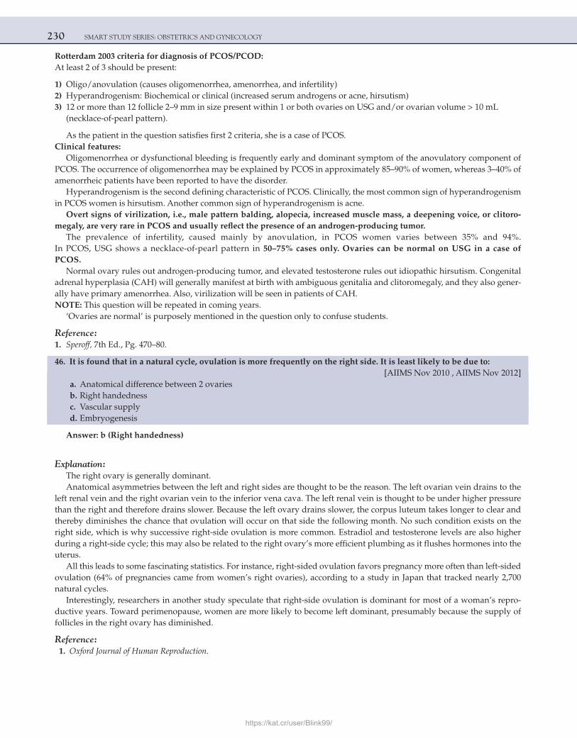
230 SMART STUDY SERIES: OBSTETRICS AND GYNECOLOGY
Rotterdam 2003 criteria for diagnosis of PCOS/PCOD:At least 2 of 3 should be present:
1) Oligo/anovulation (causes oligomenorrhea, amenorrhea, and infertility) 2) Hyperandrogenism: Biochemical or clinical (increased serum androgens or acne, hirsutism) 3) 12 or more than 12 follicle 2–9 mm in size present within 1 or both ovaries on USG and/or ovarian volume > 10 mL
(necklace-of-pearl pattern).
As the patient in the question satisfies first 2 criteria, she is a case of PCOS.Clinical features:
Oligomenorrhea or dysfunctional bleeding is frequently early and dominant symptom of the anovulatory component of PCOS. The occurrence of oligomenorrhea may be explained by PCOS in approximately 85–90% of women, whereas 3–40% of amenorrheic patients have been reported to have the disorder.
Hyperandrogenism is the second defining characteristic of PCOS. Clinically, the most common sign of hyperandrogenism in PCOS women is hirsutism. Another common sign of hyperandrogenism is acne.
Overt signs of virilization, i.e., male pattern balding, alopecia, increased muscle mass, a deepening voice, or clitoro-megaly, are very rare in PCOS and usually reflect the presence of an androgen-producing tumor.
The prevalence of infertility, caused mainly by anovulation, in PCOS women varies between 35% and 94%. In PCOS, USG shows a necklace-of-pearl pattern in 50–75% cases only. Ovaries can be normal on USG in a case of PCOS.
Normal ovary rules out androgen-producing tumor, and elevated testosterone rules out idiopathic hirsutism. Congenital adrenal hyperplasia (CAH) will generally manifest at birth with ambiguous genitalia and clitoromegaly, and they also gener-ally have primary amenorrhea. Also, virilization will be seen in patients of CAH.NOTE: This question will be repeated in coming years.
‘Ovaries are normal’ is purposely mentioned in the question only to confuse students.
Reference: 1. Speroff, 7th Ed., Pg. 470–80.
46. It is found that in a natural cycle, ovulation is more frequently on the right side. It is least likely to be due to: [AIIMS Nov 2010 , AIIMS Nov 2012] a. Anatomical difference between 2 ovaries b. Right handedness c. Vascular supply d. Embryogenesis
Answer: b (Right handedness)
Explanation:The right ovary is generally dominant.Anatomical asymmetries between the left and right sides are thought to be the reason. The left ovarian vein drains to the
left renal vein and the right ovarian vein to the inferior vena cava. The left renal vein is thought to be under higher pressure than the right and therefore drains slower. Because the left ovary drains slower, the corpus luteum takes longer to clear and thereby diminishes the chance that ovulation will occur on that side the following month. No such condition exists on the right side, which is why successive right-side ovulation is more common. Estradiol and testosterone levels are also higher during a right-side cycle; this may also be related to the right ovary’s more efficient plumbing as it flushes hormones into the uterus.
All this leads to some fascinating statistics. For instance, right-sided ovulation favors pregnancy more often than left-sided ovulation (64% of pregnancies came from women’s right ovaries), according to a study in Japan that tracked nearly 2,700 natural cycles.
Interestingly, researchers in another study speculate that right-side ovulation is dominant for most of a woman’s repro-ductive years. Toward perimenopause, women are more likely to become left dominant, presumably because the supply of follicles in the right ovary has diminished.
Reference: 1. Oxford Journal of Human Reproduction.
https://kat.cr/user/Blink99/
REPRODUCTIVE PHYSIOLOGY, ENDOCRINOLOGY, AND INFERTILITY 231
47. In polycystic ovarian syndrome, all of the following are present, EXCEPT: [AIIMS May 2008; All India 2011] a. Increase DHEAS b. Increase LH c. Increase LH:FSH ratio d. Increase prolactin
Answer: d (Increased prolactin)
Explanation:
• Polycystic ovary disease (PCOD) is a heterogenous syndrome consisting of chronic anovulation and hyperandrogenism. • Polycystic ovary syndrome (PCOS) is one of the most common female endocrine disorders and is thought to be one of
the leading causes of female subfertility. • The principal features are infertility, irregular menstruation, acne, and hirsutism. The symptoms and severity of the syndrome
vary greatly among women. While the causes are unknown, insulin resistance and obesity are strongly correlated with PCOS. • Luteinizing hormone (LH) is elevated due to chronic anovulation. • Normal FSH/LH ratio of 2:1 is reversed (1:2), and it can also be 1:3. • Androgens, including androstenedione, testosterone, and dehydroepiandrosterone (DHEAS), are elevated. • But prolactin levels per se are not elevated in patients of PCOD.
Reference: 1. Speroff, 7th Ed., Pg. 470–5.
48. A 35-year-old woman presents with primary infertility and palpable adnexal mass. Her CA125 level is 90 U/mL. The most likely diagnosis is:
[AIIMS May 2010, AIIMS May 2011] a. Epithelial ovarian Ca b. Endometrioma c. Tuberculosis d. Borderline ovarian tumor
Answer: b (Endometrioma)
Explanation:Epithelial ovarian Ca and borderline Ca mainly occur in perimenopausal and postmenopausal ladies.In ovarian tumor, CA125 is elevated, but it is in very high range (800 to >1000 U/mL).In endometriosis, it is mildly elevated (as it is in this case), and besides the patient has infertility and a palpable adnexal
mass, all these go in favor of a endometrioma (chocolate cyst of the ovary = ovarian endometriosis).Genital TB mainly affects the fallopian tube, where there would be no palpable adnexal mass.CA125 is a marker for ovarian cancer, but it may also be elevated in other cancers, including those originating in the
endometrium, fallopian tubes, lungs, breast, and gastrointestinal tract. CA125 may also be elevated in a number of relatively benign conditions, such as endometriosis, acute PID, and pregnancy. It also tends to be elevated in the presence of any inflam-matory condition in the abdominal area, both cancerous and benign.
Thus, CA125 is neither perfectly specific for cancer nor is it perfectly sensitive since not every patient with cancer will have elevated levels of CA125 in the blood.
While this test is not generally regarded as useful for large-scale screening by the medical community, a high value may be an indication that the woman should receive further diagnostic screening or treatment. Normal values range from 0 to 35 (U/mL). Elevated levels in postmenopausal women are usually an indication that further screening is necessary. In premeno-pausal women, the test is less reliable as values are often elevated due to a number of noncancerous causes, and a value >35 is not necessarily a cause for concern.
An endometrioma, or chocolate cyst, is caused by endometriosis and formed when a tiny patch of endometrial tissue bleeds, sloughs off, becomes transplanted, and grows and enlarges inside the ovaries.
It is an estrogen-dependent disease and, thus, usually affects reproductive-aged women. Endometriosis has a prevalence rate of 20–50% in infertile women and as high as 80% in women with chronic pelvic pain.
232 SMART STUDY SERIES: OBSTETRICS AND GYNECOLOGY
Transvaginal sonography is a useful method of identifying the classic chocolate cyst of the ovary. The typical appearance is that of a cyst containing low-level homogenous internal echoes consistent with old blood.
References: 1. TeLinde, 7th Ed. 2. Novak’s, 14th Ed., Pg. 1478–80.
49. Best indicator for ovarian reserve is: [AIIMS May 2011] a. LH b. LH/FSH ratio c. FSH d. Estradiol
Answer: c (FSH)
Explanation:
• In women, FSH stimulates production of eggs and estradiol during the first half of the menstrual cycle. • In men, FSH stimulates production of sperm. • Ovarian reserve means the capacity of the ovary to produce eggs. High FSH indicates that the ovarian reserve is
getting depleted and there are less oocytes remaining in the ovary. (Therefore, FSH is highest in the postmenopausal ladies.)
• FSH levels are higher than normal in women with ovarian hypofunction (and hence, it is a marker for ovarian reserve).The most widely used endocrine marker for ovarian reserve is the early follicular phase (day 2 or day 3 of menstrual cycle) FSH level.
• FSH level has been shown to be an independent predictor of IVF outcome and is a stronger predictor of poor response and the number of oocytes collected at pick-up.
Reference: 1. Speroff, 7th Ed., Pg. 444–8.
50. An infertile woman has bilateral tubal block at cornua diagnosed on hysterosalpingography. Next treatment of choice is:
[AIIMS Nov 2011 , All India 2013] a. IVF b. Laparoscopy and hysteroscopy c. Tuboplasty d. Hydrotubation
Answer: b (Laparoscopy and hysteroscopy)
Explanation:In hysterosalpingography (HSG), cavity of the uterus and fallopian tube patency can be checked.
• As it does not require anesthesia, it is the first-line investigation for checking tubal patency. • Disadvantage: While pushing the dye, there can be cornual spasm and the fallopian tubes can appear to be blocked
even if the tubes are normal/healthy So HSG cannot differentiate between cornual blocks (pathological) and cornual spasm.
Laparoscopy (with chromopertubation with methylene blue dye): Best investigation for tubal patency, as tubal patency can be confirmed under vision, and besides, any pathology can simultaneously be corrected with operative laparoscopy.
This patient has bilateral cornual blocks on HSG, and hence, a laparoscopy should be done to confirm the findings.If on laparoscopy there is a presence of cornual block, cornual catheterization (using operative hysteroscopy) should be
done simultaneously to remove the blocks.IVF is the option in inoperable cases/severely damaged tubes or if surgery fails to remove the blocks.
Reference: 1. Speroff, 7th Ed., Pg. 1013–37.
https://kat.cr/user/Blink99/
REPRODUCTIVE PHYSIOLOGY, ENDOCRINOLOGY, AND INFERTILITY 233
51. Young male presents with delayed puberty with decreased FSH, LH, and testosterone. Which of the following is NOT possible?
[All India 2012] a. Kallmann syndrome b. Klinefelter’s syndrome c. Constitutional delay d. Dax-1 gene mutation
Answer: b (Klinefelter’s syndrome)
Explanation:Decrease in serum follicle-stimulating hormone (FSH), luteinizing hormone (LH), and testosterone indicates that this is a
case of hypogonadotropic hypogonadism.
• Hypogonadism resulting from defects of the gonads is traditionally referred to as primary hypogonadism (hypergonadotropic hypogonadism). Examples include: Klinefelter syndrome, mumps, varicocele, testicular torsion, cryptorchidism, etc.In humans, Klinefelter syndrome is the most common sex chromosome disorder in males. Because of this (primary)
hypogonadism, individuals will often have a low serum testosterone level but high serum FSH and LH levels. • Hypogonadism resulting from hypothalamic or pituitary defects are termed secondary hypogonadism
(hypogonadotropic hypogonadism or central hypogonadism, referring to the central nervous system). Examples of hypothalamic defects include Kallmann syndrome. Examples of pituitary defects include hypopituitarism. Dax-1 (dosage-sensitive sex reversal, adrenal hypoplasia critical region, on chromosome X, gene 1) is a nuclear receptor protein that is encoded by the NR0B1 gene (nuclear receptor subfamily 0, group B, member 1) in humans.
Mutations in this gene result in both X-linked congenital adrenal hypoplasia and hypogonadotropic hypogonadism.
Reference: 1. Speroff, 7th Ed., Pg 404–7.
52. The LH surge occurs due to: [All India 2012] a. Markedly increased estrogen level b. Increased level of prostaglandins c. Increase in progesterone levels d. Decreased FSH levels
Answer: a (Markedly increased estrogen level)
Explanation:In females, at the time of menstruation, follicle-stimulating hormone (FSH) initiates follicular growth, specifically affecting
granulosa cells. With the rise in estrogens, luteinizing hormone (LH) receptors are also expressed on the maturing follicle that produces an increasing amount of estradiol. Eventually at the time of the maturation of the follicle, the estrogen rise leads (via the HPO axis) to the ‘positive feedback’ effect, a release of LH over a 24- to 48-hour period. This ‘LH surge’ triggers ovulation, thereby not only releasing the egg but also initiating the conversion of the residual follicle into a corpus luteum, which, in turn, produces progesterone to prepare the endometrium for a possible implantation. Luteinizing hormone is necessary to maintain luteal function for the first 2 weeks. In case of a pregnancy, luteal function will be further maintained by the action of hCG (a hormone very similar to LH) from the newly established pregnancy. Luteinizing hormone supports theca cells in the ovary that provide androgens and hormonal precursors for estradiol production.
• Ovulation occurs because of LH surge. • Onset of LH surge to ovulation = 36 hours • Onset to peak = 24 hours • Peak to ovulation = 12 hours
Pre-ovulatory estradiol levels should reach 200 pg/mL and should be maintained for 24–48 hours. Only when this is achieved, there is a positive feedback to pituitary, and then the LH surge starts.
Reference: 1. Speroff, 7th Ed., Pg. 220–4.
234 SMART STUDY SERIES: OBSTETRICS AND GYNECOLOGY
53. A female presents with infertility. There is history of dyspareunia and cyclic pain. Best investigation to be done is: [AIIMS Nov 2012] a. TVS b. Diagnostic laparoscopy c. HSG d. diagnostic hysteroscopy
Answer: b (Diagnostic laparoscopy)
Explanation:The given clinical history points to diagnosis of endometriosis.Visualization of endometriotic implants is the definitive method of diagnosis. Laparoscopy is the investigation of choice.
On USG, chocolate cyst of the ovary may be seen but USG can also be normal in patients of endometriosis. Laparoscopy is the investigation of choice for endometriosis.
Besides it also has an advantage of surgically treating the condition (chocolate cyst removal, adhesiolysis, electrofulguration of endometriotic implants) and also it can confirm the tubal patency and hence is the best investigation for this infertile patient.
Please refer the theory section for the various laparoscopy findings in case of endometriosis.Endometriosis is the presence of functional endometrium (glands and stroma) abnormally implanted in locations other
than the uterine cavity. This tissue is capable of responding to the normal cyclic hormonal changes.Symptoms of endometriosis can be variable but typically reflect the area of involvement. Such symptoms may include the
following:
• Dysmenorrhea • Menorrhagia or irregular bleeding • Pelvic pain • Lower abdominal or back pain • Dyspareunia • Infertility
References: 1. TeLinde, 7th Ed. 2. Novak’s, 14th Ed. Pg. 1478–80.
54. A 19 year old unmarried girl presents with irregular periods, acne and facial hair. The best treatment for her is: [AIIMS May 2012, AIIMS May 2013] a) clomiphene citrate b) cyclical progesterone tablets c) OC pills containing LNG d) OC pills containing cyproterone
Answer: d (OC pills containing cyproterone)
Explanation:PCOS is a heterogeneous syndrome complex characterized by chronic anovulation with androgen excess and frequently
associated with insulin resistance, resulting in menstrual irregularity, infertility, acne and hirsutism.Oral contraceptive pills are the first-line therapy (DOC) for PCOS patients with menstrual abnormalities and hirsutism, acne.OC pills will make the menstrual cycles regular and also suppress acne and hirsutism.Cyproterone is a progesterone with anti-androgenic property and hence it is best for those patients who have androgen excess.So option d) is preferred over option c).Cyclical progesterone treatment will only regularize the cycles and will not help in treatment of acne and hirsutism.Clomiphene citrate induces ovulation and hence it is the first-line treatment for infertile patients of PCOS. It is to be used
only in cases of anovulatory infertility.
Reference: 1. Speroff, 7th Ed. Pg. 475–480
https://kat.cr/user/Blink99/
235
8Menstrual Disorders, Menopause and HRT
TYPES OF ABNORMAL UTERINE BLEEDING
Dysfunctional uterine bleeding
Abnormal uterine bleeding with no demonstrable organic cause, genital or extragenital
Menorrhagia Prolonged and/or excessive uterine bleeding (>80 mL) occurring at regular intervals
Metrorrhagia Uterine bleeding occurring at completely irregular but frequent intervals, the amount being variable (intermenstrual bleeding)
Polymenorrhea Uterine bleeding occurring at regular intervals of less than 21 days
Postmenopausal bleeding Bleeding occurring more than 1 year after the last menses in a woman with ovarian failure
Postcoital bleeding Bleeding occurring after intercourse
Premenstrual spotting Scant bleeding that occurs a few days, a week before menses
Oligomenorrhea Menstrual bleeding occurring more than 35 days apart and which remains constant at that frequency
DIFFERENTIAL DIAGNOSIS OF ABNORMAL UTERINE BLEEDING
Reproductive Tract Disease
1. Complications of pregnancy a. Abortion b. Ectopic gestation c. Retained products of conception 2. Benign pelvic lesions a. Fibroids b. Polyps c. Adenomyosis and endometriosis d. Endometritis/PID e. Foreign body 3. Malignant pelvic lesions a. Cervix, endometrium, ovary, vagina, and vulva b. “Precancer”—endometrial hyperplasia
Systemic Disease
1. Coagulation disorders, for example, ITP, vWD (important cause for puberty menorrhagia) 2. Hypothyroidism/hyperthyroidism 3. Liver disease
C H A P T E R
236 SMART STUDY SERIES: OBSTETRICS AND GYNECOLOGY
Iatrogenic Causes
1. Steroids 2. Anticoagulants 3. Intra-uterine contraceptive device (IUCD)
CAUSES OF CONTACT BLEEDING
• Carcinoma cervix • Mucous polyp of cervix • Vascular ectopy of cervix specially during pregnancy, pill use • Infections—chlamydial or tubercular cervicitis • Cervical endometriosis
IMPORTANT CAUSES OF MENORRHAGIA IN DIFFERENT AGE GROUPS
Puberty Reproductive Age Perimenopausal
HPO axis immaturity Pregnancy-related complication (incomplete abortion)
Endometrial hyperplasia, carcinomas
Dysfunctional uterine bleeding(DUB)
Fibroids, polyps, adenomyosis,endometriosis
DUB, infrequent ovulation
Coagulation defects (ITP, von DUB Endocrine problems
Willebrand disease)
Endocrine abnormalities Endocrine abnormalities Fibroids, polyps, adenomyosis, endometriosis
MANAGEMENT OF MENORRHAGIA IN DIFFERENT AGE GROUPS
1. Ultrasonography should be done in all age groups. 2. Sr. TSH should also be done for all patients.
Puberty Menorrhagia
• Always rule out bleeding disorders such as ITP, vWD. • Platelet count, bleeding time, clotting time, prothrombin time, and activated partial thromboplastin time should
be done. • Medical line of management is always the first choice. • Various drugs that can be used are: tranexamic acid, ethamsylate, mefenamic acid, progesterone (oral/
injectables), and OC pills. • IV estrogen (not available in India) may be used to control heavy bleeding in acute phase to regenerate the
endometrium. • Desmopressin (IV/intranasal) is to be used for patients of von Willebrand disease or factor VIII deficiency. • D&C is used as the last resort only when all the medical methods fail to control bleeding.
Reproductive age group
• Management is directed toward treatment of the causative factor (fibroids, polyps, and endometriosis) • In cases of DUB, three cycles of hormonal manipulation is given (OC pills or cyclical progesterone) • If the menorrhagia persists then histopathological diagnosis (D&C/endometrial biopsy/hysteroscopy and
biopsy) should be made.
https://kat.cr/user/Blink99/
MENSTRUAL DISORDERS, MENOPAUSE AND HRT 237
Perimenopausal age group
• Histopathological diagnosis (D&C/endometrial biopsy/hysteroscopy and biopsy) should always be made first to rule out endometrial hyperplasia/cancer before proceeding with any treatment.
• Hysteroscopy and biopsy are preferred to blind D&C. • Management is dependent on the histology report. • If there is no evidence of malignancy, the treatment options include: Mirena, DMPA, endometrial ablation/
resection, or simple hysterectomy.
METROPATHIA HEMORRHAGICA (SCHROEDER’S DISEASE/CYSTIC GLANDULAR HYPERPLASIA)
• This is usually seen in perimenopausal women due to infrequent/irregular ovulation. • The classical presenting feature is amenorrhea followed by menorrhagia. • As there is no ovulation, there is unopposed estrogenic action on the endometrium leading to thickening of
endometrium and a period of amenorrhea. • After a variable period of time (6–8 weeks), the endometrial shedding happens (either due to decrease in
estrogen or when the endometrium outgrows it blood supply), resulting in heavy bleeding. • There is myohyperplasia, and there can be symmetric enlargement of uterus to about 8–10 weeks size and the
endometrium looks thick, congested, and polypoidal. • Histopathology: cystic glandular hyperplasia, Swiss cheese pattern (small and large empty glands with
columnar epithelium), and absence of secretory changes. • Swiss cheese pattern is also seen on ultrasonography in adenomyosis.
POSTMENOPAUSAL BLEEDING PV
• Never wait and watch. • Histopathological diagnosis should always be made first. • Fractional curettage (to rule out Ca cervix and Ca endometrium) is the investigation of choice. • There is no role for hormonal manipulation to control postmenopausal bleeding.
In postmenopausal women (even if she is on hormone replacement therapy or HRT) the endometrial thickness (ET) should be less than 4 mm. If the ET is more than 4 mm, it requires further evaluation (histology), even if the patient is asymptomatic.
UTERUS CONSERVING SURGERIES FOR DUB (ENDOMETRIAL ABLATION/RESECTION)
The various surgeries are:
1. Transcervical resection of endometrium (TCRE), in which the basal endometrium is removed using diathermy loop
3. Roller ball endometrial ablation 4. Laser (Nd: YAG) endometrial ablation 5. MEA (microwave of 9.2 GHz used for endometrial ablation) 7. Uterine thermal balloon in which hot saline/dextrose is circulated within the balloon after it is placed inside the
uterus 8. Hydrotherm ablator in which heated saline in circulated within uterine cavity
In a D/C, only superficial endometrium is removed which grows back, but in above minimally invasive surgeries the basal endometrium is destroyed so that it does not regenerate back.
238 SMART STUDY SERIES: OBSTETRICS AND GYNECOLOGY
Prerequisites
• Patient’s family should be complete • Histopathology: there should be no evidence of malignancy
Advantages
• Day care procedure • Major surgery such as hysterotomy is avoided
Results
• 40% patients will become amenorrheic. • 40% will have hypomenorrhea. • Only 20% will require hysterectomy.
PREMENSTRUAL SYMPTOMS
It is a psychosomatic disorder of unknown etiology, in which there is cyclic appearance of symptoms that regularly occur during luteal phase of each ovulatory cycle.
The most common premenstrual symptoms (PMS) are categorized into cluster by Moos:
• Anxiety: ○ Nervous tension ○ Mood swings ○ Irritability ○ Restlessness • Water retention: ○ Weight gain ○ Swelling ○ Breast tenderness ○ Abdominal bloating • Depression: ○ Crying ○ Confusion ○ Social withdrawal ○ Insomnia • Pain: ○ Cramps ○ Headache ○ Backache ○ Breast pain • Concentration: ○ Difficulty in concentrating/confusion • Autonomic reaction: ○ Dizziness, cold sweats, nausea, and hot flushes
Differential Diagnosis for PMS
• Common differential diagnosis: ○ Adjustment disorder with depressed mood ○ Affective disorders ○ Anxiety disorder ○ Substance-abuse disorder ○ Personality disorder ○ Dysmenorrhea
https://kat.cr/user/Blink99/
MENSTRUAL DISORDERS, MENOPAUSE AND HRT 239
• Less common differential diagnosis: ○ Psychosis ○ Eating disorder ○ Manic-depression disorder ○ Chronic fatigue syndrome ○ Migraine headaches ○ Irritable bowel syndrome
Treatments of PMS
Conservative Measures Inhibition of Ovulation Medications Directed at Symptoms
Salt/fluid/alcohol/caffeine restriction Oral contraceptives (especially drospirenone containing)
Fluid retention: diuretics
Counseling, emotional support GnRHa Pain: PG inhibitors
Low-fat, high-fiber diet and essential fatty acids
Danazol Mastalgia: evening primrose oil and pyridoxine
Exercise Anxiety/depression: SSRI
MENOPAUSE AND HRT
• Menopause is defined as the permanent cessation of menses for 1 year and is physiologically correlated with the decline in estrogen secretion resulting from the loss of follicular/ovarian function.
• The perimenopausal period encompasses the time before, during, and after menopause. The length of this period varies but is usually considered to last approximately 7 years, beginning with the decline in ovarian function in a woman’s 40s and continuing until she has not had menses for 1 year.
• The time of menopause is determined genetically and occurs at the median age of 51 years in West and 47 years in India.
• Menopause occurs earlier in nulliparous women, in tobacco smokers, and hysterectomized women. • Premature ovarian failure is defined as menopause occurring spontaneously before 40 years of age.
Causes of Premature Ovarian Failure:
Chromosomal etiologyIatrogenic causesRadiationChemotherapySurgical alteration of ovarian blood supplySavage syndromeInfectionsAutoimmune disordersGalactosemiaCigarette smokingIdiopathic
Menopausal Symptoms
1. Hot flushes: ○ The classic symptom associated with estrogen deficiency is the hot flash, also known as hot flush ○ This symptom is described as “recurrent, transient periods of flushing, sweating and a sensation of heat, often
accompanied by palpations, feeling of anxiety and sometimes followed by chills” ○ The entire episode usually lasts no more than 1–3 min and may recur as many as 30 times per day, although
5–10 times per day is probably more common
240 SMART STUDY SERIES: OBSTETRICS AND GYNECOLOGY
○ Hot flashes are experienced by at least half of all women during natural menopause and even more women after surgical menopause
○ In severe cases, hot flashes may be accompanied by fatigue, nervousness, anxiety, irritability, depression, and memory loss. These sensations, if occurring at night, are called as “night sweats” and can lead to interruption of sleep patterns
○ Physiologically, hot flashes correspond to marked, episodic increase in the frequency and intensity of gonado-tropin-releasing hormone (GnRH) pulses from the hypothalamus and not due to increased GnRH secretion
2. Vaginal dryness, dyspareunia, recurrent vulvovaginal infections, and urinary tract infections 3. Mood swings, irritability, and depression 4. Decreased libido 5. Memory loss 6. Osteoporosis
Risk factors for osteoporosis:Modifiable risk factors:
○ Estrogen deficiency (menopause/premature menopause/prolonged amenorrhea) ○ Low BMI ○ Prolonged immobility ○ Smoking, alcohol abuse ○ Nutritional factors ○ Secondary causes: celiac disease Nonmodifiable risk factors: ○ Age ○ Race ○ Positive family history ○ Prior fragile fracture
Osteoporosis is defined as the reduction in the quantity of bone, leading to enhanced susceptibility to fractures. Bones associated with postmenopausal fractures:
1. Spinal vertebrae 2. Radius 3. Neck of femur.
HORMONE REPLACEMENT THERAPY
Based on the results of Women’s Health Initiative (WHI) trial, the following are now the accepted indications for HRT:
1. Menopausal symptoms such as hot flushes, vaginal dryness, mood swings, irritability, etc 2. Prevention and treatment of osteoporosis 3. Decreased libido
HRT is not given for primary prevention of heart disease.The different hormones used are:
1. Estrogen (E) and progesterone (P) combination: • As unopposed estrogen is a risk factor for endometrial hyperplasia and cancer; in women with intact uterus
both E + P should be given. In hysterectomized women, only E can be given. • The most commonly prescribed oral estrogen is conjugated equine estrogen (CEE). • The most common progestin is medroxyprogesterone acetate (MPA). 2. Testosterone: • The most common indication for androgens is loss of libido. Testosterone by peripheral conversion to estrogen will also relieve the hot flushes. 3. Tibolone: • It is considered as designer HRT It is a selective tissue estrogen activity regulator (STEAR). • It has estrogenic, progestogenic, and androgenic properties. 4. Selective estrogen receptor modulators: • Raloxifene is a selective estrogen receptor modulator (SERM), which binds with higher affinity to estrogen
alpha receptor than the beta receptors.
https://kat.cr/user/Blink99/
MENSTRUAL DISORDERS, MENOPAUSE AND HRT 241
• Clinically raloxifene produces an effect similar to estrogen on skeletal and cardiovascular system, while behaving as an estrogen antagonist in the uterus and breast.
• Raloxifene maintains a favorable lipid profile and does not exert a proliferative effect on the endometrium. • Effects on bone remodeling are similar to those of estrogen; there is a decrease in the incidence of
fractures. • Unfortunately, raloxifene does not relieve hot flushes and can even worsen them. • There is increased incidence of venous thromboembolism. • Raloxifene is useful in decreasing the risk of osteoporosis.
CONTRAINDICATIONS OF HRT
• Active liver disease (hepatitis/tumor) • Thrombophilias • IHD • Complicated migraine • Complicated valvular heart disease • Breast cancer (current or past history) • Severe hypertension (systolic >160 or diastolic >100) • DM with vascular complications • History of thromboembolism/stroke/DVT
NONHORMONAL DRUGS
1. Calcium 2. Bisphosphonates • Etidronate • Alendronate • Pamidronate Risedronate 3. Calcitonin, calcitriol, and vitamin D-400 IU/day 4. Strontium
Nonhormonal drugs that relieve the hot flushes
1. Clonidine 2. Sertraline 3. Venlafaxine 4. Fluoxetine 5. Gabapentin
MULTIPLE CHOICE QUESTIONS
1. A 29-year-old female patient suffers from emotional lability and depression for about 10 days prior to her menses. She reports that once she begins to bleed she feels back to normal. The patient also gives history of premenstrual fatigue, breast tenderness, and bloating. She is on oral contraceptives to treat her symptoms since 6 months. She reports that the pills have reduced all her PMS symptoms except for the depression and emotional symptoms. Which of the following would be the best treatment for this patient’s problem?
a. Spironolactone b. Evening primrose oil c. Fluoxetine d. Vitamin B6
Answer: c (Fluoxetine)
242 SMART STUDY SERIES: OBSTETRICS AND GYNECOLOGY
Explanation:OC pills are effective in the treatment of PMS and they reduce the mastalgia and bloating.The only medications that have been shown in randomized, double-blind, placebo-controlled trials to be consistently effec-
tive in treating the emotional symptoms of PMS are the selective serotonin reuptake inhibitors. Such antidepressants include fluoxetine, sertraline, and paroxetine. Some women can be effectively treated by limiting use of the medication to the luteal phase.
Reference: 1. Speroff, 7th Ed., Pg. 535–40.
2. A 18-year-old girl has been bleeding heavily for the past 2 weeks. She experienced menarche about 3 years ago, and since that time her periods have been extremely irregular and heavy. She appears very pale and fatigued. Her blood pressure is 110/60 mmHg and the pulse is 70/min. All of the following are appropriate tests to be done, except:
a. Beta-HCG b. Bleeding time c. CBC d. Estradiol level
Answer: d (Estradiol level )
Explanation:The case presented here is a typical representation of dysfunctional uterine bleeding due to anovulation. The onset of men-
arche in young women is typically followed by approximately 5 years of irregular cycles that result from anovulation second-ary to immaturity of the hypothalamic–pituitary axis. Endometrial hyperplasia, polyps, cervical polyps or cervical pathology, and fibroids would be rare in a girl of this age. These other causes of abnormal bleeding would be more common in older women. Of course pregnancy should always be considered as a possible cause in all women of reproductive age. Appropriate laboratory tests to order in the emergency room would be:
1. Beta-HCG (to rule out pregnancy) 2. Bleeding time, clotting time, and platelet count, PT, and aPTT (20% of adolescents with dysfunctional uterine bleeding
have a coagulation defect). Always rule out bleeding disorders in patients of puberty menorrhagia 3. Blood grouping and cross match (may need a blood transfusion) 4. CBC (will show the degree of blood loss this patient has suffered)
Measuring an estradiol level would serve no utility in the workup of this patient.
Reference: 1. Speroff, 7th Ed., Pg. 554–60.
3. Which of the following medications is most useful for the treatment of premenstrual syndrome? [All India 2008] a. Progesterone b. Anxiolytics c. Vitamin B6
d. Selective serotonin reuptake inhibitors (SSRIs)
Answer: d (Selective serotonin reuptake inhibitors)
Explanation:Premenstrual syndrome is a constellation of symptoms that occur in a cyclic pattern, always in the same phase of the men-
strual cycle. These symptoms usually occur 7–10 days before the onset of menses. Examples of symptoms reported include edema, mood swings, depression, irritability, breast tenderness, increased appetite, and cravings for sweets. The etiology is unclear. Besides the treatments listed in the question, therapy has included oral contraceptives, danazol, bromocriptine, evening primrose oil, and aerobic exercise. Of all the medications studied, SSRIs have shown the greatest efficiency in PMS treatment.
Reference: 1. Speroff, 7th Ed., Pg. 535–8.
https://kat.cr/user/Blink99/
MENSTRUAL DISORDERS, MENOPAUSE AND HRT 243
4. The presentation of Asherman syndrome typically involves: [AIIMS Nov 2007] a. Hypomenorrhea b. Oligomenorrhea c. Menorrhagia d. Metrorrhagia
Answer: a (Hypomenorrhea)
Explanation:Ovulation is not affected in Asherman syndrome. Because of the decreased amount of functional endometrium, progres-
sive hypomenorrhea (lighter menstrual flow) followed by amenorrhea is commonly seen.The diagnosis can be made on honeycomb appearance (HSG) or hysteroscopy.Treatment includes hysteroscopic adhesiolysis. This is followed by insertion of IUD for few days to keep the cavity distended
to prevent adhesions, and then the patient should be given estrogen to regenerate the endometrium followed by progesterone.
Reference: 1. Speroff, 7th Ed., Pg. 419.
5. A 32-year-old patient complains of bleeding between her periods and increasingly heavy menses. Over the past 9 months, she has had two dilation and curettages (D&Cs), which have failed to resolve her symptoms, and contraceptives and antiprostaglandins have not decreased the abnormal bleeding. Of the following options, which is the most appropriate at this time?
a. Perform a hysterectomy b. Perform hysteroscopy c. Perform endometrial ablation d. Treat with a GnRH agonist
Answer: b (Perform hysteroscopy)
Explanation:In patients with abnormal bleeding who are not responding to standard therapy, hysteroscopy should be performed.
Hysteroscopy can rule out endometrial polyps or small fibroids, which, if present, can be resected.This is the advantage of hysteroscopy over blind D&C. In patients with heavy abnormal bleeding who no longer desire
fertility, an endometrial ablation can be performed. If a patient had completed childbearing and was having significant abnor-mal bleeding, a hysteroscopy rather than a hysterectomy would still be the procedure of choice to rule out easily treatable disease, and then hysterectomy could be done as last resort.
Treatment with a GnRH agonist would only temporarily relieve symptoms.
Reference: 1. Speroff, 7th Ed., Pg. 550–2.
6. A 39-year-old woman, gravida 3, para 3, complains of severe, progressive secondary dysmenorrhea and menorrhagia. Pelvic examination demonstrates a tender, diffusely enlarged uterus with no adnexal tenderness. Results of endometrial biopsy are normal. This patient most likely has:
[AIIMS May 2007] a. Endometriosis b. Endometritis c. Adenomyosis d. Uterine sarcoma
Answer: c (Adenomyosis)
Explanation:Adenomyosis is a condition in which normal endometrial glands grow into the myometrium. Symptomatic disease pri-
marily occurs in multiparous women over the age of 35 years, compared to endometriosis, in which onset is considerably
244 SMART STUDY SERIES: OBSTETRICS AND GYNECOLOGY
younger. Patients with adenomyosis complain of dysmenorrhea and menorrhagia, and the classical examination findings include a tender, symmetrically enlarged uterus without adnexal tenderness. Adenomyosis on USG has a Swiss cheese appearance.Benson and Sneden’s criteria for adenomyosis (on histology):Presence of endometrium within the myometrium at least two low-power field or 8 mm from basal endometrium.
Although patients with endometriosis can have similar complaints, the physical examination of these patients more commonly reveals a fixed, retroverted uterus, adnexal tenderness and scarring, and tenderness along the uterosacral ligaments.
Leiomyoma is the most common pelvic tumor, but the majority are asymptomatic and the uterus is irregular in shape (bos-selated). Patients with endometritis can present with abnormal bleeding, but endometrial biopsies show an inflammatory pattern.
Uterine sarcoma is rare and presents in older women with postmenopausal bleeding and nontender uterine enlargement.
Reference: 1. TeLinde, 9th Ed., Pg. 629–30.
7. The investigation of choice in a 55-year-old postmenopausal woman who has presented with postmenopausal bleeding is:
[All India 2006, 2013] a. Pap smear b. Fractional curettage c. Transvaginal ultrasound d. CA-125
Answer: b (Fractional curettage)
Explanation:Postmenopausal bleeding most commonly occurs due to atrophic changes but can also occur due to Ca endometrium or
Ca cervix.Hence, in case of postmenopausal bleeding, ruling out both endometrial and cervical cancer is always a priority. Hence,
fractional curettage is the right answer. TVS can detect uterine pathology, but histopathological diagnosis is a must in such cases. Pap is positive only in 30–50% of Ca endometrium and so not useful. TVS and CA-125 are screening methods for ovar-ian CA and hence not applicable here.
Reference: 1. TeLinde, 9th Ed., Pg. 1379.
8. Period of amenorrhea followed by massive bleeding is seen in premenopausal women with: a. Irregular ripening b. Irregular shedding c. Metropathia hemorrhagica d. All of the above
Answer: c (Metropathia hemorrhagica)
Explanation:Metropathia hemorrhagica should be regarded as a specialized form of dysfunctional uterine hemorrhage. The disease
is most prevalent in women over the age of 40 years, the maximum incidence being between the ages of 40 and 45 years. Occasionally, it develops in young girls under the age of 20 years. Parity is not related to its incidence. The symptoms are very typical. The most common complaint is continuous vaginal bleeding, which may last for many weeks. In half the cases, the continuous bleeding is preceded by a short period of amenorrhea, an interval of about 8 weeks elapsing between the last period and the onset of the continuous hemorrhage. The bleeding is always painless, since it is anovulatory. Options (a) and (b) are examples of ovulatory DUB in which period of amenorrhea is not seen.
References: 1. Shaw’s Gynec, 13th Ed., Pg. 293–94. 2. Dutta, 5th Ed.
https://kat.cr/user/Blink99/
MENSTRUAL DISORDERS, MENOPAUSE AND HRT 245
9. An 18-year-old consults you for evaluation of disabling pain during her menstrual period. The pain has been present since menarche and is accompanied by nausea and headache. History is otherwise unremarkable, and pelvic examination is normal. Initial treatment will include:
a. Ergot derivatives b. Antiprostaglandins c. GnRH analogs d. Danazol
Answer: b (Antiprostaglandins)
Explanation:Dysmenorrhea is considered secondary if associated with pelvic disease such as endometriosis, uterine myomas, or pelvic
inflammatory disease. Primary dysmenorrhea is associated with a normal pelvic examination and with ovulatory cycles. Anovulatory cycles are never painful.
The pain of dysmenorrhea is usually accompanied by other symptoms (nausea, fatigue, diarrhea, and headache), which may be related to excess of prostaglandin. The two major drug therapies effective in dysmenorrhea are oral contraceptives and antiprostaglandins (NSAIDs).
GnRH analogs would not be the first-line therapy for primary dysmenorrhea, as it induces amenorrhea in the patient and even when used for 6 months are associated with osteoporosis.
Danazol was used for the treatment of endometriosis (not used nowadays due to androgenic side effects), and ergot derivatives are for hyperprolactinemia.
NSAIDs (MC used = mefenamic acid) are the first-line and most commonly used drugs for primary dysmenorrhea. But OC pills are the best for primary dysmenorrheal, as they make the cycles anovulatory.
Reference: 1. Speroff, 7th Ed., Pg. 533–6.
10. Most common cause of postmenopausal bleeding in India is: [All India 2007] a. Carcinoma cervix b. Endometrial atrophy c. Endometrial hyperplasia d. Endometrial cancer
Answer: a (Carcinoma cervix)
Explanation:Cervical carcinoma is the most common gynecologic malignancy in Indian women, occurring at between 45 and 55 years
of age. Symptoms do not occur until late and may consist of irregular vaginal and postcoital bleeding or discharge. The most common histologic type is squamous cell carcinoma.Causes of postmenopausal bleeding:Ca cervix is the MC cause of postmenopausal bleeding in India
The common causes of postmenopausal uterine bleeding (uterine pathology) are:
Cause of Bleeding Percentage
Endometrial atrophy 60–80
Hormone replacement therapy 15–25
Endometrial polyps 2–12
Endometrial hyperplasia 5–10
Endometrial cancer 10
Bleeding from the vagina may occur because when estrogen secretion stops, the vagina dries and there is atrophy. Lesions and cracks on the vulva may also bleed. Sometimes bleeding occurs after intercourse. Bleeding can occur with or without an associated infection.
246 SMART STUDY SERIES: OBSTETRICS AND GYNECOLOGY
Reference: 1. Novak’s, 14th Ed., Pg. 1148.
11. All of the following are advantages of using raloxifene over estrogens in postmenopausal women, except: a. Reduces fracture rates b. Avoids endometrial hyperplasia c. Reduces incidence of venous thrombosis d. No increase in incidence of breast carcinoma
Answer: c (Reduces incidence of venous thrombosis)
Explanation:Raloxifene is SERM. It acts as an estrogenic agent on the bone. It reduces the occurrence of vertebral fractures by 30–50%.
There is a reduction in invasive breast cancer occurrence of about 70% in women who take raloxifene compared to placebo.Raloxifene is not associated with an increase in the risk of uterine cancer. However, there is an increased risk of venous
thromboembolism.
Reference: 1. Speroff, 7th Ed., Pgs. 663, 700.
12. All of the following appear to decrease hot flushes in menopausal women, except: [All India 2005] a. Androgens b. Raloxifene c. Isoflavones d. Tibolone
Answer: b (Raloxifene)
Explanation:Hot flushes are the subjective sensation of intense warmth of upper body and range in duration from 30 s to 5 min and
usually end in sweating. They result from withdrawal of estrogen, resulting in instability of thermoregulatory center located in the hypothalamus.
Estrogen, isoflavones (plant-derived estrogens), and tibolone relieve the hot flushes. Testosterone (androgens) is given to increase the libido, but by peripheral aromatization to estrogen it also relieves the hot flushes.
Raloxifene is a SERM, having estrogen agonist/antagonist actions at various tissue levels. It is proved to be beneficial for osteoporosis especially in patients reluctant to use estrogens with no effects on endometrium/breast. But it causes hot flushes, and there is increased risk of venous thromboembolism, which are side effects specific to raloxifene.
Reference: 1. Speroff, 7th Ed., Pg. 700.
13. A 35-year-old, mother of two children, is suffering from amenorrhea for the last 10 months. She has a history of failure of lactation following second delivery but remained asymptomatic thereafter. Skull X-ray shows “empty sella.” Most likely diagnosis is:
[All India 2004] a. Menopause b. Pituitary tumor c. Sheehan’s syndrome d. Breast fibroadenoma
Answer: c (Sheehan’s syndrome)
Explanation:Postpartum failure of lactation in a reproductive age group with pituitary necrosis causing “empty sella” appearance on
X-ray skull suggests Sheehan’s syndrome. This generally happens following PPH.Failure of lactation is the earliest manifestation of this condition, and the amenorrhea persists.
https://kat.cr/user/Blink99/
MENSTRUAL DISORDERS, MENOPAUSE AND HRT 247
Progesterone challenge test would turn out to be negative, and patient will require both estrogen + progesterone to get the menses.
Pituitary tumor causes visual disturbances, headache, galactorrhea (if prolactinoma), and posterior clinoid erosion appearance on skull X-ray. Menopause and local breast disease will not have X-ray changes in skull.
Reference: 1. Speroff, 7th Ed., Pg. 415–25.
14. Dysfunctional uterine bleeding (DUB) is seen in: a. Endometrial polyp b. Adenomyosis c. Metropathia hemorrhagica d. All of the above
Answer: c (Metropathia hemorrhagica)
Explanation:
• The term DUB is used for menorrhagia in the absence of any structural abnormality or pelvic pathology or pregnancy. So when an obvious pathology like fibroid, adenomyosis, or a polyp is detected it cannot be termed as DUB.
• Metropathia hemorrhagica is a form of DUB. Continuous uterine bleed is the most constant symptom, and this is generally preceded by amenorrhea of about 8–l0 weeks of duration.
The main pathology is anovulation. This is seen in perimenopausal age group. Histopathology reveals cystic glandular hyperplasia, Swiss cheese appearance. Most of the cases of DUB have anovulation, but ovulatory DUB is also possible.Ovulatory DUB:It is of two varieties:
• Irregular shedding of the endometrium/Halban’s disease: persistent corpus luteum, which leads to irregular shedding and bleeding with simultaneous failure of regeneration of endometrium
• Irregular ripening of the endometrium: inadequate function of corpus luteum leading to premenstrual spotting
NOTE:Halban’s theory: lymphatic spread of endometriosis.Halban’s disease: ovulatory DUB and irregular shedding of the endometrium.Halban’s operation: anterior-posterior obliteration of pouch of Douglas to prevent enterocele.
Reference: 1. TeLinde, 9th Ed., Pgs. 461, 596.
15. Ritu, 15 years old, complains of heavy periods since 2 months. O/E: wt 40 kg and BP 120/80 mmHg. All of the following investigations are indicated, except:
a. S. TSH b. Platelet count c. Bleeding and clotting time d. None of the above
Answer: d (None of the above)
Explanation:Various causes of puberty menorrhagia are:
1. HPO axis immaturity (anovulation) 2. Bleeding disorders 3. Endocrinological causes like thyroid disorders.
Always rule out bleeding disorders in patients of puberty menorrhagia. Hence, all of the above investigations are indicated.
Reference: 1. Speroff, 7th Ed., Pg. 550.
248 SMART STUDY SERIES: OBSTETRICS AND GYNECOLOGY
16. A 46-year-old P3L3 complains of menorrhagia since 3 months. Next line of management is: [AIIMS Nov 2008, All India 2013] a. D&C b. Progesterone × 6 months c. OC pills × 6 months d. Hysterectomy
Answer: a (D&C)
Explanation:In patients with menorrhagia in perimenopausal age group (40+), always make the diagnosis first before proceeding with
any treatment.It is necessary to rule out endometrial hyperplasia and cancer in this age group. Hence, histopathological examination of
endometrium is required, and therefore D&C should be done first. Alternatively, endometrial biopsy or hysteroscopy and biopsy can also be done, but always histopathological diagnosis is required in this age group.
Depending on the endometrial pathology, hormonal treatment or surgery is advised. Never directly proceed with hyster-ectomy because the type of hysterectomy to be performed (simple/radical/TAH + BSO) will depend on the diagnosis.
Progesterones (oral, injectables, and Mirena) may be used after excluding endometrial carcinomas.
References: 1. Novak’s, 14th Ed. 2. TeLinde, 9th Ed., Pg. 463–5.
17. All are evidence-based treatments for menorrhagia, except: [All India 2009; AIIMS May 2010; All India 2011] a. OC PILLS b. Ethamsylate c. Tranexamic acid d. Progesterone 5–25 days cyclically
Answer: b (Ethamsylate)
Explanation:Various options are available for medical management of menorrhagia. These include:
1. OC pills 2. Progesterones (oral/DMPA/Mirena) 3. Antifibrinolytic agents (Tranexamic acid) 4. Danazol and GnRH analogs (very rarely used) 5. NSAIDs 6. Ethamsylate (capillary stabilizers)
All these medications can control the menorrhagia. As per RCOG guidelines, tranexamic acid is now the first-line drug of choice for menorrhagia. In various clinical trials (evidence-based medicine), OC pills, tranexamic acid, and cyclical progester-one from day 5 to day 25 of menstrual cycle have been effective in menorrhagia.
The efficacy of ethamsylate in the management of menorrhagia has not been proven in clinical trials, even though it decreases the blood loss and is also used in clinical practice for menorrhagia.
Reference: 1. RCOG, ACOG Guidelines for Menorrhagia.
18. A 45-year-old postmenopausal woman with DUB has 8 mm thickness of endometrium. Next line of management is: [All India 2011, 2013] a. Follow-up and USG b. Endometrial HPE c. Hysterectomy d. Progesterone therapy
Answer: b (Endometrial HPE)
https://kat.cr/user/Blink99/
MENSTRUAL DISORDERS, MENOPAUSE AND HRT 249
Explanation:
• In postmenopausal woman, the endometrial thickness should always be <4 mm. • If it is >4 mm, then histopathological examination (HPE) of the endometrium is mandatory (even if the patient is
asymptomatic). • So, endometrial sampling or D/C or hysteroscopy and biopsy should be done first. • Rule out hyperplasia/endometrial cancer first before proceeding with the treatment. • Endometrial hyperplasia and carcinoma can only be ruled out by HPE.
Reference: 1. TeLinde, 9th Ed., Pg. 463.
19. A 30-year-old nulliparous hypertensive woman has menorrhagia. The best treatment for her is: [AIIMS May 2011] a. OCP b. Mirena c. Hysterectomy d. Transcervical resection of endometrium
Answer: b (Mirena)
Explanation:
• This is a nulliparous hypertensive lady with menorrhagia. So, hysterectomy is out of question. • Transcervical resection of endometrium (TCRE) is an option for those ladies who have finished child bearing and wish to
conserve the uterus and want to avoid a hysterectomy. • Pregnancy is not possible after TCRE, and hence, it should not be done here. • The lady is hypertensive, which is a relative contraindication for the use of COC pills. • Mirena (completely reversible form of contraceptive) is the best option for her, as it will take care of menorrhagia and
will retain the uterus for further child bearing.
NOTE: Mirena will prevent conception; so actually, the best option for this nulliparous lady would be cyclic progesterone from day 21 to day 25 (withdrawal bleeding, which occurs after progesterone is always less so as this would take care of menorrhagia and will also allow conception).
References: 1. Speroff, 7th Ed. 2. Chaudhary SK, 7th Ed.
20. In postmenopausal women, HRT is indicated for all, EXCEPT: [AIIMS Nov 2011] a. Vaginal dryness b. Hot flushes c. Coronary artery disease d. Osteoporosis
Answer: c (Coronary artery disease)
Explanation:Menopause is defined as the permanent cessation of menses for 1 year and is physiologically correlated with the decline in
estrogen secretion resulting from the loss of follicular/ovarian function.Based on the results of Women’s Health Initiative (WHI) trial, the following are now the accepted indications for HRT:
1. Menopausal symptoms such as hot flushes, vaginal dryness, mood swings, irritability, etc 2. Prevention and treatment of osteoporosis 3. Decreased libido
HRT is not to be given for primary prevention of heart disease.WHI is the largest trial conducted to date, which evaluated the benefits of HRT in postmenopausal women. The trial
concluded that HRT should not be used for heart disease prevention.
Reference:
1. Speroff, 7th Ed., Pg. 663, 700.

This page intentionally left blank
https://kat.cr/user/Blink99/
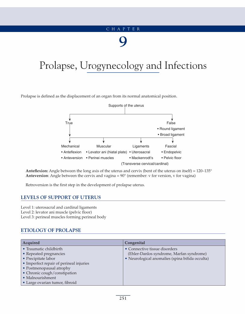
251
9Prolapse, Urogynecology and Infections
C H A P T E R
Prolapse is defined as the displacement of an organ from its normal anatomical position.
Supports of the uterus
eslaFeurT
tnemagildnuoR•
tnemagildaorB•
Mechanical Muscular Ligaments Fascial
(Transverse cervical/cardinal)
Anteflexion: Angle between the long axis of the uterus and cervix (bent of the uterus on itself) = 120–135°Anteversion: Angle between the cervix and vagina = 90° (remember: v for version, v for vagina)
Retroversion is the first step in the development of prolapse uterus.
LEVELS OF SUPPORT OF UTERUS
Level 1: uterosacral and cardinal ligamentsLevel 2: levator ani muscle (pelvic floor)Level 3: perineal muscles forming perineal body
ETIOLOGY OF PROLAPSE
Acquired Congenital
• Traumatic childbirth • Repeated pregnancies • Precipitate labor • Imperfect repair of perineal injuries • Postmenopausal atrophy • Chronic cough/constipation • Malnourishment • Large ovarian tumor, fibroid
• Connective tissue disorders(Ehler-Danlos syndrome, Marfan syndrome)
• Neurological anomalies (spina bifida occulta)
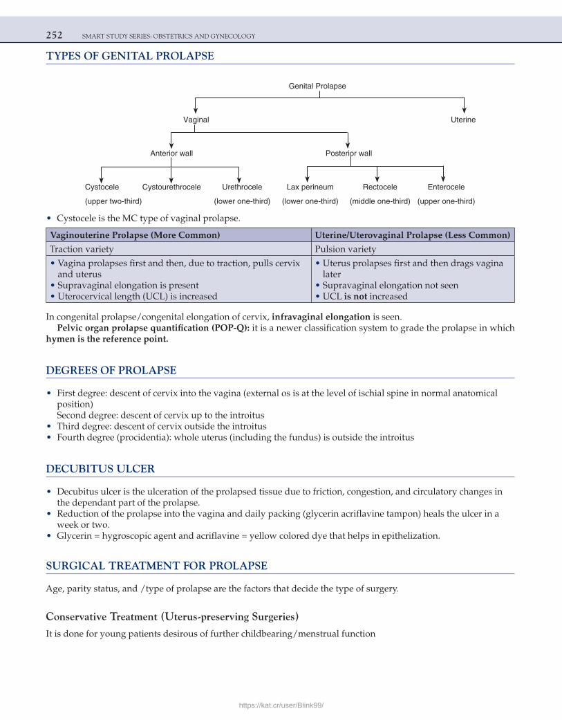
252 SMART STUDY SERIES: OBSTETRICS AND GYNECOLOGY
TYPES OF GENITAL PROLAPSE
Genital Prolapse
eniretUlanigaV
llawroiretsoPllawroiretnA
Cystocele Cystourethrocele Urethrocele Lax perineum Rectocele Enterocele
(upper two-third) (lower one-third) (lower one-third) (middle one-third) (upper one-third)
• Cystocele is the MC type of vaginal prolapse.
Vaginouterine Prolapse (More Common) Uterine/Uterovaginal Prolapse (Less Common)
Traction variety Pulsion variety
• Vagina prolapses first and then, due to traction, pulls cervix and uterus
• Supravaginal elongation is present• Uterocervical length (UCL) is increased
• Uterus prolapses first and then drags vagina later
• Supravaginal elongation not seen• UCL is not increased
In congenital prolapse/congenital elongation of cervix, infravaginal elongation is seen.Pelvic organ prolapse quantification (POP-Q): it is a newer classification system to grade the prolapse in which
hymen is the reference point.
DEGREES OF PROLAPSE
• First degree: descent of cervix into the vagina (external os is at the level of ischial spine in normal anatomical position)Second degree: descent of cervix up to the introitus
• Third degree: descent of cervix outside the introitus • Fourth degree (procidentia): whole uterus (including the fundus) is outside the introitus
DECUBITUS ULCER
• Decubitus ulcer is the ulceration of the prolapsed tissue due to friction, congestion, and circulatory changes in the dependant part of the prolapse.
• Reduction of the prolapse into the vagina and daily packing (glycerin acriflavine tampon) heals the ulcer in a week or two.
• Glycerin = hygroscopic agent and acriflavine = yellow colored dye that helps in epithelization.
SURGICAL TREATMENT FOR PROLAPSE
Age, parity status, and /type of prolapse are the factors that decide the type of surgery.
Conservative Treatment (Uterus-preserving Surgeries)
It is done for young patients desirous of further childbearing/menstrual function
https://kat.cr/user/Blink99/
PROLAPSE, UROGYNECOLOGY AND INFECTIONS 253
Transvaginal
• Fothergill’s operation • Shirodkar’s uterosacral ligament advancement
Abdominal (Sling Surgery/Cervicopexy)
• Purandare • Shirodkar • Khanna • Virkud (composite sling)
Radical Surgery
• For old patients, family complete, postmenopausal women who are medically fit for surgery • Vaginal hysterectomy with or without anterior and posterior colporrhaphy is the best surgery:
Anterior colporrhaphy: repair of cystocele and cystourethrocele
Posterior colporrhaphy: repair of rectocele and lax perineum
KEY POINTS OF VARIOUS SURGERIES
1. Fothergill’s repair (Manchester operation): Main step is amputation of cervix. ○ Initially, the operation was thought to preserve the fertility status of the patient. ○ But as it is associated with a lot of complications, it is not a preferred option nowadays. ○ Various complications include: a. Primary hemorrhage/secondary hemorrhage b. Repeated second trimester abortions due to cervical incompetence c. Preterm labor/PROM d. Cervical stenosis e. Cervical dystocia f. Infertility due to cervical factor 2. Shirodkar’s uterosacral ligament advancement surgery (modification of Fothergill’s operation): There is no
amputation of cervix, and so the complications of Fothergill’s operation are not there. It is preferred in young women desirous of further childbearing.
3. Purandare’s cervicopexy (dynamic sling and open sling): Central part of Mersilene tape is fixed anteriorly over the exposed isthmus. The two ends of tape are attached to the posterior rectus sheath.Good abdominal muscle tone is prerequisite for this surgery. If the anterior abdominal tone is poor, this surgery should not be done. Postsurgery, the uterus becomes retroverted and the POD becomes deep. Hence, enterocele is a long-term complication of this surgery. Enterocele formation can be prevented by Moschowit’s/Halban’s surgery in which POD is obliterated.
4. Shirodkar sling (static sling): Mersilene tape is placed posteriorly on the cervix and anchored to sacral promontory (anterior longitudinal ligament).On the left side, the tape has to pass below the mesentery of sigmoid colon to reach sacral promontory. On the left side, a loop is created over the psoas muscle to avoid obstruction to the rectosigmoid.
Complications:
a. Injury to sigmoid colon, mesentery, and ureters b. Hemorrhage from pre-sacral/mesenteric vessels c. Intestinal obstruction d. Injury to genitofemoral nerve (present in psoas muscle) 5. Khanna sling: Mersilene tape is anchored to anterior superior iliac spine.
254 SMART STUDY SERIES: OBSTETRICS AND GYNECOLOGY
6. Composite sling (Virkud): As the complications of Shirodkar sling are mainly on the left side in this surgery, on right side the tape is attached to sacral promontory and on left side the tape is attached to rectus sheath (left-sided Purandhare + right-sided Shirodkar).
7. Vaginal hysterectomy with pelvic floor repair: Women above 40 years who have advanced uterine prolapse with cystorectocele, have completed their families, and are not interested in further childbearing or menstruation are fit for surgery.
8. Le Fort’s repair (complete colpocleisis): It is done in very elderly postmenopausal women who are unfit for major surgery (with medical complications such as heart failure, past history of myocardial infarction, severe hyper tension, etc.).
This procedure can be performed under local anesthesia and sedation. Prior to the procedure, PAP smear and pelvic USG should be done to rule out cervical dysplasia and pelvic pathology. Vaginal sexual activity is not possible after this surgery. If sexual function is desired, Goodell-Powel surgery (partial colpocleisis) is done (modification of Le Fort’s repair).
INDICATIONS OF RING PESSARY
1. Early pregnancy (up to 18 weeks) 2. Puerperium 3. Patients unfit for surgery
It is never curative, only palliative.
VAULT PROLAPSE
It is a long-term complication of any hysterectomy and occurs more frequently after vaginal as compared to abdominal. It can be prevented by vault suspension at the time of primary surgery.
Management
• Transvaginal sacrospinous ligament fixation • Transabdominal sacrocolpopexy: mesh is attached to vault and sacral promontory
Sacrocolpopexy is considered the gold standard operation for vault prolapse.
UROGYNECOLOGY
Urinary Incontinence
Urinary incontinence is defined as objectively demonstrable involuntary loss of urine so as to cause hygienic and/ or social inconvenience for day-to-day activity.
Urethral Extra-urethral
1. SUI 2. Urge incontinence 1. Acquired 3. Mixed Fistulas
reterucipotce,.g.elatinegnoC.2
https://kat.cr/user/Blink99/
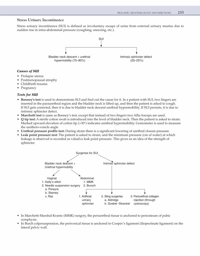
PROLAPSE, UROGYNECOLOGY AND INFECTIONS 255
Stress Urinary Incontinence
Stress urinary incontinence (SUI) is defined as involuntary escape of urine from external urinary meatus due to sudden rise in intra-abdominal pressure (coughing, sneezing, etc.).
SUI
Bladder neck descent + urethral Intrinsic sphincter defect )%52–02()%08–57(ytilibomrepyh
Causes of SUI • Prolapse uterus • Postmenopausal atrophy • Childbirth trauma • Pregnancy
Tests for SUI • Bonney’s test is used to demonstrate SUI and find out the cause for it. In a patient with SUI, two fingers are
inserted in the paraurethral region and the bladder neck is lifted up, and then the patient is asked to cough. If SUI gets corrected, then it is due to bladder neck descent urethral hypermobility. If SUI persists, it is due to intrinsic sphincter defect.
• Marchetti test is same as Bonney’s test, except that instead of two fingers two Allis forceps are used. • Q tip test: A sterile cotton swab is introduced into the level of bladder neck. Then the patient is asked to strain.
Marked upward elevation of cotton tip (>30°) indicates urethral hypermobility. Goniometer is used to measure the urethero-vesicle angle.
• Urethral pressure profile test: During strain there is a significant lowering of urethral closure pressure. • Leak point pressure test: The patient is asked to strain, and the minimum pressure (cm of water) at which
leakage is observed is recorded as valsalva leak point pressure. This gives us an idea of the strength of sphincter.
Surgeries for SUI
Bladder neck descent + Intrinsic sphincter defectUrethral hypermobility
Vaginal Abdominal1. Kelly’s stitch 1. MMK2. Needle suspension surgery 2. Bursch
a. Pereyrab. Stamey
cial 2. Sling surgeries 3. Periurethral collagenegdirdlA.aurinary injection (through
)ypocsotsycGoebel -Stoeckel.bretcnihps
• In Marchetti-Marshal-Krantz (MMK) surgery, the periurethral tissue is anchored to periosteum of pubic symphysis.
• In Burch colposuspension, the perivesical tissue is anchored to Cooper’s ligament (iliopectinate ligament) on the lateral pelvic wall.
256 SMART STUDY SERIES: OBSTETRICS AND GYNECOLOGY
MIDURETHRAL SLING
Newer Surgeries for SUITVT: tension-free vaginal typeTOT: Transobturator tape
Urge Incontinence
SensoryMotor(Detrusor Overactivity/Instability)
• UTI/cystitis/trigonitis • Urethral obstruction • Bladder stones • Bladder cancer • Suburethral diverticula • Foreign bodies
• Cerebrovascular accident • Alzheimer’s disease • Parkinsonism • Multiple sclerosis • Diabetes • Peripheral neuropathies • Autonomic neuropathies • Cauda equina lesions
Drugs Useful in Treating Detrusor Overactivity (Anticholinergic) 1. Tolterodine 2. Hyoscyamine sulfate 3. Oxybutynin chloride 4. Dicyclomine hydrochloride
Clinical Features
Stress Incontinence Urge Incontinence (Sensory) Detrusor Instability
Leakage of urine coincides with stress
Unable to control the escape of urine once there is urge to void
The incontinence may occur abruptly even without a full bladder
No prior urge to void
Amount—small Amount—large Amount—large
Patient—fully aware of it Patient—aware of the urge Patient—not aware of it
Micturition—normal Urgency and frequency Frequency and nocturia
Genitourinary Fistulas
Vesicovaginal Fistula • Prolonged and obstructed labor is the MC cause of vesicovaginal fistula (VVF) in India. • It is due to ischemic necrosis, so it develops 3–5 days following delivery. • In developed countries, the MC cause is postsurgery. • Patients with VVF present with continuous incontinence with no urge to pass urine. Patients with
ureterovaginal fistula also present with continuous incontinence, but there is an urge to pass urine. • Patients with urinary fistula may also have secondary amenorrhea (hypothalamic origin), which gets corrected
following successful repair of fistula.
MOIR’s Three Tampon (Swab) Test • Patient is placed in dorsal lithotomy or knee chest position • Three cotton tampons are placed in the vagina • Methylene blue is instilled into the bladder • Patient made to walk for 10–15 min • Tampons removed and examined
https://kat.cr/user/Blink99/
PROLAPSE, UROGYNECOLOGY AND INFECTIONS 257
Interpretation
Observation Inference
Upper most swab is soaked with urine (not with dye), lower two are dryMiddle swab is wet with dye (blue in color); other two are dryLowest swab is wet with dye (blue); other two are dry
Ureterovaginal fistulaVesicovaginal fistulaUrethrovaginal fistula
• Surgery for closure of VVF is layer technique. • The ideal time to do the surgery is 3–6 months following delivery. • Fistula formed during surgery is to be closed immediately if detected during the operation. • If the fistula is detected in the postoperative period, it is to be closed after 3–6 months. • Surgery for closure of posthysterectomy VVF = Latzko technique (layer technique + partial colpocleisis).
Menuria (menses in urine/cyclical hematuria) is seen in uterovesical fistula. Menuria is also seen in vesical endometriosis.
INFECTIONS
Feature Candidiasis Trichomonal vaginitis Bacterial vaginosis
Etiology Candida albicans Trichomonas vaginalis Gardnerella vaginalisUreaplasma urealyticumMycoplasmas
Discharge amount Scant Profuse Moderate (malodorous)
Color Curdy/cheesy white Greenish-yellow frothy Grayish white homogenous
pH of vagina <4.5 5.5–6.5 >4.7
10% KOH +secretions
— — Fishy odor (due to release of amines,acridine, and putredine) = Whiff test
Microscopy Pseudohyphae Flagellate motile organism (Hanging drop preparation)
Clue cells (vaginal epithelial cells loaded with coccobacilli)
Usual treatment Locally: clotrimazole and miconazoleOral: fluconazole
Metronidazole Metronidazole
Amsel’s Criteria for Diagnosis of Bacterial Vaginosis
Grayish white discharge
pH >4.7 Any three out of four should be present
Clue cells on microscopy
Whiff test positive
PELVIC INFLAMMATORY DISEASE
It is the infection and inflammation of the upper genital tracts, typically involving fallopian tubes, ovaries, and surrounding structures.
The primary organisms are sexually transmitted: gonococci, Chlamydia, and mycoplasma. The secondary organisms include Escherichia coli, group B Streptococcus, Klebsiella, and anaerobes.
258 SMART STUDY SERIES: OBSTETRICS AND GYNECOLOGY
Clinical Features of Acute PID
1. Rise of temperature >38°C 2. Lower abdominal tenderness 3. Tenderness on movement of the cervix 4. Adnexal mass 5. Blood: leukocytosis >10,000/mm3
6. ESR raised >15 mm/h 7. Laparoscopic evidences of tubal affection 8. Culdocentesis with purulent fluid having white cell count >30,000/mL
Stages of PID (Gainesville)
Stage 1: acute salpingitis without peritonitisStage 2: acute salpingitis with peritonitisStage 3: acute salpingitis with tubal occlusion or tubo-ovarian complexStage 4: ruptured tubo-ovarian abscessStage 5: tubercular salpingitis
Differential Diagnosis
1. Appendicitis 2. Ruptured ectopic 3. Torsion/hemorrhage/rupture of ovarian cyst 4. Endometriosis
Indications of Inpatient Antibiotic Therapy
1. Suspected pelvic abscess 2. Severe illness, temperature >38°C 3. Uncertain diagnosis—where surgical emergencies, for example, ectopic pregnancy cannot be excluded 4. Unresponsive to outpatient therapy for 48 h 5. Intolerance to oral antibiotics 6. Coexisting pregnancy 7. Patient is known to have HIV infection
CDC Guidelines for Treatments of Pelvic Inflammatory Diseases
Outpatient Treatment
Regimen AOfloxacin, 400 mg orally two times daily for 14 days or Levofloxacin, 500 mg orally once daily for 14 days With or without Metronidazole,500 mg orally two times daily for 14 daysRegimen BCefoxitin, 2 g intramuscularly, plus probenecid, 1 g orally concurrently, orCeftriaxone, 250 mg intramuscularly, orEquivalent cephalosporinPlus:Doxycycline, 100 mg orally two times daily for 14 daysWith or withoutMetronidazole, 500 mg orally twice a day for 14 daysInpatient TreatmentRegimen ACefoxitin, 2 g intravenously every 6 h orCefotetan, 2 g intravenously every 12 hPlus:Doxycycline, 100 mg orally or intravenously every 12 h
https://kat.cr/user/Blink99/
PROLAPSE, UROGYNECOLOGY AND INFECTIONS 259
Regimen BClindamycin, 900 mg intravenously every 8 hPlus:Gentamicin, loading dose intravenously or intramuscularly (2 mg/kg of body weight) followed by a maintenance dose (1.5 mg/kg) every 8 h
GENITAL TUBERCULOSIS
• It is almost always a secondary infection, with primary sites being lungs, lymph nodes, abdomen, etc. • Hematogenous route is the most common mode of spread from the primary site. • Bilateral fallopian tubes are involved in 100% of the cases. • Ampulla is the most commonly affected part of the fallopian tube. • Initial site of infection is the submucosal layer (interstitial salpingitis). • Uterus is involved in 80% of the cases. • Cornu of the uterus is commonly affected, as it is in continuity with the fallopian tube. • If the patient conceives spontaneously, ectopic pregnancy is the most likely outcome. In active tuberculosis, HSG
is contraindicated. HSG findings in a case of tuberculosis are: 1. Lead pipe tubes 2. Tobacco pouch appearance 3. Beaded tubes 4. Hydrosalpinx 5. Cornual blocks 6. Intravasation of the dye 7. Golf club tube 8. Sperm head tube 9. Uterus—honeycomb appearance (Asherman syndrome)
Treatment
• Genital tuberculosis falls in category 1. The treatment is for 6 months • Four-drug AKT (isoniazid, ethambutol, pyrazinamide, and rifampicin) • Four drugs are given for 2 months, and two drugs (INH and rifampicin) are given for 4 months • Surgery for restoration of fertility (corrective tuboplasty) is contraindicated in genital TB • IVF after completion of AKT is the treatment for infertility (provided the uterine cavity is normal) • If the endometrium is cicatrized, then IVF and surrogacy should be recommended
MULTIPLE CHOICE QUESTIONS
1. Gartner’s cyst is differentiated from cystocele by all, except: a. Not reducible b. No impulse on coughing c. Presence of rugosities of overlying vaginal mucosa d. None of the above
Answer: c (Presence of rugosities of overlying vaginal mucosa)
Explanation:The cystocele is often confused with a cyst in the anterior vaginal wall, the commonest being Gartner’s cyst (retention cyst
in remnants of Wolffian duct).Features of Gartner’s cyst are:
• Situated anteriorly or anterolaterally in vagina and of variable size • Rugosities of the overlying vaginal mucosa are lost
260 SMART STUDY SERIES: OBSTETRICS AND GYNECOLOGY
• Vaginal mucosa over it becomes tense and shiny • Not reducible • No impulse on coughing
Reference:
1. Dutta, 5th Ed., Pg. 196–8.
2. Supravaginal elongation of the cervix is associated with all, except:0 a. Vaginouterine prolapse b. Increased uterocervical length c. Extra clamps at hysterectomy d. Low pouches
Answer: d (Low pouches)
Explanation:If the supravaginal part of the cervix is well supported by Mackenrodt ligaments but the vaginal portion of the cervix
prolapses with the vagina, the supravaginal portion gets stretched and elongated. This usually happens with second- or third-degree uterine prolapse. It happens in vaginouterine variety of prolapse but does not happen in uterovaginal variety.
The uterocervical length is increased. Due to this elongation of the cervix, extra clamps may be needed during hysterec-tomy. Also, since there is elongation of the supravaginal portion of the cervix, the pouches-anterior and posterior-are found to be higher during hysterectomy.
Reference:
1. TeLinde’s, 9th Ed., Pg. 988–90.
3. Incontinence of urine is caused by all of the following, except: a. Spinal injuries b. Diabetic neuropathy c. Rectovaginal fistula d. Vesicovaginal fistula
Answer: c (Rectovaginal fistula)
Explanation:Rectovaginal fistula causes involuntary escape of flatus and/or feces into the vagina, but no urinary incontinence.
Differential Diagnosis of Urinary Incontinence
1. Extra-urethral incontinence: a. Congenital: i. Ectopic ureter ii. Bladder exstrophy iii. Other b. Acquired (fistulas): i. Ureteric ii. Vesical iii. Urethral 2. Transurethral incontinence: a. Genuine stress incontinence: i. Bladder neck displacement (anatomic hypermobility) ii. Intrinsic sphincteric dysfunction iii. Combined b. Urge incontinence i. Sensory ii. Motor c. Mixed incontinence d. Urinary retention with bladder distention and overflow e. Urethral diverticulum f. Congenital urethral abnormalities (e.g., epispadias)
https://kat.cr/user/Blink99/
PROLAPSE, UROGYNECOLOGY AND INFECTIONS 261
Reference:
1. TeLinde’s, 9th Ed., Pg. 1034.
4. A 55-year-old postmenopausal woman, presents for evaluation of troublesome urinary leakage of 6 weeks in duration. Of the following choices, which is the most appropriate first step in this patient’s evaluation?
a. Urinalysis and culture b. Urethral pressure profiles c. Intravenous pyelogram d. Cystourethrogram
Answer: a (Urinalysis and culture)
Explanation:When patients present with urinary incontinence, a urinalysis and culture should be performed first. In patients diagnosed
with a urinary tract infection, treatment should be initiated and then the patient should be reevaluated. In many patients the symptoms of urinary leakage resolve after appropriate therapy. After obtaining the history and physical examination and evaluating a urinalysis (including urine culture), initial evaluation of the incontinent patient includes a cystometrogram, checking for residual urine volume, stress test, and urinary diary. A cystometrogram is a test that determines urethral and bladder pressures as a function of bladder volume; also noted are the volumes and pressures when the patient first has the sensation of need to void, when maximal bladder capacity is reached, etc. Residual urine volume is determined by bladder catheterization after the patient has voided or by USG. When urine remains after voiding, infection and incontinence may result.
Reference:
1. TeLinde’s, 9th Ed. Pg. 1034–5.
5. The disadvantages of Marshall-Marchetti-Krantz procedure compared with other surgical alternatives for treatment of stress urinary incontinence include:
a. Urinary retention b. Increased incidence of urinary tract infections c. High failure rate d. Osteitis pubis
Answer: d (Osteitis pubis)
Explanation:There are various surgeries for correction of stress urinary incontinence. One of the abdominal procedures that suc-
cessfully cures stress incontinence is the Marshall-Marchetti-Krantz (MMK) procedure, which involves the attachment of the periurethral tissue to the symphysis pubis. However, in approximately 3% of patients undergoing the procedure, the painfully debilitating condition of osteitis pubis will develop. Treatment of this aseptic inflammation of the symphysis is suboptimal, and the course is usually chronic.
An alternative procedure (the Burch procedure) was therefore introduced; this involves the attachment of the periurethral tissue to Cooper’s ligament. The incidences of urinary retention, recurrent urinary tract infections, are essentially the same in the MMK and Burch procedures.
Other procedures commonly employed in the treatment of stress incontinence are needle suspension urethropexy (Stamey-Pereyra procedure).
The Kelly’s plication has a 5-year failure rate of approximately 50%.
Reference:
1. TeLinde’s, 9th Ed., Pg. 1057–8.
6. All are causes of detrusor instability, except: a. Idiopathic b. Diabetes c. Neuropathies d. None of the above
Answer: d (None of above)
262 SMART STUDY SERIES: OBSTETRICS AND GYNECOLOGY
Explanation:Urge incontinence due to detrusor instability (DI) is the second most common cause of urinary incontinence in adult
female, the most common being SUI.Causes of detrusor overactivity/instability are:
• Idiopathic • Cerebrovascular accident • Alzheimer’s disease • Parkinsonism • Multiple sclerosis • Diabetes • Peripheral neuropathies • Autonomic neuropathies • Cauda equina lesions
Reference:
1. TeLinde’s Gynecology, 9th Ed., Pg. 1035.
7. Which of the following conditions is most likely to be associated with a vaginal pH of 4? [All India 2004] a. Atrophic vaginitis b. Candidal vaginitis c. Trichomonas vaginitis d. Gardnerella vaginitis
Answer: b (Candidal vaginitis)
Explanation:Candida albicans, a Gram-positive yeast-like fungus, thrives on carbohydrates and likes an acid medium (pH 4.0–5.5).
Hence, candidal vaginitis is associated with a pH of <4.5.Trichomonas vaginitis often manifests itself immediately after a menstrual period during which the vaginal pH is raised.
The optimum pH for trichomonads is 5.5–6.5, and this or a slightly higher pH is usually found in the vagina when the disease is present.
Bacterial vaginosis, most often caused by Gardnerella vaginalis, is diagnosed when at least three of the following are present (Amsel’s criteria):
1. Characteristic grayish white, homogenous discharge 2. Positive “Whiff test” 3. Vaginal fluid pH >4.7 4. Clue cells
Senile (atrophic) vaginitis results from estrogen deficiency.
Reference:
1. Dutta, 5th Ed., Pg. 153–6.
8. A 45-year-old female complains of lower abdominal pain and vaginal discharge. On examination, there is cervicitis along with a mucopurulent cervical discharge. The Gram smear of the discharge shows presence of abundant pus cells, but no bacteria. The best approach to isolate the possible causative agent would be:
[All India 2005] a. Culture on chocolate agar supplemented with hemin b. Culture on McCoy cells c. Culture on a bilayer human blood agar d. Culture on vero cell lines
Answer: b (Culture on McCoy cells)
Explanation:The above picture reveals acute PID. The presence of pus cells in the absence of organism indicates chlamydial infection
(commonest STD today). It is an intracellular organism that grows only on McCoy or Hela cell cultures.
https://kat.cr/user/Blink99/
PROLAPSE, UROGYNECOLOGY AND INFECTIONS 263
It cannot be grown on other media and hence often goes unnoticed, leading to infertility later.Culture media for Candida = Sabourad’s agar, Nickerson’s mediaCulture media for Trichomonas = Kuferburg media, Feinberg–Whittington media.
NOTE:DOC for chlamydia is tetracyclines.DOC for chlamydia in pregnancy is Azithromycin followed by erythromycin. [All India 2010].
Reference:
1. Shaw’s, 12th Ed., Pg. 326.
9. Urinary tract infections can be prevented by: [All India 2007] a. Orange juice b. Grape juice c. Cranberry juice d. Strawberry juice
Answer: c (Cranberry juice)
Explanation:UTIs are often treated with antibacterial drugs. But these can have side effects, and may promote the emergence of drug-
resistant bacteria. Therefore, doctors suggest additional steps that patients can take on their own to avoid infection, including drinking cranberry juice.Mechanism of Action for UTI:
Current belief is that UTI can be prevented by inhibiting adhesion of Escherichia coli, to uroepithelial cells. Bacterial adherence to these cells is a critical step in the development of infection. It is facilitated by fimbriae. Fimbriae produce adhesins, which attach to receptors on uroepithelial cells. It is hypothesized that cranberry constituents act by preventing adhesion.
Two components of cranberry juice have been shown to inhibit the adherence of E. coli to uroepithelial cells. Fructose inhibits the adherence of type 1 fimbriated E. coli, and proanthocyanidins inhibit the adherence of P-fimbriated E. coli to uroepithelial cell.
Reference:
1. www.emedicine.com.
10. True supports of uterus are all, except: [All India 2007] a. Uterosacral ligaments b. Mackenrodt’s ligament c. Broad ligament d. Levator ani
Answer: c (Broad ligament)
Explanation:True supports
Level 1 support:Transverse cervical ligament/cardinal ligamentUterosacral ligament
Level 2:Pelvic diaphragm (levator ani)
Level 3:Perineal bodyBroad ligament and round ligaments are false supports of uterus.
264 SMART STUDY SERIES: OBSTETRICS AND GYNECOLOGY
Reference:
1. TeLinde, 9th Ed., Pgs. 932–3.
11. A 25-year-old female has a 2 cm soft, nontender swelling in the vulva, just outside the vaginal introitus. While walking she has discomfort. The treatment of choice is:
[AIIMS May 2001] a. Antibiotics b. Incision and drainage c. Marsupialization d. Surgical excision
Answer: c (Marsupialization)
Explanation:Bartholin’s gland is a compound racemose gland. It corresponds to bulbourethral/Cowper’s glands in males. Its function
is to secrete alkaline mucus during intercourse. The duct is 2 cm in length and opens into the groove between labia minora and hymen. Bartholin’s cyst is formed when its duct is blocked by inflammation or by inspissated secretion. It appears as a swelling on the inner side of the junction of the anterior two-third with the posterior one-third of the labium majus. Small cysts are asymptomatic while large cysts can cause dyspareunia and local discomfort.
Bartholin’s cyst is best treated by marsupialization. Bartholin’s gland abscess will require incision and drainage, and antibiotics.NOTE: Gartner’s duct cyst is a cystic swelling at junction of lower one-third and upper two-thirds of anterior vaginal wall.
Reference:
1. TeLinde, 9th Ed., Pg. 874.
12. A 59-year-old woman undergoes vaginal hysterectomy and anteroposterior repair for uterine prolapse. Which of the following is a complication of this procedure that often develops within 2 weeks of surgery?
[All India 2006] a. Dyspareunia b. Stress urinary incontinence c. Nonfistulous fecal incontinence d. Vault prolapse
Answer: b (Stress urinary incontinence)
Explanation:Many patients who have uterine prolapse or a large protuberant cystocele will be continent because of urethra obstruction
caused by the cystocele or prolapse. In fact, at times these patients may need to reduce the prolapse in order to void. Following surgical repair, if the urethrovesical junction is not properly elevated, urinary incontinence may result. This incontinence may present within the first few days following surgery.
Dyspareunia can be caused by shortening of the vagina or constriction at the introitus after healing is complete. If the vagi-nal vault is not properly suspended and the uterosacral ligaments plicated, vaginal vault prolapse or enterocele may occur at a later date. Fecal incontinence is not a complication of vaginal hysterectomy with repair. It may occur, however, if a fistula is formed trough unrecognized damage to the rectal mucosa.
Reference:
1. TeLinde’s, Pg. 1044–5.
13. Which surgical procedure has the highest incidence of ureteric injury? [All India 2006] a. Vaginal hysterectomy b. Abdominal hysterectomy c. Wertheim’s hysterectomy d. Subtotal hysterectomy
Answer: c (Wertheim’s hysterectomy)
https://kat.cr/user/Blink99/
PROLAPSE, UROGYNECOLOGY AND INFECTIONS 265
Explanation:Wertheim’s hysterectomy requires dissection of the periureteral tissues and removing the lymphatics surrounding the
course of the ureter. This can devascularize the ureter causing ureteric injury/fistulas. The next most common cause of ureteric injury is abdominal hysterectomy.
Subtotal hysterectomy involves removal of only the body of uterus and keeping the cervix in situ. This can only be done if hysterectomy is being done for benign conditions and not malignancy.
It is done to prevent injury to bladder and ureter.
Surgery Ureteric Injury
Vaginal hysterectomyAbdominal hysterectomyWertheim’s hysterectomy
0.1%1%
1–2%
Reference:
1. TeLinde, 9th Ed., Pg. 1093–5.
14. Among the surgeries to correct SUI, the long-term success rate is maximum with: [All India 2002, 2011] a. Burch’s colposuspension b. Stamey’s repair c. Kelly’s stitch d. Aldridge surgery
Answer: a (Burch’s colposuspension)
Explanation:
Procedure Long-Term Success Rate (%)
Burch’s colposuspension 89.5
Stamey’s repair 85
Kelly’s repair 50–60
Aldridge repair 85
Reference:
1. TeLinde, 9th Ed., Pg. 1052–6.
15. A 28-year old, nulliparous woman, with third-degree uterine prolapse and cervical elongation with good anterior abdominal wall tone, is treated with:
[All India 2002] a. Le Fort’s colpocleisis b. Fothergill’s repair c. Cervicopexy d. Hysterectomy
Answer: c (Cervicopexy)
Explanation:Vaginal hysterectomy is the surgery of choice in old women or when the family is complete.Le Fort’s colpocleisis is to be done in menopausal women who are not medically fit for surgery/anesthesia, as it can be
done under local anesthesia and sedation.Options (b) and (c) can be done, as in both the uterus is conserved, but as Fothergill’s repair is associated with a lot of
complications such as os incompetence and infertility, it is to be avoided in women desirous of future childbearing. It is rarely performed today.
Cervicopexy (sling surgery) is the surgery of choice in women desirous of future childbearing.
266 SMART STUDY SERIES: OBSTETRICS AND GYNECOLOGY
This patient has good abdominal wall tone, so Purandhare sling surgery can be done, as it is easy and there are hardly any complications.
References:
1. Dutta, 5th Ed., Pg. 197–205. 2. TeLinde, 9th Ed.
16. Shirodkar’s sling operation may be associated with all complications, except: [All India 2001] a. Ureteral kinking b. Subacute intestinal obstruction c. Enterocele d. Paresthesia over inner aspect of thigh
Answer: c (Enterocele)
Explanation:Sling operations are conservative surgeries for prolapse uterus, which are done in young patients desirous of childbearing/
menstrual function.In Shirodkar’s sling surgery, the Mersilene tape is attached on cervix posteriorly and the two ends are attached to sacral
promontory.Complications:
1. Injury to sigmoid colon, mesentery, and ureters 2. Hemorrhage from presacral/mesenteric vessels 3. Intestinal obstruction 4. Injury to genitofemoral nerve (present in psoas muscle), leading to paresthesia over inner aspect of thigh Enterocele is a
long-term complication of Purandhare sling surgery, as after the operation the uterus becomes retroverted, so the pouch of Douglas becomes deep.
References:
1. TeLinde, 9th Ed. 2. Dutta, 5th Ed., Pg. 197–205.
17. Fothergill’s repair is associated with all the following complications, except: [All India 2001] a. First trimester abortions b. Cervical dystocia c. Primary hemorrhage d. Cervical factor of infertility
Answer: a (First trimester abortions)
Explanation:Various complications of Fothergill’s surgery include:
1. Primary hemorrhage/secondary hemorrhage 2. Repeated second trimester abortions due to cervical incompetence 3. Preterm labor/PROM 4. Cervical stenosis 5. Cervical dystocia 6. Infertility due to cervical factor
Cervical incompetence always gives rise to second trimester abortions and never first trimester.
References:
1. Dutta, 5th Ed., Pg. 197–205. 2. TeLinde, 9th Ed.
https://kat.cr/user/Blink99/
PROLAPSE, UROGYNECOLOGY AND INFECTIONS 267
18. An 84-year-old lady, with history of MI and on therapy for severe hypertension and cardiac failure, is also having procidentia. The ideal surgery for her is:
a. Thiersch’s stitch b. Vaginal hysterectomy c. Le Fort’s repair d. Cervicopexy
Answer: c (Le Fort’s repair)
Explanation:Le Fort’s repair (complete colpocleisis) is done in very elderly postmenopausal women who are unfit for major surgery
(medical complications such as heart failure, past history of myocardial infarction, severe hypertension, etc.), as the procedure can be performed under local anesthesia and sedation.
Therefore, for this patient it is the ideal surgery.Prior to the procedure, PAP smear and pelvic USG should be done to rule out cervical dysplasia and pelvic pathology.
Vaginal sexual activity is not possible after this surgery. If the patient is fit for surgery (no medical complications), then vaginal hysterectomy would be the ideal surgery.
Cervicopexy is a conservative surgery for prolapse uterus, which is to be done only in young patients desirous of childbearing/menstrual function and not in menopausal women.
Thiersch’s stitch is for rectal prolapse.
References:
1. Dutta, 5th Ed., Pg. 197–205. 2. TeLinde, 9th Ed., Pg. 1003–11.
19. A G5P4L4, 30-year-old lady with 10 weeks of pregnancy, with third-degree uterine prolapse, is treated with: [All India 2013] a. Smith-Hodge pessary b. Encerclage c. Ring pessary d. Cervicopexy
Answer: c (Ring pessary)
Explanation:Option (a) is to correct retroverted gravid uterus. It can cause retention of urine at around 12–16 weeks of gestation if
spontaneous correction does not take place by 12 weeks.Option (b) is for os incompetence.Surgery for prolapse is contraindicated during pregnancy and immediately in the postpartum period. It can only be done
after the involution of the uterus is over.Ring pessary is the treatment of choice in patients with pregnancy and prolapse. It is required till 18 weeks of gestation,
after which there is generally spontaneous correction of prolapse.Ring pessary is also indicated in:
1. Puerperium period 2. Patients unfit for surgery
References:
1. Dutta, 5th Ed., Pgs. 197–205. 2. TeLinde, 9th Ed.
20. Vault prolapse is best treated with: a. A-P repair b. Sacrospinous ligament fixation c. Hysterectomy d. Le Fort’s repair
Answer: b (Sacrospinous ligament fixation)
268 SMART STUDY SERIES: OBSTETRICS AND GYNECOLOGY
Explanation:It is a long-term complication of any hysterectomy, which occurs more frequently after vaginal as compared to abdominal.
It can be prevented by vault suspension at the time of primary surgery.Management:
• Transvaginal sacrospinous ligament fixation • Transabdominal sacrocolpopexy: mesh is attached to vault and sacral promontory
Sacrocolpopexy is considered the gold standard operation for vault prolapse.Option (a) is for cystocele and rectocele. Options (c) and (d) are surgeries for prolapse uterus and not for vault prolapse.
Reference:
1. TeLinde, 9th Ed., Pgs. 1003, 1011.
21. Bonney’s test is used to demonstrate: a. Stress urinary incontinence b. Sensory urge incontinence c. Motor urge incontinence d. All of the above
Answer: a (Stress urinary incontinence)
Explanation:Bonney’s test is performed in the clinical evaluation of SUI. In the Bonney’s test, two fingers are placed in the vagina at the
UV junction on either side of the urethra and the bladder neck is elevated.On straining or coughing, absence of leakage of urine indicates a positive test. A positive test indicates that the SUI is due
to bladder neck descent and urethral hypermobility and can be corrected by bladder neck suspension surgeries.A negative test (leakage of urine) means that SUI is due to intrinsic urethral sphincteric deficiency. Marchetti test is same
as Bonney’s test, but two Allis forceps are used instead of fingers.
Reference:
1. TeLinde, 9th Ed., Pg. 1035–7.
22. Complications associated with prolapse in pregnancy include all, except: a. Abortion b. PROM c. Cervical dystocia d. None of the above
Answer d (None of the above)
Explanation:Complications of genital prolapse in pregnancy are increased risk of:
1. Abortions 2. Cervical and intra-uterine infection 3. PROM 4. Cervical dystocia 5. Prolonged labor 6. Operative interference 7. Urinary retention and UTI 8. Subinvolution 9. Sepsis
Reference:
1. TeLinde, 9th Ed. 2. Dutta Obstetrics, 6th Ed.
https://kat.cr/user/Blink99/
PROLAPSE, UROGYNECOLOGY AND INFECTIONS 269
23. The commonest site of injury to ureter is: a. Intramural portion in the bladder wall b. Behind the infundibulopelvic ligament c. Where it crosses below the uterine arteries d. Ureteric tunnel
Answer: c (Where it crosses below the uterine arteries)
Explanation:Different types of ureteric injury are:
• Ligation • Crushing • Transection • Angulation • Ischemic • Resection • Thermal/electrical
The crossing of the uterine vessels and ureter is at the level of internal os. Over here the ureter runs below the uterine vessels (water below the bridge) and the distance between the ureter and uterine vessels is only 1.5–2 cm.
The ureter can get injured at all the sites mentioned in the question but during gynecological surgeries the commonest site of injury to ureter is where it crosses below the uterine arteries.
The next common site of injury is behind the infundibulopelvic ligament at the pelvic brim.
Reference:
1. John Stud. Progress in Obstetrics and Gynecology, Vol. 16, Pg. 306.
24. Most useful investigation for VVF is [All India 2010] a. 3 swab test b. Cystoscopy c. Urine culture d. IVP
Answer: b (Cystoscopy)
Explanation:Prolonged and obstructed labor is the MC cause of VVF in India.In developed countries the MC cause is postsurgery. Predisposing risk factors include: history of pelvic irradiation, cesar-
ean section, endometriosis, prior pelvic surgery and pelvic inflammatory disease.EvaluationHistory and clinical examination are very important.Upon examination of the vaginal vault, any fluid collection noted may be tested for urea to determine the likelihood of a
diagnosis of VVF.Urine routine and culture should be done to rule out concomitant infection.If ureter involvement is suspected then IVP can be performed.Dye test can be done. Methylene blue dye is inserted in bladder and vaginal examination is done. Appearance of blue dye
in vagina indicates a VVF.The most useful investigation is cystoscopy. All patients should undergo cystourethroscopy prior to surgery.It helps to find exact location (in relation to ureteral orifices), size and number of fistulae.In cases of large fistula there could be difficulty in performing liquid based cystoscopy. In such cases air cystoscopy can be
done. The patient is given genupectoral position and air from exterior enters vagina and fills the bladder through the fistula.With the vagina filled with water/ saline, the infusion of gas through the urethra with a cystoscope produces air bubbles
in the vaginal fluid at the site of VVF (flat tyre sign).A biopsy of the fistula tract and microscopic evaluation of the urine is warranted in patients with a history of local malignancy.3 swab test is done to differentiate between VVF, ureterovaginal and urethrovaginal fistula.NOTE: Menuria (menses in urine/cyclical hematuria) is seen in utero-vesical fistula. It is a rare complication of LSCS.
Reference:
1. TeLinde, 9th Ed., Pg. 1104.
270 SMART STUDY SERIES: OBSTETRICS AND GYNECOLOGY
25. A case of obstructed labor, which was delivered by cesarean section, complains of cyclical passage of menstrual blood in urine. Which is the most likely site of fistula?
[All India 2004] a. Urethro-vaginal b. Vesico-vaginal c. Vesico-uterine d. Uretero-uterine
Answer: c (Vesico-uterine)
Explanation:Youssef’s syndrome = Utero-vesical fistulaMenuria (menses in urine/cyclical hematuria) is seen in utero-vesical fistula. Utero-vesical fistula is a rare complication
after cesarean delivery or difficult labor. It represents only about 1–4% of urogenital fistulae.A utero-vesical fistula is known to be a complication most commonly seen after cesarean delivery; other causes are curet-
tage, difficult vaginal delivery, migration of an intra-uterine contraceptive device, high delivery by forceps or, very rarely, due to malignancy, or necrosis of bladder wall directly over the dehiscence of a lower-segment cesarean-section scar. When there is inadequate mobilization of the bladder inferiorly, the bladder may be injured with delivery of a large fetal head, or it may be accidentally included in the suture used to close the uterine incision. The fistula forms when sutures are absorbed. In most of the cases, the vesical orifice of the fistula is in the supra-trigonal location in the midline and, from the genital side, just cephalad to the internal cervical os.
Reference:
1. TeLinde, 9th Ed., Pg. 1100–5.
26. During laparoscopy, the preferred site for obtaining cultures in a patient with acute pelvic inflammatory disease is: [AIIMS Nov 2004] a. Endocervix b. Pouch of Douglas c. Endometrium d. Fallopian tubes
Answer: d (Fallopian tubes)
Explanation:Pelvic inflammatory disease (PID) is an infection and inflammatory disorder of the upper female reproductive tract (the
uterus, fallopian tubes, and adjacent pelvic structures). It is initiated by infection that ascends from the vagina and cervix. Chlamydia trachomatis is the predominant sexually transmitted organism causing PID. Newer studies have shown that PID may often be polymicrobial in nature (30–40%). Other organisms that have been implicated in the pathogenesis of PID include Neisseria gonorrheae, Gardnerella vaginalis, Haemophilus influenzae, and anaerobes, such as Peptococcus and Bacteroides species.
The criterion standard for the diagnosis of PID is laparoscopy. It is significantly more specific and sensitive than clinical criteria. The minimum criteria to diagnose PID laparoscopically include tubal wall edema, visible hyperemia of the tubal surface, and the presence of exudate on the tubal surfaces and fimbriae.
It also helps to take samples for culture directly from fallopian tube, which is most preferred.Alternatively, fluid in pouch of Douglas (POD) may also be aspirated for culture.
Reference:
1. TeLinde, 9th Ed.
27. Clue cells are seen in: [All India 2006, AIIMS May 2008, AIIMS Nov 2010, All India 2011] a. Bacterial vaginosis b. Candidiasis c. Chlamydiasis d. Trichomoniasis
Answer: a (Bacterial vaginosis)
https://kat.cr/user/Blink99/
PROLAPSE, UROGYNECOLOGY AND INFECTIONS 271
Explanation:
• Clue cells are vaginal epithelial cells that get their distinctive stippled appearance by being covered with bacteria.They are a medical sign of bacterial vaginosis, particularly that caused by Gardnerella vaginalis, a group of gram-negative bacteria. This infection gives a foul, fishy-smelling grayish-white vaginal discharge; also, the vaginal pH is increased above 4.5. • Whiff test = Vaginal secretions + 10 % KOH gives rise to fishy odor. • Metronidazole is the drug of choice for bacterial vaginosis.
Reference:
1. Dutta, 5th Ed., Pg. 153–6.
28. All of the following are risk factors for vaginal candidiasis, EXCEPT: [AIIMS Nov 2010] a. Diabetes mellitus b. Pregnancy c. Hypertension d. HIV
Answer: c (Hypertension)
Explanation:A complex and intricate balance of microorganisms maintains the normal vaginal flora. Important organisms include
Lactobacilli, Corynebacteria, and yeast. Hormones further influence this micro-environment. A state of decreased estrogen, as occurs in prepuberty and postmenopause and following oophorectomy, can increase the risk of infection.
The normal postmenarchal and premenopausal vaginal pH is 5. At this pH, growth of pathogenic organisms is usually inhibited. Disturbance of the normal vaginal pH can alter the vaginal flora, leading to overgrowth of pathogens.
Vaginal candidiasis is a common cause of vaginitis. It is caused by Candida albicans. Risk factors for recurrent candidiasis include:
• Oral contraceptive/steroids use Young age at first intercourse • Diabetes • HIV or other immunocompromised states • Chronic antibiotic use • Pregnancy
Vaginal erythema with adherent thick, cottage-cheese-like vaginal discharge (the cervix usually appears normal) is seen.
Reference:
1. Dutta, 5th Ed., Pg. 153–6.
29. Wife of truck driver came with the complaint of profuse vaginal discharge since 2 days. Syndromic management is: [AIIMS May 2012, AIIMS Nov 2012] a. Azithromycin + metronidazole + fluconazole b. Azithromycin c. Fluconazole d. Metronidazole + Fluconazole
Answer: a (Azithromycin + metronidazole + fluconazole)
Explanation:In many centers, the exact etiological diagnosis of Sexually Transmitted Infections (STIs) is difficult for health care providers.
There are constraints of time and resources, increased costs and this reduces access to treatment. In addition, the sensitivity and specificity of commercially available tests can vary.
Many countries lack the equipment and trained personnel required for etiological diagnosis of STIs. To tackle this problem, a syndrome-based approach to the management of STI patients has been developed and promoted in various countries.
The syndromic management approach is based on the identification of consistent groups of symptoms and easily rec-ognized signs and the provision of treatment that will deal with the majority of the organisms responsible for producing a syndrome.
Complaints of abnormal vaginal discharge is most commonly a result of a vaginal infection. It may in rare cases be caused by STI-related cervicitis.
272 SMART STUDY SERIES: OBSTETRICS AND GYNECOLOGY
T. vaginalis, C. albicans and bacterial vaginosis (BV) are the commonest causes of vaginal infection. N. gonorrheae and C. trachomatis cause cervical infection.
The symptom of abnormal vaginal discharge is highly indicative of vaginal infection. Thus, all women presenting with vaginal discharge should receive treatment for trichomoniasis, BV and candidiasis.
If there is higher local prevalence of gonococcal and chlamydial cervicitis then the patients should also be given treatment for gonococcal and chlamydial cervicitis in addition to the treatment of vaginal infections.Recommended regimen for trichomona vaginal infections:
• Metronidazole 2 g orally, in a single dose; or Tinidazole 2 g orally, in a single dose.
Alternative regimen:
• Metronidazole 400 mg or 500 mg orally, twice daily for 7 days; or Tinidazole, 500 mg orally, twice daily for 5 days.
Recommended regimen for BV:
• Metronidazole, 400 mg or 500 mg orally, twice daily for 7 days.
Alternative regimen:
• Metronidazole 2 g orally, as a single dose.
Recommended regimen for vulvo-vaginal candidiasis:Miconazole or clotrimazole, 200 mg intravaginally, daily for 3 days; or clotrimazole 500 mg, intravaginally as a single dose;
or fluconazole 150 mg orally, as a single dose.Chlamydia trachomatis infections (other than lymphogranuloma venereum) uncomplicated anogenital infection
Recommended regimen:Doxycycline100 mg orally, twice daily for 7 days; or Azithromycin, 1 g orally, in a single dose.
NOTE:
• Doxycyline and other tetracyclines are contraindicated during pregnancy and lactation. • Current evidence indicates that 1 g single-dose therapy of azithromycin is efficacious for chlamydial infection.
Reference:
1. WHO Guidelines.
https://kat.cr/user/Blink99/
273
10Oncology and Fibroids
CANCER OF OVARY
Classification of Ovarian Cancer
Ovarian Cancer Histogenesis Frequency (%) Types Age Group
Epithelial Celomicepithelium
85–90 Serous, mucinous endometrioid, clear cell, Brenner undifferentiated
Peri/postmenopausal(45+ years)
Sex cord stromal Gonadal stromal 5–7 Granulosa cell tumor, Sertoli–Leydig tumor
Reproductive (20–40 years)
Germ cell tumors Primitive germ cells 6–7 Dysgerminoma, endodermal sinus tumor, embryonal, teratoma, chorio CA
Prepubertal-pubertal (15–20 years)
Others Metastatic Krukenberg
Serous cyst adenoma accounts for 40% of ovarian tumors, it is bilateral in 40% cases, and it can turn malignant (adenocarcinoma) in 40% cases. It is the MC ovarian tumor.
Mucinous cyst adenoma is the largest benign ovarian tumor.
Etiology of Epithelial Ovarian Cancer
Theory of incessant ovulation = the more the ovulation, the more the riskRisk factors: • Advancing age (average age 60 years) • Early menarche and late menopause • Family history of ovarian cancer • Incessant ovulation (greater risk if more ovulatory cycles) • Personal/family history of breast CA • Multiple cycles of gonadotropins/clomiphene citrate for ovulation induction • Talc and asbestosis • Low parity
Hereditary Breast, Ovarian Cancer
• Most hereditary ovarian CA is associated with mutations in BRCA 1 (tumor-suppressor gene) gene located on chromosome 17. Small proportions have mutations in BRCA 2 gene located on chromosome 13.
• The mutations are inherited in an autosomal-dominant pattern. • Hereditary ovarian CAs occur in women approximately 10 years younger than those with nonhereditary
tumors
C H A P T E R

274 SMART STUDY SERIES: OBSTETRICS AND GYNECOLOGY
Mutation Cancer Risk
BRCA-1BRCA-2BRCA-1/2
OvarianOvarianBreast
28–44%27%56–87%
Lynch II Syndrome
It includes multiple adenocarcinomas and involves a combination of familial colon CA (Lynch I); a high-rate of ovar-ian, endometrial, breast CAs; and hereditary nonpolyposis coli. It is associated with DNA-mismatched repaired gene abnormalities.
Factors Reducing the Risk of Ovarian Cancer • Use of OC pills/DMPA (since they cause anovulation) • Multiparity • Breast feeding • Pregnancy • Anovulation
Management Options in High-risk Women (BRCA1/2 Carriers) • 6 monthly TVS • OCPs (when not interested in fertility) • Prophylactic oophorectomy (as soon as family is completed) • Annual mammography
FIGO STAGING FOR OVARIAN CANCER (SURGICAL)
Stage I Growth limited to the ovaries
Stage Ia Growth limited to one ovary; no ascites containing malignant cells
No tumor on the external surface; capsule intact
Stage Ib Growth limited to both ovaries; no ascites containing malignant cells
No tumor on the external surfaces; capsules intact
Stage Ic Tumor either stage la or lb but with tumor on the surface of one or both ovaries; or with capsule rup-tured; or with ascites present containing malignant cells or with positive peritoneal washings
Stage II Growth involving one or both ovaries with pelvic extension
Stage IIa Extension and/or metastases to the uterus and/or fallopian tubes
Stage IIb Extension to other pelvic tissues (includes pelvic L nodes)
Stage IIc Tumor either stage IIa or IIb but with tumor on the surface of one or both ovaries; or with capsule(s) ruptured; or with ascites present containing malignant cells or with positive peritoneal washings.
Stage III Tumor involving one or both ovaries with peritoneal implants outside the pelvis and/or positive ret-roperitoneal or inguinal nodes. Superficial liver metastasis and proven malignant extension to small bowel or omentum (all reference to L nodes are with respect to extrapelvic L nodes)
Stage IIIa Tumor grossly limited to the true pelvis with negative nodes, but with histologically confirmed micro-scopic seeding of abdominal peritoneal surfaces
Stage IIIb Tumors of one or both ovaries with histologically confirmed implants of abdominal peritoneal sur-faces, none exceeding 2 cm in a diameter. Nodes negative
Stage IIIc Abdominal implants >2 cm in diameter or positive retroperitoneal or inguinal nodes or both
Stage IV Growth involving one or both ovaries with distant metastasis. If pleural effusion is present, there must be positive cytologic test results to allot a case to stage IV. Parenchymal liver metastasis equals stage IV
https://kat.cr/user/Blink99/
ONCOLOGY AND FIBROIDS 275
Note:
• Superficial liver mets = stage 3 • Parenchymal liver mets = stage 4 • Inguinal lymph nodes involvement with CA ovary = stage 3c • Inguinal lymph nodes involvement with CA endometrium = stage 4b
Pseudomyxoma Peritonei:
• It is a clinical term used to describe the finding of abundant mucoid or gelatinous material in the pelvis and abdominal cavity. It is most commonly secondary to a well differentiated appendiceal carcinoma. It can also be associated with ovarian mucinous carcinoma or other gastrointestinal primary carcinoma and, less commonly, to a mucocele of the appendix.
GERM CELL TUMORS
• Germ cell tumors are generally unilateral except dysgerminoma, which is bilateral in 10–15% cases. • In the first two decades of life, 70% of ovarian tumors are germ cell in origin of which one-third are malignant.
Histologic Classification of Germ Cell Tumors of the Ovary
1. Dysgerminoma (MC malignant germ cell tumor) 2. Teratoma a. Solid b. Cystic i. Dermoid cyst/mature cystic teratoma (MC benign germ cell tumor) ii. Dermoid cyst with malignant transformation iii. Monodermal and highly specialized • Struma ovarii • Carcinoid • Struma ovarii and carcinoid 3. Endodermal sinus tumor/yolk sac tumor 4. Embryonal carcinoma 5. Polyembryoma 6. Choriocarcinoma 7. Mixed forms
TUMOR MARKERS IN OVARIAN CANCER
Ovarian Tumor Tumor Marker
Endodermal sinus tumor/yolk sac tumor AFP
Epithelial ovarian tumors (especially serous) CA-125
Sertoli cell, Leydig cell, Hilus cell tumors Testosterone
Dysgerminoma LDH, alkaline phosphatase
Choriocarcinoma hCG
Mucinous tumors CEA
Granulosa cell tumor Inhibin
• The endodermal sinus tumor is unilateral in 100% of cases • Granulosa cell tumors are unilateral in 98% of cases and bilateral in only 2% of cases
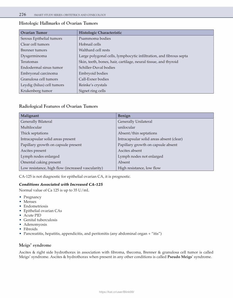
276 SMART STUDY SERIES: OBSTETRICS AND GYNECOLOGY
Histologic Hallmarks of Ovarian Tumors
Ovarian Tumor Histologic Characteristic
Serous Epithelial tumors Psammoma bodies
Clear cell tumors Hobnail cells
Brenner tumors Walthard cell rests
Dysgerminoma Large polygonal cells, lymphocytic infiltration, and fibrous septa
Teratomas Skin, teeth, bones, hair, cartilage, neural tissue, and thyroid
Endodermal sinus tumor Schiller-Duval bodies
Embryonal carcinoma Embryoid bodies
Granulosa cell tumors Call-Exner bodies
Leydig (hilus) cell tumors Reinke’s crystals
Krukenberg tumor Signet ring cells
Radiological Features of Ovarian Tumors
Malignant Benign
Generally Bilateral Generally Unilateral
Multilocular unilocular
Thick septations Absent/thin septations
Intracapsular solid areas present Intracapsular solid areas absent (clear)
Papillary growth on capsule present Papillary growth on capsule absent
Ascites present Ascites absent
Lymph nodes enlarged Lymph nodes not enlarged
Omental caking present Absent
Low resistance, high flow (increased vascularity) High resistance, low flow
CA-125 is not diagnostic for epithelial ovarian CA, it is prognostic.
Conditions Associated with Increased CA-125Normal value of Ca 125 is up to 35 U/mL
• Pregnancy • Menses • Endometriosis • Epithelial ovarian CAs • Acute PID • Genital tuberculosis • Adenomyosis • Fibroids • Pancreatitis, hepatitis, appendicitis, and peritonitis (any abdominal organ + “itis”)
Meigs’ syndrome
Ascites & right side hydrothorax in association with fibroma, thecoma, Brenner & granulosa cell tumor is called Meigs’ syndrome. Ascites & hydrothorax when present in any other conditions is called Pseudo Meigs’ syndrome.
https://kat.cr/user/Blink99/

ONCOLOGY AND FIBROIDS 277
Management
Epithelial Cancer (for All Stages 1–4)Staging laparotomy with cytoreductive surgery (hysterectomy, bilateral salpingo-oophorectomy, omentectomy, lymph node dissection, and removal of all the metastatic deposits), followed by six cycles of chemotherapy (cispla-tin/carboplatin + paclitaxel).Basic steps involved in surgical staging:
a. Send free fluid for cytology b. If no free fluid, perform peritoneal washings and send it for cytology c. Palpate all the intra-abdominal organs d. Any suspicious area on peritoneal surfaces should be biopsied e. Sample the diaphragm either by biopsy or scraping f. Perform the infracolic omentectomy g. Evaluate the pelvic and para-aortic lymph nodes. Enlarged nodes should be resected. If no metastasis are
present pelvic lymphadenectomy should be performed.
Germ Cell CancerSince it occurs in young age and since it is extremely chemosensitive, conservative surgery is advocated.
Staging laparotomy with unilateral salpingo-oophorectomy, followed by six cycles of chemotherapy (bleomy-cin, etoposide, and cisplatin).
Radiotherapy has no role in the management of ovarian CA.
KRUKENBERG TUMOR
• Accounts for 30–40% of metastatic CAs to the ovary. • Arises in the ovarian stroma and has characteristic mucin-filled signet ring cells. • The primary is most frequently located in the stomach and less commonly in the colon, breast, or biliary tract.
Rarely, the cervix and bladder may be the primary site. • They are usually bilateral and discovered when the primary is well advanced and hence survival is very poor. • Treatment of primary carcinoma does not revert to Krukenberg tumor. • The ovaries are enlarged and have a smooth surface. • The shape of the ovary is maintained. • There is no tendency of adhesion and the capsule remains intact. • Cut surface shows waxy consistency with cystic spaces due to degeneration.
MANAGEMENT OF AN ADNEXAL MASS
Adnexal mass
Premenopausal Postmenopausal
<8 cm >8 cm
clear solid areas Surgery
OC pills or wait and watch (OC pills preferred)
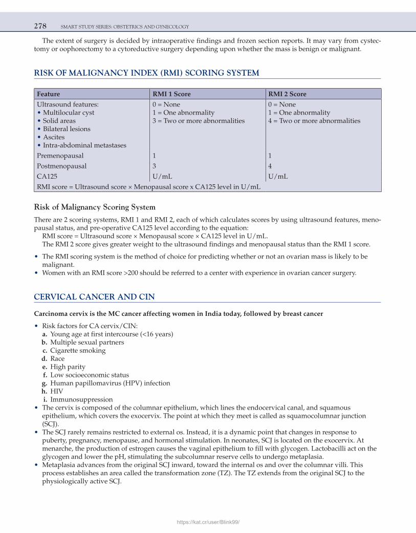
278 SMART STUDY SERIES: OBSTETRICS AND GYNECOLOGY
The extent of surgery is decided by intraoperative findings and frozen section reports. It may vary from cystec-tomy or oophorectomy to a cytoreductive surgery depending upon whether the mass is benign or malignant.
RISK OF MALIGNANCY INDEX (RMI) SCORING SYSTEM
Feature RMI 1 Score RMI 2 Score
Ultrasound features: • Multilocular cyst • Solid areas • Bilateral lesions • Ascites • Intra-abdominal metastases
0 = None1 = One abnormality3 = Two or more abnormalities
0 = None1 = One abnormality4 = Two or more abnormalities
Premenopausal 1 1
Postmenopausal 3 4
CA125 U/mL U/mL
RMI score = Ultrasound score × Menopausal score x CA125 level in U/mL
Risk of Malignancy Scoring System
There are 2 scoring systems, RMI 1 and RMI 2, each of which calculates scores by using ultrasound features, meno-pausal status, and pre-operative CA125 level according to the equation:
RMI score = Ultrasound score × Menopausal score × CA125 level in U/mL.The RMI 2 score gives greater weight to the ultrasound findings and menopausal status than the RMI 1 score.
• The RMI scoring system is the method of choice for predicting whether or not an ovarian mass is likely to be malignant.
• Women with an RMI score >200 should be referred to a center with experience in ovarian cancer surgery.
CERVICAL CANCER AND CIN
Carcinoma cervix is the MC cancer affecting women in India today, followed by breast cancer
• Risk factors for CA cervix/CIN: a. Young age at first intercourse (<16 years) b. Multiple sexual partners c. Cigarette smoking d. Race e. High parity f. Low socioeconomic status g. Human papillomavirus (HPV) infection h. HIV i. Immunosuppression • The cervix is composed of the columnar epithelium, which lines the endocervical canal, and squamous
epithelium, which covers the exocervix. The point at which they meet is called as squamocolumnar junction (SCJ).
• The SCJ rarely remains restricted to external os. Instead, it is a dynamic point that changes in response to puberty, pregnancy, menopause, and hormonal stimulation. In neonates, SCJ is located on the exocervix. At menarche, the production of estrogen causes the vaginal epithelium to fill with glycogen. Lactobacilli act on the glycogen and lower the pH, stimulating the subcolumnar reserve cells to undergo metaplasia.
• Metaplasia advances from the original SCJ inward, toward the internal os and over the columnar villi. This process establishes an area called the transformation zone (TZ). The TZ extends from the original SCJ to the physiologically active SCJ.
https://kat.cr/user/Blink99/
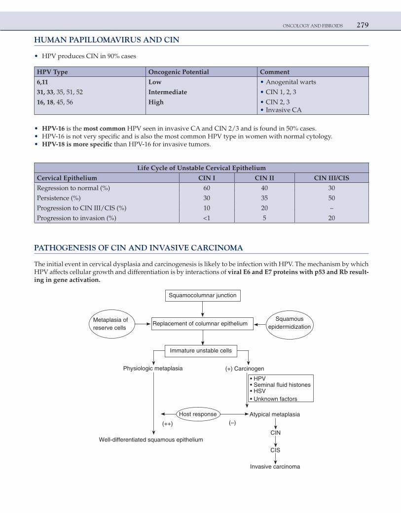
ONCOLOGY AND FIBROIDS 279
HUMAN PAPILLOMAVIRUS AND CIN
• HPV produces CIN in 90% cases
HPV Type Oncogenic Potential Comment
6,11 Low • Anogenital warts
31, 33, 35, 51, 52 Intermediate • CIN 1, 2, 3
16, 18, 45, 56 High • CIN 2, 3 • Invasive CA
• HPV-16 is the most common HPV seen in invasive CA and CIN 2/3 and is found in 50% cases. • HPV-16 is not very specific and is also the most common HPV type in women with normal cytology. • HPV-18 is more specific than HPV-16 for invasive tumors.
Life Cycle of Unstable Cervical Epithelium
Cervical Epithelium CIN I CIN II CIN III/CIS
Regression to normal (%) 60 40 30
Persistence (%) 30 35 50
Progression to CIN III/CIS (%) 10 20 –
Progression to invasion (%) <1 5 20
PATHOGENESIS OF CIN AND INVASIVE CARCINOMA
The initial event in cervical dysplasia and carcinogenesis is likely to be infection with HPV. The mechanism by which HPV affects cellular growth and differentiation is by interactions of viral E6 and E7 proteins with p53 and Rb result-ing in gene activation.
Squamocolumnar junction
Immature unstable cells
Replacement of columnar epitheliumMetaplasia of reserve cells
Squamous epidermidization
Physiologic metaplasia (+) Carcinogen
• HPV
• HSV• Unknown factors
Host response Atypical metaplasia
Well-differentiated squamous epithelium
(++) (–)
CIN
CIS
Invasive carcinoma
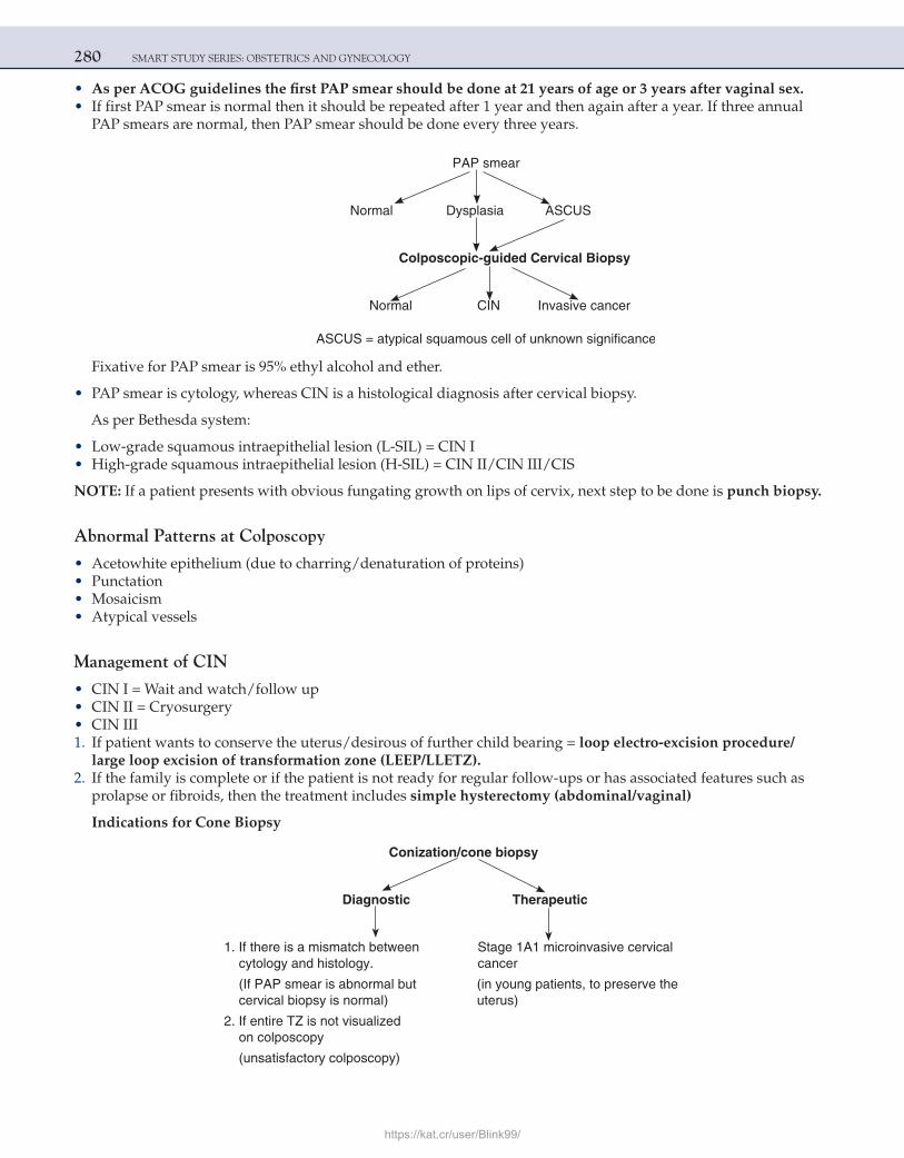
280 SMART STUDY SERIES: OBSTETRICS AND GYNECOLOGY
• As per ACOG guidelines the first PAP smear should be done at 21 years of age or 3 years after vaginal sex. • If first PAP smear is normal then it should be repeated after 1 year and then again after a year. If three annual
PAP smears are normal, then PAP smear should be done every three years.
PAP smear
Normal Dysplasia ASCUS
Colposcopic-guided Cervical Biopsy
Normal CIN Invasive cancer
Fixative for PAP smear is 95% ethyl alcohol and ether.
• PAP smear is cytology, whereas CIN is a histological diagnosis after cervical biopsy.
As per Bethesda system:
• Low-grade squamous intraepithelial lesion (L-SIL) = CIN I • High-grade squamous intraepithelial lesion (H-SIL) = CIN II/CIN III/CIS
NOTE: If a patient presents with obvious fungating growth on lips of cervix, next step to be done is punch biopsy.
Abnormal Patterns at Colposcopy
• Acetowhite epithelium (due to charring/denaturation of proteins) • Punctation • Mosaicism • Atypical vessels
Management of CIN
• CIN I = Wait and watch/follow up • CIN II = Cryosurgery • CIN III 1. If patient wants to conserve the uterus/desirous of further child bearing = loop electro-excision procedure/
large loop excision of transformation zone (LEEP/LLETZ). 2. If the family is complete or if the patient is not ready for regular follow-ups or has associated features such as
prolapse or fibroids, then the treatment includes simple hysterectomy (abdominal/vaginal)
Indications for Cone Biopsy
Conization/cone biopsy
Diagnostic Therapeutic
1. If there is a mismatch between Stage 1A1 microinvasive cervical cytology and histology. cancer
(If PAP smear is abnormal but (in young patients, to preserve thecervical biopsy is normal) uterus)
2. If entire TZ is not visualized on colposcopy
(unsatisfactory colposcopy)
https://kat.cr/user/Blink99/
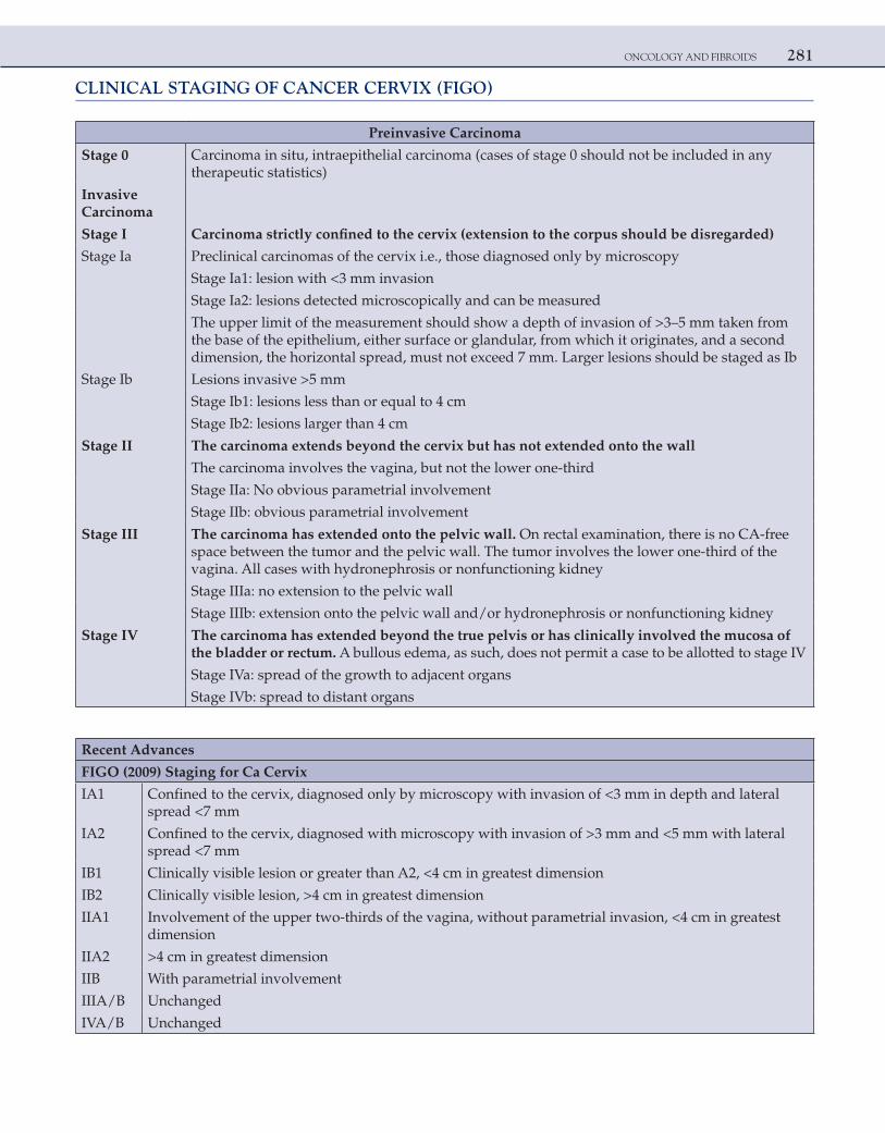
ONCOLOGY AND FIBROIDS 281
CLINICAL STAGING OF CANCER CERVIX (FIGO)
Preinvasive Carcinoma
Stage 0 Carcinoma in situ, intraepithelial carcinoma (cases of stage 0 should not be included in any therapeutic statistics)
Invasive Carcinoma
Stage I Carcinoma strictly confined to the cervix (extension to the corpus should be disregarded)
Stage Ia Preclinical carcinomas of the cervix i.e., those diagnosed only by microscopy
Stage Ia1: lesion with <3 mm invasion
Stage Ia2: lesions detected microscopically and can be measured
The upper limit of the measurement should show a depth of invasion of >3–5 mm taken from the base of the epithelium, either surface or glandular, from which it originates, and a second dimension, the horizontal spread, must not exceed 7 mm. Larger lesions should be staged as Ib
Stage Ib Lesions invasive >5 mm
Stage Ib1: lesions less than or equal to 4 cm
Stage Ib2: lesions larger than 4 cm
Stage II The carcinoma extends beyond the cervix but has not extended onto the wall
The carcinoma involves the vagina, but not the lower one-third
Stage IIa: No obvious parametrial involvement
Stage IIb: obvious parametrial involvement
Stage III The carcinoma has extended onto the pelvic wall. On rectal examination, there is no CA-free space between the tumor and the pelvic wall. The tumor involves the lower one-third of the vagina. All cases with hydronephrosis or nonfunctioning kidney
Stage IIIa: no extension to the pelvic wall
Stage IIIb: extension onto the pelvic wall and/or hydronephrosis or nonfunctioning kidney
Stage IV The carcinoma has extended beyond the true pelvis or has clinically involved the mucosa of the bladder or rectum. A bullous edema, as such, does not permit a case to be allotted to stage IV
Stage IVa: spread of the growth to adjacent organs
Stage IVb: spread to distant organs
Recent Advances
FIGO (2009) Staging for Ca Cervix
IA1 Confined to the cervix, diagnosed only by microscopy with invasion of <3 mm in depth and lateral spread <7 mm
IA2 Confined to the cervix, diagnosed with microscopy with invasion of >3 mm and <5 mm with lateral spread <7 mm
IB1 Clinically visible lesion or greater than A2, <4 cm in greatest dimension
IB2 Clinically visible lesion, >4 cm in greatest dimension
IIA1 Involvement of the upper two-thirds of the vagina, without parametrial invasion, <4 cm in greatest dimension
IIA2 >4 cm in greatest dimension
IIB With parametrial involvement
IIIA/B Unchanged
IVA/B Unchanged
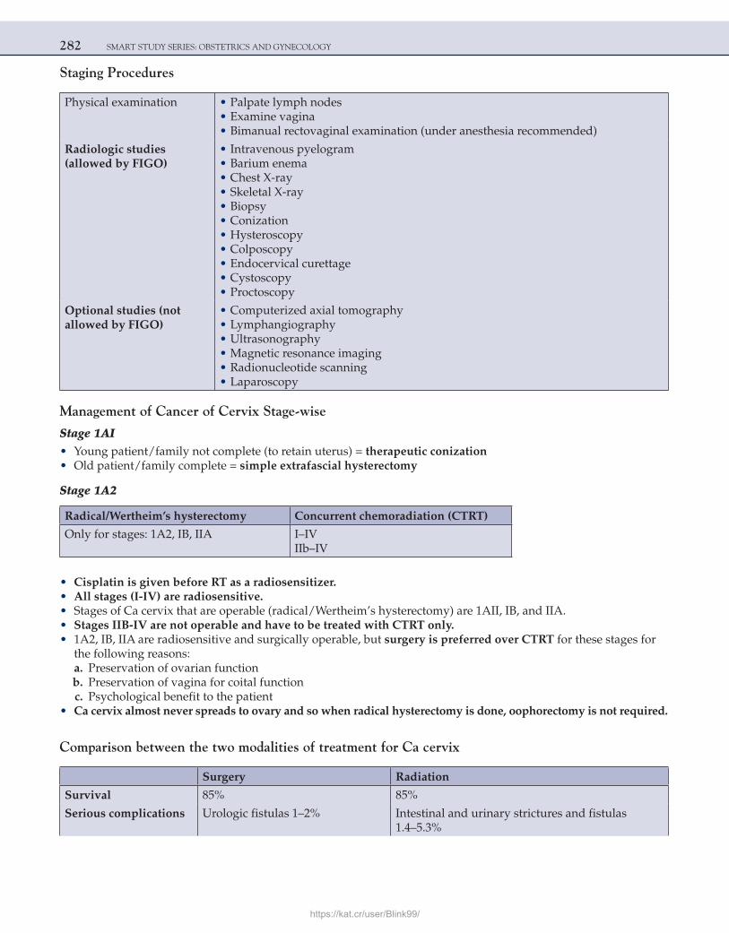
282 SMART STUDY SERIES: OBSTETRICS AND GYNECOLOGY
Staging Procedures
Physical examination • Palpate lymph nodes • Examine vagina • Bimanual rectovaginal examination (under anesthesia recommended)
Radiologic studies(allowed by FIGO)
• Intravenous pyelogram • Barium enema • Chest X-ray • Skeletal X-ray • Biopsy • Conization • Hysteroscopy • Colposcopy • Endocervical curettage • Cystoscopy • Proctoscopy
Optional studies (notallowed by FIGO)
• Computerized axial tomography • Lymphangiography • Ultrasonography • Magnetic resonance imaging • Radionucleotide scanning • Laparoscopy
Management of Cancer of Cervix Stage-wise
Stage 1AI • Young patient/family not complete (to retain uterus) = therapeutic conization • Old patient/family complete = simple extrafascial hysterectomy
Stage 1A2
Radical/Wertheim’s hysterectomy Concurrent chemoradiation (CTRT)
Only for stages: 1A2, IB, IIA I–IVIIb–IV
• Cisplatin is given before RT as a radiosensitizer. • All stages (I-IV) are radiosensitive. • Stages of Ca cervix that are operable (radical/Wertheim’s hysterectomy) are 1AII, IB, and IIA. • Stages IIB-IV are not operable and have to be treated with CTRT only. • 1A2, IB, IIA are radiosensitive and surgically operable, but surgery is preferred over CTRT for these stages for
the following reasons: a. Preservation of ovarian function b. Preservation of vagina for coital function c. Psychological benefit to the patient • Ca cervix almost never spreads to ovary and so when radical hysterectomy is done, oophorectomy is not required.
Comparison between the two modalities of treatment for Ca cervix
Surgery Radiation
Survival 85% 85%
Serious complications Urologic fistulas 1–2% Intestinal and urinary strictures and fistulas 1.4–5.3%
https://kat.cr/user/Blink99/
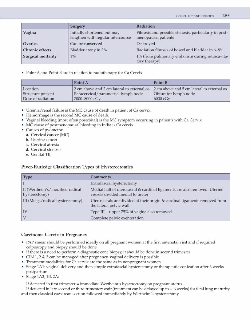
ONCOLOGY AND FIBROIDS 283
Surgery Radiation
Vagina Initially shortened but may lengthen with regular intercourse
Fibrosis and possible stenosis, particularly in post-menopausal patients
Ovaries Can be conserved Destroyed
Chronic effects Bladder atony in 3% Radiation fibrosis of bowel and bladder in 6–8%
Surgical mortality 1% 1% (from pulmonary embolism during intracavita-tory therapy)
• Point A and Point B are in relation to radiotherapy for Ca Cervix
Point A Point B
LocationStructure presentDose of radiation
2 cm above and 2 cm lateral to external osParacervical/parametrial lymph node7000–8000 cGy
2 cm above and 5 cm lateral to external osObturator lymph node6000 cGy
• Uremia/renal failure is the MC cause of death in patient of Ca cervix. • Hemorrhage is the second MC cause of death. • Vaginal bleeding (most often postcoital) is the MC symptom occurring in patients with Ca Cervix • MC cause of postmenopausal bleeding in India is Ca cervix • Causes of pyometra: a. Cervical cancer (MC) b. Uterine cancer c. Cervical atresia d. Cervical stenosis e. Genital TB
Piver-Rutledge Classification Types of Hysterectomies
Type Comments
I Extrafascial hysterectomy
II (Wertheim’s/modified radical hysterectomy)
Medial half of uterosacral & cardinal ligaments are also removed. Uterine vessels divided medial to ureter
III (Meigs/radical hysterectomy) Uterosacrals are divided at their origin & cardinal ligaments removed from the lateral pelvic wall
IV Type III + upper 75% of vagina also removed
V Complete pelvic exenteration
Carcinoma Cervix in Pregnancy
• PAP smear should be performed ideally on all pregnant women at the first antenatal visit and if required colposcopy and biopsy should be done
• If there is a need to perform a diagnostic cone biopsy, it should be done in second trimester • CIN 1, 2 & 3 can be managed after pregnancy, vaginal delivery is possible • Treatment modalities for Ca cervix are the same as in nonpregnant women • Stage 1A1: vaginal delivery and then simple extrafascial hysterectomy or therapeutic conization after 6 weeks
postpartum • Stage 1A2, 1B, 2A:
If detected in first trimester = immediate Wertheim’s hysterectomy on pregnant uterusIf detected in late second or third trimester: wait (treatment can be delayed up to 4–6 weeks) for fetal lung maturity
and then classical caesarean section followed immediately by Wertheim’s hysterectomy
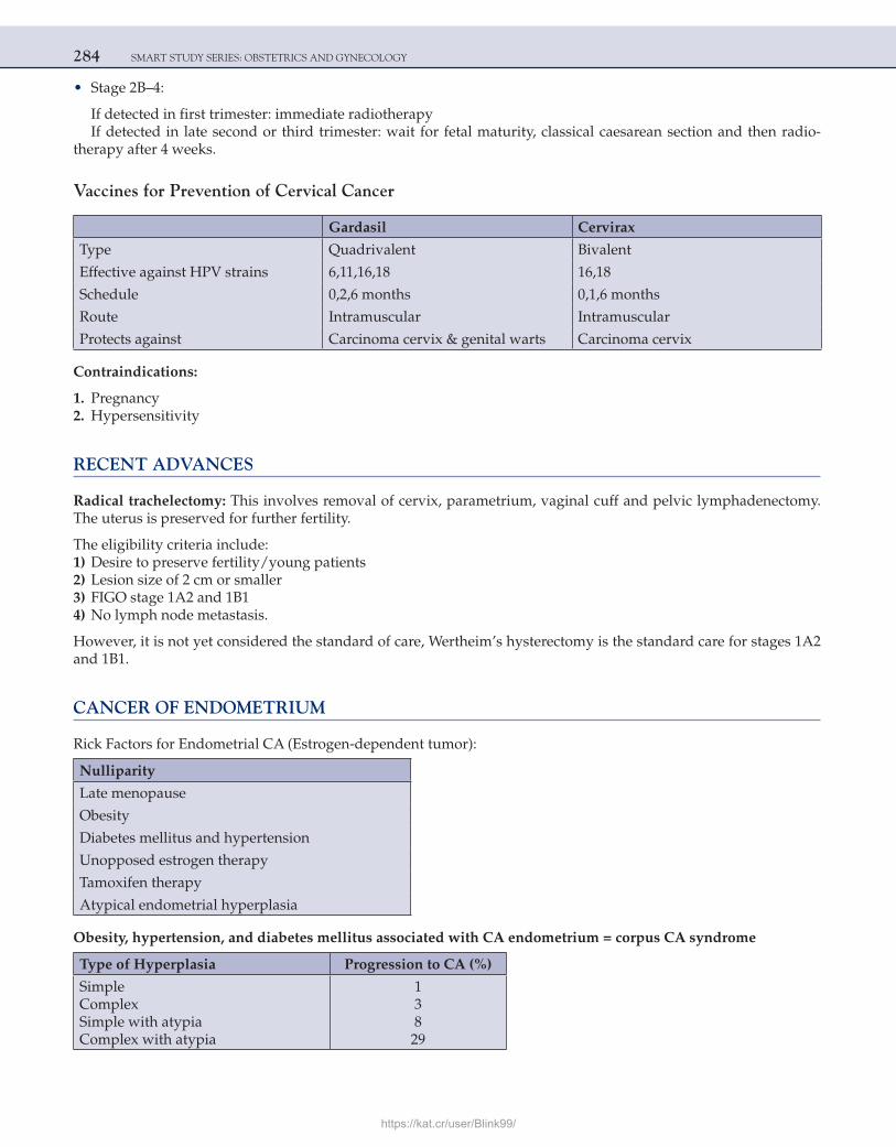
284 SMART STUDY SERIES: OBSTETRICS AND GYNECOLOGY
• Stage 2B–4:
If detected in first trimester: immediate radiotherapyIf detected in late second or third trimester: wait for fetal maturity, classical caesarean section and then radio-
therapy after 4 weeks.
Vaccines for Prevention of Cervical Cancer
Gardasil Cervirax
Type Quadrivalent Bivalent
Effective against HPV strains 6,11,16,18 16,18
Schedule 0,2,6 months 0,1,6 months
Route Intramuscular Intramuscular
Protects against Carcinoma cervix & genital warts Carcinoma cervix
Contraindications:
1. Pregnancy 2. Hypersensitivity
RECENT ADVANCES
Radical trachelectomy: This involves removal of cervix, parametrium, vaginal cuff and pelvic lymphadenectomy. The uterus is preserved for further fertility.
The eligibility criteria include: 1) Desire to preserve fertility/young patients 2) Lesion size of 2 cm or smaller 3) FIGO stage 1A2 and 1B1 4) No lymph node metastasis.
However, it is not yet considered the standard of care, Wertheim’s hysterectomy is the standard care for stages 1A2 and 1B1.
CANCER OF ENDOMETRIUM
Rick Factors for Endometrial CA (Estrogen-dependent tumor):
Nulliparity
Late menopause
Obesity
Diabetes mellitus and hypertension
Unopposed estrogen therapy
Tamoxifen therapy
Atypical endometrial hyperplasia
Obesity, hypertension, and diabetes mellitus associated with CA endometrium = corpus CA syndrome
Type of Hyperplasia Progression to CA (%)
SimpleComplexSimple with atypiaComplex with atypia
13829
https://kat.cr/user/Blink99/
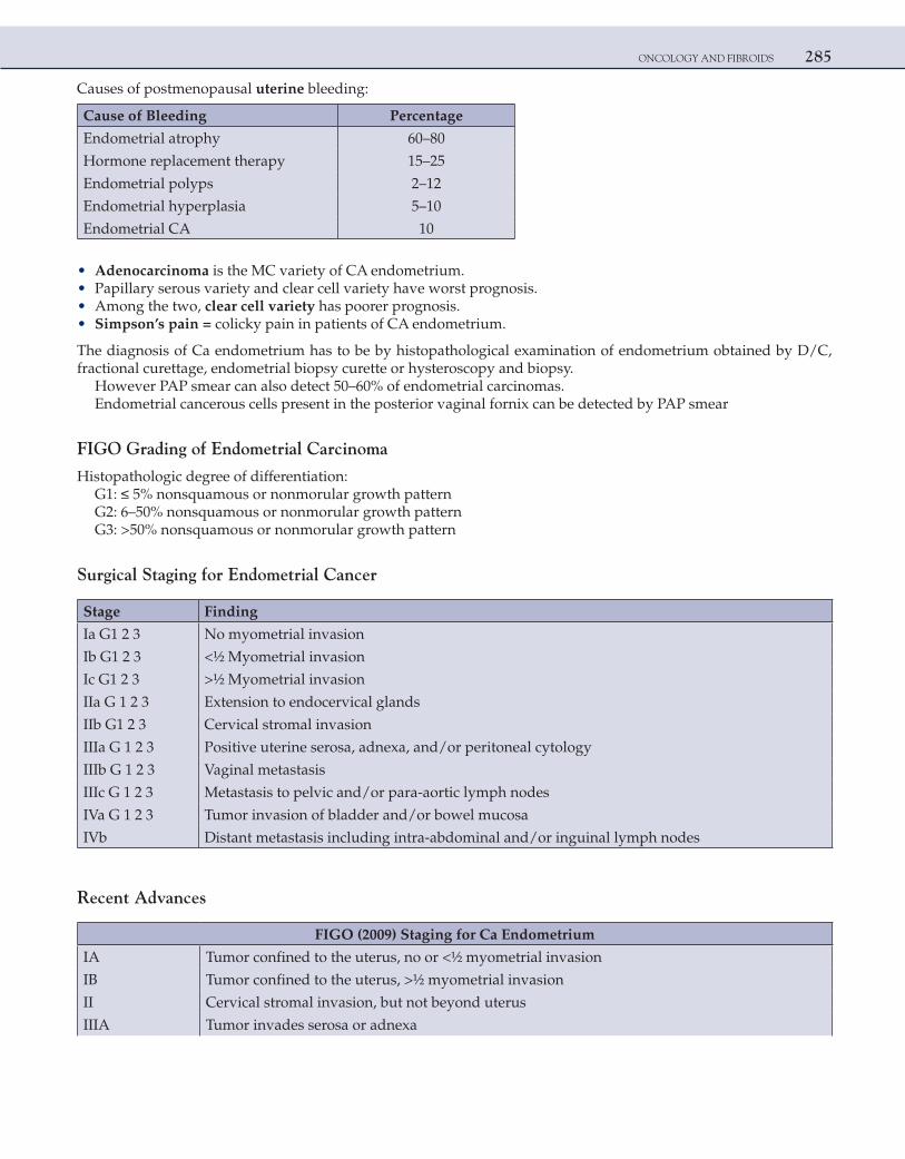
ONCOLOGY AND FIBROIDS 285
Causes of postmenopausal uterine bleeding:
Cause of Bleeding Percentage
Endometrial atrophy 60–80
Hormone replacement therapy 15–25
Endometrial polyps 2–12
Endometrial hyperplasia 5–10
Endometrial CA 10
• Adenocarcinoma is the MC variety of CA endometrium. • Papillary serous variety and clear cell variety have worst prognosis. • Among the two, clear cell variety has poorer prognosis. • Simpson’s pain = colicky pain in patients of CA endometrium.
The diagnosis of Ca endometrium has to be by histopathological examination of endometrium obtained by D/C, fractional curettage, endometrial biopsy curette or hysteroscopy and biopsy.
However PAP smear can also detect 50–60% of endometrial carcinomas.Endometrial cancerous cells present in the posterior vaginal fornix can be detected by PAP smear
FIGO Grading of Endometrial Carcinoma
Histopathologic degree of differentiation:G1: ≤ 5% nonsquamous or nonmorular growth patternG2: 6–50% nonsquamous or nonmorular growth patternG3: >50% nonsquamous or nonmorular growth pattern
Surgical Staging for Endometrial Cancer
Stage Finding
Ia G1 2 3 No myometrial invasion
Ib G1 2 3 <½ Myometrial invasion
Ic G1 2 3 >½ Myometrial invasion
IIa G 1 2 3 Extension to endocervical glands
IIb G1 2 3 Cervical stromal invasion
IIIa G 1 2 3 Positive uterine serosa, adnexa, and/or peritoneal cytology
IIIb G 1 2 3 Vaginal metastasis
IIIc G 1 2 3 Metastasis to pelvic and/or para-aortic lymph nodes
IVa G 1 2 3 Tumor invasion of bladder and/or bowel mucosa
IVb Distant metastasis including intra-abdominal and/or inguinal lymph nodes
Recent Advances
FIGO (2009) Staging for Ca Endometrium
IA Tumor confined to the uterus, no or <½ myometrial invasion
IB Tumor confined to the uterus, >½ myometrial invasion
II Cervical stromal invasion, but not beyond uterus
IIIA Tumor invades serosa or adnexa
286 SMART STUDY SERIES: OBSTETRICS AND GYNECOLOGY
FIGO (2009) Staging for Ca Endometrium
IIIB Vaginal and/or parametrial involvement
IIIC1 Pelvic node involvement
IIIC2 Para-aortic involvement
IVA Unchanged
IVB Unchanged
Management
Stage 1:
• Surgery (total abdominal hysterectomy with bilateral salpingo-oophorectomy with lymph node sampling), followed by radiotherapy
Stage 2:
• Modified radical hysterectomy, bilateral salpingo-oophorectomy with lymph node dissection, followed by radiotherapy
Stages 3 and 4:
• Debulking surgery followed by radiotherapy • Chemotherapy has no role in the management of CA endometrium • Only patients with stage 1 A, grade 1 and 2 do not require postoperative radiotherapy.
GESTATIONAL TROPHOBLASTIC NEOPLASIA
• Gestational trophoblastic neoplasia almost always develops with or follows some form of pregnancy. • Among all the cases of choriocarcinoma:
50% develop following a hydatidiform mole25% develop following an abortion20% develop following a full-term pregnancy and 5% develop following an ectopic pregnancy
• Beta-hCG is the tumor marker. The diagnosis of gestational trophoblastic neoplasia is made primarily by persistently elevated serum hCG levels.
• An important diagnostic feature of choriocarcinoma, in contrast to hydatidiform mole or invasive mole, is absence of villus pattern.
• Factors involved in malignant transformation of the chorion are unknown. In choriocarcinoma, the predisposition of normal trophoblast to invasive growth and erosion of blood vessels is greatly exaggerated.
• Metastases often develop early and are generally blood borne because of the affinity of trophoblastic cells for blood vessels.
• The MC sites of metastasis are the lungs (75–80% cases) followed by vagina in about 30–50%. • In vagina, the classical lesion is bluish purple nodule located in suburethral region. • The chest X-ray findings in case of metastasis to the lungs are: a. Cannonball metastasis b. Snowstorm appearance c. Pleural effusion
NOTE:
• “Snowstorm” on USG = vesicular mole • “Snowstorm” on chest X-ray = pulmonary metastasis of choriocarcinoma
https://kat.cr/user/Blink99/
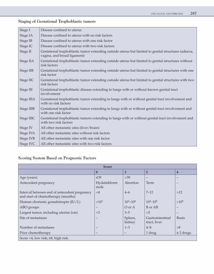
ONCOLOGY AND FIBROIDS 287
Staging of Gestational Trophoblastic tumors
Stage I Disease confined to uterus
Stage IA Disease confined to uterus with no risk factors
Stage IB Disease confined to uterus with one risk factor
Stage IC Disease confined to uterus with two risk factors
Stage II Gestational trophoblastic tumor extending outside uterus but limited to genital structures (adnexa, vagina, and broad ligament)
Stage IIA Gestational trophoblastic tumor extending outside uterus but limited to genital structures without risk factors
Stage IIB Gestational trophoblastic tumor extending outside uterus but limited to genital structures with one risk factor
Stage IIC Gestational trophoblastic tumor extending outside uterus but limited to genital structures with two risk factors
Stage III Gestational trophoblastic disease extending to lungs with or without known genital tract involvement
Stage IIIA Gestational trophoblastic tumor extending to lungs with or without genital tract involvement and with no risk factors
Stage IIIB Gestational trophoblastic tumor extending to lungs with or without genital tract involvement and with one risk factor
Stage IIIC Gestational trophoblastic tumors extending to lungs with or without genital tract involvement and with two risk factors
Stage IV All other metastatic sites (liver/brain)
Stage IVA All other metastatic sites without risk factors
Stage IVB All other metastatic sites with one risk factor
Stage IVC All other metastatic sites with two risk factors
Scoring System Based on Prognostic Factors
Score
0 1 2 4
Age (years) ≤39 >39 – –
Antecedent pregnancy Hydatidiform mole
Abortion Term –
Interval between end of antecedent pregnancy and start of chemotherapy (months)
<4 4–6 7–12 >12
Human chorionic gonadotropin (IU/L) <103 103–104 104–105 >105
ABO groups – O or A B or AB –
Largest tumor, including uterine (cm) <3 3–5 >5 –
Site of metastases – Spleen, kidney
Gastrointestinal tract, liver
Brain
Number of metastases – 1–3 4–8 >8
Prior chemotherapy – – 1 drug ≥ 2 drugs
Score <4, low risk; ≥8, high risk.
288 SMART STUDY SERIES: OBSTETRICS AND GYNECOLOGY
A GTN belongs to a high-risk category if it develops after a full-term pregnancy (postmolar pregnancy; a GTN can be a repeat molar pregnancy or a choriocarcinoma, but a GTN that develops after a full-term pregnancy is always a choriocarcinoma).
Management
• Chemotherapy is the treatment of choice. • Methotrexate is the drug of choice. • If the patient has jaundice then actinomycin D should be given. • High-risk patients and patients with stage 4 are to be treated with combination chemotherapy (EMACO
regimen): a. E = etoposide b. M = methotrexate c. A = actinomycin D d. C = cyclophosphamide e. O = vincristine (Oncovin) • EMA-CO regimen results in response rates of about 90% and survival rates of 80–100%. • The recent overall cure rate for gestational trophoblastic neoplasia of all severities is about 90%. • Women with nonmetastatic tumors or low-risk gestational trophoblastic neoplasia are cured virtually
100% of the time if single-agent chemotherapy (methotrexate) is started as soon as persistent disease is identified.
Follow-up • Weekly measurement of hCG until they are normal for 3 consecutive weeks • Monthly measurement of hCG until they are normal for 12 consecutive months for stages 1–3 • Monthly measurement of hCG until they are normal for 24 consecutive months for stage 4
PLACENTAL SITE TROPHOBLASTIC TUMOR
• It is an uncommon variant of CC. • It consists predominantly of intermediate trophoblasts. • Human placental lactogen (hPL) is the tumor marker. • They are insensitive to chemotherapy. • Hysterectomy is the most efficacious treatment for confirmed placental site trophoblastic tumor.
FIBROIDS
Fibroids are benign smooth muscle tumors arising from the myometrium. They are the MC benign tumors of uterus, and they are also the MC pelvic tumors in females.
Etiology
1. Predominantly estrogen-dependent tumors: a. Early menarche, late menopause b. Associated anovulation and PCOS c. Growing in size during pregnancy, and following menopause there is cessation of growth 2. Nulliparity (“a uterus which does not bear a baby consoles itself by having a fibroid”) 3. Deletions in chromosome 7 and t (12, 14) are associated with fibroids 4. More common in colored races 5. Infertility: Fibroids can cause infertility and infertile women are more prone to develop fibroids 6. Obesity • Smoking is protective for fibroids.
https://kat.cr/user/Blink99/
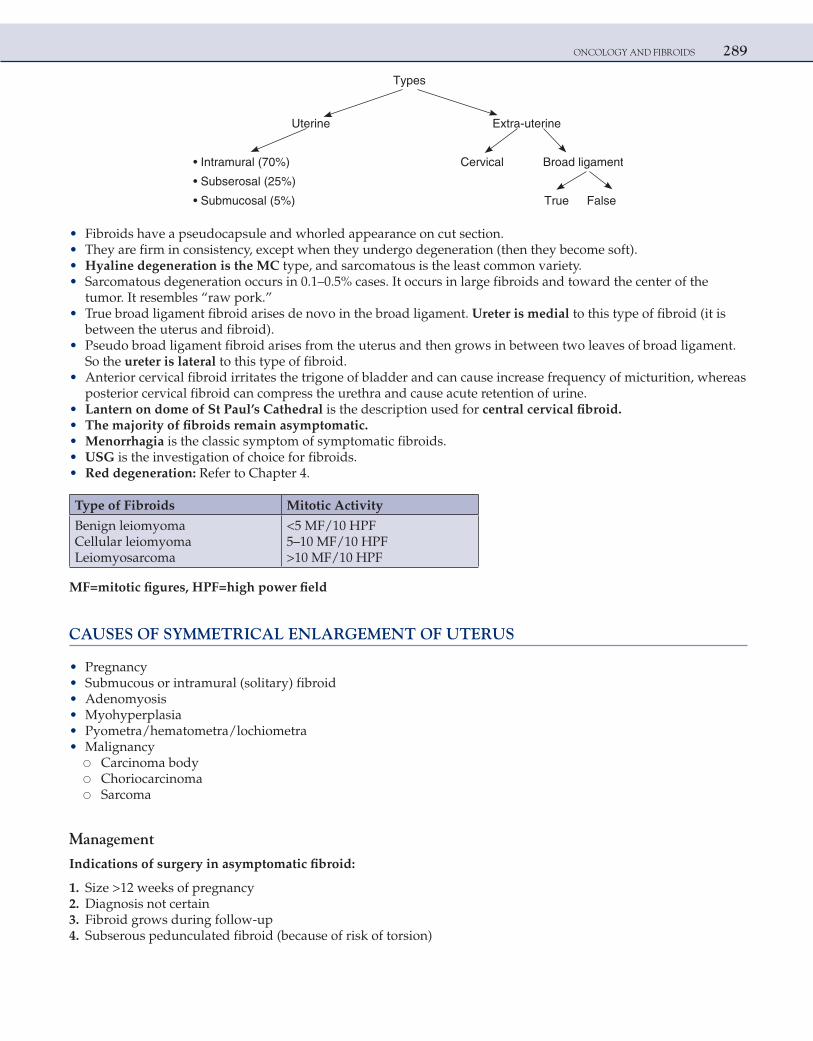
ONCOLOGY AND FIBROIDS 289
Types
eniretu-artxEeniretU
tnemagildaorBlacivreC)%07(larumartnI•
• Subserosal (25%)
eslaFeurT)%5(lasocumbuS•
• Fibroids have a pseudocapsule and whorled appearance on cut section. • They are firm in consistency, except when they undergo degeneration (then they become soft). • Hyaline degeneration is the MC type, and sarcomatous is the least common variety. • Sarcomatous degeneration occurs in 0.1–0.5% cases. It occurs in large fibroids and toward the center of the
tumor. It resembles “raw pork.” • True broad ligament fibroid arises de novo in the broad ligament. Ureter is medial to this type of fibroid (it is
between the uterus and fibroid). • Pseudo broad ligament fibroid arises from the uterus and then grows in between two leaves of broad ligament.
So the ureter is lateral to this type of fibroid. • Anterior cervical fibroid irritates the trigone of bladder and can cause increase frequency of micturition, whereas
posterior cervical fibroid can compress the urethra and cause acute retention of urine. • Lantern on dome of St Paul’s Cathedral is the description used for central cervical fibroid. • The majority of fibroids remain asymptomatic. • Menorrhagia is the classic symptom of symptomatic fibroids. • USG is the investigation of choice for fibroids. • Red degeneration: Refer to Chapter 4.
Type of Fibroids Mitotic Activity
Benign leiomyomaCellular leiomyomaLeiomyosarcoma
<5 MF/10 HPF5–10 MF/10 HPF>10 MF/10 HPF
MF=mitotic figures, HPF=high power field
CAUSES OF SYMMETRICAL ENLARGEMENT OF UTERUS
• Pregnancy • Submucous or intramural (solitary) fibroid • Adenomyosis • Myohyperplasia • Pyometra/hematometra/lochiometra • Malignancy ○ Carcinoma body ○ Choriocarcinoma ○ Sarcoma
Management
Indications of surgery in asymptomatic fibroid:
1. Size >12 weeks of pregnancy 2. Diagnosis not certain 3. Fibroid grows during follow-up 4. Subserous pedunculated fibroid (because of risk of torsion)
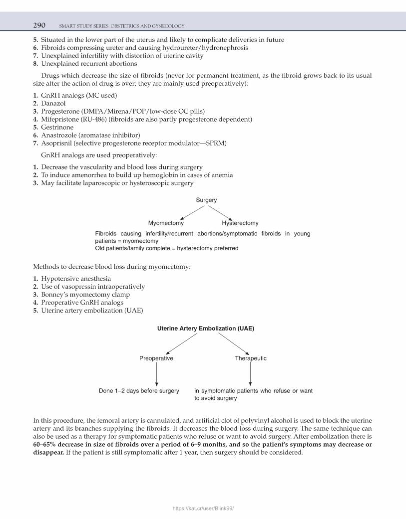
290 SMART STUDY SERIES: OBSTETRICS AND GYNECOLOGY
5. Situated in the lower part of the uterus and likely to complicate deliveries in future 6. Fibroids compressing ureter and causing hydroureter/hydronephrosis 7. Unexplained infertility with distortion of uterine cavity 8. Unexplained recurrent abortions
Drugs which decrease the size of fibroids (never for permanent treatment, as the fibroid grows back to its usual size after the action of drug is over; they are mainly used preoperatively):
1. GnRH analogs (MC used) 2. Danazol 3. Progesterone (DMPA/Mirena/POP/low-dose OC pills) 4. Mifepristone (RU-486) (fibroids are also partly progesterone dependent) 5. Gestrinone 6. Anastrozole (aromatase inhibitor) 7. Asoprisnil (selective progesterone receptor modulator—SPRM)
GnRH analogs are used preoperatively:
1. Decrease the vascularity and blood loss during surgery 2. To induce amenorrhea to build up hemoglobin in cases of anemia 3. May facilitate laparoscopic or hysteroscopic surgery
Surgery
Myomectomy Hysterectomy
patients = myomectomyOld patients/family complete = hysterectomy preferred
Methods to decrease blood loss during myomectomy:
1. Hypotensive anesthesia 2. Use of vasopressin intraoperatively 3. Bonney’s myomectomy clamp 4. Preoperative GnRH analogs 5. Uterine artery embolization (UAE)
Uterine Artery Embolization (UAE)
citueparehTevitarepoerP
Done 1–2 days before surgery in symptomatic patients who refuse or want to avoid surgery
In this procedure, the femoral artery is cannulated, and artificial clot of polyvinyl alcohol is used to block the uterine artery and its branches supplying the fibroids. It decreases the blood loss during surgery. The same technique can also be used as a therapy for symptomatic patients who refuse or want to avoid surgery. After embolization there is 60–65% decrease in size of fibroids over a period of 6–9 months, and so the patient’s symptoms may decrease or disappear. If the patient is still symptomatic after 1 year, then surgery should be considered.
https://kat.cr/user/Blink99/
ONCOLOGY AND FIBROIDS 291
Even though pregnancies have been reported after UAE, patients desirous of pregnancy is a contraindication for UAE.
RECENT ADVANCES
High-intensity focused ultrasound (HIFU or FUS) is a highly precise medical procedure using high-intensity focused ultrasound waves to heat and destroy fibroids rapidly through ablation.
Clinical HIFU procedures are typically image-guided (MRI or USG) to precisely target the fibroids before apply-ing of ultrasound energy.
When MRI is used for guidance, the technique is called magnetic resonance-guided focused ultrasound (MRgHIFU or MRgFUS). MRI is used to identify fibroids before they are destroyed by the ultrasound waves.
When USG is used to localize the fibroids, the technique is called ultrasound-guided focused ultrasound (USgFUS).
MULTIPLE CHOICE QUESTIONS
1. A 24-year-old woman presents with new-onset right lower quadrant pain, and you palpate an enlarged, tender right adnexa. Which of the following sonographic characteristics of the cyst in this patient suggests the need for surgical exploration now instead of observation for one menstrual cycle?
a. Lack of ascites b. Unilocularity c. Papillary vegetation d. Diameter of 8 cm
Answer: c (Papillary vegetation)
Explanation:Approximately 20% of ovarian neoplasms are considered malignant on pathologic examination. However, all must be
considered as placing the patient at risk. Given that most ovarian tumors are not found until significant spread has occurred, it is not unreasonable to attempt to operate on such patients as soon as there is a suspicion of tumor.
Papillary vegetation, size greater than 8 cm, ascites, possible torsion, or solid lesions within the cysts are automatic indications for exploratory laparotomy.
In a younger woman, a simple unilocular cyst can be a follicular cyst that would regress after onset of the next menstrual period. If regression does not occur, then surgery is appropriate. Doppler ultrasound imaging allows visualization of arterial and venous flow patterns superimposed on the image of the structure being examined.
Reference:
1. Novak, 14th Ed., Pg. 442.
2. A 54-year-old woman undergoes a laparotomy because of a pelvic mass. At exploratory laparotomy, a unilateral ovarian neoplasm is discovered that is accompanied by a large omental metastasis. Frozen section diagnosis confirms metastatic serous cystadenocarcinoma. The most appropriate intraoperative course of action is:
[All India 2003] a. Excision of the omental metastasis and ovarian cystectomy b. Excision of the omental metastasis and unilateral oophorectomy c. Omentectomy and bilateral salpingo-oophorectomy d. Omentectomy, total abdominal hysterectomy, and bilateral salpingo-oophorectomy
Answer: d (Omentectomy, total abdominal hysterectomy, and bilateral salpingo-oophorectomy)
Explanation:The survival of women who have ovarian carcinoma varies inversely with the amount of residual tumor left after the initial
surgery. At the time of laparotomy, a maximum effort should be made to determine the sites of tumor spread and to excise all resectable tumors (cytoreductive/debulking surgery).
292 SMART STUDY SERIES: OBSTETRICS AND GYNECOLOGY
Although the uterus and ovaries may appear grossly normal, there is a relatively high incidence of occult metastases to these organs; for this reason, they should be removed during the initial surgery. Ovarian cancer metastasizes outside the peri-toneum via the pelvic or para-aortic lymphatics, and from there into the thorax and the remainder of the body. The surgery is followed by six cycles of chemotherapy.
Reference:
1. Novak, 14th Ed., Pg. 1478–80.
3. Stage lb cervical cancer is diagnosed in a young woman. Assuming that the cancer is confirmed to the cervix and that intraoperative biopsies are negative, which of the following structure would not be removed during the radical hysterectomy?
[All India 2006] a. Uterosacral and uterovesical ligaments b. Pelvic nodes c. The entire parametrium on both sides of the cervix d. Both ovaries
Answer: d (Both ovaries)
Explanation:Radical hysterectomy is most often used as a primary treatment for early cervical cancer (stage 1A2, lB, and IIA), and
occasionally as a primary treatment for uterine cancer. In either case, there must be no evidence of spread beyond the opera-tive field, as suggested by negative intraoperative frozen-section biopsies. The procedure involves excision of the uterus, the upper third of the vagina, the uterosacral and uterovesical ligaments, and all of the parametrium, and pelvic node dissection including the ureteral, obturator, hypogastric, and iliac nodes.
Radical hysterectomy, thus, attempts to preserve the bladder, rectum, and ureters while excising as much as possible of the remaining tissue around the cervix that might be involved in microscopic spread of the disease. Ovarian metastases from cervical cancer are extremely rare. Preservation of the ovaries is generally acceptable, particularly in younger women.
Reference:
1. Novak, 14th Ed., Pg. 1428.
4. Point B in the treatment of carcinoma cervix receives the following dose of: a. 7000 cGy b. 6000 cGy c. 5000 cGy d. l0,000 cGy
Answer: b (6000 cGy)
Explanation
Point A Point B
LocationStructure presentDose of radiation
2 cm above and 2 cm lateral to external osParacervical/parametrial lymph node7000–8000 cGy
2 cm above and 5 cm lateral to external osObturator lymph node6000 cGy
Reference:
1. Novak, 14th Ed., Pg. 1428.
5. A 50-year-old woman is diagnosed with cervical cancer. Which lymph node group would be the first to be involved in metastatic spread of this disease beyond the cervix and uterus?
a. Internal iliac nodes b. Obturator nodes c. External iliac nodes d. Paracervical nodes
Answer: d (Paracervical nodes)
https://kat.cr/user/Blink99/
ONCOLOGY AND FIBROIDS 293
Explanation:The main routes of spread of cervical cancer include vaginal mucosa, myometrium, paracervical lymphatics, and direct
extension into the parametrium. The prevalence of lymph node disease correlates with the stage of malignancy. Primary node groups involved in the spread of cervical cancer include the paracervical (sentinel node), parametrial, obturator, hypogastric, external iliac, and sacral nodes, essentially in that order. Less commonly, there is involvement in the common iliac, inguinal, and para-aortic nodes.
Reference:
1. Novak, 14th Ed., Pg. 1416.
6. A patient is receiving external beam radiation for the treatment of metastatic endometrial cancer. The treatment field includes the entire pelvis. Which of the following tissues within this radiation field is the most radiosensitive?
a. Vagina b. Ovary c. Rectovaginal septum d. Bladder
Answer: b (Ovary)
Explanation:Different tissues tolerate different doses of radiation, but the ovaries are by far the most radiosensitive. They tolerate
up to 2500 rad, while the other tissues listed tolerate between 5000 and 20,000 rad. Acute side effects of excessive radiation exposure includes tissue necrosis and inflammation, resulting in enteritis, cystitis, vulvitis, proctosigmoiditis, and possible bone marrow suppression.
Chronic effects become manifest months to years after therapy, and include vasculitis, fibrosis, and deficient cellular regrowth which can result in proctitis, cystitis, fistulas, scarring, and stenosis.
The greater the fractionalization (number of portions the total dose is broken into), the better the normal tissue tolerance of that radiation dose; hence, 5000 rad of pelvic radiation is usually given in daily fractions over 5 weeks, with approximately 200 rad being administered each day.
Reference:
1. Novak. 14th Ed., Pgs. 1428–9.
7. All of the following are indications for postoperative radiotherapy in a case of carcinoma endometrium, except: [AIIMS Nov 2004, AIIMS Nov 2007] a. Myometrial invasion >½ thickness b. Positive lymph nodes c. Endocervical involvement d. Tumor positive for estrogen receptors
Answer: d (Tumor positive for estrogen receptors)
Explanation:The main treatment of CA endometrium is surgery followed by radiotherapy.Postoperative management of endometrial carcinoma based on surgical pathologic findings and stage:
Surgical Pathologic Findings Stage Postoperative Treatment
Low riskG1, G2 no myoinvasionNo cervix/isthmus invasionNo lymph vascular space invasion (LVSI)No evidence of metastasis
la G1,2 None
294 SMART STUDY SERIES: OBSTETRICS AND GYNECOLOGY
Surgical Pathologic Findings Stage Postoperative Treatment
Intermediate riskG1, G2 <50% myoinvasionG3, no myoinvasion
lbG1, 2laG3
Vaginal cuff irradiation
G3, <50% myoinvasionG1, G2 isthmus/cervix extensionG1, G2, G3 >50% myoinvasionG3, isthmus/cervix extensionG1, G2, G3 cervix invasionLVSI
lbG3llaG1, G2lcG1, G2, G3IIaG3llbG1, G2, G3
Pelvic/vaginal cuff irradiationPelvic + vaginal cuff irradiation
Positive peritoneal cytology IIIa (+cytology) Progestin/ 32P
High riskAdnexal/serosal/parametrial spread IIIaG1, G2, G3 Pelvic and vaginal irradiation
Vaginal metastasisLymph node metastasis
IIIbG1, G2, G3IIIcG1, G2, G3
Extended field radiation therapy
Bladder/rectal invasion Iva Pelvic and vaginal irradiation
Intraperitoneal spread Ivb Whole-abdomen irradiation
Reference:
1. Novak’s Gynecology, 14th Ed., Pg. 1371.
8. A 35-year-old lady with postcoital bleeding management is [AIIMS Nov 2009, All India 2013] a. Clinical examination and PAP smear b. Visual examination with lugol iodine c. Visual examination with Acetic acid d. Colposcopy
Answer: a (Clinical examination and PAP smear)
Explanation:Postcoital bleeding is typically seen in cases of Ca cervix.Whenever a patient presents with postcoital bleed, clinical examination (per speculum and per vaginal) of cervix and
vagina is mandatory.This should be followed by a PAP smear examination if no obvious lesion is seen.If an obvious growth is seen, then punch biopsy is required.Colposcopy and biopsy are required if the PAP smear shows dysplasia.Visual inspection with iodine/acetic acid is inferior to PAP smear and is done at places where facilities of PAP smear are
not available.
Reference:
1. Novak’s, 14th Ed., Pg. 464, 491.
9. A pregnant woman with fibroid uterus develops acute pain in abdomen with low-grade fever and mild leukocytosis at 28 weeks. The most likely diagnosis is:
[AIIMS Nov 2003] a. Preterm labor b. Torsions of fibroid c. Red degeneration of fibroid d. Infection in fibroid
Answer: c (Red degeneration of fibroid)
https://kat.cr/user/Blink99/
ONCOLOGY AND FIBROIDS 295
Explanation:During pregnancy, fibroid can increase in size and can undergo degeneration, especially red degeneration. Red degenera-
tion occurs most commonly in pregnancy (second half) or puerperium. It is probably vascular in origin, and infection does not play any role. Clinical features include:
1. Acute-onset pain over tumor 2. Malaise, fever 3. Rapid pulse 4. Leukocytosis
D/D:
1. Acute appendicitis 2. Twisted ovarian tumor
Treatment is conservative and consists of antibiotics, analgesics, and sedatives.
Reference:
1. Dutta, 5th Ed. Pg. 327.
10. What is the earliest commonest presenting feature of anterior cervical fibroid? a. Frequency of urine b. Bleeding c. Acute abdomen d. Constipation
Answer: a (Frequency of urine)
Explanation:Symptoms of cervical fibroid: These are predominantly due to pressure effect on surrounding structures.Anterior cervical: irritates the trigone of bladder causing frequency of micturation.Posterior cervical fibroid: retention of urine and rectal symptom in the form of constipation.Lateral cervical: vascular obstruction may lead to hemorrhoids and edema legs (rare). The ureter is pushed laterally and
below the tumor.Central cervical—predominantly bladder symptoms: the uterus sits on the top of expanded cervix (lantern on dome of St.
Paul’s).Fibroids arising from vaginal part of cervix may remain asymptomatic during nonpregnant state but produce obstruc-
tion during labor. If pedunculated, there may be a sensation of something coming down or, if infected, there may be a foul-smelling discharge per vaginum.
Reference:
1. Dutta. Gynecology, 5th Ed., Pg. 264.
11. You have a patient who has undergone an ultrasound at 20 weeks of gestation. The patient phones you immediately following the ultrasound because during the procedure the radiologist commented that she has several fibroid tumors in her uterus. As her obstetrician, you counsel the patient that all of the following are possible complications that can occur in the pregnancy as a result of leiomyomas, except:
a. Fibroid necrosis and degeneration b. Fetal malpresentation c. Progression to leiomyosarcoma d. Preterm labor
Answer: c (Progression to leiomyosarcoma)
Explanation:Uterine fibroids or myomas are benign smooth-muscle tumors of the uterus. Most women with fibroid are asymptomatic
and do not require therapy. Uterine myomas are hormonally responsive and grow in response to estrogen exposure. Therefore, during pregnancy a woman with fibroids may have an increase in size of these fibroids to the point where they outgrow their blood supply (carneous/red degeneration). In pregnancy, uterine fibroids can also be associated with fetal malpresentation
296 SMART STUDY SERIES: OBSTETRICS AND GYNECOLOGY
due to distortion of the endometrial cavity, antepartum hemorrhage, cervical dystocia, obstructed labor, postpartum atony due to inability of the uterine muscle to contract normally after delivery, and preterm labor.
Uterine leiomyosarcomas are smooth muscle malignancies characterized by more than 5 mitoses per 10 hpf Uterine leio-myosarcomas typically occur in postmenopausal women with a rapidly enlarging uterus.
Reference:
1. Novak, 14th Ed., Pg. 470.
12. Fibroid causes all the following, except: [All India 2007] a. Infertility b. Amenorrhea c. Pelvic mass d. Menorrhagia
Answer: b (Amenorrhea)
Explanation:Symptoms of fibroid are:
• Menorrhagia and dysmenorrhea • Infertility and recurrent abortions • Pain • Abdominal lump • Pressure symptoms
Around 50% women are asymptomatic.
Reference:
1. Novak, 14th Ed., Pgs. 469–70.
13. All the following are true about Krukenberg’s tumor, except: [AIIMS Nov 2000] a. Enlarged ovaries b. Bilateral c. Stomach is the most common site of primary tumor d. None of the above
Answer: d (None of the above)
ExplanationKrukenberg tumor is almost invariably bilateral.The tumor retains the shape of the normal ovary and has a peculiar solid waxy consistency, although cystic spaces due to
degeneration of the growth are common.Histologically, the tumor has a cellular or myxomatous stroma amongst which are scattered large signet ring cells.The tumors are secondary growths in the ovary and most often arise from a primary carcinoma of the stomach (70%), large
bowel (15%), and breast (6%). The tumor almost certainly arises by retrograde lymphatic spread.The ovaries are enlarged and have a smooth surface. There is no tendency of adhesion and the capsule remains intact
Reference:
1. Novak, 14th Ed., Pg. 1525.
14. Most common ovarian tumor to undergo torsion: [All India 2007] a. Benign cystic teratoma b. Dysgerminoma c. Serous adenoma d. Brenner’s tumor
Answer: a (Benign cystic teratoma)
https://kat.cr/user/Blink99/
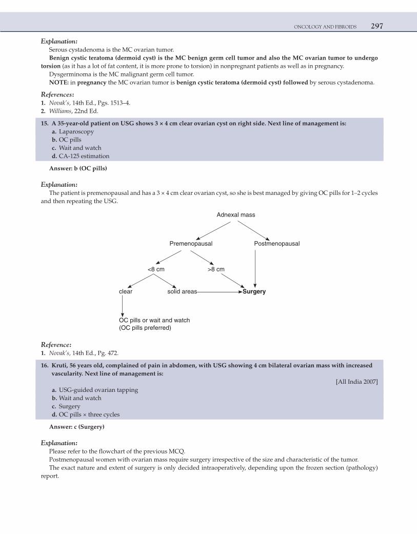
ONCOLOGY AND FIBROIDS 297
Explanation:Serous cystadenoma is the MC ovarian tumor.Benign cystic teratoma (dermoid cyst) is the MC benign germ cell tumor and also the MC ovarian tumor to undergo
torsion (as it has a lot of fat content, it is more prone to torsion) in nonpregnant patients as well as in pregnancy.Dysgerminoma is the MC malignant germ cell tumor.NOTE: in pregnancy the MC ovarian tumor is benign cystic teratoma (dermoid cyst) followed by serous cystadenoma.
References:
1. Novak’s, 14th Ed., Pgs. 1513–4. 2. Williams, 22nd Ed.
15. A 35-year-old patient on USG shows 3 × 4 cm clear ovarian cyst on right side. Next line of management is: a. Laparoscopy b. OC pills c. Wait and watch d. CA-125 estimation
Answer: b (OC pills)
Explanation:The patient is premenopausal and has a 3 × 4 cm clear ovarian cyst, so she is best managed by giving OC pills for 1–2 cycles
and then repeating the USG.
Adnexal mass
Premenopausal Postmenopausal
<8 cm >8 cm
clear solid areas Surgery
OC pills or wait and watch (OC pills preferred)
Reference:
1. Novak’s, 14th Ed., Pg. 472.
16. Kruti, 56 years old, complained of pain in abdomen, with USG showing 4 cm bilateral ovarian mass with increased vascularity. Next line of management is:
[All India 2007] a. USG-guided ovarian tapping b. Wait and watch c. Surgery d. OC pills × three cycles
Answer: c (Surgery)
Explanation:Please refer to the flowchart of the previous MCQ.Postmenopausal women with ovarian mass require surgery irrespective of the size and characteristic of the tumor.The exact nature and extent of surgery is only decided intraoperatively, depending upon the frozen section (pathology)
report.
298 SMART STUDY SERIES: OBSTETRICS AND GYNECOLOGY
Adnexal masses should never be tapped, as there is a risk of spread of tumor due to spillage of contents into the peritoneal cavity due to tapping.
Reference: 1. Novak’s, 14th Ed., Pg. 472.
17. True and false broad ligaments fibroids differentiated by anatomic position of: a. Ureter b. Internal iliac vein c. External iliac artery d. Descending cervical artery
Answer: a (Ureter)
Explanation:True broad ligament fibroid arises de novo in the broad ligament. Ureter is medial to this type of fibroid (it is between the
uterus and fibroid).Pseudo broad ligament fibroid arises from the uterus and then grows in between two leaves of broad ligament. So the
ureter is lateral to this type of fibroid.
Reference:
1. TeLinde’s, 9th Ed., Pg. 757.
18. Kamla, 30 years old, P2L2 with 3.2 × 4.1 cm fibroid uterus, complains of menorrhagia and is on symptomatic treatment since 6 months. The patient refuses surgery. Next line of management is:
a. GnRH analogs b. Danazol c. Myomectomy d. Uterine artery embolization
Answer: d (Uterine artery embolization)
Explanation:UAE can be used as a therapy for symptomatic patients who refuse or want to avoid surgery. After embolization, there is
60–65% decrease in size of fibroids over a period of 6–9 months, and so the patient’s symptoms may decrease or disappear. If the patient is still symptomatic after 1 year, then surgery should be considered.
Even though pregnancies have been reported after UAE, patient’s desire for pregnancy is a contraindication for UAE. The patient is P2L2 and is symptomatic and refuses surgery. So UAE is the best treatment for her.
Option 1: is mainly used preoperatively and is never a permanent treatment, besides it cannot be used long term.Option 2: was used in the past to decrease the size preoperatively but is hardly used now because of its androgenic side effects.Option 3: cannot be done if the patient refuses surgery.
Reference:
1. TeLinde, 9th Ed., Pg. 791–2.
19. Incidence of choriocarcinoma is seen more after: [All India 2001] a. Ectopic pregnancy b. Spontaneous abortion c. Normal delivery d. Cesarean section
Answer: b (Spontaneous abortion)
Explanation:Among all the cases of choriocarcinoma:50% develop following a hydatidiform mole25% develop following an abortion20% develop following a full-term pregnancy and 5% develop following an ectopic pregnancyAs vesicular mole is not in the options, abortion is the answer.
https://kat.cr/user/Blink99/
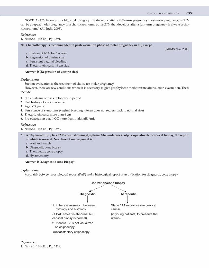
ONCOLOGY AND FIBROIDS 299
NOTE: A GTN belongs to a high-risk category if it develops after a full-term pregnancy (postmolar pregnancy, a GTN can be a repeat molar pregnancy or a choriocarcinoma, but a GTN that develops after a full-term pregnancy is always a cho-riocarcinoma) (All India 2003).
Reference:
1. Novak’s, 14th Ed., Pg. 1591.
20. Chemotherapy is recommended in postevacuation phase of molar pregnancy in all, except: [AIIMS Nov 2000] a. Plateau of hCG for 6 weeks b. Regression of uterine size c. Persistent vaginal bleeding d. Theca-lutein cysts >6 cm size
Answer: b (Regression of uterine size)
Explanation:Suction evacuation is the treatment of choice for molar pregnancy.However, there are few conditions where it is necessary to give prophylactic methotrexate after suction evacuation. These
include:
1. hCG plateaus or rises in follow-up period 2. Past history of vesicular mole 3. Age >35 years 4. Persistence of symptoms (vaginal bleeding, uterus does not regress back to normal size) 5. Theca-lutein cysts more than 6 cm 6. Pre-evacuation beta-hCG more than 1 lakh μIU/mL
Reference:
1. Novak’s, 14th Ed., Pg. 1590.
21. A 50-year-old P4L4 has PAP smear showing dysplasia. She undergoes colposcopic-directed cervical biopsy, the report of which is normal. Next line of management is:
a. Wait and watch b. Diagnostic cone biopsy c. Therapeutic cone biopsy d. Hysterectomy
Answer: b (Diagnostic cone biopsy)
Explanation:Mismatch between a cytological report (PAP) and a histological report is an indication for diagnostic cone biopsy.
Conization/cone biopsy
Diagnostic Therapeutic
1. If there is mismatch between Stage 1A1 microinvasive cervical cytology and histology cancer
(If PAP smear is abnormal but cervical biopsy is normal)
2. If entire TZ is not visualized on colposcopy
(unsatisfactory colposcopy)
young patients, to preserve theuterus)(in
Reference:
1. Novak’s, 14th Ed., Pg. 1418.
300 SMART STUDY SERIES: OBSTETRICS AND GYNECOLOGY
22. Advantages of surgery over radiotherapy in CA Cervix treatment are all, except: a. Preservation of vaginal function b. Conservation of ovaries c. Lesser surgical mortality d. None of the above
Answer: c (Lesser surgical mortality)
Explanation:Stages 1A2, IB, IIA are radiosensitive and surgically operable, but surgery is preferred over RT for these stages for the fol-
lowing reasons:
1. Preservation of ovarian function 2. Preservation of vagina for coital function 3. Psychological benefit to the patient
CA cervix almost never spreads to ovary, and so when radical hysterectomy is done, oophorectomy is not required.Mortality rate is the same 1% for both surgery and radiotherapy.
Reference:
1. Novak’s, 14th Ed., Pg. 1428.
23. Classic triad of Fallopian tube includes all, except: a. Hydrops tubae profluens b. Pelvic pain c. Bleeding PV d. Pelvic mass
Answer: c (Bleeding PV)
Explanation:Classic triad of fallopian tube cancer includes:Hydrops tubae profluens, pelvic pain and pelvic mass.This triad is seen in less than 15% cases.Bleeding PV can occur in patients of Fallopian tube cancer, but is not a part of the classic triad of fallopian tube cancer.Out of the triad the MC presenting feature of Fallopian tube cancer is persistent watery vaginal discharge (hydrops tubae
profluens)Fallopian tube cancer is managed exactly like epithelial ovarian cancer:Staging laparotomy, cytoreductive/debulking surgery followed by chemotherapy (cisplatin/carboplatin + paclitaxel).
Reference:
1. Novak’s, 14th Ed., Pg. 1528.
24. Therapeutic conization is indicated in: [AIIMS Jun 2000, All India 2013] a. Microinvasive carcinoma cervix stage 1a1 b. CIN III c. Unsatisfactory colposcopy with cervical dysplasia d. Cervical metaplasia
Answer: a (Microinvasive carcinoma cervix stage 1a1)
Explanation:Stage 1A of CA cervix is microinvasive. It is divided into 1A1 and 1A2.In stage 1A1, there is no lymph node involvement. Therapeutic conization is the surgery of choice for stage 1A1 in young
patients who are desirous of future childbearing. If the patient is old or family is complete, then this stage is treated by simple hysterectomy.
https://kat.cr/user/Blink99/
ONCOLOGY AND FIBROIDS 301
Option b = LEEP/LLETZ in young patients who are desirous of future child bearing. If the patient is old or family is com-plete, then this is treated by simple hysterectomy [All India 2010].
Option c = diagnostic conization.Option d = no treatment is required.
Reference: 1. Novak’s, 14th Ed., Pg. 1418.
25. A pregnant lady presents with genital warts. The best management for her is: [AIIMS Nov 2009] a. Imiquimod b. Trichloroacetic acid c. Podophyllin d. Cryotherapy
Answer: d (Cryotherapy)
Explanation:For reasons unknown genital warts increase in size and number during pregnancy.Treatment options during pregnancy include cryotherapy and trichloroacetic acid (TCA).Out of the two, cryosurgery is more effective than TCA and hence is preferred.Podophyllin, 5 fluorouracil, imiquimod and interferon therapy are not recommended in pregnancy because of concerns of
maternal and fetal safety (CDC 2002 guidelines).
Reference:
1. Williams, 22nd Ed., Pg. 1318.
26. Sentinel lymph node biopsy is useful for the following cancer: [All India 2010] a. cervix b. vulva c. vagina d. endometrial
Answer: b (Vulva)
Explanation:The sentinel lymph node is the hypothetical first lymph node or group of nodes reached by metastasizing cancer cells
from a primary tumor.Sentinel node biopsy technique is used in the staging of certain types of cancer to see if they have spread to any lymph
nodes. It is done using lymphoscintigraphy with technetium-99m labeled nanocolloid or isosulfan blue dye to identify a sen-tinel node that would predict the presence or absence of regional nodal metastasis.
The main advantage of this procedure is that it decreases unnecessary lymph node dissections, where it is not necessary, thereby reducing the risk of lymphedema and other complications. The main uses are in breast cancer and malignant mela-noma surgery, although it has been used in other tumor types with a degree of success.
Vulvar cancer was the first and most promising gynecological site for the sentinel lymph node biopsy strategy. As it involves a cutaneous tumor, peritumoral injections are easy and the sentinel lymph node is always located in the groin. This is another factor making the vulva an ideal site for sentinel lymph node biopsy. Preliminary studies indicate that a sentinel node can be identified in most of the patients of Ca vulva. Trials are on to determine the accuracy of negative predictive value of a uninvolved sentinel node. As of now complete inguinal-femoral lymphadenectomy is indicated in all stages of Ca vulva except stage Ia.
The role of Sentinel node detection in cervix cancer is purely investigational as of now and complete lymphadenectomy when indicated remains the standard of care.
NOTE: MC variety of vulvar cancer= Squamous cell carcinoma.MC site= labia (majora and minora) followed by clitoris.Risk factors for Ca vulva=HPV infection, cigarette smoking, lichen sclerosis, squamous hyperplasia, VIN.Most patients are asymptomatic at the time of diagnosis.
Reference:
1. Novak’s, 14th Ed., Pgs. 1562–3, 1425.
302 SMART STUDY SERIES: OBSTETRICS AND GYNECOLOGY
27. A 55-year-old lady presenting to outpatient department with postmenopausal bleeding for 3 months has a 1 × 1 cm nodule on the anterior lip of cervix. The most appropriate investigation to be done subsequently is:
[All India 2003] a. Pap smear b. Punch biopsy c. Endocervical curettage d. Colposcopy
Answer: b (Punch biopsy)
ExplanationRisk factors for Ca cervix/CIN:
a. Young age at first intercourse (<16 years) b. Multiple sexual partners c. Cigarette smoking d. Race e. High parity f. Low socio-economic status g. Human papillomavirus (HPV) infection h. HIV i. Immunosuppression
Vaginal bleeding (most often postcoital) is the MC symptom occurring in patients with Ca cervix. MC cause of postmeno-pausal bleeding in India is Ca cervix. PAP smear is a screening test.
If the PAP smear shows dysplasia, the next step to be done is cervical biopsy (preferably under colposcopy guidance).But, if a patient presents with obvious growth on lips of cervix, next step to be done is punch biopsy.
Reference
1. Novak’s, 14th Ed., Pg. 464, 491.
28. Choice of adjuvant treatment for endometrial carcinoma stage IA, grade I is: [All India 2004] a. Radiotherapy b. Chemotherapy c. Chemotherapy plus radiotherapy d. No treatment
Answer: d (No treatment)
ExplanationRick factors for endometrial Ca (estrogen-dependent tumor):
• Nulliparity • Early menarche, late menopause • Obesity • Diabetes mellitus and hypertension • PCOD • Unopposed estrogen therapy • Tamoxifen therapy • Atypical endometrial hyperplasia
Management of Ca endometrium: • Stage 1:
Surgery (total abdominal hysterectomy with bilateral salpingo-oophorectomy with lymph node sampling), followed by radiotherapy.
Only patients with stage 1A, grades 1 and 2 do not require postoperative radiotherapy. • Stage 2:
Modified radical hysterectomy, bilateral salpingo-oophorectomy with lymph node dissection, followed by radiotherapy.
https://kat.cr/user/Blink99/
ONCOLOGY AND FIBROIDS 303
• Stages 3 and 4:Debulking surgery followed by radiotherapy.Chemotherapy has no role in the management of Ca endometrium.
Reference
1. Novak’s, 14th Ed., Pg. 1371.
29. Pap smear is useful in the diagnosis of all, EXCEPT: [AIIMS May 2002] a. Gonorrhea b. Trichomonas vaginalis c. Human papilloma virus d. Inflammatory changes
Answer: a (Gonorrhea)
Explanation:The Papanicolaou test (also called Pap smear) is a screening test to detect premalignant and malignant processes in the
transformation zone. The test was invented by and named after the prominent Greek doctor Georgios Papanikolaou.Abnormal results are reported according to the Bethesda System. They include:
• Squamous cell abnormalities (SIL) ○ Atypical squamous cells of undetermined significance (ASC-US) ○ Low-grade squamous intra-epithelial lesion (LGSIL or LSIL) ○ Atypical squamous cells—cannot exclude HSIL (ASC-H) ○ High-grade squamous intra-epithelial lesion (HGSIL or HSIL) ○ Squamous cell carcinoma
• Glandular epithelial cell abnormalities ○ Atypical glandular cells not otherwise specified (AGC or AGC-NOS)
Endocervical and endometrial abnormalities can also be detected, Endocervical and endometrial abnormalities can also be detected, as can a number of infectious processes, including yeast, herpes simplex virus, and trichomoniasis. However, it is not very sensitive at detecting these infections, so absence of detection on a Pap does not mean absence of the infection.
Reference:
1. Novak’s, 14th Ed., Pg. 464, 491.
30. A 35-year-old lady has undergone radical hysterectomy for Ca cervix. Histopathology shows stage IBI with outer one-third of cervix and lower uterine segment involvement. Next line of management is:
[AIIMS May 2010] a. Follow-up b. Chemoradiation c. Chemotherapy d. Radiation
Answer: a (Follow-up)
Explanation:Stage I Carcinoma strictly confined to the cervix (extension to the corpus should be disregarded)Stage Ia Preclinical carcinomas of the cervix, i.e., those diagnosed only by microscopyStage Ia1: Lesion with <3 mm invasionStage Ia2: Lesions detected microscopically and can be measuredThe upper limit of the measurement should show a depth of invasion of >3-5 mm taken from the base of the epithelium,
either surface or glandular, from which it originates, and a second dimension, the horizontal spread, must not exceed 7 mm. Larger lesions should be staged as Ib.
Stage Ib Lesions invasive >5 mmStage Ib1: Lesions ≤ 4 cmStage Ib2: Lesions >4 cmTreatment of stage 1b1 is radical hysterectomy and that has been done for the patient.
304 SMART STUDY SERIES: OBSTETRICS AND GYNECOLOGY
Postoperative histopathology confirms that it is the same stage. (Uterus involvement does not change the staging.)Indications for postoperative chemoradiotherapy (CTRT)Postoperative CTRT to the pelvis decreases the risk of local recurrence in patients with high-risk factors, such as:
1) Positive pelvic nodes 2) Positive surgical margins 3) Residual parametrial disease
If these were present, then the answer would be chemoradiation. As these are not present, only follow-up of the patient is required.
Reference:
1. Novak’s, 14th Ed.
31. Cytogenetics is difficult in solid tumors, that too especially in carcinoma cervix, due to: [AIIMS Nov 2010] a. High mitotic activity b. Good-quality metaphase c. Specimen often not adequate d. Often contaminated and infested with infective microorganisms
Answer: d (Often contaminated and infested with infective microorganisms)
Explanation:Cytogenetics is a branch of genetics that is concerned with the study of function and structure of the cell, especially
the chromosomes. It includes routine analysis of G-banded chromosomes, other cytogenetic banding techniques, as well as molecular cytogenetics, such as fluorescent in situ hybridization (FISH) and comparative genomic hybridization (CGH).
Karyotype analyses based on G- or R-banding techniques have been widely applied to the characterization of cytogenetic abnormalities in tumor cells and have contributed significantly to the identification of recurrently involved chromosomal loci. However, the cytogenetic analysis of chromosomes from solid tumors has proven to be challenging. This is due to the often-low mitotic index, the poor quality of metaphase chromosomes, and the sheer number of cytogenetic abnormalities
Bacterial contamination is common technical problems in the isolation and extraction of DNA from clinical samples.Cytogenetics in solid tumors is done by FISH technique, most commonly which does not require dividing cells or meta-
phase nuclei.Sample adequacy in tissue biopsy is never an issue.
References:
1. Atlas of Genetics and Cytogenetics in Oncology and Haematology. 2. Robbins Textbook of Pathology.
32. A patient presents with Ca cervix with stage IIIb; treatment of choice is: [AIIMS Nov 2010] a. Chemotherapy b. Intracavitatory brachytherapy followed by external beam radiotherapy c. Wertheim’s hysterectomy d. Schauta’s operation
Answer: b (Intracavitatory brachytherapy followed by external beam radiotherapy)
Explanation:Stage-wise treatment for Ca cervix
• All stages (I–IV) are radiosensitive. • Stages of Ca cervix that are operable (radical/Wertheim’s hysterectomy) are 1A2, IB, and IIA. • Stages IIB-IV are not operable and have to be treated with RT only (brachy- and teletherapy) • Cisplatin is given before RT as a radiosensitizer.
Reference
1. Novak’s, 14th Ed., Pg. 1428.
https://kat.cr/user/Blink99/
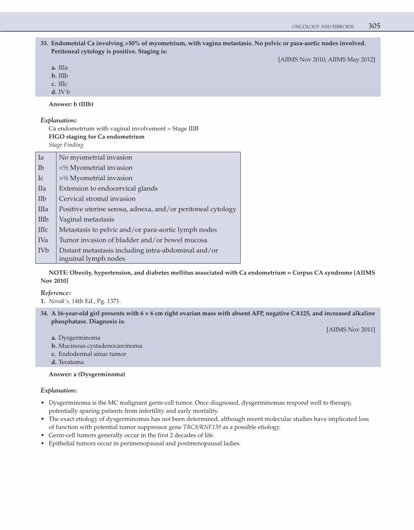
ONCOLOGY AND FIBROIDS 305
33. Endometrial Ca involving >50% of myometrium, with vagina metastasis. No pelvic or para-aortic nodes involved. Peritoneal cytology is positive. Staging is:
[AIIMS Nov 2010, AIIMS May 2012] a. IIIa b. IIIb c. IIIc d. IV b
Answer: b (IIIb)
Explanation:Ca endometrium with vaginal involvement = Stage IIIBFIGO staging for Ca endometriumStage Finding
Ia No myometrial invasion
Ib <½ Myometrial invasion
Ic >½ Myometrial invasion
IIa Extension to endocervical glands
IIb Cervical stromal invasion
IIIa Positive uterine serosa, adnexa, and/or peritoneal cytology
IIIb Vaginal metastasis
IIIc Metastasis to pelvic and/or para-aortic lymph nodes
IVa Tumor invasion of bladder and/or bowel mucosa
IVb Distant metastasis including intra-abdominal and/or inguinal lymph nodes
NOTE: Obesity, hypertension, and diabetes mellitus associated with Ca endometrium = Corpus CA syndrome [AIIMS Nov 2010]
Reference:
1. Novak’s, 14th Ed., Pg. 1371.
34. A 16-year-old girl presents with 6 × 6 cm right ovarian mass with absent AFP, negative CA125, and increased alkaline phosphatase. Diagnosis is:
[AIIMS Nov 2011] a. Dysgerminoma b. Mucinous cystadenocarcinoma c. Endodermal sinus tumor d. Teratoma
Answer: a (Dysgerminoma)
Explanation:
• Dysgerminoma is the MC malignant germ-cell tumor. Once diagnosed, dysgerminomas respond well to therapy, potentially sparing patients from infertility and early mortality.
• The exact etiology of dysgerminomas has not been determined, although recent molecular studies have implicated loss of function with potential tumor suppressor gene TRC8/RNF139 as a possible etiology.
• Germ-cell tumors generally occur in the first 2 decades of life. • Epithelial tumors occur in perimenopausal and postmenopausal ladies.
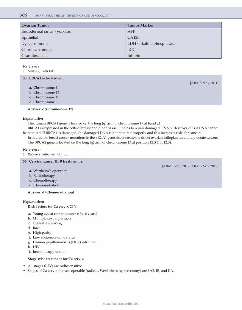
306 SMART STUDY SERIES: OBSTETRICS AND GYNECOLOGY
Ovarian Tumor Tumor Marker
Endodermal sinus /yolk sac AFP
Epithelial CA125
Dysgerminoma LDH/alkaline phosphatase
Choriocarcinoma hCG
Granulosa cell Inhibin
Reference:
1. Novak’s, 14th Ed.
35. BRCA1 is located on: [AIIMS May 2011] a. Chromosome 11 b. Chromosome 13 c. Chromosome 17 d. Chromosome 6
Answer: c (Chromosome 17)
ExplanationThe human BRCA1 gene is located on the long (q) arm of chromosome 17 at band 21.BRCA1 is expressed in the cells of breast and other tissue. It helps to repair damaged DNA or destroys cells if DNA cannot
be repaired. If BRCA1 is damaged, the damaged DNA is not repaired properly and this increases risks for cancers.In addition to breast cancer, mutations in the BRCA1 gene also increase the risk of ovarian, fallopian tube, and prostate cancers.The BRCA2 gene is located on the long (q) arm of chromosome 13 at position 12.3 (13q12.3).
Reference:
1. Robbin’s Pathology, 6th Ed.
36. Cervical cancer III B treatment is: [AIIMS May 2012, AIIMS Nov 2012] a. Wertheim’s operation b. Radiotherapy c. Chemotherapy d. Chemoradiation
Answer: d (Chemoradiation)
Explanation:Risk factors for Ca cervix/CIN:
a. Young age at first intercourse (<16 years) b. Multiple sexual partners c. Cigarette smoking d. Race e. High parity f. Low socio-economic status g. Human papillomavirus (HPV) infection h. HIV i. Immunosuppression
Stage-wise treatment for Ca cervix:
• All stages (I–IV) are radiosensitive. • Stages of Ca cervix that are operable (radical/Wertheim’s hysterectomy) are 1A2, IB, and IIA.
https://kat.cr/user/Blink99/
ONCOLOGY AND FIBROIDS 307
• Stages IIB–IV are not operable and have to be treated with RT only (brachy and teletherapy). • Cisplatin is given before RT as a radiosensitizer, hence the preferred terminology is CTRT (concurrent chemo and
radiotherapy also known as chemoradiation).
NOTE: If CTRT and RT both are in the options (as in this mcq) then CTRT (concurrent chemo and radiotherapy also known as chemoradiation) is the best option to mark.
Reference:
1. Novak’s, 14th Ed. Pg. 1428.
37. In leiomyoma of uterus, least likely change to occur is: [AIIMS Nov 2012] a. Red degeneration b. Sarcomatous change c. Calcifications d. Hyaline generation
Answer: b (Sarcomatous change)
Explanation:Various degenerative changes which occur in fibroid are:
• Hyaline degeneration is the most common (65%). The central part is the common site. • Fatty changes are rare and are found at/or after menopause. • Calcification/calcific degeneration (10%) usually occurs in postmenopausal subserosal fibroids. When whole of the
tumor is converted to a calcified mass it is called ‘womb stone.’ • Red degeneration presents almost exclusively in pregnancy with acute pain, fever and localized tenderness. It is due to
aseptic inflammation and thrombosis. It is usually managed conservatively. • Sarcomatous degeneration is the least common. The exact risk of sarcomatous change remains uncertain, but is probably
less than 0.1%.
Reference:
1. Novak’s, 14th Ed. Pg. 470.
38. BIRADS stands for: [AIIMS Nov 2012]
a. Breast Imaging Reporting and Data System b. Breast Imaging and Radiation System c. Brain Imaging Reporting and Data System d. Bone Imaging Reporting and Data System
Answer: a (Breast Imaging Reporting and Data System)
Explanation:BI-RADS stands for Breast Imaging-Reporting and Data System. It is a quality assurance tool originally designed for use
with mammography. The system is published and trademarked by the American College of Radiology (ACR).The system is designed to standardize reporting, and is used by doctors to communicate a patient’s risk of developing
breast cancer.The summary of each category is identical for all 3 modalities (mammography, MRI and USG).Category 6 was added in the 4th edition of the Mammography Atlas.BI-RADS Assessment Categories are:
• 0: Incomplete • 1: Negative • 2: Benign finding(s) • 3: Probably benign • 4: Suspicious abnormality • 5: Highly suggestive of malignancy • 6: Known biopsy – proven malignancy
308 SMART STUDY SERIES: OBSTETRICS AND GYNECOLOGY
NOTE: Metastasis of BREAST CANCER account for majority of ocular and orbital metastasis in females [AIIMS May 2013, Nov 2013]
Reference:
1. American College of Radiology (ACR) Breast Imaging Reporting and Data System Atlas (BI-RADS Atlas). Reston, Va: © American College of Radiology; 2003
39. All are causes of postmenopausal bleeding except: a. Ca endometrium b. Ca fallopian tube c. Ca ovary d. Carcinoma in situ cervix
Answer: d (Carcinoma in situ cervix)
Explanation:Endometrial cancer most often occurs in women in 6th and 7th decade, at an average age of 60 years. 75% of cases occur in
women older than 50 years. Most common presenting feature is vaginal bleeding (postmenopausal bleeding).Classic triad of fallopian tube cancer includes: Hydrops tubae profluens, pelvic pain and pelvic mass. Postmenopausal
bleeding PV can occur in patients of fallopian tube cancer.Most patients with epithelial ovarian cancer have no symptoms. When the symptoms develop they are often vague and non-
specific (upper GI complaints like dyspepsia, bloating).In advanced cases vaginal bleeding may occur in postmenopausal women.Also in granulosa cell tumor of the ovary there would be postmenopausal bleeding as it secretes estrogen.Carcinoma in situ cervix is a preclinical condition and is diagnosed with PAP smear screening and subsequent cervical biopsy.NOTE: Read all the options carefully before answering. CA cervix definitely causes postmenopausal bleeding, but the
option here is carcinoma in situ cervix.
Reference:
1. Novak’s, 14th Ed. Pg. 1528, 1418, 1362.
https://kat.cr/user/Blink99/
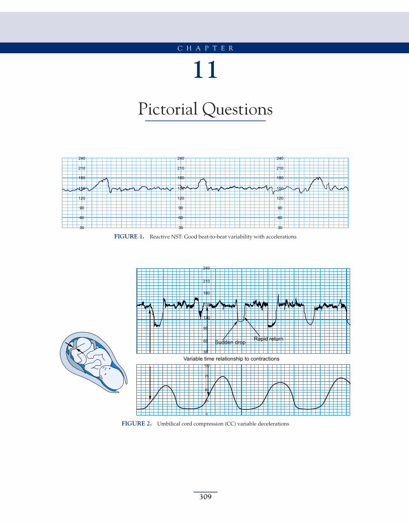
309
11Pictorial Questions
240
210
180
150
120
90
60
30
240
210
180
150
120
90
60
30
240
210
180
150
120
90
60
30
FIGURE 1. Reactive NST: Good beat-to-beat variability with accelerations
240
210
180
120
90
60
30
100
75
50
25
0
Variable time relationship to contractions
Rapid returnSudden drop
150
FIGURE 2. Umbilical cord compression (CC) variable decelerations
C H A P T E R
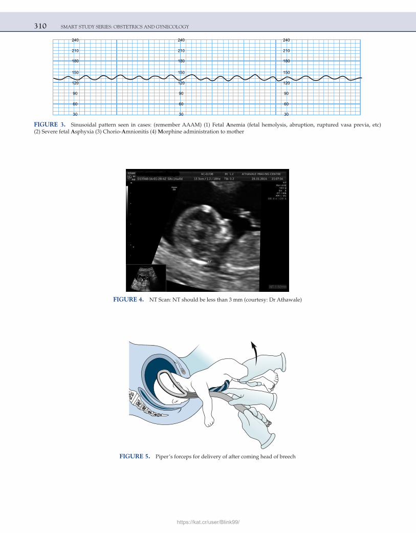
310 SMART STUDY SERIES: OBSTETRICS AND GYNECOLOGY
240
210
180
150
120
90
60
30
240
210
180
150
120
90
60
30
240
210
180
150
120
90
60
30
FIGURE 3. Sinusoidal pattern seen in cases: (remember AAAM) (1) Fetal Anemia (fetal hemolysis, abruption, ruptured vasa previa, etc) (2) Severe fetal Asphyxia (3) Chorio-Amnionitis (4) Morphine administration to mother
FIGURE 4. NT Scan: NT should be less than 3 mm (courtesy: Dr Athawale)
FIGURE 5. Piper’s forceps for delivery of after coming head of breech
https://kat.cr/user/Blink99/
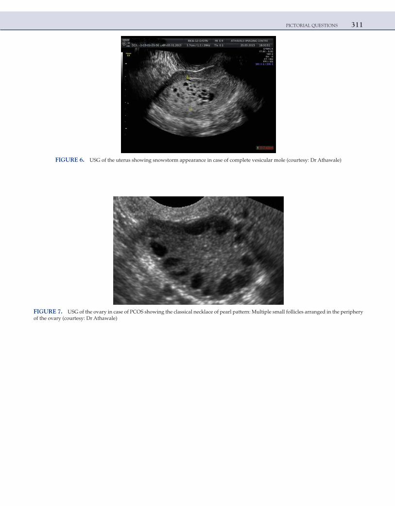
PICTORIAL QUESTIONS 311
FIGURE 6. USG of the uterus showing snowstorm appearance in case of complete vesicular mole (courtesy: Dr Athawale)
FIGURE 7. USG of the ovary in case of PCOS showing the classical necklace of pearl pattern: Multiple small follicles arranged in the periphery of the ovary (courtesy: Dr Athawale)
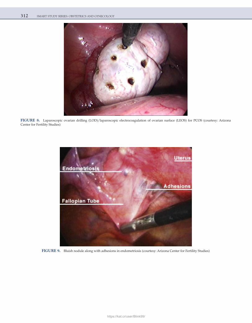
312 SMART STUDY SERIES: OBSTETRICS AND GYNECOLOGY
FIGURE 8. Laparoscopic ovarian drilling (LOD)/laparoscopic electrocoagulation of ovarian surface (LEOS) for PCOS (courtesy: Arizona Center for Fertility Studies)
FIGURE 9. Bluish nodule along with adhesions in endometriosis (courtesy: Arizona Center for Fertility Studies)
https://kat.cr/user/Blink99/
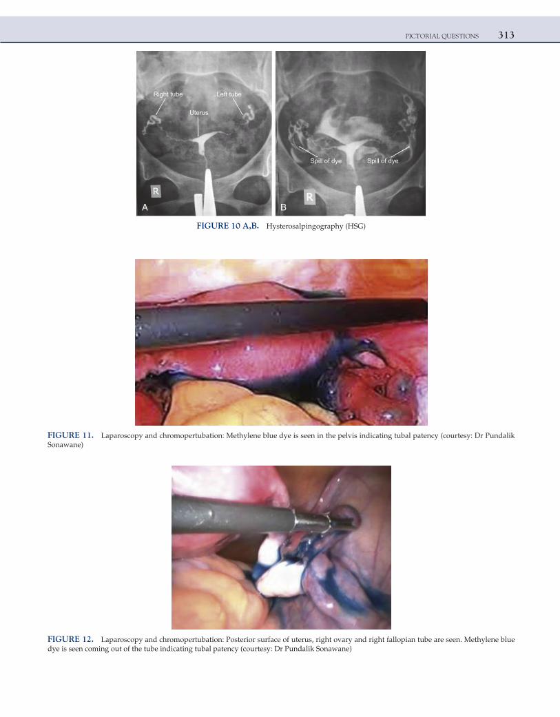
PICTORIAL QUESTIONS 313
A B
Right tube
Uterus
Left tube
Spill of dye Spill of dye
FIGURE 10 A,B. Hysterosalpingography (HSG)
FIGURE 11. Laparoscopy and chromopertubation: Methylene blue dye is seen in the pelvis indicating tubal patency (courtesy: Dr Pundalik Sonawane)
FIGURE 12. Laparoscopy and chromopertubation: Posterior surface of uterus, right ovary and right fallopian tube are seen. Methylene blue dye is seen coming out of the tube indicating tubal patency (courtesy: Dr Pundalik Sonawane)

This page intentionally left blank
https://kat.cr/user/Blink99/
315
Readers’ Reviews
1. Hello Sir, results of PG exams have been declared and I have got 14th rank in MH-CET and 78th rank in All India Exam. Sir, Smart Study in OBGYN written by you has helped me a lot in getting good ranks in all the entrance exams I gave this year. I owe my success to your book and your excellent teaching.
Thank you very much, Sir.
Saumil PandyaSpeed Mumbai
2. As regards to your book, I will call it “my savior”. It has covered all the important topics in Obstetrics and Gynecology in a very precise and simple format. I loved your book Sir.
Thanks.
Dr Tushar ShahBhatia Classes, Mumbai
3. The book is outstanding, Sir...it’s a boon to all PG aspirants. Thanks a lot.
Dr Love GoyalIAMS, Delhi
4. I thank you for writing such a fantastic book—‘Smart Study Obstetrics and Gynecology’. It is a complete book covering all the important topics required for any of the PG entrance exams and there is no need to read any other Obstetrics and Gynecology book after reading your book. Thanks and congratulations for such a student-friendly book. I was able to do almost all the Obstetrics and Gynecology questions in my PG entrance exam.
Thank you.
Dr Pooja JangidRGMC, Thane
Dr Bhatia’s Institute, Mumbai
5. It has the entire subject covered in a concise format which I feel is extremely helpful to me. Thank you, Sir.
Sourav SinhaIAMS, Kolkata
6. Your book is fabulous and very helpful.
Dr Rutvi DaveIAMS, Ahmedabad

READER’S REVIEWS316
7. Your book is awesome. I have recommended to all people I know, even to undergraduates.
Dr Nirmala RaySpeed Classes, Kolkata
8. Sir, I enjoyed your book very much…it helped me a lot. Thank you.
Dr Umang AggarwalDAMS, Mumbai
9. Thanks a lot, Sir...your book is too good...a complete book for preparation. Honestly, there is no need to look into any other book for Obstetrics and Gynecology. Hats off!
Dr Neha TulliIAMS, Delhi
10. Your book is fantastic. I wish there were books like this for other subjects as well. I could not even think of reading Obstetrics and Gynecology within such a short time.
Dr Anupam DasBhatia Classes, Kolkata
11. Thanks a lot, Sir. The presentation is very nice and the book is extremely informative. Thanks a lot for taking so much pain to compile this book. I have advised this book to all friends and juniors preparing for PG.
Dr Rahul RanjanDAMS Classes, Pune
12. Your book made things easy! It is a handy book with loads of information for PG entrance exams. Thanks a lot Sir.
Dr Vineel KolojuDr Bhatia, Hyderabad
13. Sir, your book is really great...I read it like a novel, hats off to your great job.
Dr Ranjit KumarIAMS, Bengaluru
https://kat.cr/user/Blink99/


