Embed Size (px)
Citation preview

RESEARCH Open Access
18F-florbetaben Ab imaging in mild cognitiveimpairmentKevin Ong1, Victor L Villemagne1,2,3, Alex Bahar-Fuchs1,4, Fiona Lamb1,3, Gaël Chételat1, Parnesh Raniga5,Rachel S Mulligan1, Olivier Salvado5, Barbara Putz6, Katrin Roth6, Colin L Masters3, Cornelia B Reininger6 andChristopher C Rowe1,2*
Abstract
Introduction: 18F-florbetaben and positron emission tomography were used to examine the relationships betweenb-amyloid (Ab) deposition, cognition, hippocampal volume, and white matter hyperintensities in mild cognitiveimpairment (MCI).
Methods: Forty-five MCI participants were evaluated. A neocortical standardized uptake value ratio threshold ≥
1.45 was used to discriminate high from low Ab burden. Correlations were adjusted for age, gender and years ofeducation.
Results: High Ab burden was found in 53% of MCI. Regression analyses showed standardized uptake value ratio (r= -0.51, P = 0.0015) and hippocampal volume (r = 0.60, P = 0.024) both contributing to episodic memoryimpairment in independent fashion. White matter hyperintensities correlated with nonmemory cognition, and thiscorrelation was particularly associated with Ab burden.
Conclusion: Higher Ab deposition in MCI is associated with more severe memory impairment and is contributingto early amnestic symptoms independent of hippocampal atrophy.
IntroductionThe leading etiological hypothesis of Alzheimer’s disease(AD) points to excessive brain b-amyloid (Ab) thataggregates to form extracellular plaques and vascularwall deposits [1]. With increasing prevalence and asso-ciated cost of care and the likelihood of greater benefitif therapies are applied early, earlier and more accurateidentification of AD has become a research priority.Dementia is usually preceded by a transition period of
cognitive decline commonly referred to as mild cognitiveimpairment (MCI). Characterized by an objective impair-ment of memory and/or other cognitive domains, MCI isnot severe enough to significantly interfere with activitiesof daily living [2]. The prevalence of MCI in people aged65 is believed to be 10 to 20%, with over 10% who havebeen classified as MCI converting to dementia per year[3]. Histopathologic studies on brains of MCI subjects
have shown characteristic AD pathology including Ab pla-ques and neurofibillary tangles in the majority of cases [4].MCI has been further classified based on whether memoryhas been affected (amnestic MCI) or spared (nonamnesticMCI), and whether the cognitive deficit affected is mainlyin one cognitive domain (single-domain MCI) or morethan one domain (multidomain MCI). Hence, MCI can beclassified into four clinical subtypes: nonamnestic single-domain, nonamnestic multiple domains, amnestic single-domain (asMCI), and amnestic multiple domains(amMCI). These subtypes probably differ in etiology andoutcome. Impaired episodic memory, which characterizesasMCI and amMCI, is thought to be a prodromal condi-tion for AD [3,4].The new research diagnostic criteria for AD and MCI
allow for Ab imaging in the workup of individuals withcognitive impairment [5,6]. Non-invasive Ab imaging toconfirm the presence of AD neuropathology could aid inearly differential diagnosis, identify at-risk individuals,help predict or monitor disease progression, and poten-tially evaluate the response to disease-specific therapy.
* Correspondence: [email protected] of Nuclear Medicine and Centre for PET, Austin Health, 145Studley Road, Heidelberg, VIC 3084, AustraliaFull list of author information is available at the end of the article
Ong et al. Alzheimer’s Research & Therapy 2013, 5:4http://alzres.com/content/5/1/4
© 2013 Ong et al.; licensee BioMed Central Ltd. This is an open access article distributed under the terms of the Creative CommonsAttribution License (http://creativecommons.org/licenses/by/2.0), which permits unrestricted use, distribution, and reproduction inany medium, provided the original work is properly cited.

11C-Pittsburgh Compound B (PiB) has been the mostwidely used agent in dementia research to assess Ab bur-den in vivo [7]. The major disadvantage of PiB is that it isradiolabeled with carbon-11, which has a short decayhalf-life (20 minutes) that limits its use to centers with anonsite cyclotron and 11C-radiochemistry expertise.To overcome these limitations, a number of novel
fluorine-18 Ab imaging tracers such as 18F-florbetaben(BAY 94-9172) [8-10], 18F-florbetapir (AV45) [11,12]and 18F-flutemetamol (GE067) [13,14] have been devel-oped. The 110-minute radioactive decay half-life offluorine-18 allows centralized synthesis and regional dis-tribution of these tracers as currently practiced world-wide in the supply of 18F-fluorodeoxyglucose for routineclinical positron emission tomography (PET) imaging.
18F-florbetaben (FBB; trans-4-(N-methyl-amino)-4"(2-(2-(2-[18F] fluoro-ethoxy)ethoxy)-ethoxy)stilbene),developed by Avid Radiopharmaceuticals (Philadelphia,USA) and Bayer-Schering Pharma (Berlin, Germany), hasbeen shown to bind with high affinity to Ab in brainhomogenates and selectively labeled Ab plaques and cer-ebral amyloid angiopathy (CAA) in AD tissue sections[15]. After injection into Tg2576 transgenic mice, ex vivobrain sections showed localization of FBB in regions withAb plaques as confirmed by thioflavin binding [16]. Atthe tracer concentrations achieved during human PETstudies, FBB did not show binding to a-synuclein inLewy bodies or to tau lesions in postmortem corticesfrom dementia with Lewy bodies, AD or frontotemporallobar degeneration patients [17]. In human studies, corti-cal retention of FBB was significantly higher in ADpatients compared with age-matched controls and fron-totemporal lobar degeneration patients, with bindingmatching the reported postmortem distribution of Abplaques [9]. Phase II clinical studies further confirmedthese results [8]. FBB is highly correlated with 11C-PiB(r = 0.97 with a slope of 0.71) [18], and was used todetect the presence or absence of AD pathology in thebrain in participants with a wide spectrum of neurode-generative diseases including a few MCI participants [10].Phase III studies for FBB have reached completion [19].Human postmortem studies have shown that while
soluble Ab oligomers and the density of neurofibrillarytangles strongly correlate with neurodegeneration andcognitive deficits, the density of Ab insoluble plaquesdoes not [20-24] and Αb burden as assessed by PET doesnot strongly correlate with cognitive impairment in ADpatients [25,26]. The severity of tau pathology in ADpatients is closely related to neuronal loss [27], hippo-campal atrophy [28,29] and memory impairment [30,31].Amyloid imaging studies in MCI have shown an associa-tion between Ab burden and memory [32], an associationthat is believed to be mediated by hippocampal atrophy[33]. Vascular pathology, as reflected in white matter
hyperintensities (WMH), has been shown to be asso-ciated with cognitive impairment, particularly affectingworking memory and executive function, as well asvisuospatial abilities among people with MCI [34].The purpose of this study was to characterize FBB
binding in a well-characterized MCI cohort, and toexplore the relationships of Ab burden cognitive perfor-mance, hippocampal volume (HV), and WMH.
Materials and methodsParticipantsForty-five participants fulfilling Petersen’s criteria for MCI[3] were recruited between June 2008 and December 2009from memory disorder specialists. Fifteen healthy oldercontrols and 15 patients who met National Institute ofNeurological Disorders and Stroke-Alzheimer’s Diseaseand Related Disorders Association criteria for probableAD, which were previously described in an earlier study[9], were used for comparison against the MCI cohort.Consistent with the consensus criteria for MCI at the
time of enrolment [3], all participants (and their next ofkin) reported a history of cognitive decline and hadobjective cognitive impairment on neuropsychologicalassessment but remained generally independent in dailyactivities. In addition, participants had to be at least 60years of age, had at least 7 years of formal education,spoke fluent English, were capable of giving informedconsent, had a reliable informant capable of giving a col-lateral history, were able to tolerate a brain magneticresonance imaging (MRI) scan, did not meet the NationalInstitute of Neurological Disorders and Stroke-Associa-tion Internationale pour la Recherché et l’Enseignementen Neurosciences criteria for the diagnosis of vasculardementia, and scored ≥ 24 on the Mini-Mental StateExamination (for detailed exclusion criteria, see Table S1in Additional file 1). These participants were referredfrom local specialist public and private memory disordersclinics upon being diagnosed with MCI and had no otherevidence of significant neurodegenerative disease, moder-ate or severe psychiatric illness, drug or alcohol depen-dence, or participated in any anti-Ab therapeutic trialprior to enrolment.The recruitment criterion was defined as having at least
one test score falling 1.5 standard deviations below pub-lished means. For precision, subsequent classification ofparticipants into MCI subtypes by Petersen’s criteria [3]was based instead on test scores falling 1.5 standarddeviations below the mean of a carefully screened anddemographically well-matched cohort living in the sameregion as the participants. This cohort consisted of 45healthy older participants from the Australian ImagingBiomarkers and Lifestyle flagship study of ageing [35]with no history of cognitive decline who had negativebrain PiB scans, normal brain MRI, Clinical Dementia
Ong et al. Alzheimer’s Research & Therapy 2013, 5:4http://alzres.com/content/5/1/4
Page 2 of 11

Rating = 0 and Clinical Dementia Rating sum of boxes =0, and had no psychiatric illness.Approval for the study was obtained from the Austin
Health Human Research Ethics Committee. Writteninformed consent for participation was obtained fromall participants prior to screening. Safety monitoringconsisted of clinical observation, baseline ECG, hematol-ogy and biochemistry testing and measurement of vitalsigns before and after tracer injection. Vital signs, hema-tology, and biochemistry testing were repeated 1 weekafter injection. Participants were asked about possibleadverse events after their PET scan and 1 week afterinjection.
Neuropsychological evaluationNeuropsychology evaluation was conducted within 24.5 ±15.5 days of the FBB PET scan by a licensed neuropsy-chologist. Evaluation consisted of the Mini-Mental StateExamination, the Clinical Dementia Rating, the CaliforniaVerbal Learning Test Second Edition, the Rey ComplexFigure Test (RCFT), Logical Memory I and II (WechslerMemory Scale; Story A only), the Controlled Oral WordAssociation Test, Categorical Fluency, the Boston Nam-ing Task (30-item version), Digit Symbol-coding andDigit Span.Individual composite episodic memory z scores (EM)
were generated in 44 participants by averaging the zscores for delayed recall trials of the RCFT, the CaliforniaVerbal Learning Test Second Edition, and Logical Mem-ory II. The RCFT delayed recall score was missing foranother participant and was substituted with the RCFTimmediate delay score because the relationship betweenscores on the immediate and the delayed recall trials wasvery strong (r = 0.93). Composite nonmemory z scores inall 45 participants were calculated by averaging thez scores for the Boston Naming Task, the ControlledOral Word Association Test, Categorical Fluency, DigitSpan, Digit Symbol-coding and RCFT copy [32].
Image acquisitionMagnetic resonance imagingA three-dimensional T1-weighted magnetization preparedrapid gradient echo sequence and a fluid-attenuated inver-sion recovery sequence were performed on either a 1.5 Tor a 3 T magnetic resonance scanner prior to the PETscan.18F-florbetaben imagingLabeling was carried out in the Austin Health Centre forPET, as previously described [9]. Mean specific activityat the time of injection for MCI was 60 ± 29 GBq/μmol.Imaging was performed with a three-dimensional GSOPhilips Allegro PET camera. A 2-minute transmissionscan using a rotating 137Cs source was performed for
attenuation correction immediately prior to scanning.Each MCI participant received on average 286 ± 19MBq FBB intravenously over 38 ± 17 seconds. Imageswere reconstructed using a three-dimensional RAMLAalgorithm (Philips, Cleveland, USA). Images obtainedbetween 90 and 110 minutes post injection were usedfor the analysis.
Image analysisAll image analysis was performed by experienced opera-tors blind to the clinical status and cognitive test scoresof the subjects.Extraction of HVs from the three-dimensional magne-
tization prepared rapid gradient echo MRI data in 43MCI cases was performed using a commercial, US Foodand Drug Administration-approved, fully automatedvolumetric measurement program (NeuroQuant®) [36].Preprocessing of the fluid-attenuated inversion recoveryimages was performed to correct for bias field effects andremove noise using anisotropic diffusion prior to manualsegmentation of deep WMH. Manual segmentation ofthe WMH (PR) was performed using MRIcro software[37]. The total WMH volume in each MCI subject wascalculated, as well as the number of individual lesions.All volumes were normalized for head size using the totalintracranial volume, defined as the sum of gray matter,white matter and cerebrospinal fluid volumes.Spatial normalization and co-registration of the PET
and MRI images was performed using SPM8 [38]. PETimages were processed with a semiautomatic volume ofinterest method. This method used a preset template ofnarrow cortical volume of interest that was either appliedto the spatially normalized MRI and then transferred tothe co-registered FBB scan or applied directly to the spa-tially normalized FBB scan. Minor manual adjustmentswere made to ensure that overlap with white matter andcerebrospinal fluid was minimized. Mean radioactivityvalues were obtained from the volume of interest for thecortical, subcortical and cerebellar regions. The cerebellarcortical volume of interest was placed taking care toavoid cerebellar white matter. All volume of interest pla-cement was performed by a single experienced operator(VLV) blind to the clinical status of the individuals. Nocorrection for partial volume effects was applied to thePET data.The standardized uptake value, defined as the decay-
corrected brain radioactivity concentration normalizedfor injected dose and body weight, was calculated for allregions. These values were then used to derive the stan-dardized uptake value ratio (SUVR), which was refer-enced to the cerebellar cortex. Neocortical Ab depositionwas expressed as the average SUVR of the mean for thefollowing cortical regions of interest: frontal (consisting
Ong et al. Alzheimer’s Research & Therapy 2013, 5:4http://alzres.com/content/5/1/4
Page 3 of 11
of dorsolateral prefrontal, ventrolateral prefrontal, andorbitofrontal regions), superior parietal, lateral temporal,lateral occipital, and anterior and posterior cingulate.To identify a SUVR cutoff point, a hierarchical cluster
analysis of the neocortical SUVR of FBB scans in healthycontrol participants was performed similar to that pre-viously described [10]. The cutoff value for high neocor-tical SUVR in this study was defined as ≥ 1.45.
Statistical analysisIndependent-sample t-tests were used to compare meansof MCI subtypes with healthy controls and AD patients,and to compare means within the MCI subtypes. Categori-cal differences were assessed using Fisher’s exact test.Pearson’s or Spearman’s rank correlation analyses wereconducted to assess the degree of linear relationshipbetween neuroimaging variables (SUVR, HV, WMH) withcomposite EM and nonmemory z scores, adjusting for age,gender and years of education. Data are presented asmean ± standard deviation unless otherwise stated.Adjustment for multiple testing was not performed.
Role of the funding sourceThe funding sources had no role in the data analysesand interpretation. The corresponding author had fullaccess to all data presented in this study and had finalresponsibility for the decision to submit for publication.
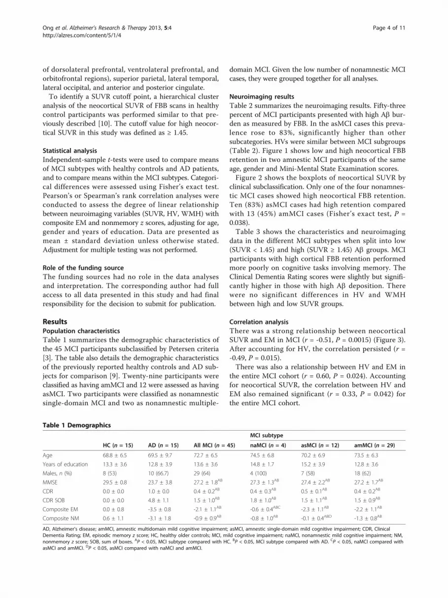
ResultsPopulation characteristicsTable 1 summarizes the demographic characteristics ofthe 45 MCI participants subclassified by Petersen criteria[3]. The table also details the demographic characteristicsof the previously reported healthy controls and AD sub-jects for comparison [9]. Twenty-nine participants wereclassified as having amMCI and 12 were assessed as havingasMCI. Two participants were classified as nonamnesticsingle-domain MCI and two as nonamnestic multiple-
domain MCI. Given the low number of nonamnestic MCIcases, they were grouped together for all analyses.
Neuroimaging resultsTable 2 summarizes the neuroimaging results. Fifty-threepercent of MCI participants presented with high Ab bur-den as measured by FBB. In the asMCI cases this preva-lence rose to 83%, significantly higher than othersubcategories. HVs were similar between MCI subgroups(Table 2). Figure 1 shows low and high neocortical FBBretention in two amnestic MCI participants of the sameage, gender and Mini-Mental State Examination scores.Figure 2 shows the boxplots of neocortical SUVR by
clinical subclassification. Only one of the four nonamnes-tic MCI cases showed high neocortical FBB retention.Ten (83%) asMCI cases had high retention comparedwith 13 (45%) amMCI cases (Fisher’s exact test, P =0.038).Table 3 shows the characteristics and neuroimaging
data in the different MCI subtypes when split into low(SUVR < 1.45) and high (SUVR ≥ 1.45) Ab groups. MCIparticipants with high cortical FBB retention performedmore poorly on cognitive tasks involving memory. TheClinical Dementia Rating scores were slightly but signifi-cantly higher in those with high Ab deposition. Therewere no significant differences in HV and WMHbetween high and low SUVR groups.
Correlation analysisThere was a strong relationship between neocorticalSUVR and EM in MCI (r = -0.51, P = 0.0015) (Figure 3).After accounting for HV, the correlation persisted (r =-0.49, P = 0.015).There was also a relationship between HV and EM in
the entire MCI cohort (r = 0.60, P = 0.024). Accountingfor neocortical SUVR, the correlation between HV andEM also remained significant (r = 0.33, P = 0.042) forthe entire MCI cohort.
Table 1 Demographics
MCI subtype
HC (n = 15) AD (n = 15) All MCI (n = 45) naMCI (n = 4) asMCI (n = 12) amMCI (n = 29)
Age 68.8 ± 6.5 69.5 ± 9.7 72.7 ± 6.5 74.5 ± 6.8 70.2 ± 6.9 73.5 ± 6.3
Years of education 13.3 ± 3.6 12.8 ± 3.9 13.6 ± 3.6 14.8 ± 1.7 15.2 ± 3.9 12.8 ± 3.6
Males, n (%) 8 (53) 10 (66.7) 29 (64) 4 (100) 7 (58) 18 (62)
MMSE 29.5 ± 0.8 23.7 ± 3.8 27.2 ± 1.8AB 27.3 ± 1.3AB 27.4 ± 2.2AB 27.2 ± 1.7AB
CDR 0.0 ± 0.0 1.0 ± 0.0 0.4 ± 0.2AB 0.4 ± 0.3AB 0.5 ± 0.1AB 0.4 ± 0.2AB
CDR SOB 0.0 ± 0.0 4.8 ± 1.1 1.5 ± 1.0AB 1.8 ± 1.0AB 1.5 ± 1.1AB 1.5 ± 0.9AB
Composite EM 0.0 ± 0.8 -3.5 ± 0.8 -2.1 ± 1.1AB -0.6 ± 0.4ABC -2.3 ± 1.1AB -2.2 ± 1.1AB
Composite NM 0.6 ± 1.1 -3.1 ± 1.8 -0.9 ± 0.9AB -0.8 ± 1.0AB -0.1 ± 0.4ABD -1.3 ± 0.8AB
AD, Alzheimer’s disease; amMCI, amnestic multidomain mild cognitive impairment; asMCI, amnestic single-domain mild cognitive impairment; CDR, ClinicalDementia Rating; EM, episodic memory z score; HC, healthy older controls; MCI, mild cognitive impairment; naMCI, nonamnestic mild cognitive impairment; NM,nonmemory z score; SOB, sum of boxes. AP < 0.05, MCI subtype compared with HC. BP < 0.05, MCI subtype compared with AD. CP < 0.05, naMCI compared withasMCI and amMCI. DP < 0.05, asMCI compared with naMCI and amMCI.
Ong et al. Alzheimer’s Research & Therapy 2013, 5:4http://alzres.com/content/5/1/4
Page 4 of 11

There was a relationship between WMH volume andnonmemory z scores (r = -0.60, P = 0.03; Spearman’s r =-0.48, P = 0.0008). This correlation was amplified in thehigh SUVR subgroup (r = -0.71, P = 0.014; Spearman’s r =-0.57, P = 0.0035), but was not present in the low SUVRsubgroup (see Figure S1 in Additional file 2). No correla-tion was found between WMH and neocortical SUVR, HVor EM.
DiscussionAb burden and memory impairmentThis study provides support for the use of FBB PET toassess brain Ab plaque levels in individuals with MCI.FBB presents with similar characteristics to PiB, includ-ing short scan acquisition time and a good safety and tol-erability profile. The longer radioactive half-life of
Table 2 Neuroimaging
MCI subtype
HC (n = 15) AD (n = 15) All MCI (n = 45) naMCI (n = 4) asMCI (n = 12) amMCI (n = 29)
Neocortical SUVR 1.26 ± 0.22 1.96 ± 0.27 1.54 ± 0.27AB 1.50 ± 0.35B 1.66 ± 0.20AB 1.49 ± 0.28B
High b-amyloid, n (%) 3 (20) 15 (100) 24 (53)B 1 (25)B 10 (83)AC 13 (45)B
Hippocampal volume (cm3) 7.1 ± 0.9 7.1 ± 0.8 7.0 ± 0.8 7.2 ± 1.0
WMH volume (cm3) 10.2 ± 11.0 13.3 ± 15.5 4.8 ± 6.6C 12.0 ± 11.4
AD, Alzheimer’s disease; amMCI, amnestic multidomain mild cognitive impairment; asMCI, amnestic single-domain mild cognitive impairment; HC, healthy oldercontrols; MCI, mild cognitive impairment; naMCI, nonamnestic mild cognitive impairment; SUVR, standard uptake value ratio; WMH, white matter hyperintensities.AP < 0.05, MCI subtype compared with HC. BP < 0.05, MCI subtype compared with AD. CP < 0.05, asMCI compared with amMCI.
Figure 1 Sagittal and transaxial 18F-florbetaben positron emission tomography images of two mild cognitive impairment participants.Representative sagittal and transaxial 18F-florbetaben positron emission tomography (PET) images of two female mild cognitive impairment participants,both of the same age (73 years old) and with the same Mini-Mental State Examination score of 27. While the PET images on the left show nonspecificretention in white matter, the PET images on the right show high cortical 18F-florbetaben retention in the typical pattern seen in Alzheimer’s disease,with highest retention in the precuneus/posterior cingulate, frontal and lateral temporal cortices. SUVR, standard uptake value ratio.
Ong et al. Alzheimer’s Research & Therapy 2013, 5:4http://alzres.com/content/5/1/4
Page 5 of 11

fluorine-18 makes FBB PET a promising clinical tool forthe detection of AD pathology in vivo.The observation that 53% of scans had high FBB reten-
tion is consistent with the prevalence of AD neuropathol-ogy at postmortem in those with MCI or in those whoprogress from MCI to dementia [39,40] and with reportsthat have used PiB PET or cerebrospinal fluid measuresto assess brain Ab in MCI [41-43]. There was a strongcorrelation between FBB retention and episodic memoryimpairment, the cognitive domain that is the best predic-tor of AD [44]. In contrast to several Ab imaging studiesusing PiB [33,45], we found the correlation to be inde-pendent of HV - suggesting that Ab might have a directeffect on memory storage and retrieval. This is supportedby functional MRI studies of the default network thathave shown a relationship between regional Ab tracerretention and disrupted synaptic activity well beyond thehippocampus in neuronal memory circuits [46].Despite the multifaceted nature of memory and other
cognitive domains affected by a wide spectrum of physicaland environmental factors, the arbitrary distinction of sin-gle-domain amnestic MCI from multidomain amnesticMCI appears to increase confidence of in vivo AD pathol-ogy. In our cohort, approximately 80% of asMCI presented
with high FBB retention. The Alzheimer’s Disease Neuroi-maging Initiative (ADNI) Grand Opportunities criterionfor late MCI, which relies on a single test score of logicalmemory [47], also has value for increasing confidence ofin vivo Ab neuropathology [48]. Indeed 24 participants inour cohort could be reclassified as late MCI, and 19 ofthese (79%) had positive scans. On the contrary, the criter-ion for early MCI [47] may have value in raising the possi-bility of neuropathology other than Ab. In our cohort,eight participants could be reclassified as early MCI usingthe ADNI Grand Opportunities criteria, and five of these(63%) had negative scans. It would be of interest to knowthe prognostic value of conversion to AD and non-ADdementia in the different classifications of MCI in ourcohort, and longitudinal follow-up of this cohort isongoing.The z scores were calculated from a demographically
matched cohort of participants with normal cognitivescores, normal brain MRI scans and negative PiB scans.Our study therefore included some subjects who did notscore 1.5 standard deviations below published means onany of the episodic memory tests, and some subjects whoperformed poorly on word list recall or complex figurerecall but not on the Logical Memory task. Consequently,
Figure 2 Boxplots of b-amyloid burden by clinical classification [3]. b-amyloid (Ab) burden in the Alzheimer’s disease (AD) group wassignificantly higher (+) compared with the mild cognitive impairment (MCI) and healthy control (HC) groups. Ab burden was high in 83% of theamnestic single-domain MCI (asMCI) participants and significantly higher than in HC (†). Only one nonamnestic MCI (naMCI) subject presentedwith high 18F-florbetaben retention. Dotted line, threshold between high and low Ab burden. amMCI, amnestic multidomain MCI; SUVR,standard uptake value ratio.
Ong et al. Alzheimer’s Research & Therapy 2013, 5:4http://alzres.com/content/5/1/4
Page 6 of 11

13 (29%) participants did not meet ADNI Grand Oppor-tunities criteria for either early or late MCI. This broaderdefinition of MCI may lead to different results. Theinclusion of MCI subjects with such a wide range ofmemory test scores may have allowed a better assessment
of the correlation of episodic memory impairment withbrain Ab burden. Significant correlation between Abdeposition and memory has been reported previously[26,32,33], but has not been consistently found in otherstudies [43,45]. In contrast to episodic memory, and
Table 3 Demographic, cognitive and neuroimaging data in the high and low 18F-florbetaben retention groups
All MCI (n = 45)
Low Ab (SUVR < 1.45) High Ab (SUVR ≥ 1.45)
Number (% of total) 21 (47) 24 (53)
Age 71.8 ± 6.1 73.5 ± 6.9
Years of education 13.5 ± 3.0 13.8 ± 4.2
MMSE 27.9 ± 1.4 26.7 ± 1.9*
CDR 0.4 ± 0.2 0.5 ± 0.0*
CDR SOB 1.3 ± 0.9 1.6 ± 1.0
Composite EM -1.4 ± 0.8 -2.8 ± 1.0*
Composite NM -1.0 ± 0.9 -0.8 ± 0.8
Neocortical SUVR 1.30 ± 0.09 1.75 ± 0.19*
Hippocampal volume (cm3) 7.4 ± 1.1 6.9 ± 0.8
WMH volume (cm3) 9.9 ± 10.2 10.5 ± 11.8
Ab, b-amyloid; CDR, Clinical Dementia Rating; EM, episodic memory z score; MCI, mild cognitive impairment; NM, nonmemory z score; SOB, sum of boxes, SUVR,standard uptake value ratio; WMH, white matter hyperintensities. *P < 0.05.
Figure 3 Relationship between b-amyloid burden and episodic memory in mild cognitive impairment. There was a significant correlationbetween b-amyloid (Ab) burden and memory impairment, which was independent of hippocampal volume. Dotted line, threshold betweenhigh and low Ab burden. amMCI, amnestic multidomain mild cognitive impairment; asMCI, amnestic single-domain mild cognitive impairment;naMCI, nonamnestic mild cognitive impairment; SUVR, standard uptake value ratio.
Ong et al. Alzheimer’s Research & Therapy 2013, 5:4http://alzres.com/content/5/1/4
Page 7 of 11
consistent with previous reports from PiB studies, noassociation was observed between neocortical FBB reten-tion and composite nonmemory scores [32], supportingthe notion that nonmemory domains at the MCI stageare not directly susceptible to Ab deposition and aremore strongly influenced by other neurodegenerativeconditions within the MCI cohort.PiB studies have shown that Ab deposition is an early
event in the development of AD, preceding the clinicalphenotype by several years [49]. Furthermore, the accumu-lation of Ab is a slow process that tends towards a plateauas dementia progresses [26,50]. The mean neocorticalSUVR in the high FBB MCI was 50% higher than inhealthy controls with low FBB (1.75 ± 0.19 vs. 1.17 ± 0.11,respectively), and 12% lower than in AD patients (1.75 ±0.19 vs. 1.96 ± 0.27, respectively). Consequently it can bepredicted that Ab burden in the MCI subjects with highFBB will reach the Ab burden typical of AD within 5 to7 years [26,51].
Hippocampal atrophyCurrent hypotheses suggest that memory decline is pre-ceded by hippocampal atrophy, which in turn is precededby Ab deposition [33,50]. While Ab deposition is a hall-mark of AD pathology, hippocampal atrophy is a com-mon feature of AD that correlates well with episodicmemory dysfunction and has emerged as a biomarker forthis condition. However, hippocampal atrophy is not spe-cific for AD and may be found in frontotemporal lobardegeneration, dementia with Lewy bodies and vasculardementia.In our MCI cohort, accounting for HV had a slight
effect on the strong correlation between Ab burden andEM. After accounting for neocortical SUVR, the correla-tion between HV and EM was still present but less signif-icant. These results suggest a direct effect of Ab onmemory networks, and are somewhat at odds with thehypothesis that hippocampal atrophy mediates Ab effectson EM [33]. This discrepancy may be explained by thedifferent approaches in the recruitment of MCI cohorts.
White matter hyperintensitiesRecent work in healthy older and vascular dementia indi-viduals suggested that Ab deposition and WMH volumeshave independent etiologies and independent impacts oncognition [52,53]. While Ab deposition is associated withaltered activity patterns in the default network duringmemory encoding tasks [46], WMH are associated with afaster decline in global cognitive performance, executivefunction and processing speed in MCI subjects [54]. Thisobservation is consistent with our finding that the major-ity (83%) of asMCI in this study had high Ab depositionand a relatively low WMH volume, where amMCI caseswho presented with a more variable FBB retention had
significantly higher WMH volumes instead. The higherWMH volumes in the amMCI subtype compared withthe asMCI subtype also suggest that cognition in theamMCI subtype is less specifically affected by Ab deposi-tion compared with the asMCI subtype for it may also beaffected by other underlying conditions associated withhigh WMH volumes [54]. In our MCI cohort there wasno direct correlation between WMH volume and Ab bur-den. An association between WMH volume and compo-site scores did present in nonmemory-related tasks butonly in the high Ab burden subjects. This observationsupports the notion that there may be a synergistic inter-action between Ab deposition and WMH on nonmem-ory-related cognitive functions [55], even though nodirect relationship between Ab deposition and nonmem-ory-related cognitive functions was found.
Clinical utility of 18F-florbetaben PET in MCIEach of the four MCI subtypes has been proposed to beassociated with an increased risk of developing a particulartype of dementia [3]. One study showed that while mostamnestic MCI progressed to AD, nonamnestic MCI wasmore likely to progress to other types of dementia [56]. Inthe current study, 21 (47%) MCI cases had low Ab burden.Our findings suggest that the cognitive impairment inthese MCI participants might not be related to Ab deposi-tion, and other factors such as depression [57], cerebrovas-cular disease [54], or non-AD pathologies [10,25] shouldbe considered. A significant proportion of individuals withMCI do not progress to dementia or return to normal[56]. These individuals will probably be in our group withlow FBB retention as shown in longitudinal PiB imagingstudies of MCI, but longitudinal follow-up of our cohort isrequired to confirm this hypothesis. In addition to Abdeposition, environmental factors, brain and/or cognitivereserve and the presence of other age-related diseases mayinfluence and modulate the development and progressionof cognitive impairment. To ascertain the clinical utility ofAb imaging will require follow-up of participants in longi-tudinal studies. Such studies are underway, including theADNI and Australian Imaging Biomarkers and Lifestyle(AIBL) trial. Longitudinal follow-up of the present cohortis also in progress.
LimitationsLimitations of the present study include the relativelysmall numbers and the single-center setting. Findingsfrom this study warrant validation in a larger multicentercohort. Moreover, given the wide day-to-day variance ofcognitive test scores, longitudinal studies will be neededto further corroborate our initial findings with regards tothe association between cognition and neuropathology.Another limitation that might hinder comparison withsimilar studies is the highly characterized normal cohort
Ong et al. Alzheimer’s Research & Therapy 2013, 5:4http://alzres.com/content/5/1/4
Page 8 of 11

used to generate the z scores that, with a smaller var-iance, results in more stringent cutoff values. Our cohortmay therefore include participants with minor deficitswho would be otherwise classified as normal when pub-lished norms standards are applied. On the contrary, allsubjects in the study were referred from memory disor-der specialists with a clinical diagnosis of MCI, so theyrepresent the patient population likely to be investigatedwith Ab imaging. Another limitation of the study is thatthe brain volumetric assessments pool data obtained onMRI scanners with different field strengths. Given itsrelevance in memory performance, this study focused onthe regional atrophy of the hippocampus, but corticalatrophy in other regions of the brain - such as the poster-ior cingulate gyrus or the parietal or frontal lobes - mightpossibly explain some additional variance in memoryimpairment, thus affecting the observed relationship withAb. Further studies assessing regional brain atrophy andits relation to cognition and Ab are needed to help eluci-date the potential interplay between these differentfactors.
ConclusionHigher Ab deposition in MCI as measured by FBB isassociated with more severe memory impairment and isindependently correlated with episodic memory impair-ment after adjusting for hippocampal volume. Moreover,the use of FBB may prove useful in the early differentialdiagnosis of MCI, identifying subjects with and withoutbrain Ab, potentially aiding early therapeutic interven-tions as well as helping to predict prognosis.
Additional material
Additional file 1: Table S1 presenting exclusion criteria.
Additional file 2: Figure S1 showing the relationship between WMHand nonmemory scores in MCI subjects with low and high Ab. Therewas a significant correlation between WMH and nonmemory scores inMCI subjects with high Ab in the brain, but the association was notpresent in the low Ab subgroup. naMCI, nonamnestic mild cognitiveimpairment.
AbbreviationsAβ: β-amyloid; AD: Alzheimer’s disease; ADNI: Alzheimer’s DiseaseNeuroimaging Initiative; amMCI: amnestic multidomain mild cognitiveimpairment; asMCI: amnestic single-domain mild cognitive impairment; EM:episodic memory z scores; FBB: 18F-florbetaben; HV: hippocampal volume;MCI: mild cognitive impairment; MRI: magnetic resonance imaging; PET:positron emission tomography; PiB: 11C-Pittsburgh Compound B; RCFT: ReyComplex Figure Test; SUVR: standard uptake value ratio; WMH: white matterhyperintensities.
Authors’ contributionsKO contributed to participant recruitment, patient screening, data collection,data analysis, and wrote the paper. VLV and CCR contributed to dataanalysis, and provided comments and critical revision to the paper. AB-F andFL performed neuropsychology assessments and provided comments to the
paper. GC provided comments to the paper. PR performed white matterhyperintensity analyses, the method of which was standardized by OS. CBRand BP developed the protocol for clinical trial designs involving FBB. KRperformed the statistical analyses. RSM produced FBB in-house. VLV, CLMand CCR are the principal investigators of this study who contributed to trialdesign. All authors read and approved the manuscript for publication
Competing interestsVLV and CCR were consultants for Bayer Schering Pharma. RSM receivedresearch support from Bayer Schering Pharma. CBR, BP and KR are BayerSchering Pharma employees. The remaining authors declare that they haveno competing interests.
AcknowledgementsThis study was sponsored by Bayer Schering Pharma, Berlin, Germany, andfunded in part by NHMRC grant 509166. Dr Kerryn Pike assisted withneuropsychology assessments. Ms Narelle Langdon, Mr David Baxendale, DrJudith Adams and Ms Svetlana Pejoska assisted in participant screening andrecruitment. Mr Gareth Jones organized and pre-processed the PET and MRIdigital data.
Author details1Department of Nuclear Medicine and Centre for PET, Austin Health, 145Studley Road, Heidelberg, VIC 3084, Australia. 2Department of Medicine,University of Melbourne, Parkville, VIC 3010, Australia. 3Mental HealthResearch Institute, 155 Oak Street, Parkville, VIC 3052, Australia. 4Centre forResearch on Aging, Health, and Wellbeing, 63 Eggleston Road, TheAustralian National University, Acton, ACT 2600, Australia. 5CSIROPreventative Health National Research Flagship, The Australian e-HealthResearch Centre - BioMedIA, Herston, QLD 4029, Australia. 6Bayer PharmaAG, Müllerstraße 178, 13353 Berlin, Germany.
Received: 9 August 2012 Revised: 5 December 2012Accepted: 6 December 2012 Published: 16 January 2013
References1. Masters CL, Beyreuther K: The neuropathology of Alzheimer’s disease in
the year 2005. In Neurodegenerative Diseases: Neurobiology, Pathogenesisand Therapeutics. Edited by: Beal MF, Lang AE, Ludolph AC. Cambridge:Cambridge University Press; 2005:433-440.
2. Winblad B, Palmer K, Kivipelto M, Jelic V, Fratiglioni L, Wahlund LO,Nordberg A, Backman L, Albert M, Almkvist O, Arai H, Basun H, Blennow K,de Leon M, DeCarli C, Erkinjuntti T, Giacobini E, Graff C, Hardy J, Jack C,Jorm A, Ritchie K, van Duijn C, Visser P, Petersen RC: Mild cognitiveimpairment - beyond controversies, towards a consensus: report of theInternational Working Group on Mild Cognitive Impairment. J Intern Med2004, 256:240-246.
3. Petersen RC: Mild cognitive impairment as a diagnostic entity. J InternMed 2004, 256:183-194.
4. Petersen RC, Doody R, Kurz A, Mohs RC, Morris JC, Rabins PV, Ritchie K,Rossor M, Thal L, Winblad B: Current concepts in mild cognitiveimpairment. Arch Neurol 2001, 58:1985-1992.
5. Albert MS, Dekosky ST, Dickson D, Dubois B, Feldman HH, Fox NC, Gamst A,Holtzman DM, Jagust WJ, Petersen RC, Snyder PJ, Carrillo MC, Thies B,Phelps CH: The diagnosis of mild cognitive impairment due toAlzheimer’s disease: recommendations from the National Institute onAging-Alzheimer’s Association workgroups on diagnostic guidelines forAlzheimer’s disease. Alzheimers Dement 2011, 7:270-279.
6. Dubois B, Feldman HH, Jacova C, Dekosky ST, Barberger-Gateau P,Cummings J, Delacourte A, Galasko D, Gauthier S, Jicha G, Meguro K,O’Brien J, Pasquier F, Robert P, Rossor M, Salloway S, Stern Y, Visser PJ,Scheltens P: Research criteria for the diagnosis of Alzheimer’s disease:revising the NINCDS-ADRDA criteria. Lancet Neurol 2007, 6:734-746.
7. Klunk WE, Engler H, Nordberg A, Wang Y, Blomqvist G, Holt DP,Bergstrom M, Savitcheva I, Huang GF, Estrada S, Ausen B, Debnath ML,Barletta J, Price JC, Sandell J, Lopresti BJ, Wall A, Koivisto P, Antoni G,Mathis CA, Langstrom B: Imaging brain amyloid in Alzheimer’s diseasewith Pittsburgh Compound-B. Ann Neurol 2004, 55:306-319.
8. Barthel H, Gertz HJ, Dresel S, Peters O, Bartenstein P, Buerger K, Hiemeyer F,Wittemer-Rump SM, Seibyl J, Reininger C, Sabri O: Cerebral amyloid-betaPET with florbetaben (18F) in patients with Alzheimer’s disease and
Ong et al. Alzheimer’s Research & Therapy 2013, 5:4http://alzres.com/content/5/1/4
Page 9 of 11

healthy controls: a multicentre phase 2 diagnostic study. Lancet Neurol2011, 10:424-435.
9. Rowe CC, Ackerman U, Browne W, Mulligan R, Pike KL, O’Keefe G, Tochon-Danguy H, Chan G, Berlangieri SU, Jones G, Dickinson-Rowe KL, Kung HP,Zhang W, Kung MP, Skovronsky D, Dyrks T, Holl G, Krause S, Friebe M,Lehman L, Lindemann S, Dinkelborg LM, Masters CL, Villemagne VL:Imaging of amyloid beta in Alzheimer’s disease with 18F-BAY94-9172, anovel PET tracer: proof of mechanism. Lancet Neurol 2008, 7:129-135.
10. Villemagne VL, Ong K, Mulligan RS, Holl G, Pejoska S, Jones G, O’Keefe G,Ackerman U, Tochon-Danguy H, Chan JG, Reininger CB, Fels L, Putz B,Rohde B, Masters CL, Rowe CC: Amyloid imaging with 18F-florbetaben inAlzheimer disease and other dementias. J Nucl Med 2011, 52:1210-1217.
11. Clark CM, Schneider JA, Bedell BJ, Beach TG, Bilker WB, Mintun MA,Pontecorvo MJ, Hefti F, Carpenter AP, Flitter ML, Krautkramer MJ, Kung HF,Coleman RE, Doraiswamy PM, Fleisher AS, Sabbagh MN, Sadowsky CH,Reiman EP, Zehntner SP, Skovronsky DM: Use of florbetapir-PET forimaging beta-amyloid pathology. JAMA 2011, 305:275-283.
12. Wong DF, Rosenberg PB, Zhou Y, Kumar A, Raymont V, Ravert HT,Dannals RF, Nandi A, Brasic JR, Ye W, Hilton J, Lyketsos C, Kung HF,Joshi AD, Skovronsky DM, Pontecorvo MJ: In vivo imaging of amyloiddeposition in Alzheimer disease using the radioligand 18F-AV-45(florbetapir [corrected] F 18). J Nucl Med 2010, 51:913-920.
13. Vandenberghe R, Van Laere K, Ivanoiu A, Salmon E, Bastin C, Triau E,Hasselbalch S, Law I, Andersen A, Korner A, Minthon L, Garraux G,Nelissen N, Bormans G, Buckley C, Owenius R, Thurfjell L, Farrar G,Brooks DJ: 18F-flutemetamol amyloid imaging in Alzheimer disease andmild cognitive impairment: a phase 2 trial. Ann Neurol 2010, 68:319-329.
14. Wolk DA, Grachev ID, Buckley C, Kazi H, Grady MS, Trojanowski JQ,Hamilton RH, Sherwin P, McLain R, Arnold SE: Association between in vivofluorine 18-labeled flutemetamol amyloid positron emission tomographyimaging and in vivo cerebral cortical histopathology. Arch Neurol 2011,68:1398-1403.
15. Zhang W, Oya S, Kung MP, Hou C, Maier DL, Kung HF: F-18 stilbenes asPET imaging agents for detecting beta-amyloid plaques in the brain.J Med Chem 2005, 48:5980-5988.
16. Zhang W, Oya S, Kung MP, Hou C, Maier DL, Kung HF: F-18polyethyleneglycol stilbenes as PET imaging agents targeting Aβaggregates in the brain. Nucl Med Biol 2005, 32:799-809.
17. Fodero-Tavoletti MT, Rowe CC, McLean CA, Leone L, Li QX, Masters CL,Cappai R, Villemagne VL: Characterization of PiB binding to white matterin Alzheimer disease and other dementias. J Nucl Med 2009, 50:198-204.
18. Villemagne VL, Mulligan RS, Pejoska S, Ong K, Jones G, O’Keefe G, Chan JG,Young K, Tochon-Danguy H, Masters CL, Rowe CC: Comparison of 11C-PiBand 18F-florbetaben for Aβ imaging in ageing and Alzheimer’s disease.Eur J Nucl Med Mol Imaging 2012, 39:983-989.
19. Sabbagh M, Seibyl J, Akatsu H, Ouchi Y, Beach T, Charny A, Barthel H,Senda K, Muryama S, Iishi K, Leverenz J, Ghetti B, Ironside J, Roth K,Hoffmann A, Schulz-Schaeffer W, Reininger C, Sabri O: Multicentre phase3 trial on florbetaben for beta-amyloid brain PET in Alzheimer’s disease.Alzheimer’s Dement 2012, 8:P90.
20. Arriagada PV, Growdon JH, Hedley-Whyte ET, Hyman BT: Neurofibrillarytangles but not senile plaques parallel duration and severity ofAlzheimer’s disease. Neurology 1992, 42:631-639.
21. Delaere P, Duyckaerts C, Brion JP, Poulain V, Hauw JJ: Tau, paired helicalfilaments and amyloid in the neocortex: a morphometric study of15 cases with graded intellectual status in aging and senile dementia ofAlzheimer type. Acta Neuropathol 1989, 77:645-653.
22. Dickson DW: Neuropathological diagnosis of Alzheimer’s disease: aperspective from longitudinal clinicopathological studies. Neurobiol Aging1997, 18:S21-S26.
23. Duyckaerts C, Brion JP, Hauw JJ, Flament-Durand J: Quantitativeassessment of the density of neurofibrillary tangles and senile plaquesin senile dementia of the Alzheimer type. Comparison ofimmunocytochemistry with a specific antibody and Bodian’s protargolmethod. Acta Neuropathol 1987, 73:167-170.
24. McLean CA, Cherny RA, Fraser FW, Fuller SJ, Smith MJ, Beyreuther K,Bush AI, Masters CL: Soluble pool of Aß amyloid as a determinant ofseverity of neurodegeneration in Alzheimer’s disease. Ann Neurol 1999,46:860-866.
25. Rowe CC, Ng S, Ackermann U, Gong SJ, Pike K, Savage G, Cowie TF,Dickinson KL, Maruff P, Darby D, Smith C, Woodward M, Merory J, Tochon-
Danguy H, O’Keefe G, Klunk WE, Mathis CA, Price JC, Masters CL,Villemagne VL: Imaging beta-amyloid burden in aging and dementia.Neurology 2007, 68:1718-1725.
26. Villemagne VL, Pike KE, Chetelat G, Ellis KA, Mulligan RS, Bourgeat P,Ackermann U, Jones G, Szoeke C, Salvado O, Martins R, O’Keefe G,Mathis CA, Klunk WE, Ames D, Masters CL, Rowe CC: Longitudinalassessment of Aβ and cognition in aging and Alzheimer disease. AnnNeurol 2011, 69:181-192.
27. Bondareff W, Mountjoy CQ, Roth M, Hauser DL: Neurofibrillarydegeneration and neuronal loss in Alzheimer’s disease. Neurobiol Aging1989, 10:709-715.
28. Bobinski M, Wegiel J, Wisniewski HM, Tarnawski M, Reisberg B, De Leon MJ,Miller DC: Neurofibrillary pathology - correlation with hippocampalformation atrophy in Alzheimer disease. Neurobiol Aging 1996, 17:909-919.
29. Chetelat G, Rowe CC, Villemagne VL: Amyloid imaging in Alzheimer’sdisease: relationships to memory deficits, brain atrophy, andhypometabolism. ENJ 2012, 4:61-68.
30. Guillozet AL, Weintraub S, Mash DC, Mesulam MM: Neurofibrillary tangles,amyloid, and memory in aging and mild cognitive impairment. ArchNeurol 2003, 60:729-736.
31. Gomez-Isla T, Price JL, McKeel DW Jr, Morris JC, Growdon JH, Hyman BT:Profound loss of layer II entorhinal cortex neurons occurs in very mildAlzheimer’s disease. J Neurosci 1996, 16:4491-4500.
32. Pike KE, Savage G, Villemagne VL, Ng S, Moss SA, Maruff P, Mathis CA,Klunk WE, Masters CL, Rowe CC: Beta-amyloid imaging and memory innon-demented individuals: evidence for preclinical Alzheimer’s disease.Brain 2007, 130:2837-2844.
33. Mormino EC, Kluth JT, Madison CM, Rabinovici GD, Baker SL, Miller BL,Koeppe RA, Mathis CA, Weiner MW, Jagust WJ: Episodic memory loss isrelated to hippocampal-mediated beta-amyloid deposition in elderlysubjects. Brain 2009, 132:1310-1323.
34. Nordahl CW, Ranganath C, Yonelinas AP, DeCarli C, Reed BR, Jagust WJ:Different mechanisms of episodic memory failure in mild cognitiveimpairment. Neuropsychologia 2005, 43:1688-1697.
35. Ellis KA, Bush AI, Darby D, De Fazio D, Foster J, Hudson P,Lautenschlager NT, Lenzo N, Martins RN, Maruff P, Masters C, Milner A,Pike K, Rowe C, Savage G, Szoeke C, Taddei K, Villemagne V, Woodward M,Ames D: The Australian Imaging, Biomarkers and Lifestyle (AIBL) study ofaging: methodology and baseline characteristics of 1112 individualsrecruited for a longitudinal study of Alzheimer’s disease. Int Psychogeriatr2009, 21:672-687.
36. Kovacevic S, Rafii MS, Brewer JB: High-throughput, fully automatedvolumetry for prediction of MMSE and CDR decline in mild cognitiveimpairment. Alzheimer Dis Assoc Disord 2009, 23:139-145.
37. Rorden C, Brett M: Stereotaxic display of brain lesions. Behav Neurol 2000,12:191-200.
38. Good CD, Johnsrude IS, Ashburner J, Henson RN, Friston KJ, Frackowiak RS:A voxel-based morphometric study of ageing in 465 normal adulthuman brains. Neuroimage 2001, 14:21-36.
39. Bennett DA, Schneider JA, Bienias JL, Evans DA, Wilson RS: Mild cognitiveimpairment is related to Alzheimer disease pathology and cerebralinfarctions. Neurology 2005, 64:834-841.
40. Jicha GA, Parisi JE, Dickson DW, Johnson K, Cha R, Ivnik RJ, Tangalos EG,Boeve BF, Knopman DS, Braak H, Petersen RC: Neuropathologic outcomeof mild cognitive impairment following progression to clinical dementia.Arch Neurol 2006, 63:674-681.
41. Petersen RC, Aisen PS, Beckett LA, Donohue MC, Gamst AC, Harvey DJ,Jack CR Jr, Jagust WJ, Shaw LM, Toga AW, Trojanowski JQ, Weiner MW:Alzheimer’s Disease Neuroimaging Initiative (ADNI): clinicalcharacterization. Neurology 2010, 74:201-209.
42. Rabinovici GD, Jagust WJ: Amyloid imaging in aging and dementia:testing the amyloid hypothesis in vivo. Behav Neurol 2009, 21:117-128.
43. Rowe CC, Ellis KA, Rimajova M, Bourgeat P, Pike KE, Jones G, Fripp J,Tochon-Danguy H, Morandeau L, O’Keefe G, Price R, Raniga P, Robins P,Acosta O, Lenzo N, Szoeke C, Salvado O, Head R, Martins R, Masters CL,Ames D, Villemagne VL: Amyloid imaging results from the AustralianImaging, Biomarkers and Lifestyle (AIBL) study of aging. Neurobiol Aging2010, 31:1275-1283.
44. Golomb J, Kluger A, Ferris SH: Mild cognitive impairment: historicaldevelopment and summary of research. Dialogues Clin Neurosci 2004,6:351-367.
Ong et al. Alzheimer’s Research & Therapy 2013, 5:4http://alzres.com/content/5/1/4
Page 10 of 11

45. Jack CR Jr, Lowe VJ, Weigand SD, Wiste HJ, Senjem ML, Knopman DS,Shiung MM, Gunter JL, Boeve BF, Kemp BJ, Weiner M, Petersen RC: SerialPIB and MRI in normal, mild cognitive impairment and Alzheimer’sdisease: implications for sequence of pathological events in Alzheimer’sdisease. Brain 2009, 132:1355-1365.
46. Sperling RA, Laviolette PS, O’Keefe K, O’Brien J, Rentz DM, Pihlajamaki M,Marshall G, Hyman BT, Selkoe DJ, Hedden T, Buckner RL, Becker JA,Johnson KA: Amyloid deposition is associated with impaired defaultnetwork function in older persons without dementia. Neuron 2009,63:178-188.
47. Aisen PS, Petersen RC, Donohue MC, Gamst A, Raman R, Thomas RG,Walter S, Trojanowski JQ, Shaw LM, Beckett LA, Jack CR Jr, Jagust W,Toga AW, Saykin AJ, Morris JC, Green RC, Weiner MW: Clinical core of theAlzheimer’s Disease Neuroimaging Initiative: progress and plans.Alzheimers Dement 2010, 6:239-246.
48. Bahar-Fuchs A, Villemagne V, Ong K, Chetelat G, Lamb F, Reininger CB,Woodward M, Rowe CC: Prediction of amyloid-beta pathology inamnestic mild cognitive impairment with neuropsychological tests.J Alzheimers Dis 2012, doi: 10.3233/JAD-2012-121315.
49. Sperling RA, Aisen PS, Beckett LA, Bennett DA, Craft S, Fagan AM,Iwatsubo T, Jack CR Jr, Kaye J, Montine TJ, Park DC, Reiman EM, Rowe CC,Siemers E, Stern Y, Yaffe K, Carrillo MC, Thies B, Morrison-Bogorad M,Wagster MV, Phelps CH: Toward defining the preclinical stages ofAlzheimer’s disease: recommendations from the National Institute onAging-Alzheimer’s Association workgroups on diagnostic guidelines forAlzheimer’s disease. Alzheimers Dement 2011, 7:280-292.
50. Jack CR Jr, Knopman DS, Jagust WJ, Shaw LM, Aisen PS, Weiner MW,Petersen RC, Trojanowski JQ: Hypothetical model of dynamic biomarkersof the Alzheimer’s pathological cascade. Lancet Neurol 2010, 9:119-128.
51. Rinne JO, Brooks DJ, Rossor MN, Fox NC, Bullock R, Klunk WE, Mathis CA,Blennow K, Barakos J, Okello AA, Rodriguez Martinez de Liano S, Liu E,Koller M, Gregg KM, Schenk D, Black R, Grundman M: 11C-PiB PETassessment of change in fibrillar amyloid-beta load in patients withAlzheimer’s disease treated with bapineuzumab: a phase 2, double-blind, placebo-controlled, ascending-dose study. Lancet Neurol 2010,9:363-372.
52. Lee JH, Kim SH, Kim GH, Seo SW, Park HK, Oh SJ, Kim JS, Cheong HK,Na DL: Identification of pure subcortical vascular dementia using11C-Pittsburgh compound B. Neurology 2011, 77:18-25.
53. Hedden T, Van Dijk KR, Shire EH, Sperling RA, Johnson KA, Buckner RL:Failure to modulate attentional control in advanced aging linked towhite matter pathology. Cereb Cortex 2012, 22:1038-1051.
54. Debette S, Markus HS: The clinical importance of white matterhyperintensities on brain magnetic resonance imaging: systematicreview and meta-analysis. BMJ 2010, 341:c3666.
55. Marchant NL, Reed BR, Decarli CS, Madison CM, Weiner MW, Chui HC,Jagust WJ: Cerebrovascular disease, beta-amyloid, and cognition inaging. Neurobiol Aging 2011.
56. Busse A, Hensel A, Guhne U, Angermeyer MC, Riedel-Heller SG: Mildcognitive impairment: long-term course of four clinical subtypes.Neurology 2006, 67:2176-2185.
57. Aggarwal NT, Wilson RS, Beck TL, Bienias JL, Bennett DA: Mild cognitiveimpairment in different functional domains and incident Alzheimer’sdisease. J Neurol Neurosurg Psychiatry 2005, 76:1479-1484.
doi:10.1186/alzrt158Cite this article as: Ong et al.: 18F-florbetaben Ab imaging in mildcognitive impairment. Alzheimer’s Research & Therapy 2013 5:4.
Submit your next manuscript to BioMed Centraland take full advantage of:
• Convenient online submission
• Thorough peer review
• No space constraints or color figure charges
• Immediate publication on acceptance
• Inclusion in PubMed, CAS, Scopus and Google Scholar
• Research which is freely available for redistribution
Submit your manuscript at www.biomedcentral.com/submit
Ong et al. Alzheimer’s Research & Therapy 2013, 5:4http://alzres.com/content/5/1/4
Page 11 of 11
![In vivo quantification of hypoxic and metabolic status of NSCLC tumors using [18F]HX4 and [18F]FDG-PET/CT imaging](https://img.pdfslide.net/doc/110x75/6361863ea514f501cd0ccf94/in-vivo-quantification-of-hypoxic-and-metabolic-status-of-nsclc-tumors-using-18fhx4.jpg)


![Characterization of biological features of a rat F98 GBM model: A PET-MRI study with [18F]FAZA and [18F]FDG](https://img.pdfslide.net/doc/110x75/636265e51c30095c3e0eb9aa/characterization-of-biological-features-of-a-rat-f98-gbm-model-a-pet-mri-study-1702216971.jpg)
![Biodistribution, pharmacokinetics and PET Imaging of [18F]FMISO, [18F]FDG and [18F]FAc in a sarcoma- and inflammation-bearing mouse model](https://img.pdfslide.net/doc/110x75/6337a4712d5148431a056e3d/biodistribution-pharmacokinetics-and-pet-imaging-of-18ffmiso-18ffdg-and-18ffac.jpg)
![The metabolic anatomy of Parkinson's disease: Complementary [18F]fluorodeoxyglucose and [18F]fluorodopa positron emission tomographic studies](https://img.pdfslide.net/doc/110x75/634c1d4b526bce8bbe0c08da/the-metabolic-anatomy-of-parkinsons-disease-complementary-18ffluorodeoxyglucose-1700043549.jpg)
![Pilot Evaluation of Anti-1-amino-2-[18F] fluorocyclopentane-1-carboxylic acid (anti-2-[18F] FACPC) PET-CT in Recurrent Prostate Carcinoma](https://img.pdfslide.net/doc/110x75/635129f7dbae8689420ce276/pilot-evaluation-of-anti-1-amino-2-18f-fluorocyclopentane-1-carboxylic-acid-anti-2-18f.jpg)
![PET imaging with [18F]fluoroethoxybenzovesamicol ([18F]FEOBV) following selective lesion of cholinergic pedunculopontine tegmental neurons in rat](https://img.pdfslide.net/doc/110x75/634aa5b56bb2dc8f25054806/pet-imaging-with-18ffluoroethoxybenzovesamicol-18ffeobv-following-selective.jpg)


![Biologically stable [18F]-labeled benzylfluoride derivatives](https://img.pdfslide.net/doc/110x75/634b0a65e2b881b8bf01abb5/biologically-stable-18f-labeled-benzylfluoride-derivatives.jpg)


