Embed Size (px)
Citation preview

Seediscussions,stats,andauthorprofilesforthispublicationat:https://www.researchgate.net/publication/285967469
Caseouslymphadenitis:Epidemiology,diagnosis,andcontrol
ArticleinIIOABJournal·January2011
CITATIONS
16
READS
114
10authors,including:
DayanaRibeiro
Friedrich-Alexander-UniversityofErlangen-N…
12PUBLICATIONS66CITATIONS
SEEPROFILE
MarcosBryanHeinemann
UniversityofSãoPaulo
128PUBLICATIONS1,070CITATIONS
SEEPROFILE
VascoAristondeCarvalhoAzevedo
FederalUniversityofMinasGerais
413PUBLICATIONS8,772CITATIONS
SEEPROFILE
AuroraMariaGuimarãesGouveia
FederalUniversityofMinasGerais
61PUBLICATIONS437CITATIONS
SEEPROFILE
Allin-textreferencesunderlinedinbluearelinkedtopublicationsonResearchGate,
lettingyouaccessandreadthemimmediately.
Availablefrom:AlessandroGuimaraes
Retrievedon:11May2016

The IIOAB Journal ISSN: 0976-3104
©IIOAB-India OPEN ACCESS Vol. 2; Issue 2; 2011: 33-43 33
REVIEW: VETERINARY MICROBIOLOGY
CASEOUS LYMPHADENITIS: EPIDEMIOLOGY, DIAGNOSIS, AND CONTROL
Alessandro de Sá Guimarães1,3
, Filipe Borges do Carmo1,3
, Rebeca Barbosa Pauletti1, Nubia Seyffert
2,
Dayana Ribeiro2, Andrey Pereira Lage
1,3, Marcos Bryan Heinemann
1,3, Anderson Miyoshi
2, Vasco
Azevedo2,3
and Aurora Maria Guimarães Gouveia1,3
* 1Laboratório de Sanidade de Ovinos e Caprinos, Departamento de Medicina Veterinária Preventiva, Escola de
Veterinária, Universidade Federal de Minas Gerais, UFMG. Av. Antônio Carlos 6627 Caixa Postal 567, Campus da
UFMG CEP 30123-970, Belo Horizonte – Minas Gerais, BRAZIL 2Laboratório de Genética Celular e Molecular, Departamento de Biologia Geral, Instituto de Ciências Biológicas,
Universidade Federal de Minas Gerais, UFMG. Av. Antônio Carlos 6627, Campus da UFMG CEP 30123-970 Belo
Horizonte – Minas Gerais, BRAZIL 3Grupo de Extensão da Pesquisa em Ovinos e Caprinos – GEPOC - Escola de Veterinária, Universidade Federal de
Minas Gerais, UFMG. Av. Antônio Carlos 6627 Caixa Postal 567, Campus da UFMG CEP 30123-970, Belo Horizonte
– Minas Gerais, BRAZIL
Received on: 4th
-Nov-2010; Revised on: 27th
-Nov -2010; Accepted on: 29th
-Nov -2010; Published on: 12th
-Jan-2011. *Corresponding author: Email: [email protected] Tel: +55 -31- 3221-6966; Fax: +55 -31- 3221-6966
_____________________________________________________
ABSTRACT
Caseous lymphadenitis, caused by Corynebacterium pseudotuberculosis, is one of the most important
diseases of sheep and goats, causing considerable losses for herd owners. Due to the chronic and
generally subclinical nature of infection, control is difficult and prevalence in animals and herds is
high. This review describes the principal characteristics of C. pseudotuberculosis, including
pathogenesis, epidemiology and principal manifestations of caseous lymphadenitis, as well as
management practices, diagnostic tests and vaccination as disease control tools.
______________________________________________________
Keywords: Caseous lymphadenitis; Corynebacterium pseudotuberculosis; sheep; goat
[I] INTRODUCTION
Caseous lymphadenitis is a chronic and subclinical disease of sheep and goat of worldwide distribuition, presenting high animal and flock prevalences. Corynebacterium pseudotuberculosis, its causal agent, affects sheep and goats, though it can also infect cattle and horses, and rarely, humans; thus, it is considered an occupational zoonosis. The pathogen has been isolated from other species, including pigs, buffaloes, deers, porcupines, llamas, camels and laboratory animals [1, 2].
Distributed throughout much of the world, this disease is found in North and South America, Australia, New Zealand, Europe, Asia and Africa; it causes considerable economic losses, from condemnation of skins and carcasses because of abscesses, to expressive losses in reproductive efficiency, and in wool, meat and milk production. It is the main cause of condemnation of sheep carcasses in slaughterhouses in Australia, one of the world’s largest producers of meat and wool [3, 4, 5]. This disease is characterized by abscessing of the lymph nodes; both
superficial and visceral. In the superficial form, the peripheral lymph
nodes swell and abscess, while in the visceral form there are systemic
The IIOAB Journal ISSN: 0976-3104
©IIOAB-India OPEN ACCESS Vol. 2; Issue 2; 2011: 33-43 34
complications that can lead to chronic thinning [6]. C.
pseudotuberculosis is easily disseminated throughout the herd by normal
management practices and by environmental contamination [7].
[II] CLASSIFICATION OF CORYNEBACTERIUM PSEUDOTUBERCULOSIS
Corynebacterium pseudotuberculosis belongs to the genus
Corynebacterium, family Corynebacteriaceae, suborder
Corynebacterineae, order Actinomycetales, subclass
Actinobacteridae, class Actinobacteria [8]. The genus
Corynebacterium belongs to the Actinomycetes group, which also
includes the genera Mycobacterium, Nocardia and Rhodococcus
[2]. Though the species of these genera, also denominated the
CMN group, are quite diverse, they have some characteristics in
common, such organization of the cell wall, composed
principally of peptidoglycans, arabinogalactan, and mycolic
acids, and a high proportion of guanine and cytosine in the
genome (G + C = 47 - 74%). The CMN group includes many
species of medical and veterinary importance, including
Mycobacterium tuberculosis, M. bovis and M. leprae, etiological
agents of human and bovine tuberculosis, and of leprosy,
respectively, and C. pseudotuberculosis.
The bacterium Corynebacterium pseudotuberculosis is classified
into two biovars [9], the biovar Ovis, which mainly affects sheep
and goats, causing superficial and visceral abscesses, and the
biovar Equi, which mainly affects horses, causing ulcerating
lymphangitis of the distal extremities, ventral abscesses of the
thorax and abdomen, and furunculosis1. The existence of these
two biovars has been confirmed by biomolecular techniques [10,
11, 12, 13].
Corynebacterium pseudotuberculosis is a Gram-positive,
nonencapsulated, nonsporing, fímbriated bacterium.14 The cell
wall is composed of mesodiaminopimelic, arabinogalactan and
corinomycolic acids (lipids), similar to mycolic acid from
Mycobacterium tuberculosis, but it is not acid-alcohol resistant
[14]. The attenuation generated by successive passages is due to
thinning of this lipid layer [15].
In stained smears, the rods appear isolated and have pleomorphic
forms, from coccoids to filamentous rods, grouped in parallel
cells or in a format similar to Chinese letters [16]. According to
Collet [17], the microorganism, when removed from culture,
does not appear pleomorphic; this was also found for 208 strains
of C. pseudotuberculosis isolated and identified at the Escola de
Veterinária da Universidade Federal de Minas Gerais, obtained
from cultures of caseous material collected at a slaughterhouse.
The cells are small (0.5-0.6 µm x 1.0- 3.0 µm), facultative
anaerobes and generally contain metachromatic granules [14,
17].
Corynebacterium pseudotuberculosis is identified by its
morphology, colony characteristics, and biochemical features,
mainly carbohydrate fermentation. It produces catalase, sulfidric
acid, phospholipase D (PLD) and hydrolyzes urea. Nitrate
reduction varies; it differentiates biovar Ovis, which is nitrate
reductase negative, from biovar Equi, nitrate reductase positive
[9]. In sheep blood agar, incubated at 37°C, cream-colored
colonies, with a β-hemolysis zone, are observed after 48 h. It
presents a reverse CAMP test, because there is inhibition of β-
hemolysis by Staphylococus aureus and synergy with
Rhodococcus equi [14, 16]. In liquid culture, it forms a surface
film, though the culture remains clear; this film is broken by
agitation, forming flakes [14]. The principal characteristics of C.
pseudotuberculosis that are important for its identification are
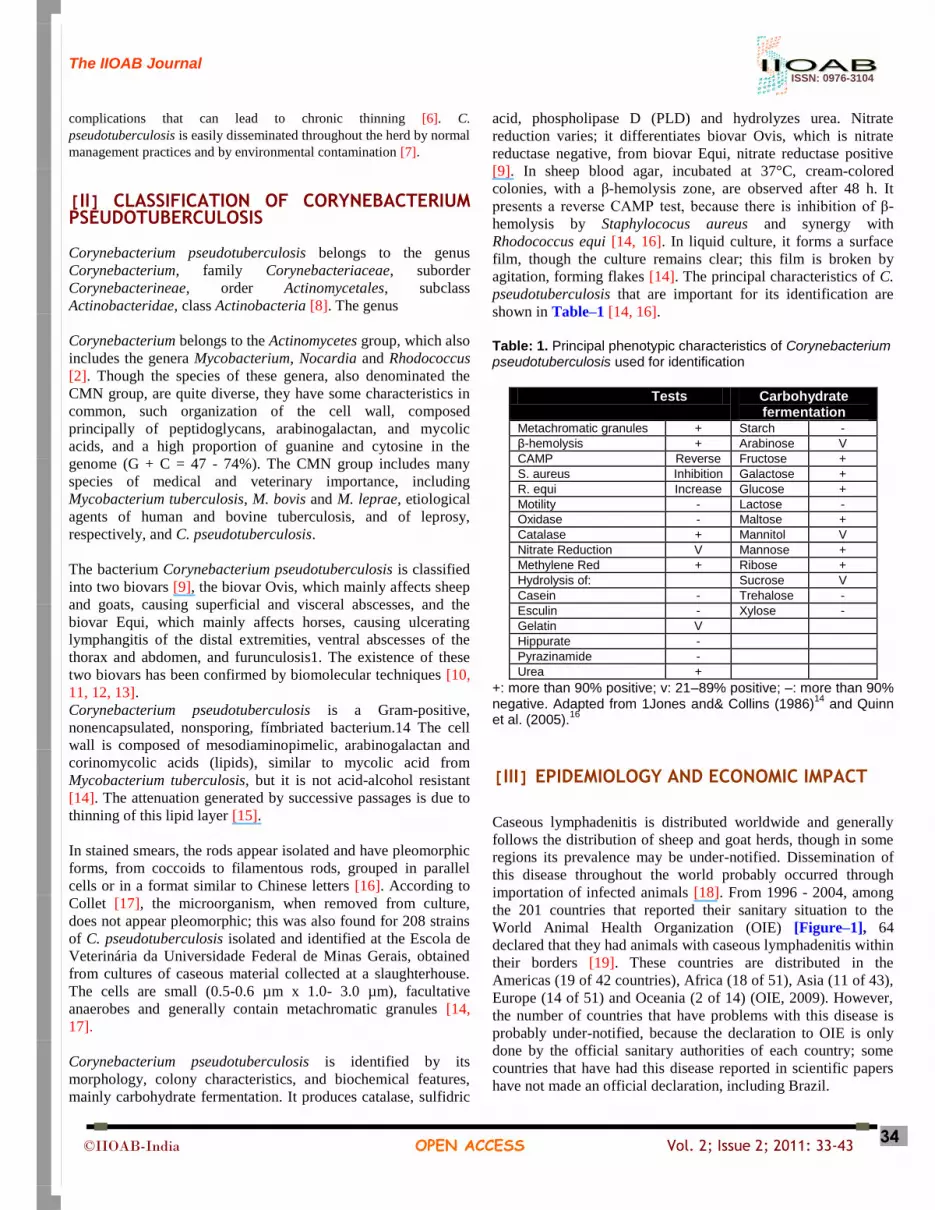
shown in Table–1 [14, 16].
Table: 1. Principal phenotypic characteristics of Corynebacterium pseudotuberculosis used for identification
Tests Carbohydrate fermentation
Metachromatic granules + Starch -
β-hemolysis + Arabinose V
CAMP Reverse Fructose +
S. aureus Inhibition Galactose +
R. equi Increase Glucose +
Motility - Lactose -
Oxidase - Maltose +
Catalase + Mannitol V
Nitrate Reduction V Mannose +
Methylene Red + Ribose +
Hydrolysis of: Sucrose V
Casein - Trehalose -
Esculin - Xylose -
Gelatin V
Hippurate -
Pyrazinamide -
Urea +
+: more than 90% positive; v: 21–89% positive; –: more than 90% negative. Adapted from 1Jones and& Collins (1986)
14 and Quinn
et al. (2005).16
[III] EPIDEMIOLOGY AND ECONOMIC IMPACT
Caseous lymphadenitis is distributed worldwide and generally
follows the distribution of sheep and goat herds, though in some
regions its prevalence may be under-notified. Dissemination of
this disease throughout the world probably occurred through
importation of infected animals [18]. From 1996 - 2004, among
the 201 countries that reported their sanitary situation to the
World Animal Health Organization (OIE) [Figure–1], 64
declared that they had animals with caseous lymphadenitis within
their borders [19]. These countries are distributed in the
Americas (19 of 42 countries), Africa (18 of 51), Asia (11 of 43),
Europe (14 of 51) and Oceania (2 of 14) (OIE, 2009). However,
the number of countries that have problems with this disease is
probably under-notified, because the declaration to OIE is only
done by the official sanitary authorities of each country; some
countries that have had this disease reported in scientific papers
have not made an official declaration, including Brazil.

The IIOAB Journal ISSN: 0976-3104
©IIOAB-India OPEN ACCESS Vol. 2; Issue 2; 2011: 33-43 35
Prevalences of caseous lymphadenitis as high as 61% were found
in Australia [20]; however, more recent studies indicate a
prevalence of 20 - 30%, after vaccination began 5. In the USA,
prevalences of up to 43% have been estimated [21], similar to the
range of 21 - 36% found among sheep in Quebec province in
Canada [4]. In Alberta, also in Canada, vaccination was effective
in the reduction of the prevalence of infection.3 In the United
Kingdom, 45% of the producers that were interviewed reported
abscesses in their sheep [22].
Fig: 1. Map with countries that reported their sanitary situation as a caseous lymphadenitis to the World Animal Health Organization (OIE), from 1996 - 2004
Fig: 2. Condemnation of sheep carcass at slaughterhouse inspection. (a) Pre-scapular lymph node. (b) Superficial lymph node.
Arrows indicate pre-scapular lymph nodes with caseous material, characteristic of caseous lymphadenitis in federally inspected slaughterhouse

The IIOAB Journal ISSN: 0976-3104
©IIOAB-India OPEN ACCESS Vol. 2; Issue 2; 2011: 33-43 36
In Brazil, the first report of caseous lymphadenitis was made by
Duport in 1918 [23]. Epidemiological studies have estimated that
most Brazilian herds are infected and that clinical prevalence
exceeds 30%. In goats, Pinheiro [24] reported 66.9% of animals
to have clinical signs of caseous lymphadenitis in Ceará. In Rio
de Janeiro, prevalence was reported to vary from 3.6-100% [25]
and in a seroepidemiological ELISA study made by our group, in
the State of Minas Gerais, we found prevalence figures of 75.8%
for sheep [26] and 78.9% for goats [27]. In an ELISA analysis
for C. pseudotuberculosis in 805 serum samples from sheep from
a federally-inspected slaughterhouse in Minas Gerais; we found
377 positive animals, and a high frequency of alterations in the
lymph nodes and internal organs [Figure–2]. This confirms the
great economical importance of C. pseudotuberculosis infection
for the sheep industry due to the high rate of carcass
condemnation. Various molecular techniques have been used to
type C. pseudotuberculosis, including RFLP of chromosomal
DNA [11], RFLP of ribosomal 16S DNA [10, 11], ribotyping
[10, 11], PFGE (pulsed-field gel electrophoresis) [12, 13] and
RAPD (randomly amplified DNA polymorphisms) [28, 29].
Though these various techniques have been useful for separating
the biovars Ovis and Equi, the species C. pseudotuberculosis has
been found to be genetically very homogeneous. The two
techniques that have given promising results for typing C.
pseudotuberculosis strains are PFGE and RAPD.
Pulsed-field electrophoresis was used to characterize 50 strains
of C. pseudotuberculosis isolated from goats, sheep and horses in
the United Kingdom [12]; six “pulsetypes” were observed, which
allowed the researchers to determine the origin of an outbreak of
caseous lymphadenitis. However, in a study of 36 sheep samples
and six goat samples from Australia, Canada, Eire, Holland and
Northern Ireland, the same research team reported four different
“pulsetypes”, with the conclusion that these C.
pseudotuberculosis strains, both those from sheep and goats,
were quite homogeneous [13]. RAPDs were useful in a study of
54 strains of C. pseudotuberculosis isolated from horses in four
different states of the USA, identifying 10 different genotypes
[28]. Also, RAPDs made with other initiators made it possible to
define eight genotypes among 61 strains of C.
pseudotuberculosis isolated from goats in Poland, with a
diversity index of 0.539 [29].
The importance of caseous lymphadenitis in Brazil can be
estimated by the increase in the participation of goats and sheep
in national animal husbandry and its relationship with the
economic impact of this disease. Brazil has 16.628.571 sheep and
9.355.220 goats, totaling 25,983,791 animals [30]. The economic
losses include decreased milk production, decreased weight gain,
reduced value of skins due to scarring, and the cost of the drugs
and labor needed to treat superficial abscesses. Losses are
increased when the affected lymph nodes are in critical areas
(jaw, crural region, udder) negatively affecting chewing,
locomotion and milk and meat production; however, economic
losses due to this disease have not yet been computed. In
industry, losses are due to the lower percent utility of carcasses
from affected animals, damage to skins, along with the need for
detailed inspection of carcasses. In the Brazilian Northeast,
where goat and sheep husbandry are important sources of food
and income, the situation is even more critical because of the
type of vegetation (spiny) and the low level of schooling of the
farmers [31, 24]. It is also becoming more of a problem in the
Southeastern, Northern and Midwestern regions, in which this
activity is increasing rapidly, negatively affecting the meat-
processing industries [26, 32].
[IV] SOURCES OF INFECTION AND FORM OF
TRANSMISSION The main source of infection is infected animals, with or without
clinical symptoms; these animals contaminate the soil, water,
feed, pastures and facilities with nasal secretions, feces and pus
from abscesses that drain spontaneously [Figure–3]. Infected
animals that do not present clinical symptoms can eliminate the
bacteria through their respiratory tract. Evaluation of the
coefficients of transmission of C. pseudotuberculosis by
respiratory tract infection and by pus from spontaneously-
draining abscesses, using a mathematical model of transmission,
showed that pulmonary abscesses have a small coefficient of
transmission, but they are more important for maintaining the
infection in the herd (endemic phase) [33].
Transmission can occur through direct or indirect contact or
through wounds that come into contact with pus from the
abscesses of sick animals [34]. Materials that are used in the
management of the animals, such as during castration,
identification with ear tags or by tattooing, contact with an
uncauterized umbilical stump, and drainage of abscesses, can
transmit the agent [Figure–3]. Vectors such as insects (especially
flies) should be considered in the transmission of the disease,
since C. pseudotuberculosis has been isolated from the bodies of
domestic flies (mechanical vector) and from fly intestines and
feces (biological vector). This bacterium has also been isolated
from flies contaminated with milk from cows with mastitis in
Israel [35, 36, 37]. In horses, flies have considerable
epidemiological importance in the dissemination of C.
pseudotuberculosis, because the higher frequency of infection in
this species occurred during periods when there are large
populations of flies [11].
Corynebacterium pseudotuberculosis survives long periods in the
soil. Through experimental contaminations of soil and of sheep
and goat facilities, it was found that C. pseudotuberculosis can
survive up to eight months at various temperatures [7]. In
bedding straw, it can remain viable for three weeks, during two
months in hay, four months in shearing stalls and for more than
eight months in the soil. This bacterium has been isolated after
five months in places where there has been contamination with
pus [34] and the concentration of viable microorganisms in the
purulent material is estimated to be from 106 to 107 bacteria per
gram of pus; consequently, environmental contamination due to a
leaking abscess is very high and persistent [38].

The IIOAB Journal ISSN: 0976-3104
©IIOAB-India OPEN ACCESS Vol. 2; Issue 2; 2011: 33-43 37
Fig: 3. Environmental and animal contamination with abscess suppurated in sheep. (A) Two caeous abscesses, one
spontaneous opened and other closed. (B) Ocular and cutaneous infection of sheep with caseous material
The use of barbed-wire fences or troughs and posts with sharp,
cutting edges can cause lesions in the skin of the animals,
opening passage for the entry of bacteria [26]. On farms that rear
sheep for wool, the equipment and facilities used for shearing
can transmit C. pseudotuberculosis among animals. Immersion
baths immediately after shearing can disseminate the infectious
agent, because these solutions can harbor bacteria for up to 24 h
[39]. In the Brazilian Northeast, where non-wool sheep
predominate almost completely, shearing and tail removal are not
common and the sheep are rarely ear tagged [24]; however, the
bacteria can penetrate through the respiratory system,
transcutaneously or through skin wounds caused by the caatinga
vegetation of this region [31].
Goat and sheep meat producers tend to make few periodic
inspections of their herds because of the extensive type of rearing
system, in which they do not identify individual animals, arguing
that these animals are slaughtered within a short time interval.
Conversely, goat milk producers tend to identify animals
individually and are more likely to detect abscesses during daily
contact, favoring the control of caseous lymphadenitis in these
herds; this is proved by the fact that 103 (36.3%) of the 284 goat
farmer interviewed in Minas Gerais, Brazil, have reported this
disease in their herds, while only 13 (6.1%) of the 213 sheep
farmers state the same [40]. In this State most goat herds are for
milk production, while most sheep flocks are for meat production
[40].
Corynebacterium pseudotuberculosis is sensitive to common
disinfectants, such as hypochlorite, formalin and cresol; however,
the surfaces should be cleaned before disinfection, because
organic matter interferes with the action of these agents [41].
Iiodine is recommended for chemical disinfection of wounds in
order to reduce bacterial transmission after surgical draining of
the abscesses [42].
[V] PATHOGENICITY AND VIRULENCE
FACTORS
Corynebacterium pseudotuberculosis is a facultative intracellular bacterium, multiplying within macrophages and surviving the action of phagolysosomic enzymes, because of the external lipid layer of the cell wall [2, 18]. After penetrating into the host, which generally occurs through the oral, nasal and ocular mucosa, or through skin wounds, the agent disseminates freely or within macrophages, mainly through the afferent lymphatic system, to local lymph nodes and internal organs. This process depends on the ability of the agent to infect macrophages, resist phagolysosomes and kill cells, liberating new bacteria and causing necrosis [43]. Three minutes after intraperitoneal inoculation in mice, phagocytic vacuoles are observed; after an hour, 60-80% of the goat macrophages contain bacteria, and two hours after inoculation, acid phosphatase is present in the vesicles containing the bacteria [44]. A strong local reaction occurs four hours after challenge in sheep [45], and a few hours later macrophages are degenerated and polymorphonuclear cell infiltrates containing bacteria are seen [46, 44, 47]. A day after experimental cutaneous infection, microabscesses develop in draining lymph nodes, and pyogranulomas are formed three to 10 days post-infection [48, 49, 6]. The lipid cell layer of the bacteria is pyogenic, but not immunogenic. This same layer makes phagocytosis of the bacteria difficult, increasing its virulence (cytoxicity), and survival inside macrophages; abscesses form through the release of lysosomal enzymes1. Besides participating in pathogenicity,

The IIOAB Journal ISSN: 0976-3104
©IIOAB-India OPEN ACCESS Vol. 2; Issue 2; 2011: 33-43 38
mycolic acid appears to be important for the survival of this bacterium in the environment [50]. Phospholipase D (PLD) increases vascular permeability and bacterial survival in the host. It is important for the dissemination of the bacteria from the location of the primary infection (local lymph node) to other organs (lungs, regional lymph nodes, mesenteric limph nodes, etc.), because it lyses mammal cell membranes, rich in phospholipids, causing microhemorrhages and vascular lesions, withincreased vascular permeability [2].
[VI] IMMUNE RESPONSE
Immunity against C. pseudotuberculosis is complex and involves cellular and humoral immune responses [51]. Studies point to a greater cellular immune response, chiefly a Th1 response, because of the facultative intracellular nature of the microorganism, with production of gamma-interferon (IFN-γ) and other cytokines that are important for controlling infection [52, 53, 54]. The humoral immune response is observed to present, from 6 to 11 days post-infection, a low production of IFN-γ, which significantly increases thereafter [55]. Inflammatory cytokines, such as TNF-α and IL-6, are mainly produced at the site of inoculation, while T cell-associated cytokines, such as IFN-γ, are chiefly produced in drainage lymph nodes [47].
[VII] CLINICAL SIGNS
Caseous lymphadenitis in its superficial form is characterized by infection of external lymph nodes, such as the submandibular, parotid, pre-scapular, subiliac, popliteal and supramammary lymph nodes, while the visceral form is characterized by abscessing of internal organs, such as lungs, liver, kidneys, uterus, spleen and internal lymph nodes, such as the mediastinal and bronchial lymph nodes. These two forms can coexist; however, other less common sites can be involved, such as mammary gland, scrotum, the central nervous system and joints. Internal abscesses are normally associated with weight loss and weakness, known in sheep as thin-ewe syndrome. The mature abscesses easily leak through fistulas, releasing purulent whitish-green discharges into the environment or into the affected organ. Abscesses usually recur, months or years later, in the same animal, due to the failure to eliminate the infection [1]. In some cases, infections produce few characteristic clinical signs, and a post-mortem examination becomes necessary for diagnosis; this makes it difficult to obtain objective data about disease prevalence [38]. Differences in the place of the abscesses between sheep and goats have been reported, the visceral form being more frequent among sheep and the superficial form among goats [7]. External abscesses in the lymph nodes of the head and neck are more common in goats, while the subiliac and pre-scapular lymph nodes are more commonly affected in sheep [7, 42]. Differences in the appearance of abscess content have also been reported
between sheep and goats; in sheep the contents have a laminar form when cut, similar to the layers of an onion, caused by the formation of layers of fibrous tissue and thick caseous material, while abscesses in goats have a thin and pasty exudate [7]. However, onion-like abscesses were not always present in sheep. Sheep carcass inspection at a federally inspected slaughterhouse in Minas Gerais, Brazil, showed that most of the abscesses in sheep were located in the head and neck lymph nodes and their content was essentially pasty. Isolation of C. pseudotuberculosis from these materials confirms the infection status of the animals. It is possible that older abscesses become more consistent, with a tendency towards fibrosis and calcification, progressing to an onion-like appearance, independent of animal species. In horses, there have been reports of abortions and cases of mastitis associated with visceral abscesses.16 In Israel, this bacterium was isolated from subcutaneous abscesses in milking cows; which could occur in outbreaks and cases of mastitis, affecting the whole mammary gland, resulting in total loss of milk production [56].
[VIII] CLINICAL AND LABORATORY DIAGNOSIS
Abscesses in goats and sheep are very suggestive of caseous lymphadenitis, especially if animals of the same lot have similar clinical signs, however bacterial isolation is necessary to identify the causative agent, since other bacteria such as Arcanobacterium pyogenes, Staphylococcus aureus subsp. anaerobius, Actinobacillus licheniformis and Pasteurella multocida, can be found in abscesses [57]. In animals with respiratory problems, a thoracic X-ray can reveal masses in the pulmonary parenchyma and lymph nodes; which also must be confirmed by culture of tracheal washes [58]. The use of aspirating puncture with a fine needle in the diagnosis of C. pseudotuberculosis was evaluated [59]. It proved to be easily performed, to have a low cost and to cause little damage to the tissues when compared to histopathology. It allows presumptive cytological diagnosis of the infection, before the affected lymph nodes abscessed, aiding in early adoption of prophylactic measures for the rest of the flock. Gram and Giemsa staining can be used for cytological identification of the microorganism. Although Gram staining is not primarily indicate for staining tissues, the bluish color taken on by C. pseudotuberculosis, in contrast with the reddish color of the cellular and inflammatory material from the aspirated lymph nodes, helps in the identification of the infectious agent [6]. In order to make a definitive diagnosis of caseous lymphadenitis, the agent should be isolated from purulent material from abscessed lymph nodes samples from live animals. Besides aspirating puncture, the material can be obtained by excision after trichotomy and careful antiseptic cleaning of the skin [17, 42]. It can also be collected at necropsy or during slaughter, when internal abscesses, affecting the liver, lungs, intestine,

The IIOAB Journal ISSN: 0976-3104
©IIOAB-India OPEN ACCESS Vol. 2; Issue 2; 2011: 33-43 39
kidneys, internal lymph nodes and other tissues, become accessible [60]. In the laboratory, after isolation, the identification of C. pseudotuberculosis is done by its morphology, staining characteristics, profile and fermentation of various carbohydrates [14]. The main phenotypic characteristics of C. pseudotuberculosis used for identification are shown in Table–1. Various diagnostic techniques have been developed for caseous lymphadenitis in goats and sheep, such as serological neutralization for antitoxins, immunodiffusion in agar gel, indirect hemagglutination, complement fixation and hypersensitivity tests [25, 1, 18]. Immunoenzymatic tests (ELISA), using bacterial cells, toxins and secreted proteins of C. pseudotuberculosis, such as PLD [61, 62, 63, 64], have been reported to be effective in caseous lymphadenitis control and eradication programs. Indirect ELISA based on secreted proteins has shown a diagnostic sensitivity and specificity of 93.5% and 100%, respectively, in the diagnosisof caseous lymphadenitis in small ruminants [63]. Detection of INF-γ by ELISA, an indicator of cell-mediated immunity, has been used for diagnosis of infection by C. pseudotuberculosis, with a sensitivity of 91% and a specificity of 98%, demonstrating its potential for use in caseous lymphadenitis eradication programs [51, 65]. Molecular techniques have also been used for the diagnosis of caseous lymphadenitis. Polymerase chain reaction (PCR), used to identify C. pseudotuberculosis, is an alternative to conventional diagnostic methods, with the advantage of being faster and more specific [66]. Multiplex PCR based on amplification of the genes 16S rDNA, rpoB and pld, presented 94.6% diagnostic sensitivity, for C. pseudotuberculosis isolates as well as for clinical material [67]. It facilitates the diagnosis by differentiating C. pseudotuberculosis from other pathogens present in abscesses, chiefly C. ulcerans [67]. Recently, the genome of two C. pseudotuberculosis strains isolated from goats and sheep has been sequenced by a Minas Gerais Genome Network and Pará Genomic and Proteomic Network. The genomic data will help to identify new specific targets, useful in the diagnosis as well as in the development of drugs and vaccines and in the understanding of C. pseudotuberculosis pathogenicity mechanisms.
[IX] DIFFERENTIAL DIAGNOSIS
Pyogranulomatous lesions, such as found in actinobacillosis, tuberculosis and superficial abscesses caused by Staphylococcus aureus and Actinomyces pyogenes, must be differentiated from caseous lymphadenitis [17]. The superficial form of the disease should also be differentiated from submandibular edema caused by parasites, Fasciola hepatica and Haemonchus sp., salivary cysts, lymphosarcoma and subcutaneous inoculation of vaccines.
The debilitating visceral form can be clinically similar to chronic parasitism, thinning due to abnormal waste of teeth, alveolar periodontitis, malnutrition and chronic diseases, such as pulmonary adenomatosis, neoplasias and scrapie [17]. Pneumonias caused by Mycobacterium bovis, Pasteurella haemolytica, Pasteurella multocida or ovine progressive pneumonia, due to Maedi-Visna virus infection, can make the diagnosis of caseous lymphadenitis even more difficult [58]. In sheep, orchitis and epidydimitis caused by C. pseudotuberculosis needs to be differentiated from similar lesions caused by Brucella ovis, Actinobacillus seminis, Histophilus ovis and Pasteurella spp [17, 68].
[X] TREATMENT Treatment of affected animals consists of the drainage of abscesses, followed by cleansing and chemical cauterization, usually with 10% iodine, or even removal of the affected superficial lymph nodes [69]. Although it is an important control measure, this procedure might not be as effective as expected due to the presence of internal abscesses. Drainage of the abscess should be done in a way that avoids environmental contamination, with disinfection of the surgical material before and after the procedure, and all of the disposable materials should be incinerated and buried, including plastics and paper used to cover the area. Another treatment option is antibiotic therapy, which is not very efficient, even though C. pseudotuberculosis is sensitive in vitro to almost all antibiotics that have been tested. The intracellular location of the bacteria and the formation of biofilm in natural infections reduces drug efficacy, making antimicrobials inefficient under these conditions [7, 70]. The inefficacy and high cost of antibiotic treatment make it an inviable option for herd-level disease management.
[XI] CONTROL AND PROPHYLAXIS
An effective program for the control of caseous lymphadenitis should be based on clinical inspection and periodic serology of all animals in the flock, which includes recently-acquired animals and those that return to the herd, culling the ones that have clinical signs or that are serologically positive. Once infected, an animal hardly eliminates the C. pseudotuberculosis [71]. The main source of infection for a flock is introduction of infected or abscessed animals into a herd, which results in a high frequency of abscesses after two or three years. This stresses the importance of employing biosecurity procedures in all flocks, chiefly during the introduction of animals. Measures designed to reduce the environmental risk of wounding should also be adopted, such as the use of smooth wire fences, troughs and facilities without sharp edges, disinfection of surgical, ear tagging and shearing instruments, systematic use of

The IIOAB Journal ISSN: 0976-3104
©IIOAB-India OPEN ACCESS Vol. 2; Issue 2; 2011: 33-43 40
individual disposable needles, effective control of insects, and disinfection of newborns’ navels and any other wounds with 10% iodine. Although it is not recommended to be applied to swelled lymph nodes because of its irritating and caustic action on tissues (skin, mucosa and lungs), 10% formaldehydeshould be used for disinfection of herd facilities [7, 1, 18]. All control programs should be based on sanitary education of herd owners and technical personnel, otherwise success will be compromised. Information about losses throughout the production cycle, as well as concerning the zoonotic potential of C. pseudotuberculosis, should be supplied to the people who work with the herds directly or indirectly, reinforcing their importance in the success of the control program. Control measures vary with the prevalence of infection. In countries free of this disease, importation should only be permitted from herds that have been certified free of caseous lymphadenitis for three years, all animals should be tested by ELISA before importing and they should initially be placed in quarantine. In countries with low disease prevalence, the clinically affected animals should be separated and submitted to ELISA testing, lambs and kids should be reared away from their mothers, and installations and equipment should be well disinfected. In countries with a high incidence, rigorous sanitary measures should be implemented, associated with vaccination [7, 17]. Disease eradication can be achieved in endemically-infected herds by initially discarding all animals that have clinical signs and those that are positive in serological tests [6]; however, this is difficult to accomplish because of the rapid dissemination of the agent within the herd and the difficulty in identifying animals that have a subclinical form of the disease [60, 66].
[XII] VACCINATION
Given that caseous lymphadenitis treatment is ineffective and expensive, the best strategy for control and prevention of the disease is immunization, as it was observed in countries with high prevalence of infection [5]. The vaccines commercially available have different relevant features that should be considered on their use. Not all of the vaccines licensed for use in sheep have the same efficiency in goats, and normally it is necessary to adjust the vaccination program to the flock conditions. Also, the protection provided by vaccination is only partial, as external and internal abscess development can still occur [1]. The principal component of C. pseudotuberculosis used in the formulation of vaccines is PLD. The rationale for its use as a vaccination antigen is the good rates of protection obtained after immunization of goats and sheep with this toxin. Most of the commercial vaccines against C. pseudotuberculosis use inactivated PLD associated to antigens of other pathogens, such as Clostridium tetani, Clostridium perfringens type D, Clostridium novyi, Clostridium chauvoei and Clostridium
septicum, along with some vaccines that are associated with the endectocide moxidectin. Such a formulation is the basis of the Glanvac vaccine (Vetrepharm, Inc London), licensed for use in sheep and goats in Canada, Australia and New Zealand and the Biodectin vaccine (Fort Dodge Austrália PTY LTD), also licensed in Brazil for use in sheep. The Glanvac vaccine has been evaluated in various countries [72]. Vaccination of sheep and goats with Glanvac resulted in protection against experimentally-induced infection with C. pseudotuberculosis, evidenced by a decrease in the number of lesions [73]. Another commercial vaccine that has been evaluated, Caseous D-T (PBS Animal Health, USA), has two formulations, one that only contains toxoids (clostridial and from C. pseudotuberculosis) and another that is a combination of clostridial toxoids and the bacterium C. pseudotuberculosis. Preliminary results indicate that this second formulation confers better protection against experimental infection than the first, reducing the number of internal and external lesions [74]. The use of PLD toxoid for the immunization of goats can have some negative consequences, including reduced milk production, fever, ventral edema, ataxia and convulsions; therefore, recommendations for its use in this species should be made with restrictions [1]. The partial protection provided by immunization of goats and sheep with commercial vaccines is associated with the type of immune response elicited. Protection against C. pseudotubeculosis is mainly dependent on immune response that involves INF-γ production and cytotoxic T-cells. A humoral response alone is insufficient to protect the animal, and a good cellular response is not achieved with inactivated vaccines [75]. Hence, various attempts have been made to obtain an attenuated vaccine that is effective against caseous lymphadenitis [76, 52]. Attenuation can happen naturally or through manipulations using temperature, chemical and genetic (recombinant) agents. With this type of vaccine strategy, the microorganism maintains its capacity to replicate, mimicking natural infection and producing humoral and cellular responses. Also, this is the type of vaccine that confers the best and longest-lasting immune response, due to its similarity to natural infection [75]. Techniques such as deletion of multiple genes involved in virulence, and insertion of fragments that interrupt these genes in the pathogen, practically eliminate the risk that the pathogen can revert to its virulent form [77]. Live vaccines that have been attenuated in the laboratory (recombinants) usually have the PLD gene as a target for attenuation, because of its importance as a virulence factor [78]. In Brazil, the Bahia State Agency for Agricultural Development (EBDA) developed a vaccine based on strain 1002, a naturally attenuated strain, which is currently commercially available. It stimulates significant protection levels, 83%, in vaccine trials; however, immunization still presents collateral effects, such as local reactions, and field trials have not been as successful as the initial vaccine tests, presenting highly variable protection levels [79]. Another attenuated live vaccine, LinfoVac (Laboratórios Vencofarma do Brasil Ltda), licensed for use in sheep and goats, is also currently available in Brazil. The results obtained in the

The IIOAB Journal ISSN: 0976-3104
©IIOAB-India OPEN ACCESS Vol. 2; Issue 2; 2011: 33-43 41
field with these attenuated vaccines demonstrate the need to develop a more effective and safe vaccine [75].
[XIII] CONCLUSIONS
Caseous lymphadenitis continues to be an important challenge for sheep and goat industries, limiting their profitability. The intense market and movement of small ruminants, without the necessary biosecurity measures, are important obstacles to the control of caseous lymphadenitis, maintaining its prevalence at high levels, which indicates that specific control measures must be adopted. There are various difficulties affecting the development and application of effective diagnosis and more effective immunogens need to be made available as vaccines. Thus, great efforts need to be made by all players in sheep and goat industries to control this awful disease. CONFLICT OF INTEREST The authors declare that they have no conflict of interest.
ACKNOWLEDGEMENT We are indebeted with JPS Mol for helping with the figures. ASG, APL and VA have scholarships from the Conselho Nacional de Desenvolvimento Científico e Tecnológico – CNPq. This work was supported by the Fundação de Amparo à Pesquisa do Estado de Minas Gerais – FAPEMIG (CVZ APQ 3283-5.04/07) and Conselho Nacional de Desenvolvimento Técnico e Científico – CNPq. The study sponsors had no involvement in the review design; in the collection, analysis and interpretation of data; in the writing of the manuscript; and in the decision to submit the manuscript for publication..
REFERENCES
[1] Williamson LH. [2001] Caseous lymphadenitis in small
ruminants. Vet Clin North Am 17: 359–371.
[2] Dorella FA, Pacheco LGC, Oliveira, SC, et al. [2006]
Corynebacterium pseudotuberculosis: microbiology,
biochemical properties, pathogenesis and molecular studies
of virulence. Vet Rec 37: 201–218.
[3] Stanford K, Brogden KA, McClelland LA et al. [1997] The
incidence of caseous lymphadenitis in Alberta sheep and
assessment of impact by vaccination with commercial and
experimental vaccines. Can J Vet Res 62: 38–43.
[4] Arsenault JO, Girard C, Dubreuil P et al. [2003] Prevalence
of and carcass condemnation from maedi-visna,
paratuberculosis and caseous lymphadenitis in culled sheep
from Quebec, Canada. Prev Vet Med 59: 67–81.
[5] Paton MW, Walker SB, Rose IR et al. Paton MW, Walker
SB, Rose IR et al. [2003] Prevalence of caseous
lymphadenitis and usage of caseous lymphadenitis vaccines
in sheep flocks. Aust Vet J 81: 91–95.
[6] Radostits OM, Gay, CC, Blood DC et al. [2002] Clínica
Veterinária - Um Tratado de Doenças dos Bovinos, Ovinos,
Suínos, Caprinos. 9th edn. Guanabara Koogan, Rio de
Janeiro.
[7] Brown CC, Olander HJ. [1987] Caseous lymphadenitis of
goats and sheep: a review. Vet Bull 57: 1–11.
[8] Stackebrandt E, Rainey FA, Ward-Rainey NL. [1997]
Proposal for a new hierarchic classification system,
Actinobacteria classis nov. Int J Syst Bacteriol 47: 479 –
491.
[9] Biberstein EL, Knight HD, Jang S. [1971] Two biotypes of
Corynebacterium pseudotuberculosis. Vet Rec 89: 691–692.
[10] Sutherland SS, Hart RA, Buller NB. [1996] Genetic
differences between nitrate-negative and nitrate-positive
Corynebacterium pseudotuberculosis strains using
restriction fragment length polymorphisms. Vet Microbiol
49: 1–9.
[11] Costa LRR, Spier SJ, Hirsh DC. [1998] Comparative
molecular characterization of Corynebacterium
pseudotuberculosis of different origin. Vet Microbiol 62:
135–143.
[12] Connor KM., Quire M, Baird G et al. [2000]
Characterization of United Kingdom isolates of
Corynebacterium pseudotuberculosis using pulsed-field gel
electrophoresis. J Clin Microbiol 38: 2633–2637.
[13] Connor KM, Fontaine MC, Rudge K et al. [2007] Molecular
genotyping of multinational ovine and caprine
Corynebacterium pseudotuberculosis isolates using pulsed-
field gel electrophoresis. Vet Res 38: 613–623.
[14] Jones D, Collins MD. [1986] Irregular, nonsporing Gram-
positive rods. In: Sneath, P.H.A. et al. Bergey’s Manual of
Systematic Bacteriology. 2nd edn Baltimore: Williams and
Wilkins, p. 1261–1282.
[15] Hard GC. [1975] Comparative toxic effect on the surface
lipid of Corynebacterium ovis on peritoneal macrophages.
Infec Immun 12: 4139–1449.
[16] Quinn PJ, Markey BK, Carter ME et al. [2005]
Microbiologia Veterinária e Doenças Infecciosas. 1st edn.
Artmed, Porto Alegre.
[17] Collett MG, Bath GF, Cameron,CM. [1994]
Corynebacterium pseudotuberculosis infections. In: Coetzer,
J.; Thomson, G.R. Infectious Diseases of Livestock with
Special Reference to Southern Africa. 2nd edn. Capetown:
Oxford University Press, p. 1387–1395.
[18] Baird GJ, Fontaine MC. [2007] Corynebacterium
pseudotuberculosis and its role in ovine caseous
lymphadenitis. J Comp Pathol 137: 179–210.
[19] OIE – World Organization for Animal Health. [2009].
http://www.oie.int/hs2/sit_mald_cont.asp?c_mald=156andc_
cont=6andannee=2004. Accessed 26 sept.
[20] Middleton MJ, Epstein VM, Gregory GG. [1991] Caseous
lymphadenitis on Flanders Island: prevalence and
management surveys. AustVet J 68: 311-312.
[21] Stoops SG, Renshaw HW, Thilsted JP. [1984] Ovine
caseous lymphadenitis: disease prevalence, lesion
distribution, and thoracic manifestations in a population of
mature culled sheep from western United States. Am J Vet
Res 45: 557–561.
[22] Binns SH, Bairley M, Green LE. [2002] Postal survey of
ovine caseous lymphadenitis in the United Kingdom
between 1990 and 1999. Vet Rec 150: 263–268.
[23] Garcia M, Araújo WP, Carvalho VM et al. [1987]
Isolamento e identificação do Corynebacterium
pseudotuberculosis em ovinos e caprinos nos Estados de São
Paulo e Minas Gerais. Fac Med Vet Zootec Univ de São
Paulo 24: 23–25.
The IIOAB Journal ISSN: 0976-3104
©IIOAB-India OPEN ACCESS Vol. 2; Issue 2; 2011: 33-43 42
[24] Pinheiro RR, Gouveia AMG, Alves FSF et al. [2000]
Aspectos epidemiológicos da caprinocultura cearense. Arq
Bras MedVet Zootec 52: 534–543.
[25] Langenegger CH, Langenegger J, Scherer PO. [1991]
Prevalência e diagnóstico comparativo da linfadenite
caseosa em caprinos do Estado do Rio de Janeiro. Pesq Vet
Brasil 11: 31–34.
[26] Guimarães AS, Seyffert N, Bastos BL et al. [2009] Caseous
lymphadenitis in sheep flocks of the state of Minas Gerais,
Brazil: prevalence and management surveys. Small Rumin
Res 87: 86–91
[27] Guimarães AS, Pacheco LGC et al. [2010] High
seroprevalence of caseous lymphadenitis in Brazilian goat
herds revealed by Corynebacterium pseudotuberculosis
secreted proteins-based ELISA. Res Vet Sc 88: 50–55.
[28] Foley JE, Spier SJ, Mihalyi J et al. [2004] Molecular
epidemiologic features of Corynebacterium
pseudotuberculosis isolated from horses. Am J Vet Res 65:
1734–1737.
[29] Stefańska I, Rzewuska M, Binek M. [2008] Evaluation of
three methods for DNA fingerprinting of Corynebacterium
pseudotuberculosis strains isolated from goats in Poland.
Polish J Microbiol 57: 105–112.
[30] Instituto Brasileiro de Geografia e Estatística. Censo
agropecuário 2008. http: www.ibge.gov.br. Accessed 10 jul
2009.
[31] Unanian MM, Feliciano Silva AED, Pant KP et al. [1985]
Abscesses and caseous lymphadenitis in goats in tropical
semi-arid north-east Brazil. Trop An Health Prod 17: 57–62.
[32] Guimarães AS, Gouveia AMG, Abreu AB et al. [2009]
Características zoossanitárias das caprinoculturas de leite e
corte em Minas Gerais. Rev Vet e Zootec. em Minas 101:
23–29.
[33] O’Reilly KM., Green LE., Malone FE et al. [2008]
Parameter estimation and simulations of a mathematical
model of Corynebacterium pseudotuberculosis transmission
in sheep. Prev Vet Med 83: 242–259.
[34] Nairn ME., Robertson JP. [1974] Corynebacterium
pseudotuberculosis infection of sheep: role of skin lesions
and dipping fluids. Aust Vet J 50: 537-–542.
[35] Yeruham I, Braverman Y, Shpigel NY et al. [1996] Mastitis
in dairy cattle caused by Corynebacterium
pseudotuberculosis and the feasibility of transmission by
houseflies. Vet Q 18: 87–89.
[36] Braverman Y, Chizov-Ginzburg A, Saran A et al. [1999]
The role of houseflies (Musca domestica) in harbouring
Corynebacterium pseudotuberculosis in dairy herds in
Israel. Rev. Sci. Tech. O I E (Off Int. Epizoot) 18: 681–690.
[37] Spier SJ, Leutenegger CM, Carroll SP et al. [2004] Use of a
real-time polymerase chain reaction-based fluorogenic 5'
nuclease assay to evaluate insect vectors of
Corynebacterium pseudotuberculosis infections in horses.
Am J Vet Res 65: 829–834.
[38] Brown CC, Olander HJ, Alves SF. [1987] Synergistic
hemolysis-inhibition titers associated with caseous
lymphadenitis in a slaughterhouse survey of goats and sheep
in Northeastern Brazil. Can J Vet Res 51: 46–49.
[39] Rizvi S, Green LE, Glover MJ. [1997] Caseous
lymphadenitis: An increasing cause for concern. Vet Rec
140: 586–587.
[40] Guimarães AS. [2006] Caracterização da caprinovinocultura
em Minas Gerais. (Masters thesis of the Escola de
Veterinária da Universidade Federal de Minas Gerais).
[41] Ismail AA, Hamid YMA. [1972] Studies on the effect of
some chemical disinfectants used in veterinary practice in
Corynebacterium ovis. J Egyptian Vet Med Assoc 32: 195–
202.
[42] Smith MC, Sherman D. [1994] Caseous Lymphadenitis.
Goat Medicine.1st edn. Lea and Febier, Iowa.
[43] Batey RG. [1986] Pathogenesis of caseous lymphadenitis in
sheep and goats. Aust Vet J 63: 269–272.
[44] Tashjian JJ, Campbell SG. [1983] Interaction between
caprine macrophages and Corynebacterium
pseudotuberculosis: an electron microscopy study. Am J Vet
Res 44: 690–693.
[45] Pepin M, Pardon P, Marly J et al. [1988] Corynebacterium
pseudotuberculosis infection in adult ewes by inoculation in
the external ear. Am J Vet Res 49: 459–463.
[46] Hard GC. [1969] Electron microscopic study of the
differentiation of mouse peritoneal macrophages stimulated
by Corynebacterium ovis infection. Lab Invest 21: 309–315.
[47] Guilloteau L, Pepin M, Pardon P et al. [1990] Recruitment
of 99m-technetium- or 111-indium-labelled
polymorphonuclear leucocytes in experimentally induced
pyogranulomas in lambs. J Leukocyte Biol 48: 343–352.
[48] Ellis JA, Hawk DA, Holler LD et al. [1990] Diferential
antibody responses to Corynebacterium pseudotuberculosis
in sheep with naturally acquired caseous lymphadenitis. J
AmVet Med Assoc 196: 1609–1613.
[49] Pepin M, Seow HF, Corner L et al. [1997] Cytokine gene
expression in sheep following experimental infection with
various strains of Corynebacterium pseudotuberculosis
differing in virulence. Vet Res 28: 149–163.
[50] West DM, Bruere AN, Ridler AL. [2002] Caseous
lymphadenitis. In: The Sheep: Health, Disease and
Production. Foundation for Veterinary Continuing
Education. Massey University, New Zealand.
[51] Prescott JF, Menzies PI, Hwang YT. [2002] An interferon-
gamma assay for diagnosis of Corynebacterium
pseudotuberculosis infection in adult sheep from a research
flock. Vet Microbiol 88: 287–297.
[52] Simmons CP, Dunstan SJ, Tachedjian M et al. [1998]
Vaccine potential of attenuated mutants of Corynebacterium
pseudotuberculosis. Infect Immun 66: 474–479.
[53] Lan DTB, Makino S, Shirahata T et al. [1999] Tumor
necrosis factor and γ interferon are required for the
development of protective immunity to secondary
Corynebacterium pseudotuberculosis infection in mice. J
Vet Med Sci 61: 1203–1208.
[54] El-Enbaawy MI, Saad MM, Selim SA. [2005] Humoral and
cellular immune responses of a murine model against
Corynebacterium pseudotuberculosis antigens. Egyptian J
Immun 12: 13–20.
[55] Paule BJA, Azevedo V, Regis LF, et al. [2003]
Experimental Corynebacterium pseudotuberculosis primary
infection in goats: kinetics of IgG and interferon-γ
production, IgG avidity and antigen recognition by Western
blotting. Vet Immunol Immunopathol 96: 129–139.
[56] Yeruham D, Elad S, Friedman SP. [2003] Corynebacterium
pseudotuberculosis infection in Israeli dairy cattle.
Epidemiol Infect 131: 947–955.
[57] Pekelder JJ. [2003] Caseous lymphadenitis. In: Martin,
W.B.; Aitken, I.D. Diseases of Sheep. 3th edn Blackwell
Science, Oxford.
[58] Pugh DG. [2004] Clínica de ovinos e caprinos. 1rt edn Roca,
São Paulo.

The IIOAB Journal ISSN: 0976-3104
©IIOAB-India OPEN ACCESS Vol. 2; Issue 2; 2011: 33-43 43
[59] Ribeiro MG, Dias Junior JG, Paes AC et al. [2001] Punção
aspirativa com agulha fina no diagnóstico de
Corynebacterium pseudotuberculosis na linfadenite caseosa
caprina. Arq Inst Biol São Paulo 68: 23–28.
[60] Riet-Correa F, Schild AL, Méndez MC et al. [2001]
Doenças de ruminantes e eqüinos. 1st edn. Varela, São
Paulo.
[61] Ter Laak EA, Bosch J, Bijl GC et al. [1992] Double-
antibody sandwich enzyme-linked immunosorbent assay and
immunoblot analysis used for control of caseous
lymphadenitis in goats and sheep. Am J Vet Res 53: 1125–
1132.
[62] Dercksen DP, Brinkhof JMA, Dekker-Nooren T et al.
[2000] A comparison of four serological tests for the
diagnosis of caseous lymphadenitis in sheep and goats. Vet
Microbiol 75: 167–175.
[63] Carminati R, Bahia R, Costa LFM et al. [2003]
Determinação da sensibilidade e da especificidade de um
teste de ELISA indireto para o diagnóstico de linfadenite
caseosa em caprinos. R Cienc Méd Biol 2: 88–93.
[64] Binns SH, Green LE, Bailey M. [2007] Development and
validation of an ELISA to detect antibodies to
Corynebacterium pseudotuberculosis in ovine sera. Vet
Microbiol 20: 169–179.
[65] Sunil V, Menzies PI, Shewen PE, Prescott JF. [2008]
Performance of a whole blood interferon-gamma assay for
detection and eradication of caseous lymphadenitis in sheep.
Vet Microbiol 30: 288–297.
[66] Çetinkaya B, Karahan M, Atil E et al. [2002] Identification
of Corynebacterium pseudotuberculosis isolates from sheep
and goats by PCR. Vet Microbiol 88: 75–83.
[67] Pacheco LGG, Pena RR, Castro TLP et al. [2007] Multiplex
PCR assay for identification of Corynebacterium
pseudotuberculosis from pure cultures and for rapid
detection of this pathogen in clinical samples. J Med
Microbiol 56: 1–7.
[68] Saunders VF, Redacliff LA, Berg T et al. [2007] Multiplex
PCR for the detection of Brucella ovis, Actinobacillus
seminis and Histophilus somni in ram semen. Aust Vet J 85:
72–77.
[69] Nozaki CN, Faria MAR, Machado TMM. [2000] Extirpação
cirúrgica dos abscessos da linfadenite caseosa em caprinos.
Arq Inst Biol 67: 187–189.
[70] Olson ME, Ceri H, Morck DW et al. [2002] Biofilm
bacteria: formation and comparative susceptibility to
antibiotics. Can J Vet Res 66: 86–92.
[71] Campbell SG., Ashfaq MK, Tashjian JJ. [1982] Caseous
lymphadenitis in goats in the USA. In: Proceedings 3rd
International Conference on Goat Production and Disease.
Tucson. Arizona 449–454.
[72] Eggleton DG, Middleton HD, Doidge CV et al. [1991]
Immunization against ovine caseous lymphadenitis:
comparison of Corynebacterium pseudotuberculosis
vaccines with and without bacterial cells. Aust Vet J 68:
317–319.
[73] Fontaine MC, Baird G, Connor KM et al. [2006]
Vaccination confers significant protection of sheep against
infection with a virulent United Kingdom strain of
Corynebacterium pseudotuberculosis. Vaccine 24: 5986–
5996.
[74] Piontkowski MD, Shivvers DW. [1998] Evaluation of a
commercially available vaccine against Corynebacterium
pseudotuberculosis for use in sheep. J Am Vet Med Assoc
212: 1765–1768.
[75] Dorella FA, Pacheco LGC, Seyffert N et al. [2009] Antigens
of Corynebacterium pseudotuberculosis and prospects for
vaccine development. Exp Rev of Vaccines 8: 205–213.
[76] Hodgson ALM, Tachedjian M, Corner LA et al. [1994]
Protection of sheep against caseous lymphadenitis by use of
a single oral dose of live recombinant Corynebacterium
pseudotuberculosis. Infec Immun 62: 5275–5280
[77] Jiskoot W, Kersten GFA., Beuvery EC. [2002] Vaccine. In:
Crommelin DJA, Sindelar RD. Pharmaceutical
Biotechnology – An introduction for pharmacists and
pharmaceutical scientists. 2nd edn. London: Taylor and
Francis Group. P.259–282.
[78] Carne HR, Onon EO. [1978] Action of Corynebacterium
ovis exotoxin on the endothelial cells of blood vessels.
Nature 271: 246–248.
[79] Hage JA. [2000] Vacina da EBDA é novidade mundial.
EMBRAPA - Pesquisa Estadual em Foco 05/08: 9


