Embed Size (px)
Citation preview

Spine www.spinejournal.com 891
BIOMECHANICS
SPINE Volume 40 , Number 12 , pp 891 - 901 ©2015, Wolters Kluwer Health, Inc. All rights reserved.
DOI: 10.1097/BRS.0000000000000888
Study Design. Microstructural investigation of compression-induced herniation of the fl exed lumbar disc. Objective. To provide a microstructural analysis of the mechanisms of annular wall failure in healthy discs subjected to fl exion and a rate of compression comparable with the maximum rate at which the muscles of the spinal column can generate a force. Summary of Background Data. Clinical evidence indicates the involvement of the endplate in herniation. It is known that both an elevated rate of compression and a fl exed posture are necessary to cause disc failure either within the midspan of the annulus or at the annular-endplate interface. However, the question of what effect a sudden or “surprise” loading might have on the mode of failure is, as yet, unanswered. Methods. Twenty-four healthy mature ovine lumbar motion segments were compressed to failure in high physiological fl exion (10º). This occurred over approximately 5 mm of crosshead displacement in 0.75 seconds that resulted in a displacement rate of 400 mm/min (defi ned as a “surprise” rate) and was intended to simulate the maximum rate at which the muscles of the spinal column can generate a force. The damaged discs were then analyzed microstructurally. Results. Fifty-eight percent of discs suffered annular-endplate junction rupture, 25% suffered midspan annular rupture, and the
From the * Experimental Tissue Mechanics Laboratory, Department of Chemical and Materials Engineering, University of Auckland, Auckland, New Zealand; and † Department of Orthopaedic Surgery, Auckland City Hospital, Auckland, New Zealand.
Acknowledgment date: October 14, 2014. Revision date: January 21, 2015. Acceptance date: March 6, 2015.
The manuscript submitted does not contain information about medical device(s)/drug(s).
AO Foundation (Project S-12-24B) grant funds were received to support this work.
Relevant fi nancial activities outside the submitted work: consultancy, patents, employment
Address correspondence and reprint requests to Neil D. Broom, PhD, Department of Chemical and Materials Engineering, University of Auckland, Private Bag 92019, Auckland 1142, New Zealand; E-mail: [email protected]
Lumbar disc herniation is the most common cause of sciatica, and disruption of the outer disc annulus, even without actual herniation, may stimulate nerve endings
in this region leading to low back pain. 1–3 The majority of biomechanical studies attempting to reproduce disc hernia-tions have compressed motion segments in a fi xed posture until failure occurs. Unfl exed segments tend to fail via end-plate fracture or vertebral body compression, 4–9 whereas fl ex-ion increases the likelihood of annular failure, 10–13 especially when combined with an elevated rate of loading. 14 Micro-structural analysis has shown that this annular failure tends to initiate in the mid-to-outer region of the disc wall, and this may be a direct consequence of the profi le of the endplates. The endplate profi le displays a raised apical ridge that occurs approximately midway through the annulus, and it has been proposed that this ridge places the fi bers originating from its apex at the greatest strain when the disc is fl exed. 14 Failure can occur either within the midspan of the annulus or at the annular-endplate interface, with a majority of in vitro her-niations occurring via the former mode of failure. 14 In con-trast, recent clinical evidence indicates that the majority of
balance of 17% endplate fracture. Microstructural analysis indicated that annular rupture initiated at the endplate apical ridge in the mid-to-outer region of the annulus in both annular-endplate and midspan annulus rupture. Conclusion. Motion segments subjected to a “surprise” loading rate are likely to fail via some form of annular rupture. Failure under such sudden loading occurs mostly via rupture of the annular-endplate junction and is thought to arise from a rate-induced mechanostructural imbalance between the annulus and the endplate. Key words: ovine lumbar motion segments , high rate of compres-sion , sudden loading , “surprise” loading , fl exion , microstructural analysis , annular disruption , annular-endplate rupture , mechanism of herniation . Level of Evidence: N/A Spine 2015;40:891–901
“Surprise” Loading in Flexion Increases the Risk of Disc Herniation Due to Annulus-Endplate Junction Failure
A Mechanical and Microstructural Investigation
Kelly R. Wade , PhD * Peter A. Robertson , MD † Ashvin Thambyah , PhD * and Neil D. Broom , PhD *
Copyright © 2015 Wolters Kluwer Health, Inc. Unauthorized reproduction of this article is prohibited.
SPINEIP1411_LR 891SPINEIP1411_LR 891 21/05/15 10:43 AM21/05/15 10:43 AM

BIOMECHANICS “Surprise” Loading Causes Annular-Endplate Rupture • Wade et al
892 www.spinejournal.com June 2015
herniations, about 65%, involve the endplate. 15 It is possible that this inconsistency may arise from a difference between in vitro and in vivo loading rates.
Previous studies have shown that rapid loading of motion segments held in the neutral posture is likely to result in dam-age to the vertebral endplate. 16 It has also been shown that rapid internal pressurization achieved by injecting a gel into the nucleus axially via an adjacent vertebra increases the like-lihood of annular-endplate failure in fl exed samples. 17 How-ever, this technique did not account for the changes in defor-mation in the disc associated with compressive loading and fl exion as would occur in vivo .
To date, the effects of clinically relevant loading rates that would occur in real life, such as a sudden lift, a sudden resistance to extension when pulling on an object, or a sud-den unexpected restraint of a dropping load, remain largely unexplored. Electromyographic studies have shown that the maximum rate at which the muscles of the spinal column can generate a force suffi cient to lift or catch a signifi cant weight in response to this “surprise” loading is around 0.5 to 1 seconds. 18 , 19 Traumatic situations such as a fall could obvi-ously involve still higher rates.
This new study was aimed at understanding to what extent sudden or “surprise” rates of compressive loading might infl uence the modes of failure of the disc-endplate system in physiologically fl exed healthy discs.
MATERIALS AND METHODS Lumbar spines from skeletally mature female ovine animals, 3 to 5 years of age, were stored for up to 6 months at − 28 ° C, thawed before testing, and extraneous soft tissues removed prior to dissection into L1–L2, L3–L4, and L5–L6 motion segments. Spines with visible evidence of degeneration 20 based on a macroscopic inspection of any transected disc were excluded. The facets were maintained to preserve physiologi-cal constraints during compression testing. After rehydration for 20 hours in physiological saline at 4 ° C, the motion seg-ments were potted with dental plaster in stainless steel cups. Using a custom-built rig ( Figure 1 ), the segments were fl exed 10 ° and then compressed to failure. This posture represented the non-injurious physiological limit of fl exion of the ovine lumbar spine as might occur in an overload situation. 10 , 14
Preliminary studies indicated that around 5 mm of cross-head displacement was required to load a motion segment to failure. Based on the previously noted electromyographic studies 18 , 19 into “surprise” loading, the tests were conducted in approximately 0.75 seconds, which required a compression rate of 400 mm/min. All compression tests were terminated either when the load dropped by 20% or when a displace-ment of 5 mm was reached, whichever occurred fi rst. Video and sound recordings provided both a macroscopic visualiza-tion of the disc response and audible evidence of rupture that, when combined with the load-displacement curves, enhanced detection of the failure process. Failure data were analyzed for statistical signifi cance using Fisher exact test, t tests, and analysis of variance in SPSS software (version 21; IBM Corp., Armonk, NY).
After testing, each motion segment was carefully trimmed to isolate the disc and its adjacent endplates and then fi xed in 10% formalin for 7 days before being decalcifi ed in 10% formic acid for at least 14 days. These samples were then cut into quadrants, examined macroscopically, and then cryosec-tioned into 30- μ m thick slices and microscopically analyzed for damage in their fully hydrated state. Failure modes were initially categorized into vertebral fracture or annulus/annu-lar-endplate rupture, the latter further classifi ed according to severity.
Twenty-four motion segments from 9 healthy ovine lum-bar spines were successfully tested according to the aforemen-tioned criteria. All data obtained from the motion segments compressed at 400 mm/min were compared with those using a lower rate of 40 mm/min but otherwise conducted under identical conditions. 14
RESULTS The loading data and failure modes shown in Table 1 and summarized in Table 2 indicate that the majority of samples tested at 400 mm/min suffered annular-endplate junction rup-ture (58%). A further 25% suffered midspan annular rupture and the balance endplate fracture (17%). This is signifi cantly different to those samples tested previously at 40 mm/min under identical conditions 14 (see Table 2 , P = 0.012). Analy-sis using analysis of variance and t tests found that spinal level
Copyright © 2015 Wolters Kluwer Health, Inc. Unauthorized reproduction of this article is prohibited.
Figure 1. Rig used to compress motion segments in fl exion (ϴ) as set by roller height difference. Vertical pillars constrain the upper plate to a single plane of motion, thus eliminating torsion. (Based on design used in a previous study, 14 used with permission.)
SPINEIP1411_LR 892SPINEIP1411_LR 892 21/05/15 10:43 AM21/05/15 10:43 AM

BIOMECHANICS “Surprise” Loading Causes Annular-Endplate Rupture • Wade et al
Spine www.spinejournal.com 893Copyright © 2015 Wolters Kluwer Health, Inc. Unauthorized reproduction of this article is prohibited.
TABLE 1. Failure Data From 24 Motion Segments (9 Spines) Compressed in Flexion (10 ° ) at 400 mm/min
Motion Segment
Failure Load (kN) Endplate Fusion Failure Mode
Transverse Location of Wall Failure Wall Damage Morphology
Spine 1 L3–L4 12.24 Fused Disc wall Posterolateral MS + SL
Spine 1 L5–L6 10.72 Fused Disc wall Posterior AE + SL + OT + EC
Spine 2 L1–L2 8.87 Unfused Disc wall Posterior AE + SL + MT + EC
Spine 2 L3–L4 11.65 Unfused Endplate Central N/A
Spine 3 L1–L2 8.21 Unfused Disc wall Central AE + SL + OT + MT + EC
Spine 3 L3–L4 10.43 Unfused Endplate N/A N/A
Spine 3 L5–L6 11.61 Fused Disc wall Posterior AE + SL + MT + EC
Spine 4 L1–L2 5.92 Unfused Endplate N/A N/A
Spine 4 L3–L4 8.12 Unfused Disc wall Posterior AE + SL + MT + OT + EC
Spine 5 L1–L2 7.23 Unfused Disc wall Posterior AE + SL + MT + OT + EC
Spine 5 L3–L4 10.47 Unfused Disc wall Posterolateral MS + SL
Spine 5 L5–L6 7.22 Unfused Disc wall Posterior AE + SL + MT + OT + EM
Spine 6 L1–L2 9.96 Unfused Disc wall Posterior MS + NP
Spine 6 L3–L4 10.92 Fused Disc wall Posterolateral MS + SL
Spine 6 L5–L6 10.29 Fused Disc wall Posterior AE + SL + MT + OT + EM
Spine 7 L1–L2 13.29 Unfused Disc wall posterior AE + SCM + MT + EC
Spine 7 L3–L4 13.72 Fused Disc wall posterior AE + SL + MT + OT + EC
Spine 7 L5–L6 12.63 Fused Disc wall Posterolateral MS + SL
Spine 8 L1–L2 10.95 Fused Endplate N/A N/A
Spine 8 L3–L4 12.91 Fused Disc wall Posterior AE + SL + MT + OT + EB
Spine 8 L5–L6 12.40 Fused Disc wall Posterior AE + SCM + NP + MT + OT + EC
Spine 9 L1–L2 9.82 Fused Disc wall Posterior AE + IT + MT + EC
Spine 9 L3–L4 10.53 Fused Disc wall Posterior AE + SCM + MT + OT + EC
Spine 9 L5–L6 8.61 Fused Disc wall Posterior MS + SL
Failures are classifi ed according to morphology and transverse location. First, decide on the overall annular failure mode: MS = midspan annular rupture; AE = annular-endplate rupture. Then classify the extent of the damage: ( > 1 of these can be present!): SCM = subcritical failure, that is, the mid/outer annulus has been torn but no inner disc contents are extruded; NP = nuclear protrusion, that is, nucleus is extruded into the annular tear; SL = subligamentous nuclear extrusion; OT = outer annulus/endplate tearing; MT = mid annulus/endplate tearing; IT = inner annulus/endplate tearing (likely to be present in conjunction with major inner disc extrusion); EM = annulus has torn at the calcifi ed cartilage; EC = annulus has torn at the cement line (cartilaginous endplate-bone); EB = annulus has pulled a chip of bone out with it; N/A = not applicable.
TABLE 2. Summary of Failure Mode in Relation to Compression Rate
Rate Endplate Fracture
Herniation
Midspan Annular Rupture Annular-Endplate Rupture
400 mm/min 17% (4)83% (20)
25% (6) 58% (14)
40 mm/min 42% (10)58% (14)
42% (10) 16% (4)
SPINEIP1411_LR 893SPINEIP1411_LR 893 21/05/15 10:43 AM21/05/15 10:43 AM

BIOMECHANICS “Surprise” Loading Causes Annular-Endplate Rupture • Wade et al
894 www.spinejournal.com June 2015
and loading rate had no signifi cant infl uence on the load at failure ( P > 0.05). Fisher exact test showed that there was no relationship between spinal level and failure mode.
The loading curves shown in Figure 2 reinforce the simi-larity in the overall behavior of the discs up to the point of annular failure. Curves A and B from samples that suffered herniations at 400 mm/min and 40 mm/min, 14 respectively, are very similar in gradient up to the point of initial failure (see arrows).
In Figures 3 to 6 , it can be seen that annular rupture occurs consistently adjacent to the endplate ridge apex in the mid-to-outer region of the annulus in both annular-endplate and midspan modes of failure. In either case, if the sample is com-pressed beyond the point of incipient failure, the inner disc contents ( i.e ., the inner annulus and nucleus) will be extruded through this initial point of failure as can be observed in Figures 5 and 6C .
Figure 3 shows sections from a disc with no outward signs of failure, although the gradient of the load-displacement
Copyright © 2015 Wolters Kluwer Health, Inc. Unauthorized reproduction of this article is prohibited.
Figure 2. Typical load versus displacement curves from 2 motion seg-ments that suffered disc wall failure. Curve A was loaded at 400 mm/min and failed via rupture of the annular-endplate junction. Curve B was loaded at 40 mm/min (data from a previous study, 14 used with permis-sion) and which failed via midspan annular rupture. The arrows indi-cate the approximate commencement of audible fi ber rupture.
Figure 3. A series of central sagittal (A) and offset (B–D) sections (amount of offset indicated in top right-hand corner of each image) through the posterio-mediolateral region of a disc that exhibited no outward signs of damage during loading. The annular endplate junction has ruptured as indicated by the arrows, accompanied by tearing and disruption between the lamellae of the outer annulus between the asterisks.
SPINEIP1411_LR 894SPINEIP1411_LR 894 21/05/15 10:43 AM21/05/15 10:43 AM

BIOMECHANICS “Surprise” Loading Causes Annular-Endplate Rupture • Wade et al
Spine www.spinejournal.com 895
curve (actual curve A in Figure 2 ) had begun to decrease, which suggested the onset of damage. This incipient failure was confi rmed by structural analysis showing the annulus disrupted but with little displacement of the inner disc mate-rial ( Figure 3A–D ). Higher magnifi cation indicates that dam-age occurs throughout the cartilaginous endplate from the calcifi ed cartilage down to the cement line ( Figure 4A, B ). The series of images in Figure 5A–D are from a more exten-sively compressed disc that suffered annular endplate failure together with extrusion of the inner disc contents, thus pro-ducing a transligamentous herniation.
Figure 6A and B shows an incipient midspan failure that has commenced in the annular wall adjacent to the ridge apex
of the endplate and which now contains inner disc material progressively extruded through the defect. The end stage of this process is shown in Figure 6C , where the annulus has suf-fered midspan rupture with extruded material displacing the posterior longitudinal ligament.
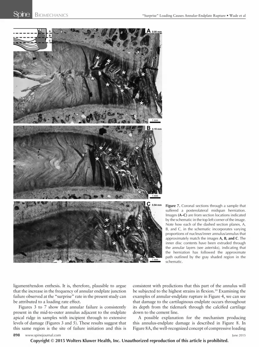
Figure 7 shows a sequence of 3-staged coronal sections ( Figure 7A–C ) through a posterolateral herniation. This type of herniation typically followed a largely radial path from the initial site of rupture as illustrated in the inset schematic. The extruded material has tracked through the midspan region of the annulus as indicated by the asterisks. Note that Figure 7A shows the nucleus proper on the left-hand-side blending into the now ruptured annulus. Figure 7B shows the nucleus-inner
Copyright © 2015 Wolters Kluwer Health, Inc. Unauthorized reproduction of this article is prohibited.
Figure 4. ( A, B ) Higher magnifi cation views of the boxed regions from Figure 3 A and B , respectively. The endplate can be seen to have ruptured between the tidemark (boundary between the annulus and calcifi ed cartilage—see arrows marked TM indicating this) and the cement line (boundary between the cartilaginous and vertebral endplate—see arrows marked CL indicating this). Arrows denote tidemark and cement line, respectively.
SPINEIP1411_LR 895SPINEIP1411_LR 895 21/05/15 10:43 AM21/05/15 10:43 AM

BIOMECHANICS “Surprise” Loading Causes Annular-Endplate Rupture • Wade et al
896 www.spinejournal.com June 2015
annulus blending into the now ruptured annulus. Figure 7C , because of its much greater coronal offset combined with the curvature of the posterolateral wall, now shows the intact inner annulus blending into the ruptured annular wall.
DISCUSSION The results from this new study show that 83% of the motion segments compressed in fl exion at the “surprise” rate of 400 mm/min suffered herniation compared with a herniation rate of 58% previously observed in motion segments similarly compressed but at an order of magnitude lower rate of 40 mm/min (see Table 2 ). This “surprise” rate also favored rup-ture of the annulus-endplate junction ( P = 0.012), although the average load at failure was not signifi cantly different for either the mode of failure ( P > 0.05) or the loading rate ( P > 0.05). Also, 70% (14/20) of the herniations at this “surprise” rate were due to rupture of the annulus-endplate junction, which is comparable with the 65% reported clinically by
Rajasekaran et al . 15 However, it should be noted that many of the endplate junction failures reported in the aforementioned article included bone evulsions that were not confi ned to the plane of the endplate and thus in contrast to those observed in the present study. It is possible that this difference is related in part to the lower density of the human vertebra 21 and the inevitably greater variability in the mechanical overload envi-ronment that has led to the clinically reported failures form-ing the basis of the study by Rajasekaran et al .
Both the general shape and the stiffness of the loading curves across the 10-fold difference in loading rate ( Figure 2 ) were similar. It should be noted that both of these rates would have been suffi cient to ensure that viscoelastic creep of the disc was minimized. 22 These experimental fi ndings, therefore, indicate that despite a similar load-bearing ability, the rate at which the fl exed motion segment is compressed will result in different structures within the disc-endplate complex being rendered vulnerable to damage.
Copyright © 2015 Wolters Kluwer Health, Inc. Unauthorized reproduction of this article is prohibited.
Figure 5. A series of central sagittal (A) and offset (B–D) sections (amount of offset indicated in top right-hand corner of each image) through the posteriomediolateral region of a disc that suffered annular-endplate rupture (see asterisks) and was compressed to the point of transligamentous herniation. The annulus-endplate junction has ruptured at the tidemark and at the cement line as shown in Figure 3. The fl ow of inner disc mate-rial (inner annulus and nucleus) is indicated by arrows. Note that some of the extruded material was lost from images A and B, as it had fl owed out of the section plane.
SPINEIP1411_LR 896SPINEIP1411_LR 896 21/05/15 10:43 AM21/05/15 10:43 AM

BIOMECHANICS “Surprise” Loading Causes Annular-Endplate Rupture • Wade et al
Spine www.spinejournal.com 897
So Why Is the Mode of Failure So Infl uenced by Loading Rate? It has been suggested that the stiffness and ultimate strength of biological tissues varies with loading rate, although the exact nature of the relationship between rate and mechanical
properties is still the subject of ongoing experimentation. Soft tissues such as tendon and ligament show a signifi cant increase in failure at the soft-hard junction or enthesis with increasing loading rate, 23 , 24 and there are obvious structural similarities between the annular-endplate junction and the
Copyright © 2015 Wolters Kluwer Health, Inc. Unauthorized reproduction of this article is prohibited.
Figure 6. Sagittal sections through 2 different discs suffering midspan failure of the posterior annulus. A, The incipient stage of failure is shown, in which the asterisks mark the location of annular rupture. B, Enlarged view of this same region, clearly indicating how the inner disc contents have been extruded into this defect as shown by the arrows. Further compression would lead to propagation of this defect due to this extrusion of the inner disc material as indicated by the arrows (C) , which is from a more extensively compressed sample with the midspan annular rupture marked by asterisks.
SPINEIP1411_LR 897SPINEIP1411_LR 897 21/05/15 10:43 AM21/05/15 10:43 AM
BIOMECHANICS “Surprise” Loading Causes Annular-Endplate Rupture • Wade et al
898 www.spinejournal.com June 2015
ligament/tendon enthesis. It is, therefore, plausible to argue that the increase in the frequency of annular endplate junction failure observed at the “surprise” rate in the present study can be attributed to a loading rate effect.
Figures 3 to 7 show that annular failure is consistently present in the mid-to-outer annulus adjacent to the endplate apical ridge in samples with incipient through to extensive levels of damage ( Figures 3 and 5 ). These results suggest that this same region is the site of failure initiation and this is
consistent with predictions that this part of the annulus will be subjected to the highest strains in fl exion. 14 Examining the examples of annular-endplate rupture in Figure 4 , we can see that damage to the cartilaginous endplate occurs throughout its depth from the tidemark through the calcifi ed cartilage down to the cement line.
A possible explanation for the mechanism producing this annulus-endplate damage is described in Figure 8 . In Figure 8A , the well-recognized concept of compressive loading
Copyright © 2015 Wolters Kluwer Health, Inc. Unauthorized reproduction of this article is prohibited.
Figure 7. Coronal sections through a sample that suffered a posterolateral midspan herniation. Images (A–C) are from section locations indicated by the schematic in the top left corner of the image. Note how each of the dashed section planes, A, B, and C, in the schematic incorporates varying proportions of nucleus/inner annulus/annulus that approximately match the images A, B, and C . The inner disc contents have been extruded through the annular layers (see asterisks), indicating that the herniation has followed the approximate path outlined by the gray shaded region in the schematic.
SPINEIP1411_LR 898SPINEIP1411_LR 898 21/05/15 10:44 AM21/05/15 10:44 AM

BIOMECHANICS “Surprise” Loading Causes Annular-Endplate Rupture • Wade et al
Spine www.spinejournal.com 899
generating a hydrostatic pressure within the disc nucleus is illustrated schematically. The boxed areas shown enlarged in Figures 8B and E identify the regions that can rupture and lead to herniation. Figure 8E shows the arrangement of forces at the annulus-endplate junction as the disc is loaded. The functional nucleus, 25 , 26 which behaves hydrostatically under load, expands outward against the annulus wall, increasing the in-plane tensile stresses in the fi ber bundles.
We propose that if this occurs rapidly, a micromechanical compliance imbalance will arise within the cartilaginous end-plate between the soft tissue of the annulus and the hard ver-tebral endplate, potentially rendering this junction vulnerable to failure ( Figure 8E ). This effect will be most severe adjacent to the endplate in the region of the endplate ridge apex where the annular fi bers will experience the highest level of strain 14 ( Figure 8E ). Because this imbalance will occur at the micro-structural level within the cartilaginous endplate, it does not have a readily detectable infl uence on the overall mechanical response of the disc. However, it will determine how failure is initiated structurally in the samples, resulting in the annular endplate damage illustrated schematically in Figure 8F and imaged microstructurally in Figures 3 to 5 .
Mid-span annular rupture did still occur at the high load-ing rate used in the present study, although at a substantially reduced frequency compared with that observed previously at the lower rate. 14 This possibly arises from pre-existing defects in the annular wall such as a variation in lamellar density or an incomplete lamella 27 that propagates when this region of the disc is strained in the fl exed posture. A tensile load would be generated in the fi ber bundles as shown schematically ( Fig-ure 8B ), leading to the tearing shown in ( Figure 8C, D ). The reduced incidence of this failure mode at high rates is likely to be a consequence of these defects needing to be large enough to fail in preference to the annulus-endplate junction. Inner disc material is then extruded into these tears as evident in the incipient stages of failure ( Figure 6A, B ), with further com-pression leading to eventual full-scale herniation ( Figure 6C ).
Posterolateral rupture as shown in Figure 7 is likely to occur via a subtly different mechanism. As illustrated in Figure 8G–I , the oblique-counter oblique fi bers in the annular layers in this region are not stretched equally when the disc is fl exed. Rather, one oblique population will be strained while the other relaxed. We propose that if incomplete lamellae or other similar defects are present within the loaded set of these
Copyright © 2015 Wolters Kluwer Health, Inc. Unauthorized reproduction of this article is prohibited.
Figure 8. Schematics illustrating potential herniations mechanisms. A, Combined fl exion and compression generates hydrostatic pressure in the functional nucleus (which includes inner annulus as shown by the gray dashed line in E) and creates in-plane tension within the annulus. Tension in the annulus proper (B) can also lead to failure at weak points and midspan rupture ( C and D ). When applied rapidly, this tension creates a micromechanical compliance imbalance within the cartilaginous endplate between the soft tissue of the annulus and the hard vertebral endplate (E) which will then render it vulnerable to rupture (F) . From the transverse view of the disc (G) , the posterolateral views, unfl exed (H) and fl exed (I) , show that fl exion will differentially strain alternate fi ber populations in the posterolateral region of the disc. This would lead to failure of initia-tion at weak points in these strained lamellae, resulting in the approximate herniation path (shown shaded in G).
SPINEIP1411_LR 899SPINEIP1411_LR 899 21/05/15 10:44 AM21/05/15 10:44 AM

BIOMECHANICS “Surprise” Loading Causes Annular-Endplate Rupture • Wade et al
900 www.spinejournal.com June 2015
➢ Key Points
“Surprise” loading in fl exion is likely to cause some form of annular rupture. The annulus-endplate junction is the most vulner-
able region of the disc under “sudden” loading. This vulnerability arises from a rate-induced
mechanostructural imbalance between the an-nulus and the endplate.
differentially strained fi ber bundles in the midouter region, these could act as initiation sites for the herniation process. This correlates with the common clinical fi nding of posterolat-eral herniation patterns. 28 , 29 Once failure begins at these sites, it will advance through the next most highly strained lamellae toward the disc periphery as inner disc material is extruded.
After damage to the annulus by the mechanisms described previously, herniation will progress as follows: in the discs that suffered incipient failure of the mid-to-outer annulus/annular-endplate junction ( Figure 3 ), there will be little disruption of the inner annulus and nucleus and no external evidence of herniation. However, in the more extensively loaded samples, the inner disc contents will be extruded through this initiation site and eventually breach the outer annular wall to produce a transligamentous herniation 12 , 14 , 17 ( Figures 5 and 6C ).
Although speculative, it is worth considering what effect the presence of degeneration would have on these proposed failure mechanisms. Although a full description of the degen-erative process is outside the scope of this article, we would draw attention to the fi ndings of Adams and Roughley, 30 which show that 30- to 40-year-old human discs are most susceptible to herniation. They proposed that this is due to fi ssuring of the outer annular wall, which weakens it, rendering it more vulnerable to the pressure exerted by the inner disc contents. It is possible that this fi ssuring would provide initiation sites for midspan herniation and reduce the likelihood of annular-endplate failure. Thus, on this basis, annular-endplate failure would be more likely to occur in healthy discs subjected to overloading. Conversely, it is possible that the incipient form of annular-endplate failure, as is illustrated in Figure 3 , could be responsible for the initiation of rim lesions that are known to play an important role in the degenerative process. 30–34
A limitation of the present study is the use of the ovine lumbar spine model. However, this model has been shown to be generally similar to the human lumbar spine in terms of anatomy, 35 , 36 biochemistry, 36 and biomechanics. 37 Despite the fact that sheep are quadrupedal, their spinal column is known to experience axial loading (as does the human spine) because of the muscle reaction forces acting upon it. 21 Also, given the similarity in profi le between the ovine and human endplates with both possessing an apical ridge, 35 , 36 , 38 , 39 it is likely that comparable mechanisms of damage initiation and herniation will occur in humans, although further investigations will be required to confi rm this. Also, all tissues, for logistical rea-sons relating to collection from the abattoir, were subjected to a single freeze/thaw cycle. However, such a treatment has been shown to have a negligible effect on the mechanical properties of the components of the intervertebral disc. 40–42 Finally, at the microlevel, given that the same soft-hard tissue complexities exist, the present study, by employing healthy ovine motion segments, offers a detailed analysis of failure at a scale of structural resolution that is simply not accessible with macro-level techniques.
CONCLUSION Motion segments subjected to a “surprise” loading, compara-ble with the maximum rate at which the muscles of the spinal
column can generate a force while in fl exion, are vulnerable to herniation. These herniations occurred mostly via rupture of the annular-endplate junction, and this is thought to be due to a rate-induced mechanostructural imbalance between the compliant annulus and the rigid endplate. These ruptures seemed to initiate in the mid-to-outer annular region which, due to the profi le of the endplate apical ridge, is considered to be most highly strained when the disc is in fl exion. Loading rate thus plays a critical role in the etiology of herniation.
References 1. Porchet F , Wietlisbach V , Burnand B , et al. Relationship between
severity of lumbar disc disease and disability scores in sciatica patients . Neurosurgery 2002 ; 50 : 1253 – 60 .
2. Weber H . Lumbar disc herniation. A controlled, prospective study with ten years of observation . Spine 1983 ; 8 : 131 – 40 .
3. Arts MP , Peul WC , Koes BW , et al. Management of sciatica due to lumbar disc herniation in the Netherlands: a survey among spine surgeons . J Neurosurg 2008 ; 9 : 32 – 9 .
4. Yoganandan N , Maiman DJ , Pintar F , et al. Microtrauma in the lum-bar spine: a cause of low back pain . Neurosurgery 1988 ; 23 : 162 – 8 .
5. Rolander SD , Blair WE . Deformation and fracture of the lumbar vertebral end plate . Orthop Clin North Am 1975 ; 6 : 75 – 81 .
6. Roaf R . A study of the mechanics of spinal injuries . J Bone Joint Surg Br 1960 ; 42-B : 810 – 23 .
7. Lin HS , Liu YK , Adams KH . Mechanical response of the lumbar intervertebral joint under physiological (complex) loading . J Bone Joint Surg Am 1978 ; 60A : 41 – 55 .
8. Brown T , Hansen RJ , Yorra AJ . Some mechanical tests on the lum-bosacral spine with particular reference to the intervertebral discs; a preliminary report . J Bone Joint Surg Am 1957 ; 39A : 1135 – 64 .
9. Lundin O , Ekström L , Hellström M , et al. Injuries in the adoles-cent porcine spine exposed to mechanical compression . Spine 1998 ; 23 : 2574 – 9 .
10. Adams MA , Hutton WC . Prolapsed intervertebral disc: a hyperfl ex-ion injury . Spine 1982 ; 7 : 184 – 91 .
11. Adams MA , Hutton WC . Gradual disc prolapse . Spine 1985 ; 10 : 524 – 31 .
12. Adams MA , Hutton WC . The mechanics of prolapsed interverte-bral disc . Int Orthop 1982 ; 6 : 249 – 53 .
13. McNally DS , Adams MA , Goodship AE . Can intervertebral disc prolapse be predicted by disc mechanics ? Spine 1993 ; 18 : 1525 – 30 .
14. Wade KR , Robertson PA , Thambyah A , et al. How healthy discs herniate: a biomechanical and microstructural study investigat-ing the combined effects of compression rate and fl exion . Spine 2014 ; 39 : 1018 – 28 .
15. Rajasekaran S , Bajaj N , Tubaki V , et al. ISSLS Prize winner: the anatomy of failure in lumbar disc herniation: an in-vivo, multi-modal, prospective study of 181 subjects . Spine 2013 ; 38 : 1491 – 500 .
16. Yingling VR , Callaghan JP , McGill SM . Dynamic loading affects the mechanical properties and failure site of porcine spines . Clin Biomech 1997 ; 12 : 301 – 5 .
Copyright © 2015 Wolters Kluwer Health, Inc. Unauthorized reproduction of this article is prohibited.
SPINEIP1411_LR 900SPINEIP1411_LR 900 21/05/15 10:44 AM21/05/15 10:44 AM

BIOMECHANICS “Surprise” Loading Causes Annular-Endplate Rupture • Wade et al
Spine www.spinejournal.com 901
17. Veres SP , Robertson PA , Broom ND . ISSLS prize winner: how load-ing rate infl uences disc failure mechanics: a microstructural assess-ment of internal disruption . Spine 2010 ; 35 : 1897 – 908 .
18. Dolan P , Adams MA . The relationship between EMG activity and extensor moment generation in the erector spinae muscles during bending and lifting activities . J Biomech 1993 ; 26 : 513 – 22 .
19. Mannion AF , Adams MA , Dolan P . Sudden and unexpected loading generates high forces on the lumbar spine . Spine 2000 ; 25 : 842 – 52 .
20. Thompson JP , Pearce RH , Schechter MT , et al. Preliminary evalu-ation of a scheme for grading the gross morphology of the human intervertebral disc . Spine 1990 ; 15 : 411 – 5 .
21. Smit TH . The use of a quadruped as an in vivo model for the study of the spine—biomechanical considerations . Eur Spine J 2002 ; 11 : 137 – 44 .
22. Race A , Broom ND , Robertson P . Effect of loading rate and hydra-tion on the mechanical properties of the disc . Spine 2000 ; 25 : 662 – 9 .
23. Danto MI , Woo SLY . The mechanical properties of skeletally mature rabbit anterior cruciate ligament and patellar tendon over a range of strain rates . J Orthop Res 1993 ; 11 : 58 – 67 .
24. Pioletti DP , Rakotomanana LR , Leyvraz PF . Strain rate effect on the mechanical behavior of the anterior cruciate ligament-bone com-plex . Med Eng Phys 1999 ; 21 : 95 – 100 .
25. Adams MA , McNally DS , Chinn H , et al. Posture and the compres-sive strength of the lumbar spine . Clin Biomech 1994 ; 9 : 5 – 14 .
26. McNally DS , Adams MA . Internal intervertebral disc mechanics as revealed by stress profi lometry . Spine 1992 ; 17 : 66 – 73 .
27. Schollum ML , Robertson PA , Broom ND . ISSLS prize winner: microstructure and mechanical disruption of the lumbar disc annu-lus: part I: a microscopic investigation of the translamellar bridging network . Spine 2008 ; 33 : 2702 – 10 .
28. Ebeling U , Reulen HJ . Are there typical localisations of lumbar disc herniations? A prospective study . Acta Neurochir 1992 ; 117 : 143 – 8 .
29. Maezawa S , Muro T . Pain provocation at lumbar discogra-phy as analyzed by computed tomography/discography . Spine 1992 ; 17 : 1309 – 15 .
30. Adams MA , Roughley PJ . What is intervertebral disc degeneration, and what causes it? Spine 2006 ; 31 : 2151 – 61 .
31. Vernon Roberts B , Pirie CJ . Degenerative changes in the interverte-bral discs of the lumbar spine and their sequelae . Rheumatol Reha-bil 1977 ; 16 : 13 – 21 .
32. Vernon-Roberts B , Moore RJ , Fraser RD . The natural history of age-related disc degeneration: the pathology and sequelae of tears . Spine 2007 ; 32 : 2797 – 804 .
33. Osti OL , Vernon-Roberts B , Fraser RD . 1990 Volvo Award in experimental studies: anulus tears and intervertebral disc degen-eration: an experimental study using an animal model . Spine 1990 ; 15 : 762 – 7 .
34. Osti OL , Vernon-Roberts B , Moore R , et al. Annular tears and disc degeneration in the lumbar spine: a post- mortem study of 135 discs . J Bone Joint Surg Br 1992 ; 74 : 678 – 82 .
35. Wilke HJ , Kettler A , Wenger KH , et al. Anatomy of the sheep spine and its comparison to the human spine . Anat Rec 1997 ; 247 : 542 – 55 .
36. Reid JE , Meakin JR , Robins SP , et al. Sheep lumbar intervertebral discs as models for human discs . Clin Biomech 2002 ; 17 : 312 – 4 .
37. Wilke HJ , Kettler A , Claes LE . Are sheep spines a valid biomechani-cal model for human spines? Spine 1997 ; 22 : 2365 – 74 .
38. Twomey LT , Taylor JR . Age changes in lumbar vertebrae and inter-vertebral discs . Clin Orthop Relat Res 1987 ;224: 97 – 104 .
39. Francois RJ , Dhem A . Microradiographic study of the normal human vertebral body . Acta Anat 1974 ; 89 : 251 – 65 .
40. Goh KL , Chen Y , Chou SM , et al. Effects of frozen storage tempera-ture on the elasticity of tendons from a small murine model . Animal 2010 ; 4 : 1613 – 7 .
41. Smeathers JE , Joanes DN . Dynamic compressive properties of human lumbar intervertebral joints: a comparison between fresh and thawed specimens . J Biomech 1988 ; 21 : 425 – 33 .
42. Linde F , Sorensen HCF . The effect of different storage meth-ods on the mechanical properties of trabecular bone . J Biomech 1993 ; 26 : 1249 – 52 .
Copyright © 2015 Wolters Kluwer Health, Inc. Unauthorized reproduction of this article is prohibited.
SPINEIP1411_LR 901SPINEIP1411_LR 901 21/05/15 10:44 AM21/05/15 10:44 AM


