Embed Size (px)
Citation preview
A fourth mandible and associated dental remains of Gigantopithecus blacki from the EarlyPleistocene Yanliang Cave, Fusui, Guangxi, South China
Yingqi Zhanga*, Changzhu Jina, Reiko T. Konob, Terry Harrisonc and Wei Wangd
aKey Laboratory of Vertebrate Evolution and Human Origins, Institute of Vertebrate Paleontology and Paleoanthropology (IVPP),Chinese Academy of Sciences, P.O. Box 643, Beijing 100044, P.R. China; bDepartment of Anthropology, National Museum of Nature andScience, Tsukuba 305-0005, Japan; cDepartment of Anthropology, Center for the Study of Human Origins, New York University,New York, NY 10003, USA; dGuangxi Museum of Nationalities, Nanning 530021, P.R. China
(Received 13 December 2014; accepted 25 February 2015)
Dentognathic remains of Gigantopithecus blacki from the newly discovered Early Pleistocene locality of Yanliang Cave,Guangxi, South China are described. These include an incomplete mandible, only the fourth discovered and the first knownfrom a site other than Liucheng, as well as 25 isolated teeth. Comparisons of the Yanliang mandible show that the bestpreserved part of the right corpus is morphologically similar to the left side of the Liucheng Mandible III. In addition, theYanliang mandible and the Liucheng Mandible III share a similar degree and pattern of wear on the premolars and molars.The partially resorbed alveolus for the right M2 in the Yanliang mandible indicates antemortem tooth loss, which is the firstrecord of its kind for Gigantopithecus blacki. Comparisons of the enamel–dentine junction morphology show that theisolated upper premolars from Yanliang are similar to those of Gigantopithecus blacki from Early Pleistocene sites, anddiffer from the more specialised form from the Middle Pleistocene Hejiang Cave. This supports the biochronologicalevidence that Yanliang Cave is Early Pleistocene in age.
Keywords: Gigantopithecus blacki; hominoid; Pleistocene; antemortem tooth loss; enamel–dentine junction
Introduction
The extinct giant ape Gigantopithecus blacki was initially
named by von Koenigswald (1935) based on an isolated
right M3 obtained from a Hong Kong drugstore, where it
was being sold for traditional Chinese medicine. Over two
decades later, three teeth of Gigantopithecus blacki were
discovered in situ in Hei Cave, Daxin County, Guangxi
(Pei and Woo 1956). These finds shed important light on
the provenance and geological age of Gigantopithecus.
Later that same year, two partial mandibles of Gigan-
topithecus blackiwere discovered in situ, with the help of a
local farmer, in the Gigantopithecus Cave (Xiaoyan Cave)
of Liucheng County, Guangxi nearly 300 km NE of Hei
Cave (Pei 1957). The following field seasons at the site
yielded a third mandible (Pei and Li 1958) and over 1000
isolated teeth (Woo 1962). During the 1970s, several other
Gigantopithecus-bearing localities were reported (Chang
et al. 1973, 1975; Hsu et al. 1974), but the collections were
limited to isolated teeth. This first wave of discoveries,
primarily by researchers from the Institute of Vertebrate
Paleontology and Paleoanthropology (IVPP), resulted in a
much improved understanding of the phylogenetic
relationships and palaeobiology of Gigantopithecus (e.g.
Weidenreich 1945; von Koenigswald 1952, 1958; Woo
1962; Eckhardt 1975; White 1975; Szalay and Delson
1979; Zhang 1982; Ciochon et al. 1990; Daegling and
Grine 1994; Han and Zhao 2002; Kelley 2002; Cameron
2003; Zhao and Zhang 2013). However, the lack of cranial
and postcranial material of Gigantopithecus blacki, due to
preservational and taphonomic factors (White 1975), has
seriously hampered a more comprehensive understanding
of the species. More recent excavations at cave sites in
Guangxi by researchers of the IVPP and the Natural
History Museum of Guangxi (NMHG) (Wang et al. 2005;
Wang 2009; Jin et al. 2009; Takai et al. 2014; Zhang, Jin,
et al. 2014; Zhang, Kono, et al. 2014) have also yielded
collections that consist entirely of dental remains. Over the
past decade, our team, led by Changzhu Jin, has
systematically investigated caves in the Chongzuo area
in Guangxi, and has excavated more than 10 Pleistocene
sites with abundant fossil remains (Jin et al. 2009; Takai
et al. 2014; Zhang, Jin, et al. 2014; Zhang, Kono, et al.
2014). Eight of these sites have yielded the remains of
Gigantopithecus blacki. The newly discovered remains are
all isolated teeth except for an incomplete mandible of
Gigantopithecus blacki from Yanliang Cave discovered in
2011. This is only the fourth partial mandible of
Gigantopithecus blacki recovered, and the only one
known from a site beyond Liucheng. In view of its
importance, the Yanliang mandible is described in the
present paper, along with the isolated teeth from the site.
We dedicate this paper to Dr. Yukimitsu Tomida, an old
q 2015 Taylor & Francis
*Corresponding author. Email: [email protected]
Historical Biology, 2016
Vol. 28, Nos. 1–2, 95–104, http://dx.doi.org/10.1080/08912963.2015.1024115
Dow
nloa
ded
by [
New
Yor
k U
nive
rsity
] at
16:
17 0
6 O
ctob
er 2
015

friend and mentor to the lead author, on the occasion of his
retirement next year from National Museum of Nature and
Science (NMNS).
Materials and methods
Institutional abbreviations
IVPP, Institute of Vertebrate Paleontology and Paleoan-
thropology, Chinese Academy of Sciences, Beijing,
China; PA, palaeoanthropological collection numbers of
IVPP; KLVEHO, Key Laboratory of Vertebrate Evolution
and Human Origins, Chinese Academy of Sciences,
Beijing, China; IHEP, Institute of High Energy Physics,
Chinese Academy of Sciences, Beijing, China; NHMG,
Natural History Museum of Guangxi, Nanning, Guangxi,
China; NMNS, National Museum of Nature and Science,
Tokyo, Japan.
Location, age and geological context of the material
The Gigantopithecus blacki specimens (Table 1) described
here were obtained from Yanliang Cave (2281305400N,10783603500E), Fusui County, Chongzuo City, Guangxi,
during the spring and autumn field seasons in 2011. Yan
et al. (2014) and Zhu et al. (2014) list at least 40mammalian
taxa associated with Gigantopithecus blacki, and consider
the age of the fauna to be Early Pleistocene basedmainly on
biostratigraphic correlations and the relative elevation of
the cave systems (Yan et al. 2014; Zhu et al. 2014).
Measurements and methods
The molar morphology terminology used in this paper is
adapted fromSzalay andDelson (1979). Crown dimensions
of the original specimens were measured to the nearest
0.1mmwith a sliding vernier caliper (Mitutoyo, Kanagawa
Prefecture, Japan) by R.T. Kono. Crown mesiodistal (MD)
length and buccolingual (BL) breadth were measured using
the methods described in White (1977), Suwa (1990) and
Suwa et al. (2009).. For all molars and fourth premolars,
MD was measured along the MD axis of the crown that
roughly bisects the tooth. BL is the maximum breadth
measured along an axis perpendicular to the MD axis.
In either case, the caliper jawswere applied to the specimen
vertical to the occlusal plane. For the P3s, we measured the
longest diameter,maximumoblique diameter at crown base
(MAXOB), instead of MD and the maximum diameter
perpendicular to MAXOB, PP, instead of BL. For the P3s,
MD was not measured at the bisecting axis, but at the
maximum width, maximus mesiodistal length, which
usually corresponds to the position of preparacrista (see
Zhang, Jin, et al. 2014; Zhang, Kono, et al. 2014).
Microfocus computed tomography (micro-CT) of the
Gigantopithecus blackimandible and upper premolars was
Table 1. Specimen list and measurements (mm) of Gigantopithecus blacki from Yanliang Cave, Fusui, Guangxi.
IVPP no. Field no. Identification MD MAXOB MDMAX BL PP
PA1601-1 CFLGYL201111-800 Right P3 22.1 15.7Right M1 20.1 17.9Right M3 21.6 17.3
PA1601-2 CFLGYL201111-801 Left M2 20.6 19.7Left M3 20.3 17.8
PA1602.1 CFLGYL201111-812 Left I1 15.0 15.1PA1602.2 FLGY201104-GY-1-7 Right C1 16.1 15.9PA1602.3 FLGY201104-GY-1-33 Right C1 17.0 15.8PA1602.4 CFLGYL201111-808 Left P3 14.8 20.9PA1602.5 CFLGYL201111-809 Left P3 13.7 20.3PA1602.6 CFLGYL201111-810 Right P3 17.1PA1602.7 CFLGYL201111-803 Left M1/2
PA1602.8 CFLGYL201111-802 Left M3 20.5 22.1PA1602.9 CFLGYL201111-815 Left I1 9.7PA1602.10 FLGY201104-GY-1-26 Left I2 8.4 11.1PA1602.11 FLGY201104-GY-1-36 Left I1 7.8 10.6PA1602.12 FLGY201104-GY-1-336 Left I1 7.2 9.5PA1602.13 FLGY201104-GY-1-6 Left P3 20.9 15.4PA1602.14 FLGY201104-GY-1-322 Left P3 19.2 15.3PA1602.15 CFLGYL201111-811 Right P3PA1602.16 FLGY201104-GY-1-2 Left P4 14.8 17.0PA1602.17 FLGY201104-GY-1-323 Left P4 17.0 18.6PA1602.18 CFLGYL201111-804 Right M1 19.2 17.3PA1602.19 FLGY201104-GY-1-3 Left M1 18.9 16.8PA1602.20 FLGY201104-GY-1-5 Left M1 17.8 17.3PA1602.21 CFLGYL201111-805 Right M2 21.1 18.4PA1602.22 FLGY201104-GY-1-1 Left M3 25.3 21.4
96 Y. Zhang et al.
Dow
nloa
ded
by [
New
Yor
k U
nive
rsity
] at
16:
17 0
6 O
ctob
er 2
015
carried out using the 225-kV micro-CT system (developed
by IHEP) at the KLVEHO. The specimens were scanned
with beam energy of 140 kV and a flux of 100mA at a
detector resolution of around 40–120mm per pixel using a
3608 rotation with a step size of 0.58 and an unfiltered
aluminum reflection target. A total of 720 transmission
images were reconstructed in a 1024 £ 1024 matrix of at
most 781 slices using a two-dimensional reconstruction
software developed by IHEP.
Systematic palaeontology
Order Primates Linnaeus, 1758
Superfamily Hominoidea Gray, 1825
Family Hominidae Gray, 1825
Subfamily Ponginae Elliot, 1913
Genus Gigantopithecus von Koenigswald, 1935
Species Gigantopithecus blacki von Koenigswald, 1935
(Figures 1 and 2)
For a list of newly referred material, see Table 1.
Description
Mandibular material
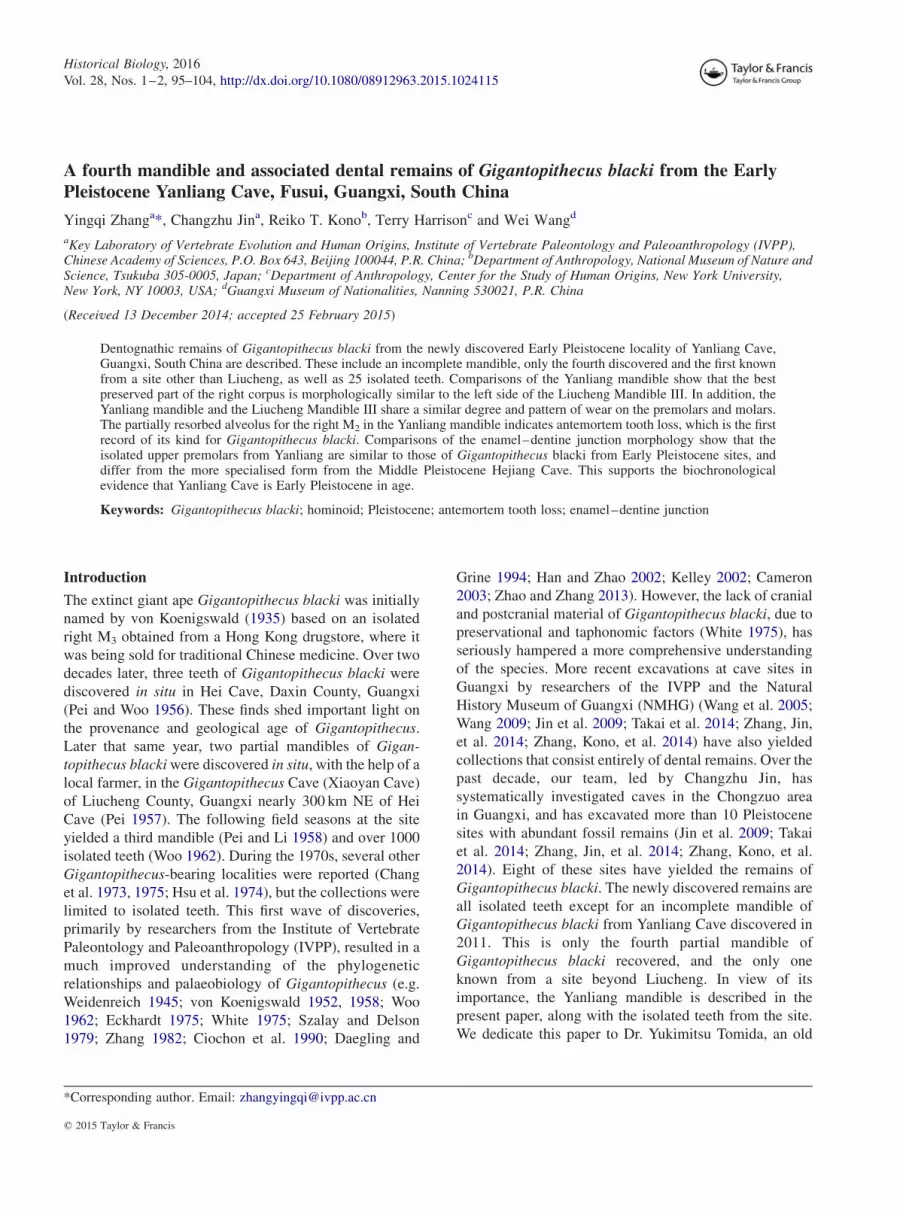
The material consists of two mandibular fragments that
belong to a single individual, PA1601-1 (Figure 1(A)) and
PA1601-2 (Figure 1(B)). PA1601-1 is a partially complete
right corpus with P3–M1 and M3, a crushed symphyseal
region, and anterior part of the left corpus with C–P4. The
highly crushed symphysis and the anterior part of the left
corpus are cemented together by matrix. The symphysis is
badly deformed, but the alveoli for right I1–C are arranged
approximately in their original anatomical positions. The
upper part of the right corpus below M1–M3, is best
preserved. The right corpus is undistorted, but heavily
weathered with irregular cracks. Unfortunately, the lower
margin of the corpus is not preserved, so it is not possible
to measure its height. Posteriorly, it is preserved
approximately to the junction of the corpus and ramus,
Figure 1. Mandibular remains of Gigantopithecus blacki from Yanliang Cave in comparison to Mandible III from the GigantopithecusCave, Liucheng, Guangxi. A, PA1601-1, a partial mandible with right P3–M1, M3 and right C1–P4; B, PA1601-2 (occlusal view), a partialleft corpus with M1–3 belonging to the same individual as PA1601-1; C, Mandible III (PA83); D, Mandible I (PA77). 1, Occlusal view; 2,right lateral view; 3, left medial view.
Historical Biology 97
Dow
nloa
ded
by [
New
Yor
k U
nive
rsity
] at
16:
17 0
6 O
ctob
er 2
015

similar to the condition in the Liucheng Mandible III
(Figure 1(C)). The right P3 and P4 and their alveolar region
are slightly displaced anteriorly compared with the better-
preserved posterior portion. The premolars are displaced
from their alveoli and exhibit excessive exposure of the
roots. The roots of the right M1 are also exposed buccally
due to breakage of the alveolar bone. The corpus is robust,
and the lateral eminence is strongly developed opposite
M2, which contributes to the development of a wide
extramolar sulcus. A small aperture resembling a mental
foramen is located inferior to the posterior root of P3, but
its position is much higher than in the Liucheng mandibles.
Using serial CT scan data, we have been able to confirm
that it is not a mental foramen. On the medial surface of the
corpus, a well-developed triangular torus envelops the
roots of M3. A broad and deep concavity is present just
posterior to this torus. The wall of this concavity is
positioned close to the distal root apex of the posterior
molar. PA1601-2 is the posterior part of the left corpus
with M1–3. Only the alveolar portion of the body below the
molars is preserved. Due to its fragility, it was detached
from PA1601-1 during excavation. Because no mean-
ingful morphological features are preserved on the partial
corpus, the specimen was consolidated by plaster and
moulded into the shape of a mandibular corpus.
All the teeth are moderately to heavily worn. Most of
them have large areas of dentine exposure on the buccal
cusps, except for the right M3, which has dentine, exposed
Figure 2. Dental remains ofGigantopithecus blacki from Yanliang Cave. A, PA1602.1, left I1; B, PA1602.2, right C1; C, PA1602.3, rightC1; D, PA1602.4, left P3; E, PA1602.5, left P3; F, PA1602.6, right P3; G, PA1602.7, left M1/2; H, PA1602.8, left M3; I, PA1602.9, left I1; J,PA1602.10, left I2; K, PA1602.11, left I1; L, PA1602.12, left I1; M, PA1602.13, left P3; N, PA1602.14, left P3; O, PA1602.15, right P3; P,PA1602.16, left P4; Q, PA1602.17, left P4; R, PA1602.18, right M1; S, PA1602.19, left M1; T, PA1602.20, left M1; U, PA1602.21, rightM2; V, PA1602.22, left M3. 1, Occlusal view; 2, lingual view; 3, buccal view; 4, mesial view; 5, distal view; occlusal view for the others.
98 Y. Zhang et al.
Dow
nloa
ded
by [
New
Yor
k U
nive
rsity
] at
16:
17 0
6 O
ctob
er 2
015

distally instead. The basal cross-sectional outline of the
left canine is a buccolingually elongated oval shape. It is
heavily worn, with a large flat wear facet oriented
perpendicular to the longitudinal axis of the tooth.
A ledge-like enamel fold is present distolingually at the
distal shoulder. Based on the size and morphology of the
canine, the mandible belongs to a female individual. M2 is
larger than M1 and M3. M3 tapers distally giving it an
overall smaller size than M2. The alveolus of the missing
right M2 is half -closed anteriorly, and posteriorly there is a
wide and shallow depression surrounded by pathological
bone. The depression continues to the base of the buccal
root of M3, and the mesial face of the M3 does not show
any interstitial contact facet. These observations indicate
that the right M2 had been lost during the individual’s
lifetime.
Upper incisor
PA1602.1 (Figure2(A)) is an unworn left upper central
incisor. The root has been broken off about 5mm from the
buccal cervical margin. Its crown is robust and squat, with
a somewhat triangular outline and nearly equal MD and
BL dimensions. The buccal face is slightly convex. There
are three distinct mammelons on the broad incisal margin
of the tooth, which gives the incisal margin a zigzagged
appearance. On the lingual side, the apical half of the
crown is concave. Two marginal ridges, originating from
the mesial and distal angles, respectively, border the
central lingual fovea. The cervical half of the crown bulges
lingually, and basal tubercules are present where the two
marginal ridges meet. The cervical margin extends
substantially onto the root buccally and lingually.
Two additional specimens, PA1602.23 and
PA1602.24, both more worn than PA1602.1, are identified
as left upper central incisors. It can be estimated that about
two-thirds of the crown have been lost through wear in
both specimens, but they preserve relatively complete
roots. Their roots are approximately 20mm in length when
measured from the buccal cervical line. In both specimens,
remnants of interstitial contact facets can be observed on
the mesial and distal margins, and these help to determine
the side. The wear facets, sloping to the distal side, and the
occlusal outlines support the side identification.
Upper canines
PA1602.2 (Figure 2(B)) is a moderately worn right upper
canine that can be assumed to have belonged to a female
individual. The root has been gnawed by porcupines. The
occlusal wear facet, forming a rounded dentine pit about
5 mm in diameter, is almost perpendicular to the
longitudinal axis of the tooth. The basal cross-sectional
outline is nearly circular. The mesial groove is narrow and
shallow. The distal shoulder is more developed than the
mesial one. The lingual cingulum can be observed leading
from the distal shoulder, and several irregular pillars
originate from the cingulum and pass towards the crown
apex. The enamel margin extends substantially along the
root on the buccal side. The enamel is quite thick, with a
maximum thickness of 2.4mm on the buccal rim of the
occlusal facet.
PA1602.3 (Figure 2(C)) is a right upper canine germ of
a presumed female individual. A sharp ridge marks the
distal side of the tooth, which runs towards the cervix and
ends at a moderately developed distal shoulder. A lingual
marginal ridge runs from the distal shoulder, gives rise to
several irregular ridges towards the crown apex, and ends
at a lingual tubercle. A well-developed mesial groove
originates from the mesial shoulder, but it is less well-
developed than its distal counterpart. The cross-sectional
contour at the cervix is nearly circular but slightly
mesiodistally elongated.
Upper premolars
PA1602.4 (Figure 2(D)) is a moderately worn left P3 with
dentine exposure on the protocone and paracone. It has an
asymmetric triangular occlusal outline. There is not much
morphological detail left on the occlusal surface. The
paracone is more elevated than the protocone. The buccal
moiety is longer than the lingual moiety because of the
mesially projecting preparacrista. All the roots are
missing.
PA1602.5 (Figure 2(E)) is a slightly worn left P3 with a
tiny dentine pit on the paracone. Only the robust lingual
root, approximately 28mm in length, is preserved in its
entirety. The tooth was three rooted because two separate
root canals can be observed on the fractured surface of the
buccal root. A strong invagination in the middle of the
buccal root surface suggests that the divergence between
the two radicles occurred more apically. The protocone
and paracone are connected by a mesially convex crest,
which also separates a large central fovea distally from a
well-developed mesial fovea. The preprotocrista meets the
preparacrista at the buccomesial corner of the crown and
forms an angular projection. The postprotocrista runs
buccally to meet the postparacrista. These two crests
delimit the distal margin of the central fovea, with the
hypoprotocrista and hypoparacrista representing the
mesial margin.
PA1602.6 (Figure 2(F)) is a moderately worn right P3
with dentine exposures on the protocone and paracone.
Almost all the roots are missing and the pulp chamber is
filled with matrix. Enamel hypoplasia is evident on the
buccal wall of the tooth. The large anterior fovea, divided
by a small longitudinal ridge, is bordered distally by the
hypoprotocrista and the hypoparacrista and mesially by
Historical Biology 99
Dow
nloa
ded
by [
New
Yor
k U
nive
rsity
] at
16:
17 0
6 O
ctob
er 2
015

the preprotocrista and the preparacrista. The base of the
lingual wall is heavily worn with dentine exposure.
Upper molars
PA1602.7 (Figure 2(G)) is a lightly worn left M1/2 without
dentine exposure. The crown is mesiodistally longer than
broad. The roots are missing, and there is slight damage to
the crown along the distal and mesial walls and at the
paracone tip. In occlusal view, the outline of the crown is
rhomboidal, with the buccal cusps more mesially placed
than the lingual cusps. The protocone is the largest of the
main cusps, with the other three being subequal in size.
The crista obliqua is intersected by a weak groove running
between the paracone and hypocone. A narrow transver-
sely aligned mesial fovea is present, delimited by a mesial
marginal ridge, the preparacrista and the two mesial cusps.
The posthypocrista and the postmetacrista do not quite
meet, leaving the distal fovea open distally.
PA1602.8 (Figure 2(H)) is a slightly worn left M3
without dentine exposure. The roots are mostly missing,
but the fractured surface indicates the presence of three
roots. The crown tapers distally, but its MD and BL
dimensions are similar. The protocone is the largest cusp,
and the metacone is relatively small. A ledge-like
Carabelli’s cusp is present, encircling the mesiolingual
corner of the protocone. The mesial marginal ridge,
together with the protocone and the paracone, delimits a
narrow mesial fovea. The postmetacrista acts as the distal
border of the distal fovea. Pit -like hypoplastic features are
present on the mesiobuccal and lingual faces.
PA1602.25 represents half of a moderately worn M1/2
crown. It was difficult to determine whether this is the
mesial half of a left molar or the distal half of right molar.
We provisionally recognise it as the latter because the
exposed dentine patch on the lingual cusp is more
marginally situated, indicating that the remaining lingual
cusp is a hypocone rather than a protocone. If this
identification is correct, the specimen is most probably a
first molar because the MD corner forms a very acute
angle. Also, the BL breadth of this tooth would have been
greater than in the other two upper molars.
Lower incisors
PA1602.12 (Figure 2(L)) is a heavily worn left I1 with
considerable dentine exposure. The root is fully preserved.
The root surface is concave both mesially and distally, but
is more strongly so on the distal side. The crown narrows
basally from the apex in lingual view. The buccal face of
the tooth is slightly convex. A modest mesial marginal
ridge borders the lightly concave lingual face, but there is
no basal tubercle. Enamel thickness, measured on the
buccal rim of the occlusal wear facet, is 1.2mm.
PA1602.9 (Figure 2(I)) is a heavily worn left I2 with
extensive dentine exposure. The crown is asymmetrical.
The worn occlusal surface slopes down towards the mesial
side, which is an atypical wear pattern. However, exactly
the same pattern can be seen on the lateral incisors of the
Liucheng Mandible III. A shallow fovea occupies the
central part of the lingual face, and is bordered basally by a
narrow cingulum. The root is long, robust and mesiodis-
tally compressed. The distal face of the root is only faintly
grooved. The mesial notch of the cervical enamel line is
more pronounced than its distal counterpart.
PA1602.10 (Figure 2(J)) and PA1602.11 (Figure 2(K))
are moderately worn left I2 and I1, respectively. Both
crowns exhibit dentine exposure. The two incisors are
likely to belong to a single individual because of the close
fit between the interstitial contact facets and the thick
enamel exposed on the occlusal wear facet (2.0 and
1.7mm, respectively, on the buccal rim of the facet). The
roots of the teeth have been gnawed by porcupines. The
distal portion of the incisal edge slopes basally. There is no
cingulum; instead, a low median basal bulge and a distal
fovea are present. Again, the mesial cervical notches are
more pronounced than their distal counterparts.
Lower premolars
PA1602.13 (Figure 2(M)) is a moderately worn left P3.
It is bicuspid with the protoconid larger than the
metaconid. The cusps are separated by a median
longitudinal groove. In occlusal view, the crown is
rhomboidal in shape. The occlusal surface of the crown is
worn nearly flat. The protoconid has a dentine pit about
3mm in diameter. The enamel thickness measured on the
buccal rim of the wear facet is 2.7mm, although it is
probably somewhat exaggerated by the obliquity of the
wear. The preprotocristid and the premetacristid meet
mesially and delimit the mesial fovea. The postprotocris-
tid extends lingually to the distolingual margin of the
metaconid and forms the distal margin of the distal fovea.
The mesiobuccal face of the crown bulges buccally and
bears a smooth honing facet produced by occlusion with
the upper canine. The lingual face of the crown is nearly
vertical. The cervix on the mesiobuccal face extends far
inferiorly onto the root. The two roots are only partially
preserved due to porcupine gnawing. The mesial and
distal roots are subequal in size.
PA1602.14 (Figure 2(N)), a left P3 and PA1602.15
(Figure 2(O)), a right P3 exhibit a similar morphology to
PA1602.13 (Figure 2(M)), except that the honing facet is
less distinct. Both specimens bear prominent hypoplasial
grooves, especially on the buccal faces. The roots of
PA1602.14 are almost completely preserved. The buccal
root is compressed mesiodistally, while the lingual root is
buccolingually compressed. The roots of PA1602.15 are
100 Y. Zhang et al.
Dow
nloa
ded
by [
New
Yor
k U
nive
rsity
] at
16:
17 0
6 O
ctob
er 2
015

not preserved, and the crown is damaged mesially and
distally.
PA1602.16 (Figure 2(P)) and PA1602.17 (Figure 2(Q))
are unworn left P4s. The former is smaller than the latter.
The occlusal outline is trapezoidal, with the mesial moiety
broader than the distal moiety. The two main cusps are
transversely aligned and separated by a narrow median
fissure. The metaconid is more elevated than the
protoconid. The mesial fovea is narrow and groove-like,
and bordered by the preprotocristid, premetacristid and the
two main cusps. The distal marginal ridge, which bears
three or four small tubercles, is separated from the main
cusps by a deep groove. The talonid basin occupies the
distal one-third of the crown. It is deep and well defined,
being much larger than the mesial fovea.
Lower molars
PA1602.20 (Figure 2(T)) and PA1602.18 (Figure 2(R)) are
moderately worn left and right M1s, respectively. They
both have considerable dentine exposure on the buccal
cusps. Enamel thickness measured at the buccal rim of the
protoconid wear facet, is 2.4 and 2.1mm, respectively.
The following description is mainly based on the nearly
unworn left M1, PA1602.19 (Figure 2(S)). The roots of all
three teeth are not well preserved. The crown is elongated
mesiodistally with some degree of waisting between the
mesial and distal cusps. The metaconid is the largest
cusp. The protoconid is slightly smaller, followed by the
hypoconid and entoconid, with the hypoconulid being the
smallest cusp. The crown exhibits pronounced buccal
flare. The metaconid is rectangular in occlusal outline.
Both the preprotocristid and the premetacristid extend
medially to meet each other. Along with the mesial cusps,
these two crests delimit the narrow groove-like mesial
fovea. A trenchant longitudinal groove separates the
protoconid from the metaconid. A deep transverse groove
passes between the protoconid and hypoconid, and extends
two -thirds down the buccal face of the crown.
PA1602.21 (Figure 2(U)) is a moderately worn right
M2 with only a tiny dentine pit on the hypoconid. The roots
have been mostly removed by gnawing. The crown is not
as mesiodistally elongated as in M1. The buccal wall
exhibits strong flare. A longitudinal groove passes
between protoconid and metaconid, and a transverse one
passes between the protoconid and hypoconid. The two
grooves meet at a right angle near the distal margin of the
protoconid. The metaconid is the largest cusp, followed by
the hypoconid, protoconid and hypoconulid. The entoco-
nid is the smallest cusp. The thickness of the enamel on the
buccal rim of the hypoconid wear facet is 2.8mm, but
the value may be overestimated due to the obliquity of
the wear. This specimen possibly belongs to the same
individual as PA1602.18 (Figure 2(R)), because the
interstitial contact facets correspond well and the degree of
wear is consistent.
PA1602.22 (Figure 2(V)) is an unworn left M3 with the
roots missing. The tooth is long relative to its breadth. The
largest cusp is the metaconid. The main crest of the
metaconid originates at the buccodistal corner and passes
buccally then distally (like a ‘deflecting wrinkle’), to join
the entoconid. The protoconid is the next largest cusp,
followed by the hypoconid. The hypoconulid is much
smaller, and is buccally displaced. All three buccal cusps
are separated from each other by deep fissures. The groove
passing between the protoconid and hypoconid extends far
down on the buccal wall. Two accessory cupsules are
present between the entoconid and the hypoconulid. The
distalmost cuspule, which is the largest of the two,
probably corresponds to a sixth cusp. A groove passes
mesiodistally to separate the buccal cusps from the lingual
cusps. The mesial fovea is represented by a transverse
fissure bordered by the preprotocristid, premetacristid and
the mesial cusps.
Comparisons and discussion
Because the Yanliang mandible (Figure 1(A),(B)) is
poorly preserved, not much can be determined about its
morphological characteristics. However, comparisons
with the Liucheng mandibles, especially Liucheng
Mandible III with comparable anatomical features
preserved to those of the Yanliang mandible, do allow
for several interesting observations to be made. The best
preserved part of the right corpus is morphologically
similar to the left side of Liucheng Mandible III (Figure 1
(C)). In superior view, they both share a strongly laterally
flaring ramus root with a wide extramolar sulcus lateral to
M3. It might be more flared in the Liucheng mandible, but
since the bone surface of the Yanliang specimen is eroded,
we cannot be certain. Medially, it is not possible to discern
anatomical details of the Yanliang specimen, such as the
course of the mylohyoid line or the development of the
transverse torus. The posterior part of the corpus is better
preserved, and there is a well-developed toral structure at
the level of M3, and a distinctive concavity just posterior to
it. Unfortunately, the corresponding part of the mandible is
not well preserved in Liucheng Mandible III, so it is not
possible to make a direct comparison. Nevertheless, the
remaining bone surface of the latter specimen suggests that
a similar structure was present.
The degree and pattern of wear seen on the premolars
and molars of the Yanliang mandible is similar to that seen
in Liucheng Mandible III. Only the right M3 differs,
probably because the M3 of the Yanliang mandible is
tilted. When compared with Liucheng Mandible I (from an
older adult female, Figure 1(D)), the premolars are much
more worn in the Yanliang mandible. The wear facet on
Historical Biology 101
Dow
nloa
ded
by [
New
Yor
k U
nive
rsity
] at
16:
17 0
6 O
ctob
er 2
015

the Yanliang canine is distinctive compared to the two
Liucheng mandibles in being flat and almost perpendicular
to the long axis of the tooth.
The Middle and Late Pleistocene hominins with severe
antemortem tooth loss (AMTL) are often regarded as
evidence of human-like behaviours, such as conspecific
care or cooking (e.g. Lordkipanidze et al. 2005; Lebel
et al. 2001). However, AMTL is not unique to hominins,
but also occurs in non-human primates (Gilmore 2013).
The occurrence of AMTL in fossil hominoids is seldom
reported. The antemortem loss of the right M2 in PA1601-
1 is indicated by the partially resorbed socket. The vertical
CT section of the Mandible III (Figure 3(C),(D)) shows the
socket of the left M2 is filled by matrix, while that of
PA1601-1 (Figure 3(A),(B)) shows that the space between
the right M1 and M3 is much wider than normal and that
resorption has partially filled the alveolus of the lost right
M2 with bony tissue. There is an interstitial contact facet
on the distal wall of the Yanliang right M1, but the facet on
the mesial wall of the right M3 is not discernable. This
indicates that the right M2 was lost before eruption of the
right M3. The anteriorly tilted right M3 also supports this
assumption. The malocclusion was probably caused by the
lack of resistance from the neighbouring M2. Wear on the
right M3 confirms that even though it was tilted, it was still
effective in chewing. However, the pattern of wear is not
typical; only the hypoconulid was heavily worn with a
large oval dentine exposure about 6mm in diameter. The
evidence suggests that the individual represented by
PA1601-1 and PA1601-2 must have survived for a
considerable period of time with impaired masticatory
capabilities.
Zhang, Jin, et al. (2014) and Zhang, Kono, et al. (2014)
analysed the upper premolar enamel–dentine junction
(EDJ) complexity of Gigantopithecus blacki from Hejiang
Cave, dated to 400–320 ka, as well as those from Early
Pleistocene sites and drugstore collections. They con-
cluded that the Hejiang Cave material represented a
specialised form of Gigantopithecus blacki limited to the
Middle Pleistocene and differing in premolar morphology
complexity from those from the Early Pleistocene. The
Middle Pleistocene Hejiang Cave upper premolars
demonstrate a more complicated and crenulated outer
enamel surface than those in the comparative Early
Figure 3. CT scanning sections for PA1601-1 and Liucheng Mandible III, and the EDJ morphology of PA1602.4, PA1602.5 andPA1602.6. A, CT section for PA1601-1; B, CT section for PA1601-1; C, CT section for Mandible III; D, CT section for the Mandible III;E, EDJ morphology of PA1602.4; F, EDJ morphology of PA1602.5; and G, EDJ morphology of PA1602.6.
102 Y. Zhang et al.
Dow
nloa
ded
by [
New
Yor
k U
nive
rsity
] at
16:
17 0
6 O
ctob
er 2
015

Pleistocene and drugstore samples due to the presence of
secondary crests that radiate from the paracone and
protocone and that originate from the preparacrista and
postparacrista. The EDJ morphology of all three P3s from
Yanliang (Figure 3(E)–(G)) was compared with those
analysed by Zhang, Jin, et al. (2014) and Zhang, Kono,
et al. (2014). The overall EDJ morphology of all three P3s
falls in the variation range of Gigantopithecus blacki
illustrated by Zhang, Jin, et al. (2014) and Zhang, Kono,
et al. (2014). The major crests on the Yanliang P3s are not
as sharp as those from Hejiang Cave. Unlike the Hejiang
P3s, no minor crests are present on the Yanliang premolars,
including those that radiate from the paracone. The
postparacrista and the postprotocone crista on the Yanliang
P3s extend medially and join the distal margin of the
occlusal fovea. The preprotocone crista joins the parastylar
crest on all of the P3s from Yanliang Cave. The wide sulcus
that interrupts the mesial margin of the occlusal fovea is
not present on any the P3s from Yanliang Cave. The EDJ
morphology of the P3s from Yanliang Cave resembles that
of Gigantopithecus blacki from Early Pleistocene cave
sites, and it can be distinguished from the more specialised
morphology seen in the P3s from Hejiang Cave. This
evidence is consistent with the Early Pleistocene age
designation of the Yanliang Cave (Yan et al. 2014; Zhu
et al. 2014).
Acknowledgements
We thank Yemao Hou for arranging the CT scanning; YihongLiu, Yuan Wang, Min Zhu and Yaling Yan for their hard work inthe field; and Wenshu Sun for preparation of the fossil material.We extend our gratitude to Gareth Dyke, Eric Delson and oneanonymous reviewer for their helpful comments on an earlierversion of the manuscript.
Disclosure statement
No potential conflict of interest was reported by the authors.
Funding
The research was supported by the Key Research Program of theChinese Academy of Sciences (kzzd-ew-03), the NationalScience Fund for Talent Training in Basic Science [grantnumber J1210008], National Natural Science Foundation ofChina [grant numbers 41072013 and 41202017] and the Programof China Geological Survey [grant number 1212011220519].
References
Cameron DW. 2003. A functional and phylogenetic interpretation of thelate Miocene Siwalik hominid Indopithecus and the ChinesePleistocene hominid Gigantopithecus. Himalayan Geol. 24:19–28.
Chang Y, Wang L, Dong X, Chen WY. 1975. Discovery of aGigantopithecus tooth from Bama District in Kwangsi. VertPalasiatica. 13:148–154.
Chang Y, Wu M, Liu C. 1973. New discovery of Gigantopithecus teethfrom Wuming, Kwangsi. Chin Sci Bull. 18:130–133.
Ciochon RL, Piperno DR, Thompson RG. 1990. Opal phytoliths found onthe teeth of the extinct ape Gigantopithecus blacki: implications forpaleodietary studies. Proc Nat Acad Sci USA. 87(20):8120–8124.doi:10.1073/pnas.87.20.8120.
Daegling DJ, Grine FE. 1994. Bamboo feeding, dental microwear, anddiet of the Pleistocene ape Gigantopithecus blacki. South Afr J Sci.90:527–532.
Eckhardt RB. 1975. Gigantopithecus as a hominid. In: Tuttle RL, editor.Paleoanthropology, morphology and palaeoecology. Mouton: TheHague; p. 105–127.
Gilmore CC. 2013. A comparison of antemortem tooth loss in humanhunter-gatherers and non-human catarrhines: implications for theidentification of behavioral evolution in the human fossil record. AmJ Phys Anthropol. 151(2):252–264. doi:10.1002/ajpa.22275.
Han K, Zhao L. 2002. Dental caries of Gigantopithecus blacki fromHubei Province of China. Acta Anthropol Sin. 21:191–197.
Hsu C-H, Han K-X, Wang L-H. 1974. Discovery of Gigantopithecusteeth and associated fauna in Western Hopei. Vert Palasiatica. 12:293–309.
Jin C, Qin D, Pan W, Tang Z, Liu J, Wang Y, Deng C, Zhang Y, DongW,Tong H. 2009. A newly discovered Gigantopithecus fauna fromSanhe Cave, Chongzuo, Guangxi, South China. Chin Sci Bull. 54(5):788–797. doi:10.1007/s11434-008-0531-y.
Kelley J. 2002. The hominoid radiation in Asia. In: Hartwig WC, editor.The primate fossil record. Cambridge: Cambridge University Press;p. 369–384.
Lebel S, Trinkaus E, Faure M, Fernandez P, Guerin C, Richter D, MercierN, Valladas H, Wagner GA. 2001. Comparative morphology andpaleobiology of Middle Pleistocene human remains from the Bau deL’Aubesier, Vaucluse, France. Proc Nat Acad Sci USA. 98(20):11097–11102. doi:10.1073/pnas.181353998.
Lordkipanidze D, Vekua A, Ferring R, Rightmire GP, Agusti J, KiladzeG, Mouskhelishvili A, Nioradze M, de Leon MSP, Tappen M,Zollikofer CPE. 2005. Anthropology: the earliest toothless homininskull. Nature. 434(7034):717–718. doi:10.1038/434717b.
Pei W. 1957. Discovery of Gigantopithecus mandible and other materialin Liu-cheng district of central Kwangsi in South China. VertPalasiatica. 1:65–72.
Pei W, Li Y. 1958. Discovery of a third mandible of Gigantopithecus inLiu-Cheng, Kwangsi, South China. Vert Palasiatica. 2:193–200.
Pei WC, Woo JK. 1956. New materials of Gigantopithecus teeth fromSouth China. Acta Palaeontol Sin. 4:477–490.
Suwa G. 1990. A comparative analysis of hominid dental remains fromthe Shungura and Usno Formations, Omo Valley, Ethiopia [PhDthesis]. Berkeley, CA: University of California.
Suwa G, Kono RT, Simpson SW, Asfaw B, Lovejoy CO, White TD.2009. Paleobiological implications of the Ardipithecus ramidusdentition. Science. 326(5949):69, 94–99. doi:10.1126/science.1175824.
Szalay FS, Delson E. 1979. Evolutionary history of the primates. NewYork, NY: Academic Press.
Takai M, Zhang Y, Kono RT, Jin C. 2014. Changes in the composition ofthe Pleistocene primate fauna in southern China. Quat Int. 354:75–85. doi:10.1016/j.quaint.2014.02.021.
von Koenigswald GHR. 1935. Eine fossile Saugetierfauna mit Simia ausSudchina [A fossil mammalian fauna including Simia form SouthChina]. Proceedings of the Koninklijke Nederlandse Akademie vanWetenschappen. SerB Palaeontol Geol Phys Chem Anthropol. 38:872–879.
von Koenigswald GHR. 1952. Gigantopithecus blacki von Koenigswald,a giant fossil hominoid from the Pleistocene of Southern China.Anthropol Pap Am Mus Nat Hist. 43:292–325.
von Koenigswald GHR. 1958. Gigantopithecus and Australopithecus.Leech. 28:101–105.
Wang W. 2009. New discoveries of Gigantopithecus blacki teeth fromChuifeng Cave in the Bubing Basin, Guangxi, South China. J HumEvol. 57(3):229–240. doi:10.1016/j.jhevol.2009.05.004.
Wang W, Potts R, Hou Y, Cheng Y, Wu H, Yuan B, Huang W. 2005.Early Pleistocene hominid teeth recovered in Mohui Cave in BubingBasin, Guangxi, South China. Chin Sci Bull. 50(23):2777–2782.doi:10.1360/982004-614.
Historical Biology 103
Dow
nloa
ded
by [
New
Yor
k U
nive
rsity
] at
16:
17 0
6 O
ctob
er 2
015

Weidenreich F. 1945. Giant early man from Java and South China.Anthropol Pap Am Mus Nat Hist. 40:1–134.
White TD. 1975. Geomorphology to paleoecology: Gigantopithecusreappraised. J Hum Evol. 4(3):219–233. doi:10.1016/0047-2484(75)90009-3.
White TD. 1977. New fossil hominids from Laetolil, Tanzania. Am JPhys Anthropol. 46(2):197–229. doi:10.1002/ajpa.1330460203.
Woo J-K. 1962. The mandibles and dentition of Gigantopithecus.Paleontol Sin New Ser D. 11:1–94.
Yan Y, Wang Y, Jin C, Mead JI. 2014. New remains of Rhinoceros(Rhinocerotidae, Perissodactyla, Mammalia) associated with Gigan-topithecus blacki from the Early Pleistocene Yanliang Cave, Fusui,South China. Quat Int. 354:110–121. doi:10.1016/j.quaint.2014.01.004.
Zhang Y. 1982. Variability and evolutionary trends in tooth size ofGigantopithecus blacki. Am J Phys Anthropol. 59(1):21–32. doi:10.1002/ajpa.1330590104.
Zhang Y, Jin C, Cai Y, Kono R, Wang W, Wang Y, Zhu M, Yan Y. 2014.New 400–320 ka Gigantopithecus blacki remains from HejiangCave, Chongzuo City, Guangxi, South China. Quat Int. 354:35–45.doi:10.1016/j.quaint.2013.12.008.
Zhang Y, Kono RT, Jin C, WangW, Harrison T. 2014. Possible change indental morphology in Gigantopithecus blacki just prior to itsextinction: evidence from the upper premolar enamel–dentinejunction. J Hum Evol. 75:166–171. doi:10.1016/j.jhevol.2014.06.010.
Zhao LX, Zhang LZ. 2013. New fossil evidence and diet analysis ofGigantopithecus blacki and its distribution and extinction in SouthChina. Quat Int. 286:69–74. doi:10.1016/j.quaint.2011.12.016.
Zhu M, Schubert BW, Liu J, Wallace SC. 2014. A new record of thesaber-toothed catMegantereon (Felidae, Machairodontinae) from anEarly Pleistocene Gigantopithecus fauna, Yanliang Cave, Fusui,Guangxi, South China. Quat Int. 354:100–109. doi:10.1016/j.quaint.2014.06.052.
104 Y. Zhang et al.
Dow
nloa
ded
by [
New
Yor
k U
nive
rsity
] at
16:
17 0
6 O
ctob
er 2
015


