Embed Size (px)
Citation preview

UNIVERSIDAD DE GRANADA
DEPARTAMENTO DE FISIOLOGÍA
INSTITUTO DE NUTRICIÓN Y TECNOLOGÍA DE LOS ALIMENTOS “JOSÉ MATAIX VERDÚ”
PROGRAMA DE DOCTORADO NUTRICIÓN HUMANA Y CIENCIA DE LOS ALIMENTOS
Functional and structural aspects of
aged pancreas related to the nature of
dietary fat supplemented or not with
coenzyme Q10
TESIS DOCTORAL
ADRIÁN GONZÁLEZ ALONSO
2016


Este trabajo de Tesis Doctoral ha sido realizado en el
Instituto de Nutrición y Tecnología de los Alimentos “José
Mataix Verdú” situado en el Centro de Investigación
Biomédica de la Universidad de Granada como miembro
del Grupo de investigación “Fisiología y Bioquímica
Nutricional del Estrés Oxidativo” (CTS-627).


Durante la realización del siguiente trabajo, el Ldo. Adrián González Alonso fué
financiado por las siguientes fuentes:
Ayudas para contratos predoctorales de Formación del Profesorado Universitario
(FPU) (Diciembre 2010 – Noviembre 2014)
Ayudas para la realización de estancias breves en el extranjero para beneficiarios de
FPU. Ministerio de Educación, Cultura y Deporte (Febrero 2014 – Abril 2014)


Proyectos de investigación que han financiado este estudio:
Título del proyecto: Análisis de expresión génica y causas de muerte en ratas con
diferente longevidad y envejecimiento por ingesta de diferentes fuentes grasas
(oliva, girasol y pescado) suplementadas o no con coenzima Q. Entidad financiadora:
Ministerio de Ciencia e Innovación. Referencia: AGL2008-01057/ALI. Investigador
responsable: Dr. José Luis Quiles Morales. Duración: de 01/01/2009 a 31/12/2011.
Título del proyecto: Estudio multidisciplinar del papel de los aceites de oliva virgen,
girasol y pescado y del coenzima Q10 en el proceso de envejecimiento. Entidad
financiadora: Consejería de Innovación, Ciencia y Empresa. Junta de Andalucía
(Referencia P05-AGR 832). Duración: de 2006 a 2009. Investigador responsable: Dr.
José Luis Quiles Morales.


D. José Luis Quiles Morales. Catedrático de la Universidad de Granada,
Departamento de Fisiología.
Dª. María del Carmen Ramírez Tortosa. Catedrática de la Universidad de
Granada, Departamento de Bioquímica y Biología Molecular II.
CERTIFICAN:
Que los trabajos de investigación que se exponen en la Memoria de Tesis
Doctoral: “Functional and structural aspects of aged pancreas related
to the nature of dietary fat supplemented or not with coenzyme Q10”,
han sido realizados bajo nuestra dirección por la licenciada en Ciencia y
Tecnología de los Alimentos D. Adrián González Alonso y la encontramos
conforme para ser presentada y aspirar al Grado de Doctor por la Universidad
de Granada con el Tribunal que en su día se designe.
Y para que conste, en cumplimiento de las disposiciones vigentes,
extendemos el presente en Granada a cinco de Marzo de dos mil catorce.
Dr. D. José Luis Quiles Morales
Dra. Dª. María del Carmen Ramírez Tortosa


El doctorando Adrián González Alonso y los directores de la tesis Dr. D. José
Luis Quiles Morales, y la Dra. Dª. María del Carmen Ramírez Tortosa,
garantizamos, al firmar esta tesis doctoral, que el trabajo ha sido realizado por
la doctoranda bajo la dirección de los directores de la tesis y hasta donde
nuestro conocimiento alcanza, en la realización del trabajo, se han respetado
los derechos de otros autores a ser citados, cuando se han utilizado sus
resultados o publicaciones.
En Granada, a uno de Julio de dos mil dieciséis
Director/es de la Tesis Doctorando
JOSÉ LUIS QUILES MORALES ADRIÁN GONZÁLEZ ALONSO
MARÍA DEL CARMEN RAMÍREZ TORTOSA


MEMORIA QUE PRESENTA EL LDO. ADRIÁN GONZÁLEZ ALONSO PARA ASPIRAR AL
GRADO DE DOCTOR POR LA UNIVERSIDAD DE GRANADA
ESTA TESIS DOCTORAL HA SIDO REALIZADA BAJO LA DIRECCIÓN DE:
Dr. D. José Luis Quiles Morales Dra. Dª. María del Carmen Ramírez Tortosa
Ldo. D. Adrián González Alonso
Granada, 2016


AGRADECIMIENTOS
Si alguien me hubiera dicho durante me etapa de niñez o desde mi etapa de
adolescente que me iba a encontrar en la situación en la que me encuentro, le habría
dicho que ―tú tienes mucha fe‖, que es lo que se suele decir en mi tierra a alguien que
tiene expectativas muy improbables, pero uno no sabe a dónde le va a llevar la vida,
pero si empiezo a recapitular y echo la mirada atrás, puedo ver que terminé mis estudios
obligatorios, el Bachiller de ciencias de la salud, comencé la Universidad estudiando
Biología y la termine con Ciencia y Tecnología de los Alimentos y finalmente los
estudios de posgrado, ¡Vaya! Aquella persona tenía razón, yo pensaba en ese momento
que a estas alturas estaría… pues no me acuerdo muy bien cuales eran mis expectativas,
era solo un chaval de barrio cuyas preocupaciones eran cumplir mis obligaciones
estudiantiles, jugar al futbol, balonmano, incluso atletismo y pasármelo bien, sin
preocuparme mucho en lo que vendría después, y pasado los años esa es una de las
cosas en las que no he cambiado, el no pensar en lo que puede venir después, sino en lo
que tengo en el presente, lo que en términos futbolísticos se conoce como ―partido a
partido‖.
Evidentemente el camino hasta llegar hasta aquí no ha salido como se suele decir
un camino de rosas, ha sido un camino lleno de sacrificios, pero también de
satisfacciones y aprendizaje. Quien me conozca bien, sabrá que soy una persona que le
cuesta mostrar incluso en privado mis sentimientos más profundos, pues en público ya
ni os digo, de hecho en algún momento pensé en no escribir esta nota de
agradecimientos con la excusa de que me parecía una ―cursilada ―, pero reflexioné y
pensé que quizás sea el momento para mostrar mis agradecimientos, no solo a aquellas
personas que han formado y forman de mi entorno o de mi vida, es de justicia acordarse
de las instituciones que me han ayudado a conseguir lo que tengo.
Empezando cronológicamente, he de agradecer al colegio Los Pinos de
Algeciras por la educación recibida en mi etapa desde los 4 a 15 años de edad, en
especial a los muchos profesores que tuve durante esa etapa, los cuales me inculcaron
valores muy positivos que sigo conservando hoy, y a mis compañeros de clase de
entonces, con algunos de los cuales sigo manteniendo el contacto aunque sea por
Facebook.
Agradecer también al I.E.S Levante de Algeciras, sus profesores y mis
compañeros de entonces, por los 2 años que estudié allí, sin duda uno de los institutos
con mayor tasa de aprobados en la prueba de selectividad de la comarca, además de
haberme preparado para los primeros años de mi etapa universitaria.
Como no, mostrar mi profundo agradecimiento a todas las instituciones
relacionadas con la Universidad de Granada, como la Facultad de
Ciencias y de Farmacia y a todo el profesorado y personal de administración y
conserjería que me facilitaron mi labor, y en general a la propia universidad, ya que no
sólo me ha aportado los conocimientos que debo tener, sino lo que es más importante,

que me ha guiado para utilizar los recursos y herramientas que una persona de mi
formación ha de saber manejar, además de instruirme en valores como la solidaridad, el
bien común, la justicia social, la reflexión y otros tantos valores positivos que
difícilmente habría podido adquirir en cualquier otro lugar, y todo junto me ha hecho ser
no sólo una persona apta y útil para la sociedad, sino una persona que se siente realizada
para mi vida de ahora en adelante.
Otra institución a la que le debo gran parte de lo conseguido es el Ministerio de
Educación, Cultura y Deporte, por facilitarme las ayudas económicas necesarias para
poder seguir formándome y que sin duda me han facilitado muchísimo mi labor y mi
realización personal, que al fin y al cabo es una apuesta que hace la sociedad española
para invertir en gente como yo, para que este País siga mejorando en todo los aspectos,
y que por lo tanto me siento en deuda con el pueblo de España y haré todo lo posible
por devolverlo durante mi etapa laboral.
Agradezco enormemente a la Universidad de Reading por facilitarme la
realización de las estancias en el extranjero en sus instituciones y edificios, en especial a
la Profesora Julie Lovegrove que aceptó mi solicitud sin apenas tener referencias mías, a
Jane y Salisu que estaban siempre atentos a mí, facilitándome mi adaptación al
―Department of Food and Nutritional Sciences‖ y siendo mi guía durante dicha estancia.
Allí también conocí a gente que me ayudaron mucho a sentirme lo mejor posible fuera
de casa, muchas gracias Luis, Isidro, Yianna y especialmente Natalia, que durante los
primeros días de mi estancia fue esencial con su ayuda y predisposición a lo que hiciera
falta, mil gracias.
Como no, agradecer al Departamento de Fisiología de la Facultad de Farmacia
que me acogió desde el primer momento y me hizo sentirme parte suya, especialmente
por facilitarme una experiencia de la que me siento muy realizado, que es la de impartir
clases universitarias. Muchas gracias a todo el equipo de profesorado, a los compañeros
becarios, pero en especial a Elisa y Encarna, 2 piezas claves que facilitan enormemente
el trabajo docente y de investigación de los que estamos ahí.
Agradecimientos también para el Centro de Investigaciones Biomédicas, lugar
de mi rutina durante este tiempo, para los conserjes del edificio, el equipo de
administración, el equipo de limpieza y las distintas unidades que nos allanaban nuestra
labor de manera notable. En especial me siento agradecido al Instituto de Nutrición y
Tecnología de los Alimentos ―José Mataix Verdú‖, institución de la que formo parte de
manera orgullosa, en especial a algunos con los que han estado a mi lado el día a día y
con los que me he sentido muy a gusto, como Mari Ángeles, Laura, Elenita, Alicia,
Silvia, Jesús, Tomás y Paco. Mención especial para Javi, un talento que desborda
humor, alegría, una capacidad de trabajo fuera de lo común, buen rollo y sobretodo su
ayuda incondicional y desinteresada, gracias por todo ello Javi. Mis compañeros de
grupo y laboratorio también son de muy especial mención, con todos ellos he vivido
momentos muy bueno, que perdurarán en mi memoria, por la buena sintonía que hemos
tenido, por el compañerismo tan profundo que vivimos en el 120, por las mañanas y
tardes de risas que hemos vivido a pesar de las jornadas intensas de laboratorio que nos
hemos metido, muchas gracias Sergio, fuiste un gran apoyo durante mi primera etapa en
el grupo y alguien del que aprender, muchas gracias Patri, aparte de ser una gran

compañera, admiro tu capacidad de superar las adversidades que la vida pone, muchas
gracias Nuri, por tu nobleza y bondad, muchas gracias Naroa, por darle alegría y buen
rollo al grupo. Muchas gracias Mario, una persona a la que admiro por su enorme
talento, su compañerismo incondicional y su humor a todas horas del día; muchas
gracias Alfonso, por tu humildad y modestia a pesar de las enormes virtudes que posees,
y gracias Julio que has sido como un compañero más a pesar de que dabas consejos de
director de tesis, gracias a todos, me he sentido como si estuviera dentro de una familia.
Me siento enormemente agradecido a mis directores de tesis, Pepe, Mayca y César, por
darme la oportunidad de emprender esta aventura y apostar por mí frente a otros, a pesar
de mis defectos y mis debilidades, me siento un privilegiado porque lo que aquí he
aprendido, no habría podido aprenderlo en otro lugar, os estaré eternamente agradecido.
Tengo que agradecer también a mis amigos, por ser como son, por sentirme
querido y aceptado, porque de ellos he aprendido valores, enseñanzas, otros puntos de
vista, a valorar la amistad entre otras cosas. Muchas gracias a mis amigos de la carrera
Juanma, Iván, Chema, Jorge, Susana, Marga, Irene, Diego, Jaime, Worty, Alejandra,
Omar, Rocío, María, Javi, Javito, Inma, Fran, Ginés, Alejandro, Rubén, Azahara y
Miriam, ellas 2 en especial porque eran compañeras de sufrimiento y les agradezco la
enorme ayuda que me dieron a la hora de realizar los trámites burocráticos que supone
el pedir una beca FPU o las estancias en el extranjero. Del mismo modo también me
siento agradecido por otros amigos que forman parte de mi vida y mi ser, Rafa, Roberto,
Lechera, Alberto, Cangrejo, Javi, Fran, Nazaret, y Manolo, sé que me falta más gente
pero también se lo agradezco.
Durante estos últimos años tengo que agradecer a cierta familia que me han
aceptado como a uno más de ellos, gracias Miguel, Eleuteria, Antonia, Eleuterio, Rocío,
Javi, Miguel Angel, María José y los pequeños Javi y Alba, no os imagináis lo
afortunado que me siento al haberos conocido, os siento como a mi propia familia
porque vosotros me lo habéis transmitido a mí, además de ser unas personas con una
honestidad, cariño y saber estar fuera de lo común, mil gracias.
Las lágrimas me inundan, porque solo pensar en lo que mi familia es para mí lo
es todo, la emoción que emerge sobresale más allá de lo común, y en lo profundamente
agradecido y afortunado que me siento de teneros a mi lado, muchas gracias a mis tíos y
tías, Berto, Jaime, Mari, Carlos, Fabi, Ali, Paco, Pepe, muchas gracias a mis primos y
primas, Hugo, Maite, Guillermo, Fernando, Esther y Pablo, Miriam, Mariola y Ester.
Abuela, no sabes lo que te quiero y admiro, todo el enorme sacrificio que has vivido
durante tu vida no tiene nombre, criando a 7 hijos y nietos y nietas, como yo, pasando
por momentos muy difíciles, los que la vida te pone, estoy muy orgulloso de ti abuela y
cada día que estés con nosotros es un regalo. Irene, Hermana, estoy muy orgulloso de ti,
todavía recuerdo el día que naciste y cuando te cogí en brazos por primera vez, fue
maravilloso, y el verte crecer hasta el día de hoy, en el que ya veo a mi hermana
pequeña como una mujer madura, con una capacidad de trabajo admirable y con unas
ideas y valores excepcionales me hace sentir tremendamente orgulloso. No me puedo
olvidar de los que hoy ya no están, mis abuelos Jaime, Pepe y Rosario que se fueron
cuando yo era muy pequeño, pero que todavía tengo grabado momentos de inmensa
felicidad cuando estuve a su lado y que daría lo que fuera porque me estuvieran
observando desde allá donde quiera que estén y que se sintieran orgullosos de mí, de
todos los pasos que he dado en mi vida, tanto de los aciertos como los fracasos. Papá,
Mamá, que puedo decir de vosotros, en primer lugar gracias por haberme dado la vida,
por haber tenido una infancia muy feliz, por inculcarme valores tan positivos que me
hacen ser la persona que soy, por inculcarme lo importante que es la educación, la
formación y sobretodo el valor de ser honesto y buena persona, el tremendo sacrificio
que ha supuesto el irme a otra ciudad a iniciar mis estudios universitarios, esos son los
mejores regalos que unos padres pueden dar a sus hijos, lo máximo que un hijo puede
aspirar, y vosotros me lo habéis dado, os estaré eternamente agradecido. Y a ti Mar, mi
Mar, son sólo 4 años, 3 meses y 11 días desde que te conocí, pero ya no puedo vivir sin
ti, sin tus besos, sin tus caricias, sin tu mirada de complicidad, sin tu sonrisa ni tus
abrazos, especialmente los mañaneros que hacen que vea que esto no es un sueño sino
una realidad, una bendita realidad. Te agradezco todo lo que haces por mí en el día a
día, soportar mis defectos, el sentirme tremendamente querido, tus enseñanzas y ayudas,
gracias por todo amor y por todo lo que venga, te quiero.


TABLE OF CONTENTS
ABBREVATIONS 25
SUMMARY 31
INTRODUCTION 39
I DEFINITION AND BEGINNINGS OF AGING 41
II AGING THEORIES 42
III NUTRITION AND AGING 46
III.1 CALORIC RESTRICTION AND DELAY OF AGING PROCESS 46
III.2 EFFECTS OF ANTIOXIDANTS SUPPLEMENTATION 47
IV BIOLOGICAL MEMBRANES RELATED TO DIETARY FAT 47
V RELEVANCE OF DIETARY FAT ON HEALTH MAINTENANCE 51
V.1 EFFECTS ON CARDIOVASCULAR SYSTEM 51
V.2 EFFECTS ON DIGESTIVE SYSTEM 51
V.3 EFFECTS ON DIABETES 52
V.4 EFFECTS ON CANCER 52
V.5 EFFECTS ON AGING PROCESS 53
VI COENZYME Q 58
VI.1 DISCOVERY OF COQ 58
VI.2 SYNTHESIS OF COQ 59
VI.3 DISTRIBUTION AND BIOAVAILABILITY OF COQ 61
VI.4 MECHANISM OF COQ ACTION AND ITS RELATION TO AGING 62
VI.5 AGING AND PHYSIOPATHOLOGY IN RELATION TO COENZYME Q 63
VI.6 ANTIOXIDANT FUNCTIONS AND COQ DEFICIENCY 66
VI.7 CARDIAC FAILURE AND ITS RELATION TO COQ USAGE 67
VII THE PANCREAS 67
VII.1 ANATOMY AND HISTOLOGY OF PANCREAS 67
VII.2 ROLES OF PANCREAS 69
VII.2.1 Role of exocrine pancreas 69
VII.2.2 Role of endocrine Pancreas 71
VII.2.2.1 Endocrine pancreas and carbohydrate metabolism 71
VII.2.2.2 Endocrine pancreas and lipid metabolism 72
VII.2.2.3 Endocrine Pancreas and protein metabolism 73
VIII NON-NEOPLASTIC DISORDERS OF THE PANCREAS 74
VIII.1 PANCREATITIS 74
VIII.1.1 Acute Pancreatitis 74
VIII.1.2 Chronic pancreatitis 76
VIII.2 DIABETES MELLITUS 77
VIII.2.1 Type 1 diabetes 77
VIII.2.2 Type 2 diabetes 79
VIII.2.3 Other diabetes types 80
IX NEOPLASTIC DISORDERS OF THE PANCREAS 81

IX.1 TUMORS IN THE EXOCRINE PANCREAS 81
IX.2 TUMORS IN THE ENDOCRINE PANCREAS 85
IX.2.1 Functional pancreatic neuroendocrine tumors 85
IX.2.2 Nonfunctional pancreatic neuroendocrine tumors 86
X AGE-RELATED CHANGES OF THE PANCREAS 87
X.1 AGE-RELATED STRUCTURAL CHANGES OF THE PANCREAS 88
X.2 AGE-RELATED FUNCTIONAL CHANGES OF THE PANCREAS 89
XI RISK FACTORS FOR PANCREATIC DISEASES 90
XI.1 CIGARETTE SMOKING 90
XI.2 ALCOHOL CONSUMPTION 91
XI.3 OBESITY AND OVERWEIGHT 92
XI.4 PHYSICAL ACTIVITY 93
XI.5 DIET 94
HYPOTHESIS AND OBJECTIVES 99
MATERIALS AND METHODS 103
I MATERIALS 105
I.1 CHEMICAL PRODUCTS 105
I.2 EQUIPMENT 105
I.3 SOFTWARE 107
II METHODS 107
II.1 EXPERIMENTAL DESIGN: ANIMALS AND DIET 107
II.2 TOTAL PROTEIN EXTRACTION AND DETERMINATION IN PANCREAS TISSUE 110
II.3 DETERMINATION OF CIRCULATING FATTY ACID PROFILE 111
II.4 HISTOPATHOLOGICAL ANALYSIS 113
II.5 IMMUNOHISTOCHEMISTRY FOR INSULIN AND GLUCAGON EXPRESSION 113
II.6 PROLIFERATION ASSAY: KI-67 IMMUNOSTAINING 114
II.7 PROTEIN, HORMONE AND METABOLITE DETERMINATIONS 114
II.7.1 Total insulin, glucagon and leptin 114
II.7.2 Parameters obtained by plasma extracted 115
II.7.2.1 Circulating glucose 115
II.7.2.2 Circulating triglycerides 115
II.7.2.3 Circulating cholesterol 116
II.7.2.4 Circulating total lipids 117
II.7.2.5 Circulating enzymatic markers 117
II.7.2.5.1 Circulating phosphatase alkaline 117
II.7.2.5.2 Circulating creatine kinase 118
II.7.2.5.3 Circulating glutamyl oxaloacetic transaminase 118
II.7.2.5.4 Circulating glutamic pyruvic transaminase 119
II.7.2.5.5 Circulating lactate dehydrogenase 119
II.7.2.6 Circulating Creatinine 120
II.7.2.7 Circulating lactate 120
II.7.2.8 Homeostasis model assessment index (HOMA) 121
II.8 PROTEIN CARBONYLS ASSAY 121
II.9 REVERSE TRANSCRIPTION–POLYMERASE CHAIN REACTION 122

II.10 STATISTIC 122
RESULTS 125
I DIETARY FAT EFFECTS ON PANCREAS 127
I.1 BODY WEIGHT EVOLUTION AND ADAPTATION TO DIET 127
I.2 CIRCULATING HORMONE LEVELS AND BIOCHEMICAL PARAMETERS 129
I.3 HISTOLOGICAL EXAMINATION 129
I.3.1 Pancreatic Parenchyma 129
I.3.2 Immunohistochemical assessment of pancreatic islets 131
I.4 PANCREATIC INSULIN AND GLUCAGON CONTENT 131
I.5 EXPERIMENTAL APPROACH TO EXPLAIN THE OBSERVED PANCREATIC CHANGES 132
II. DIETARY COENZYME Q EFFECTS ON PANCREAS 135
II.1 BODY WEIGHT EVOLUTION AND ADAPTATION TO THE DIET 135
II.2 CIRCULATING HORMONE LEVELS AND BIOCHEMICAL PARAMETERS 135
II.3 HISTOLOGICAL EXAMINATION 138
II.3.1 Pancreatic Parenchyma 138
II.3.2 Immunohistochemical assessment of pancreatic islets 138
II.4 PANCREATIC INSULIN AND GLUCAGON CONTENT 138
DISCUSSION 145
I. DIETARY FAT EFFECTS ON AGE-RELATED CHANGES OF PANCREAS 147
II. DIETARY COQ EFFECTS ON AGE-RELATED CHANGES OF PANCREAS 150
CONCLUSIONS 155
REFERENCES 161
APPENDIX 1: CONTENT ORIGINALITY REPORT 187
APPENDIX 2: CURRICULUM VITAE 193
APPENDIX 3: RELATED PUBLICATIONS 203


25
ABBREVIATIONS
4-AP: 4-aminophenazone
AC: alcohol consumption
ADP: adenosine diphosphate
AIDS: acquired immune deficiency
syndrome
Akt: protein kinase B
ALA: α-linolenic acid
ALT: Alanine Tranferase
AMP: adenosine monophosphate
ANOVA: analysis of variance
AP: acute pancreatitis
AR: antigen retrieval
ATP: adenosine triphosphate
BMI: body mass index
BRCA2: breast cancer type 2
susceptibility protein
cAMP: cyclic adenosine
monophosphate
CCK: cholecystokinin
CD4: cluster of differentiation 4
CD8: cluster of differentiation 8
CDH1: cadherin 1 type 1
CDKN2A: cyclin-dependent kinase
inhibitor 2A
cDNA: complementary DNA
CF: cardiac failure
CFTR: cystic fibrosis transmembrane
conductance regulator
CHE: cholesterol esterase
CHOD: cholesterol oxidase
CK: creatin kinase
CMV: cytomegalovirus
c-myc: myelocytomatosis viral
oncogene homolog
CoQ: coenzima Q
CP: chronic pancreatitis
CR: caloric restriction
CS: cigarette smoking
CT: computed tomography
CTLA-4: cytotoxic T-lymphocyte
antigen 4
CV: cardiovascular
CVB: coxsackievirus B
CVD: cardiovascular disease
DAB: 3,3’-diaminobenzidine
DGLA: dihomo-Gamma-Linolenic Acid
DHA: docosahexaenoic acid
DM: diabetes mellitus
DMBA: 7,12-
dimethylbenz[a]anthracene
DNA: deoxyribonucleic acid
DNPH: 2,4-dinitrophenylhydrazine
EDTA: ethylenediaminetetraacetic acid
EGFR: epidermal growth factor
receptor
26
ELISA: enzyme-linked immunoSorbent
assay
EPA: eicosapentaenoic acid
EPHA3: EPH receptor A3
ER: endoplasmic reticulum
ERCP: endoscopic retrograde
cholangiopancreatography
EVOO: extra virgin olive oil
FAMMM: familial atypical mole
melanoma
FBXW7: F-box and WD repeat domain
containing 7
FFA: free fatty acid
FOF: fish oil fed
G6PDH: glucose-6P-dehydrogenase
GAD65: glutamic acid decarboxylase
GH: growth hormone
GK: glycerol-kinase
GLUT: glucose transporter
GOD: glucose oxidase
GOT: glutamyl oxaloacetic
transaminase
GPO: glycerol phosphate oxidase
GPT: glutamic pyruvic transaminase
GTP: guanosine triphosphate
H2O2: hydrogen peroxide
HCl: chlorhydric acid
HFD: high fat diet
HDL: high density lipoprotein
HLA: human leukocyte antigen
HK: hexoquinase
HMG-CoA: 3-hydroxy-3-
methylglutaryl-coenzyme A
HOMA: homeostasis model assessment
index
HPLC: high-performance liquid
chromatography
HPOO: high phenolic olive oil
Hsp70: 70 kilodalton heat shock protein
HT: hydroxytyrosol
IA-2: islet antigen 2
IA-2 β : islet antigen 2 β
I-CAM1: Intercellular Adhesion
Molecule 1
IDH1: isocitrate dehydrogenase 1
IFAPA: Institute for Agricultural and
Fisheries of Andalusia
IL-2: interleukine 2
IL-6: interleukine 6
IL2RA: interleukin 2 receptor alpha
IPMNs: intraductal papillary mucinous
neoplasms
IR: insulin resistance
IRS: insulin receptor substrate
JNKs: c-Jun N-terminal kinases
KRAS: Kirsten rat sarcoma viral
oncogene homolog
LA: linoleic acid
LDH: lactate dehydrogenase

27
LDL: low density lipoprotein
LO: lactate oxidase
LPL: lipoprotein-lipase
MCNs: mucinous cystic neoplasms
MCP-1: monocyte chemoattractant
protein–1
MD: mediterranean diet
MDH: malate dehydrogenase
MEN1: multiple endocrine neoplasia
type 1
METC: mitochondrial electronic
transport chain
MLH1: mutL homolog 1
MRC: mitochondrial respiratory chain
mRNA: messenger ribonucleic acid
mROS: mitochondrial reactive oxygen
species
mtDNA: mitochondrial
Deoxyribonucleic acid
MUC1: mucin 1 cell surface associated
MUC2: mucin 2 cell surface associated
MUFA: monounsaturated fatty acid
NAD+: nicotinamide adenine
dinucleotide (oxidizes form)
NADH: nicotinamide adenine
dinucleotide (reduced form)
NADPH: nicotinamide adenine
dinucleotide phosphate
NEFA: non-esterified fatty acid
NF1: neurofibromin 1
NHL: non-Hodgkin lymphoma
NYHA: New York Heart Association
OD: optical density
p-NPP: p-nitrophenylphosphate
p27Kip1: cyclin-dependent kinase
inhibitor 1B
PA: physical activity
PAL: phosphatase alkaline
PanIn: pancreatic intraepithelial
neoplasia
PaSC: pancreatic stellate cell
PC: pancreatic cancer
PDAC: pancreatic ductal
adenocarcinoma
PDK1: phosphoinositide-dependent
kinase type1
PGE2: prostaglandin E2
PGI3: prostaglandin I3
PI3K: phosphoinositide 3-kinase
PLA2: phospholipase A2
PNETs: pancreatic neuroendocrine
tumors
POD: peroxidase
PP: pancreatic polypeptide
PPAR: peroxisome proliferator-
activated receptor
PSTI: pancreatic secretory trypsin
inhibitor
PTPN22: Protein tyrosine phosphatase
non-receptor type 22

28
PUFA: polyunsaturated fatty acid
PYY: peptide tyrosine-tyrosine
RBP4: retinol-binding protein 4
RER: rough endoplasmic reticulum
RIA: radio-immune assay
RNA: ribonucleic acid
ROS: reactive oxygen species
SAP: severe acute pancreatitis
SCN: serous cystic neoplasm
SEM: standard error of the mean
SFA: saturated fatty acids
Sfrp4: secreted frizzled-related protein 4
SIRS: systemic Inflammatory response
syndrome
SMAD4: SMAD family member 4
SMARCA4: SWI/SNF related, matrix
associated, actin dependent regulator of
chromatin, subfamily a, member 4
SOD: superoxide dismutase
SOF: sunflower oil fed
SPINK1: serine protease inhibitor
Kazal-type 1
STK11: serine/threonine kinase 11
T1D: type 1 diabetes
T2D: type 2 diabetes
TAG: triacylglyceride
TBP: tributylphosphine
TFA: trans-fatty acid
TNF-α: tumor necrosis factor
TP53: tumor protein 53
TXA2: thromboxane A2
TXA3: thromboxane A3
UFA: unsaturated fatty acid
US: ultrasonography
V-CAM1: Vascular cell adhesion
protein 1
VHL: von Hippel–Lindau syndrome
VIP: vasoactive intestinal peptide
VLDL: very low-density lipoprotein
VOOF: virgin olive oil fed
WDHA: watery diarrhea, hypokalemia,
and achlorhydria
WHO: world health organization
WHR: waist-hips ratio
Wnt: wingless/integrated
WT-2: Wilms tumor 2


SUMMARY
31
SUMMARY
Today, aging entails a challenge for scientific community, as longevity increases
generation after generation, leading to the appearance of new causes of death to which
society faces. Aging can be defined from biological point of view as a common
physiologic process of multicellular organisms, which the individual suffers over time a
progressive and endogen decline of the efficacy of physiologic processes after
reproductive phase, so that sooner or later it becomes non-viable keeping the stability of
biochemical reactions as well as body functions which depends on metabolism.
Although many theories have been proposed in order to explain this
phenomenon, only a couple of them have been considered as feasible, among them the
free-radical theory of aging proposed by Harman in 1956, whereby free radicals
produced through aerobic respiration generate damages on biomolecules of organism,
which accumulate over time, triggering in a gradual loss of maintenance of homeostatic
functions.
Aging process can be aggressive or moderate depending on genetic and
environmental factor. It is calculated that genetic factor comprises around 25% of aging
process, whilst the remaining 75% corresponds to environmental factors, which means
that it is quite more relevant lifestyle or pollution on aging than genetic. Within these
moldable factors, nutrition is getting much relevance about its role on aging. The first
evidence why nutrition was related to such process was thanked to caloric restriction,
which is the decrease of caloric ingestion without nutrient deficit. Many studies
performed on experimentation animals reached a conclusion which those subjects
underwent caloric restriction were observed a significantly increased longevity in
respect to those which were fed with a balanced caloric ingestion. In addition to caloric
restriction, antioxidant supplementation was researched in order to verify if it possessed
a similar effect to caloric restriction, obtaining positive results in some cases but
negative in other ones.
Over last years, dietary fat is getting attention as feasible modulator of aging
process, as body fat has not a predetermined genetic component like proteins, but it
must be contributed through diet. In fact it was observed that lipid component of
cellular membranes from tissues and organs have fatty acid profile similar to which is
contributed by dietary fat, what means that there is an adaptation process.
On other hand, many studies have searched for a correlation between longevity and
unsaturation degree of fatty acids of cellular membranes from tissues and organs,
observing that the lesser degree of fatty acids unsaturation, the most longevity of such
specie was observed. An explanation of this phenomenon is that there is a greater
susceptibility to be oxidized by free radicals as the unsaturation degree is higher in a
fatty acid. So, it may be concluded that depending on nature of dietary fat source
throughout subject’s life, it influences on aging process of a determined tissue or organ,
understood as the damage accumulated by its biomolecules, and therefore it may be
accelerated or braked the dysfunction of itself due to consequence of aging.
Thus, many experiments have been performed in which animals were fed with
the same diet but differed in nature of fatty component, either olive oil, fish oil,

SUMMARY
32
sunflower oil, coconut oil, lard among others. So that it was pretended observing the
differential effect that a determined dietary fat source may exert on longevity, and
moreover how it could affect to functionality and structure of determined tissue or
organ.
With this background it was disposed to investigate which role dietary fat could
possess about age-related signs at pancreatic level. For this purpose, Wistar rats were
used as experimental model. Rats were fed with a standard diet differing in the fat
component: virgin olive oil, fish oil or sunflower oil, resulting in 3 experimental groups.
Moreover, half of the animals from each group, were supplemented with 0.7mg/kg·day
of coenzyme Q (CoQ) in order to ascertain which effects may be obtained on age-
related process together which a specific dietary fat. Animals were sacrificed at 6 or 24
months of age by decapitation followed to cervical dislocation. Blood plasma was
obtained in order to analyze the fatty acid profile by gas chromatography and for
determination of several biochemical parameters. Moreover, pancreatic tissue was
collected in order to evaluate total proteins content, an histopathological and
immunohistochemical analysis, as well as several markers of pancreas health evaluated
by Enzyme-Linked ImmunoSorbent Assay (ELISA), and RT-PCR techniques.
As results, it can be outlined that there are signs by which it may exist a
differential affectation in either, endocrine or exocrine compartment depending on
dietary fat source, so that being the group fed with virgin olive oil that was offered the
best results. On one hand, it was observed in non-supplemented groups that sunflower
group showed the most signs of age-related alterations in the endocrine compartment, as
it was found a high number of β-cells joined by high levels of insulin and leptin, typical
of pre-pathologic states. Meanwhile in the exocrine compartment, fish oil group showed
the most relevant alterations, such as acinar atrophy, fibrosis signs, fatty infiltration or
hyperplasia among others. Concerning CoQ supplemented groups, it was observed that
sunflower group achieved better results compared with its counterpart, ranking as
results obtained by virgin olive oil group globally. However this did not happen in fish
oil group, as there were no significant changes in respect to its counterpart.
Overall, from this study, it can be extracted that depending on nature of ingested
dietary fat source throughout life the age-related effects suffer by pancreas may be
different depending on it is considered endocrine or exocrine pancreas. So that,
sunflower oil led to more evident age-related sign in endocrine pancreas, and fish oil did
in exocrine pancreas. Coenzyme Q supplementation in sunflower oil group was able to
prevent the age-related signs observed in its counterpart in the endocrine pancreas but it
was not able to do it in the fish oil group in the exocrine pancreas. Anyway it must be
ascertained the mechanism by which coenzyme Q may be beneficial about structure and
function of both pancreatic compartments.


RESUMEN
35
RESUMEN
A día de hoy, el envejecimiento supone un desafío para la comunidad científica,
ya que generación tras generación la longevidad aumenta, y ello trae consigo la
aparición de nuevas causas de mortalidad a las que la sociedad se enfrenta. El
envejecimiento puede definirse desde el punto de vista biológico como un proceso
fisiológico común a todos los organismos multicelulares donde el sujeto en cuestión
sufre con el tiempo un descenso progresivo y endógeno de la eficacia de los procesos
fisiológicos tras la fase reproductiva, de manera que llega un punto en el que es inviable
mantener la estabilidad de las reacciones bioquímicas y las funciones corporales
dependientes del metabolismo.
Aunque se han propuesto diversas teorías para explicar los mecanismos del
envejecimiento, sólo unas pocas han sido consideradas como factibles, entre ellas la
teoría del envejecimiento por radicales libres enunciada por Harman en 1956, por la que
los radicales libres generados durante la respiración aeróbica producen daños en las
biomoléculas del organismo, daño que se va acumulando con el paso del tiempo,
produciendo esa pérdida gradual del mantenimiento de las funciones homeostáticas.
El que el proceso de envejecimiento sea más agresivo o más moderado va a
depender de factores genéticos y de factores externos o ambientales. La genética se
considera que es responsable entorno a un 25% de dicho proceso, mientras que el
ambiente sería resonsab le del 75% restante. Esto implica que son mucho más relevantes
factores como el estilo de vida o la contaminación ambiental que la genética del
individuo desde el punto de vista del envejecimiento. Dentro de los factores modulables
del envejecimiento, en las últimas décadas la nutrición está adquiriendo gran relevancia
en el ámbito de la investigación. La primera evidencia por la que la nutrición se
relacionaba con dicho proceso fue gracias a la restricción calórica, es decir, la
disminución de la ingesta calórica sin déficit de nutrientes. Diversos estudios realizados
en animales de experimentación llegaron a la conclusión de que aquellos sujetos
sometidos a restricción calórica tenían una longevidad significativamente mayor con
respecto a aquellos que tenían un aporte calórico equilibrado. Además de la restricción
calórica, se estudió si la suplementación con antioxidantes podría tener un efecto
parecido al de la restricción calórica, obteniendo resultados positivos sólo en algunos
casos.
En los últimos años, nuestro grupo de investigación ha trabajado
consistentemente desde el punto de vista de la grasa de la dieta como posible
moduladora del proceso de envejecimiento. La grasa corporal no tiene un componente
genético predeterminado como las proteínas, sino que tiene que ser aportado a través de
la dieta. Así, se ha observado que el componente lipídico de las membranas celulares de
los tejidos y órganos tiene un perfil de ácidos grasos que refleja el aportado por el
componente graso de la dieta, es decir, existe un proceso de adaptación.
Por otra parte, diversos estudios han buscado una correlación entre la longevidad
y el grado de insaturación de los ácidos grasos presentes en las membranas celulares de
tejidos y órganos, observándose cómo a menor grado de insaturación de los ácidos
grasos presente en dichas membranas, mayor era la longevidad de la especie en

RESUMEN
36
cuestión, dándose como explicación la mayor susceptibilidad a ser oxidado por radicales
libres cuanto menos insaturado se encuentra un ácido graso. Por lo que se podría
concluir que la naturaleza de la fuente grasa de la dieta, ingerida a lo largo de la vida de
un sujeto, tendría efecto sobre el proceso de envejecimiento de un tejido u órgano.
Dicho efecto podría entenderse, al menos en parte, como el daño acumulado que sufren
sus estructuras, lo cual podría acelerar o frenar sus pérdidas funcionales asociadas al
envejecimiento. Esto ha motivado la realización de diversos estudios en los cuales
animales de experimentación eran alimentados con dietas similares en las que se
cambiaba la naturaleza del componente graso de la misma, tales como aceite de oliva,
aceite de pescado, aceite de girasol, aceite de coco, manteca de cerdo etc. De esta
manera se ha pretendido ver el efecto diferencial que una fuente grasa podría ejercer
sobre la longevidad por una parte, y por otra como podría afectar a la funcionalidad y
estructura de un tejido u órgano en concreto.
Con todos estos antecedentes, en el presente estudio se dispuso investigar qué
papel podría tener la grasa de la dieta sobre signos relacionados con el envejecimiento a
nivel pancreático, para ello se usó como modelo experimental ratas Wistar que fueron
alimentadas con una dieta estándar, pero que variaban en la fuente grasa, pudiendo ser
aceite de oliva virgen, aceite de girasol o aceite de pescado. Además, dichos grupos se
dividieron en función de que estuviesen suplementados o no con coenzima Q en una
dosis de 0.7 mg/kg·día, para averiguar qué efectos se podrían obtener con dicha
suplementación al complementarse con una determinada fuente grasa en lo que
concierne a procesos relacionados con la edad. Los animales se sacrificaron a los 6 o 24
meses de edad, extrayendo sangre para la obtención de plasma, el cual sirvió para
determinar el perfil lipídico por cromatografía de gases y para la determinación de
varios parámetros bioquímicos, y por otra lado el páncreas del animal, usado para la
determinación de proteínas totales, análisis histopatológico, análisis
inmunohistoquímico, técnicas de radioinmunoensayo y técnicas de RT-PCR. Con
respecto a los resultados obtenidos, se observó que la grasa de la dieta afectaba de forma
diferenciada al páncreas exocrino y al endocrino. En relación a los grupos sin
suplementación con coenzima Q, los mejores resultados se obtuvieron en aquellos
animales alimentados con aceite de oliva virgen. Por su parte, los animales alimentados
con aceite de girasol fueron los que mostraron mayores signos de alteración en el
páncreas endocrino, ya que se halló un alto número de β-células acompañado de altos
niveles de insulina y leptina, propios de estados prepatológicos. Mientras que en el
páncreas exocrino, el grupo que mostró alteraciones más relevantes fue el alimentado
con aceite de pescado, hallándose atrofia acinar, fibrosis, infiltración grasa, hiperplasia
y otras alteraciones. La suplementación con coenzima Q obtuvo una mejora de los
parámetros del páncreas endocrino en el grupo alimentado con aceite de girasol con
respecto a su homólogo sin suplementar, equiparándolo a los resultados que se
obtuvieron con los grupos alimentados con aceite de oliva virgen. Sin embargo, en el
páncreas exocrino, el grupo alimentado con aceite de pescado, que fue el que más
alteraciones sufrió, la suplementación con coenzima Q no hizo surgir la mejora
esperada. Como conclusión global, se puede extraer de este estudio que en función de la
naturaleza de la grasa ingerida en la dieta a largo de toda la vida, los efectos que sufre el

RESUMEN
37
páncreas son distintos en función de que hablemos del compartimento exocrino o
endocrino, de manera que el aceite de girasol genera indicios de alteraciones en el
compartimento endocrino, mientras que el aceite de pescado lo fue en el compartimento
exocrino. La suplementación de los mismos grupos con coenzima Q solo revirtió los
efectos en el grupo alimentado con aceite de girasol, pero no en el grupo alimentado con
aceite de pescado. En cualquier caso, se requieren más estudios para tratar de esclarecer
en su totalidad los mecanismos por los que el coenzima Q podría ser beneficioso en la
estructura y función de ambos compartimentos pancreáticos durante el envejecimiento.


introduction


INTRODUCTION
41
I DEFINITION AND BEGINNINGS OF AGING
Aging is an endogenous, progressive, unique and individual process which
occurs in each person. It is necessarily related to social, cultured and historical factors
that possess a relevant social impact, since it historically involves seniors of several
generations. Due to its multi-faceted character, aging comprises many approaches, such
as physical, emotional, political, social, economic, ideological, cultural, and historical
among others. The body of theses aspects guides the different ways of living and
knowing aging, as well as, the cultural determination take differentiated form in time
and space. Its social relevance firstly due to a large number of individuals over 65
(about 20%), the remarkable increase of individuals over 80, in addition to the rise of
chronic aging-related illness (1). Another differential one concerns to the social status
which individuals take place. People who live in locals with proper sanitation, in clean
residences which they can reach a balanced nutrition, efficient health services, nets of
collective transport that serve to population demands and net of competent teaching in
all grades. All these premises favor seniors so as to live and age healthier and more
dignified than those ones excluded from above mentioned services.
Finding an accurate and exhaustive definition of aging could be a hard job, but
biologically it can be defined as a physiological process which is common to all
multicellular organisms, wherein the person, over time, suffers an endogenous and
progressive descent on the efficacy of physiological processes after reproductive phase
of life, so that it becomes unable in order to maintain both the stability of biochemical
reactions and the body functions depending on metabolism against alterations from
external environment (2). It should be considered that aging process has an added
difficulty, which it consists in the separation of the process itself from the illness which
derivate from aging (1).
All the variety of living beings have a parameter named maximum life, which is
defined as the tangible maximum age at which is able to reach the individuals of each
species. In human beings case, this parameter was set at 110-120 years old. Currently, a
tiny part of human beings are able to reach the maximum age, mostly because of
infectious illness and a poor and inadequate nutrition (1).
From the Industrial Revolution began in the eighteenth century, the society
underwent an improvement from the social and technological point of view, which
completely changed the life expectancy of the population. The so-called industrialized
countries suffered an annual increase of 3 months in life expectancy. This increase is
sustained even in present days. However, it is important to consider the large increase in
the cost of health as well as the emergence of previously unknown diseases, such as
Alzheimer disease, cancer, and cardiovascular diseases, among others. Therefore, it
could be said that the half-life increased to a greater instance, in the most advanced
societies in comparison with primitive, even though the maximum life is the same (2).
Aging, therefore, is a process that involves a number of constants that determine
its intensity. Inhibition of the process could make clear by suppressing the cause that

INTRODUCTION
42
generates oxidative damage at the genetic level. When a reduction of aging is given by
single mutations that extend life, it is observed a decrease in fertility. Food also plays a
main role. This is a source of damage, once many of the genes that are involved in the
reduction of the aging process, are also involved in the response to changes in nutrient
levels. It has been shown that the reduction of food intake decreases aging in a variety
of organisms ranging from yeast to mammals (1).
II AGING THEORIES
The study of aging, multidisciplinary in nature, has been characterized by a large
variety of theories, by a wide literature of phenomenological character and the absence
of a firm establishment of the primary causes that produce it. Numerous theories have
been proposed in order to explain how and why the aging phenomenon occurs, although
many of them lack of sufficient empirical support to be considered as such. In general,
any theory which claims to be credible, it must satisfy a number of conditions (3): it
must be able to explain how an organism loses its ability to maintain homeostasis in the
latter part of his life; it must be able to clarify the basis for the wide variations in the
length of life cohort genetic strains and species; it must be able to identify the factors
responsible for life extension by single mutations or through experimental regimens
such as caloric restriction (CR) in rodents and changes on environmental temperature in
poikilotherms organism; must demonstrate that the degree of aging can be manipulated
by varying the factors that are suspected of causing senescence.
In 1928 Pearl proposed "The rate of living theory‖ based on the observation that
species with high metabolic rate had often shorter lives, and therefore the life
expectancy is inversely rational to the metabolic rate of the species according to this
theory. Although initially the link between metabolism and longevity was unknown, in
1956 Harman proposed biochemical explanation for this relationship postulating the
"free radical theory of aging". The theory suggests that normal aging results from
random tissue damage mediated by endogenous oxidants. Although the concept of
endogenous oxidant initially resulted in a dispute, the discovery in 1969 of the enzyme
known as superoxide dismutase (SOD) (4) provided base to this theory, demonstrating
the first evidence of generation ―in vivo‖ of superoxide radical and the subsequence
requirement by cell of creating an antioxidant defense (5). According to this theory, a
high metabolism leads to high free radical generation, accelerating aging and shortening
longevity, the link between Pearl’s hypothesis and Harman’s theory. Over time Harman
was focusing his theory on mitochondria as the main source of production of free
radicals damage, being the mitochondria the target of damage caused by free radicals
(6). Use of SOD as tool for locating the subcellular compartments which produce
superoxide radical (O2·-) led to the conclusion that mitochondria is the main source on
production of endogenous oxidants (7). Thus, from the idea that the mitochondrion
produces most of the energy consumed in the cell and accordingly consumes a large
volume of intracellular oxygen, the theory of free radicals was bonded to the rate of

INTRODUCTION
43
living theory early in the century (8). The study of oxidant production, oxidative stress
and antioxidant systems led to the confirmation of the theory proposed by Harman:
"free radicals produced during aerobic respiration cause oxidative damage accumulation
accelerating the process of aging and cell death."
Later, Miquel and coworkers (9) proposed the ―theory of mitochondrial aging‖
which enounces that the progressive damage to mitochondrial Deoxyribonucleic acid
(DNA) is performed by reactive oxygen species (ROS). Being as it is currently known
that some ROS are not free radicals, today it is known as ―Aging theory of oxidative
stress‖.
In addition to previous theories mentioned, there is other group of theories which
have been proposed and own certain similarities with the theory of oxidative stress and
that somehow could be explained by it. Together they form the group of ―theories of
damage accumulation‖ (2) where it could be included on it the ―theory of crossover‖
(the random crossover between proteins and DNA alters cellular function); ―theory of
catastrophic failure‖ (an accumulation of random damage on protein biosynthesis);
―theory of glycosylation‖ (production of glycosylated proteins leads to a severe
disruption of cellular functions); ―theory of longevity determinants‖ (aging is caused by
metabolism products and aging degree is due to the ability of discarding waste
products); ―theory of membrane hypothesis‖ (damage on cellular membrane lead to a
decrease on ability for discarding waste product, to decreased protean biosynthesis and
to loss of water from cytoplasm, triggering a decrease of enzymatic activity).
It could be set a group of mathematical and physical-mathematical theories.
Normal or young state is characterized by the existence of a large number of regulatory
factors interacting chaotically, and aging is characterized by loss of complexity and
tendency to move towards non-chaotic, simpler and dynamic systems, leading in
ultimately to a loss of the adaptive capacity of body. More than an explicative theory,
the enounce tallies to a model based on chaos theory. The ―theory of entropy‖ enounces
that some mechanism such as caloric restriction, reduce the degree of entropy
production, delivering energy slower and delaying molecular deterioration (10). In so-
called evolutionist theories, such as ―theory of immediate surviving‖, aging happens
because nature selects the gens which possess value of immediate surviving, but in
long-term they have deleterious consequences (11). It is emphasized that the theories
mentioned are not mutually exclusive and it seems clear that the intrinsic and extrinsic
processes that lead to aging have been established, through evolution, to provide
maximum lifespan same for all of a species. Evolution by natural selection has led to
human being to adapt to live in different conditions to which today forms the lifestyle of
most humans. So that certain elements of intrinsic aging process may be considered
resulting from a lack of adaptation, maybe this explains that the problems as vascular
diseases caused by the diets of modern power and osteoarthritic joints that have never
adapted to the standing.

INTRODUCTION
44
More than 300 hypothesis have been proposed in order to explain the processes
that occur in senescence (12), within these ones exists other group of theories called
―immunological theories‖ where aging is originated by decrease of the ability of
immune system for creating antibodies: as the immune response decreases, the ability of
the system to discriminate between their constituents and others is also reduced, with an
increase of autoimmune reactions. This theory has the disadvantage that only can be
applied to the immune system and would not rule out the possibility that these changes
are secondary to others earlier, for example, hormone-like. ―The neuroendocrine
theory‖ is based on the fact that there is no any part of the body which can act aisled
from endocrine and nervous system, therefore if any of them is disturbed, the remaining
systems will be affected one way or another. However, as the immunological theory,
this one lacks of universality because not all organisms own a neuroendocrine system,
and in spite of it, they age (10). Besides, many other theories have been proposed so as
to explain aging, among them, the ―genetic theories of aging‖ (13). Genetic theories are
varied and generally propose that aging is continuing the process of development and
differentiation, being a sequence of events encoded in the genome. These theories are
based on a fact which the aging would be the late consequence in the expression of
genes selected by evolution because they increase reproductive success. The power that
favors the selection of beneficial genes are displayed more in young people because
these are the ones that are responsible for reproduction and genetic transmission, so that
no one considers what happens to these genes in mature age. Thus, a gene that promotes
reproduction but detrimental in the long-term will not be selected for deletion.
Within this group of theories is found the ―hypothesis of disposable soma‖,
which enounces that energy throughout life must be used preferentially for
reproduction, at the expense of repairing mechanism itself, which capacity would be
exceeded soon at the time of overpassing reproductive age. This theory proposes that
the main candidates which determine the life expectancy of a species from the genetic
point of view are those genes that regulate the repair and maintenance of somatic cells
(14).The study of gene expression in rodents related to aging, reveals genes that alter
their expression with age, or which expression is altered by interventions such as calorie
restriction, affecting the rate of aging (15). So, it is not surprising that the fact which the
most of these genes are involved in both cellular response to stress or oxidative damage
pathways.
According to the hypothesis of disposable soma, natural selection favors those
genes that act in the early stages of life allowing the reproduction of the species,
compared to those genes that are responsible for preserving the non-germ cell or
disposable soma (11, 16). Therefore, somatic lineages of all animals (unlike germ cells),
are which decline and degenerate with age causing the phenotypic changes which is
recognized as aging (17).
At the cellular and molecular level, disposable soma theory predicts that the
effort for cellular maintenance and repairing processes vary in direct ratio to longevity.
There are numerous studies which support this idea, for instance, it is found that the
INTRODUCTION
45
long-life rodent species which ROS production is low, oxidative damage levels in
proteins is lower and cellular concentration of some antioxidant enzymes is higher in
regard to short-life ones. Likewise, it was also observed a direct correlation between life
expectancy and repairing ability in mammals (18). Hence, it could be concluded that
quality of repairing and cellular maintenance mechanism is determined by ability for
facing to any oxidative stress. Comparisons between the functional capacity of cell
cultures in response to a variety of stressors, demonstrate that cells taken from long-life
species are more resistant to oxidative stress than those made of short-life species (8).
In general, it could be affirmed that aging genetic has 2 main ways:
The first one proposes that aging is programmed under control of genes which
sort smartly a number of changes in cell that ultimately get it to fall and auto-
destruction. Its mechanism of action is based on activation of pernicious genes and/or
deactivation of others beneficial for life. Theories based on the genome suggest that
aging is genetically programmed, either because the original program ("somatic
mutation theory") is altered or because cellular changes associated with aging are
included within the incorporated instructions in DNA from birth, that is to say, they are
part of normal develop. Other implicit possibility in these theories is that those genes
which encode the production of a protein with useful function, adaptive to a certain age,
they become harmful to an elderly (pleiotrophy) (19). Aging is, on this as well, a
multifactorial phenomenal.
The second one is based on changes or damage suffered by DNA by a more or
less random process. The cells present these repair mechanisms but are not 100%
efficient. The damage will occur due to the effect of free radicals on the genetic material
either by DNA demethylation or fragments telomere losses.
Some scientists think that the keys to aging must be sought in the process of cell
division, an idea that leads to the "telomere theory of aging" (20). According to the
hypothesis formulated by Olovnikovk (1971), the telomere shortening in each of
cellular division cycle is the responsible of the limitation on the proliferation of culture
cells, what is denominated ―Hayflick’s limit‖ found by Hayflick and Moorhead (1961).
Studies on in vitro cells have shown that certain cultures loss overtime the division
ability. It is possible because such cells specialize in each cellular division cycle until
they loss its replication ability, thus reaching what is known as replicative senescence.
For this group, the biological timing that controls the life of all cells is precisely
the telomere, a long DNA fragment consisting of the repetition of a series of 6 or 7
bases in high Guanine, located at the ends of all eukaryotic chromosomes. The
telomeres function is protecting the chromosomal ends, preventing that fusions occur
between them or that the genetic material could recombine improperly. Every time cell
divides, the telomere progressively shorten to until nearly disappear, reaching cell the
state of senescence in which the division capacity and other metabolic functions are lost.

INTRODUCTION
46
In 1998, Hayflick enounced a newfangled hypothesis based on telomere
shortening and subsequent loss of cellular replicative capacity, which exclusively
determines extension of the life of each specie, However aging process is determined by
the accumulation over time of cell damage. This hypothesis is based on the followings
observations:
• The length of telomeres in cells characteristic of patients with premature aging (such
as Hutchinson-Gilford´s progeria and Werner’s syndrome), is initially shortened in
correlation with a Hayflick limit visibly diminished. Patients show remarkable
characteristics of accelerated aging at younger ages compared to healthy individuals.
• Telomere cells from individuals extremely old never reach critical length, which
indicates that telomere shortening has not got a direct link to aging
In order to establish a correlation between aging and this theory, Hayflick
propose that senescence and subsequent demise do not need to reach Hayflick limit in
all organism tissues. Depletion of proliferative potential in certain areas of some organs
may be sufficient to promote the emergence of certain diseases. The blend of these
disorders increases gradually with age, leading to aging. Apparently, telomere
shortening and the blocking of cellular replication occur irregularly in different tissues
and organs and at different speed in each person. This fact leads to corresponding organ
dysfunction as in the case of neuroendocrine glands, heart and hematopoietic tissue,
increasing susceptibility to suffer various diseases which rate of appearance and severity
increases with age, eventually leading to death. There are evidences about telomere
shortening in certain tissues promotes the appearance of disorders such as hypertension,
diabetes mellitus non-insulin dependent, atherosclerosis and cancer (21).
According to this theory, aging could be defeated by obstructing and blocking
the process which telomeres are shortened. There is a gen which controls production of
enzyme named as telomerase, a ribonucleoprotein which catalyzes the addition of DNA
fragments to telomeres, thus the preservation of its length remains assured, this fact
allows infinite cellular replication, conferring it immortality. Nevertheless, the
telomerase is not innocuous, because if it was not allowed to cell reproducing, that
could trigger the expansion of a carcinogenic tumor. At the moment it has been
demonstrated in vitro that cells may live longer if telomeres from chromosomes are
artificially extended; but still to be seen if an entire organism may enjoy a longer life
through telomeres using, without exhibiting the risk involved.
INTRODUCTION
47
III NUTRITION AND AGING
III.1 CALORIC RESTRICTION AND DELAY OF AGING PROCESS
Some observations demonstrate that reduction of caloric intake positively
influence the longevity of several species. Limiting food intake was first proposed in
1935 by Macey et al. From this point it was made a description of increased longevity
or lifetime in several species and rodents. It was also observed that a delay of
emergence of some diseases related to age was promoted (1). The most accepted
hypothesis so as to explain such phenomenon is based on the possible reduction of
oxidative stress. It was observed through experiments that mice fed by a restricted
caloric diet generated less oxidative stress than its homologues fed ad libitum, besides
exhibiting less incidence on lipid, protein and DNA oxidation (22). Many of the
changes on gene expression during aging, including the increase of heat shock proteins
or amelioration on expression of protein induced by stress of Hsp70 (70 kilodalton heat
shock protein), are observed. In addition to this it was observed that caloric restriction
confers to rodents a higher ability to resist the different types of physiological stress,
improves thermotolerance and reduces damage induced by heat in aged rats (1).
Caloric restriction, therefore, can be considered as an efficient and valuable
mechanism in order to be used in therapies against aging and, consequently against
diseases bound to it (23). However its unviability becomes evident because there are
some ethic obstacles besides being impracticable in order to implement in society (1).
III.2 EFFECTS OF ANTIOXIDANTS SUPPLEMENTATION
The feasible anti-aging therapy through use of antioxidant is one of the
alternatives so as to obstruct aging process. Among the first studies described it is
highlighted Miquel and Ecónomos’s about the properties of thiazolidine carboxylate,
which increases vitality and life expectancy in mice (24). Oral glutathione intake versus
descent of immune activity associated with aging was described by Fukurawa (25).
Furthermore, throughout scientific development is was observed the use of numerous
antioxidants in experiments which aimed countering aging process, as the case of
vitamins C and E, coenzyme Q, some herbal extracts rich in flavonoids and
polyphenols, among others (2, 26). Results about these antioxidants were positive in
regards to decrease of oxidative stress associated with aging. Nevertheless, regarding to
longevity increase, only some cases were observed positive effects (27). Knowledge of
the pharmacological properties of the substances used in the studies would be important
for greater effectiveness in terms of therapy based on antioxidants, especially regarding
its absorption, tissue distribution and metabolism. It must be also considered the role
which ROS perform on cellular signaling, so antioxidant doses must be adjusted in
order to avoid changes on redox state that could lead to alteration of cellular function.
The use of synthetic antioxidant substances which are mimetic of SOD and catalase are
solving part of propounded problems. These substances are being tested successfully in
part, and they are been demonstrated to be effective on longevity increase in mice and
nematode Caenorabditis elegans (28).

INTRODUCTION
48
IV BIOLOGICAL MEMBRANES RELATED TO DIETARY FAT
Some substances, such as proteins, have its cellular content defined in part by a
genetic component. In fat case it does not occur, so some factors such as age,
physiological state, cellular type, antioxidant capacity, metabolic activity and especially
diet, are factors which affect to the type and amount of fat at corporal level. According
to mentioned above, it seems evident that ingested fat through diet affects
preponderantly to fatty acids composition of tissue lipids. Thus, fat type will affect at
every level the fatty acid production, where its composition, genetic factors, regulation
factors and adaptation to environment will be which lead to the organism to alter
cellular content of different fatty acids in different organs and corporal tissues (29, 30).
According to Quiles et al. (30), dietary fat relatively determines fatty acid
composition of membrane, even desaturase system also contribute for such event
through inhibition of different components of various fatty acids series. However, it is
relevant to mention that the tissues are distinct, so this phenomenon is observed
distinctly in the several biological membranes presented in the organism (31).
Some fatty acids are converted in animal tissues into polyunsaturated fatty acids
(PUFA). Among fatty acids, it is observed different series (table IV.1) the division into
series is done according to position of last double bound observed in each member or
group. Linoleic, linolenic and oleic acid initiate 3 relevant series, n-6, n-3 and n-9,
respectively, once the double bound is on the 6, 3 and 9 atoms of each formed fatty
acid.
Linoleic acid is Dihomo-Gamma-Linolenic Acid (DGLA-C20:3n-6) precursor,
which in turn comes from the formation of type1 prostaglandins and arachidonic acid
(C20:4n-6), which in turn precedes type2 prostaglandins formation.
Table IV.1. different components of fatty acid series n-6; n-3; and n-9
n-6 Series n-3 Series n-9 Series
Linoleic C18:2n-6 Linolenic C18:3n-3 Oleic C18:1n-9
Eicosatrienoic C18:3n-6 Eicosatrienoic C20:4n-3 Eicosadienoic C20:2n-9
Eicosatetranoic C20:4n-6 Eicosapentanoic C20:5n-3 Eicosatrienoic C20:3n-9
Docosatetranoic C22:4n-6 Docosapentanoic C22:5n-3 Docosatrienoic C22:3n-9
Docosapentanoic C22:5n-6 Docosahexaenoic C22:6n-3 Docosatetraenoic C22:4n-9

INTRODUCTION
49
Linoneic acid precedes eicosapentaenoic (EPA-C20:5n-3) acid which in turn,
come from type 3 prostagladines. Other n-3 series member is docosahexaenoic acid
(DHA-C22:6n-3), relevant for brain and retina development.
Oleic acid in standard conditions is presented in low ratio compared to other
lipid tissues. But its importance and rate markedly increase when it is observed a
decrease of essential fatty acids levels. This fact can be tested, as example, observing a
tissue increase of 5,8,11 eicosaenoic acid (C20:3n-9) in substitution of arachidonic acid
in such circumstances. From palmitic and stearic acid, by Δ9 desaturase action, is
formed in the body palmitoleic (C16:1n-9) and oleic (C18:1n-9) acid, and thanks to
elongation process, remaining monoeoic fatty acids will be produced, which relevance
is linked to production of cerebral myelin (32).
Several experiments demonstrated that olive oil stimulates membrane formations
where presence of n-3 PUFA is plentiful compared to rich diets in sunflower oil (mainly
formed on n-6 PUFA) as fat source (33-35). It was also observed a decrease of palmitic
and stearic acid, as well as a significant decrease of linoleic and arachidonic acid in
plasma and other organs (36). However, dietary olive oil stimulates monounsaturated
fatty acids contents in membranes and also increases Δ9 desaturase activity.
Lower unsaturated fatty acids levels are observed in rich diet in
monounsaturated fatty acids (MUFA), such as oleic acid, if it is compared to diets with
significant amounts of n-6 PUFA and saturated fatty acids –SFA- (37-39).
Some studies could determinate there is no difference between unsaturated fatty
acids (UFA) and SFA ratios in membrane phospholipids from rats fed with three
different fat sources, that lead us to guess that this variation is not related to the amount
of UFA molecules, but it is related to saturation degree of itself (40).
It was also determined through specific studies that the dietary effect on fatty
acids tissue composition is possibly caused by a competition among PUFA, MUFA and
SFA for enzymatic systems, both elongation, as in the desaturation, without
underestimating other factors such susceptibility to peroxidation in membranes, the
organ in question, membrane fluidity or specified receptors. It have been shown that
there is a Δ9 desaturase specific affinity for fatty acids which own a greater number of
double bonds (C18:1n-9<C18:2n-6<C18:3n-3), that explains partially the lower n-9
derived levels in eukaryotic cells (32).
About rich diets in n-3 PUFA it was observed a displacement in the ratio of n-6
fatty acids series, including polyunsaturated ones. There is a ratio between 2 series (n-
9/n-6) which suggests n-9 fatty acids series have more facility for being incorporated to
the detriment of the n-6 ones. Diets based on SFA increase levels of MUFA and those
diets based on n-3 PUFA increase the n-3/n-6 ratio (41).
Maintenance of membrane fluidity is achieved in mammals by a homeostatic
mechanism of control and compensation which keeps an optimal relation between

INTRODUCTION
50
unsaturated and saturated fatty acids ratio (30, 32, 42). This argument is demonstrated
once several studies which diet was shifted in relation to saturation degree of ingested
fat were not observed variations as expressive as on saturation of membrane lipids as an
increase in n-6 series provoked a decreased in n-3 series and vice versa (30, 40).
Quiles et al (30) also observed in an experiment which rats fed during eight
weeks with a fat amount (olive oil) equivalent to 8% overall, obtaining variations in
stearic acid levels. MUFA levels were higher in rats fed with olive oil compared to
those ones fed with sunflower oil. Specimens fed with sunflower oil showed higher n-6
PUFA proportions in mitochondrial membranes, whilst those fed with olive virgin oil
had reported higher levels of n-3 PUFA. Both results were justified based on
competence among fatty acids series.
Lipid oxidation at mitochondrial membrane level of rats is modulated in
accordance to dietary fat, so MUFA induce to a minor susceptibility to such
peroxidation. MUFA seem also to exert an important role in peroxidation. It tried to
find out the different organs adaptability to different diets regarding fat source. It seems
that some organs such liver and muscles, adapted better to lipid profile than heart. Free
radicals production depended mainly on type of tissue and secondarily on the diet in
liver and muscle. In heart did not occur such phenomenon. α-tocopherol had an
antioxidant action significantly smaller than MUFA against lipid peroxidation (31).
Antioxidant action against lipid peroxidation seems to own a great importance in
heart when specimens are fed with diet rich in monounsaturated exhibits higher
amounts of such fatty acids (40).
If it is attempted to be more specific in regard to the action of MUFA like
modulators of membrane and specific tissues. It was observed in several studies that
C14:0 levels are equivalent in all rat groups, C16:0 varies according to diet and C18:0
have a higher dependence on tissue adaptability. Thus, those animal which are fed with
diet which the fat source presents higher amounts of C16:0 increases MUFA levels,
what implies stimulation on ∆9 desaturase activity. It was observed in muscle that oleic
acid is no displaced by neither n-3 or n-6 PUFA due to a reduced ability to synthesize
and/or incorporation by muscle. Linolenic acid levels depend on what was ingested by
diet, however, araquidonic acid levels depend on tissue requirements, and therefore,
although linolenic acid is rich, this does not convert into araquidonic acid (43).
If it is considered some beginnings, it could be said that fatty acid composition
of biological membranes varies according to type of fat source administered in diet.
Taking into account some directives: 1) it is observed there is a tendency which ratio of
SFA remains stable, like C18:0, whose levels depend mostly on requirement of each
tissue. Concerning to C16:0, it is observed an adaptability according to diet
composition, having a special ability to activate ∆9 desaturase. 2) diets rich in n-9 fatty
acids series have been observed a leaning to the increase of MUFA, especially when
oleic acid is administered. 3) for n-6 series, its membrane levels only seem to increase
when a great dose of its own agents are administered. 4) n-3 series is noted when a great
INTRODUCTION
51
dose of its own agents are administered, when there is a n-6 deficiency, once both
compete for ∆6 desaturase, conditioning n3/n-6 ratio (44).
V RELEVANCE OF DIETARY FAT ON HEALTH MAINTENANCE
It is widely demonstrated that dietary fat contributes to the health maintenance outcome,
however it will be focused on health aspects which are related directly or indirectly to
the pancreas.
V.1 EFFECTS ON CARDIOVASCULAR SYSTEM
The studies which relate dietary fat composition with cardiovascular disease
possess a great relevance because it is known that diets rich in animal fats (those which
contains great amounts of saturated fatty acids), generate problems on lipid metabolism,
which lead ultimately to atherosclerosis problems (36, 45, 46). On the other hand, UFA,
especially those derived from fish (rich in n-3 PUFA) seem to own antiatherosclerotic
effects. Many studies have revealed a lower ratio of cardiovascular diseases occurrence
in countries which assume a Mediterranean diet pattern, whose main dietary fat source
is olive oil, rich in MUFA. Thus, if a saturated diet is replaced by a monounsaturated or
a polyunsaturated diet, this results in a decrease of plasma cholesterol, as well as low
density lipoprotein (LDL) cholesterol levels. A diet rich in MUFA prevents oxidative
impairment to lipoprotein in a greater extent that does a polyunsaturated one. Dietary
fatty acids have a great influence on lipoprotein oxidation, exerting a great effect on
adhesion molecules and others inflammatory factors. Thus, dietary oleic acid decreases
genetic expression of endothelium adhesion protein (V-CAM1) (47).
When n-3 PUFA are ingested, a higher proportion of ecosanoids derived from
EPA are formed. That may be a thrombogenesis preventive factor, because allow
prostaglandins I3 (PGI3) formation, which works as a vasodilator and platelet
antiaggregating and it also allows thromboxane A3 (TXA3) formation, hardly with
vasoconstrictor and proagreggant effect. Likewise, EPA and DHA suppress
prostaglandins I2 (PGI2) formation from arachidonic acid, which owns similar PGI3
and thromboxane A2 (TXA2) effects, due to its vasoconstrictor ability and platelet
aggregating. Thereby they own an antithrombogenic character and therefore it is
justified in part that population eating higher amounts of fish have less cardiovascular
diseases occurrence. Oleic acid from olive oil shows a more obvious effect than EPA
derived from fish, since oleic acid produce additionally an increase of PGI3 and TXA3
levels, and decreases simultaneously PGI2 and TXA2 synthesis (45, 48).
V.2 EFFECTS ON DIGESTIVE SYSTEM
Experiments performed on animals and humans, related to digestive secretions
(gastric, pancreatic and bile), both under fasting and in response to food intake, showed
after an adaptation or in response to diets rich in olive oil certain positive effects.
Among those positive effects, authors found a decrease of intragastric acidity after

INTRODUCTION
52
intake of mixture of diets containing virgin olive oil in opposition to PUFA rich diets
(49). It has been described variations on fatty acids profile related to eicosanoid
biosynthesis and inflammatory response. It has been observed that such changes may
have a beneficial effect on intestinal inflammatory disease after altering the lipid
environment required in order to the generation of mediators related to anti-
inflammatory response, because of being more resisting to lipid peroxidation process.
Virgin olive oil alters basal and postprandial levels of several gastrointestinal peptides
such as colecystoquinine (CCK), polypeptide Y (PPY), gastrin and Polipeptide P (PP),
which are related to this disease. It is possible that this beneficial effect is due to the
standardization of the gastrointestinal peptides pattern (50). It also reduces cytokine
productions, the expression of adhesion molecules and levels of other endothelial
markers on this pathology. Monounsaturated fatty acids and phenolic compounds from
olive oil are aspirants to increase the apoptotic threshold of intestinal cells, thus
decreasing necrosis and exerting a protective role (51). Studies about the role of dietary
fat on pancreas will be discussed in subsequent sections.
V.3 EFFECTS ON DIABETES
Several studies in recent years have showed that virgin olive oil intake reduces
glucose levels in blood, due to a better using of glucose and likewise, through a
decrease in insulin requirements. Garg in 1998 performed a meta-analysis of ten studies
performed on type 2 diabetics that compared effect of a diet rich in MUFA and other
rich in carbohydrates. It was demonstrated that the first one reduced fasting and
postprandial glucose levels, without changing insulin levels in fasting. Furthermore,
diets rich in virgin olive oil reduced glucose and insulin profile in 24 hours and it was
also found a reduction in the daly requirements of the hormone (52).
V.4 EFFECTS ON CANCER
When talking about fat and cancer, it should be considered both, amount and
quality of fat and fatty acids, and the list of fatty acids displayed in the type of fat source
(53). In this sense, there are experimental evidences that point the possible existence of
certain level of n-6 PUFA, which can define the difference in the etiologic ability of
different fat types concerning cancer genesis (54). It seems that a 4% of diet weight as
linoleic acid (corresponding to 8% of overall energy) define a limit, below which,
certain fatty acids have a different carcinogenetic ability, and whilst over such limit,
everyone have the same ability.
Virgin olive oil seems to be a fat with palpable advantages in relation to cancer.
From the point of view of tumor initiation, the role of the fat is characterized by
oxidative factors, such as free radical production conditioned by fat unsaturation. In
relation to cancer growing and progression, it is required an increase of vascularization
and parallel the immune system must be altered, in this process certain metabolites from
arachidonic acid are essential, among it is emphasized prostaglandin E2 (PGE2), which
is generated by derivatives from linoleic acid. Virgin olive oil would ameliorate
growing by a minor production of this one. In metastasis, genetic expression of

INTRODUCTION
53
carcinogenetic adhesion factor cells (E-cadherines) is diminished. Other group of
adhesion molecules to vascular endothelium such as Intercellular Adhesion Molecule 1
(I-CAM1), V-CAM1 and E-selectine, are overexpressed. It is unknown the precise role
of fatty acids, but it set up a clear correlation between oxidative state and free radical
role on this overexpression (2, 47, 55-57).
Breast cancer has been the subject of many studies in relation to dietary fat
intake, specifically concerning virgin olive oil intake, achieving very interesting results.
Recently, Moral et al. (58) have carried out an experiment by feeding female Sprague-
Dawleys rats on a low-fat, high-corn oil, or high extra-virgin olive oil (EVOO) diet
from weaning, and with further induction of mammary tumours by administration of
7,12 dimethylbenz[a]anthracene (DMBA). These authors observed that those rats fed
with high-corn oil reported a higher proliferation/apoptosis ratio and modifications in
the expression profile, which help in order to create a microenvironment to produce
malignant cells. Previously, Escrich et al. had analyzed data from 16 experimental
studies about the effects of dietary lipids on mammary carcinogenesis in an animal
model. They concluded that EVOO, when is mildly ingested throughout life, is the
choice that seems to be the most healthy, and positively takes influence on breast cancer
prevention, especially on tumor progression (59).
Granados et al. also approached the benefits of virgin olive oil components on
breast cancer. These authors did not focused on the fatty component of the oil, but on a
particular compound of the minor fraction of EVOO named hydroxytyrosol (HT). They
also induced breast cancer by DMBA administration to female Sprague-Dawley rats.
Results showed that those rats treated with HT inhibited the tumour growth and cell
proliferation. In addition to this, HT modified the expression of some genes related to
cell proliferation, apoptosis and the wingless/integrated (Wnt) signaling pathway, which
resulted in a high expression of secreted frizzled-related protein 4 (Sfrp4) (60). In the
same study, Granados et al. (61) observed that the negative effects of doxorubicin (a
common chemotherapeutic agent administered in patients of breast cancer), such as
cardiac alterations at the cellular and mitochondrial level, was ameliorated by
administration of HT in female Sprague-Dawleys rats which previously were induced
mammary tumors and subsequently treated with doxorubicin.
Virgin olive oil has not been only tested on animal models of breast cancer.
Hashim et al. have tested possible positive effects on colon cancer cells. They had
shown that the phenolic extract of virgin olive could inhibit invasion of HT115 colon
cancer cells in vitro. Later they tested this phenolic extract on Balb-c mouse model,
observing similar results as in vitro model (62).
V.5 EFFECTS ON AGING PROCESS
More and more studies have proved that the aging process has a modifiable
factor which depends on our lifestyle. One of the most relevant factor which researchers
are increasingly studying is diet, where dietary fat is getting attention in the last years so
that according to the intake of a specific fatty acids or group of lipids in diet, either as a

INTRODUCTION
54
supplement or long-life, it could be exerted a different outcome on aging process, and
therefore on health.
When it is talked about dietary fat and aging, it must be considered either
quantity as well as quality. It seems to be clear, through numerous studies, that a high-
fat diet prolonged in time can trigger in obesity, which could early lead to age-related
diseases such as diabetes, atherosclerosis, hypertension or neurodegenerative diseases
among others (63, 64).On the other hand, a low-fat diet prolonged in time may be more
feasible to be a low-caloric diet, or commonly known as caloric restriction (CR), which
in turn, it was previously said that CR has proved to be a way in order to extend
longevity as studies show (65, 66).
Obesity is usually observed in individuals who intake more calories than they do
spend over time. This fact provokes that adipose tissue increases. Recent studies have
observed that a change of the adipokine profile according to the obesity degree, so that
the most amount of adipose tissue, the most delivery of pro-inflammatory adipokines is
done from adipose tissue, which results in a modification of preadipocyte number and
function and a macrophages infiltration in adipose tissue. This is believed to be a
mechanism that produces a major prevalence of age-related diseases at early age such as
diabetes, atherosclerosis, hypertension or neurodegenerative diseases (67).
On the other hand, in non-obesity conditions or normal weight, the quality of
dietary fat over time may involve a different susceptibility of suffering age-related
diseases, and therefore that could lead to a modification of life span. It was previously
mentioned that oxidative stress can take part in the aging process, what it is known as
―free radical theory of aging‖, where ROS have a pivotal role in order to oxidize
biological molecules and therefore that could be the way which ROS are involved. It
was also mentioned that biological membranes can adapt its fatty acid profile from
dietary fat at long-term. These two premises are linked so that according to the degree
of unsaturation of biological membranes, it could be modified the susceptibility of such
membranes to oxidative alterations, and thus affecting the production of reactive species
such as ROS.
Experiments found that the most unsaturated is a fatty acid, the most susceptible
it is to oxygen radical damage because double bonds (68). Therefore, decreasing the
degree of fatty acid unsaturation in biological membranes, it might decrease its
sensitivity to lipid peroxidation, which in turn, might protect even other molecules
against lipid-derived damage (31). Supporting what recently mentioned, some studies
showed that the degree of fatty acid unsaturation of mammalian tissues is negatively
correlated with maximum longevity (69, 70). In fact, several studies support this
premise, because it has been found that long-lived animals, such as birds and humans,
have a lower degree of total tissue and mitochondrial fatty acid unsaturation than short-
lived ones (71-75). In addition to this, it was experimentally observed that when the
degree of fatty acid unsaturation decreased in liver and brain, the oxidative damage in
mitochondrial deoxyribonucleic acid (mtDNA) also was lower (76). Furthermore, this

INTRODUCTION
55
decrease in the degree of fatty acid unsaturation in long-lived animals was accompanied
by a low sensitivity to lipid peroxidation in cells from another tissues like skeletal
muscle, heart, liver and brain (70, 77).
A comparative study of dietary fat and longevity fulfilled the expectation (78)
about the studies previously mentioned, in which C57BL/6J mice were fed different fat
sources, soybean oil, fish oil or lard, rich in n-6 PUFA, n-3 PUFA or MUFA/SFA,
respectively at CR conditions. It was observed an increase in life span in those mice
whose diet was low in n3-PUFA and those ones that were fed on a high MUFA or SFA
diet. However, another comparative study of dietary fat performed on SAMP8 mice that
were fed with different fat sources, namely lard, soybean oil or fish oil, an opposite
result was obtained, as those mice which were fed with a diet rich in PUFA achieved a
greater longevity (79). Likewise, a recent review (80) recruited studies which focused
on dietary lipids and longevity, showing that dietary PUFA may affect to longevity.
According to this review, arachidonic acid (n-6 PUFA) may extend longevity by
inducing apoptosis of cancer cells. Moreover, In C. elegans, a diet rich n-6 PUFA
enhance longevity and resistance against nutrient deprivation by inducing autophagy.
Other way through which dietary fat might take influence on longevity, or at
least on healthy aging, is through its involvement on age-related dysfunctions such as
cardiovascular diseases (CVD), bone health, immunological decline, mental and
cognitive diseases and muscle function.
In regards to mental health, fish oil consumption has proved to be a feasible tool
in order to improve this aspect in the elderly. Most of the studies and reviews show a
positive association between regular fish oil consumption or supplementation and
reduction of age-related decline of mental and cognitive functions (81). This association
improves when the intervention begins as soon as possible (82). A potential mechanism
which could explain this fact may be because DHA is involved in regulation of synapsis
and neuroinflammation (83). However some studies did not found such evidences. In
that sense, a study carried out by Sergeant et al. (84) found that fish oil supplementation
did not improved the age-related declines in memory or in brain inflammatory status. A
systematic review pointed out that these controversial results may be explained by a
different methodology, concluding that n-3 PUFA consumption may trigger in a
decreased risk of cognitive problem in the elderly (85). Meanwhile virgin olive oil
consumption has been under study concerning mental health, either by itself or by
adherence to Mediterranean diet. Specifically this edible oil, rich in polyphenols was
administered to C57Bl/6J mice in a long-term study in order to assess cognitive, motor,
and emotional behavior. Authors observed that virgin olive oil reversed age-related
dysfunctions in motor coordination and contextual memory by differentially affecting
several brain areas (86). Other study tested virgin olive oil in order to assess cognitive
function in old people during 6 and a half years of treatment, observing an improvement
of cognitive function due to the virgin olive oil treatment.

INTRODUCTION
56
Cardiovascular diseases are one of the leading causes of premature death in
developed societies. Diverse parameters affecting the prevalence of these diseases, such
as cholesterol levels, arterial stiffness, blood pressure or diabetes, have been analyzed
with respect to dietary fat type. An approach these aspects have been recently reviewed
(80). This review has focused the antetion on that those diets enriched in UFA had high
levels of high density lipoprotein (HDL) cholesterol, reduced levels of LDL
cholesterol, lower blood pressure, and better insulin sensitivity, and therefore a reduced
CVD risk. On the other hand, dietary trans-fats triggered inflammatory responses,
which lead to a higher cardiovascular (CV) risk. However, they did not found consensus
about dietary SFA.
Fish oil has been widely studied in relation to CV health thank to its high content
in n-3 PUFA, either as supplementation or as fish consumption. Supplementation in fish
oil during 4 weeks is observed to improve lipid metabolism and endothelial function in
postmenopausal women with type 2 diabetes (T2D) (87). Furthermore supplementation
in fish oil administered in late-life during 4 months ameliorated the aged-related
declines in diastolic function in aged rats (84). Retrospective surveys have also
approached consumption of fish, fish oil or liver fish oil and CV function. Concerning
previously mentioned, it was observed a reduced risk of coronary diseases in old
women who consume liver fish oil at elderly stage, and this protective role is improved
at this stage if consumption is done throughout life (88). Other similar study performed
on volunteers around 59 years on average concluded that consumption of fish more than
once per month is associated with a lower risk of heart failure (89). However when it
was assessed consumption of fish oil long-life in respect to arterial stiffness, it was not
observed an association between both, although an association was found between high
levels of plasmatic n-3 PUFA and lower arterial stiffness (90).
Reviews have also involved in analyzing studies about ingestion of n-3 PUFA
and CV health. Most of them match positive CV effects in relation to n-3 PUFA
consumption, either lowering heart failure risk (91), reducing triacylglyceride (TAG)
levels, improving endothelial function and control of inflammatory processes or
exerting an antiarrhythmic action (92). Nevertheless, Patel et al. pointed that these
positive effects of n-3 PUFA on CV health are limited to cultures with high ingestion of
n-3 PUFA and the ethnicity could also control availability from dietary n-3PUFA (93).
In addition, those possible positive effects could be highly influenced by an ingestion of
a proper dose (92).
Although fish oil is the most studied fat source in respect to CV health, virgin
olive oil has been also considered about this aspect. Most of those studies highlight the
protective role either olive oil itself or its phenolic fraction. A review recruited the main
latest results from epidemiological studies and dietary interventions in large cohort
which were mainly performed in Southern European countries (94). From them, they
conclude that diets rich in virgin olive oil have a cardiovascular protective effect and
observing a decreased risk of mortality close to 45% among the highest consumers.
Moreover, virgin olive oil consumption is related to lower T2D risk in women, which in

INTRODUCTION
57
turn, is a risk factor for CVD (95). This positive effect was enhanced by replacing other
dietary fats such mayonnaise, margarine or butter contain by virgin olive oil. Studies are
pointed to the phenolic fraction of olive oil as additional CV benefits; in fact, they are
associated with a wide spectrum of bioactive properties such as improvement of lipid
profile, anti-inflammatory, vasodilation, anti-platelet aggregation and antioxidant effects
(94). Accordingly with recently said, individuals at high CV risk who ingest virgin olive
oil, are associated with lower CV risk, specially its extra-virgin variety. Similarly,
when dietary fat from a typical American diet was replaced by this edible oil in
overweight aged people, there were a widely decreased of systolic pressure and a strong
trend toward incremented levels of HDL (96). Moreover a supplementation in 25
ml/day of virgin olive oil in a wide range of aged population during 12 weeks improved
the anti-atherogenic activity of HDL through regulating or maintaining cholesterol
homeostasis (97). A very similar study to the previous one showed that consumption of
the same dose of virgin olive oil increased the anti-inflammatory effects of HDL
cholesterol, reducing the age-relate decline in anti-atherogenic activity (98).
Furthermore, there is an ―artificial‖ variety of olive oil with higher fraction of phenolic
compound than the virgin olive oil, namely high phenolic olive oil (HPOO), which a
review focused on it regarding its beneficial properties on CV health, observing that the
consumption of this variety of olive oil produces small benefits on systolic pressure, so
it may be considered as a nutraceutical in CV prevention (99).
Other aspect which declines with age is the immune system. It is observed a
worse lymphocyte functioning and proliferation, a decreased antibody formation among
other effects. That could lead to be more susceptible to infections which tend to have
more severe consequences. A recent review, analyzed five studies which aimed the
effect of n-3 PUFA on immune function in the elderly, concluding that those fatty acids
even at low doses could be enough to improve the immune response of aged people
(85). Virgin olive oil could improve this function at elderly according to a study which
observed that the replacement of American dietary fat by virgin olive oil during three
months led to a moderate enhancement in T cell proliferation, besides a positive
correlation between increased production of T cell cytokine interleukine 2 (IL-2) and
the magnitude of increase plasma oleic acid (96).
Other essential age-related regard in the elderly is muscular function. It seems
that quality of fat type might take influence on age-related loss of skeletal muscle mass.
A study compared the effect of several type fat and dietary percentage of fat energy in
relation to skeletal muscle mass in women aged 18-79. They concluded that a diet with
a high PUFA/SFA index corresponded to a lower loss of skeletal muscle mass, but
when MUFAs, trans-fatty acids (TFAs) and SFAs index were high corresponded to a
worse maintenance of skeletal muscle mass (100). Accordingly, a recent review (85)
affirms that supplementation with n-3 PUFA seems to be very useful and promising in
order to promote the protein turnover during healthy aging. However, a study which
investigated associations between plasma PUFA and muscle parameters such muscle

INTRODUCTION
58
size, intermuscular adipose tissue and muscle strength, did not achieve consistent
relations among each other (101).
Bone health is a subject which concerns in the elderly, as a good bone health is
essential for a better quality of life at this stage of life. Although there are no much
researches about this, it seems that a regularly great intake of fish oil in the elderly may
be related to lower osteoporotic fracture risk, particularly in men (102), suggesting that
critical time periods for n-3 PUFA consumption may differ by sex. But Farina et al.
observed that long-term fatty fish supplementation contributed to the maintenance of
femoral neck bone mineral density in both sex (103). Of interest is the study conducted
by Bullón et al. which concerned about the role of different type of dietary fat in
relation to age-related alveolar bone loss (a feature of periodontal disease). The
mechanisms involved in this fact are related to an ablation of the cell capacity to adapt
to aging, where MUFA, as virgin olive oil, as well as n-3 PUFA might allow
mitochondrial maintaining turnover through biogenesis or autophagy (104).
VI COENZYME Q
VI.1 DISCOVERY OF COQ
Within mitochondrial respiratory chain is allocated a component chemically
defined as representative of liposoluble quinones, which performs a central role on
adenosine triphosphate (ATP) synthesis during oxidative phosphorylation process. It
was discovered in 1995 by Fernenstein et al., and was named as ubiquinone. However,
in 1957, was discovered a compound which was named as coenzyme Q10 (CoQ10) (105).
Folkers et al. determined that ubiquinone and CoQ10 structure was similar (Figure VI.1).
Regardless of the fact of its discovery in the 50’s, the knowledge and understanding of
its action mechanism in biological systems and diseases is still limited, so its therapeutic
use is not fully assessed. Therapeutically, this molecule is used in the treatment of
neuropathies, myopathies, immunological perturbations, in certain diseases with a
cardiovascular origin and in chronic fatigue syndrome.
CH3
CH3
H3CO
H3CO
O
O
H
10
Figure VI.1. Coenzyme Q10 Structure

INTRODUCTION
59
VI.2 SYNTHESIS OF COQ
Ubiquinone is the only lipid endogenously synthetized by mammals which
presents a redox function. It is presented in a wide set of tissues which exhibit a specific
intracellular distribution in each of them. Two functions are attributed to CoQ, as
electron transporter in the mitochondrial electronic transport chain (mETC), as well as
liposoluble antioxidant (106). In the last decade the scientific interest for CoQ was
increased, especially since numerous diseases have been related to alterations of
mitochondrial function and oxidative stress. The decrease of its tissue concentrations
was observed in several studies about aging in different organs that raised the idea that
the dietary supplementation could potentially provide physiological advances, leading
to positive benefits on health.
The ubiquinone or CoQ is one of the representative ones from a group of
homologues which have as common characteristic a lateral chain, different at size, on
the sixth position, which in addition, is constituted by a variable number of isoprenoid
units. Chemically is defined by 2,3-dimethoxy,5-methyl,6 parabenzoquinone
polyisoprene. The CoQ10 which has been found in humans has a polyisoprene chain
comprised of 10 isoprene units (with 5 carbons each one) and 50 carbons overall.
Methoxy and methyl groups contribute the specificity of its enzymatic action. The
quinonic rings do not permit that additional reaction occur to thiol groups in cells,
presents in glutathion, thioredoxine or thiotic acid. The functional group is the quinonic
ring. The isoprenoid chain is responsible for the lipophilic nature of the molecule and its
ubication inside biological membranes. Through the reduction from quinone to quinol it
is generated the electronic transport (107).
The percentage of compound as ubiquinol form varies between 30-90%,
depending on the tissue and on its cellular metabolic state (108). Ubiquinol is more
hydrophilic, so that its head is pressed on the membrane surface. The change of
position, which depends on the oxide-reduction, alters the structural and enzymatic
properties in the membrane. Phospholipase activity in the membrane can be determined
by redox state (109).
It is about one of the final product from mevalonate pathway that in turn, it is
one of the products from farnesyl pyrophosphate (FPP) metabolic pathway, a common
substrate of dolichyll-P (110). In contrast to other isoprenoids, the ubiquinone is formed
by a monoisoprenoid half, where is observed the presence of a benzoate ring, from
tyrosine, which in turn comes from phenylalanine. The aromatic ring derives from diet,
once mammals are not able to produce such substances (111). After the condensation
between the ring and the isoprenoid lateral chain, several substitutions and
modifications on the benzoate ring occur. These steps of the enzymatic sequence are not
fully understood in mammals, nevertheless it is known that it is included on it 1
decarboxylation, 3 hidroxilation, 2 O-methylations and 1 C-methylation in order to
obtain the final product.
INTRODUCTION
60
Most of studies which researched clarifying the full ubiquinone synthesis were
performed on bacteria and yeast. The enzymes implicated were not isolated, neither
purified or cloned yet from mammals. In vitro, the reconstitutions of metabolic
pathways are not totally identified yet, due to the difficulty of detection and
determination on involved enzymatic reactions. In contrast to bacteria, mammalian
transphenyltranferase has more affinity to geranyl-PP rather than FPP as substrate
(112). An only enzyme, FPP synthetase, is responsible for formation of FPP from
dimethylalil-PP and isopenthenyl pyrophosphate (113). It is not certainly known yet if
there is any other intermediary or if FPP synthetase is the responsible to geranyl
pyrophosphate formation.
In humans and the great part of mammals, a predominance of CoQ10 compared
to CoQ9 is observed (114). CoQ9 levels correspond to 2-7% of overall in humans.
However, in rats, the percentage of CoQ10 is about 10-30% of overall. Because
ubiquinone has structural characteristics similar to liposoluble vitamins, a possibility
admitted was that they have similar properties, being therefore, a substance not
produced by mammals. However, the nutritional restriction of ubiquinone did not lead
to a decrease on its levels in rats, what determined a very possible biosynthesis by
mammals. The ubiquinone is also absorbed through feed, therefore it may be
assimilated at intestinal level. Kalen et al. demonstrated that that CoQ is produced by
hepatic microsomals from rats, presenting relevant concentrations in golgi apparatus
and lysosomes (115). The incubation of bacteria under controlled conditions in mammal
tissues, in the presence of radioactive 4-hydroxibenzoate, demonstrated a polypropenyl-
4-hydroxibenzoate accumulation (116). It was suggested that de next step
(hydroxylation or decarboxylation) could be limiting. Nevertheless, there are 2
possibilities, the production of polypropenyl-4-hydroxibenzoate as final product or 4-
hydroxibenzoyl-CoA, which would be the required substrate for polypropenyl-4-
hydroxibenzoate formation that in turn would be later converted for the next step of
CoQ formation. In case of mammals, 4-hydroxibenzoate could be not the formed
substrate and instead, it is observed the production of more completed benzoates for
catecholamine catabolism, such as vinyl acid (117). It is important to reiterate that
mammals, in contrast to bacteria and yeast, are not able to synthetize aromatic rings,
turning into a limiting factor to CoQ production, therefore it must be correct by one way
or another.
Its transport, when is analyzed in the blood circulation, reveals that about 60% of
this molecule is transported by LDL and less than 30% by HDL2, however, it has been
observed that the impossibility of achieving the ubiquinone from a exogenous source
triggers that the corporal tissues have to synthetize it, because they are ready to do it. It
is noteworthy that endogenous origin of CoQ is not transported or reallocated in the
organism. As exogenous source, it is noted meat and poultry as its main sources, taking
into account that for human being the most important production is the endogenous
source. In relation to oral supplementation, it is observed an increase of its plasma
levels, noticing concentrations peaks between the fifth and the tenth hour after

INTRODUCTION
61
ingestion. Its high molecular weight and low hydrosolubility triggers that its absorption
by intestinal tract is slow. 4 days after ingestion is obtained stable a 90% of levels. The
level of plasmatic clearance is low, and its expectancy in blood is even 34±5 hours.
Expectancy in different tissues varies between 49-125 hours, similar to cholesterol,
dolychol and phospholipids expectancy. Regarding brain, the expectancy is about 90
hours, whilst dolychol and cholesterol expectancy are not much higher, what shows the
importance of terminal regular of this parameter. When ubiquinone and dolychol are
marked by radioactive H-mevalonate, hepatic levels and its daily turnover cannot be
limited because bile secretion takes care of 86% of ubiquinone and 60% of dolychol.
Therefore, its excretion is mostly carried out through bile tract (118). It argued that CoQ
fate when is ingested orally, it is processed in subcellular organelles, especially at
mitochondrial matrix and inner membranes level after approximately 72 hours. CoQ
tissue levels vary substantially among different organs, these levels seem to be higher in
high metabolic aerobic tissues, which in turn are those tissues with higher ability to
produce free radicals (119). Compounds identified in urine and feces displayed intact
the short chain (5-7 carbon atoms) quinonic ring, with a carboxyl group on ω-ending.
The great most was at conjugated form, disulfate form or glucoronidase.
VI.3 DISTRIBUTION AND BIOAVAILABILITY OF COQ
Ubiquinone bioavailability in blood is about 2-3% (120). The increase from
twice to four times on this concentration can be achieved by administration of high
doses. Bioavailability in rats for liver and kidney is increase when it is exogenously
administered (121).
Low increase in organs is due to the physic situation of ubiquinone, which is
located at the membrane hydrophobic side, between two phospholipid parts (122). It
was observed the presence of other isoprenoids (dolichol and dolichyll-phosphate)
which also surround it. These three isoprenoids determine and limit the space between
them, and definitely the membrane fluidity and permeability. In standard conditions,
membrane functional structure is already saturated by these compounds. Concentration
of such long chain isoprenoids in subcellular organelles present a variation of 0, 4-1.5
μg/mg of protein, excluding Golgi and lysosomes complex, which present between 4
and 7 μg/mg (123).
The subcellular fraction of rat liver, points that most of endogenous CoQ
concentration is presented in the mitochondria (124). When supplementation happens,
the higher extramitochondrial levels are found at golgi complex and lysosomes (125).
Great part of benefit effects are observed by an increase at blood level, thus
altogether with α-tocopherol can confer an additional protection against lipid
peroxidation, microangiopathies, temporal monoischemies and inflammatory reactions
(126).

INTRODUCTION
62
Genetic mutations, aging, cancer and some specific drugs can reduce serum and
tissue ubiquinone concentrations. The amount of CoQ so as to vary serum
concentrations must be an equal administration or upper 100mg/day in humans.
VI.4 MECHANISM OF COQ ACTION AND ITS RELATION TO AGING
Within mitochondrial respiratory chain, CoQ performs an electron transport role.
Its activity let processing reversible interactions among nicotinamide adenine
dinucleotide (NADH), succinate dehydrogenase and the cytochrome systems. When it
acts, do it as proton transference agent (redox) coming in a oxide-reduction cycle at the
mitochondrial transport chain, where it is presented as semiquinone, in addition to its
absolute reduced form (ubiquinol CoQ10H2) or oxidized form (ubiquinone CoQ10) (119).
Other dehydrogenases, donate equivalent reducers to cytochrome systems, toward CoQ.
CoQ is an essential part of ATP producer machinery, which provides energy to
muscle for its contraction and maintaining of vital cellular functions. Most of the ATP
production is processed inside of mitochondrial membrane, where CoQ is found. CoQ
transfers electrons from primary substrates to oxidase system while it transfers protons
toward outer side of mitochondrial membrane. This transference promotes a protonic
gradient through the membrane, whilst protons back to inner by ATP producer
enzymatic machinery. CoQ is destined to guide protein enzymatic complexes and it
externally delivers protons to the membrane, besides capturing electrons and protons
within mitochondrial membrane (127).
There are two protein complexes at membrane where electrons and protons are
transferred by CoQ intervention. The first one is a primary reductase which reduces
CoQ by NADH assistance (Complex I). During reduction process, four protons are
transported through membrane by action of any CoQ reduced (127). Details of this
process are not still totally clear, but it was proposed that CoQ reduces and reoxides
twice before electronic transference into CoQ in quinol form, where the electron travels
around the liposoluble membrane toward a second complex, which promotes the quinol
oxidation one more time (Complex III) (128). Meanwhile at complex I there is a cyclic
oxidation-reduction-reoxidation, including a protonic delivering to the outer membrane
side, which it will be correctly redirected within this process. Newly four protons are
transported by membrane in each oxidation of quinonic cycle.
A simple form of energetic conversion based on CoQ oxidation and a reduction
event is found in lysosomes (129). In this case, protonic transference through lysosomal
membrane acidifies the inner part of the cell, performing work against protonic gradient.
It is not generated ATP because lysosomes do not possess ATP sintetase. Lysosome
acidification actives some hydrolytic enzymes for cellular digestion. Enzymatic
complex involves the reduction of CoQ by NADH action in cytoplasm with reoxidation
of quinol by oxygen action.
As mentioned above, CoQ10 is transported by lipoproteins in circulation as
ubiquinol (reduced form). Such compound acts as antioxidant agent because it reduces

INTRODUCTION
63
the oxidative level of the fatty acids transported on lipoproteins, what allows during
those processes, its transformation into ubiquinone.
In oxidative stress situations, where the plasma is subjected to oxidative stress, it
is observed that the first antioxidant substance to be expended is ubiquinone. An
important role is assigned to CoQ regarding preventing initiation or propagation of lipid
peroxidation in plasma and membrane lipoproteins. Moreover, when it is referred to
damage on nucleic bases of DNA, it is confirmed its protective action during such
oxidation.
The oxidative stress mediated by ischemia/reperfusion phenomenon seems to
suffer an important antioxidant action by CoQ. Some studies, which experimented with
animal models, proved that ubiquinone is able to promote a protective action against
coronary hyperactivity of ischemia/reperfusion. It was affirmed that the previous
administration of CoQ prevents regular function of cardiac endothelium, simultaneously
the free radical level decreased as the reperfusion beginning. In a canine model it was
observed that the malondialdehyde production is totally eliminated, besides reducing
oxidative damage in an ischemia/reperfusion situation on dogs treated with CoQ10
(130).
CoQ administration in vivo in rabbits improves their ability to produce ATP,
besides warranting the maintaining of these phosphate levels, due to a better
preservation of myocardial contractible function, together with a decrease of creatine
phosphoquinase delivering on ischemia/reperfusion phenomenon. There are also studies
which prove by CoQ10 treatment, in aisled heart models, an improvement on diastolic
function during reperfusion, the maintenance of high ATP levels, preservation of the
coronary vasodilation produced by sodium nitroprusside and increase of coronary flow.
The improved cardiac mechanic and metabolic recovering thanks to CoQ10, has proved
that it ameliorates the intracellular charge of calcium, which suppresses the
phosphorylation in the mitochondria (119). Analysis of electronic microscopy about
heart treated with ubiquinone have observed low calcium phosphate levels in the
intramitochondrial matrix, what proves a prevention related to calcium overload (131).
VI.5 AGING AND PHYSIOPATHOLOGY IN RELATION TO COENZYME Q
Concentration of ubiquinone varies according to age and depends on the organ
in question (132). It is observed an increase on its concentration, between 20-30 years,
and a gradual decrease thereafter, so this reduction provokes that an individual 80 years
old has the same concentration as one’s which is 1 year old. In experimental animals are
not always produced an age-depending reduction which it observed in humans. Other
native component from mevalonate pathway such as cholesterol, keeps its concentration
stable throughout life, but in disease, and concentrations of dolichol are continuously
increased.
With the exception of skeletal muscle, concentration of ubiquinone as protein
base in mitochondria does not decrease so pronouncedly with age. Consequently,
INTRODUCTION
64
variations of CoQ contents are reflected by variation of mitochondrial content or by
extra-mitochondrial variations. Therefore, it is premature to affirm that the decrease on
its concentration is only related to age. It could be a deficit syndrome or a general
simple adaptation of organism created by amelioration of metabolism with age. It would
be necessary to develop a model for determining the partial deficiency, enumerating
consequences of such reduction in the tissues, in the mitochondrial function and in its
antioxidant status. In situations which ubiquinone blood levels are modified through
diets or drugs, which alters its biosynthesis, cellular proportion does not vary (133).
Tissues have efficient mechanisms for keeping reduced CoQ, which is a
prerequisite for maintaining its antioxidant function. During the carcinogenic process in
rats and humans, there are important variations in concentrations of ubiquinone (134).
In models of induction to carcinogenic through use of acetyl aminofluoren,
precarcinogenic changes can be identified. The nodules which sometimes are
developed, they are converted into hepatocarcinomes. In pre-cancer state,
concentrations of ubiquinone is double than usual, whilst when disease is started, levels
in rats are 25% lower than regular levels. In humans this decrease is about 50%.
Theorically, the changes observed in concentration of ubiquinone reflect a cellular
adaptation to the oxidative increase. That was experimented during the pre-carcinogenic
period, whilst in carcinogenic state, developed below, it is observed an adaptation to the
new metabolic conditions.
In some neurodegenerative diseases lipid changes on mevalonate pathway
products have been observed (135). Surprisingly, the lipid changes keep the same
pattern in spite of etiologist differences in question. It was promoted a decrease of
dolichol concentrations and an increase of dolichyll-P and ubiquinone concentrations.
Cholesterol levels do not suffer alterations. The finding that α-tocopherol concentrations
increase in situ, confirms the hypothesis of the increase of liposoluble antioxidant levels
as cellular adaptive response due to the increase of free radicals. Oxidative stress is
involved in numerous neurodegenerative diseases (136). Such vents may be interpreted
as a ubiquinone deficiency; in this case the supplementation might be valid. In
experimental models about Huntigton´s disease, Parkinson and lateral amyotrophic
sclerosis, point the beneficial effects of supplementation with CoQ10 (137).
An infrequent mioencephalopathy was diagnosed in three brothers in which
severe neural and muscle symptoms were observed. It was observed in vitro that
mitochondrial respiration was obstructed. An analysis of fibroblast and lymphoblast
revealed the absence of CoQ. The resulting products derived from mevalonate,
cholesterol and dolichol, had its parameters unaltered in spite of the punctual mutation
which limited ubiquinone production. Supplementation with CoQ led to a dramatic
change on quality life of the brothers, allowing that they did not need their wheelchairs
and recovering their ability for moving, besides recovering utterance, writing and logic
reasoning abilities. It is clear that administrated ubiquinone blocked initial deficiency,
distributing in subcellular organelles and membranes which suffered CoQ deficit (138,
139).

INTRODUCTION
65
Aside from being involved in energetic preservation cycle of mETC, it has been
suggested that CoQ delivers electrons to other electrolytic pathway considered
important within physiopathologic process which contributes for establishment of age-
depending oxidative stress. It is talked about ubisemiquinones redox cycle (140).
Ubisemiquinones redox cycle can be determined by spectrophotometry in
oxygen absence and its presence. The ubisemiquinones identified did not suffer redox
alterations caused by oxygen in untouched mitochondria. However, under some
circumstances, especially when the mitochondrial membrane is not untouched, could
generate superoxide anion (O2-) due to arising of an electronic escape via, which make
suffer an autoxidation process to ubisemiquinones (141).
Those specific conditions are observed when the redox potential values transfer
an electron to O2. It was observed a change in ubiquinone redox cycle which remains as
the deprotonated form once it losses one electron. Redox cycle associated with the
cytosolic side of complex III interacts with cytochrome b566 redox center. Electrostatic
interaction with hemo center of cytochrome b566, promotes a heavy changes on
negatives values, which allows transferring an electron to oxygen (142). Nevertheless,
the prevention of autoxidation is presented in the untouched mitochondrial membrane.
In mitochondrial membranes which suffer molecular alterations in its phospholipidic
bases induced by accumulation of toluene, it is more common than in the ubiquinone
redox cycle (143). O2·- radical is lineally increased in the cytosol aqueous side, in the
ubisemiquinone redox cycle at the inner mitochondrial membrane. Structural disorders
were induced due to toluene administration.
The great variation of CoQ concentration in different organ, besides variation
which depends on the cells belonging to the same organ, shows that it is talked about a
substance produced (observing its amounts) according to requirements and metabolic
efficiency, besides cellular type in question (144).
One of the regulator enzymes of the mevalonate pathway is 3-hydroxy-3-
methylglutaryl-coenzyme A (HMG-CoA) reductase which basically takes control of
cholesterol production. In hypercholesterolemia treatments are used HMG-CoA
reductase inhibitors (145). Use of these medicines for long times, such novastatine,
besides a decrease of cholesterol, it is also observed a decrease of ubiquinone only in
plasma. In case of rats, it is observed that such decrease can be processed in several
tissues (146). However use of esculistaline1, inhibitor of squalene synthase, increases
biosynthesis and in turn, ubiquinone levels as well (147).
Other condition where is observed the increase in ubiquinone synthesis, is
during body adaptation to cold process (148). Mediator would be thyroxine, a thyroid
hormone. This hormone increases the metabolism, promoting a change at cellular
metabolic level (149). That leads to promote a change in respiratory activity, increasing
ubiquinone synthesis.

INTRODUCTION
66
VI.6 ANTIOXIDANT FUNCTIONS AND COQ DEFICIENCY
CoQ is located at the membranes which are intimately related to unsaturated
lipid chain, and it acts inactivating those free radicals presented as quinol form (150).
Quantitatively it has an occurrence in tissues from 3 to 30 times higher than tocopherol
amount (121). More important is the presence of many enzymes located at the cellular
membranes which reduce any quinone CoQ radical generated by reaction with any lipid
or ROS.
Three known enzymes perform the role of keeping CoQ in its reduced form in
plasma and endogenous membranes(151): NADH cytochrome b5 reductase;
NADH/nicotinamide adenine dinucleotide phosphate (NADPH) oxide reductase (DT-
diaphorase) and NADPH CoQ reductase (152). In mitochondria, NADH and succinate
dehydrogenate can keep CoQ partially reduced. Reductases I and III presented in
membranes can be especially important concerning the transference of an electron for
reducing any semiquinone regenerated by reaction between a quinol and a radical. The
DT-diaforase reduces through transference of two electrons to any formed intermediary
quinone, without intermediary formation of semiquinone.
In oxidative stress conditions induced by nutritional lacks of selenium and α-
tocopherol, it is observed a great increase of CoQ in membranes (153). Similar
decreases of α-tocopherol induced by peroxisomal proliferation also widely increase
amounts of CoQ (121). Some mutant yeasts which suffer a CoQ deficiency, exhibit
higher levels of lipid peroxidation compared to regular yeasts (154).
Other clear demonstration of free radicals elimination is observed in the
treatment which through CoQ usage of skin creams in old people. Free radical
luminescence is eliminated at the time which cream is applied on skin (155). Quinol can
act recovering topheryl radicals generated in lipid reactions or in ROS, reducing newly
to tocopherol (156). Being CoQ absent in membrane, this reconversion is slowly
promoted. There are some evidences which electron-CoQ-depending transported
through membrane can be used for regenerating ascorbate from ascorbate radical
(monodihydroxiascobate) (157). This is processed at the ascorbate locate out of cells.
In healthy individuals, CoQ is synthesized in cells from tyrosine (phenylalanine)
and mevalonate pathway (158). Only four registered cases report fails in the genetic
types on its synthesis (159). Apart from decrease of its biosynthesis, other factors affect
to CoQ levels and functions (160). This includes increase of its degradation or changes
in lipid membranes which impede the quinonic movement (161). Change of CoQ
content in some tissues and membranes during aging is variable. During aging in rats is
observed a selective decrease in CoQ levels in skeletal muscle mitochondria (162), and
however an increase in brain (163). Changes in other membranes need more
clarification regarding its gradual loss of function and antioxidant capacity.

INTRODUCTION
67
Normal levels of CoQ in blood are about 1 μg/ml (164). The increment up to 2
μg/ml is feasible due to supplementation above 100mg/day. This increment could be
profitable in conditions which some tissues require an increment of CoQ.
VI.7 CARDIAC FAILURE AND ITS RELATION TO COQ USAGE
CoQ10 levels in both, blood and myocardia tissue, in patients with cardiac failure
are lowers when are compared to individuals free of such disease. In a pioneering
experiment, Langsjoen found a symptomatic improvement on 19 patients in CF (cardiac
failure) III and IV of the NYHA (New York Heart Association), due to the daily
administration of 100 mg of CoQ10 by oral via. In the protocol it was established a first
period of four weeks of patient stabilization who after they were selected randomly for
the administration of CoQ10 or placebo, during 12 weeks. At the end of this lapse, it was
newly crossed treatments during another 12 additional weeks (119). This study let
observing an improvement of cardiac failure symptomatology once the ventricular
function improved markedly, what let to patients an improvement in their life quality.
Those patients also improved, in lesser extent, in relation to exercise tolerance, assessed
by cycloergometre.
VII THE PANCREAS
The pancreas has a central role both in the digestive process as well as the
consumption process. This dual function can be performed due to the presence of 2
compartments, the exocrine pancreas and the endocrine one. The exocrine compartment
is the most abundant, corresponding to 98% of pancreatic mass and is formed by
interstitial mesenchymal, ductal and acinar cells, the latter producing enzymes
responsible for food digestion in the duodenum. The endocrine compartment is formed
by aggregates cells (also called islets of Langerhans) disseminated throughout the
exocrine structure and compound of different cell types specialized in the biosynthesis
and secretion of specific hormones: insulin-producing β-cells (around 60% of islet
mass), glucagon-producing α-cells, pancreatic polypeptide-producing δ-cells, and
finally, ghrelin-synthesizing E-cells (only present embryogenesis) (165).
VII.1 ANATOMY AND HISTOLOGY OF PANCREAS
The pancreas is a pinkish white organ which lies in the upper abdomen behind
the stomach. It is part of the gastrointestinal system that makes and secretes digestive
enzymes into the intestine, and also an endocrine organ that makes and secretes
hormones into the blood to control energy metabolism and storage throughout the body,
therefore, according to the specific function, the pancreas can be sorted as exocrine and
endocrine pancreas. The exocrine pancreas is the portion of the pancreas that makes and
secretes digestive enzymes into the duodenum. This includes acinar and duct cells with
associated connective tissue, vessels, and nerves connected by stroma. The exocrine
components comprise more than 95% of the pancreatic mass. Meanwhile, the endocrine
INTRODUCTION
68
pancreas comprises the portions of the pancreas (the islets) that make and secrete
insulin, glucagon, somatostatin and pancreatic polypeptide into the blood. Islets
correspond to 1-2% of pancreatic mass.
In order to go deep into the microscopic structure of the pancreas, it is required
to stain tissue sections, the most used staining is the Hematoxylin and Eosin, where it
can be observed that genetic material (DNA and ribonucleic acid (RNA)) stains blue,
and most protein and carbohydrate stain pink to red (mainly used for exocrine
pancreas); fat is extracted by organic solvents used in processing leaving unstained
spaces. However when it is wanted to differentiate specific hormones separately such
glucagon, insulin or somatostatin which are in α, β and δ-cells respectively (in
pancreatic islet), it is used the immunoperoxidase staining, with the proper antigen in
each case; there, the hormone in question stains brown. But when it is wanted to
observe a section where those three hormones are simultaneously detected in the
pancreatic islet, it is appealed to the triple-immmunolabeling staining, where β-cells
stain pink, α-cells brown and δ-cells blue; it can be also observed the predominance of
β-cells and the typical location of the δ-cells at the peripheria of clusters β-cells (166).
Sections for light microscopy are most often made from formalin-fixed paraffin-
embedded tissue and the sections are usually 4 or 5 micrometers (μm) thick. Thinner (1
μm) sections of plastic embedded tissues (prepared for electron microscopy) may also
be used for light microscopy.
As previously said, the exocrine pancreas can be stained by hematoxilyn and
eosin, showing that it is a complex tubular network where pancreatic acini are not
arranged in clusters like grapes at the ends of a branching duct system but rather as an
anastomosing tubular network that at some termini form classic acini. Centroacinar cells
are typically located at the junction of an acinus or acinar tubule with a small ductule,
but they may be interspersed within an acinar tubule. Acinar cells stain blue at their base
because of the high content of RNA and the presence of nuclei. They are pink at their
apex (lumenal aspect) where there is a high content of zymogen proteins (digestive
enzymes). The nuclei of centroacinar cells are sometimes seen within an acinus. If it is
performed a toluidine staining, it could be observed the presence of numerous round
empty capillaries in the interstitial space which would indicate that the pancreas was
perfused with fixative and blue granules in the acinar cells represent zymogen granules,
a digestive enzyme (166).
The components of the duct system are the main pancreatic duct (duct of
Wirsung), interlobular ducts that drain into the main duct throughout the pancreas and
intralobular ducts (sometimes called intercalated ductules) that link acinar tubules to the
interlobular ducts. The intralobular ducts and ductues are ordinarily seen only at the
level of light and electron microscopy. Enzymes from acinar cells are released into a
bicarbonate-rich solution that is secreted by the centroacinar and ductal cells and flows
from the acini and acinar tubules to the intralobular ducts, then into the interlobular
ducts and main duct, and finally into the duodenum at the major or minor papillae. The
integrity of the duct system is of key importance in preventing entry of the exocrine

INTRODUCTION
69
enzymes into the interstitial space where they may be activated and cause tissue damage
manifest as pancreatitis. The main and interlobular ducts have thick dense collagenous
walls. The connective tissue component of the duct wall becomes progressively thinner
as the ducts branch and become narrower. Intercellular tight junctions, also called
zonula occludens, between duct cells, centroacinar cells and acinar cells play a major
role in preventing leakage of the duct system (167).
Regarding endocrine pancreas, most islets (islets of Langerhans) that
collectively comprise the endocrine pancreas are too small to be seen by gross
examination. Islets vary greatly in size, ~70% are in the size range of 50-250 μm in
diameter in humans with an average in the range of 100-150 μm (168). Smaller islets
are dispersed throughout the acinar lobules and largest islets lie along the main and
interlobular ducts of the pancreas. Most islets are spherical or ellipsoid, but they can be
irregular in shape, sometimes reflecting the pressure of an adjacent structure, often a
duct, or limitation by a tissue plane. Several reports provide support for the presence of
a higher population density of islets in the tail of the pancreas than in the head and body
(169-171) although another finds no difference (172). In adult humans the number of
islets is calculated to be 500,000-1 million whereas there are far fewer in smaller
animals (167, 173). Islets comprise 1-2% of the pancreas in adults of most mammalian
species. In addition to the islets, isolated islet cells may be found dispersed in the acinar
lobules or in association with ducts.
VII.2 ROLES OF PANCREAS
As previously mentioned, pancreas has a dual function in the digestive process
as well as the consumption of the resulting nutrients from the digestive process. These
roles are performed by exocrine pancreas and endocrine pancreas respectively.
VII.2.1 Role of exocrine pancreas
The main function of exocrine pancreas is the secretion of pancreatic juice,
which contains the enzymes responsible for digestion of the most part of total
macronutrient from diet. This secretion not only happens after an intake of a meal,
known as digestive phase, but it also does during fasting, which is known as
interdigestive phase, and concur when the upper intestinal tract is empty of food.
The functional unit of the exocrine pancreas consists of an acinus and its
draining ductule. The ductal system extends from the lumen of the acinus to the
duodenum, briefly the smaller ducts transport the product from the acini, which
converge into bigger ducts (intercalated ducts) successively, going by pancreatic ductal
system. As result, all the secretion from acini converges into duodenum. The acinar
cells of the acinus undertake synthesizing, storing, and secreting digestive enzymes.
Thanks to the receptors for hormones and neurotransmitters located at the basolateral
membrane, it is stimulated the secretion of these enzymes (174), which are stored in
zymogen granules at the apical region of the cell, and when is required the release of

INTRODUCTION
70
these granules, the microvilli of the apical surface and filamentous actin meshwork
triggers the exocytosis of the content zymogen granules (175).
In brief, the digestive enzymes can be sorted according to the substrate nature
(protein, lipid, carbohydrate and genetic material), namely proteolytic, amylolytic,
lipolytic and nuclease digestive enzymes respectively. Within amylolytic enzymes,
amylase is the most abundant enzyme in the pancreatic secretion, that is involved in
hydrolyzing complex carbohydrates such as starch by rupture of 1,4 glycoside linkages
at any other attachment point between carbon 1 and oxygen. As product of such reaction
it is obtained maltose and maltotriose and separately more complex glucides such as
α-dextrins, which contain 1,6 glycosidic linkages which cannot be hydrolyzed by
amylase. For this reason the brushborder enzymes conclude the hydrolysis of the
products of amylase digestion to glucose, which finally is transported across the
intestinal absorptive epithelial cell by a Na+ coupled transport (176, 177).
Regarding lipolytic enzymes included in the pancreatic secretion, the two most
representative enzymes are lipase and phospholipase. The pancreatic lipase takes charge
of hydrolyzing any triglyceride molecule from diet to a monoglyceride with a fatty acid
esterified to glycerol at carbon level 2 and separately two fatty acids cleaved from
carbon 1 and 3 (178). Meanwhile pancreatic phospholipase hydrolyzes the
phosphatidylcholine molecule by cleavage of the fatty acid ester linkage at carbon 2
level, resulting in a free fatty acid and lysophosphatidylcholine (179).
The catalysis of proteins is performed by proteases, which are divided into two
groups, the endopeptidases and the exopeptidases. Both are found as inactive precursors
in the pancreas, but when they are released into the duodenum, trypsin takes charge of
activating it. Within endopeptidases are found the trypsin itself, chymotrypsin and
elastase, which act cleaving internal peptide bonds within a peptide chain. Meanwhile
exopeptidases such as carboxypeptidases act cleaving peptide bonds located at the
terminal carboxyl of a peptide chain.
All digestive enzymes of pancreatic secretion are susceptible to damage the
pancreatic tissues itself, for avoiding that, there are mechanisms which prevent the self-
digestion of the pancreas. One of them includes storage and packing in acidic zymogen
granules to inhibit activity, another one includes synthesis and storage as inactive
precursor forms. In order to prevent intracellular activation of trypsin before being
secreted into duodenum, it is required a trypsin inhibitor, known as pancreatic secretory
trypsin inhibitor (PSTI). It is a 56-aminoacid which operating mechanism forming a
relatively stable form complex with the enzyme close to its catalytic site, so that makes
that trypsin remains inactive (180), avoiding the pancreatic autodigestion which triggers
in disorders such as pancreatitis (179, 181).
Another cell that is becoming important because of its role in pathologic states is
the pancreatic stellate cell (PaSC) (182-187). This is a very slender star shaped (hence
the name stellate) cell that drapes itself around the acinar and ductular structures as well
as the islets of Langerhans. The role of PaSCs in normal function is probably to lay

INTRODUCTION
71
down the basement membrane to direct proper formation of the epithelial structures.
Their role in pathologic states, such as chronic pancreatitis (CP) and pancreatic cancer,
has been of considerable interest. In these diseases, the PaSC is transformed into a
proliferating myofibroblastic cell type that synthesizes and secretes extracellular matrix
proteins, proinflammatory cytokines and growth factors. These actions of the
transformed PaSCs are central to the inflammatory and fibrosing pathologic processes
of chronic pancreatitis and are procarcinogenic for pancreatic cancer. In fact, the
myofibroblastic transformed state of the PaSC is emerging as a key participant in both
the rate of growth of the cancer and the development of resistance to chemotherapy.
VII.2.2 Role of endocrine Pancreas
As previously mentioned, the endocrine pancreas houses 4 cellular types, β-cell
α-cell, δ-cell and E-cell, which synthetize, store and secrete specific hormones (insulin,
glucagon, pancreatic polypeptide and ghrelin respectively) in order to facilitate storage
of foodstuffs (by release of insulin following a meal), and to provide a mechanism for
mobilization of foodstuffs (by release of glucagon during periods of fasting), therefore
the pancreatic hormones regulate the lipid, carbohydrate and protein metabolism.
VII.2.2.1 Endocrine pancreas and carbohydrate metabolism
When it is talked about pancreas and carbohydrate, it is mainly referred to
glucose which is a small, polar and, thus, water-soluble monosaccharide. Its
physiological importance greatly outweighs its size for two reasons. The first one is that
it has multiple metabolic pathways. The second is that neurons have an absolute
nutritional requirement for a continuous supply from it, in its absence they would die.
Thus, homoeostatic regulation of the concentration of extracellular fluid glucose is vital.
It is achieved primarily by the actions of the hormones secreted by the pancreas, insulin
and glucagon, although other hormones are also involved. Loss of insulin secretion or
resistance to its actions (termed insulin resistance) causes an increase in circulating
glucose concentrations and the disease diabetes mellitus (DM). As it would be expected
from the multiple metabolic pathways involving glucose, DM is a multifaceted
metabolic disease although it is defined solely in terms of an elevated blood glucose
concentration (188).
Glucose is present in polysaccharides (such as starch), disaccharides (such as
sucrose, maltose and lactose) or alone as monosaccharide. The more complex
carbohydrate the more digestion is required in order to convert into simple
carbohydrates such as maltose, lactose, glucose, or fructose which are absorbed across
the hydrophobic cell membrane of the gut involves specialized transport protein know
as glucose transporter (GLUT).
The major targets for the anabolic actions of insulin are the liver, adipose tissue
and muscle. At the cellular level, such target cells possess specific insulin receptors.

INTRODUCTION
72
Within the liver and insulin, it stimulates glycogen synthetase and moreover
inhibits glycogen phosphorylase, all this makes trigger in a glycogen synthesis, but it is
not observed an increase of glucose uptaking into hepatocytes, which is corroborated by
the not observed changes on GLUT2 tranporters. However, insulin is able to stimulate
the synthesis of intracellular GLUT4 transporters in muscle and adipose tissue, resulting
in an increase of its cell-surface expression, so that induces a quick uptake of glucose.
This glucose will be used for producing glycogen in muscle, and fatty acids for its
storage as triglyceride in adipose tissue. In addition to this, insulin promotes the
synthesis of muscle tissue from amino acids. Simultaneously, insulin inhibits the
catabolic metabolism suppressing the breakdown of glycogen in the liver or the release
of amino acids from muscle or free fatty acids from adipose tissue (188).
Glucagon has opposing actions to those of insulin, promoting mobilization of
fuels, particularly glucose. Its primary action is on the liver where it binds to a seven
transmembrane G-protein-linked glycoprotein receptor and stimulates the production of
cyclic adenosine monophosphate (cAMP). It may also activate the phosphatidylinositol
signaling pathway. Through a subsequent cascade of intracellular events, glucagon
ultimately stimulates the breakdown of glycogen to glucose and the production of
glucose from amino acids (gluconeogenesis). In addition, it stimulates the release of free
fatty acids from adipose tissue and, when these penetrate the liver, glucagon directs
their metabolic fate. Rather than being used for the synthesis of triglycerides, they are
shunted towards β-oxidation and the formation of ketoacids (see below). Thus, glucagon
is both a hyperglycemic and a ketogenic hormone (188).
By and large, it is the molar ratio of insulin to glucagon in the portal blood that
governs the metabolic state of the liver. As insulin concentrations fall during the
postabsorptive state, its inhibitory effect on glycogenolysis is removed whilst the
increasing concentration of serum glucagon reduces glycogen synthesis by inactivating
(by phosphorylation) glycogen synthetase. In the same way that glycogen synthesis and
breakdown is controlled by the molar ratio of insulin to glucagon, so too is
gluconeogenesis. Thus, in the post-absorptive state, glycolysis is reduced and
gluconeogenesis increases. In the early post-absorptive state, glycogenolysis and
gluconeogenesis occur simultaneously but, as the glycogen stores become exhausted,
gluconeogenesis becomes the sole source of glucose. Other hormones that also play
major roles in the regulation of blood glucose concentrations include cortisol,
adrenaline and growth hormone, all of which act to raise blood glucose concentrations
and are, thus, considered to be counter-regulatory and in excess diabetogenic (188).
VII.2.2.2 Endocrine pancreas and lipid metabolism
Lipids are stored as triglycerides (three fatty acids attached to a glycerol
molecule) and transported around the body associated with proteins such as
lipoproteins. The overall action of insulin on the adipocyte is to stimulate fat storage
and inhibit mobilization. The effects of locally injected insulin on the accumulation of
triglyceride into adipocytes are remarkable. Whilst this was likely to have been caused

INTRODUCTION
73
by de novo lipogenesis from glucose, it is generally believed that on a Western diet,
triglycerides are usually accumulated in adipocytes by uptaking from plasma. This
process is also stimulated by insulin-mediated activation of key enzymes inducing
hydrolysis of very low-density lipoproteins (VLDL) and chylomicron triglycerides into
nonesterified or free fatty acids (FFAs), thus making them available for uptaking into
adipocytes. As insulin concentrations fall, FFAs are released from adipocytes into the
circulation. Taken up by the liver, they can be esterified to form triacylglycerol stored in
the liver and used either for hepatic energy needs or as the basis of VLDL formation.
Alternatively, the FFAs may undergo β-oxidation that also produces energy for hepatic
metabolism together with acetoacetate and 3β-hydroxybutyrate. These ketones are
exported into the blood. The relative concentrations of insulin and glucagon regulate the
rates of esterification and β-oxidation (188).
The uptake of FFAs into mitochondria, where β-oxidation occurs, is facilitated
by the enzyme carnitine O-palmitoyltransferase-1. The activity of this enzyme is
inhibited by malonyl-coenzyme A, and when insulin concentrations are high so too are
the concentrations of malonyl-CoA. Thus, β-oxidation is inhibited and ketone body
formation is low. The reverse occurs when the insulin:glucagon ratio is reduced and
ketone body formation increases. This, together with a reduction in insulin suppressive
actions on the release of fatty acids from adipocytes and increased gluconeogenesis,
fuels ketone body formation. The presence and degree of ketosis is a sensitive indicator
of circulating insulin concentrations (188).
The interactions between glucose metabolism and the fatty acid cycle were noted
more than 30 years ago. Elevated concentrations of free or non-esterified fatty acids
(NEFA) increase lipid oxidation and decrease the activity of pyruvate dehydrogenase
and phosphofructokinase, enzymes involved in glycolysis. The resulting gain in glucose
6-phosphate leads to a decrease in glucose consumption and oxidation. There are also
evidences that increased concentrations of FFAs decrease non-oxidative glucose
metabolism, for instance, glucose to lactate (188).
The changes in the concentrations of FFAs are translated into changes in the
expression of genes by a family of nuclear transcription factors, the peroxisome
proliferator-activated receptors (PPAR). After binding FFAs, these heterodimerize with
the retinoid X receptor and interact with specific response elements in DNA. There are
three PPARs (α, β, γ) in the family and they share strong structural similarities with
other nuclear receptors such as those for steroids and tri-iodothyronine. PPARγ plays an
important role in the adipocyte.
VII.2.2.3 Endocrine Pancreas and protein metabolism
The importance of proteins (or, more correctly, their constituent amino acids) in
the control of blood glucose concentrations is that they can be converted to glucose by
gluconeogensis or form ketoacids by ketogenesis. Alternatively they can be degraded
and the released of ammonium incorporated into urea in the liver via a biochemical
pathway known as the urea cycle. In turn, this cycle is linked to the energy-producing

INTRODUCTION
74
citric acid cycle. Thus, whatever the catabolic fate of proteins, they are all energy
producing pathways. Insulin promotes the uptake of amino acids (specially the essential
amino acids leucine, valine, isoleucine, tyrosine and phenylalanine) into muscle and
stimulates protein synthesis. Simultaneously, it prevents protein breakdown and the
release of certain amino acids from muscle. Glucagon predominantly acts on the liver,
stimulating the extraction of amino acids from the circulation and increasing the activity
of the gluconeogenic enzymes whilst decreasing the activity of the glycolytic enzymes
(188).
VIII NON-NEOPLASTIC DISORDERS OF THE PANCREAS
In brief, non-neoplastic disorders of the pancreas include congenital
abnormalities of the pancreas such as aplasia, hypoplasia, ductal abnormalities, pancreas
divisum, annular pancreas and congenital cyst. It also includes hereditary diseases such
as cystic fibrosis, infectious diseases, enzymatic deficiencies or diabetes mellitus among
others. Inflammatory processes take part in non-neoplastic disorders of the pancreas,
which acute and chronic pancreatitis are the most representative non-neoplastic lesions
of the pancreas. Finally, it could be included among non-neoplastic disorders of the
pancreas, acquired lesions/disease such as heterotopic pancreas, pseudocyst, true non-
neoplastic cyst and age-related alteration among others.
In this section it is focused on pancreatitis and diabetes because of its high
incidence in population. Age-related alterations will be mentioned in subsequent
sections.
VIII.1 PANCREATITIS
Pancreatitis is an inflammation of the pancreas due to a leakage of pancreatic
enzymes such as trypsin which attacks pancreatic tissue that in turn provokes release of
inflammatory mediator, responsible for such inflammation in the tissue which can
endanger the patient´s health. The most common pancreatitis is chronic and acute
pancreatitis, furthermore other type of pancreatitis less common is groove and
autoimmune pancreatitis.
VIII.1.1 Acute Pancreatitis
Acute pancreatitis (AP) is an infrequent acute inflammatory disorder of the
pancreas in which the common characteristics are abdominal pain and abnormal levels
of pancreatic enzymes observed in biological fluids such as blood and urine. Its
presentation ranges from a self-limiting mild disorder to a more severe and fulminant
disease. Gallstones and alcohol misuse are the main risk factors for acute pancreatitis
(189). Other factors, possibly genetic, could likely play a part, including smoking,
which might increase the risk of acute pancreatitis (190-192). Anatomical/functional
disorders (e.g. pancreas divisum or sphincter of Oddi dysfunction), autoimmune
conditions (systemic lupus erythematosus), hypercalcemia/hyperparathyroidism,

INTRODUCTION
75
pancreatic infections, and accidental/iatrogenic trauma can also cause AP (193, 194).
Gallstones as well as alcohol abuse represent about 75-85% of pancreatitis causes, due
to the obstruction of the common bile duct (195).
In case of gallstone-induced pancreatitis, the obstruction responsible for the
disease can be located at the bile duct, the pancreatic duct, or both. Such obstruction
provokes a higher ductal pressure which in turn, affects the regulation on activation of
digestive enzymes (196). A useful method which may help us to predict biliary origin of
pancreatitis is the measurement of serum pancreatic enzyme concentration, being that
some of them such as ALT (Alanine transferase), is found three time at least over
normal upper limit (197). Furthermore, excessive alcohol consumption, is considered
the second most frequent cause of AP, however the correlation between them is not
enough clear yet, although Gorelick (198) through experimental models which were
observed that ethanol may provoke that acinar cells are more sensitive to
cholecystokinin stimulation. However, AP has also a genetic component (199) which
falls on two genes, SPINK1, whose absence or mutation lead to a fail in the inhibition
of trypsin activity, and beside the gene mutation with dysfunction of the cystic fibrosis
transmembrane conductance gene (CFTR). Both alterations might promote alcoholic
pancreatitis (196).
In respect to acute pancreatitis pathophysiology, although there is no a
consensus about, most researchers believe that AP is caused by the unregulated
activation of trypsin within pancreatic acinar cells (196). Histopathological changes in
AP include edema of the pancreatic lobules, peripancreatic fat necrosis, and neutrophil
infiltration of interstitial spaces, while necrotic and hemorrhagic areas are seen in severe
acute pancreatitis (SAP) with necrosis (200). Because pancreatic biopsy is difficult and
the late presentation in patients, changes at cellular level are determined from animal
studies. Inflammatory changes in AP begin in acinar cells (201). The pathophysiology
of AP comprises a complex cascade of events. Trypsinogen activation within the
pancreas is the critical event in AP. Trypsin activates the complement system and the
kallikrein-kinin cascade as well as coagulation and fibrinolysis (202). Inflammatory
cytokines have been long considered responsible for the systemic manifestations and
complications of systemic inflammatory response syndrome (SIRS) in AP (203).
Cytokines cause local and distant upregulation of adhesion molecules that further
trigger inflammatory cascades via leukocyte migration, complement activation,
neutrophil degranulation, and production of phospholipase A2 (PLA2), nitric oxide, and
oxygen radicals (202). These events together cause the characteristic clinical picture of
SIRS in AP (204).
Regarding microscopic features of AP, all the alterations observed have as
common origin the release of digestive enzymes from acinar cells. The lipase released
provokes fat necrosis in peripancreatic tissues as well as subcutaneous fat. It is also
observed the presence of pseudocysts, resulting from duct rupture. The formation of
abscesses have been observed, whose origin comes from the necrotisation of connective
tissue as a consequence of the autodigestion and activity of mixed bacterial flora. If AP

INTRODUCTION
76
is interstitial, it may be detected by the presence of a dispersed acute inflammatory cell
infiltrate where are found polymorphonuclear leukocytes within interstitial tissue,
edema, and fibrinous exudates. Secondary metaplastic changes, such as mucous cell
metaplasia, hyperplasia, and squamous metaplasia might be observed as well. However,
if the AP is hemorrhagic, the essential feature is the presence of necrosis signs involving
all elements of the pancreas, including acini, ducts, interstitial tissue, vascular
structures, nerves, islets of Langerhans, and adipose tissue (205).
VIII.1.2 Chronic pancreatitis
Whatever its etiology, CP is characterized by typical signs of alteration
including irregular fibrosis, destruction, and distortion of the pancreatic ducts with loss
of exocrine and endocrine parenchyma. Alcohol abuse is the main reason of its
emergence, meanwhile other groups of pancreatitis disease such as hereditary, tropical,
autoimmune, and idiopathic pancreatitis comprise the remaining causes, highlighting
idiopathic pancreatitis, which accounts for 10% to 30% of cases (206). About 20% of
patients are observed endocrine or exocrine dysfunction and the absence of abdominal
pain (207). Exocrine dysfunction leads to maldigestion causing patients to present
diarrhea, steatorrhoea, and weight loss.
CP is a multifactorial disorder where two probably incident are the responsible
for the beginning of this disorder. The first incident is related to a decrement of
bicarbonate secretion due to a functional impairment which, and the second one, may be
caused by genetics factors, where the mutation of cystic fibrosis transmembrane
conductance regulator and serine protease inhibitor kazal type 1could take part, and
additionally, the mentioned functional impairment might be related to tumor, strictures
and sphincter of Oddi dysfunction (208). The second incident involves the premature
activation of enzymes within pancreatic juice within pancreatic gland, observing in
there alterations such as fat necrosis and hemorrhage, which trigger fibrosis events, duct
alterations and an altered pancreatic secretion. This second incident has been also
related to a genetic component. All these damages, altogether, ultimately triggers an
irreversible harm to the organ (209-211).
At the microscopic level, the histologic features of CP include preservation of
the normal lobular architecture of the gland, irregular loss of acinar and ductal tissue
combined with various types of duct alterations (dilatation and ectasia, cyst formation,
and the presence of inspissated secretions, calculi and duct epithelial alterations such as
hyperplasia or metaplasia), and a degree of chronic inflammation and fibrosis. However
the lobular architecture keeps intact, what makes us distinguish from duct-type
adenocarcinoma, although it may contain various degree of atrophy according to disease
progression. Inflammatory processes are observed at this level, being that it is observed
the existence of a marked eosinophil infiltration, inflammation at perineural and
intraneural level, and perineural fibrosis, all them typical features of CP. It is also barely
observed hyperplasia and hypertrophy process on nervous tissue (205).

INTRODUCTION
77
It can be appreciated that in spite of all the alterations suffered by pancreas, the
islets of Langerhans seem to be resistant to the effects of CP, at most it has been
observed slight morphologic alterations, however as the diseases progresses, it suffers a
reduction in number and progressive atrophy, its β-cells in number remains diminished,
even sometimes it could be seen islet cell hyperplasia. Concerning the affectation to the
other hormone-producer cells in endocrine pancreas, α-cells and PP-cells are increased
as well, but Δ-cells keep uniform its numbers. As the degree of acinar atrophy
augments, it is also observed potential signs of endocrine neoplasm due to the
apparition of hyperplasia symptoms along with a higher number in the islets. Finally in
the later stage of the disease, the islets of Langerhans may disappear as well (205).
VIII.2 DIABETES MELLITUS
DM has become in one of the diseases with greater occurrence worldwide. It is
about a metabolic disease which common thread is the deregulation of blood glucose,
leading to abnormal higher levels. If it is maintained over time, the risk of suffering
damage to any tissues or organs, in some case irreversible, increases considerably.
Some of the consequences of not intercepting this abnormality are related to loss of
vision, renal failure, amputations of parts of the body, cardiovascular risks, sexual
dysfunction, cerebrovascular disease, hypertension and abnormalities of lipoprotein
metabolism among others. The cause of this hyperglycemia is related to 2 events, which
are able to trigger the disease lonely, although sometimes both simultaneously concur to
trigger. The first one results from a defect about insulin secretion by β-cells in order to
satisfy the demand. The second one is related to insulin insensitivity from the insulin
receptor in tissues such as liver, muscle and adipose tissues, which are insulin-accepters,
affecting to carbohydrate, fat, and protein metabolism. These events lead to early
symptoms such as polyuria, polydipsia, weight loss or blurred vision which warn us to
remedy (212).
The vast majority of cases of diabetes can be classified into two broad
etiopathogenetic categories, type 1 diabetes (T1D) and T2D, which differ in that type 1
cause is due to an absolute deficiency of insulin secretion whilst type 2 is due to a
combination of resistance and an inadequate compensatory insulin secretory response.
Epidemiologically, nowadays about 6-7% of adult population suffer diabetes that results
in 285 million people worldwide, of which no more than 10% correspond to T1D and
about 90% of cases correspond to T2D (213).
VIII.2.1 Type 1 diabetes
As previously said, the main cause of T1D is an autoimmune destruction of β-
cells from endocrine pancreas. So people who are affected by this disease are insulin-
dependent. For this reason, this kind of diabetes it characterized by affecting to young
and non-obese people although it may happen at any stage of life and at any physical
stage. Apparently the autoimmune disorder that involves the destruction of the β-cells
activated by lymphocytes T CD4+ and CD8+ and macrophages infiltrating the
pancreatic islets (214). For this reason, we help us of markers which point the existence

INTRODUCTION
78
of immune destruction of β-cell by antibodies related to islet cell, insulin, GAD65
(glutamic acid decarboxylase) or tyrosine phosphatase islet antigens (IA-2 and IA-2β)
(212). Other markers not related to immune destruction such as low or very low levels
of C-peptide in plasma which is indicative of ketoacidosis provoked by a poor insulin
secretion, may suggest the existence of the disease, especially in children and adolescent
(212).
The susceptibility of suffering T1D depends on a genetic predisposition in the
individual in combination to environmental factors. Regarding genetic factors, there are
few genetic explanations which lead to T1D, such as rare monogenic forms (mutations
in Foxp3 transcription factor) (215) or allelic expressions in certain chromosomal
region, where the human leukocyte antigen (HLA) region on chromosome p21 is a
critical susceptibility locus for many human autoimmune diseases, including T1D (216,
217). The HLA proteins are located on the cell surfaces that help the immune system to
distinguish body's normal cells from foreign infectious and non-infectious agents. In
T1D, an abnormality in the HLA proteins leads to an autoimmune reaction against the
β-cells (213). Other alleles confirmed could lead to T1D such as PTPN22, CTLA-4, or
IL2RA (Protein tyrosine phosphatase non-receptor type 22, Cytotoxic T-Lymphocyte
Antigen 4 and interleukin 2 receptor alpha respectively) (213). An individual, who
possesses any genetic predisposition, requires of an environmental trigger which finally
culminates in a T-cell-mediated destruction of pancreatic β-cell, that includes viral
infection and cow´s milk protein.
Studies in humans and animal models clearly suggest that viral infections can
modulate autoimmune responses (218-220). Evidence from epidemiological studies and
animal models demonstrate a dual role of viruses as inducers or accelerators of disease
(221), as well as a protective role supporting the so-called 'hygiene hypothesis.' Several
viral infections such as coxsackievirus B, varicella-zoster, enterovirus, rotavirus,
cytomegalovirus, rubella and mumps suggest putative triggering agents. In particular,
studies into enteroviruses that target β-cells support the hypothesis that certain viral
infections can trigger β-cell autoimmunity (222, 223). Enteroviruses have been shown
to target and destroy islet β-cells in vitro (224) and anti-enterovirus antibodies (as well
as enterovirus RNA) are more frequently found in newly-diagnosed cases of T1D as
compared to healthy individuals (225-228). Ultimately, epidemiological and
experimental evidence supports the hypothesis that enteroviruses play a role in the
development of T1D by altering β-cell function and viability (223). Nevertheless, a
better understanding of the cellular and molecular mechanisms underlying the
interaction between virus and host is required to better understand its role in T1D
development.
Concerning cow´s milk protein, due to it contains bovine insulin and it is often
an infant's most common source of foreign complex proteins in most developed
countries, and its potential role as a trigger for T1D has been studied. Bovine insulin is
very similar to human insulin, differing only by three amino acids. It is possible that in
young patients exhibiting β-cell autoimmunity, the initial immune response to bovine

INTRODUCTION
79
insulin may change in nature to become a response to endogenous insulin. Nevertheless,
further studies are needed to make a conclusion on the role of cow’s milk in T1D
pathogenesis (229).
There is another group of T1D which the autoimmune destruction of β-cell is not
the common cause, the idiopathic T1D. It is characterized by no known etiologies. It is
curious that the most of patients which suffers this disease are African or Asian
ancestry, this fact corroborates that it is strongly inherited. Although these patients are
not detected signs of autoimmunity, they have permanent insulipenia and therefore
liable to ketoacidosis, so they continuously require insulin replacement (212).
VIII.2.2 Type 2 diabetes
T2D, formerly known as non-insulin-dependent diabetes mellitus or adult-onset
diabetes, is the most prevalent diabetes accounting for approximately 90% of all
diabetes worldwide (212). Although the incidence and prevalence increases with age
(230), it is increasingly suffering T2D population under 40 years old (231), as lifestyle
and genetic factors influence on triggering the disease (232, 233). This tendency
increase the morbidity and mortality and increments the risk of development premature
microvascular and macrovascular disesase. For this reason changes on lifestyle as diet
(234) or physical exercise are considered the best therapy to defeat the T2D.
The primary event to trigger the disease is the insulin resistance (prediabetes
state), it means that human tissues do not respond appropriately when insulin is present
because insulin receptors in target cells of specific tissues do not act properly, that leads
to glucose plasma levels are increased (hyperglycemia). So as to compensate insulin
resistance, pancreatic islets are forced to raise its cell mass and insulin secretion, but
there is a point which β-cells of endocrine pancreas became unable to compensate the
insulin resistance which may lead to development T2D.
There are many risk factors so as to trigger the insulin resistance (IR) such as
obesity, aging, β-cell dysfunction, tissue lipid accumulation, oxidative stress,
endoplasmic reticulum (ER) stress in β-cell, tissue inflammation and physical inactivity
(235). Response to insulin concerns to signaling pathway which includes the molecular
complexes involved in this issue, such as insulin receptor substrate (IRS),
Phosphoinositide 3-kinase (PI3K), phosphoinositide-dependent kinase 1(PDK1) or
protein kinaseB (Akt). All of them play a pivotal role about metabolic actions of insulin
in many cell types, according to several studies revealed (236). At the systemic insulin
resistance level, a study suggested that it exists an interstitial communication (237),
which in addition to the traditional hormonal and nervous mediation, it seems that it
could be also mediated by the generation and secretion of bioactive molecules. One of
the most studied bioactive molecules is adipokine, which is biosynthesized by
adipocyte. It is known several types of adipokines such as leptin, tumor necrosis factor
alpha(TNF-α), resistin, adiponectin and RBP4 (retinol-binding protein 4) among others.
All of them are related to inflammatory processes, so that it is found that some
adipokines can be pro-inflammatory or anti-inflammatory. Apparently, the most degree
INTRODUCTION
80
of obesity, the most pro- inflammatory pattern is released from adipocytes, and
therefore, it is more likely the development of insulin resistance. Monocyte
chemoattractant protein–1, (MCP1) a pro-inflammatory adipokine, had an increased
secretion in obese mice. This one may trigger a macrophage recruitment which in turn,
may promote the secretion of more pro-inflammatory adipokines, which ultimately
might affect the gene expression in adipocytes, leading to systemic insulin resistance
(236). FFA is a common feature of insuline resistance as well, so FFA are also potential
mediators of insulin resistance associated with obesity because release of FFA is related
to adipocyte size, which in turn, is related to obesity grade. It is suggested that diet-
induced obesity and insulin resistance could be determined by the transport of FFAs
into cells and its intracellular availability (238).
As consequence of the initial insulin resistance, it is observed high levels of
plasma glucose (hyperglycemia) as well as high FFA plasma levels, which in turn, are
related to the β-cell dysfunction (239). Both levels increased in T2D are named as
glucolipotoxicity. This glucolipotoxicity in turn may cause ER-stress within pancreatic
islets (240). It is known that ER takes care of the most protein synthesis including pro-
insulin in β-pancreatic cells, which is massively produced during chronically high
glucose levels, exerting extra stress on ER. But if this high protein production does not
decrease up to regular levels, ER-stress might end up with cell apoptosis (241). In
addition to this, hyperglycemia may also lead to generation of ROS, promoting
oxidative stress in β-cell, which origin is related to alteration of the mitochondrial
activity of mETC by mROS generation (involving complex I-IV) (242). This oxidative
stress leads to a synthesis and releasing of pro-inflammatory mediators (cytokines and
chemokines) which cause β-cell dysfunction leading to insulin resistance and
inflammation attributed to T2D. Other studies on diabetic mice hypothesize that the
reduced pancreatic β-cell mass apparent with insulin resistance is because of an
alteration in cell cycle progression (243) due to an accumulation of p27Kip1 (Cyclin-
dependent kinase inhibitor 1B) in β-cell nucleus, which is an inhibitor of cyclin, thereby
preventing β-cell regeneration.
VIII.2.3 Other diabetes types
Type 1 and 2 diabetes comprise the 95% of diabetes disease, which etiological
causes have been explained. However the remaining 5% of the disease are due to other
etiological causes.
Genetic causes: within this group it is found an autosomal dominant mode of
inheritance. Mutations in certain genes involved in the regulation of insulin secretion
have been shown to be the cause of hyperglycemia and, insulin resistance is very rare
(244). Additionally, diabetes can be a cause of genetic defects of insulin action due to a
disruption between receptor binding of insulin and the subsequent cascade of event.
Disease of pancreas: hyperglycemia may occur during an episode of acute
pancreatitis, cystic fibrosis or due to excessive production of some hormones that

INTRODUCTION
81
antagonize insulin action such as cortisol, GH (growth hormone), glucagon and
catecholamines due to a tumor in the pancreas.
Finally, without inquiring more, other diabetes are induced by chemical, drug or
infections.
IX NEOPLASTIC DISORDERS OF THE PANCREAS
Neoplasm is an alteration in proliferation, and often, in cellular differentiation
that is manifested by the generation of a mass named as tumor or cyst. Neoplasm is
commonly known as cancer. These tumors are grossly manifested, located and
delimited, altering the organ architecture. Sometimes these tumors are only visible
microscopically. Pancreas, like any other organ, is susceptible to suffer a neoplastic
process in its tissues. It can be located at exocrine pancreas with 98-99% of occurrence
or at endocrine pancreas with 1-2% of occurrence. It is noted that both occurrences are
directly proportional to the percentage which each one takes place in the pancreas.
IX.1 TUMORS IN THE EXOCRINE PANCREAS
Tumors in the exocrine pancreas include those tumors that are related to the
pancreatic ductal and acinar cells and their stem cells. As previously said, almost all
pancreatic tumors have an exocrine origin, and almost all tumors of exocrine pancreas
emerge from the ductal epithelium (>90%), leading to pancreatic ductal
adenocarcinoma (PDAC), or commonly known as pancreatic cancer (PC) (245). PC has
a high mortality due to most patients do not present with symptoms until after the
cancer has spread to other organs and due to the vast majority of pancreatic cancers do
not respond to existing chemo- and radiation therapies (246). Risk factor for PC can be
classified into non-genetics and genetics. Regarding non-genetic risk factors, the most
significant which have been studied are age, smoking, physical habits, diet, obesity, race
and alcohol consumption. PC is mainly a disease of the elderly, so it is a rare disease
under 40, being the median age at diagnosis 73 years (247). Smoking is by the far the
leading preventable case of PC, doubling the risk of PC and it is believed that it is
responsible for at least one in four cases of PC (248-250). Diets high in meat, sedentary
and abdominal obesity are other modifiable risk factors which increased the PC
occurrence (247). In alcohol consumption case, studies on it had several controversial
results (251). Other risk factors which were studied and increase risk of PC are low
serum folate levels, long-standing diabetes Mellitus and CP (248). However genetic risk
factors are no modifiable. Within this group it is found a family history of the disease
where it is more probably developing the disease providing there is a strong family
history of PC (252). The reason of this fact is the existence of determined germ-lines
which harbor an autosomal dominant inheritance of a rare allele which are denominated
as genetic syndrome of cancers. Among the most studied germ-lines which have been
studied to increase PC risk they are found germ-line BRCA2 (breast cancer type 2
susceptibility protein), STK11 (serine/threonine kinase 11) for Peutz-Jeghers syndrome,
INTRODUCTION
82
CDKN2A (cyclin-dependent kinase inhibitor 2A) for familial atypical mole melanoma
(FAMMM) syndrome, PRSS1 (protease serine 1) for hereditary pancreatitis or MLH1
(mutL homolog 1) for Lynch syndrome (245, 247). Family history comprises 7-10% of
total PC, so the sporadic PCs comprise remaining, that is mutations in cancer-causing
genes. A study carried out by Jones et al. (253) achieved revealing the sequencing of
23,219 transcripts representing 20,661 protein-coding genes in 24 ductal
adenocarcinomas of the pancreas, where the most significant mutated genes were KRAS,
presented in more than 95% of PC which encodes a membrane-bound guanosine
triphosphate (GTP) binding protein; CDKN2A which encodes 2 tumor suppressor
proteins (P16INK4A and P14ARF) and it is inactivated in most of the PDAC cases;
TP53 (tumor protein 53) is inactivated in about 75% or more of PC cases and it is a
stress-inducible transcription factor that exerts this protective impact by the induction of
cell cycle arrest as well as apoptosis in damaged cells. Functional loss of the TP53
protein activates cellular survival and division when DNA damage is presented,
facilitating the accumulation. SMAD4 is inactivated in more than half pancreatic
cancers; loss of SMAD4 function cancels the SMAD4-dependent TGF-β (transforming
growth factor beta) pathway resulting to unregulated cellular proliferation. Those were
the 4 most frequent mutations in PC, but they found numerous hills involving
SMARCA4 (SWI/SNF related, matrix associated, actin dependent regulator of
chromatin, subfamily a, member 4), CDH1 (cadherin 1 type 1), EPHA3 (EPH receptor
A3), FBXW7 (F-box and WD repeat domain containing 7), EGFR (epidermal growth
factor receptor), IDH1 (isocitrate dehydrogenase 1), and NF1 (neurofibromin 1). All
these mutations alter 12 core signaling pathways: K-Ras, TGFβ, JNKs, Integrin,
Wnt/Notch, Hedgehog, small GTPase pathways, control of G1/S phase transition,
apoptosis, DNA damage control, invasion and hemophilic cell adhesion (254).
Pathologically, 3 histologically defined precursors to invasive adenocarcinoma
of the pancreas have been identified. These include microscopic, non-cystic ―pancreatic
intraepithelial neoplasia‖ (PanIn), cystic ―intraductal papillary mucinous neoplasms‖
(IPMNs) and ―mucinous cystic neoplasms‖ (MCNs) (246).
PanINs are microscopic lesions in the smaller (<5 mm) pancreatic ductal cells. It
is found papillary as well as flat appearance, with columnar to cuboidal cells and
presenting a wide range of amounts of mucin. However PanINs are sorted according to
the degree of cytologic and architectural atypia, so that they are established 3 subgroups
(PanIN-1, PanIN-2, and PanIN-3) from no dysplasia, to high-dysplasia up to invasive
carcinoma (255, 256). These PanINs lesions harbor many of the genetic alterations for
PC, for instance PanIN-1early low-grade lesions occur typically because of mutations of
KRAS2 genes. PanIN-2 intermediate lesions are related to inactivating mutations in the
p16/CDKN2A genes. Finally PanIN-3 late lesions are related to inactivating mutations
of some genes such as SMAD4, TP53, and BRCA2 (257). Likewise, telomere shortening
is an event that was related to the contribution to accumulation of chromosomal
abnormalities in PanIN-1 lesions (258).

INTRODUCTION
83
In IPMNs case, they are considered epithelial neoplasms which produce mucine
with papillary structure. They emerge from inside the main pancreatic duct or any of its
branches (255), so they are distinguished from PanINs by the diameter of the ducts in
which they arise, typically greater than 10 mm in size. Other ways to distinguish in
respect to PanINs are, the phenotypic expression of one kind or another mucin, namely
MUC1 (mucin 1 cell surface associated) in most PanINs lesions and MUC2 (mucin 2
cell surface associated) in IPMNs lesions, and moreover, the gene product from STK11
is significantly more common on IPMNs lesions (259), whereas it is less common on
those lesions the inactivation of SMAD4 gene (247).
The third histologically distinct precursor lesion in the pancreas is MCN, which
as a curiosity the most part emerge in women. Unlike IPMNs, MCNs have no
communication with larger pancreatic ducts, but its distinctive characteristic is an
ovarian-type stroma (260). Similarly to IPMNs, they are defined as cystic neoplasms
composed of mucin-producing columnar epithelial cells that may be arranged to form a
single layer of benign epithelium or may present gross papillary projections that harbor
the full spectrum of cellular modifications from low-grade atypia to carcinoma in situ
and associated with invasive carcinoma (261). Despite not having been many genetic
studies on MCNs, apparently they possess similar alterations found in PDAC but at
lower frequency (258).
It seems to be clarified that some noninvasive IPMNs and MCNs could become
invasive adenocarcinoma over time. Likewise, it is estimated that patients enduring an
invasive lesion are usually around 3-5 years older that its noninvasive homologue
(IPMNs and MCMs)
PDAC or PC is composed of a firm and high sclerotic mass with poorly defined
edges, which long branches of carcinoma expanded beyond the main tumor. Focusing
on the light-microscopic level, it is found that PC possesses an infiltrating gland-
forming neoplastic epithelium with a severe desmoplastic reaction, which intensity
provokes that a minor part of mass cells formed by PC are neoplastic cells. PCs are
extremely infiltrate neoplasm, in fact vascular and perineural invasion are found in most
part of surgically resected cancers and metastases in tissues such as regional lymph
nodes and hepatic tissue among others (260). PC has a number of histologic variants
described, where are mainly included carcinomas such as colloid carcinoma, hepatoid
carcinoma, medullary carcinoma or adenosquamous carcinoma among others (257),
which differ in having distinct pathogenesis and better or worse prognosis (260).
In addition to PDAC, IPMN and MCN, in the exocrine pancreas there is a group
of neoplastic disorders, either benign or malignant tumors or cyst, which have an
occurrence around 1% each one of total neoplastic disorders of pancreas.
Solid-pseudopapillary neoplasm: Most solid-pseudopapillary neoplasms strike
women in their 20s and 30s. They are characterized by having a small mass, a solid or
cystic lesion or a large almost completely cystic mass, revealed by imaging, in addition
to a filling of necrotic an hemorrhagic material. Structurally is observed by light

INTRODUCTION
84
microscopy that a scarce cohesion among its cells, which form a thin layer on the blood
vessels. It has been also observed the presence of diverse cells such as foam cells, clear
cells, cholesterol clefts and eosinophilic hyaline globules (262). More than 90% of solid
pseudopapillary neoplasms have mutations in exon 3 of the β-catenin gene (263). These
mutations interfere with the degradation of the β-catenin protein, and as a result, the β-
catenin protein abnormally accumulates in the neoplastic cells and is translocated to the
nucleus where it increases the transcription of c-myc (myelocytomatosis viral oncogene
homolog) and cyclin D1 (264), resulting in a high prevalence of solid-pseudopapillary
neoplasms exhibit an altered nuclear profile of labeling with antibodies to the β- catenin
protein. Furthermore, an unclear mechanism whereby the disruption of β-catenin
interferes with e-cadherin provoke that the most part of solid-pseudopapillary
neoplasms cannot express the extracellular domain of e-cadherin, which loss, may be
the reason that explains the special dyscohesiveness of the cells happening in this
neoplasm.
Pancreoblastoma: Pancreatoblastomas are neoplasm as well, but with a
distinctive morphology. It has 2 common components, the first one is the existence of
neoplastic cells with acinar differentiation and the second one is the presence of
squamoid nests. It can be also observed other elements such as immature primitive cells
as well as neoplastic cells in differentiation processes on endocrine, ductal and
mesenchymal tissue. Genetically it observed a loss of copy from the short arm of
chromosome 11 near the WT-2 (Wilms tumor 2) gene locus (11p15.5) (265). Likewise
molecular analyses revealed an existing parentage with hepatoblastomas and
nephroblastomas, what suggests a primitive link of these neoplasms that nowadays
emerge from different organs (262).
Acinar cell carcinoma: These kinds of neoplasms are characterized by the
synthesis of exocrine enzymes such as trypsin, chymotrypsin or lipase by neoplastic
cells (263). Acinar carcinomas of the pancreas are cellular neoplasms with minimal
fibrous stroma. The cells are often pyramidal in shape, with abundant granular apical
cytoplasm and the nuclei typically have single prominent nucleoli. In contrast to PDAC,
most of these carcinoma do not harbor KRAS gene mutations, nor do they harbor
mutations in the TP53, p16/CDKN2A, or SMAD4 genes (264). Molecularly, acinar cell
carcinoma possesses a loss of chromosome 11p and mutations on and β-catenin gene,
suggesting thereby a greater parentage with pancreoblastoma than ductal
adenocarcinomas.
Serous cystic neoplasm (SCN): SCN are benign cyst-producing neoplasms. Its
cysts are composed by cuboidal cells forming a line, and which cytoplasm is clear
(260). It has been reported by genetic analyses that some sporadic serous cystic
neoplasms harbor somatic mutations in the VHL (von Hippel–Lindau syndrome) gene
(266). Other neoplasms with VHL mutations often have clear cell morphology. Some
SNC also have a prominent vasculature (267).

INTRODUCTION
85
IX.2 TUMORS IN THE ENDOCRINE PANCREAS
Tumors in the endocrine pancreas are rare tumors currently known as pancreatic
neuroendocrine tumors (PNETs). It is thought that PNETs emerge from multipotential
stem cells from pancreatic ductal epithelium more frequent than from islet Langerhans.
Epidemiologically PNETs account for 1-2% of all pancreatic tumors, but several studies
have observed that the incidence and prevalence is increasing in recent years. PNETs
increase with age and mostly occur in the fourth to sixth decades of life, although may
manifest at any age. Although most PNETs are spontaneous, they can also be associated
with hereditary endocrinopathies, including multiple endocrine neoplasia type 1
(MEN1), VHL, neurofibromatosis, and tuberous sclerosis (268). Like exocrine tumors
of the pancreas, for its location it is required imaging techniques such as biophasic
computed tomography scan or magnetic resonance, which often detect the primary
tumor, as well as metastatic disease in the liver and lymph nodes. Endoscopic
ultrasound is extremely helpful detecting small lesions (269).
Although WHO established a classification according to histological criteria
such as the degree of differentiation, the size of the tumor, the presence or absence of
angioinvasion and proliferation index, PNETs may be broadly categorized as functional
or nonfunctional.
IX.2.1 Functional pancreatic neuroendocrine tumors
These kinds of tumors have an altered physiology that facilitates its recognition,
moreover as it is usual that each kind of functional pancreatic neuroendocrine tumor has
associated an elevated level of a specific hormone, it can be detected by commercial
assays (270).
Insulinomas are the most common of the functional PNETs, are usually benign,
and present classically with Whipple’s triad: symptoms of hypoglycemia, documented
hypoglycemia at the time of symptoms, and relief of symptoms with glucose
administration. The symptoms from insulinomas may be either neuroglycopenic
symptoms, such as confusion, seizure, obtundation and coma, or may be hypoglycemic-
induced symptoms, such as palpitations, diaphoresis, and tachycardia (268).
Gastrinomas: The consequences of this tumor are related to gastrointestinal
alterations, as it is observed gastric acid hypersecretion, peptic ulceration and diarrhea.
Only half of them are malignant. Laboratory analyses revealed an elevated fasting
serum gastrin and a positive secretin stimulation test (271).
Glucagonomas: It is characterized by being composed of necrolytic migratory
eritrema after a skin biopsy of its inner edge, what may be helpful in order to detect, but
it is usually not required because positive cases are related to elevated glucagon levels
(272). It is often associated with DM, and the greater part of them are malignant,
presenting metastatic processes (273), usually presented with necrolytic migratory
erythema, and may also be associated with DM. Elevated glucagon levels above 1000

INTRODUCTION
86
pg/ml may be useful for diagnostic, as well as skin biopsy (272). The most part of
glucagonomas are malignant and metastatic event usually happens in these patients
(273).
VIPoma: It is known as Verner-Morrison syndrome, WDHA syndrome (watery
diarrhea, hypokalemia, and achlorhydria), and pancreatic cholera syndrome as well.
VIPoma patients suffers several kind of diarrhea, hypokalemia because of a remarkable
loss of potassium at fecal level and metabolic acidosis as bicarbonate loss are presented
at fecal level as well. It can be detected by the elevated levels of serum VIP (vasoactive
intestinal peptide), but due to its intermittence it is required repeated fasting VIP levels
in order to assure a strong clinical suspicion (274).
Somatostatinoma syndrome: It is the least common of the mentioned well
recognized functional PNETs, occurring in less than 1 in 40 million people (270). The
symptoms which are observed include steatorrhea, diabetes, and cholelithiasis. In
respect to its location, most of them are presented in the pancreatic head in
periampullary region. For its detection, it is required measurement of somatostatin
levels in serum (259).
IX.2.2 Nonfunctional pancreatic neuroendocrine tumors
These kinds of tumors comprise around two-thirds of PNETs. In contrast to
functional ones, they cannot be detected by altered hormone levels in serum, but most
of them produce peptide that allows its detection. Both types of PNETs secrete
substances of different nature which do not cause specific syndrome, but in case of
chromogranin A, a neuroendocrine secretory protein, might be useful for the detection
of both PNETs. Furthermore, it could be also useful for the progression of the disease
after treatment and monitoring. Anyway, it is not a universal biomarker for these kinds
of tumor, but it is the most sensitive until the date (268).
Mesenchymal tumors of the pancreas: the great part of pancreatic neoplasm arise
from pancreatic epithelium cells but, there is a small group of pancreatic mesenchymal
tumors, while rare, can derive from the connective, lymphatic, vascular and neural
tissues of the pancreas which account for 1-2% of all pancreatic tumors and are sorted
regarding their histologic source (275). Subsequently it briefly mentions the main
mesenchymal tumors of the pancreas.
Lymphangioma: lymphangioma has an uncertain histogenesis, some theories
proposed suggesting an inherited abnormality or an embryologic or traumatic origin,
whereas other authors believe lymphangioma is a true neoplastic lesion. It is typically
multicystic with a varying range of size, is presented a peripheral thin capsule of fibrous
tissue although calcifications are rare. Pancreatic lymphangiomas can rise within the
parenchyma or it could be linked to the organ by a pedicle that can be liable to torsion
(276).

INTRODUCTION
87
Lipoma: they are uncommon in the pancreas. It is characterized by its histology,
as it is composed of mature adipose cells which forming a lobules and are restricted by
a slight collagen capsule, what makes differing from lipomatosis (277).
Pancreoblastoma: pancreoblastoma was previously mentioned within exocrine
tumors of the pancreas, but according to what was mentioned, pancreoblastoma in rare
occasion may have a mesenchymal origin.
Schwannoma: these kinds of tumor arise from an unusual site of origin,
Schwann cells. This tumor provokes rare lesions because of the envelope of nervous
fibers, and only few cases are considered benign (278). It comprises of two elements, an
organized cellular component and a loose hypocellular component (276), known as
Antoni type A area and Antoni type B area respectively.
Neurofibroma: this variant of non-functional PNETs is related to
Recklinghausen disease. Structurally it is composed of fibroblast, Schwann cells and
neural components. Exceptionally is located at pancreas, in fact, A regional diffusion
from celiac plexus involvement in type 1 neurofibromatosis is the most common finding
(276).
Lymphoma: most known as Non-Hodgkin lymphoma (NHL), has a poor
pancreatic location (<1%), being the pancreas much more involved by systemic
lymphoma than being primary or predominant site (279). However the 20% of cases are
related to an extranodal location. It is observed a predominant structure which is
uniform, hypodense and hypovascular (279).
Sarcoma: it is an infrequent pancreatic malignance, characterized by a high
vascularity and early metastatic diffusion, especially to hepatic tissue (280). Although
most of pancreatic malignance has a sarcomatous appearance, actually are anaplastic
carcinomas, while real types of sarcomas such as primary undifferentiated sarcomas,
fibrosarcomas or leiomyosarcomas have been described (276). It is thought that this
tumor may emerge from the pancreatic duct as well as blood vessels in the pancreas
(280).
X AGE-RELATED CHANGES OF THE PANCREAS
Pancreas as well as other tissues and organs, suffers certain changes in its
structure associated to age, which make it more susceptible to age-related diseases such
diabetes, pancreatitis or pancreatic cancer, and others. These alterations in the pancreas,
previously mentioned, are not exclusive of aging people, as the current trend is that
becoming more young people are suffering this age-related diseases. The cause of this
trend seems to be related to lifestyle factors, such as diet, physical habits, smoking or
alcohol consumption.

INTRODUCTION
88
X.1 AGE-RELATED STRUCTURAL CHANGES OF THE PANCREAS
Investigations conducted in animals showed that with advancing age, the
pancreas undergoes alterations, such as atrophy, fibrosis and fatty infiltration (281).
Similarly, in 112 unselected autopsies of adults without clinical manifestation of
pancreatic disease, an increase in lipomatosis, fibrosis and both ductal and ductal
epithelial alterations were observed in old age (282). A further finding of this study was
that generalized atherosclerosis was correlated with the presence of lipomatosis and
fibrosis (282). Lipomatosis is a fatty replacement, in this condition, the pancreas shows
near-total atrophy of acinar cells by fat, although clinical pancreatic insufficiency
usually does not occur (205). These morphological alterations were accompanied by a
steady decrease in the mean weight of the gland, starting at the age of 40 years, but due
to lipomatosis, there is usually an increase in the weight of the gland. Lipomatous
atrophy, what is also known as Shwachman syndrome, is part of a systemic syndrome
with symptoms of pancreatic insufficiency, recurrent infections, neutropenia and
abnormalities on skeletal muscle and skin, but pathologically it is similar to lipomatosis
(205). Other changes reported in the elderly are the obtainment of a smaller size
pancreas and an increase in the duct diameter (283).
At the microscopy level, the pancreatic ducts may show a dilation of peripheral
ducts where it can be observed amounts augmented of thickened secretions. Likewise it
is observed signs of fibrosis at periductular level and amplification of duct epithelial
cells which show mucus cell hyperplasia (205, 284). All these increase with age, both in
frequency and intensity, and usually appear after the age of 50 (284).
With regards to acinar cells changes, aging leads to many modifications at many
levels. Firstly, at nuclear level, it is observed a reduction in size and diverse alteration of
its shape. At the cytoplasm level, there is general vacuolization and lesser production of
zymogen granules. In respect to acini, it is observed a generalized dilation and reduction
of the acini height (205).
Several studies based on imaging, such as computed tomography (CT) (285),
endoscopic retrograde cholangiopancreatography (ERCP) (286) and ultrasonography
(US) (287, 288), have identified a series of structural alterations. Of particular note is a
Japanese study that assessed the parenchymal perfusion of the normal pancreas using
dynamic CT in correlation with the patients’ demographic characteristics (289). The
authors reported that the pancreas perfusion, ranging from 0.554 to 1.698 ml·min-1
·ml-1
,
decreased with the patient’s age. They attributed this fact to the increasing atrophy of
the exocrine pancreas. Conversely, an increase in the fatty tissue with age was unlikely
to be the cause of the reduced perfusion, as pancreatic parenchymal density measured
before the injection of contrast material did not correlate with the perfusion measured
by dynamic CT or with age.

INTRODUCTION
89
X.2 AGE-RELATED FUNCTIONAL CHANGES OF THE PANCREAS
In animal studies, it was shown that the pancreatic function was diminished in
older rats as compared with younger animals (290). In particular, in the basal condition,
enzymatic and protein concentration in pancreatic tissue was not significantly different
in the 2 age groups. Interestingly, after 7 days of a diet enriched with fat or starch, lipase
and amylase concentration increased by about 25% in young, but not in aging rats. The
authors concluded that aging induces modest changes in pancreatic digestive enzymes
which are unlikely to be physiologically important, but the pancreas of aging rats did
not adapt to changes in dietary intake as well as those from young rats (290). Another
study performed in aged rat investigated whether aging could affect pancreatic exocrine
secretion and its action mechanism. Authors concluded that pancreatic exocrine
secretion was reduced with age, and that endogenous insulin secretion and/or action was
involved in that phenomenon (291). The same conclusion was drawn from studies
conducted in humans. In older humans, pancreatic secretion was not diminished upon
initial stimulation with either secretin or cholecystokinin (292, 293) this was
demonstrated by Gullo et al. using either a secretin-cerulein test (292) or a tubeless, a
noninvasive pancreatic function test such as the pancreolauryl test (293). However upon
repeated stimulation, pancreatic secretion droped significantly in older persons as
compared with younger control individuals (294). Thus, it appears that in the elderly,
the pancreas might be able to function well under unstressed conditions. This was
confirmed in a study By Arora et al. (295) which showed that fat malabsorption did not
occur in elderly humans up to the age of 91 years, 24-hour fecal fat excretion on a diet
of 100g fat/day was found to be 2.8 g for both age groups of 19-44 and 70-91 years.
However, in a Swedish study, it was found that elderly volunteers developed mild
steatorrhea when the dietary fat content was increased to rather uncharacteristically high
levels of 115-120 g/day (296).
With regards to carbohydrate digestion and/or absorption, a study (297) was
interpreted as showing decrease carbohydrate digestive or absorptive capacity with
aging. Mixed carbohydrate meals containing about 200 g of carbohydrate were fed to
elderly individuals and young control subjects. Breath hydrogen test were subsequently
carried out, and in the elderly group, the prevalence of positive test was about 60 %. In
the control group breath hydrogen test was negative, even in subjects on the 200 g
carbohydrate meal. Interpreting findings of carbohydrate maldigestion/malabsorption
with as a sign of pancreatic insufficiency is complicated by the increased numbers of
bacteria that can overgrow in the intestine of adults as a result of hypochlorhydria of
aging (298). Hypochlorhydria happens in advanced age as a result of high prevalence of
atrophic gastritis, which affects as many as 10-30 of elderly person over 60 (299). It is
known that Helicobacter pylori infection is the main cause of atrophic gastritis. The
decreased acid secretion in persons with atrophic gastritis results in increased
survivability of swallowed bacteria in the stomach and small intestine, which in younger
normochlorhydric persons would be destroyed by stomach acid. Thus, an abnormal
result on a breath test in an elderly person might not reflect carbohydrate malabsorption,

INTRODUCTION
90
but rather simply exposure of the carbohydrate load to the bacteria residing in the small
intestine.
The demonstration that pancreatic function is well preserved with aging has the
following practical consequence: encountering true pancreatic insufficiency in an
elderly person should raise concerns as to whether a significant disease process is under
way in the pancreas.
XI RISK FACTORS FOR PANCREATIC DISEASES
Aging itself may be considered as a risk factor for enduring the most common
diseases of pancreas such as diabetes, pancreatitis, or pancreatic cancer, which are
regarded as age-related diseases. Aging is not a risk factor which it could be controlled a
priori. On the other hand, certain controllable risk factors such as physical activity,
obesity, smoking, alcohol consumption and diet have been showed to be able of
preventing or favoring the pancreatic diseases mentioned, and therefore if those
controllable factors work in order to reduce the risk of enduring a pancreatic disease not
only in the young and mid-life, but it may delay the age-related process to such diseases
in aging.
XI.1 CIGARETTE SMOKING
Cigarette smoking (CS) is probably one of the most injurious habits for the
public health, causing millions of death worldwide every year and increasing costs on
health. It can affect to the health of the pancreas by several ways. Numerous studies
have found a strong relation between CS and PC. A study analyzed data from 12 case-
control studies including 6507 pancreatic cancer cases and 12890 controls. The results
confirmed that CS is associated with a twofold increased risk of PC and that the risk
increases with the number of cigarettes smoked and duration of smoking (300). Other
similar studies obtained such results (301-306). Some of these studies also observed that
smoking cessation decreases such risk, becoming up to non-smoker risk for PC elapsed
20 years. Even possibilities for surviving after a PC are decreased for smokers with
respect to non-smokers (307).
CS is also related to an increase of enduring diabetes type 2 according to a
systematic review and meta-analysis (308) of studies which researched for this
relationship, even though mechanisms must be clarified. It was studied about nicotine
effects, finding that this substance affect to insulin action and sensitiveness and
therefore is connected with T2D (309). Alike, smoking cessation reduces the risk to
endure diabetes type as well as the negative effects on insulin action and sensitiveness
(309-312).
Pancreatitis has an association with CS, either AP or CP. Many studies confirm
that CS is an independent risk factor for AP (190, 313, 314). Previously was thought
that CS only enhanced the effects of alcohol consumption for AP. Although the

INTRODUCTION
91
mechanism is not clarified enough, there is a dose-response relationship. Smoking
cessation is needed for diminishing the risk for AP. Alike CS increases the risk for CP
according to several studies (313, 315-317). Smoking once a patient is enduring CP is
associated with morphologic and functional signs of more severe disease, observing
pancreatic exocrine insufficiency, calcifications and severe morphologic changes. It was
observed that such calcifications may be caused by nicotine or its metabolites, however
only few studies explain possible mechanism of action. Like AP, in CP, smoking is a
trigger itself but it enhances the effects produced by alcohol consumption (synergy) and
likewise, it has been shown a dose-response relationship.
In addition to the known pancreatic diseases mentioned above which CS is
related to, it has been observed that CS is associated with pancreatic fibrosis,
specifically the intralobular pancreatic fibrosis (318), so somehow smoking accelerates
the aging of the pancreas.
XI.2 ALCOHOL CONSUMPTION
Consumption of alcoholic drinks is a regular habit, which moderate consume
may be compatible with a healthy life in some cases, but heavy consumption is
associated with overall mortality, cardiomyopathy, hypertension, acute cerebrovascular
events, gastrointestinal diseases, and cancer (319). Therefore, the pancreas is a target.
Tobacco is a well-known risk factor for PC, however the role of alcohol
consumption (AC) on PC is controversial. What is in agreement is that heavy AC
enhances the effect of tobacco, but some authors found a positive association between
heavy AC and increased risk for PC (320, 321), even such association was found in a
study which subjects consumed 3 liquor alcoholic drinks per day (322). On the other
hand, it is thought that heavy AC is not individually involved in PC, but only enhancing
other risk factors such as smoking (323).
AC is involved in the development of pancreatitis. AC is the second common
cause for AP after gallstones (324). Although it is reversible, if AC continues, may lead
to CP, which AC is the first cause for it and which effects are chronic and irreversible
(321). However only 3% of heavy drinkers develop CP, so authors insinuate that AC
increases the sensibility for injuring the pancreas through other factors, either genetic or
environmental (324, 325). Several studies aimed how much amount of alcohol is
required for developing pancreatitis. Dufour et al. (326) concluded that CP development
is proportional to the dose and duration of alcohol consumption (minimum, 6-12 years
of approximately 80 g of alcohol per day). Nevertheless the data may be different
depending on the geographical situation or race (324). A therapeutic strategy is
cessation of AC, it is the outset for the reversion of the injuries generated by AC such as
pancreatic fibrosis (327).
With regards to the AC influence on diabetes Mellitus, authors are controversial
about some aspects. On the one hand, in 2009 a review that selected meta-analyses,
review articles and observational studies about this association, concluded that moderate

INTRODUCTION
92
consumption of alcohol decreases the risk of T2D by 30%, whilst heavy drinkers have
the same or slightly higher risk than abstainers (328). They attributed this beneficial
effect of alcohol intake to an ethanol-mediated improvement of insulin sensitivity. A
later study on Nord-Trøndelag population concluded the same (329). On the other hand,
an experimental study performed on Swedish population (330) deducted that such
positive effect of moderate consumption of alcohol on T2D risk was only appreciated in
women, and concerning heavy drinkers, they observed an increased risk of pre-diabetes
and TD2; heavy chronic consumption of alcohol deteriorates glucose tolerance and
insulin resistance (331). In any case authors do not recommend become a moderate
alcohol drinker to people who have a predisposition or risk of T2D, pre-diabetes or
suffering T2D, especially if the patients are overweight, suffering from hypertension or
hypertriglyceridemia.
XI.3 OBESITY AND OVERWEIGHT
Obesity can be measured by body mass index (BMI), that is the ratio of weight
in kilograms (kg) to height in squared meters (m2), where a BMI of 18.5 to <25 is
normal, 25 to <30 is overweight and from 30 onwards is obese. Obesity can be also
quantified by adiposity measurement such as waist circumference and waist to hip ratio
(WHR) (5). Obesity was not considered as a disease until last few decades, when
researchers associated obesity with cardiovascular diseases, T2D, pulmonary
dysfunction, gallbladder disease liver disease musculoskeletal disease and increased risk
of certain forms of cancer. Likewise, obesity may take part on pancreatic diseases.
A review (332) analyzed epidemiological case-control, cohort and prospective
cohort studies which searched an association between obesity (as BMI or WHR) and
PC. This review found a positive association between obesity/high BMI and PC risk.
Particularly they observed that the risk increases 10% for a 5 kg/m2 unit increase in
BMI. They focus on the possible mechanisms for this association on hormonal and
inflammatory effects of adipose tissue, increased exposure to carcinogens as a result of
increased food consumption and diminished physical activity. Other study confirmed
this positive association on people who are 50-71 years old (333). Another systematic
review and meta-analysis of prospective studies (334) also researched for the
association between BMI and pancreatic cancer risk, but in addition to this, they
researched which role had abdominal fatness. The conclusion they reached was that
both, regular and abdominal fat are connected with increased risk for PC, where they
specify that non-linear association is observed in the overall analysis with increased risk
above a BMI of 25 but most pronounced above a BMI of 35 (in nonsmokers). Besides
this increased risk for PC due to obesity, obese people have a poorer prognosis and
survival than non-obese people once the PC is diagnosed (332).
Develop of pancreatitis due to obesity has been also studied. The most of these
studies observed the role of obesity on PC development. A study observed that obesity
may act as an independent risk factor for PC, which was shown in population-based
case-control study in Sweden by comparing 462 patients admitted for a first episode of

INTRODUCTION
93
AP with 1781 control subjects (335). But if the reason of this relationship is due to total
adiposity or abdominal adiposity is controversial. In a meta-analysis high BMI has been
associated with an increased risk of acute pancreatitis (336), but a population-based
prospective cohort study concluded that abdominal adiposity, but not total adiposity, is
an independent risk factor for the development of acute pancreatitis (337). The possible
mechanism which obesity could increase the risk for AC is due to a greater release of
pro-inflammatory mediators such as leptin and resistin than anti-inflammatory mediator
such as adiponectin by adipose tissue at obesity state. When there is an accumulation of
adipose tissue at abdominal level, the release of these pro-inflammatory mediators are
drained into the portal system reaching the liver which somehow alters the conversion
of trypsinogen into trypsin (338). Furthermore, a review confirmed that obesity has a
clinically relevant impact on the course and outcome of AP. In regards to the impact of
obesity on CP development there are no many studies about, in fact only a review found
a suggestion which either obesity as well as overweight are additional risk factors for
development of alcohol-induced CP (339).
Association between obesity and T2D is well documented (212), in particular
central obesity. Actually in a recent study was concluded that overweight and obesity
confers an increased risk for diabetes at any BMI status (340), and duration of obesity is
another additional risk factor for develop of T2D (341). In summary, obesity, in
particular central obesity, is the most predominant risk factor for insulin resistance,
which in turns, leads to T2D. As said above, the adipose tissue releases more pro-
inflammatory cytokines than anti-inflammatory cytokines in obesity state, which in
turns provokes the infiltration of macrophages into adipose tissue, which are attributed
to exert a central role on development of insulin resistance (342).
XI.4 PHYSICAL ACTIVITY
Physical activity (PA) is widely demonstrated to be a healthy habit, contributing
to cardiovascular positive effects and delay of age-related disease among others. Several
studies researched for the positive effects of PA on pancreas physiology about its
functionality and lower risk in pancreatic diseases such as PC and T2D.
With regards to the association between PA and PC, recent meta-analyses and
reviews have approached to this relation (333, 343, 344). There was not found a strong
evidence which PA is an independent risk factor for PC, but indirectly PA have a role to
prevent this disease, because PA is demonstrated to prevent overweight and adiposity,
and diabetes, which are well-established risk factors for PC. But there were not studies
about the role of PA on patients with PC, but recently, a case study (345) aimed to
determine the safety and efficacy of a 6-month supervised exercise program in a
pancreatic cancer patient undergoing adjuvant treatment, concluding that PA led to
improvements in a variety of patient outcomes during adjuvant therapy for pancreatic
cancer, suggesting PA like a part of therapy in PC patients.
What seems to be clear is the beneficial effect of PA on diabetes disease,
especially in T2D, which reviews are agree about PA reduces the risk of insulin

INTRODUCTION
94
resistance and T2D, increases insulin action in skeletal muscle from obese and insulin
resistant individual and halve the risk of progressing to T2D over a 5-year period (346).
Likewise, PA is considered the single intervention with greatest efficacy to reduce the
risk of virtually all chronic diseases simultaneously (346). Other authors reviewed if the
risk factors for T2D may be corrected by the positive effects from regular PA, observing
positive effects on different parameters, such as reduced blood pressure, better arterial
response, adjustment on alterations such as dysglycaemia and dyslipidaemia,
enhancement of antioxidant defenses which lead to lower oxidative damages, improving
the markers of anti-inflammatory profile in respect to pro-inflammatory profile ones and
amelioration of dysfunction on islet of Langerhans triggered by T2D (347). Although
the guidelines for managing PA for are controversial, there is a clear consensus that
regular exercise of the type and form traditionally advocated (∼30–60 min of moderate
intensity exercise 5–7 days per week) (346). In patients with T1D, the glycaemia is not
well regulated because β-cells are impaired, so insulin cannot exert its function on
glycemic levels in blood. The possible beneficial effect of regular PA on these kinds of
patients have been also studied, showing that there is enough evidence which regular
PA could be recommended as part of the management of patients with type 1 diabetes
being as PA improved some parameters related to T1D disease, However further studies
will have to search in which form and duration of PA is the most convenient for these
patients (348). Nevertheless is observed that in order to avoid some risk of adverse
responses to exercise in T1D patients, is required a careful screening, pre-exercise
preparation, and appropriate exercise prescription (349).
XI.5 DIET
Increasing number of studies demonstrate the role of diet on health. In summary,
those studies usually search for the contribution of a certain dietary pattern, food group,
food product or a specific nutrient on the general health or wellness, or on a certain
disease.
A well-establish studied dietary pattern is the Mediterranean diet (MD). There is
a vast amount of literature that demonstrates strict relation between Mediterranean diet
and health status in both general population and patients with a manifested disease.
Adherence to the MD has been shown to have a protective role on cardiovascular
diseases and neurodegenerative diseases such as Alzheimer and Parkinson.
Furthermore, some kinds of neoplastic diseases such as colorectal cancer, breast cancer
and prostate endometrial and pancreas tumors have a lower risk. Diverse studies have
been also demonstrated that adherence to the MD markedly reduces the risk for T2D.
As a consequence of these beneficial effects of this adherence, the MD is able to
decrease the risk of mortality but also is able to determine a better and longer life with a
better psychological wellbeing and a higher perceived health status (350).
Likewise, the pancreas has been under study with regards to the positive or
negative influence of a specific diet on the health and functional aspects of this organ.
Great part of these studies observe the influence of a modified diet regarding its

INTRODUCTION
95
macronutrient composition (carbohydrates, proteins or fat), the quality of a
macronutrient or the caloric content of the diet on the pancreas.
A high content in sugar or sucrose in diet is associated with a higher
susceptibility of suffering metabolic alterations that can lead to injure pancreatic β-cells,
which in turn, implies to T2D (351). Although there is no a significant direct
relationship between diets high in glycemic index, glycemic load, total carbohydrates or
sucrose and PC risk (352), high-sugar diet play a role concerning development of
obesity (351), which in turn was previously associated with increased risk of PC, so
indirectly high-sugar diet may have a role on PC. At microscopic level, a study carried
out on male Sprague-dawley breeder rats with a high-sucrose diet, was observed
pancreatic alterations such as hyalinization with focal inflammatory infiltrates, a greater
macrophage infiltration in the pancreatic interstitium, a not-significant higher
infiltration of macrophages within the islets and significantly less insulin staining in the
islets (353).
Alike, a high-fat diet has been studied in animal models, observing harmful
results. It has been observed an increment of insulin levels, corresponding to a pre-
diabetes state (354). In addition to this, a high-fat diet or hypercaloric diet may promote
a mutation concerning Kras genes, which ultimately promotes to neoplastic pancreatic
lesions (355). Furthermore, these high-fat diets may induce to endopancreatic or
exopancreatic abnormalities, increase of pro-inflammatory cytokines, fibrogenesis and
may also promote to pancreatic fibrosis due to a higher oxidative stress (356).
The fatty requirements in order to achieve a balanced diet are about 25-35% of
total ingest. However there are no much studies about the prevalence of a specific fat
type that influence in pancreatic health or function at recommended fat-diet levels.
A feasible baseline is the fact that pancreatic cellular membranes composition is
markedly influenced by lipid pattern from ingested diet. Simultaneously, this lipid
pattern can modulate the secretory activity of pancreatic acinar cells which involve, at
least in part, calcium signaling (357). So the studies focused on how a particular fat
supplementation such as olive oil, sunflower oil or fish oil among aothers, influences to
the pancreas functionality, more specifically to β-cells glucose sensitivity, either after
the patient suffers diabetes mellitus or after the patient suffers any pathology that affects
to pancreas, can be related to the aging process.
Olive oil is a dietary fat rich in MUFA, especially in oleic acid (18:0) and it is
the predominant fat in the Mediterranean diet. It has been under study due to its healthy
properties on many diseases. With Regards to the possible benefit of olive oil about
pancreatic function and health, a couple studies considered. In 2004 Martínez et al.
(358) demonstrated that a diet supplemented with virgin olive oil can modify pancreatic
cell function as assessed by [Ca2+
]i mobilization and amylase release evoked by
secretagogues in rat pancreatic acinar cells. In other study a supplementation with olive
oil in diet on ovariectomized rats improved insulin stimulation after glucose ingest.
Histological studies revealed a standardization of Langerhans islets size in pancreatic

INTRODUCTION
96
tissue with respect to those rats which were not supplemented in such olive oil (359).
Oleic acid from peanut was efficient in order to revert inhibitory effects of insulin
production, mediated by inflammatory cytokine TNF-α (352). TNF-α is produced by
adipose tissue, this is why the obesity is a risk factor to suffer diabetes mellitus (360). In
addition to this, it has been observed an inverse relationship between PC and olive oil
consumption in a meta-analysis (361) or by adherence to Mediterranean diet (362),
which highlights the role of olive oil. Furthermore it has been observed an improvement
of the management of diabetes in patients with cardiovascular diseases which were
adhered to the Mediterranean diet, where olive oil has a primordial role (363, 364).
When the olive oil administered is extra virgin (EVOO), in addition to oleic acid
effect, it must be considered the antioxidant effect from the organic fraction of this fat
source. In fact, a study performed on metabolic tissues such as liver and β-cells from
endocrine pancreas in rats, was observed that a diet rich in EVOO directly protected
against oxidative stress through its antioxidant compounds as well as through indirect
mechanisms that involves a higher expression and activity of certain enzymes with
antioxidant activities. Such positives effects were the improvement of insulin secretion
mediated by glucose, protection of lipid membrane from liver and β-cell peroxidation
and higher expression of catalase and glutathione peroxidase at Langerhans islets (365).
Fish oil is a dietary fat mostly compounded by PUFA, in which the most
representative ones are α-linolenic acid (ALA) (C18:3), EPA (C20:5) and DHA
(C22:6). These fatty acids have been extensively studied and were found certain
benefits on cardiovascular diseases, prevention of chronic diseases, mental illness and
positive association with certain cancer types. It has been also studied possible effects
on the pancreatic function and health (366).
A study investigated the impact of elevated tissue omega-3 (n-3) PUFA status on
age-related glucose intolerance utilizing the fat-1 transgenic mouse model which can
endogenously synthesize n-3 PUFA from omega-6 (n-6) PUFA, and they provided that
elevated tissue n-3 PUFA status may improve age-related glucose intolerance,
suggesting that beneficial effects of elevated tissue n-3 fatty acid status in the
prevention and treatment of age-related chronic metabolic diseases, such as insulin
resistance and type 2 diabetes (367). Another study observed that fish oil
supplementation had positive effects on insulin sensibility in healthy rats only when
were administered for a long-term period (368). However, according to a systematic
review (369) there were not found an inverse association between fish oil consumption
and decrease of PC risk.
Sunflower oil is a dietary fat rich in n-6 PUFA, mainly linoleic acid. It has been
used on acute administration studies in order to prevent gastric impairments caused by
injection of indomethacin. Likewise, its supplementation on diet had positive effects on
plasma lipid profile (370). However, the contrary effect is also suggested, being that
increased pro-inflammatory impact by a major presence of interleukine 6 (IL-6) in white
adipose tissue of mice which were fed with a sunflower oil-enriched diet (371).

INTRODUCTION
97
To elucidate these contradictory results about anti- or pro- inflammatory
properties, a study assessed the impact of sunflower oil supplementation on
inflammatory state and insulin resistance condition using high fat diet-induced obese
mice C57BL/6 male mice after 8 weeks of experiment. It was concluded that sunflower
oil supplementation has pro-inflammatory effects in macrophages and additionally it
was promoted insulin-sensitive peripheral tissues and induction of insulin resistance in
vivo and in vitro. In addition to this, although the lipid profile improved, the
inflammatory state and insulin resistance did not improve or even worsened (372).
Nevertheless not many comparative studies of dietary fats related to the
pancreatic function and health have been performed. An early approach was performed
by Tinahones et al. (373), who carried out a comparative study of the effects of different
dietary fatty acids (olive oil, sunflower oil, soybean oil, fish oil and palmitic acid (16:0)
+ soybean oil) on the insulin secretion during 30 days. Although they found that diet
rich in sunflower oil with little supplementation of polyunsaturated fatty acids secreted
greater amounts of insulin and important changes took place in the fatty acid
composition of the phospholipids (like marker of fatty acid modification), there was no
linear correlation between the fatty acid composition and the secretion or content of
insulin. Díaz et al. (374) observed in vivo, a different pattern of secretory activity of the
exocrine pancreas in rats exclusively fed by virgin olive oil or sunflower oil as fat
source during 8 weeks.
However until now, there no was a comparative study of 3 of the most consumed
dietary unsaturated fats (fish oil, virgin olive oil or sunflower oil) related to pancreatic
function and pancreatic integrity for a long-term period and in relation to aging.


HYPOTHESIS AND OBJECTIVES

101
The hypothesis for the present Doctoral Thesis was that age-related damage on
pancreatic tissue can be differently modulated depending on a specific dietary fat type
(virgin olive oil, fish oil or sunflower oil) administered throughout life. Likewise, it is
hypothesized that coenzyme Q10 administration could have a complementary role
together with dietary fat in order to modulate age-related processes at the pancreas.
According to this hypothesis the following objectives were proposed:
1º To compare the effects of feeding rats lifelong on different dietary fat sources (virgin
olive, sunflower of fish oils) on the aged pancreas.
2º To ascertain if coenzyme Q10 supplementation to every experimental dietary fat
might exert in the rat any additional effect on the pancreas during aging to those
attributed to a single dietary fat type
3º To investigate putative mechanisms behind observed effects of dietary fat and/or
coenzyme Q10 administration on aged pancreas.


Materials and methods


MATERIALS AND METHODS
105
I MATERIALS
I.1 CHEMICAL PRODUCTS
Reagents and solvents used for the analytical measurements came from Fluka (St.
Louis, Missouri, USA), Merck Millipore (Hohenbrunn, Germany), Sigma-Aldrich (St.
Louis, Missouri, USA), Panreac (Barcelona, Spain), Boehringer (Manheim, Germany),
Roche (Basel, Switzerland), Bio-Rad (Hercules, California, USA), Cayman Chemical
Company (Michigan, USA), Macherey-Nagel (Durën, Germany), and Thermo Scientific
(Rockfor, Illinois, USA). Standards to fatty acids determinations, Coenzyme Q9 and
CoQ10 were supplied by Sigma-Aldrich and Fluka. High-performance liquid
chromatography (HPLC) quality reagents and solvents were acquired in laboratories
Merck and Panreac. Antibodies for histopathological analyses were adquired from Dako
(Denmark), and antibodies for the same purpose were adquired from Máster
Diagnóstica (Granada, Spain).
Concerning experimental dietary components, virgin olive oil came from ―Venta
del Llano‖, from Research and Training Institute for Agricultural and Fisheries of
Andalusia (IFAPA); sunflower oil was bought in a local supermarket, and fish oil was
acquired from Roche (Basel, Switzerland). The other components used to elaborate
diets were purchased to Musal Chemical (Granada, Spain).
I.2 EQUIPMENT
-20°C freezer Liebher (Liebher, Biberach, Denmark).
-80°C freezer Revco VLT-1786-5-VUA (Revco, Asheville, North Carolina,
USA).
4°C refrigerator LIEBHERR (Ochsenhausen, Alemania).
Automatic cutter model PT 10-35 POLYTRON (Kinematical AG, Lucerne,
Switzerland).
Automatic pipettes NICHIPET EX (Nichiryo, Tokyo, Japan).
Automatic staining equipment Leica Autostainer XL (Leica, Wetzlar, Germany).
AutostainerPlusLink (Dako, Demark)
Cold plate Leica EG1150C (Leica, Wetzlar, Germany).
Computer Asus P6-P8H61E, Mini Tower, Core i3 2100 3.1 Ghz.
Dako Automation Hematoxylin Histological Staining Reagent (Dako Denmark
A/S, Denmark).
Digital scale GRAM PRECISION (Gram Precisión, SL. Barcelona, Spain).
DP72 camera Olympus from an Olympus BX41 microscope (x20 objective).
Dry heat bath Dry block BD200-RE OVAN (Esper Group, Badalona, Spain).
Electron microscope Carl Zeiss EM10C (Oberkochen, Germany).
Electronic micropipettes EPPENDORF RESEARCH® PRO. (Eppendorf
®,
Hamburg, Denmark).
Eppendorfs microcentrifuge MiniSpin®
Rotor F45-12-11 (Eppendorf®,
Hamburg, Denmark).
Fully Automated Glass Coverslipper Leica CV5030 (Leica, Wetzlar, Germany).

MATERIALS AND METHODS
106
Gas-liquid chromatograph model HP-5890 Series II (Hewlett Packard, Palo
Alto, California, USA).
Heater MEMMERT (Memmert, Schwabach, Denmark).
High precision electronic scale SARTORIUS BP110S (Sartorius AG, Götingen,
Denmark).
Histological bath for inclusion in paraffin Termofin (JP Selecta, Barcelona,
Spain).
HPLC system:
- 3mm ODS Supelcosil column (Beckman Instruments, Fullerton,
California, USA Sigma-Aldrich, St. Louis, Missouri, USA).
- Analytical cell model 5011 (Thermo Scientific, Rockfor, Illinois, USA).
- Beckman System Gold 125 pump (Beckman Instruments, Fullerton,
California, USA).
- Electrochemical detector ESA Coulochem III (Thermo Scientific,
Rockfor, Illinois, USA).
- Guard cell model 5020 (Thermo Scientific, Rockfor, Illinois, USA).
- Water 717 plus autosampler (Milford, Massachusetts, USA).
Ice machine SCOTSMAN AF-10 (Scotsman Ice Systems, Vernon Hills, Illinois,
USA).
Incubator (JP Selecta, Barcelona, Spain).
Magnetic shaker with heater, AGIMATIC-E. (JP Selecta, S.A, (Barcelona,
Spain).
Microplate’s reader SYNERGY HT, Multidetection Microplate Reader (BioTek
Instruments, Inc. Higland Park, Vermont, USA).
Microplate’s Shaker Heidolph Titramax 100 150-1350 1/min (Heidolph
Instruments GmbH & Co.KG, Schwabach, Germany).
Microtome blades 250 µm thick (Feather Safety Razor Co, Osaka, Japan).
Microtome Leica RM2235 (Leica, Wetzlar, Germany).
Optical microscope ECLIPSE E600 (Nikon Co., Tokio, Japan).
pH-metre GLP21 (, Crison, (Barcelona, Spain).
Platform-shaker Heidolph Polymax 1040 2-50 1/min (Heidolph Instruments
GmbH & Co.KG, Schwabach, Germany).
Potter-Elvehjem PTFE glass tube (Sigma-Aldrich, St. Louis, Missouri, USA).
PTM 2330 F.S column (Supelco Inc, Bellefonte, Palo Alto, California, USA).
Refrigerated centrifuge BECKMAN model J-21 (Beckman Coulter, Inc.
Fullerton, Palo Alto, California, USA).
Scan Speed MaxiVac Alpha (Neutec Group, New York, USA).
Teflon pestle (Heidolph Instruments GmbH & Co.KG, Schwabach, Germany).
Thermal Cycler of Applied Biosystems model 2720 (Life Technologies
Corporation, Carlsbad, California, USA).
UP50H sonicator (Dr Hielscher, Teltow, Germany)
Ultracut S ultramicrotome EM UC6 (Leica, Illinois, USA).
Vortex VWR (VWR International Eurolab S.L., Barcelona, Spain).

MATERIALS AND METHODS
107
I.3 SOFTWARE
Windows 7 Home Premium.
SPSS 20.0 for Windows (SPSS Inc., Chicago, Illinois, USA).
Image J 1.46r, (Rasband, 2008)
AnaliSYS Image Processing software (Olympus)
II METHODS
II.1 EXPERIMENTAL DESIGN: ANIMALS AND DIET
Thirty six male Wistar rats (Rattus norvegicus) initially weighing 80-90 grams
were used for this study. The rats were randomly assigned into six experimental groups,
and match-fed from weaning until 24 months of age on a semi-synthetic and
isoenergetic diet according to the AIN93 criteria described by Reeves et al. (375, 376)
but modified in relation to the dietary fat source (see below) and the addition or not of
CoQ10 (0,7 mg/kg·day). Modified AIN-93G diet was administered to rats from weaning
to 2 months of age and AIN-93M was administered for the rest of the experiment.
Components of AIN-93G (caloric content 401,4 Kcal/100g) and of AIN-93M (caloric
content 362,4 Kcal/100g) are shown in tables II.1, II.2 and II.3. As indicated above,
AIN93 diets were modified in relation to the fat source. Original AIN93 diets are
composed by soya oil. We replaced this oil by virgin olive oil (virgin olive oil fed
group), sunflower oil (sunflower oil fed group) or fish oil (fish oil fed group,) to
formulate the experimental diets and groups. Table II. 4 shows the fatty acid profile of
experimental oils. Diets were prepared weekly and stored in darkness at 4ºC to avoid
lipid peroxidation. Animals were placed in a climate-controlled room (20ºC, 12 h
dark/12h light cycle) for two years in collective cages, in groups of three animals per
cage, with free access to water. Diet was delivered ad libitum for the first two months
and then at 25 grams/rat·day for the rest of the experiment (in order to avoid
overweight). Food intake was indirectly monitored through the weekly body weight
control and daily spillage monitoring.

MATERIALS AND METHODS
108
Table II. 1. AIN93 diets composition.
COMPONENTS AIN-93G (%) AIN-93M (%)
Starch 38,7 46,6
Casein 20,0 14,0
Dextrose 13,2 15,5
Sucrose 10,0 10,0
Oil 8,0 4,0
Fiber (cellulose) 5,0 5,0
Mineral mixture 3,5 3,5
Vitamins mixture 1,0 1,0
L-cysteine 0,3 0,18
Methionine - -
Choline birtrate 0,25 0,25
Table II.2. Vitamin mixture content.
COMPONENTS AIN-93G (%) AIN-93M (%)
Nicotinic acid 0,3 0,3
Panthotenate Ca 0,16 0,16
Pirydoxine-HCl 0,07 0,07
Thiamine-HCl 0,06 0,06
Riboflavin 0,06 0,06
Folic Acid 0,02 0,02
D-Biotin 0,002 0,002
Vitamin K 0,0075 0,0075
Vitamin B-12 0,25 0,25
Vitamin A 0,08 0,08
Vitamin D 0,025 0,025
Vitamin E 1,5 1,5
Powder sucrose 97,5 97,5
Table II.3. Mineral mixture content.
COMPONENTS AIN-93G (%) AIN-93M (%)
Essentials Elements
Calcium biphasic phosphate - -
Calcium carbonate anhydride,
40,04% Ca 35,7 35,7
Potassium monophasic phosphate,
22,76% P; 28,73% K 19,6 25,0
Tripotassium monohydrate citrate 7,1 2,8
Sodium chloride 7,4 7,4
Potassium sulfate 4,7 4,7
Manganese oxide 2,4 2,4
Ferric Citrate 0,606 0,606
Zinc carbonate 0,165 0,165

MATERIALS AND METHODS
109
Magnesium carbonate 0,063 0,063
Cupric carbonate 0,03 0,03
potassium Iodate 0,001 0,001
potassium Selenate 0,001025 0,001025
Ammonium paramolybdate 0,000795 0,000795
Potential Beneficial Elements
Sodium metasilicate 0,145 0,145
Potassium chrome sulphate 0,0275 0,0275
Lithium chloride 0,00174 0,00815
Boric acid 0,00815 0,00635
Sodium fluorouridine 0,00635 0,00318
Nickel carbonate 0,00318 0,00174
Ammonium vanadate 0,00066 0,00066
Powder sucrose 22,1 21,0
Table II.4. Fatty-acid composition of experimental oils (g/100g).
Fatty Acid Composition Virgin Olive Oil Sunflower Oil Fish Oil
Myristic acid (C14:0) 0,0 0,1 7,2
Palmitic acid (C16:0) 8,3 6,4 17,1
Stearic acid (C18:0) 3,2 4,7 2,7
Lignoceric acid (C24:0) 0,0 0,1 0,3
Total SFA 12,6 11,5 30,5
Hexadecenoic acid (C16:1n9) 1,1 0,1 9,6
Oleic acid (C18:1n9) 77,7 24,2 15,1
Nervonic acid (C24:1n9) 0,0 0,0 0,9
Total MUFA 83,7 24,4 30,1
Linoleic acid (C18:2n6) 3,2 62,8 2,8
DGLA (C20:3n6) 0,1 0,9 0,1
AA (C20:4n6) 0,0 0,0 2,1
EPA (C20:5n3) 0,2 0,1 18,6
DHA (C22:6n3) 0,0 0,0 10,5
Total PUFA 3,7 64,1 39,4
n-3 PUFA 0,4 0,4 31,3
n-6 PUFA 3,3 63,7 8,2
n-6/n-3 PUFA ratio 5,5 171,0 0,3
Oleic/Linoleic ratio 24,1 0,4 5,4
AA: arachidonic acid, DGLA: dihomo-γ-linolenic acid, DHA: docosahexaenoic acid, EPA:
eicosapentaenoic acid, MUFA: monounsaturated fatty acids, PUFA: polyunsaturated fatty acids, SFA:
saturated fatty acids
The animals were treated in accordance with the guidelines of the Spanish Society
for Laboratory Animals and the experiment was approved by the Ethical Committee of
the University of Granada (permit number 20-CEA-2004). The rats were killed by
cervical dislocation followed by decapitation, at the same time of the day to avoid any

MATERIALS AND METHODS
110
circadian fluctuation. Blood was collected in ethylenediaminetetraacetic acid (EDTA)-
coated tubes and the plasma was centrifuged at 1750 xg for 10 min. Plasma samples
were stored at -80ºC until analyses. After exsanguination, pancreatic tissue was excised
and properly preserved according to the analyses to be done (see forward).
II.2 TOTAL PROTEIN EXTRACTION AND DETERMINATION IN PANCREAS TISSUE
The total protein extraction was performed with the ReadyPrepTM
protein
extraction kit (Bio-Rad, Hercules, California, USA). Fifty milligrams of pancreatic
tissue were added to 1ml of extraction buffer, 10µl of tributylphosphine (TBP)
reduction agent and 10µl of a protease inhibitor, and were homogenized with a
mechanic homogenizer, formed by a Teflon pestle (Heidolph Instruments GmbH &
Co.KG, Schwabach, Germany) and glass potter-Elvehjem (Sigma-Aldrich, St. Louis,
Missouri, USA). The homogenate was centrifuged at 16.000 xg for 30 minutes at room
temperature. The supernatant was transferred to a clean tube, and the pellet was
discarded. Aliquots were made and stored at -80°C until its determination.
Total protein determination was performed using the RC DC (reducing agent and
detergent compatible) protein assay kit (Bio-Rad, Hercules, California, USA).
The reagents were:
-DC Reagent S
-DC Reagent A
-DC Reagent B
-Reagent I
-Reagent II
Five microliters of DC Reagent S were added to 250µl of DC Reagent A, this
solution is referred to as Reagent A’. Each standard or sample assayed required 127µl of
Reagent A’. A standard curve was prepared with 3 dilutions of a protein standard from
0,2mg/ml to 1,5mg/ml. 25µl of standards and 1:10 diluted protein extraction was
pipetted into microfuge tubes. One hundred and twenty five µl of RD Reagent I were
added into each tube, vortexed and incubated for 1 minute at room temperature. Then,
125µl of RC Reagent II was added into each tube, vortexed and spinned at 14.000 xg for
7 minutes. After centrifugation, supernatants were discarded and 127µl of Reagent A’
were added into each tube, vortexed and incubated at room temperature for 5 minutes.
The microfuge tubes were newly vortexed and 1ml of DC Reagent B were added into
each tube and vortexed immediately. Microfuge tubes were incubated at room
temperature for 15 minutes. After the 15 minutes incubation, 200µl of standards and
samples were added into a microplate and absorbances were read at 750nm in the
microplate’s reader Synergy HT Bio-Tek.

MATERIALS AND METHODS
111
II.3 DETERMINATION OF CIRCULATING FATTY ACID PROFILE
Fatty acid analysis of biological specimens by gas-liquid chromatography requires
solvent extraction, purification, and derivatization procedures that are both lengthy and
heavy. Lepage and Roy proposed in 1986 a technique that avoids most of the
preparative steps and consists in one step methylation and transesterification of samples.
One hundred microliters of plasma sample were added with 2 ml of a
methanol:benzene (4:1) mixture and 200µl of acetyl chloride. Tubes were capped and
vortexed, and they were incubated for 1 hour in a 100°C bath. After this time, the tubes
were removed and allowed to cool at room temperature. Next, 2 ml of a solution of 5%
potassium carbonate were added to stop reaction and neutralize. After that, for 15 min at
2500 xg centrifugation in order to separate the phases was performed. The upper
benzenic phase was removed with a Pasteur pipette and preserved with the lipid fraction
in a separate tube.
Thereafter, tubes were evaporated in a speed vacuum system (Scan Speed
MaxiVac Alpha, Neutec Group, New York, USA) and the lipids remain in the bottom of
the tube were resuspended in 50µl of hexane for analysis. A gas-liquid chromatograph
model HP-5890 Series II (Hewlett Packard, Palo Alto, California, USA) equipped with
a flame-ionization detector was used to analyze the fatty acids.
Chromatography was performed using a SPTM 2330 F.S column (Supelco Inc,
Bellefonte, Palo Alto, California, USA), with 60-m-long, 32 mm id and 20 mm
thickness. The method has a duration of 40 minutes and the following temperature
ramp: initial temperature; 160°C for 5 min; 6°C/min to 195°C; 4°C/min to 220°C;
2°C/min to 230°C; hold 12 min at 230°C; and finally 14°C/min to 160°C.
Fatty acids analyzed were: C14:0, C16:0, C16:1n9, C18, C18:1n9, C18:2n6,
C20:3n6, C20:4n6, C24:0, C24:1n9, C22:6n3. Post-analytical calculations were done to
determinate SFA, MUFA, PUFA, n-6 PUFA and n-3 PUFA. The results have been
expressed as a percentage of total fatty acids detected in the chromatogram. Figure 1
represents a typical chromatogram showing peaks and names of the fatty acids
identified.

MATERIALS AND METHODS
112
Figure II.1. Typical chromatogram showing peaks and names of the fatty acids identified

MATERIALS AND METHODS
113
II.4 HISTOPATHOLOGICAL ANALYSIS
The pancreatic tissue taken from each rat was fixed in 10% formalin and
embedded in paraffin, and then several 4 µm sections were cut for histological
examinations, Masson´s Trichrome staining, Perl´s stain and immunohistochemistry.
For histological examinations, the sections were stained with haematoxylin and eosin,
Masson´s Trichrome and Perls staining. Evaluation of the pathological changes was
performed by two experienced pathologists who were blinded to the treatment groups.
When there were discrepancies, they discussed among themselves and reached a
consensus outcome. Total fields per section (the full slide) were observed and the entire
available pancreas was studied in order to avoid a focal observation of the same. Acinar
atrophy was scored as 0, absent; 1, cytoplasmic basophilia reduction, acinar size
reduction and reduction in the content of acinar zymogen granules; 2, acinar
vacuolization, early destruction of acini, inflammation and early onset of fibrosis; 3,
moderate; 4, severe with metaplasia and/or acinar dilatation (transformation of acini in
cystic/ductular structures coated with cuboidal or flattened cells or the formation of
small glands lined by cuboidal cells in a microcystic pattern or "honeycomb"). Acinar
fat infiltration (presence of adipocytes in the pancreatic parenchyma) was scored as 0,
absent; 1, very low number of cells and aggregates; 2, low; 3, moderate; 4, severe.
Ductular hyperplasia was scored according to its presence as 1, absent; 2, present.
Acinar fibrosis was scored as 0, absent; 1, perivenular fibrosis with few septa and thin;
2, poor, thin septa with fibrous incomplete bridges between regions; 3, thin septa with
extensive bridges between regions; 4, septa thickened with numerous bridges and
nodular appearance. Periinsular fibrosis was scored as 0, absent; 1, focal and mild
increase of peripheral collagen deposition; 2, slight increase but diffuse; 3, moderate
increase; 4, severe increase. Iron deposition was scored as 0, absent; 1, splashed positive
macrophages; 2, mild positive macrophage accumulation; 3, moderate accumulations; 4,
numerous Perls-positive macrophage accumulation.
II.5 IMMUNOHISTOCHEMISTRY FOR INSULIN AND GLUCAGON EXPRESSION
Insulin and glucagon immunohistochemistry were used to identify β and α cells,
assess their density (percentage of β and α cells per islet area) and distribution in islets.
For all immunohistochemical techniques, pancreatic sections were dewaxed in xylene
and rehydrated through graded alcohols to water. Antigen retrieval was performed with
the PTLink module (Dako, Demark) using Dako low-pH Antigen Retrieval (AR) fluid
(Dako, Demark) followed by several washes in water before being placed onto an
Autostainer Plus Link (Dako, Demark) where the remainder of the
immunohistochemical staining was performed using Envision ™ FLEX (Dako,
Demark). Briefly, sections were first placed in washing buffer followed by blockade of
endogenous peroxidase with 3% hydrogen peroxide for 5 min. Then, the primary
antibody Insulin (Clonal 2D11-H5)(Master Diagnóstica© MAD-021340Q, Granada,
Spain) and polyclonal Glucagon (Master Diagnóstica©MAD-021325Q) were applied
for 20 min, followed by another buffer wash and visualized with 3-3’ aminobenzidine

MATERIALS AND METHODS
114
(DAB) for 10 min. Following a water wash, sections were counterstained in
haematoxylin for 7 min, washed in water, dehydrated and coverslipped.
For the image analysis, islets were defined as clusters of seven or more β cells in
association with other morphologically identifiable endocrine cells. All data were
obtained from immunostained sections. Islet image capture was performed using a
DP72 camera Olympus from an Olympus BX41 microscope (x20 objective) and
analysed using AnaliSYS Image Processing software (Olympus , Tokyo, Japan). Islets
were identified and images stored. Individual islet perimeters, islet size, total islet cell
number, number and area of insulin positive (β) cells, (α) cells and the percentage of
islet insulin and glucagon staining were semi-automatically determined. All insulin and
glucagon positive cells in tissue sections were counted, including faintly positive cells.
II.6 PROLIFERATION ASSAY: KI-67 IMMUNOSTAINING
Paraffin-embedded pancreas sections were sectioned (3 µm thickness), placed on
a glass slide, deparaffinized and rehydrated through a graded alcohol series. After the
antigen-retrieval procedure at pH 6 (Dako Target Retrieval Solution. Dako, Glostrup,
Denmark), primary rabbit antibody Ki-67 (clone SP6. Máster Diagnóstica, Granada,
Spain) was used for 10 min. Once antigen-antibody reaction was performed in an
automatic immunostaining system (Autostainer Plus Link. Dako, Glostrup, Denmark),
sections were blocked and treated with immunodetection solution, consisting in a horse
radish peroxidase and biotinylated secondary antibody for 30 min. Diluted 1:50 liquid
DAB (Dako, Glostrup, Denmark) was used as a chromogenic agent and sections were
counterstained in Meyer’s hematoxylin. Negative controls were carried out by the same
procedure without antibody. Positive exogenous controls were pharyngeal tonsil.
Results were evaluated as the percentage of positive cells per islet in a total of ten islets
by an experienced pathologist.
II.7 PROTEIN, HORMONE AND METABOLITE DETERMINATIONS
II.7.1 Total insulin, glucagon and leptin
Frozen pancreatic portions (around 100 mg each) were used in order to analyze
total insulin, total glucagon and total leptin. Previously, the frozen pancreatic portions
were thawed and homogenized in 1 mL of alcohol:acid (50 parts of 95% ethanol:1 part
of 10.2 N HCl) and then sonicated using a UP50H sonicator (Dr Hielscher, Teltow,
Germany) for 3-5 pulses in an ice bath. After an overnight incubation at 4⁰C, the
homogenates were centrifuged at 650 xg for 30 minutes at 4⁰C as indicated in (377).
After that Total insulin was measured by radio-immuno assay (RIA) using the Coat-A-
Count kit (Siemens, Los Angeles, CA). Total glucagon was measured by RIA using the
Millipore kit (Billerica, MA). Leptin was determined by ELISA (LINCO, St Charles,
MO). The protein content of the pancreatic homogenates was determined using the Bio-
Rad assay.

MATERIALS AND METHODS
115
POD
GOD
II.7.2 Plasma clinical biochemistry
All the followings determinations was performed by commercial kits (Spinreact,
Barcelona)
II.7.2.1 Circulating glucose
Glucose oxidase (GOD) catalyzes the oxidation of glucose to gluconic acid
according to reference (378). The formed hydrogen peroxide (H2O2) is detected by a
chromogenic oxygen acceptor, phenol, 4-aminophenazone (4-AP) in the presence of
peroxidase (POD). The intensity of the color formed is proportional to the glucose
concentration in the sample.
Β-D-Glucose + O2 + H2O Gluconic acid + H2O2
H2O2 + Phenol + 4-AP Quinone + H2O
For such determinations were required 3 types of vials: the blank vial, the
standard vial and the sample vials. The blank vial was only added 1ml of reagent
(Phenol, GOD, POD and 4-AP). For standard vial was added 1ml of reagent and 10µl of
glucose standard (100mg/dL). For each sample vial was added 1ml of reagent and 10µl
of sample. All vials were mixed and incubated for 10 minutes at 37ºC. Subsequently,
the absorbance (A) was measured by spectrophotometry at 505nm with a centimeter
cuvette in respect to blank vial. Spectrophotometer was adjusted to zero with distilled
water.
Calculation of each concentration was obtained by the following formula:
II.7.2.2 Circulating triglycerides
Circulating triglycerides was determined according to reference (379) from
coupled reactions of reactions of lipoprotein-lipase, glycerol-kinase, glycerol phosphate
oxidase and peroxidase, giving Quinone as final product which is proportional to the
amount of triglycerides

MATERIALS AND METHODS
116
GK
GPO
POD
CHOD
POD
CHE
LPL
Triglycerides + H2O Glycerol + Fatty acids
Glycerol + ATP Glycerol-3-P + ADP
Glycerol-3-P + O2 Dihydroxyacetone-P + H2O2
H2O2 + 4-AP + p-clorofenol Quinone + 4 H2O
LPL: Lipoprotein-lipase ; GK: Glycerol-kinase; GPO: glicerol phosphate oxidase
For such determination, were required 3 types of vials: the blank vial, the
standard vial and the sample vials. The blank vial was only added 200µl of enzymatic
mixture(LPL, GK, GPO, POD and 4-AP). For the standard vial were added 2µl of
triglyceride standards (200mg/dL) and 200 µl of enzymatic mixture. For each sample
vial was added 2µl of each sample and 200 µl of enzymatic mixture. All vials was
shaken and incubated for 5 minutes at 37ºC. Subsequently, absorbance was measured by
spectrophotometry at 505nm with a centimeter cuvette in respect to blank vial.
Spectrophotometer was adjusted to zero with distilled water.
Calculation of each concentration was obtained by the following formula:
II.7.2.3 Circulating cholesterol
Circulating cholesterol was measured by a commercial kit based on enzymatic-
colorimetric reactions according to reference (379).
cholesterol esters + H2O Cholesterol + Fatty acids
Cholesterol + O2 4-colesterone + H2O2
H2O2 + 4-AF + phenol Quinonimine + H2O
CHE: cholesterol esterase; CHOD: cholesterol oxidase
For such determinations were required 3 types of vials: the blank vial, the
standard vial and the sample vials. For the blank vial was only added 50µl of enzymatic
mixture (POD, CHE, CHOD and 4-aminoantipyrine). For the standard vial was added
2,5µl of cholesterol standards (200mg/dL) and 50 µl of enzymatic mixture. For each
sample vial was added 2,5µl of sample and 50µl of enzymatic mixture. All vials were
shaken and absorbance (A) was determined by spectrophotometry at 505nm after

MATERIALS AND METHODS
117
PAL
mg/dL x 0.0258 = mmol/ L (SI)
incubation at 37ºC for 5 minutes, using cuvettes of a centimeter thick. The
spectrophotometer was adjusted to Zero with distilled water.
Calculation of each concentration was obtained by the following formula:
II.7.2.4 Circulating total lipids
Concentration of circulating total lipids was measured through a colorimetric
commercial kit according to reference (380). Total lipids are treated with sulfuric acid
and subsequently with phosphovainilline, giving pinkness color proportional to lipid
concentration in sample. For such determinations were required a blank, a standard and
the sample tube. In the sample tubes were added 2µl of each sample and 50µl of sulfuric
acid, in the blank tube was added 50µl of sulfuric acid, and in the standard tube 2µl of
lipid standard (750mg/dL) and 50µl of sulfuric acid were added.
Tubes were shaken and got into water-bath canner for 10 minutes at 100ºC. Later,
it was taken 8µl of supernatant and was added 160µl of phosphovainilline. After
vigorous shaking, they were incubated for 15 minutes at 37ºC. Absorbance (A) was
measured by spectrophotometry at 520nm in respect to blank.
Calculation of each concentration was obtained by the following formula:
II.7.2.5 Circulating enzymatic markers
Circulating enzymatic markers were measured as indicated in (381).
II.7.2.5.1 Circulating phosphatase alkaline
Phosphatase alkaline (PAL) catalyzes p-nitrophenylphosphate (p-NPP)
hydrolysis at pH 10,4 releasing p-nitrophenol and phosphate according the following
reaction:
p-NPP + H2O p-Nitrophenol + phosphate
Initially it is dissolved a compressed of p-NPP on 15ml of buffer. In order to
measure PAL, 75l of working reactive and 7,5µl of sample were added into an

MATERIALS AND METHODS
118
CK
HK
G6PDH
ΔE/min x 4127 = U/L
eppendorf. Eppendorf content was mixed and was subsequently measured in
spectrophotometer at first and fourth minute after mixture. Such measurements were
made in respect to air at 405nm. Calculation of each concentration was determined the
mean of increment of extinction per minute (ΔE/min).
ΔE/min x 3300 = U/L
II.7.2.5.2 Circulating creatine kinase
Concentration of plasma creatine kinase (CK) was determined by a Kinetic test
after activation through N-acetylcysteine.
Creatin-phosphate + ADP Creatine + ATP
ATP + Glucose Glucose-6-P + ADP
Glucose-6-P + NADP+ Gluconate-6-P + NADPH + H+
HK: Hexoquinase; G6PDH: Glucose-6P-Dehydrogenase
Oxidation from NADP+ to NADPH is directly proportional to CK activity. For
such determination was performed a semi-microtest. Firstly a compressed which
contents NADP+, adenosine diphosphate (ADP), adenosine monophosphate (AMP),
diadenosine-5-P, hexoquinase, G6PDH, N-acetylcysteine, and creatin-phosphate is
dissolved into 2,5ml of buffer. 62,5 µl from this monoreagent and 2,5µl of sample were
added and mixed in an eppendorf, immediately incubated for 2 minutes and measured in
spectrophotometer.
It was also measured after 5 minutes from mixture. Such measurements were
made in respect to air at 340nm. In order to calculate such concentration it was
determined the mean of increment of extinction per minute (ΔE/min), taking into
account that such determination was performed at 25-30 ºC
II.7.2.5.3 Circulating glutamyl oxaloacetic transaminase
Concentration of plasma Circulating glutamyl oxaloacetic transaminase (GOT)
was determined by a kinetic test according to the following reactions:

MATERIALS AND METHODS
119
GOT
MDH
ΔE/min x 1750 = U/L
GPT
LDH
ΔE/min x 1750 = U/L
α-ketoglutarate + aspartate L-glutamate + Oxaloacetate
Oxaloacetate + NADH + H+ Malate + NAD+
MDH: Malate Dehydrogenase
Oxidation from NADH to NAD+ is directly proportional to GOT activity. For
such determination was performed a semi-microtest. Firstly a compressed which
contents NADH, lactate dehydrogenase (LDH), MDH and α-ketoglutarate is dissolved
into 15ml of buffer. 75µl from this monoreagent and 7,5µl of sample were added and
mixed into a eppendorf. One minute after mixture reading were taken in
spectrophotometer at 1 and 3 minutes. Such measurements were made in respect to air
at 340nm with a centimeter cuvette.
In order to calculate such concentration it was determined the mean of increment
of extinction per minute (ΔE/min)
II.7.2.5.4 Circulating glutamic pyruvic transaminase (GPT)
Concentration of plasma GPT was determined by a commercial kit based on
kinetic test.
α-ketoglutarate + L-Alanine L-glutamate + Pyruvate
Pyruvate + NADH + H+ Lactate + NAD+
Oxidation from NADH to NAD+ is directly proportional to GPT activity. For such
determination was performed a semi-microtest. Firstly a compressed which contents
NADH, LDH, and α-ketoglutarate is dissolved into 15ml of buffer. 75µl from this
monoreagent and 7,5µl of sample were added and mixed in a eppendorf. After mixture,
reading were taken in spectrophotometer at 1 and 4 minutes. Such measurements were
made in respect to air at 340nm with a centimeter cuvette at room temperature.
In order to calculate such concentration it was determined the mean of increment
of extinction per minute (ΔE/min).

MATERIALS AND METHODS
120
ΔE/min x 4925 = U/L
LDH
II.7.2.5.5 Circulating lactate dehydrogenase
Concentration of plasma LDH was determined by a commercial kit based on
kinetic test.
Pyruvate + NADH + H+ Lactate + NAD+
Oxidation from NADH to NAD+ in addition to a decrease of absorbance at
340nm is directly proportional to LDH activity. For such determination, firstly a
compressed which contents NADH is dissolved into 3ml of buffer. 75µl from this
monoreagent and 7,5µl of sample were added and mixed in a eppendorf. One minute
after mixture, reading was taken in spectrophotometer at 1 and 3 minutes. Such
measurements were made in respect to air at 340nm. Such measurements were made in
respect to air at 340nm.
In order to calculate such concentration it was determined the mean of increment
of extinction per minute (ΔE/min)
II.7.2.6 Circulating Creatinine
The creatinine assay is based on Jaffé direct reaction of creatinine with alkaline
picrate forming a red complex which intensity is directly proportional to creatinine
concentration of sample (382). For such determinations were required 3 types of vials:
the blank vial, the standard vial and the sample vials. The blank vial was only added
1ml of working reagent (picric acid and sodium hydroxide). For standard vial was
added 1ml of working reagent and 100µl of creatinine standard (2mg/dL). For each
sample vial was added 1ml of working reagent and 100µl of sample. Every vial was
mixed before starting and measurements were taken at 30 seconds (A1) and 90 seconds
(A2) in the spectrophotometer, from which we can obtain its remainder (ΔA = A2 – A1) .
Measurements were made at 492nm with a centimeter cuvette at 37ºC.
Spectrophotometer was adjusted to zero in respect to the blank vial.
In order to calculate such concentration, it was used the following equation:
Δ𝐴 𝑠𝑎𝑚𝑝𝑙𝑒
ΔA 𝑠𝑡𝑎𝑛𝑑𝑎𝑟𝑑 𝑥 𝑠𝑡𝑎𝑛𝑑𝑎𝑟𝑑 𝑐𝑜𝑛𝑐𝑒𝑛𝑡𝑟𝑎𝑡𝑖𝑜𝑛 𝑚𝑔 𝑑𝐿 𝑐𝑟𝑒𝑎𝑡𝑖𝑛𝑖𝑛𝑒 𝑖𝑛 𝑡 𝑒 𝑠𝑎𝑚𝑝𝑙𝑒

MATERIALS AND METHODS
121
POD
LO
II.7.2.7 Circulating lactate
The method consists in the oxidation of lactate by lactate oxidase (LO) to
pyruvate and H2O2 according to reference (383). Which under the influence of POD, 4-
4-AP and 4-chlorophenol form a red quinone compound.
L-lactate + O2 + H2O Pyruvate + H2O2
2 H2O2 + 4-AP + 4-Clorophenol Quinone + H2O
The intensity of the color formed is proportional to the lactate concentration in the
sample. For such determinations were required 3 types of vials: the blank vial, the
standard vial and the sample vials. The blank vial was only added 1ml of working
reagent (made of a buffer with 4-clophenol and an enzymatic component with LO, POD
and 4-AP). For standard vial was added 1ml of working reagent and 10µl of lactate
standard (10mg/dL). For each sample vial was added 1ml of working reagent and 10µl
of sample. Every vial was mixed and subsequently incubated for 5 minutes at 37ºC.
Then, the absorbance (A) was read at 505nm with a centimeter cuvette.
Spectrophotometer was adjusted to zero in respect to distilled water.
In order to calculate such concentration, it was used the following equation:
II.7.2.8 Homeostasis model assessment index (HOMA)
HOMA was calculated according to (384) by the equation:
HOMA = fasting glucose (nmol/L) x fasting insuling (µUl/ml)/22.5
II.8 PROTEIN CARBONYLS ASSAY
Oxidative stress and ROS lead protein oxidation producing carbonyl groups
(aldehydes and ketones) on protein side chains (especially of proline, arginine, lysine
and threonine) when they are oxidized. The usage of protein carbonyl groups as
biomarkers of oxidative stress has some advantages in comparison with the
measurement of other oxidation products because of the relative early formation and the
relative stability of carbonylated proteins.
A 𝑆𝑎𝑚𝑝𝑙𝑒 − 𝐴 𝐵𝑙𝑎𝑛𝑘
A 𝑆𝑡𝑎𝑛𝑑𝑎𝑟𝑑 − 𝐴 𝐵𝑙𝑎𝑛𝑘 𝑥 𝑆𝑡𝑎𝑛𝑑𝑎𝑟𝑑 𝑐𝑜𝑛𝑐𝑒𝑛𝑡𝑟𝑎𝑡𝑖𝑜𝑛 𝑚𝑔 𝑑𝐿 𝑙𝑎𝑐𝑡𝑎𝑡𝑒 𝑖𝑛 𝑡 𝑒 𝑠𝑎𝑚𝑝𝑙𝑒

MATERIALS AND METHODS
122
The levels of protein carbonyl groups in pancreas were assessed using Protein
Carbonyl Kit (Cayman Chemical Company, Michigan, USA), which is a convenient
colorimetric assay for the measurement of oxidized proteins. Protein samples are
derivatized by making use of the reaction between 2,4-dinitrophenylhydrazine (DNPH)
and protein carbonyls. Formation of a Schiff base produces the corresponding
hydrazone which can be analyzed spectrophotometrically at 360-385 nm.
Briefly, 100µl of pancreas protein extract were transferred to two tubes. One tube
was the sample tube and the other was the control tube. After adding 400µl of DNPH to
the sample tube and 400µl of 2.5 M HCl to the control tube, both of them were
incubated in the dark at room temperature for 1 h, stirring them shortly every 15
minutes. Afterwards, 0.5 ml of 20% trichloroacetic acid was added to each tube and
incubated in ice for 5 min. This mixture was centrifuged at 10.000 xg for 10 min at 4ºC,
obtaining a pellet that was resuspended in 0.5 ml of 10% trichloroacetic acid and
incubated in ice for 5 min and again centrifuged at 10.000 xg for 10 min at 4ºC. The
pellet obtained was resuspended in 0.5 ml of (1:1) ethanol/ethyl acetate mixture and
centrifuged at 10.000 xg for 10 min at 4ºC twice. Finally, the pellet obtained was
resuspended in 250µl of guanidine hydrochloride and centrifuged at 10.000 xg for 10
min at 4ºC, obtaining a supernatant of which 220µl were transferred to a 96-well plate
and absorbance read in a microplate’s reader SYNERGY HT BIO-TEK at 370 nm.
Total protein concentration in pancreatic samples extracts was measured using Pierce®
BCA Protein Assay (Thermo scientific, Rockford, Illinois, USA) following the
manufactures recommendations. The results were expressed as nmol of carbonyl
proteins per mg of total proteins in the extract.
II.9 REVERSE TRANSCRIPTION–POLYMERASE CHAIN REACTION
Pancreatic portions, previously preserved in RNAlater and stored at -80°C were
used. Total RNA was extracted using RNeasy Lipid Tissue Mini Kit (Qiagen , Hilden,
Germany) following manufacturer’s condition. The quantity and purity of the RNA
were determined form the absorbance at 260/280 nm. 20ng of total RNA was reverse-
transcribed into cDNA using Multiscribe enzyme (Applied Biosystems, Foster city,
USA) in accordance with the manufacturer’s protocol. Abi 7900 Real-Time PCR
(polymerase chain reaction) system and real time PCR kit (TaqMan® Gene Expression
Assays, Applied Biosystems, Foster city, USA) were employed based on the
manufacturer’s instruction. Probes for Bcl2, Bad and TNFα were used, as well as for
18S RNA, which was used as an internal control. PCR thermal cycling were
development in three steps. Step 1 at 50°C during 2 minutes. Step 2 at 95°C during 10
minutes and a finally step with 40 cycles at 95°C during 15 seconds and 60°C during 1
minute.

MATERIALS AND METHODS
123
II.10 STATISTICAL ANALYSES
Data are expressed as means ± standard error of the mean (SEM) of 6 animals.
For quantitative parameters, prior to any statistical analysis, all variables were checked
for normality and homogeneity of variance using the Kolmogorov-Smirnoff and the
Levene tests, respectively. When a variable was found not to follow normality, it was
log-transformed and reanalyzed. To evaluate differences in the means between groups
one way analysis of variance (ANOVA) adjusted by Bonferroni correction was
performed. For semiquantitative parameters (histological analysis), differences were
analyzed by the Kruskal-Wallis test, and a Mann-Whitney U test was used a posteriori
to evaluate mean differences between groups. For all statistical analysis, a value of
P<0.05 was considered significant. Data were analysed using IBM® SPSS® Statistics
20 (IBM Corp. Armonk, New York, USA) statistical package.


Results


RESULTS
127
In order to facilitate the designation of each group, it will be used the following
abbreviated nomenclature. Animal group fed on virgin olive oil, fish oil or sunflower oil
will be named as VOOF, FOF, or SOF, respectively. Their homologues supplemented
with CoQ10 will be named as VOOFQ, FOFQ or SOFQ, respectively. Results and
discussion obtained were performed separately between non-supplemented groups and
supplemented groups according to the annexed papers, although in some cases
differences between both groups may be mentioned.
I. DIETARY FAT EFFECTS ON PANCREAS
I.1 BODY WEIGHT EVOLUTION AND ADAPTATION TO THE DIET
After the 24 month follow-up period, non-supplemented rats did not present
significant differences of weight, being FOF animals those that presented the highest
values (604 ± 23.3 g), followed by VOOF rats (534±51.8 g) and finally by SOF animals
(508.3±19.5 g). From the observation of food spillage, no differences concerning food
intake were inferred between dietary groups.
As the differences among groups are according to dietary fat sources, which each one
has a predominant fatty acid, it was required to verify a proper adaptation to the diet of
the animals in order to attribute the possible differences among groups, so a circulating
fatty acid profile was performed (Table I.1). As global result, it was observed that the
adaptation expected was achieved, so that levels of circulating MUFAs was significant
higher in VOOF than levels found in SOF as well as FOF, being oleic acid (C18:1n9)
the most affluent in VOOF animals. On the other hand, levels of PUFAs were
significantly higher in FOF and SOF groups in respect to VOOF group, even though
SOF animals obtained significantly higher levels of linoleic (C18:2n6) and arachidonic
(C20:4n6) acids, and FOF animals did it with significantly higher levels of
eicosapentaenoic (C20:5n3) and docosahexaenoic (C22:6n3) acids. Altogether, the
percentage of circulating MUFAs was significantly higher in VOOF rats, circulating n-6
PUFAs were significantly higher in SOF animals, and n-3 PUFAs were higher in FOF
rats. Circulating saturated fatty acids levels were also measured, obtaining similar
results in all groups, being the most representative fatty acids, palmitic (C16:0) and
stearic (C18:0) acids, in this order.

RESULTS
128
Table I.1: Circulating fatty acid profile of VOOF, SOF and FOF rats
Fatty acid VOOF SOF FOF
Myristic (C14:0) 0.7±0.1 0.6±0.0 1.1±0.1*
Palmitic (C16:0) 20.9±1.3 20.8±0.5 25.8±1.3*
Stearic (C18:0) 12.4±0.5 18.8±0.5* 9.9±0.5*
Lignoceric (C24:0) 0.8±0.1 1.6±0.4* 0.8±0.3
Saturated 37.2±1.5 38.5±1.1 40.6±1.7
Hexadecenoic (C16:1n9) 5.5±0.5 3.6±0.6* 6.7±0.3*
Oleic (C18:1n9) 26.9±5.3 10.5±5.3* 11.9±3.6*
Nervonic (C24:1n9) 0.9±0.2 1.0±0.3 1.5±0.2
Total MUFA 40.6±1.8 27.9±3.3* 31.6±2.4*
Linoleic (C18:2n6) 7.3±0.4 14.7±2.1* 2.3±0.7*
DGLA (C20:3n6) 0.3±0.1 0.4±0.1 0.2±0.0
AA (C20:4n6) 6.7±0.2 15.7±1.9* 5.8±0.4*
EPA (C20:5n3) 0.2±0.1 0.1±0.0* 8.4±1.1*
DHA (C22:6n3) 1.9±0.2 0.6±0.3* 9.0±0.6*
Total PUFA 22.2±0.7 33.6±2.5* 27.8±1.3*
n-3PUFA 2.4±0.1 1.3±0.2 18.3±1.2*
n-6PUFA 19.8±0.7 32.2±2.7* 9.4±0.4*
Notes: Results are expressed as percentage of total fatty acids. AA: arachidonic acid, DGLA:
dihomo-γ-linolenic acid, DHA: docosahexaenoic acid, EPA: eicosapentaenoic acid, FOF: fish oil
fed group; MUFA: monounsaturated fatty acids, PUFA: polyunsaturated fatty acids, SFA:
saturated fatty acids; SOF: sunflower oil fed group; VOOF: virgin olive oil fed group. (*) P< .05
respect to VOOF rats.

RESULTS
129
I.2 CIRCULATING HORMONE LEVELS AND BIOCHEMICAL PARAMETERS
Concerning circulating hormone levels (insulin and leptin), it was found
significant differences on leptin levels in SOF animals, obtaining higher levels than
VOOF and FOF animals, which obtained very similar results. No differences between
groups were found concerning insulin values (Table I.2).
Biochemical parameters were analyzed in order to compare glycemic and lipid
levels, including triglycerides, cholesterol and phospholipids. Values were not
significantly different among the groups, although VOOF displayed the highest values
on circulating total lipid levels. In addition to this, biochemical parameters served for
measuring markers of several tissue injuries, such as GPT (marker of hepatic injury),
alkaline phosphatase (marker of liver, bile ducts and bone injury), GOT (marker of
muscle, heart and liver injury), CK (marker of Skeletal muscle injury), LDH (marker of
hemolysis and general tissue integrity), and creatinine (marker of renal injury).
Likewise, comparing VOOF with SOF and FOF independently, no significant
differences were found on these parameters, even obtaining very similar values,
excepting alkaline phosphatase and GTP which were significantly higher in FOF group
(Table I.2). Finally homeostasis model assessment (HOMA) values did not showed
significant pathological situation of insulin resistance or loss of insulin sensitivity in all
groups of animals (Table I.2).
I.3 HISTOLOGICAL EXAMINATION
I.3.1 Pancreatic Parenchyma
Figure I.1 represents the histology of the pancreatic endocrine and exocrine
compartment. The most important features have been quantified and represented in
figure I.2.
In the endocrine pancreas, the group which displayed the less injured histology
was VOOF group (reference group), without even signs of fibrosis (Figures I.1A and
I.2F). On the other hand, SOF group showed grade 2 peri-insular fibrosis with
macrophage infiltrates (Figure I.1B) and FOF animals displayed grade 4 peri-insular
fibrosis with clear macrophage infiltrates (Figure I.1C) and even iron deposits (Figure
I.2G). However, only in FOF it was found significant differences with respect to VOOF
group (P<0.05).
Concerning exocrine pancreas, VOOF showed again the less injuried pancreas
(Figure I.1J), without evident signs of acinar atrophy, fat infiltration, dilatation, fibrosis
(Figures I.2A-C and E) or ductal hyperplasia (Figure I.2D). On the contrary, the other
groups showed some signs of alteration. In SOF case, it was observed grade 3 acinar
atrophy with vacuolization and interstitial lymphocyte infiltrates (Figure I.1K).
Additional significant histological signs included ductal hyperplasia (Figure I.2D),

RESULTS
130
whereas no significant differences with respect to VOOF rats observed regarding
atrophy, dilation, fat infiltration or fibrosis (Figure I.2A-C and E). In FOF case, it was
observed grade 4 acinar atrophy with clear signs of cystic structure and ducts coated by
plate cells (Figure I.1L). Likewise, it was found significant differences compared with
VOOF group about acinar atrophy, fat infiltration (which was also significantly
different to SOF), dilation, fibrosis (Figure I.2A-C and E) and ductal hyperplasia (figure
I.2D).
Table I.2: Plasma circulating parameters in 24 months old rats fed on virgin olive, sunflower or
fish oils.
Parameter VOOF SOF FOF
Insulin (pg/mL) 190.9±47.7 330.2±98.5 246.6±69.8
Leptin (pg/mL) 17490.6±660.2 30650.9±7888.6* 14029.1±2520.7
Glucose (mM) 6.7±0.6 6.8±0.6 6.1±0.4
HOMA 1.5±0.4 2.3±0.6 1.7±0.5
Triglycerides (mM) 1.9±0.6 1.4±0.2 1.2±0.2
Cholesterol (mM) 2.5±0.4 2.2±0.3 1.8±0.3
Phospholipids (mM) 1.3±0.3 1.0±0.1 0.8±0.1
Total lipids (mg/dL) 493.1±83.2 434.1±62.3 339.7±65.6
ALP (U/mL) 63.5±8.3 57.2±6.6 104.6±9.2*
CK (U/mL) 399.2±143.6 433.1±104.8 485.4±82.5
LDH (U/mL) 438.1±67.4 460.3±43.2 384.0±51.2
GOT (U/mL) 18.5±2.1 17.7±3.6 20.2±1.3
GPT (U/mL) 5.6±0.7 6.7±1.1 9.5±1.2*
Proteins (g/dL) 6.4±0.6 5.6±0.2 5.9±0.1
Creatinine (μM) 112.7±20.1 76.4±8.9 101.9±10.5
Lactate (mM) 3.1±0.3 3.6±0.6 3.1±0.3
Notes: ALP = alkaline phosphatase; CK = creatine kinase; FOF: fish oil fed group; GOT =
glutamyl oxaloacetic transaminase; GPT = glutamic pyruvic transaminase; HOMA =
homeostasis model assessment; LDH = lactate dehydrogenase; SOF: sunflower oil fed group;
VOOF = virgin olive oil–fed group. (*) P < .05 respect to VOOF rats.

RESULTS
131
Fig
ure
I.1
. E
ffec
ts o
f fe
edin
g r
ats
for
twenty
fo
ur
mo
nth
s o
n d
iffe
rent
die
tary
fat
sourc
es
(vir
gin
oli
ve,
su
nfl
ow
er o
r fi
sh o
ils)
on h
isto
logic
al c
hanges
in
the
pan
crea
s. A
: T
ricr
om
e M
asso
n (
TM
) st
ainin
g i
n t
he p
ancr
eas
fro
m v
irgin
oli
ve o
il f
ed (
VO
OF
) an
imal
s. N
orm
al p
ancr
eati
c is
let.
No
ev
iden
ce o
f
fib
rosi
s. B
: T
M s
tain
ing i
n t
he p
ancr
eas
fro
m s
unfl
ow
er o
il f
ed (
SO
F)
anim
als.
Pan
crea
tic
isle
t w
ith p
erii
nsu
lar
fib
rosi
s gra
de
2.
Mac
rop
hag
es w
ith
th
e
pig
ment
hem
osi
der
in a
re p
rese
nt.
C:
TM
sta
inin
g i
n t
he
pancr
eas
fro
m f
ish o
il f
ed (
FO
F)
anim
als.
Pan
crea
tic
isle
t w
ith p
erii
nsu
lar
fib
rosi
s gra
de
4.
Mac
rop
hag
es w
ith t
he
pig
ment
hem
osi
der
in a
re p
rese
nt.
D:
Imm
uno
his
toch
em
istr
y (
IHC
) o
f p
ancr
eas
fro
m V
OO
F a
nim
als
wit
h i
nsu
lin a
nti
bo
dy.
E:
IHC
of
pan
crea
s fr
om
SO
F a
nim
als
wit
h i
nsu
lin a
nti
bo
dy.
F:
IHC
of
pancr
eas
fro
m F
OF
anim
als
wit
h i
nsu
lin a
nti
bo
dy.
G:
IHC
of
pancr
eas
fro
m
VO
OF
anim
als
wit
h g
luca
go
n a
nti
bo
dy.
H:
IHC
of
pan
creas
fro
m S
OF
anim
als
wit
h g
luca
go
n a
nti
bo
dy.
I: I
HC
of
pan
crea
s fr
om
FO
F a
nim
als
wit
h
glu
cago
n a
nti
bo
dy.
J: H
emato
xyli
n &
Eo
sin (
HE
) st
ainin
g i
n t
he
pan
crea
s fr
om
VO
OF
anim
als.
No
rmal
exo
crin
e p
ancr
eas.
Mac
rop
ha
ges
wit
h t
he
pig
ment
hem
osi
der
in a
re p
rese
nt.
K:
HE
sta
inin
g i
n t
he
pan
crea
s fr
om
SO
F a
nim
als.
Acin
ar v
acuo
lati
on,
inte
rsti
cial
lym
pho
cyti
c in
filt
rati
on.
Aci
nar
atro
ph
y g
rad
e 3
. L
: H
E s
tain
ing i
n t
he
pan
crea
s fr
om
FO
F a
nim
als.
Aci
nar
tra
nsf
orm
atio
n w
ith q
uis
tic
or
duct
ula
r st
ruct
ure
s c
oat
ed w
ith f
latt
ened
cel
ls.
Aci
nar
atr
op
hy g
rad
e 4
. E
xce
pt
for
A (
bar
=5
0µ
m),
bar
= 1
00
µm
.

RESULTS
132
Figure I.2. Histological and immunohistochemistry (IHC) examinations in the pancreases of rats fed for 24 mo on different dietary
fat sources (virgin olive, sunflower, or fish oils). (A–G) Data from histological examination. (H–K) Data from IHC analysis. Data
are presented as mean ± SEM. Columns not sharing superscript letters are statistically different (P < 0.05). A.U. = arbitrary units.

RESULTS
133
I.3.2 Immunohistochemical assessment of pancreatic islets
The immunohistochemical analysis was performed in order to ascertain β and α
cells distribution and density (percentage of β and α cells per islet area) by using
glucagon and insulin immunohistochemistry, which thus, identify cell type (Figures I.1
and I.2). In this sense, it was observed significant higher values of insulin
immunopositive area per islet in the SOF group (Figures I.1E, I.2H and I.2I) with
respect to VOOF one (Figures I.1D, I.2H and I.2I). This increase may be correlated with
an increase in the number of cells positive to the hormone per islet (more than an
increment of delivery rate of insulin from cell). The number of insulin-positive cells in
the FOF group obtained intermediated values between VOOF and SOF (Figures I.1F
and I.2H and I.2I). However, concerning number of glucagon-positive cells no
significant differences were found (Figures I.1G-I, I.2J and I.2K), suggesting that the
islet changes seems to be β-cell specific.
I.4 PANCREATIC INSULIN AND GLUCAGON CONTENT
The histological and immunohistological studies suggested that the main
alterations in the endocrine pancreas were found in the SOF group. To confirm the
previous finding, a complementary analysis was performed, consisting in a
measurement of insulin and glucagon pancreatic levels. As Table I.3 shows, no
significant differences in insulin levels between VOOF and FOF were found, but SOF
insulin levels doubled VOOF ones. Glucagon levels were similar in all groups.
Table I.3: Total insulin and glucagon contents in pancreas of 24 months old rats fed on virgin
olive, sunflower or fish oils.
Parameter VOOF SOF FOF
Insulin (pg/μg protein) 8028.7±2293.3 15166.6±3469.9* 9143.6±2271.8
Glucagon (pg/μg protein) 115.4±13.3 121.4±20.0 119.7±12.3
Data are presented as mean ± standard error of mean. Abbreviations: FOF: fish oil–fed group;
SOF: sunflower oil–fed group; VOOF: virgin olive oil–fed group. (*) P < .05 respect to VOOF
rats.
I.5 MECHANISTIC APPROACH TO THE OBSERVED PANCREATIC CHANGES
Proliferation, apoptotic, inflammation and oxidative stress markers were
analyzed (Table I.4) in order to elucidate which feasible mechanism may be involved in
the previous results in rats fed with different oils. To measure proliferation events, ki-67
test were performed, but no differences in number were found, suggesting very similar
proliferation events in all groups. Expression of mitochondrial apoptotic markers was

RESULTS
134
also analyzed through Bcl2 and Bad, obtaining similar results in all groups. However,
VOOF rats displayed significant lower amounts of tumor necrosis factor-α expression
and protein carbonyl presence than SOF and FOF groups.
Table I.4: Ki67 proliferation assay, mRNA levels of apoptosis and inflammation related genes
as well as protein oxidation in the pancreas of VOOF, SOF and FOF rats at the end of the
experimental procedure (2 years).
Parameter VOOF SOF FOF
Ki67 (positive cells/islet) 0.30±0.07 0.22±0.07 0.32±0.04
Bcl2 (RQ) 1.05±0.12 1.01±0.25 1.17±0.12
Bad (RQ) 1.28±0.12 1.03±0.06 1.17±0.05
TNF (RQ) 0.69±0.17 1.34±0.32* 1.48±0.19*
Protein carbonyls (nmol/mg) 62.6±9.1 138.9±22.8* 177.5±19.5*
Notes: FOF = fish oil–fed group; mRNA = messenger RNA; RQ = relative quantity; SOF =
sunflower oil–fed group; TNFα = tumor necrosis factor α; VOOF = virgin olive oil–fed group
(*)p < .05 respect to VOOF rats.

RESULTS
135
II. DIETARY COENZYME Q EFFECTS ON PANCREAS
II.1 BODY WEIGHT EVOLUTION AND ADAPTATION TO THE DIET
Concerning supplemented groups, significant differences for body weight were
observed in FOFQ group, being the group with the highest values (624± 20.0 g), the
remaining supplemented animals, VOOFQ and SOFQ, led to similar body weight (496
± 20.0 g) and (499.0 ± 6.6 g), respectively. From the observation of food spillage, no
differences concerning food intake were inferred between groups. As its homologues
without CoQ supplementation, the circulating fatty acid profile was assessed to verify a
proper adaptation to the diet in order to attribute the possible differences among groups
to them. Very similar results with respect to non-supplemented groups were obtained
(Table II.1), indicating as well, a proper adaptation to the dietary fats.
II.2 CIRCULATING HORMONE LEVELS AND BIOCHEMICAL PARAMETERS
Significantly lower concentration of insulin were found in FOFQ group
compared to VOOFQ group. Higher, but not significantly different, concentration of
leptin was found in FOFQ compared with SOFQ and VOOFQ groups (Table II.2).
Concerning the rest of biochemical parameters, very similar levels among
groups were found with respect to glycaemia and serum lipids, including triglycerides,
cholesterol and phospholipids. However, for total lipids, a lower concentration was
observed in FOFQ group, and likewise HOMA index were lower in this group (Table
II.2), which agree with the low insulin levels found in this group. Concerning the
markers for tissue damage, FOFQ animals showed significantly higher levels of GOT
with respect to SOFQ animals. Moreover, FOFQ group reported significantly higher
levels of GPT with respect to SOFQ and VOOFQ groups (Table II.2).

RESULTS
136
Table II.1. Circulating fatty acid profile of VOOF, SOF and FOF rats supplemented
with coenzyme Q.
Fatty acid VOOFQ SOFQ FOFQ
Myristic (C14:0) 0.63±0.04 0.56±0.02 1.06±0.11*
Palmitic (C16:0) 20.13±1.13 20.90±0.92 24.40±1.44*
Stearic (C18:0) 12.63±0.49 13.47±0.28 9.77±0.17*
Lignoceric (C24:0) 0.94±0.27 1.69±0.30* 1.25±0.15
SFA 37.28±2.13 38.69±1.35 38.25±1.36
Hexadecenoic (C16:1n9) 4.64±0.55 3.48±0.18* 6.48±0.61*
Oleic (C18:1n9) 13.26±5.80 4.94±3.05 6.09±3.65
Nervonic (C24:1n9) 0.88±0.37 1.28±0.23 1.46±0.26
MUFA 38.70±4.05 28.21±2.34* 34.62±2.26
Linoleic (C18:2n6) 8.42±2.03 14.64±0.56* 2.60±0.50*
DGLA (C20:3n6) 0.28±0.09 0.36±0.05 0.19±0.05
AA (C20:4n6) 8.38±1.56 17.01±0.65* 5.78±0.51*
EPA (C20:5n3) 0.25±0.11 0.05±0.02* 9.05±0.67*
DHA (C22:6n3) 1.78±0.38 0.26±0.14* 7.95±0.54*
PUFA 24.03±2.64 33.10±1.21* 27.13±1.07
n-3PUFA 2.26±0.43 0.78±0.21* 18.06±0.86*
n-6PUFA 21.77±2.77 32.32±1.05* 9.07±0.56*
Notes: Results are expressed as percentage of total fatty acids. AA: arachidonic acid,
DGLA: dihomo-γ-linolenic acid, DHA: docosahexaenoic acid, EPA: eicosapentaenoic
acid, MUFA: monounsaturated fatty acids, PUFA: polyunsaturated fatty acids, SFA:
saturated fatty acids.FOFQ = fish oil–fed group plus CoQ10; SOFQ = sunflower oil–
fed group plus CoQ10; VOOFQ = virgin olive oil–fed group plus CoQ10. All
supplemented with CoQ. (*) P< 0.05 respect to VOOFQ rats.
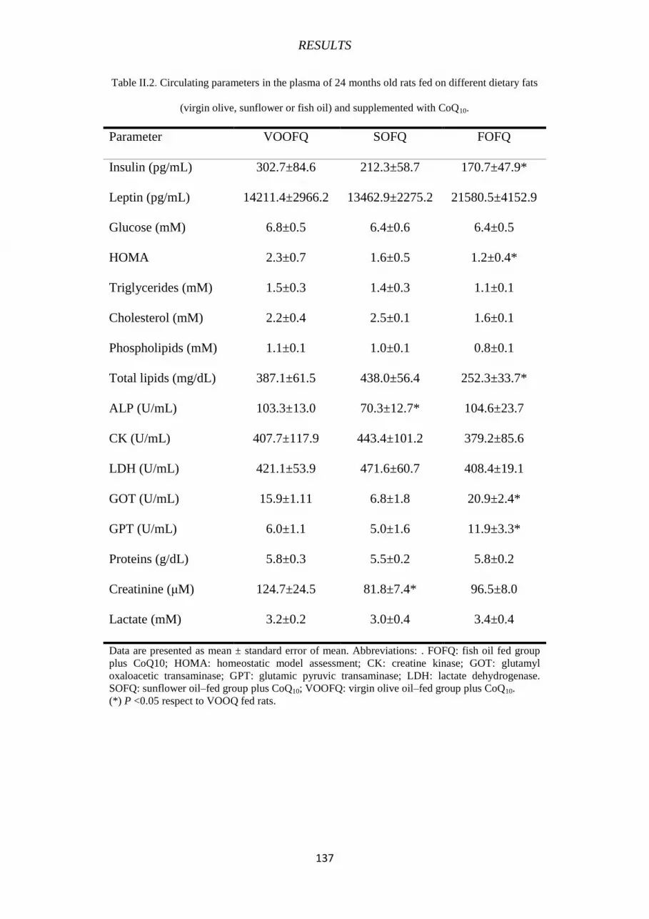
RESULTS
137
Table II.2. Circulating parameters in the plasma of 24 months old rats fed on different dietary fats
(virgin olive, sunflower or fish oil) and supplemented with CoQ10.
Parameter VOOFQ SOFQ FOFQ
Insulin (pg/mL) 302.7±84.6 212.3±58.7 170.7±47.9*
Leptin (pg/mL) 14211.4±2966.2 13462.9±2275.2 21580.5±4152.9
Glucose (mM) 6.8±0.5 6.4±0.6 6.4±0.5
HOMA 2.3±0.7 1.6±0.5 1.2±0.4*
Triglycerides (mM) 1.5±0.3 1.4±0.3 1.1±0.1
Cholesterol (mM) 2.2±0.4 2.5±0.1 1.6±0.1
Phospholipids (mM) 1.1±0.1 1.0±0.1 0.8±0.1
Total lipids (mg/dL) 387.1±61.5 438.0±56.4 252.3±33.7*
ALP (U/mL) 103.3±13.0 70.3±12.7* 104.6±23.7
CK (U/mL) 407.7±117.9 443.4±101.2 379.2±85.6
LDH (U/mL) 421.1±53.9 471.6±60.7 408.4±19.1
GOT (U/mL) 15.9±1.11 6.8±1.8 20.9±2.4*
GPT (U/mL) 6.0±1.1 5.0±1.6 11.9±3.3*
Proteins (g/dL) 5.8±0.3 5.5±0.2 5.8±0.2
Creatinine (μM) 124.7±24.5 81.8±7.4* 96.5±8.0
Lactate (mM) 3.2±0.2 3.0±0.4 3.4±0.4
Data are presented as mean ± standard error of mean. Abbreviations: . FOFQ: fish oil fed group
plus CoQ10; HOMA: homeostatic model assessment; CK: creatine kinase; GOT: glutamyl
oxaloacetic transaminase; GPT: glutamic pyruvic transaminase; LDH: lactate dehydrogenase.
SOFQ: sunflower oil–fed group plus CoQ10; VOOFQ: virgin olive oil–fed group plus CoQ10.
(*) P <0.05 respect to VOOQ fed rats.

RESULTS
138
II.3 HISTOLOGICAL EXAMINATION
II.3.1 Pancreatic Parenchyma
FOFQ group showed the highest number of alterations in pancreatic
parenchyma. It was observed a clear acinar atrophy (Figure II.1C) accompanied by
signs of inflammation (Figure II.1, F and I), and fibrosis, which was affecting the acinar
(Figure II.2c) and the peri-insular compartment (Figure II.2f). Moreover, evidences of
hyperplasia in the ductal region (Figure II.2i) as well as certain degree of acinar fat
infiltration (Figure 1L) were detected in FOFQ group. Concerning SOFQ and VOOFQ
groups, these showed a lower degree of alterations, without significant differences
between them for most of the analyzed parameters (Figure II.3). VOOFQ group showed
light signs of fibrosis (Figure II.2a), slight evidences of fat depots (Figure II.1j, k) and
the ductal tissue and pancreatic blood vessel did not present signs of metaplasia and
hyperplasia (Figure II.2 h, g). The only remarkable difference was that a slight degree of
acinar atrophy was detected in VOOFQ group (Figure II.1a) whereas in SOFQ there
were many samples which did not show such alteration (Figure II.1b).
II.3.2 Immunohistochemical assessment of pancreatic islets
Likewise as its non-supplemented counterparts, immunohistochemical analyses
were performed in order to ascertain cell type and distribution in pancreatic islets. Thus,
insulin and glucagon immunohistochemistry, identifying β (Figure II.4a) and α-cells
(Figure II.4b) were evaluated. Supplemented groups did not reported significant
differences concerning these parameters (Figure II.4c).
II.4 PANCREATIC INSULIN AND GLUCAGON CONTENT
The purpose of this analysis was to gain more insight into how histological
changes could affect the endocrine pancreas compartment. For this reason, the main
hormones produced by this compartment (insulin and glucagon) were measured. All
groups obtained similar contents of both hormones, without significant differences
among them (Table II.3).

RESULTS
139
Table II.3: Total insulin and glucagon contents in pancreas VOOF, SOF and FOF rats supplemented
with coenzyme Q at the end of the experimental procedure (2 years).
Parameter VOOFQ SOFQ FOFQ
Insulin (pg/μg protein) 18902.5±6989.5 14144±4588.5 19576±3720.3
Glucagon (pg/μg protein) 151.8±12.8 143±37.2 129.6±23.8
Data are presented as mean ± standard error of mean. Abbreviations: FOFQ: fish oil fed group
supplemented on CoQ10; SOFQ: sunflower oil fed group supplemented on CoQ10; VOOF: virgin
olive oil fed group supplemented on CoQ10. (*) P < .05 respect to VOOFQ rats.

RESULTS
140
Fig
ure
II.
1.
His
tolo
gic
al s
ecti
ons
of
pancre
as f
rom
24
mo
nth
s o
ld r
ats
fed
on d
iffe
rent
die
tary
fat
sourc
es
(vir
gin
oli
ve,
su
nfl
ow
er o
r fi
sh o
ils)
sup
ple
men
ted
wit
h
Co
enzym
e Q
10
af
ter
hem
ato
xil
in
and
eo
sin
stai
nin
g
for
asse
ssm
ent
of
acin
ar
atro
ph
y,
par
ench
ym
al
and
p
erid
uct
al
infl
am
mat
ion
and
ac
inar
fa
t in
filt
rati
on,
resp
ecti
vel
y.
(A)
Min
imal
acin
ar a
tro
ph
y (
ind
icat
ed b
y a
yel
low
cir
cle)
; (B
) N
orm
al p
ancr
eati
c ti
ssues
; (C
) S
ever
e a
cinar
atr
op
hy,
gra
de
4;
(D,E
) A
bse
nce
of
infl
am
mati
on;
(F)
Infl
am
mat
ory
cel
ls (
its
pre
sence
is
ind
icat
ed b
y w
hit
e ar
row
s);
(G)
Ab
sence
of
infl
am
mat
ion;
(H)
Min
imal
in
flam
mat
ion w
ith s
om
e in
flam
mat
ory
cell
s (i
nd
icat
ed b
y a
wh
ite
arro
w);
(I)
Mo
re s
ever
e i
nfi
ltra
te (
ind
icat
ed b
y a
bla
ck a
rro
w)
aro
un
d t
he
duct
(in
dic
ated
by a
blu
e ar
row
); (
J,K
) V
ery l
ow
nu
mb
er o
f fa
tty
cell
s and
aggre
gat
es (
gra
de
1–
2)
(ind
icat
ed b
y w
hit
e ar
row
s);
(L)
Sev
ere
fat
dep
osi
ts (
gra
de
3–4). Y
ellow bar = 200 μm; Black bar = 50 μm; Green
bar = 500 μm.
FO
FQ
: fi
sh o
il f
ed g
roup
plu
s C
oQ
10;
SO
FQ
: su
nfl
ow
er o
il–
fed
gro
up
plu
s C
oQ
10;
VO
OF
Q:
vir
gin
oli
ve
oil–
fed
gro
up
plu
s C
oQ
10.

RESULTS
141
Fig
ure
II.
2.
His
tolo
gic
al s
ecti
ons
of
pan
cre
as f
rom
24
mo
nth
s o
ld r
ats
fed
on d
iffe
rent
die
tary
fat
so
urc
es (
vir
gin
oli
ve,
su
nfl
ow
er o
r fi
sh o
ils)
sup
ple
men
ted
wit
h
Co
enzym
e Q
10 a
fter
Tri
chro
me
Mass
on (
a–
f),
Hem
ato
xil
in a
nd
eo
sin
(g–
i) a
nd
Per
ls s
tain
ing (
j–l)
fo
r as
sess
ment
of
acin
ar a
nd
per
i-in
sula
r fi
bro
sis,
duct
al
hyp
erp
lasi
a and
iro
n d
epo
sits
, re
spec
tivel
y.
(a)
Min
imal
acin
ar f
ibro
sis
(ind
icat
ed b
y a
yell
ow
cir
cle)
; (b
) N
orm
al p
ancr
eat
ic a
cini;
(c)
Aci
nar
fib
rosi
s g
rad
e 3
; (d
,e)
Ab
sence
of
per
i-in
sula
r fi
bro
sis;
(f)
Mo
der
ate
fib
rosi
s; (
g,h
) A
bse
nce
of
duct
ula
r h
yp
erp
lasi
a; (
i) D
uct
ula
r h
yp
erp
lasi
a (
ind
icat
ed b
y b
lack a
rro
ws)
; (j
,k)
Ver
y l
ow
nu
mb
er o
f m
acro
phag
es
stai
ned
fo
r hem
osi
der
in (
gra
de
1)
wit
h p
eri-
insu
lar
dis
trib
uti
on (
ind
icat
ed b
y w
hit
e ar
row
s);
(l)
Hig
h n
um
ber
of
mac
rop
hag
es s
tain
ed f
or
hem
osi
der
in (
gra
de
3–
4),
(hem
osi
der
in d
epo
site
d a
re i
nd
icat
ed b
y w
hit
e ar
row
s).
Blu
e b
ar =
10
0 μ
m.
FO
FQ
: fi
sh o
il f
ed g
roup
plu
s C
oQ
10;
SO
FQ
: su
nfl
ow
er o
il–
fed
gro
up
plu
s C
oQ
10;
VO
OF
Q:
vir
gin
oli
ve
oil–
fed
gro
up
plu
s C
oQ
10.

RESULTS
142
Figure II.3. Data from histological evaluations of pancreas from rats fed on different dietary fat sources
(virgin olive, sunflower or fish oils) supplemented with Coenzyme Q10 . Data are presented as mean ±
SEM. A.U. = arbitrary units. (*) P < 0.05 in respect to VOOFQ fed rats. FOFQ: fish oil fed group plus
CoQ10; SOFQ: sunflower oil–fed group plus CoQ10; VOOFQ: virgin olive oil–fed group plus CoQ10.
VOOFQ SOOFQ FOFQ VOOFQ SOOFQ FOFQ

RESULTS
143
Fig
ure
II.
4.
Imm
uno
his
toch
em
ical
ass
ess
men
t in
the
pan
crea
s o
f 2
4 m
onth
s o
ld r
ats
fed
on d
iffe
rent
die
tary
fat
so
urc
es (
vir
gin
oli
ve,
su
nfl
ow
er o
r fi
sh
oil
s) s
up
ple
mente
d w
ith C
oen
zym
e Q
10.
(a)
His
tolo
gic
al s
ect
ions
wit
h p
ancr
eati
c is
les
imm
uno
stai
ned
fo
r glu
cago
n;
(b)
His
tolo
gic
al s
ecti
ons
wit
h
pan
crea
tic isles im
munostained for insu
lin; (c) Quantitative data from histological evaluations of α
- and β
-cel
l p
osi
tiv
e d
ensi
ty a
nd
are
a. D
ata
are
presented as mea
n ± SEM
. Scare bar = 100 μm.
FO
FQ
: fi
sh o
il f
ed g
roup
plu
s C
oQ
10;
SO
FQ
: su
nfl
ow
er o
il–
fed
gro
up
plu
s C
oQ
10;
VO
OF
Q:
vir
gin
oli
ve
oil–
fed
gro
up
plu
s C
oQ
10.
VO
OFQ
SO
OFQ
F
OFQ
V
OO
FQ
S
OO
FQ
FO
FQ
VO
OFQ
S
OO
FQ
FO
FQ
VO
OFQ
S
OO
FQ
FO
FQ


discussion


DISCUSSION
147
I. DIETARY FAT EFFECTS ON AGE-RELATED CHANGES OF PANCREAS
One of the aims of this study was to investigate the role of life-long feeding on
diets varying only in the fat source. This type of dietary habits, in which diet is the same
for the whole life is unlike to happens as it in the human population. However, people
ha preferences for certain foods that constitute the basis of their diet. Dietary fat is one
of these foods and a rat model might be very useful as a direct and easy method to
observe harmful and beneficial effects derived from the exclusive intake of a particular
fat type for a long period. Likewise, the rat model let us accessing to certain type of
biological samples (pancreas in this case) which would be more difficult in humans, and
therefore this animal model can help us to understand how diet affects organ histology
in different histopathological situations. In order to avoid pathological situations such
obesity or type 2 diabetes mellitus, rats were administered a balanced diet whose caloric
components were controlled. In this sense, circulating parameters were measured (Table
I.2). Other variables that could contribute to the observed differences, such as genetic
predisposition or the rest of dietary nutrients were overruled because they should
contribute equally in all the experimental groups as they were not modified. Dietary fats
administered to each experimental group presented particular characteristics. Although
fish oil had the highest amount of saturated fatty acids (Table I.1), this is not a key point
because the circulating saturated fatty acid profile were similar in the three experimental
groups after long-term consumption. Concerning unsaturated fatty acids, virgin olive oil
group presented the highest amount of circulating MUFA, sunflower oil presented the
highest amount of n-6 PUFA and fish oil group presented the highest amount of n-3
PUFA. This confirms the adaptation of animals to dietary fatty component, and could
led to assume that found differences between groups might be attributed, at least in part,
to dietary fat.
A remarkable finding of this study was the fact that the type of dietary fat
affected to the exocrine and endocrine compartments of the pancreas in a different way.
In this context, having VOOF islets as reference, SOF islets showed the highest number
of β-cells, increased insulin content and hyperleptinemia. These signs along with other
parameters are typical in alterations such as obesity, glucose intolerance, and insulin
resistance, disruption of the adipoinsular axis or prediabetes (385). Nevertheless, SOF
rats presented balanced levels of circulating glucose and insulin, suggesting there is no a
prepathological state (386). In this sense, it cannot be given a clear explanation about
the hyperleptinemia and if it was somehow correlated with the high content of insulin in
islets. A possible explanation (only concerning islet tissue) may be that the high number
of β-cells in SOF compared with VOOF could be interpreted as an increased replicative
capacity of β-cells or a longer life span of these cells due to a reduced apoptosis.
However, it has been well documented that β-cell replication decreases with age in
rodents and humans (387, 388), but in compensation presents a minimal apoptosis
(389). Proliferation events were very similar in all groups according to Ki-67+ assay on
pancreases. Likewise, the expression of apoptotic markers were very similar in all
DISCUSSION
148
groups for Bad and Bcl2, but the expression of TNF-α apoptotic marker was lower in
VOOF than SOF and FOF. All these observations together may suggest that the
mitochondrial apoptotic pathway was not active in any of the of the animal groups, but
in the cytoplasmic pathway, where TNF-α is most likely involved (390), it seems to be
instrumental in the changes observed in SOF and FOF groups. In SOF group, it is
probably that many of the positive insulin cells observed might be apoptotic, but it is
required to perform functional analyses such as experiments in isolated islets so as to
verify this hypothesis. Moreover SOF and FOF also obtained higher levels of oxidative
stress by protein carbonyl assay compared with VOOF. So, these observations on
endocrine pancreas are preliminary data, and therefore should be interpreted with
caution.
Concerning the exocrine pancreas, it seems to be affected by a fish oil diet and
in a lower way by a sunflower oil diet. The histopathological features typical observed
in pancreatic fibrosis in the elderly (391) are very similar to what it was observed on
FOF animals. In turn, consumption of olive oil at long-term suggests a possible delay in
exocrine pancreatic aging. It is clear that the development of pancreatic fibrosis is a
complex process where pancreatic stellate cells play an instrumental role. These cells
which were initially characterized in rat and human pancreas (392, 393) are presented
in the periacinar region and display droplets of lipids within the cytoplasm, and keeping
a close connection with liver stellate cells which lead to hepatic fibrosis (394). The
initiation of a fibrosis process requires the releasing of cytokines and growth factor from
pancreatic resident cells or infiltrated inflammatory cells such as macrophages (395-
397). If macrophage phagocyte damage cells release additional cytokines, it triggers to
proliferation and transformation induction of stellate cells into myofibroblasts, which
are responsible for producing extracellular matrix deposition and therefore fibrosis
(398). Accordingly, we hypothesize that nutrients may take part in modulating the
function of these cells, as it was described for glucose (399). So it is tempting to
speculate that certain types of fatty acids, like those present in fish oil, might affect
stellate cell function. In any case, further experiments are required to confirm this
hypothesis.
In summary, according to results from the present study, the endocrine
compartment of the pancreas was mostly negatively affected by a diet based on
sunflower oil, meanwhile the exocrine compartment was affected in a greater extent by
a fish oil diet. According to previous studies, it was observed that virgin olive oil
preserved the integrity and functionality of the pancreas in the elderly, reinforcing its
beneficial effects, especially in endocrine pancreas. In this sense, interventional and
epidemiological studies proved that diets rich in MUFA are able to improve insulin
sensitivity (400-404) and lower glycaemia and insulin requirements in type 2 diabetes
patients (405). Likewise, in a cross-sectional study performed in South of Spain, an oral
glucose tolerance test performed in 530 individuals in order to calculate insulin
resistance using homeostasis model assessment showed a positive association between
MUFA consumption and β-cell function (406). Nevertheless, it is not clear the precise

DISCUSSION
149
mechanisms by which virgin olive oil induced this beneficial effect on endocrine
pancreas. This must be investigated in the future.. Virgin olive oil is observed to have
positive effects in other tissues and organs, which indirectly may improve endocrine and
exocrine pancreas (407). In this sense, virgin olive oil is rich in antioxidants, such as
vitamin E or phenolic compounds, and antithrombotic agents that can improve
endothelial function and reduce the risk of cardiovascular disease, insulin resistance
(408-410) and possibly, pancreatitis (this study). Therefore, mechanisms responsible for
virgin olive oil effects could be more complex than expected, and may be due to the
synergistic effect of several compounds and not exclusively by oleic acid or MUFA.

DISCUSSION
150
II. DIETARY COQ EFFECTS ON AGE-RELATED CHANGES OF PANCREAS
The second objective of the present study aimed to gain more insight about the
effects after the administration of a chronic low dosage of CoQ10 in the animals fed on
the same diets as for the first objective and in the same conditions. According to that,
animals from this second part of the study were fed in the same schedule as for the first
part but with a supplement of CoQ10.
Like in the previous study, CoQ10-supplemented animals were fed on a
controlled caloric intake pattern in order to avoid obesity or type 2 diabetes. Also, food
spillage was indirectly monitored. FOFQ group displayed significant higher weight at
sacrifice compared with the other two groups (non-supplemented animals fed on fish oil
led to a higher weight too, but differences were not significant). CoQ10-supplemented
rats were analyzed for plasma fatty acid profile in order to ascertain adaptation to
dietary fat. Results from fatty acid profile analysis lead to conclude that animals
properly adapted to different dietary fat sources.
Concerning circulating hormone levels, when compared with non-supplemented
rats, it was observed that SOF led to higher leptin concentration in comparison with the
other two dietary fat sources. However, as for CoQ10-supplemented animals are
concerned, no differences were found between FOFQ and the other two groups of rats
supplemented on CoQ10. Somehow CoQ10 supplementation led to lower leptin levels
associated to a SO-based diet. Leptin is produced and released primarily by adipocytes
whose circulating levels are directly proportional to total body fat mass (386), but also
to the percentage of body fat and BMI (411). With the present methodological
approach, it is not possible to establish a clear explanation about the CoQ effect on
circulating leptin levels when rats are maintaining on a sunflower oil-based diet. On the
other hand, lower levels of insulin, and lower HOMA index were found in FOFQ
animals in respect to VOOFQ rats. However, no clear differences were observed
between animals fed on similar dietary fat in absence of CoQ supplementation for the
most of the parameters analyzed. Thus, from the point of view of circulating hormones
and biochemical parameters, it is very difficult establish the role of CoQ
supplementation in the three studied fats, but in any case neither group of supplemented
rats showed a significant pathological situation of insulin resistance or loss of insulin
sensitivity (412, 413).
Histological assessment of exocrine pancreas showed that FOFQ animals
presented similar alterations in pancreas parenchyma as did non-supplemented rats fed
on the same dietary fat. So CoQ10 supplementation did not prevented alterations due to a
life-long feeding non the n-3PUFA based diet. No significant differences on this tissue
compartment were observed between SOFQ and VOOFQ. When compared
supplemented and non-supplemented groups for these two dietary fats, better results
were found for sunflower oil fed animals when they were supplemented on CoQ10.
DISCUSSION
151
Virgin olive oil fed animals showed similar results irrespective of the supplementation
with CoQ10. The histological alteration degree observed in fish oil fed animals,
supplemented or not, resemble to those observed in pancreatic fibrosis of the elderly
(391). The higher degree of inflammation and fibrosis in pancreas found in animals fed
on fish oil seems contradictory with those from animals studies where n-3 fatty acid-
rich diets shows anti-inflammatory effects, although pancreas was not directly studied
(414-417). Other studies have indicated an absence anti-inflammatory effect after
feeding on n-3PUFA (418) or even pro-oxidant and pro-inflammatory effects for these
fatty acids (419, 420). In addition to this, it has been also observed that some treatments
based on these fatty acids can decrease life-span (421-424). So, the amount, the animal
model, age or treatment time seems to be pivotal factors affecting n-3PUFA effects that
need to be explored in more detail. Conversely, long-term consumption of diets based
on virgin olive oil and sunflower oil supplemented with CoQ, would delay exocrine
pancreatic aging in the present model, as these animals did not displayed a typical
fibrotic pancreas of the elderly. In this sense, as previously explained above, pancreatic
stellate cells are a key factor on the fibrotic process at this organ. In such condition
oxidative stress is directly involved. Different antioxidant therapies have been tested
demonstrating decreased pancreatic stellate cells and proinflammatory factors both in
vitro (425, 426) and in vivo (395, 425-432). The findings from the present study can be
associated with such facts, so that the improvement of certain aging indicators produced
in SOFQ animals, might be due to the long-term addition of CoQ comparing with their
corresponding non-supplemented counterparts. Nevertheless, in animals fed on fish oil
and virgin olive oil, such supplementation seems to have no effect over pancreatic
parenchyma. On the one hand, it could be hypothesized that the fish oil effect on the
exocrine pancreas was due to a mechanism not related to oxidative stress, or at least that
this was not exclusive. On the other hand, it is possible that CoQ does not act as a
specific antioxidant for this particular process and/or not have enough to counteract fish
oil effects. Concerning virgin olive oil, it might be so beneficial for this pancreatic
compartment that CoQ supplements hardly have additional effects, but curiously the
degree of acinar atrophy was lower in sunflower oil fed rats supplemented with CoQ
which suggests that this combination could be even more beneficial. In any case, more
studies are needed in order to understand mechanisms underlying differences among
dietary fats and CoQ effects.
In regards to endocrine pancreas, it is interesting to note that CoQ-supplemented
groups did not show significant differences among them. However, for the non-
supplemented groups, sunflower oil fed animals led to significantly higher values, when
compared to those fed on virgin olive oil, for the β-cell area, as well as a higher number
of cell per islet. Age-related disorders of pancreatic endocrine function usually start
with a prediabetic state of insulin resistance in which the adaptive response of
pancreatic β-cells results in the presence of high levels of circulating insulin
(hyperinsulinemia) in order to maintain normoglycemia. Hyperinsulinemia can be
achieved by combining different mechanisms that involve expanding β-cells mass by
hyperplasia and hypertrophy, enhancing insulin biosynthesis and increasing insulin
DISCUSSION
152
secretion in response to sustained extracellular nutrients demand (433-437). However in
sunflower oil fed rats from the present study there are no signs of β-cells hypertrophy
nor hyperplasia at all. This finding suggests that CoQ addition to diet prevented age-
related β-cells alterations associated to long-life consumption of a sunflower oil rich
diet under our experimental conditions. Leptin has been shown to have a proliferative
effect on β-cells cultures under certain conditions (438-442). As previously mentioned,
it is not possible to establish an explanation for CoQ effect on leptin level, but this
effect on β-cells might derive from it, since this group displayed lower levels than in the
non-supplemented counterparts.
Results on pancreatic contents of insulin and glucagon, in spite of observing
similar values in the three groups, either insulin or glucagon, some inferences can be
extracted from these results. First, FOFQ animals in which alterations at the peri-insular
region level were observed, they did not yet lead to endocrine dysfunctions. Moreover,
compared with non-supplemented animals, it was observed that total insulin content of
supplemented rats fed on virgin olive oil and fish oil was double with respect to their
non-supplemented counterparts. However, for sunflower oil-fed rats both, supplemented
and non-supplemented, presented similar insulin content, although in non-supplemented
rats that was associated with a higher β-cell mass. It has been hypothesized that β-cell
hypertrophy could be an adaptive response to the aging situation in an attempt to
maintain correct insulin levels in an organ with diminished replicative capacities (440)
which would explain the observations done in non-supplemented rats. In turn, in CoQ-
supplemented rats, histological measures related to β-cells were similar among groups
and similar to those found in supplemented animals fed on fish or virgin olive oils. This
suggests that CoQ may contribute to specifically increase the content of insulin
regardless dietary fat type, but without affecting glucagon that maintained similar levels
among dietary groups in both studies. HOMA values, insulin circulating levels and
glycaemia were similar to those found in absence of CoQ, which suggests that there is
no decrease in insulin secretion levels or demands by other tissues. Another possibility
is that CoQ improves β-cells capacity to restore insulin reserves. However, because of
our experimental design focused pancreas, it was not possible to elucidate it.


CONCLUSIOns


157
CONCLUSIONS
1º Life-long feeding on a 4% of virgin olive oil led to a good maintaining of pancreatic
microstructure through aging. Both, exocrine and endocrine compartments were
benefited from the continued intake of this dietary fat when compared with the other fat
sources. Supplementation of virgin olive oil with coenzyme Q10 did not result in a
subsequent improvement in the results obtained with the non-supplemented fat.
2º The intake throughout life with fish oil, rich in n-3PUFA, as dietary fat, resulted in
alterations in exocrine pancreas compared to that observed with animals fed on MUFA
or n-6PUFA based diets. These alterations were not prevented by supplementation of
fish oil with a low dosage of coenzyme Q10.
3º The use of sunflower oil as the exclusive dietary fat source throughout life led to
alterations in the endocrine pancreas during aging, in relation to that observed in
animals fed on virgin olive or fish oils. Sunflower oil supplementation with coenzyme
Q10 prevented such alterations, approaching results to those found after the intake of
virgin olive oil.
4º Despite that diets with a unique fat source over very long periods of time are not
likely to occur in humans, animal models like the one presented here, could be very
useful to examine possible effects of fat types predominant in a particular diet. In
addition, these models could help to understand how diet can model organ histology in
different physiopathological situations considering the difficulty to obtain certain
human samples such as pancreas. Based on this approach, it was reported that different
pancreatic compartments were affected according to the fat composition consumed,
emphasizing the importance of dietary fatty acids in determining pancreatic structure.
Moreover, chronic CoQ supplementation together with dietary fat type could play an
important role in modulating aging in the pancreas, affecting different compartments of
this organ in opposite ways.

158
CONCLUSIONES
1º La alimentación de por vida con un 4% de aceite de oliva virgen dio lugar a un
adecuado mantenimiento de la microestructura del páncreas durante el envejecimiento.
Tanto los compartimentos endocrino como exocrino se beneficiaron de la ingesta
continuada de esta grasa de la dieta en comparación con las otras fuentes grasas. La
suplementación de aceite de oliva virgen con coenzima Q10 no dio lugar a una posterior
mejora de los resultados obtenidos con la grasa sin suplementar.
2º La ingesta durante toda la vida de aceite de pescado, rico en ácidos grasos
poliinsaturados de la serie n-3, dio lugar a alteraciones en el páncreas exocrino en
comparación con lo observado en los animales alimentados mayoritariamente con
dietas basadas en ácidos grasos monoinsaturados o poliinsaturados de la serie n-6. Estas
alteraciones no fueron impedidas por la administración de aceite de pescado
suplementado con una dosis baja de coenzima Q10.
3º El uso de aceite de girasol como fuente de grasa exclusiva en la dieta a lo largo de
toda la vida condujo a alteraciones en el páncreas endocrino durante el envejecimiento,
en relación a la observado en animales alimentados con aceites de oliva virgen o
pescado. La administración de coenzima Q10 junto al aceite de girasol evitó tales
alteraciones, acercándose entonces a los resultados obtenidos tras la ingesta de aceite de
oliva virgen.
4º A pesar de que el seguimiento de una dieta basada en una única fuente de grasa
durante períodos de tiempo muy largos es poco probable que ocurra en los seres
humanos, los modelos animales como el que aquí se presenta, podrían ser de gran
utilidad a la hora de examinar los posibles efectos de un tipo de grasa predominante en
una dieta en particular. Además, estos modelos podrían ayudar a entender cómo la dieta
puede modelar la histología de un órgano bajo diferentes situaciones fisiopatológicas
considerando la dificultad para obtener ciertas muestras humanas, tales como ocurre con
el páncreas. Sobre la base de este enfoque, se ha podido constatar que los diferentes
compartimentos del páncreas se vieron afectados de acuerdo con la composición de la
grasa consumida, haciendo hincapié en la importancia de los ácidos grasos de la dieta en
la determinación de la estructura de este órgano. Por otra parte, la administración
crónica de suplementos de CoQ10 a dosis bajas junto con el tipo de grasa de la dieta
podría desempeñar un papel importante en la modulación de envejecimiento en el
páncreas, afectando a los distintos compartimentos de este órgano de manera a veces
opuesta.

159


REFERENCES


REFERENCES
163
1.Quiles JL, Ochoa JJ, Huertas JR, Mataix J. Coenzyme Q supplementation protects
from age-related DNA double-strand breaks and increases lifespan in rats fed on a
PUFA-rich diet. Experimental gerontology. 2004;39(2):189-94.
2.Halliwell B, Gutteridge JMC. Free radicals in biology and medicine. Oxford: Oxford
university press; 1999.
3.Sohal RS, Mockett RJ, Orr WC. Mechanisms of aging: an appraisal of the oxidative
stress hypothesis. Free radical biology & medicine. 2002;33(5):575-86.
4.McCord JM, Fridovich I. Superoxide dismutase. An enzymic function for
erythrocuprein (hemocuprein). The Journal of biological chemistry. 1969;244(22):6049-
55.
5.Yu BP. Cellular defenses against damage from reactive oxygen species. Physiological
reviews. 1994;74(1):139-62.
6.Harman D. The biologic clock: the mitochondria? Journal of the American geriatrics
society. 1972;20(4):145-7.
7.Chance B, Sies H, Boveris A. Hydroperoxide metabolism in mammalian organs.
Physiological reviews. 1979;59(3):527-605.
8.Kapahi P, Boulton ME, Kirkwood TB. Positive correlation between mammalian life
span and cellular resistance to stress. Free radical biology & medicine. 1999;26(5-
6):495-500.
9.Miquel J, Economos AC, Fleming J, Johnson JE, Jr. Mitochondrial role in cell aging.
Experimental gerontology. 1980;15(6):575-91.
10.Gutiérrez-Robledo LM. Envejecimiento. Rev Fac Med. 1998;41(5):198-206.
11.Kirkwood TB, Austad SN. Why do we age? Nature. 2000;408(6809):233-8.
12.Medvedev ZA. An attempt at a rational classification of theories of ageing.
Biological reviews of the Cambridge philosophical society. 1990;65(3):375-98.
13.Finch CE, Ruvkun G. The genetics of aging. Annual review of genomics and human
genetics. 2001;2:435-62.
14.Lithgow GJ, Kirkwood TB. Mechanisms and evolution of aging. Science.
1996;273(5271):80.
15.Lee CK, Klopp RG, Weindruch R, Prolla TA. Gene expression profile of aging and
its retardation by caloric restriction. Science. 1999;285(5432):1390-3.
16.Kirkwood TB, Rose MR. Evolution of senescence: late survival sacrificed for
reproduction. Philosophical transactions of the Royal society of London series B,
biological sciences. 1991;332(1262):15-24.
17.Guarente L, Kenyon C. Genetic pathways that regulate ageing in model organisms.
Nature. 2000;408(6809):255-62.
18.Kirkwood TB. DNA, mutations and aging. Mutation research. 1989;219(1):1-7.
19.Catania J, Fairweather, DS. Genetic and genome factors in aging. Reviews in clinical
gerontology,. 1991;1:99-111.
20.Mikhelson VM. Replicative mosaicism might explain the seeming contradictions in
the telomere theory of aging. Mechanisms of ageing and development.
2001;122(13):1361-5.
21.Aviv A, Aviv H. Telomeres, hidden mosaicism, loss of heterozygosity, and complex
genetic traits. Human genetics. 1998;103(1):2-4.
22.Finkel T, Holbrook NJ. Oxidants, oxidative stress and the biology of ageing. Nature.
2000;408(6809):239-47.
23.Roth GS, Ingram DK, Lane MA. Calorie restriction in primates: will it work and
how will we know? Journal of the American geriatrics society. 1999;47(7):896-903.

REFERENCES
164
24.Miquel J, Economos AC. Favorable effects of the antioxidants sodium and
magnesium thiazolidine carboxylate on the vitality and life span of Drosophila and
mice. Experimental gerontology. 1979;14(5):279-85.
25.Furukawa T, Meydani SN, Blumberg JB. Reversal of age-associated decline in
immune responsiveness by dietary glutathione supplementation in mice. Mechanisms of
ageing and development. 1987;38(2):107-17.
26.Sastre J, Pallardo FV, Garcia de la Asuncion J, Vina J. Mitochondria, oxidative
stress and aging. Free radical research. 2000;32(3):189-98.
27.Quiles JL, Ochoa JJ, Ramirez-Tortosa C, Battino M, Huertas JR, Martin Y, et al.
Dietary fat type (virgin olive vs. sunflower oils) affects age-related changes in DNA
double-strand-breaks, antioxidant capacity and blood lipids in rats. Experimental
gerontology. 2004;39(8):1189-98.
28.Melov S. Therapeutics against mitochondrial oxidative stress in animal models of
aging. Annals of the New York academy of sciences. 2002;959:330-40.
29.Clandinin MT, Field CJ, Hargreaves K, Morson L, Zsigmond E. Role of diet fat in
subcellular structure and function. Canadian journal of physiology and pharmacology.
1985;63(5):546-56.
30.Quiles JL, Huertas JR, Manas M, Ochoa JJ, Battino M, Mataix J. Oxidative stress
induced by exercise and dietary fat modulates the coenzyme Q and vitamin A balance
between plasma and mitochondria. International journal for vitamin and nutrition
research Internationale Zeitschrift fur Vitamin- und Ernahrungsforschung Journal
international de vitaminologie et de nutrition. 1999;69(4):243-9.
31.Mataix J, Quiles JL, Huertas JR, Battino M, Manas M. Tissue specific interactions of
exercise, dietary fatty acids, and vitamin E in lipid peroxidation. Free radical biology &
medicine. 1998;24(4):511-21.
32.Suzuki H, Yamazaki M, Arai S, Nagao A, Terao J. Effect of lard, palm and rapeseed
oils life conservation in aged mice. Mechanisms of ageing and development.
1991;60(3):267-74.
33.Giron MD, Mataix FJ, Suarez MD. Long-term effects of dietary monounsaturated
and polyunsaturated fatty acids on the lipid composition of erythrocyte membranes in
dogs. Comparative biochemistry and physiology Comparative physiology.
1992;102(1):197-201.
34.Navarro MD, Periago JL, Pita ML, Hortelano P. The n-3 polyunsaturated fatty acid
levels in rat tissue lipids increase in response to dietary olive oil relative to sunflower
oil. Lipids. 1994;29(12):845-9.
35.Escudero A, Montilla JC, Garcia JM, Sanchez-Quevedo MC, Periago JL, Hortelano
P, et al. Effect of dietary (n-9), (n-6) and (n-3) fatty acids on membrane lipid
composition and morphology of rat erythrocytes. Biochimica et biophysica acta.
1998;1394(1):65-73.
36.Periago JL, Suarez MD, Pita ML. Effect of dietary olive oil, corn oil and medium-
chain triglycerides on the lipid composition of rat red blood cell membranes. The
Journal of nutrition. 1990;120(9):986-94.
37.Spector AA, Yorek MA. Membrane lipid composition and cellular function. Journal
of lipid research. 1985;26(9):1015-35.
38.Masi I, Giani E, Galli C, Tremoli E, Sirtori CR. Diets rich in saturated,
monounsaturated and polyunsaturated fatty acids differently affect plasma lipids,
platelet and arterial wall eicosanoids in rabbits. Annals of nutrition & metabolism.
1986;30(1):66-72.
39.Seiquer I, Manas M, Martinez-Victoria E, Ballesta MC, Mataix FJ. Long-term
influence of dietary fat (sunflower oil, olive oil, lard and fish oil) in the serum fatty acid

REFERENCES
165
composition and in the different lipidic fractions, in miniature swine. International
journal for vitamin and nutrition research Internationale Zeitschrift fur Vitamin- und
Ernahrungsforschung Journal international de vitaminologie et de nutrition.
1996;66(2):171-9.
40.Huertas JR, Battino M, Lenaz G, Mataix FJ. Changes in mitochondrial and
microsomal rat liver coenzyme Q9 and Q10 content induced by dietary fat and
endogenous lipid peroxidation. FEBS letters. 1991;287(1-2):89-92.
41.Ayre KJ, Hulbert AJ. Dietary fatty acid profile affects endurance in rats. Lipids.
1997;32(12):1265-70.
42.Gibson RA, McMurchie EJ, Charnock JS, Kneebone GM. Homeostatic control of
membrane fatty acid composition in the rat after dietary lipid treatment. Lipids.
1984;19(12):942-51.
43.Soriguer FJ, Tinahones FJ, Monzon A, Pareja A, Rojo-Martinez G, Moreno F, et al.
Varying incorporation of fatty acids into phospholipids from muscle, adipose and
pancreatic exocrine tissues and thymocytes in adult rats fed with diets rich in different
fatty acids. European journal of epidemiology. 2000;16(6):585-94.
44.Martin F. [effect of adaptation to the type of dietary fat and physical activity on the
composition of microsomal and mitochondrial membranes of various organs] [doctoral
thesis]. Granada: Universidad de Granada; 2002. Spanish.
45.Navarro MD, Hortelano P, Periago JL, Pita ML. Effect of dietary olive and
sunflower oils on the lipid composition of the aorta and platelets and on blood
eicosanoids in rats. Arteriosclerosis and thrombosis : a journal of vascular biology /
American heart association. 1992;12(7):830-5.
46.Ramirez-Tortosa C, Lopez-Pedrosa JM, Suarez A, Ros E, Mataix J, Gil A. Olive oil-
and fish oil-enriched diets modify plasma lipids and susceptibility of LDL to oxidative
modification in free-living male patients with peripheral vascular disease: the Spanish
Nutrition Study. The British journal of nutrition. 1999;82(1):31-9.
47.Aguilera CM, Ramirez-Tortosa MC, Mesa MD, Gil A. [Protective effect of
monounsaturated and polyunsaturated fatty acids on the development of cardiovascular
disease]. Nutricion hospitalaria. 2001;16(3):78-91.
48.Sirtori CR, Crepaldi G, Manzato E, Mancini M, Rivellese A, Paoletti R, et al. One-
year treatment with ethyl esters of n-3 fatty acids in patients with hypertriglyceridemia
and glucose intolerance: reduced triglyceridemia, total cholesterol and increased HDL-C
without glycemic alterations. Atherosclerosis. 1998;137(2):419-27.
49.Serrano P, Yago MD, Manas M, Calpena R, Mataix J, Martinez-Victoria E.
Influence of type of dietary fat (olive and sunflower oil) upon gastric acid secretion and
release of gastrin, somatostatin, and peptide YY in man. Digestive diseases and
sciences. 1997;42(3):626-33.
50.Naranjo JA, Martinez-Victoria E, Valverde A, Yago MD, Manas M. Effect of age on
the exocrine pancreatic secretion of the preruminant milk-fed goat. Archives of
physiology and biochemistry. 1997;105(2):144-50.
51.Sureda A, Arranz R, Iriondo A, Carreras E, Lahuerta JJ, Garcia-Conde J, et al.
Autologous stem-cell transplantation for Hodgkin's disease: results and prognostic
factors in 494 patients from the Grupo Espanol de Linfomas/Transplante Autologo de
Medula Osea Spanish Cooperative Group. Journal of clinical oncology : official journal
of the American Society of Clinical Oncology. 2001;19(5):1395-404.
52.Garg A. High-monounsaturated-fat diets for patients with diabetes mellitus: a meta-
analysis. The American journal of clinical nutrition. 1998;67(3 Suppl):577S-82S.
53.Quiles JL, Ochoa, J.J, Huertas, J.R., Mataix, J. [mitochondrial aspects of aging: role
of dietary fat type and oxidative stress]. Endocrinología y nutrición. 2004;51(3):107-20.

REFERENCES
166
54.Proceedings of a workshop on new developments on dietary fat and fiber in
carcinogenesis (optimal types and amounts of fat or fiber). Preventive medicine.
1987;16(4):449-595.
55.Rose MR, Mueller LD, Long AD. Pharmacology, genomics, and the evolutionary
biology of ageing. Free radical research. 2002;36(12):1293-7.
56.Quiles JL, Huertas JR, Manas M, Ochoa JJ, Battino M, Mataix J. Dietary fat type
and regular exercise affect mitochondrial composition and function depending on
specific tissue in the rat. Journal of bioenergetics and biomembranes. 2001;33(2):127-
34.
57.Sugimura T. Nutrition and dietary carcinogens. Carcinogenesis. 2000;21(3):387-95.
58.Moral R, Escrich R, Solanas M, Vela E, Ruiz de Villa MC, Escrich E. Diets high in
corn oil or extra-virgin olive oil differentially modify the gene expression profile of the
mammary gland and influence experimental breast cancer susceptibility. European
journal of nutrition. 2015.
59.Escrich E, Moral R, Solanas M. Olive oil, an essential component of the
Mediterranean diet, and breast cancer. Public health nutrition. 2011;14(12A):2323-32.
60.Granados-Principal S, Quiles JL, Ramirez-Tortosa C, Camacho-Corencia P,
Sanchez-Rovira P, Vera-Ramirez L, et al. Hydroxytyrosol inhibits growth and cell
proliferation and promotes high expression of sfrp4 in rat mammary tumours. Molecular
nutrition & food research. 2011;55 Suppl 1:S117-26.
61.Granados-Principal S, El-Azem N, Pamplona R, Ramirez-Tortosa C, Pulido-Moran
M, Vera-Ramirez L, et al. Hydroxytyrosol ameliorates oxidative stress and
mitochondrial dysfunction in doxorubicin-induced cardiotoxicity in rats with breast
cancer. Biochemical pharmacology. 2014;90(1):25-33.
62.Hashim YZ, Worthington J, Allsopp P, Ternan NG, Brown EM, McCann MJ, et al.
Virgin olive oil phenolics extract inhibit invasion of HT115 human colon cancer cells in
vitro and in vivo. Food & function. 2014;5(7):1513-9.
63.Freeman LR, Haley-Zitlin V, Rosenberger DS, Granholm AC. Damaging effects of a
high-fat diet to the brain and cognition: a review of proposed mechanisms. Nutritional
neuroscience. 2014;17(6):241-51.
64.Tchkonia T, Morbeck DE, Von Zglinicki T, Van Deursen J, Lustgarten J, Scrable H,
et al. Fat tissue, aging, and cellular senescence. Aging cell. 2010;9(5):667-84.
65.Barzilai N, Gupta G. Revisiting the role of fat mass in the life extension induced by
caloric restriction. The journals of gerontology Series A, Biological sciences and
medical sciences. 1999;54(3):B89-96; discussion B7-8.
66.Masoro EJ. Caloric restriction and aging: controversial issues. The journals of
gerontology Series A, Biological sciences and medical sciences. 2006;61(1):14-9.
67.Zamboni M, Rossi AP, Fantin F, Zamboni G, Chirumbolo S, Zoico E, et al. Adipose
tissue, diet and aging. Mechanisms of ageing and development. 2014;136-137:129-37.
68.Bielski BH, Arudi RL, Sutherland MW. A study of the reactivity of HO2/O2- with
unsaturated fatty acids. The Journal of biological chemistry. 1983;258(8):4759-61.
69.Pamplona R, Portero-Otin M, Ruiz C, Gredilla R, Herrero A, Barja G. Double bond
content of phospholipids and lipid peroxidation negatively correlate with maximum
longevity in the heart of mammals. Mechanisms of ageing and development.
2000;112(3):169-83.
70.Pamplona R. Membrane phospholipids, lipoxidative damage and molecular integrity:
a causal role in aging and longevity. Biochimica et biophysica acta.
2008;1777(10):1249-62.
71.Pamplona R, Prat J, Cadenas S, Rojas C, Perez-Campo R, Lopez Torres M, et al.
Low fatty acid unsaturation protects against lipid peroxidation in liver mitochondria

REFERENCES
167
from long-lived species: the pigeon and human case. Mechanisms of ageing and
development. 1996;86(1):53-66.
72.Pamplona R, Portero-Otin M, Riba D, Requena JR, Thorpe SR, Lopez-Torres M, et
al. Low fatty acid unsaturation: a mechanism for lowered lipoperoxidative modification
of tissue proteins in mammalian species with long life spans. The journals of
gerontology Series A, Biological sciences and medical sciences. 2000;55(6):B286-91.
73.Pamplona R, Barja G. Highly resistant macromolecular components and low rate of
generation of endogenous damage: two key traits of longevity. Ageing research reviews.
2007;6(3):189-210.
74.Hulbert AJ. Membrane fatty acids as pacemakers of animal metabolism. Lipids.
2007;42(9):811-9.
75.Pamplona R, Barja G, Portero-Otin M. Membrane fatty acid unsaturation, protection
against oxidative stress, and maximum life span: a homeoviscous-longevity adaptation?
Annals of the New York academy of sciences. 2002;959:475-90.
76.Pamplona R, Portero-Otin M, Sanz A, Requena J, Barja G. Modification of the
longevity-related degree of fatty acid unsaturation modulates oxidative damage to
proteins and mitochondrial DNA in liver and brain. Experimental gerontology.
2004;39(5):725-33.
77.Hulbert AJ, Pamplona R, Buffenstein R, Buttemer WA. Life and death: metabolic
rate, membrane composition, and life span of animals. Physiological reviews.
2007;87(4):1175-213.
78.Lopez-Dominguez JA, Ramsey JJ, Tran D, Imai DM, Koehne A, Laing ST, et al.
The Influence of Dietary Fat Source on Life Span in Calorie Restricted Mice. The
journals of gerontology Series A, Biological sciences and medical sciences.
2015;70(10):1181-8.
79.Ueda Y, Wang MF, Irei AV, Sarukura N, Sakai T, Hsu TF. Effect of dietary lipids
on longevity and memory in the SAMP8 mice. Journal of nutritional science and
vitaminology. 2011;57(1):36-41.
80.Lee D, Hwang W, Artan M, Jeong DE, Lee SJ. Effects of nutritional components on
aging. Aging cell. 2015;14(1):8-16.
81.Huhn S, Kharabian Masouleh S, Stumvoll M, Villringer A, Witte AV. Components
of a Mediterranean diet and their impact on cognitive functions in aging. Frontiers in
aging neuroscience. 2015;7:132.
82.Molfino A, Gioia G, Rossi Fanelli F, Muscaritoli M. The role for dietary omega-3
fatty acids supplementation in older adults. Nutrients. 2014;6(10):4058-73.
83.Denis I, Potier B, Heberden C, Vancassel S. Omega-3 polyunsaturated fatty acids
and brain aging. Current opinion in clinical nutrition and metabolic care.
2015;18(2):139-46.
84.Sergeant S, McQuail JA, Riddle DR, Chilton FH, Ortmeier SB, Jessup JA, et al.
Dietary fish oil modestly attenuates the effect of age on diastolic function but has no
effect on memory or brain inflammation in aged rats. The journals of gerontology Series
A, Biological sciences and medical sciences. 2011;66(5):521-33.
85.Ubeda N, Achon M, Varela-Moreiras G. Omega 3 fatty acids in the elderly. The
British journal of nutrition. 2012;107 Suppl 2:S137-51.
86.Pitozzi V, Jacomelli M, Catelan D, Servili M, Taticchi A, Biggeri A, et al. Long-
term dietary extra-virgin olive oil rich in polyphenols reverses age-related dysfunctions
in motor coordination and contextual memory in mice: role of oxidative stress.
Rejuvenation research. 2012;15(6):601-12.
87.Kondo K, Morino K, Nishio Y, Kondo M, Nakao K, Nakagawa F, et al. A fish-based
diet intervention improves endothelial function in postmenopausal women with type 2

REFERENCES
168
diabetes mellitus: a randomized crossover trial. Metabolism: clinical and experimental.
2014;63(7):930-40.
88.Haraldsdottir A, Torfadottir JE, Valdimarsdottir UA, Aspelund T, Harris TB, Launer
LJ, et al. Fish and fish-liver oil consumption in adolescence and midlife and risk of
CHD in older women. Public health nutrition. 2016;19(2):318-25.
89.Wilk JB, Tsai MY, Hanson NQ, Gaziano JM, Djousse L. Plasma and dietary omega-
3 fatty acids, fish intake, and heart failure risk in the Physicians' Health Study. The
American journal of clinical nutrition. 2012;96(4):882-8.
90.Reinders I, Murphy RA, Song X, Mitchell GF, Visser M, Cotch MF, et al. Higher
Plasma Phospholipid n-3 PUFAs, but Lower n-6 PUFAs, Are Associated with Lower
Pulse Wave Velocity among Older Adults. The Journal of nutrition.
2015;145(10):2317-24.
91.Djousse L, Akinkuolie AO, Wu JH, Ding EL, Gaziano JM. Fish consumption,
omega-3 fatty acids and risk of heart failure: a meta-analysis. Clinical nutrition.
2012;31(6):846-53.
92.Bilato C. n-3 fatty acids and cardiovascular disease: the story is not over yet. Aging
clinical and experimental research. 2013;25(4):357-63.
93.Patel JV, Tracey I, Hughes EA, Lip GY. Omega-3 polyunsaturated acids and
cardiovascular disease: notable ethnic differences or unfulfilled promise? Journal of
thrombosis and haemostasis : JTH. 2010;8(10):2095-104.
94.Amiot M. Olive oil and health effects: from epidemiological studies to the molecular
mechanisms of phenolic fraction. OCL. 2014;21(5):D512.
95.Guasch-Ferre M, Hruby A, Salas-Salvado J, Martinez-Gonzalez MA, Sun Q, Willett
WC, et al. Olive oil consumption and risk of type 2 diabetes in US women. The
American journal of clinical nutrition. 2015;102(2):479-86.
96.Rozati M, Barnett J, Wu D, Handelman G, Saltzman E, Wilson T, et al. Cardio-
metabolic and immunological impacts of extra virgin olive oil consumption in
overweight and obese older adults: a randomized controlled trial. Nutrition &
metabolism. 2015;12:28.
97.Helal O, Berrougui H, Loued S, Khalil A. Extra-virgin olive oil consumption
improves the capacity of HDL to mediate cholesterol efflux and increases ABCA1 and
ABCG1 expression in human macrophages. The British journal of nutrition.
2013;109(10):1844-55.
98.Loued S, Berrougui H, Componova P, Ikhlef S, Helal O, Khalil A. Extra-virgin olive
oil consumption reduces the age-related decrease in HDL and paraoxonase 1 anti-
inflammatory activities. The British journal of nutrition. 2013;110(7):1272-84.
99.Hohmann CD, Cramer H, Michalsen A, Kessler C, Steckhan N, Choi K, et al.
Effects of high phenolic olive oil on cardiovascular risk factors: A systematic review
and meta-analysis. Phytomedicine : international journal of phytotherapy and
phytopharmacology. 2015;22(6):631-40.
100.Welch AA, MacGregor AJ, Minihane AM, Skinner J, Valdes AA, Spector TD, et
al. Dietary fat and fatty acid profile are associated with indices of skeletal muscle mass
in women aged 18-79 years. The Journal of nutrition. 2014;144(3):327-34.
101.Reinders I, Song X, Visser M, Eiriksdottir G, Gudnason V, Sigurdsson S, et al.
Plasma phospholipid PUFAs are associated with greater muscle and knee extension
strength but not with changes in muscle parameters in older adults. The Journal of
nutrition. 2015;145(1):105-12.
102.Harris TB, Song X, Reinders I, Lang TF, Garcia ME, Siggeirsdottir K, et al. Plasma
phospholipid fatty acids and fish-oil consumption in relation to osteoporotic fracture

REFERENCES
169
risk in older adults: the Age, Gene/Environment Susceptibility Study. The American
journal of clinical nutrition. 2015;101(5):947-55.
103.Farina EK, Kiel DP, Roubenoff R, Schaefer EJ, Cupples LA, Tucker KL. Protective
effects of fish intake and interactive effects of long-chain polyunsaturated fatty acid
intakes on hip bone mineral density in older adults: the Framingham Osteoporosis
Study. The American journal of clinical nutrition. 2011;93(5):1142-51.
104.Bullon P, Battino M, Varela-Lopez A, Perez-Lopez P, Granados-Principal S,
Ramirez-Tortosa MC, et al. Diets based on virgin olive oil or fish oil but not on
sunflower oil prevent age-related alveolar bone resorption by mitochondrial-related
mechanisms. PloS one. 2013;8(9):e74234.
105.Crane FL, Hatefi Y, Lester RL, Widmer C. Isolation of a quinone from beef heart
mitochondria. Biochimica et biophysica acta. 1957;25(1):220-1.
106.Ernster L. Lipid peroxidation in biological membranes: mechanisms and
implications. In: Yagi, K. Active oxygens, lipid peroxides and antioxidants. Boca
Ratón: CRC Press; 1993. 1-38 p.
107.Lenaz G, Fato R, Degli Esposti M, Rugolo M, Parenti Castelli G. The essentiality
of coenzyme Q for bioenergetics and clinical medicine. Drugs under experimental and
clinical research. 1985;11(8):547-56.
108.Yamamoto Y, Yamashita S. Plasma ratio of ubiquinol and ubiquinone as a marker
of oxidative stress. Molecular aspects of medicine. 1997;18 Suppl:S79-84.
109.Lopez-Lluch G, Barroso MP, Martin SF, Fernandez-Ayala DJ, Gomez-Diaz C,
Villalba JM, et al. Role of plasma membrane coenzyme Q on the regulation of
apoptosis. BioFactors. 1999;9(2-4):171-7.
110.Grunler J, Ericsson J, Dallner G. Branch-point reactions in the biosynthesis of
cholesterol, dolichol, ubiquinone and prenylated proteins. Biochimica et biophysica
acta. 1994;1212(3):259-77.
111.Olson RE, Rudney H. Biosynthesis of ubiquinone. Vitamins and hormones.
1983;40:1-43.
112.Teclebrhan H, Olsson J, Swiezewska E, Dallner G. Biosynthesis of the side chain
of ubiquinone:trans-prenyltransferase in rat liver microsomes. The Journal of biological
chemistry. 1993;268(31):23081-6.
113.Poulter CD, Rilling, HC. Prenyl transferases and isomerases; In: Porter, J.
Biosynthesis of isoprenoide compounds. New York: John Wiley; 1981.
114.Aberg F, Appelkvist EL, Dallner G, Ernster L. Distribution and redox state of
ubiquinones in rat and human tissues. Archives of biochemistry and biophysics.
1992;295(2):230-4.
115.Appelkvist EL, Kalen, A., Dallner, G. G. Biosynthesis and regulation of coenzyme
Q. In: Biomedical and clinical aspects of coenzyme Q. Folkers, K., Litarru, G. P. Eds.,
Elsevier science publishers, 1991; 6:141-50. p. 141-50.
116.Yamamoto T, Shimizu S, Sugawara H, Momose K, Rudney H. Identification of
regulatory sites in the biosynthesis of ubiquinone in the perfused rat heart. Archives of
biochemistry and biophysics. 1989;269(1):86-92.
117.Fedurov VF. [Ubiquinone and sterol metabolism in the liver of rats maintained on a
diet short of aromatic amino acids]. Voprosy pitaniia. 1977(6):23-5.
118.Elmberger PG, Kalen A, Brunk UT, Dallner G. Discharge of newly-synthesized
dolichol and ubiquinone with lipoproteins to rat liver perfusate and to the bile. Lipids.
1989;24(11):919-30.
119.Marasco S, Salvatore, P., Franklin, L. [clinic use of coenzyme Q10]. Antioxidantes
y calidad de vida [Internet].

REFERENCES
170
120.Ramasarma, T. Metabolism of coenzime Q. In : Lenaz, G., ed. Coenzime Q.
Chichester: John Wiley, 1985; 131-42. .
121.Turunen M, Appelkvist EL, Sindelar P, Dallner G. Blood concentration of
coenzyme Q(10) increases in rats when esterified forms are administered. The Journal
of nutrition. 1999;129(12):2113-8.
122.Ulrich EL, Girvin ME, Cramer WA, Markley JL. Location and mobility of
ubiquinones of different chain lengths in artificial membrane vesicles. Biochemistry.
1985;24(10):2501-8.
123.Kalen A, Norling B, Appelkvist EL, Dallner G. Ubiquinone biosynthesis by the
microsomal fraction from rat liver. Biochimica et biophysica acta. 1987;926(1):70-8.
124.Aberg F, Zhang Y, Teclebrhan H, Appelkvist EL, Dallner G. Increases in tissue
levels of ubiquinone in association with peroxisome proliferation. Chemico-biological
interactions. 1996;99(1-3):205-18.
125.Turunen M, Sindelar P, Dallner G. Induction of endogenous coenzyme Q
biosynthesis by administration of peroxisomal inducers. BioFactors. 1999;9(2-4):131-9.
126.Ernster L, Dallner G. Biochemical, physiological and medical aspects of
ubiquinone function. Biochimica et biophysica acta. 1995;1271(1):195-204.
127.Brandt U. Proton translocation in the respiratory chain involving ubiquinone--a
hypothetical semiquinone switch mechanism for complex I. BioFactors. 1999;9(2-
4):95-101.
128.Yu CA, Zhang L, Deng KP, Tian H, Xia D, Kim H, et al. Structure and reaction
mechanisms of multifunctional mitochondrial cytochrome bc1 complex. BioFactors.
1999;9(2-4):103-9.
129.Gille L, Nohl H. The existence of a lysosomal redox chain and the role of
ubiquinone. Archives of biochemistry and biophysics. 2000;375(2):347-54.
130.Yokoyama H, Lingle DM, Crestanello JA, Kamelgard J, Kott BR, Momeni R, et al.
Coenzyme Q10 protects coronary endothelial function from ischemia reperfusion injury
via an antioxidant effect. Surgery. 1996;120(2):189-96.
131.Matsumoto, H. and Miyawaki F: Effect of Coenzyme Q10 pretreatment on
myocardial preservation. Heart Transplantation, 1984; 3: 160-165. .
132.Kalen A, Appelkvist EL, Dallner G. Age-related changes in the lipid compositions
of rat and human tissues. Lipids. 1989;24(7):579-84.
133.Aberg F, Zhang Y, Appelkvist EL, Dallner G. Effects of clofibrate, phthalates and
probucol on ubiquinone levels. Chemico-biological interactions. 1994;91(1):1-14.
134.Eggens I, Elmberger PG, Low P. Polyisoprenoid, cholesterol and ubiquinone levels
in human hepatocellular carcinomas. British journal of experimental pathology.
1989;70(1):83-92.
135.Guan Z, Soderberg M, Sindelar P, Prusiner SB, Kristensson K, Dallner G. Lipid
composition in scrapie-infected mouse brain: prion infection increases the levels of
dolichyl phosphate and ubiquinone. Journal of neurochemistry. 1996;66(1):277-85.
136.Halliwell B, Gutteridge JM, Cross CE. Free radicals, antioxidants, and human
disease: where are we now? The Journal of laboratory and clinical medicine.
1992;119(6):598-620.
137.Beal MF, Matthews RT. Coenzyme Q10 in the central nervous system and its
potential usefulness in the treatment of neurodegenerative diseases. Molecular aspects
of medicine. 1997;18 Suppl:S169-79.
138.Dallner G, Sindelar PJ. Regulation of ubiquinone metabolism. Free radical biology
& medicine. 2000;29(3-4):285-94.

REFERENCES
171
139.Dallner, G.: Regulatory aspects of coenzyme Q metabolism. Stockholm University:
Abstracts of the second conference of the International coenzyme Q10 association.
Frankfurt, Germany, 2000a; 1-3.
140.Thelin A, Schedin S, Dallner G. Half-life of ubiquinone-9 in rat tissues. FEBS
letters. 1992;313(2):118-20.
141.Andersson M, Elmberger PG, Edlund C, Kristensson K, Dallner G. Rates of
cholesterol, ubiquinone, dolichol and dolichyl-P biosynthesis in rat brain slices. FEBS
letters. 1990;269(1):15-8.
142.Ding H, Moser CC, Robertson DE, Tokito MK, Daldal F, Dutton PL. Ubiquinone
pair in the Qo site central to the primary energy conversion reactions of cytochrome bc1
complex. Biochemistry. 1995;34(49):15979-96.
143.Nohl H, Staniek K, Gille L. Imbalance of oxygen activation and energy metabolism
as a consequence or mediator of aging. Experimental gerontology. 1997;32(4-5):485-
500.
144.Runquist M, Parmryd I, Thelin A, Chojnacki T, Dallner G. Distribution of branch
point prenyltransferases in regions of bovine brain. Journal of neurochemistry.
1995;65(5):2299-306.
145.Elmberger PG, Kalen A, Lund E, Reihner E, Eriksson M, Berglund L, et al. Effects
of pravastatin and cholestyramine on products of the mevalonate pathway in familial
hypercholesterolemia. Journal of lipid research. 1991;32(6):935-40.
146.Low P, Andersson M, Edlund C, Dallner G. Effects of mevinolin treatment on
tissue dolichol and ubiquinone levels in the rat. Biochimica et biophysica acta.
1992;1165(1):102-9.
147.Thelin A, Peterson E, Hutson JL, McCarthy AD, Ericsson J, Dallner G. Effect of
squalestatin 1 on the biosynthesis of the mevalonate pathway lipids. Biochimica et
biophysica acta. 1994;1215(3):245-9.
148.Aithal HN, Joshi VC, Ramasarma T. Effect of cold exposure on the metabolism of
ubiquinone in the rat. Biochimica et biophysica acta. 1968;162(1):66-72.
149.Pedersen S, Tata JR, Ernster L. Ubiquinone (coenzyme Q) and the regulation of
basal metabolic rate by thryoid hormones. Biochimica et biophysica acta. 1963;69:407-
9.
150.Takahashi T, Okamoto T, Mori K, Sayo H, Kishi T. Distribution of ubiquinone and
ubiquinol homologues in rat tissues and subcellular fractions. Lipids. 1993;28(9):803-9.
151.Crane FL. Biochemical functions of coenzyme Q10. Journal of the American
college of nutrition. 2001;20(6):591-8.
152.Villalba JM, Navas P. Plasma membrane redox system in the control of stress-
induced apoptosis. Antioxidants & redox signaling. 2000;2(2):213-30.
153.Navarro F, Arroyo A, Martin SF, Bello RI, de Cabo R, Burgess JR, et al. Protective
role of ubiquinone in vitamin E and selenium-deficient plasma membranes. BioFactors.
1999;9(2-4):163-70.
154.Poon WW, Do TQ, Marbois BN, Clarke CF. Sensitivity to treatment with
polyunsaturated fatty acids is a general characteristic of the ubiquinone-deficient yeast
coq mutants. Molecular aspects of medicine. 1997;18 Suppl:S121-7.
155.Hoppe U, Bergemann J, Diembeck W, Ennen J, Gohla S, Harris I, et al. Coenzyme
Q10, a cutaneous antioxidant and energizer. BioFactors. 1999;9(2-4):371-8.
156.Arroyo A, Kagan VE, Tyurin VA, Burgess JR, de Cabo R, Navas P, et al. NADH
and NADPH-dependent reduction of coenzyme Q at the plasma membrane.
Antioxidants & redox signaling. 2000;2(2):251-62.

REFERENCES
172
157.Villalba JM, Crane, F.L., Navas, P. Anti-oxidative role of ubiquinone in the animal
plasma membrane. In: Asard, H. Plasma membrane redox system and their role in
biological stress and diseases. Dordrecht: Kluwer, 1998, pp.247-266.
158.Schultz JR, Clarke , CF. Functional roles of ubiquinona. In: Cardenas, E., Packer,
L. (eds): Mitochondrial oxidants and ageing. New York: M. Dekker, 1999; 95-118.
159.Rotig A, Appelkvist EL, Geromel V, Chretien D, Kadhom N, Edery P, et al.
Quinone-responsive multiple respiratory-chain dysfunction due to widespread
coenzyme Q10 deficiency. Lancet. 2000;356(9227):391-5.
160.Nakamura T, Ohno T, Hamamura K, Sato T. Metabolism of coenzyme Q10: biliary
and urinary excretion study in guinea pigs. BioFactors. 1999;9(2-4):111-9.
161.Kagan VE, Nohl, H. and Quinn, P. J. Coenzyme Q: its role in scavenging and
generation of radical membranes. In: Cadenas, E. Handbook of antioxidants. N. Y. M.
Dekker, 1996; 157-201. .
162.Lass A, Kwong L, Sohal RS. Mitochondrial coenzyme Q content and aging.
BioFactors. 1999;9(2-4):199-205.
163.Battino M, Bompadre S, Leone L, Villa RF, Gorini A. Coenzymes Q9 and Q10,
vitamin E and peroxidation in rat synaptic and non-synaptic occipital cerebral cortex
mitochondria during ageing. Biological chemistry. 2001;382(6):925-31.
164.Eriksson JG, Forsen TJ, Mortensen SA, Rohde M. The effect of coenzyme Q10
administration on metabolic control in patients with type 2 diabetes mellitus.
BioFactors. 1999;9(2-4):315-8.
165.Murtaugh LC, Melton DA. Genes, signals, and lineages in pancreas development.
Annual review of cell and developmental biology. 2003;19:71-89.
166.Longnecker D. Anatomy and Histology of the Pancreas. The Pancreapedia.
Exocrine pancreas knowledge base. Longnecker, D. 2014.
167.Korc M. Normal function of the endocrine pancreas. Chapter 38. In: The pancreas:
biology, pathobiology, and disease, second edition, edited by Go VLW, et al. Raven
press Ltd., New York, pp. 751-758, 1993.
168.Hellman B. Actual distribution of the number and volume of the islets of
Langerhans in different size classes in non-diabetic humans of varying ages. Nature.
1959 Nov 7;184(Suppl 19):1498–1499.
169.Rahier J, Guiot Y, Goebbels RM, Sempoux C, Henquin JC. Pancreatic beta-cell
mass in European subjects with type 2 diabetes. Diabetes, obesity & metabolism.
2008;10 Suppl 4:32-42.
170.Wittingen J, Frey CF. Islet concentration in the head, body, tail and uncinate
process of the pancreas. Annals of surgery. 1974;179(4):412-4.
171.Hellman B. The frequency distribution of the number and volume of the islets of
Langerhans in man. Acta societatis medicorum upsaliensis. 64: 432-460, 1959.
172.Yoon KH, Ko SH, Cho JH, Lee JM, Ahn YB, Song KH, et al. Selective beta-cell
loss and alpha-cell expansion in patients with type 2 diabetes mellitus in Korea. The
Journal of clinical endocrinology and metabolism. 2003;88(5):2300-8.
173.Longnecker D, Wilson, GL,. Pancreas. In: Handbook of toxicologic pathology,
edited by Haschek-Hock WM and Rousseaux CG. Academic press Inc, San Diego, pp.
253-278, 1991.
174.Williams JA. Intracellular signaling mechanisms activated by cholecystokinin-
regulating synthesis and secretion of digestive enzymes in pancreatic acinar cells.
Annual review of physiology. 2001;63:77-97.
175.Muallem S, Kwiatkowska K, Xu X, Yin HL. Actin filament disassembly is a
sufficient final trigger for exocytosis in nonexcitable cells. The Journal of cell biology.
1995;128(4):589-98.

REFERENCES
173
176.Kimmich GA. Membrane potentials and the mechanism of intestinal Na(+)-
dependent sugar transport. The Journal of membrane biology. 1990;114(1):1-27.
177.Wright EM, Martin MG, Turk E. Intestinal absorption in health and disease--
sugars. Best practice & research Clinical gastroenterology. 2003;17(6):943-56.
178.Hofmann AF, Borgstrom B. Hydrolysis of long-chain monoglycerides in micellar
solution by pancreatic lipase. Biochimica et biophysica acta. 1963;70:317-31.
179.Whitcomb DC, Gorry MC, Preston RA, Furey W, Sossenheimer MJ, Ulrich CD, et
al. Hereditary pancreatitis is caused by a mutation in the cationic trypsinogen gene.
Nature genetics. 1996;14(2):141-5.
180.Pubols MH, Bartelt DC, Greene LJ. Trypsin inhibitor from human pancreas and
pancreatic juice. The Journal of biological chemistry. 1974;249(7):2235-42.
181.Gorry MC, Gabbaizedeh D, Furey W, Gates LK, Jr., Preston RA, Aston CE, et al.
Mutations in the cationic trypsinogen gene are associated with recurrent acute and
chronic pancreatitis. Gastroenterology. 1997;113(4):1063-8.
182.Omary MB, Lugea A, Lowe AW, Pandol SJ. The pancreatic stellate cell: a star on
the rise in pancreatic diseases. The Journal of clinical investigation. 2007;117(1):50-9.
183.Apte MV, Pirola RC, Wilson JS. Battle-scarred pancreas: role of alcohol and
pancreatic stellate cells in pancreatic fibrosis. Journal of gastroenterology and
hepatology. 2006;21 Suppl 3:S97-S101.
184.Vonlaufen A, Phillips PA, Xu Z, Goldstein D, Pirola RC, Wilson JS, et al.
Pancreatic stellate cells and pancreatic cancer cells: an unholy alliance. Cancer research.
2008;68(19):7707-10.
185.Bachem MG, Zhou S, Buck K, Schneiderhan W, Siech M. Pancreatic stellate cells--
role in pancreas cancer. Langenbeck's archives of surgery / Deutsche Gesellschaft fur
Chirurgie. 2008;393(6):891-900.
186.Bachem MG, Zhou Z, Zhou S, Siech M. Role of stellate cells in pancreatic
fibrogenesis associated with acute and chronic pancreatitis. Journal of gastroenterology
and hepatology. 2006;21 Suppl 3:S92-6.
187.Pandol S, Edderkaoui M, Gukovsky I, Lugea A, Gukovskaya A. Desmoplasia of
pancreatic ductal adenocarcinoma. Clinical gastroenterology and hepatology : the
official clinical practice journal of the American gastroenterological association.
2009;7(11 Suppl):S44-7.
188.Nussey S, Whitehead, S. Endocrinology: An integrated approach. Oxford: BIOS
scientific publishers; 2001. Chapter 2, The endocrine pancreas. Available from:
http://www.ncbi.nlm.nih.gov/books/NBK30/.
189.Lankisch PG, Apte M, Banks PA. Acute pancreatitis. Lancet. 2015;386(9988):85-
96.
190.Sadr-Azodi O, Andren-Sandberg A, Orsini N, Wolk A. Cigarette smoking, smoking
cessation and acute pancreatitis: a prospective population-based study. Gut.
2012;61(2):262-7.
191.Lindkvist B, Appelros S, Manjer J, Berglund G, Borgstrom A. A prospective cohort
study of smoking in acute pancreatitis. Pancreatology : official journal of the
international association of pancreatology. 2008;8(1):63-70.
192.Tolstrup JS, Kristiansen L, Becker U, Gronbaek M. Smoking and risk of acute and
chronic pancreatitis among women and men: a population-based cohort study. Archives
of internal medicine. 2009;169(6):603-9.
193.Carroll JK, Herrick B, Gipson T, Lee SP. Acute pancreatitis: diagnosis, prognosis,
and treatment. American family physician. 2007;75(10):1513-20.
194.Mitchell RM, Byrne MF, Baillie J. Pancreatitis. Lancet. 2003;361(9367):1447-55.

REFERENCES
174
195.Lankisch P. Epidemiology of acute pancreatitis. In: Buchler MW, Uhl W, Friess H,
Malfertheiner P, eds. Acute pancreatitis: novel concepts in biology and therapy. Berlin:
Blackwell Science, 1999:145–53.
196.Frossard JL, Steer ML, Pastor CM. Acute pancreatitis. Lancet.
2008;371(9607):143-52.
197.Tenner S, Dubner H, Steinberg W. Predicting gallstone pancreatitis with laboratory
parameters: a meta-analysis. The American journal of gastroenterology.
1994;89(10):1863-6.
198.Gorelick FS. Alcohol and zymogen activation in the pancreatic acinar cell.
Pancreas. 2003;27(4):305-10.
199.Whitcomb DC. Genetic polymorphisms in alcoholic pancreatitis. Digestive
diseases. 2005;23(3-4):247-54.
200.Bassi C, Falconi M, Sartori N, Bonora A, Caldiron E, Butturini G, et al. The role of
surgery in the major early complications of severe acute pancreatitis. European journal
of gastroenterology & hepatology. 1997;9(2):131-6.
201.Go V, DiMagno, EP., Gardner, JD. The pancreas: biology, pathobiology, and
disease. 2nd ed. New York, Raven Press, 1993.
202.Elfar M, Gaber LW, Sabek O, Fischer CP, Gaber AO. The inflammatory cascade in
acute pancreatitis: relevance to clinical disease. The Surgical clinics of North America.
2007;87(6):1325-40, vii.
203.Guice KS, Oldham KT, Remick DG, Kunkel SL, Ward PA. Anti-tumor necrosis
factor antibody augments edema formation in caerulein-induced acute pancreatitis. The
Journal of surgical research. 1991;51(6):495-9.
204.Norman J. The role of cytokines in the pathogenesis of acute pancreatitis. American
journal of surgery. 1998;175(1):76-83.
205.Wenig BM, Heffess CS. Chapter 34 - inflammatory, infectious, and other non-
neoplastic disorders of the pancreas in: Goldblum, Robert D. Odzejohn R. Surgical
pathology of the gastrointestinal tract, liver, biliary tract, and pancreas (Second Edition).
Philadelphia: W.B. Saunders; 2009. p. 877-907.
206.Choueiri NE, Balci NC, Alkaade S, Burton FR. Advanced imaging of chronic
pancreatitis. Current gastroenterology reports. 2010;12(2):114-20.
207.Layer P, Yamamoto H, Kalthoff L, Clain JE, Bakken LJ, DiMagno EP. The
different courses of early- and late-onset idiopathic and alcoholic chronic pancreatitis.
Gastroenterology. 1994;107(5):1481-7.
208.Cohn JA, Bornstein JD, Jowell PS. Cystic fibrosis mutations and genetic
predisposition to idiopathic chronic pancreatitis. The Medical clinics of North America.
2000;84(3):621-31, ix.
209.Friess H, Yamanaka Y, Buchler M, Beger HG, Do DA, Kobrin MS, et al. Increased
expression of acidic and basic fibroblast growth factors in chronic pancreatitis. The
American journal of pathology. 1994;144(1):117-28.
210.Di Sebastiano P, di Mola FF, Di Febbo C, Baccante G, Porreca E, Innocenti P, et al.
Expression of interleukin 8 (IL-8) and substance P in human chronic pancreatitis. Gut.
2000;47(3):423-8.
211.Chey W. Neurohormonal control of the exocrine pancreas. Current opinion in
gastroenterology 1997;13:375–80.
212.American diabetes association: Diagnosis and Classification of Diabetes Mellitus.
Diabetes care, volume 33, supplement 1, january 2010.
213.Ahmad SI. Prologue. Diabetes. An old disease, a new insight. Advances in
experimental medicine and biology. 2012;771:xxvii-xxxiii.

REFERENCES
175
214.Phillips JM, Parish NM, Raine T, Bland C, Sawyer Y, De La Pena H, et al. Type 1
diabetes development requires both CD4+ and CD8+ T cells and can be reversed by
non-depleting antibodies targeting both T cell populations. The review of diabetic
studies : RDS. 2009;6(2):97-103.
215.van Belle TL, Coppieters KT, von Herrath MG. Type 1 diabetes: etiology,
immunology, and therapeutic strategies. Physiological reviews. 2011;91(1):79-118.
216.Nerup J, Platz P, Andersen OO, Christy M, Lyngsoe J, Poulsen JE, et al. HL-A
antigens and diabetes mellitus. Lancet. 1974;2(7885):864-6.
217.Noble JA, Valdes AM, Cook M, Klitz W, Thomson G, Erlich HA. The role of HLA
class II genes in insulin-dependent diabetes mellitus: molecular analysis of 180
Caucasian, multiplex families. American journal of human genetics. 1996;59(5):1134-
48.
218.Jaidane H, Sane F, Gharbi J, Aouni M, Romond MB, Hober D. Coxsackievirus B4
and type 1 diabetes pathogenesis: contribution of animal models. Diabetes/metabolism
research and reviews. 2009;25(7):591-603.
219.Bach JF. The effect of infections on susceptibility to autoimmune and allergic
diseases. The New England journal of medicine. 2002;347(12):911-20.
220.Goldberg E, Krause I. Infection and type 1 diabetes mellitus - a two edged sword?
Autoimmunity reviews. 2009;8(8):682-6.
221.Christen U, von Herrath MG. Do viral infections protect from or enhance type 1
diabetes and how can we tell the difference? Cellular & molecular immunology.
2011;8(3):193-8.
222.Knip M, Veijola R, Virtanen SM, Hyoty H, Vaarala O, Akerblom HK.
Environmental triggers and determinants of type 1 diabetes. Diabetes. 2005;54 Suppl
2:S125-36.
223.Hober D, Sauter P. Pathogenesis of type 1 diabetes mellitus: interplay between
enterovirus and host. Nature reviews Endocrinology. 2010;6(5):279-89.
224.Roivainen M, Ylipaasto P, Savolainen C, Galama J, Hovi T, Otonkoski T.
Functional impairment and killing of human beta cells by enteroviruses: the capacity is
shared by a wide range of serotypes, but the extent is a characteristic of individual virus
strains. Diabetologia. 2002;45(5):693-702.
225.Hyoty H. Enterovirus infections and type 1 diabetes. Annals of medicine.
2002;34(3):138-47.
226.Haverkos HW, Battula N, Drotman DP, Rennert OM. Enteroviruses and type 1
diabetes mellitus. Biomedicine & pharmacotherapy = Biomedecine &
pharmacotherapie. 2003;57(9):379-85.
227.Yin H, Berg AK, Tuvemo T, Frisk G. Enterovirus RNA is found in peripheral
blood mononuclear cells in a majority of type 1 diabetic children at onset. Diabetes.
2002;51(6):1964-71.
228.Sarmiento L, Cabrera-Rode E, Lekuleni L, Cuba I, Molina G, Fonseca M, et al.
Occurrence of enterovirus RNA in serum of children with newly diagnosed type 1
diabetes and islet cell autoantibody-positive subjects in a population with a low
incidence of type 1 diabetes. Autoimmunity. 2007;40(7):540-5.
229.Vaarala O, Atkinson MA, Neu J. The "perfect storm" for type 1 diabetes: the
complex interplay between intestinal microbiota, gut permeability, and mucosal
immunity. Diabetes. 2008;57(10):2555-62.
230.Action to Control Cardiovascular Risk in Diabetes Study G, Gerstein HC, Miller
ME, Byington RP, Goff DC, Jr., Bigger JT, et al. Effects of intensive glucose lowering
in type 2 diabetes. The New England journal of medicine. 2008;358(24):2545-59.

REFERENCES
176
231.Home P, Mant J, Diaz J, Turner C. Management of type 2 diabetes: summary of
updated NICE guidance. British medical journal. 2008;336(7656):1306-8.
232.Barnett AH, Eff C, Leslie RD, Pyke DA. Diabetes in identical twins. A study of
200 pairs. Diabetologia. 1981;20(2):87-93.
233.Knowler WC, Saad MF, Pettitt DJ, Nelson RG, Bennett PH. Determinants of
diabetes mellitus in the Pima Indians. Diabetes care. 1993;16(1):216-27.
234.Warburton DER, Nicol CW, Bredin SSD. Health benefits of physical activity: the
evidence. CMAJ : Canadian medical association journal. 2006;174(6):801-9.
235.Akash MS, Rehman K, Chen S. Role of inflammatory mechanisms in pathogenesis
of type 2 diabetes mellitus. Journal of cellular biochemistry. 2013;114(3):525-31.
236.Kasuga M. Insulin resistance and pancreatic beta cell failure. The Journal of clinical
investigation. 2006;116(7):1756-60.
237.Abel ED, Peroni O, Kim JK, Kim YB, Boss O, Hadro E, et al. Adipose-selective
targeting of the GLUT4 gene impairs insulin action in muscle and liver. Nature.
2001;409(6821):729-33.
238.Maeda K, Cao H, Kono K, Gorgun CZ, Furuhashi M, Uysal KT, et al.
Adipocyte/macrophage fatty acid binding proteins control integrated metabolic
responses in obesity and diabetes. Cell metabolism. 2005;1(2):107-19.
239.Poitout V, Robertson RP. Glucolipotoxicity: fuel excess and beta-cell dysfunction.
Endocrine reviews. 2008;29(3):351-66.
240.Cnop M, Igoillo-Esteve M, Cunha DA, Ladriere L, Eizirik DL. An update on
lipotoxic endoplasmic reticulum stress in pancreatic beta-cells. Biochemical society
transactions. 2008;36(Pt 5):909-15.
241.Eizirik DL, Cardozo AK, Cnop M. The role for endoplasmic reticulum stress in
diabetes mellitus. Endocrine reviews. 2008;29(1):42-61.
242.Lambert AJ, Brand MD. Inhibitors of the quinone-binding site allow rapid
superoxide production from mitochondrial NADH:ubiquinone oxidoreductase (complex
I). The Journal of biological chemistry. 2004;279(38):39414-20.
243.Uchida T, Nakamura T, Hashimoto N, Matsuda T, Kotani K, Sakaue H, et al.
Deletion of Cdkn1b ameliorates hyperglycemia by maintaining compensatory
hyperinsulinemia in diabetic mice. Nature medicine. 2005;11(2):175-82.
244.Velho G, Froguel P. Maturity-onset diabetes of the young (MODY), MODY genes
and non-insulin-dependent diabetes mellitus. Diabetes & metabolism. 1997;23 Suppl
2:34-7.
245.Muniraj T, Jamidar PA, Aslanian HR. Pancreatic cancer: a comprehensive review
and update. Disease-a-month : DM. 2013;59(11):368-402.
246.Matthaei H, Dal Molin M, Maitra A. Identification and analysis of precursors to
invasive pancreatic cancer. Methods in molecular biology. 2013;980:1-12.
247.Maitra A, Hruban RH. Pancreatic cancer. Annual review of pathology. 2008;3:157-
88.
248.Lowenfels AB, Maisonneuve P. Epidemiology and risk factors for pancreatic
cancer. Best practice & research clinical gastroenterology. 2006;20(2):197-209.
249.Stolzenberg-Solomon RZ, Pietinen P, Barrett MJ, Taylor PR, Virtamo J, Albanes
D. Dietary and other methyl-group availability factors and pancreatic cancer risk in a
cohort of male smokers. American journal of epidemiology. 2001;153(7):680-7.
250.Villeneuve PJ, Johnson KC, Hanley AJ, Mao Y. Alcohol, tobacco and coffee
consumption and the risk of pancreatic cancer: results from the Canadian Enhanced
Surveillance System case-control project. Canadian cancer registries epidemiology
research group. European journal of cancer prevention : the official journal of the
European cancer prevention organisation. 2000;9(1):49-58.

REFERENCES
177
251.Genkinger JM, Spiegelman D, Anderson KE, Bergkvist L, Bernstein L, van den
Brandt PA, et al. Alcohol intake and pancreatic cancer risk: a pooled analysis of
fourteen cohort studies. Cancer epidemiology, biomarkers & prevention : a publication
of the American association for cancer research, cosponsored by the American society
of preventive oncology. 2009;18(3):765-76.
252.Amundadottir LT, Thorvaldsson S, Gudbjartsson DF, Sulem P, Kristjansson K,
Arnason S, et al. Cancer as a complex phenotype: pattern of cancer distribution within
and beyond the nuclear family. PLoS medicine. 2004;1(3):e65.
253.Jones S, Zhang X, Parsons DW, Lin JC, Leary RJ, Angenendt P, et al. Core
signaling pathways in human pancreatic cancers revealed by global genomic analyses.
Science. 2008;321(5897):1801-6.
254.Saiki Y, Horii A. Molecular pathology of pancreatic cancer. Pathology
international. 2014;64(1):10-9.
255.Hruban RH, Adsay NV, Albores-Saavedra J, Compton C, Garrett ES, Goodman
SN, et al. Pancreatic intraepithelial neoplasia: a new nomenclature and classification
system for pancreatic duct lesions. The American journal of surgical pathology.
2001;25(5):579-86.
256.Hruban RH, Takaori K, Klimstra DS, Adsay NV, Albores-Saavedra J, Biankin AV,
et al. An illustrated consensus on the classification of pancreatic intraepithelial
neoplasia and intraductal papillary mucinous neoplasms. The American journal of
surgical pathology. 2004;28(8):977-87.
257.Maitra A, Fukushima N, Takaori K, Hruban RH. Precursors to invasive pancreatic
cancer. Advances in anatomic pathology. 2005;12(2):81-91.
258.van Heek NT, Meeker AK, Kern SE, Yeo CJ, Lillemoe KD, Cameron JL, et al.
Telomere shortening is nearly universal in pancreatic intraepithelial neoplasia. The
American journal of pathology. 2002;161(5):1541-7.
259.House MG, Yeo CJ, Schulick RD. Periampullary pancreatic somatostatinoma.
Annals of surgical oncology. 2002;9(9):869-74.
260.Hruban RH, Pitman MB, Klimstra DS. Tumors of the pancreas. In: Atlas of tumor
pathology. 4th Series. Fascicle 6. Washington, DC: Armed forces institutes of
pathology; 2007.
261.Basturk O, Coban I, Adsay NV. Pancreatic cysts: pathologic classification,
differential diagnosis, and clinical implications. Archives of pathology & laboratory
medicine. 2009;133(3):423-38.
262.Hruban RH, Adsay NV. Molecular classification of neoplasms of the pancreas.
Human pathology. 2009;40(5):612-23.
263.Shi C, Daniels JA, Hruban RH. Molecular characterization of pancreatic
neoplasms. Advances in anatomic pathology. 2008;15(4):185-95.
264.Abraham SC, Klimstra DS, Wilentz RE, Yeo CJ, Conlon K, Brennan M, et al.
Solid-pseudopapillary tumors of the pancreas are genetically distinct from pancreatic
ductal adenocarcinomas and almost always harbor beta-catenin mutations. The
American journal of pathology. 2002;160(4):1361-9.
265.Kerr NJ, Chun YH, Yun K, Heathcott RW, Reeve AE, Sullivan MJ.
Pancreatoblastoma is associated with chromosome 11p loss of heterozygosity and IGF2
overexpression. Medical and pediatric oncology. 2002;39(1):52-4.
266.Mohr VH, Vortmeyer AO, Zhuang Z, Libutti SK, Walther MM, Choyke PL, et al.
Histopathology and molecular genetics of multiple cysts and microcystic (serous)
adenomas of the pancreas in von Hippel-Lindau patients. The American journal of
pathology. 2000;157(5):1615-21.

REFERENCES
178
267.Thirabanjasak D, Basturk O, Altinel D, Cheng JD, Adsay NV. Is serous
cystadenoma of the pancreas a model of clear-cell-associated angiogenesis and
tumorigenesis? Pancreatology : official journal of the International Association of
Pancreatology. 2009;9(1-2):182-8.
268.Milan SA, Yeo CJ. Neuroendocrine tumors of the pancreas. Current opinion in
oncology. 2012;24(1):46-55.
269.Dixon E, Pasieka JL. Functioning and nonfunctioning neuroendocrine tumors of the
pancreas. Current opinion in oncology. 2007;19(1):30-5.
270.Kennedy EP, Brody JP, Yeo CJ. Neoplasms of the endocrine pancreas. In
Mulholland MW, Lillemoe KD, Doherty GM, et al., editors. Greenfield’s surgery:
scientific principles and practice. 5th ed. Philadelphia: Lippincott Williams & Wilkins;
2010.
271.Wilcox CM, Seay T, Arcury JT. Zollinger-Ellison syndrome: presentation, response
to therapy, and outcome. Digestive and liver disease : official journal of the Italian
society of gastroenterology and the Italian association for the study of the liver.
2011;43(6):439-43.
272.Jabbour SA. Skin manifestations of hormone-secreting tumors. Dermatologic
therapy. 2010;23(6):643-50.
273.Eldor R, Glaser B, Fraenkel M, Doviner V, Salmon A, Gross DJ. Glucagonoma and
the glucagonoma syndrome - cumulative experience with an elusive endocrine tumour.
Clinical endocrinology. 2011;74(5):593-8.
274.Adam N, Lim SS, Ananda V, Chan SP. VIPoma syndrome: challenges in
management. Singapore medical journal. 2010;51(7):e129-32.
275.Kloppel G, Maillet B. Classification and staging of pancreatic nonendocrine
tumors. Radiologic clinics of North America. 1989;27(1):105-19.
276.Ferrozzi F, Zuccoli G, Bova D, Calculli L. Mesenchymal tumors of the pancreas:
CT findings. Journal of computer assisted tomography. 2000;24(4):622-7.
277.Katz DS, Hines J, Math KR, Nardi PM, Mindelzun RE, Lane MJ. Using CT to
reveal fat-containing abnormalities of the pancreas. American journal of roentgenology.
1999;172(2):393-6.
278.Lee JY, Kim IO, Kim WS, Kim CW, Yeon KM. CT and US findings of
pancreatoblastoma. Journal of computer assisted tomography. 1996;20(3):370-4.
279.Ferrozzi F, Campani R, Garlaschi G, Campodonico F. [Extranodal lymphomas:
computerized tomography features and differential diagnosis]. La Radiologia medica.
1997;93(4):429-41.
280.Zalatnai A, Kovacs M, Flautner L, Sipos B, Sarkady E, Bocsi J. Pancreatic
leiomyosarcoma. Case report with immunohistochemical and flow cytometric studies.
Virchows Archiv : an international journal of pathology. 1998;432(5):469-72.
281.Andrew W. Senile changes in the pancreas of wistar institute rats and of man with
special regard to the similarity of locule and cavity formation. The American journal of
anatomy 1944;74:97-127.
282.Stamm BH. Incidence and diagnostic significance of minor pathologic changes in
the adult pancreas at autopsy: a systematic study of 112 autopsies in patients without
known pancreatic disease. Human pathology. 1984;15(7):677-83.
283.Schmitz-Moormann P, Otte CA, Ihm P, Schmidt G. [Comparative radiological and
morphological study of the human pancreas. III. Morphometric investigation of the
major pancreatic duct (author's transl)]. Zeitschrift fur gastroenterologie.
1979;17(4):256-63.

REFERENCES
179
284.Schmitz-Moormann P, Hein J. [Changes of the pancreatic duct system associated
with aging: their relations to parenchyma (author's transl)]. Virchows Archiv A,
Pathological anatomy and histology. 1976;371(2):145-52.
285.Heuck A, Maubach PA, Reiser M, Feuerbach S, Allgayer B, Lukas P, et al. Age-
related morphology of the normal pancreas on computed tomography. Gastrointestinal
radiology. 1987;12(1):18-22.
286.Hastier P, Buckley MJ, Dumas R, Kuhdorf H, Staccini P, Demarquay JF, et al. A
study of the effect of age on pancreatic duct morphology. Gastrointestinal endoscopy.
1998;48(1):53-7.
287.Perret RS, Sloop GD, Borne JA. Common bile duct measurements in an elderly
population. Journal of ultrasound in medicine : official journal of the American institute
of ultrasound in medicine. 2000;19(11):727-30; quiz 31.
288.Glaser J, Stienecker K. Pancreas and aging: a study using ultrasonography.
Gerontology. 2000;46(2):93-6.
289.Tsushima Y, Kusano S. Age-dependent decline in parenchymal perfusion in the
normal human pancreas: measurement by dynamic computed tomography. Pancreas.
1998;17(2):148-52.
290.Greenberg RE, Holt PR. Influence of aging upon pancreatic digestive enzymes.
Digestive diseases and sciences. 1986;31(9):970-7.
291.Jiang ZE, Jiang C, Chen B, Koh CS, Yong JH, Park DH, et al. Age-associated
changes in pancreatic exocrine secretion of the isolated perfused rat pancreas.
Laboratory animal research. 2013;29(1):19-26.
292.Gullo L, Priori P, Daniele C, Ventrucci M, Gasbarrini G, Labo G. Exocrine
pancreatic function in the elderly. Gerontology. 1983;29(6):407-11.
293.Gullo L, Ventrucci M, Naldoni P, Pezzilli R. Aging and exocrine pancreatic
function. Journal of the American geriatrics society. 1986;34(11):790-2.
294.Bartos V, Groh J. The effect of repeated stimulation of the pancreas on the
pancreatic secretion in young and aged men. Gerontologia clinica. 1969;11(1):56-62.
295.Arora S, Kassarjian Z, Krasinski SD, Croffey B, Kaplan MM, Russell RM. Effect
of age on tests of intestinal and hepatic function in healthy humans. Gastroenterology.
1989;96(6):1560-5.
296.Werner I, Hambraeus L. The digestive capacity of elderly people; in Carsol LA
(ed):Nutrition in Old age. Uppsala, Almqvist and Wicksel, 1972, pp 55-60.
297.Feibusch JM, Holt PR. Impaired absorptive capacity for carbohydrate in the aging
human. Digestive diseases and sciences. 1982;27(12):1095-100.
298.Saltzman JR, Kowdley KV, Pedrosa MC, Sepe T, Golner B, Perrone G, et al.
Bacterial overgrowth without clinical malabsorption in elderly hypochlorhydric
subjects. Gastroenterology. 1994;106(3):615-23.
299.Hurwitz A, Brady DA, Schaal SE, Samloff IM, Dedon J, Ruhl CE. Gastric acidity
in older adults. Jama. 1997;278(8):659-62.
300.Bosetti C, Lucenteforte E, Silverman DT, Petersen G, Bracci PM, Ji BT, et al.
Cigarette smoking and pancreatic cancer: an analysis from the International Pancreatic
Cancer Case-Control Consortium (Panc4). Annals of oncology : official journal of the
European society for medical oncology / ESMO. 2012;23(7):1880-8.
301.Iodice S, Gandini S, Maisonneuve P, Lowenfels AB. Tobacco and the risk of
pancreatic cancer: a review and meta-analysis. Langenbeck's archives of surgery /
Deutsche Gesellschaft fur Chirurgie. 2008;393(4):535-45.
302.Ansary-Moghaddam A, Huxley R, Barzi F, Lawes C, Ohkubo T, Fang X, et al. The
effect of modifiable risk factors on pancreatic cancer mortality in populations of the
Asia-Pacific region. Cancer epidemiology, biomarkers & prevention : a publication of
REFERENCES
180
the American Association for Cancer Research, cosponsored by the American Society
of Preventive Oncology. 2006;15(12):2435-40.
303.Katanoda K, Marugame T, Saika K, Satoh H, Tajima K, Suzuki T, et al. Population
attributable fraction of mortality associated with tobacco smoking in Japan: a pooled
analysis of three large-scale cohort studies. Journal of epidemiology / Japan
Epidemiological Association. 2008;18(6):251-64.
304.Lynch SM, Vrieling A, Lubin JH, Kraft P, Mendelsohn JB, Hartge P, et al.
Cigarette smoking and pancreatic cancer: a pooled analysis from the pancreatic cancer
cohort consortium. American journal of epidemiology. 2009;170(4):403-13.
305.Matsuo K, Ito H, Wakai K, Nagata C, Mizoue T, Tanaka K, et al. Cigarette
smoking and pancreas cancer risk: an evaluation based on a systematic review of
epidemiologic evidence in the Japanese population. Japanese journal of clinical
oncology. 2011;41(11):1292-302.
306.Zou L, Zhong R, Shen N, Chen W, Zhu B, Ke J, et al. Non-linear dose-response
relationship between cigarette smoking and pancreatic cancer risk: evidence from a
meta-analysis of 42 observational studies. European journal of cancer. 2014;50(1):193-
203.
307.Pelucchi C, Galeone C, Polesel J, Manzari M, Zucchetto A, Talamini R, et al.
Smoking and body mass index and survival in pancreatic cancer patients. Pancreas.
2014;43(1):47-52.
308.Willi C, Bodenmann P, Ghali WA, Faris PD, Cornuz J. Active smoking and the risk
of type 2 diabetes: a systematic review and meta-analysis. Jama. 2007;298(22):2654-64.
309.Xie XT, Liu Q, Wu J, Wakui M. Impact of cigarette smoking in type 2 diabetes
development. Acta pharmacologica Sinica. 2009;30(6):784-7.
310.Thankappan KR, Mini GK, Daivadanam M, Vijayakumar G, Sarma PS, Nichter M.
Smoking cessation among diabetes patients: results of a pilot randomized controlled
trial in Kerala, India. BMC public health. 2013;13:47.
311.Chang SA. Smoking and type 2 diabetes mellitus. Diabetes & metabolism journal.
2012;36(6):399-403.
312.Nagrebetsky A, Brettell R, Roberts N, Farmer A. Smoking cessation in adults with
diabetes: a systematic review and meta-analysis of data from randomised controlled
trials. BMJ open. 2014;4(3):e004107.
313.Alexandre M, Pandol SJ, Gorelick FS, Thrower EC. The emerging role of smoking
in the development of pancreatitis. Pancreatology : official journal of the International
association of pancreatology. 2011;11(5):469-74.
314.Yuhara H, Ogawa M, Kawaguchi Y, Igarashi M, Mine T. Smoking and risk for
acute pancreatitis: a systematic review and meta-analysis. Pancreas. 2014;43(8):1201-7.
315.Luaces-Regueira M, Iglesias-Garcia J, Lindkvist B, Castineira-Alvarino M, Nieto-
Garcia L, Larino-Noia J, et al. Smoking as a risk factor for complications in chronic
pancreatitis. Pancreas. 2014;43(2):275-80.
316.Brock C, Nielsen LM, Lelic D, Drewes AM. Pathophysiology of chronic
pancreatitis. World journal of gastroenterology. 2013;19(42):7231-40.
317.Andriulli A, Botteri E, Almasio PL, Vantini I, Uomo G, Maisonneuve P, et al.
Smoking as a cofactor for causation of chronic pancreatitis: a meta-analysis. Pancreas.
2010;39(8):1205-10.
318.van Geenen EJ, Smits MM, Schreuder TC, van der Peet DL, Bloemena E, Mulder
CJ. Smoking is related to pancreatic fibrosis in humans. The American journal of
gastroenterology. 2011;106(6):1161-6; quiz 7.
319.Giacosa A, Adam-Blondon AF, Baer-Sinnott S, Barale R, Bavaresco L, Di Gaspero
G, et al. Alcohol and wine in relation to cancer and other diseases. European journal of

REFERENCES
181
cancer prevention : the official journal of the European cancer prevention Organisation.
2012;21(1):103-8.
320.Lucenteforte E, La Vecchia C, Silverman D, Petersen GM, Bracci PM, Ji BT, et al.
Alcohol consumption and pancreatic cancer: a pooled analysis in the International
Pancreatic Cancer Case-Control Consortium (PanC4). Annals of oncology : official
journal of the European Society for Medical Oncology / ESMO. 2012;23(2):374-82.
321.Herreros-Villanueva M, Hijona E, Banales JM, Cosme A, Bujanda L. Alcohol
consumption on pancreatic diseases. World journal of gastroenterology.
2013;19(5):638-47.
322.Gapstur SM, Jacobs EJ, Deka A, McCullough ML, Patel AV, Thun MJ.
Association of alcohol intake with pancreatic cancer mortality in never smokers.
Archives of internal medicine. 2011;171(5):444-51.
323.Duell EJ. Epidemiology and potential mechanisms of tobacco smoking and heavy
alcohol consumption in pancreatic cancer. Molecular carcinogenesis. 2012;51(1):40-52.
324.Yadav D, Lowenfels AB. The epidemiology of pancreatitis and pancreatic cancer.
Gastroenterology. 2013;144(6):1252-61.
325.DiMagno MJ, DiMagno EP. Chronic pancreatitis. Current opinion in
gastroenterology. 2013;29(5):531-6.
326.Dufour MC, Adamson MD. The epidemiology of alcohol-induced pancreatitis.
Pancreas. 2003;27(4):286-90.
327.Vonlaufen A, Phillips PA, Xu Z, Zhang X, Yang L, Pirola RC, et al. Withdrawal of
alcohol promotes regression while continued alcohol intake promotes persistence of
LPS-induced pancreatic injury in alcohol-fed rats. Gut. 2011;60(2):238-46.
328.Pietraszek A, Gregersen S, Hermansen K. Alcohol and type 2 diabetes. A review.
Nutrition, metabolism, and cardiovascular diseases : NMCD. 2010;20(5):366-75.
329.Rasouli B, Ahlbom A, Andersson T, Grill V, Midthjell K, Olsson L, et al. Alcohol
consumption is associated with reduced risk of Type 2 diabetes and autoimmune
diabetes in adults: results from the Nord-Trondelag health study. Diabetic medicine : a
journal of the British diabetic association. 2013;30(1):56-64.
330.Cullmann M, Hilding A, Ostenson CG. Alcohol consumption and risk of pre-
diabetes and type 2 diabetes development in a Swedish population. Diabetic medicine :
a journal of the British diabetic association. 2012;29(4):441-52.
331.Kim SJ, Kim DJ. Alcoholism and diabetes mellitus. Diabetes & metabolism
journal. 2012;36(2):108-15.
332.Bracci PM. Obesity and pancreatic cancer: overview of epidemiologic evidence and
biologic mechanisms. Molecular carcinogenesis. 2012;51(1):53-63.
333.Stolzenberg-Solomon RZ, Adams K, Leitzmann M, Schairer C, Michaud DS,
Hollenbeck A, et al. Adiposity, physical activity, and pancreatic cancer in the National
Institutes of Health-AARP Diet and Health Cohort. American journal of epidemiology.
2008;167(5):586-97.
334.Aune D, Greenwood DC, Chan DS, Vieira R, Vieira AR, Navarro Rosenblatt DA,
et al. Body mass index, abdominal fatness and pancreatic cancer risk: a systematic
review and non-linear dose-response meta-analysis of prospective studies. Annals of
oncology : official journal of the European society for medical oncology / ESMO.
2012;23(4):843-52.
335.Blomgren KB, Sundstrom A, Steineck G, Wiholm BE. Obesity and treatment of
diabetes with glyburide may both be risk factors for acute pancreatitis. Diabetes care.
2002;25(2):298-302.

REFERENCES
182
336.Hong S, Qiwen B, Ying J, Wei A, Chaoyang T. Body mass index and the risk and
prognosis of acute pancreatitis: a meta-analysis. European journal of gastroenterology &
hepatology. 2011;23(12):1136-43.
337.Sadr-Azodi O, Orsini N, Andren-Sandberg A, Wolk A. Abdominal and total
adiposity and the risk of acute pancreatitis: a population-based prospective cohort study.
The American journal of gastroenterology. 2013;108(1):133-9.
338.Davor timac and Neven Franji (2012). Obesity and Acute Pancreatitis, Acute
Pancreatitis, Prof. Luis Rodrigo (Ed.), ISBN: 978-953-307-984-4, InTech, DOI:
10.5772/26839. Available from: http://www.intechopen.com/books/acute-
pancreatitis/obesity-and-acute-pancreatitis.
339.Nitsche C, Simon P, Weiss FU, Fluhr G, Weber E, Gartner S, et al. Environmental
risk factors for chronic pancreatitis and pancreatic cancer. Digestive diseases.
2011;29(2):235-42.
340.Twig G, Afek A, Derazne E, Tzur D, Cukierman-Yaffe T, Gerstein HC, et al.
Diabetes risk among overweight and obese metabolically healthy young adults.
Diabetes care. 2014;37(11):2989-95.
341.Hu Y, Bhupathiraju SN, de Koning L, Hu FB. Duration of obesity and overweight
and risk of type 2 diabetes among US women. Obesity. 2014;22(10):2267-73.
342.Zeyda M, Stulnig TM. Obesity, inflammation, and insulin resistance--a mini-
review. Gerontology. 2009;55(4):379-86.
343.Behrens G, Jochem C, Schmid D, Keimling M, Ricci C, Leitzmann MF. Physical
activity and risk of pancreatic cancer: a systematic review and meta-analysis. European
journal of epidemiology. 2015;30(4):279-98.
344.Clague J, Bernstein L. Physical activity and cancer. Current oncology reports.
2012;14(6):550-8.
345.Cormie P, Spry N, Jasas K, Johansson M, Yusoff IF, Newton RU, et al. Exercise as
medicine in the management of pancreatic cancer: a case study. Medicine and science in
sports and exercise. 2014;46(4):664-70.
346.Bird SR, Hawley JA. Exercise and type 2 diabetes: new prescription for an old
problem. Maturitas. 2012;72(4):311-6.
347.Teixeira-Lemos E, Nunes S, Teixeira F, Reis F. Regular physical exercise training
assists in preventing type 2 diabetes development: focus on its antioxidant and anti-
inflammatory properties. Cardiovascular diabetology. 2011;10:12.
348.Chimen M, Kennedy A, Nirantharakumar K, Pang TT, Andrews R, Narendran P.
What are the health benefits of physical activity in type 1 diabetes mellitus? A literature
review. Diabetologia. 2012;55(3):542-51.
349.Burr JF, Shephard RJ, Riddell MC. Physical activity in type 1 diabetes mellitus:
assessing risks for physical activity clearance and prescription. Canadian family
physician Medecin de famille canadien. 2012;58(5):533-5.
350.Sofi F, Macchi C, Abbate R, Gensini GF, Casini A. Mediterranean diet and health.
BioFactors. 2013;39(4):335-42.
351.Moreira PI. High-sugar diets, type 2 diabetes and Alzheimer's disease. Current
opinion in clinical nutrition and metabolic care. 2013;16(4):440-5.
352.Aune D, Chan DS, Vieira AR, Navarro Rosenblatt DA, Vieira R, Greenwood DC,
et al. Dietary fructose, carbohydrates, glycemic indices and pancreatic cancer risk: a
systematic review and meta-analysis of cohort studies. Annals of oncology : official
journal of the European society for medical oncology / ESMO. 2012;23(10):2536-46.
353.Roncal-Jimenez CA, Lanaspa MA, Rivard CJ, Nakagawa T, Sanchez-Lozada LG,
Jalal D, et al. Sucrose induces fatty liver and pancreatic inflammation in male breeder

REFERENCES
183
rats independent of excess energy intake. Metabolism: clinical and experimental.
2011;60(9):1259-70.
354.Honma T, Shinohara N, Ito J, Kijima R, Sugawara S, Arai T, et al. High-fat diet
intake accelerates aging, increases expression of Hsd11b1, and promotes lipid
accumulation in liver of SAMP10 mouse. Biogerontology. 2012;13(2):93-103.
355.Dawson DW, Hertzer K, Moro A, Donald G, Chang HH, Go VL, et al. High-fat,
high-calorie diet promotes early pancreatic neoplasia in the conditional KrasG12D
mouse model. Cancer prevention research. 2013;6(10):1064-73.
356.Yan MX, Ren HB, Kou Y, Meng M, Li YQ. Involvement of nuclear factor kappa B
in high-fat diet-related pancreatic fibrosis in rats. Gut and liver. 2012;6(3):381-7.
357.Yago MD, Gonzalez MV, Martinez-Victoria E, Mataix J, Medrano J, Calpena R, et
al. Pancreatic enzyme secretion in response to test meals differing in the quality of
dietary fat (olive and sunflowerseed oils) in human subjects. The British journal of
nutrition. 1997;78(1):27-39.
358.Martinez MA, Lajas AI, Yago MD, Redondo PC, Granados MP, Gonzalez A, et al.
Dietary virgin olive oil enhances secretagogue-evoked calcium signaling in rat
pancreatic acinar cells. Nutrition. 2004;20(6):536-41.
359.Saleh NK, Saleh HA. Olive oil improved the impairment of in vitro insulin-
stimulated glucose uptake by diaphragm in ovariectomized female Wistar rats.
Experimental gerontology. 2010;45(12):964-9.
360.Vassiliou EK, Gonzalez A, Garcia C, Tadros JH, Chakraborty G, Toney JH. Oleic
acid and peanut oil high in oleic acid reverse the inhibitory effect of insulin production
of the inflammatory cytokine TNF-alpha both in vitro and in vivo systems. Lipids in
health and disease. 2009;8:25.
361.Psaltopoulou T, Kosti RI, Haidopoulos D, Dimopoulos M, Panagiotakos DB. Olive
oil intake is inversely related to cancer prevalence: a systematic review and a meta-
analysis of 13,800 patients and 23,340 controls in 19 observational studies. Lipids in
health and disease. 2011;10:127.
362.Bosetti C, Turati F, Dal Pont A, Ferraroni M, Polesel J, Negri E, et al. The role of
Mediterranean diet on the risk of pancreatic cancer. British journal of cancer.
2013;109(5):1360-6.
363.Salas-Salvado J, Bullo M, Babio N, Martinez-Gonzalez MA, Ibarrola-Jurado N,
Basora J, et al. Reduction in the incidence of type 2 diabetes with the Mediterranean
diet: results of the PREDIMED-Reus nutrition intervention randomized trial. Diabetes
care. 2011;34(1):14-9.
364.Ceriello A, Esposito K, La Sala L, Pujadas G, De Nigris V, Testa R, et al. The
protective effect of the Mediterranean diet on endothelial resistance to GLP-1 in type 2
diabetes: a preliminary report. Cardiovascular diabetology. 2014;13:140.
365.Oliveras-Lopez MJ, Berna G, Carneiro EM, Lopez-Garcia de la Serrana H, Martin
F, Lopez MC. An extra-virgin olive oil rich in polyphenolic compounds has antioxidant
effects in OF1 mice. The Journal of nutrition. 2008;138(6):1074-8.
366.Riediger ND, Othman RA, Suh M, Moghadasian MH. A systemic review of the
roles of n-3 fatty acids in health and disease. Journal of the American dietetic
association. 2009;109(4):668-79.
367.Romanatto T, Fiamoncini J, Wang B, Curi R, Kang JX. Elevated tissue omega-3
fatty acid status prevents age-related glucose intolerance in fat-1 transgenic mice.
Biochimica et biophysica acta. 2014;1842(2):186-91.
368.Hirabara SM, Folador A, Fiamoncini J, Lambertucci RH, Rodrigues CF, Jr., Rocha
MS, et al. Fish oil supplementation for two generations increases insulin sensitivity in
rats. The Journal of nutritional biochemistry. 2013;24(6):1136-45.

REFERENCES
184
369.Qin B, Xun P, He K. Fish or long-chain (n-3) PUFA intake is not associated with
pancreatic cancer risk in a meta-analysis and systematic review. The Journal of
nutrition. 2012;142(6):1067-73.
370.Odabasoglu F, Halici Z, Cakir A, Halici M, Aygun H, Suleyman H, et al. Beneficial
effects of vegetable oils (corn, olive and sunflower oils) and alpha-tocopherol on anti-
inflammatory and gastrointestinal profiles of indomethacin in rats. European journal of
pharmacology. 2008;591(1-3):300-6.
371.Laugerette F, Vors C, Peretti N, Michalski MC. Complex links between dietary
lipids, endogenous endotoxins and metabolic inflammation. Biochimie. 2011;93(1):39-
45.
372.Masi LN, Martins AR, Rosa Neto JC, do Amaral CL, Crisma AR, Vinolo MA, et
al. Sunflower oil supplementation has proinflammatory effects and does not reverse
insulin resistance in obesity induced by high-fat diet in C57BL/6 mice. Journal of
biomedicine & biotechnology. 2012;2012:945131.
373.Tinahones FJ, Pareja A, Soriguer FJ, Gomez-Zumaquero JM, Cardona F, Rojo-
Martinez G. Dietary fatty acids modify insulin secretion of rat pancreatic islet cells in
vitro. Journal of endocrinological investigation. 2002;25(5):436-41.
374.Diaz RJ, Yago MD, Martinez-Victoria E, Naranjo JA, Martinez MA, Manas M.
Comparison of the effects of dietary sunflower oil and virgin olive oil on rat exocrine
pancreatic secretion in vivo. Lipids. 2003;38(11):1119-26.
375.Reeves PG, Nielsen FH, Fahey GC, Jr. AIN-93 purified diets for laboratory
rodents: final report of the American Institute of Nutrition ad hoc writing committee on
the reformulation of the AIN-76A rodent diet. The Journal of nutrition.
1993;123(11):1939-51.
376.Reeves PG. Components of the AIN-93 diets as improvements in the AIN-76A diet.
The Journal of nutrition. 1997;127(5 Suppl):838S-41S.
377.Duttaroy A, Zimliki CL, Gautam D, Cui Y, Mears D, Wess J. Muscarinic
stimulation of pancreatic insulin and glucagon release is abolished in m3 muscarinic
acetylcholine receptor-deficient mice. Diabetes. 2004;53(7):1714-20.
378.Trinder P. Determination of blood glucose using an oxidase-peroxidase system with
a non-carcinogenic chromogen. Journal of clinical pathology. 1969;22(2):158-61.
379.Bucolo G, David H. Quantitative determination of serum triglycerides by the use of
enzymes. Clinical chemistry. 1973;19(5):476-82.
380.Naito HK, David JA. Laboratory considerations: determination of cholesterol,
triglyceride, phospholipid, and other lipids in blood and tissues. Laboratory and
research methods in biology and medicine. 1984;10:1-76.
381.Young D, Friedman, RB. Effects of Disease on Clinical Laboratory Tests.
Washington, DC: AACC Press; 2001
382.Bowers LD, Wong ET. Kinetic serum creatinine assays. II. A critical evaluation
and review. Clinical chemistry. 1980;26(5):555-61.
383.Weisshaar D, Gossrau E, Faderl B. [Normal ranges of alpha-HBDH, LDH, AP, and
LAP as measured with substrate-optimated test charges]. Die Medizinische Welt.
1975;26(9):387-92.
384.Wallace TM, Matthews DR. The assessment of insulin resistance in man. Diabetic
medicine : a journal of the British diabetic association. 2002;19(7):527-34.
385.Jones HB, Nugent D, Jenkins R. Variation in characteristics of islets of Langerhans
in insulin-resistant, diabetic and non-diabetic-rat strains. International journal of
experimental pathology. 2010;91(3):288-301.

REFERENCES
185
386.Sattar N, Wannamethee SG, Forouhi NG. Novel biochemical risk factors for type 2
diabetes: pathogenic insights or prediction possibilities? Diabetologia. 2008;51(6):926-
40.
387.Finegood DT, Scaglia L, Bonner-Weir S. Dynamics of beta-cell mass in the
growing rat pancreas. Estimation with a simple mathematical model. Diabetes.
1995;44(3):249-56.
388.Meier JJ, Butler AE, Saisho Y, Monchamp T, Galasso R, Bhushan A, et al. Beta-
cell replication is the primary mechanism subserving the postnatal expansion of beta-
cell mass in humans. Diabetes. 2008;57(6):1584-94.
389.Teta M, Long SY, Wartschow LM, Rankin MM, Kushner JA. Very slow turnover
of beta-cells in aged adult mice. Diabetes. 2005;54(9):2557-67.
390.Chu WM. Tumor necrosis factor. Cancer letters. 2013;328(2):222-5.
391.Kloppel G, Detlefsen S, Feyerabend B. Fibrosis of the pancreas: the initial tissue
damage and the resulting pattern. Virchows Archiv : an international journal of
pathology. 2004;445(1):1-8.
392.Apte MV, Haber PS, Applegate TL, Norton ID, McCaughan GW, Korsten MA, et
al. Periacinar stellate shaped cells in rat pancreas: identification, isolation, and culture.
Gut. 1998;43(1):128-33.
393.Bachem MG, Schneider E, Gross H, Weidenbach H, Schmid RM, Menke A, et al.
Identification, culture, and characterization of pancreatic stellate cells in rats and
humans. Gastroenterology. 1998;115(2):421-32.
394.Bedossa P, Paradis V. Liver extracellular matrix in health and disease. The Journal
of pathology. 2003;200(4):504-15.
395.Andoh A, Takaya H, Saotome T, Shimada M, Hata K, Araki Y, et al. Cytokine
regulation of chemokine (IL-8, MCP-1, and RANTES) gene expression in human
pancreatic periacinar myofibroblasts. Gastroenterology. 2000;119(1):211-9.
396.Luttenberger T, Schmid-Kotsas A, Menke A, Siech M, Beger H, Adler G, et al.
Platelet-derived growth factors stimulate proliferation and extracellular matrix synthesis
of pancreatic stellate cells: implications in pathogenesis of pancreas fibrosis. Laboratory
investigation; a journal of technical methods and pathology. 2000;80(1):47-55.
397.Saurer L, Reber P, Schaffner T, Buchler MW, Buri C, Kappeler A, et al.
Differential expression of chemokines in normal pancreas and in chronic pancreatitis.
Gastroenterology. 2000;118(2):356-67.
398.Shek FW, Benyon RC, Walker FM, McCrudden PR, Pender SL, Williams EJ, et al.
Expression of transforming growth factor-beta 1 by pancreatic stellate cells and its
implications for matrix secretion and turnover in chronic pancreatitis. The American
journal of pathology. 2002;160(5):1787-98.
399.Czako L, Hegyi P, Rakonczay Z, Jr., Wittmann T, Otsuki M. Interactions between
the endocrine and exocrine pancreas and their clinical relevance. Pancreatology :
official journal of the international association of pancreatology. 2009;9(4):351-9.
400.Maron DJ, Fair JM, Haskell WL. Saturated fat intake and insulin resistance in men
with coronary artery disease. The Stanford Coronary Risk Intervention Project
Investigators and Staff. Circulation. 1991;84(5):2020-7.
401.Soriguer F, Esteva I, Rojo-Martinez G, Ruiz de Adana MS, Dobarganes MC,
Garcia-Almeida JM, et al. Oleic acid from cooking oils is associated with lower insulin
resistance in the general population (Pizarra study). European journal of endocrinology /
European federation of endocrine societies. 2004;150(1):33-9.
402.Mayer EJ, Newman B, Quesenberry CP, Jr., Selby JV. Usual dietary fat intake and
insulin concentrations in healthy women twins. Diabetes care. 1993;16(11):1459-69.

REFERENCES
186
403.Ryan M, McInerney D, Owens D. Diabetes and the Mediterranean diet: a beneficial
effect of oleic acid on insulin sensitivity, adipocyte glucose transport and endothelium-
dependent vasoreactivity. Monthly journal of the association of physicians.
2000;93(2):85-91.
404.Marshall JA, Bessesen DH, Hamman RF. High saturated fat and low starch and
fibre are associated with hyperinsulinaemia in a non-diabetic population: the San Luis
Valley Diabetes Study. Diabetologia. 1997;40(4):430-8.
405.Garg A, Bonanome A, Grundy SM, Zhang ZJ, Unger RH. Comparison of a high-
carbohydrate diet with a high-monounsaturated-fat diet in patients with non-insulin-
dependent diabetes mellitus. The New England journal of medicine. 1988;319(13):829-
34.
406.Rojo-Martinez G, Esteva I, Ruiz de Adana MS, Garcia-Almeida JM, Tinahones F,
Cardona F, et al. Dietary fatty acids and insulin secretion: a population-based study.
European journal of clinical nutrition. 2006;60(10):1195-200.
407.Umezawa M, Higuchi K, Mori M, Matushita T, Hosokawa M. Effect of dietary
unsaturated fatty acids on senile amyloidosis in senescence-accelerated mice. The
journals of gerontology Series A, Biological sciences and medical sciences.
2009;64(6):646-52.
408.Serra-Majem L, de la Cruz JN, Ribas L, Salleras L. Mediterranean diet and health:
is all the secret in olive oil? Pathophysiology of haemostasis and thrombosis.
2003;33(5-6):461-5.
409.Madigan C, Ryan M, Owens D, Collins P, Tomkin GH. Dietary unsaturated fatty
acids in type 2 diabetes: higher levels of postprandial lipoprotein on a linoleic acid-rich
sunflower oil diet compared with an oleic acid-rich olive oil diet. Diabetes care.
2000;23(10):1472-7.
410.Perona JS, Vogler O, Sanchez-Dominguez JM, Montero E, Escriba PV, Ruiz-
Gutierrez V. Consumption of virgin olive oil influences membrane lipid composition
and regulates intracellular signaling in elderly adults with type 2 diabetes mellitus. The
journals of gerontology Series A, Biological sciences and medical sciences.
2007;62(3):256-63.
411.Considine RV, Sinha MK, Heiman ML, Kriauciunas A, Stephens TW, Nyce MR, et
al. Serum immunoreactive-leptin concentrations in normal-weight and obese humans.
The New England journal of medicine. 1996;334(5):292-5.
412.Zethelius B, Lithell H, Hales CN, Berne C. Insulin sensitivity, proinsulin and
insulin as predictors of coronary heart disease. A population-based 10-year, follow-up
study in 70-year old men using the euglycaemic insulin clamp. Diabetologia.
2005;48(5):862-7.
413.Flier JS. Leptin expression and action: new experimental paradigms. Proceedings of
the national academy of sciences of the United States of America. 1997;94(9):4242-5.
414.Bargut TC, Mandarim-de-Lacerda CA, Aguila MB. A high-fish-oil diet prevents
adiposity and modulates white adipose tissue inflammation pathways in mice. The
Journal of nutritional biochemistry. 2015;26(9):960-9.
415.Kalupahana NS, Claycombe K, Newman SJ, Stewart T, Siriwardhana N, Matthan
N, et al. Eicosapentaenoic acid prevents and reverses insulin resistance in high-fat diet-
induced obese mice via modulation of adipose tissue inflammation. The Journal of
nutrition. 2010;140(11):1915-22.
416.Kalupahana NS, Claycombe KJ, Moustaid-Moussa N. (n-3) Fatty acids alleviate
adipose tissue inflammation and insulin resistance: mechanistic insights. Advances in
nutrition. 2011;2(4):304-16.

REFERENCES
187
417.Cai A, Hutchison E, Hudson J. Metabolic enrichment of omega-3 polyunsaturated
fatty acids does not reduce the onset of idiopathic knee osteoarthritis in mice.
Osteoarthritis Research Society. 2014;22(9):1301-9.
418.Boulis TS, Rochelson B, Novick O, Xue X, Chatterjee PK, Gupta M, et al. Omega-
3 polyunsaturated fatty acids enhance cytokine production and oxidative stress in a
mouse model of preterm labor. Journal of perinatal medicine. 2014;42(6):693-8.
419.Lionetti L, Mollica MP, Sica R, Donizzetti I, Gifuni G, Pignalosa A, et al.
Differential effects of high-fish oil and high-lard diets on cells and cytokines involved
in the inflammatory process in rat insulin-sensitive tissues. International journal of
molecular sciences. 2014;15(2):3040-63.
420.Chen CC, Ho CY, Chaung HC, Tain YL, Hsieh CS, Kuo FY, et al. Fish omega-3
fatty acids induce liver fibrosis in the treatment of bile duct-ligated rats. Digestive
diseases and sciences. 2013;58(2):440-7.
421.Church MW, Jen KL, Anumba JI, Jackson DA, Adams BR, Hotra JW. Excess
omega-3 fatty acid consumption by mothers during pregnancy and lactation caused
shorter life span and abnormal ABRs in old adult offspring. Neurotoxicology and
teratology. 2010;32(2):171-81.
422.Liu Y, Zhang D, Wu Y, Ji B. Docosahexaenoic acid aggravates photooxidative
damage in retinal pigment epithelial cells via lipid peroxidation. Journal of
photochemistry and photobiology B, Biology. 2014;140:85-93.
423.Spindler SR, Mote PL, Flegal JM. Dietary supplementation with Lovaza and krill
oil shortens the life span of long-lived F1 mice. Age. 2014;36(3):9659.
424.Valencak TG, Ruf T. N-3 polyunsaturated fatty acids impair lifespan but have no
role for metabolism. Aging cell. 2007;6(1):15-25.
425.Kikuta K, Masamune A, Satoh M, Suzuki N, Satoh K, Shimosegawa T. Hydrogen
peroxide activates activator protein-1 and mitogen-activated protein kinases in
pancreatic stellate cells. Molecular and cellular biochemistry. 2006;291(1-2):11-20.
426.Masamune A, Kikuta K, Satoh M, Suzuki N, Shimosegawa T. Green tea
polyphenol epigallocatechin-3-gallate blocks PDGF-induced proliferation and migration
of rat pancreatic stellate cells. World journal of gastroenterology. 2005;11(22):3368-74.
427.Tanioka H, Mizushima T, Shirahige A, Matsushita K, Ochi K, Ichimura M, et al.
Xanthine oxidase-derived free radicals directly activate rat pancreatic stellate cells.
Journal of gastroenterology and hepatology. 2006;21(3):537-44.
428.Kikuta K, Masamune A, Satoh M, Suzuki N, Shimosegawa T. 4-hydroxy-2, 3-
nonenal activates activator protein-1 and mitogen-activated protein kinases in rat
pancreatic stellate cells. World journal of gastroenterology. 2004;10(16):2344-51.
429.Masamune A, Watanabe T, Kikuta K, Satoh K, Shimosegawa T. NADPH oxidase
plays a crucial role in the activation of pancreatic stellate cells. American journal of
physiology Gastrointestinal and liver physiology. 2008;294(1):G99-G108.
430.Asaumi H, Watanabe S, Taguchi M, Tashiro M, Otsuki M. Externally applied
pressure activates pancreatic stellate cells through the generation of intracellular
reactive oxygen species. American journal of physiology gastrointestinal and liver
physiology. 2007;293(5):G972-8.
431.Masamune A, Satoh M, Kikuta K, Suzuki N, Satoh K, Shimosegawa T. Ellagic acid
blocks activation of pancreatic stellate cells. Biochemical pharmacology.
2005;70(6):869-78.
432.Zhou CH, Lin L, Zhu XY, Wen T, Hu DM, Dong Y, et al. Protective effects of
edaravone on experimental chronic pancreatitis induced by dibutyltin dichloride in rats.
Pancreatology : official journal of the international association of pancreatology.
2013;13(2):125-32.

REFERENCES
188
433.Jiang F, Liao Z, Hu LH, Du YQ, Man XH, Gu JJ, et al. Comparison of
antioxidative and antifibrotic effects of alpha-tocopherol with those of tocotrienol-rich
fraction in a rat model of chronic pancreatitis. Pancreas. 2011;40(7):1091-6.
434.Nolan CJ, Prentki M. The islet beta-cell: fuel responsive and vulnerable. Trends in
endocrinology and metabolism. 2008;19(8):285-91.
435.Steil GM, Trivedi N, Jonas JC, Hasenkamp WM, Sharma A, Bonner-Weir S, et al.
Adaptation of beta-cell mass to substrate oversupply: enhanced function with normal
gene expression. American journal of physiology Endocrinology and metabolism.
2001;280(5):E788-96.
436.Gonzalez AM, Garcia T, Samper E, Rickmann M, Vaquero EC, Molero X.
Assessment of the protective effects of oral tocotrienols in arginine chronic-like
pancreatitis. American journal of physiology gastrointestinal and liver physiology.
2011;301(5):G846-55.
437.Liu YQ, Jetton TL, Leahy JL. beta-Cell adaptation to insulin resistance. Increased
pyruvate carboxylase and malate-pyruvate shuttle activity in islets of nondiabetic
Zucker fatty rats. The Journal of biological chemistry. 2002;277(42):39163-8.
438.Jetton TL, Lausier J, LaRock K, Trotman WE, Larmie B, Habibovic A, et al.
Mechanisms of compensatory beta-cell growth in insulin-resistant rats: roles of Akt
kinase. Diabetes. 2005;54(8):2294-304.
439.Islam MS, Morton NM, Hansson A, Emilsson V. Rat insulinoma-derived pancreatic
beta-cells express a functional leptin receptor that mediates a proliferative response.
Biochemical and biophysical research communications. 1997;238(3):851-5.
440.Tanabe K, Okuya S, Tanizawa Y, Matsutani A, Oka Y. Leptin induces proliferation
of pancreatic beta cell line MIN6 through activation of mitogen-activated protein
kinase. Biochemical and biophysical research communications. 1997;241(3):765-8.
441.Islam MS, Sjoholm A, Emilsson V. Fetal pancreatic islets express functional leptin
receptors and leptin stimulates proliferation of fetal islet cells. International journal of
obesity and related metabolic disorders : journal of the international association for the
study of obesity. 2000;24(10):1246-53.
442.Khalaileh A, Gonen-Gross T, Magenheim J, Nir T, Porat S, Salpeter S, et al.
Determinants of pancreatic beta-cell regeneration. Diabetes, obesity & metabolism.
2008;10 Suppl 4:128-35.


APPENDIX 1: CONTENT
ORIGINALITY REPORT


APPENDIX 1: CONTENT ORIGINALITY REPORT
193
In order to verify the thesis originality, it was subjected to a detection analysis by
―Turnitin Ephorus‖ software version 2.3.5 (Turnitin LLC, Utrech, Netherlands, 2015).
Such analysis was performed separately by sections, indicating number of matching
words to the total, matching percentage per section, and the sources from matching.
Matching words from own texts were not considered. Table 1 Shows the summary of
the results.
Table 1. Content percentage of the present PhD thesis that matches that of other published documents.
Sources:
I: Body mass index, abdominal fatness and pancreatic cancer risk: a systematic review
and non-linear dose–response meta-analysis of prospective studies. Annals of Oncology
23: 843–852, 2012.
II: Molecular classification of neoplasms of the pancreas. Human Pathology (2009) 40,
612–623.
III: Mesenchymal Tumors of the Pancreas: CT Findings. Journal of Computer Assisted
Tomography 24(4), July/August 2000, pp 622-627.
IV: Pathogenesis of type 1 diabetes mellitus: interplay between enterovirus and host.
Nature Reviews Endocrinology 6, 279-289 (May 2010).
V: https://en.wikipedia.org/w/index.php?title=SMARCD2&oldid=679040448
VI:https://www1.ephorus.com/reportjs/rest/restricted/documents/download/document?d
oceGuid=a9414b7e-567c-4577-b6a5-2f976dab45b6
I 26
II 19
III 16
IV 13
V 12
Hypothesis and Objectives 237 0 0%
VI 401
VII 40
VIII 35
IX 34
X 17
Results 2342 11 0,47% XI 11
Discussion 2665 0 0%
Conclusions 597 0 0%
SectionMatching words of
sourceSource
Percentage per
section
Matching
wordsTotal Words
Introduction
Materials and Methods
27397 86 0,31%
5863 527 8,99%

APPENDIX 1: CONTENT ORIGINALITY REPORT
194
VII: Variation in characteristics of islets of Langerhans in insulin-resistant, diabetic and
non-diabetic-rat strains. Int. J. Exp. Path. (2010), 91, 288–301
VIII:http://www.tdx.cat/bitstream/handle/10803/11052/GarciaAlonso2de2.pdf?sequenc
e=2
IX: Effect of quercetin and desferrioxamine on 6-hydroxydopamine (6-OHDA) induced
neurotoxicity in striatum of rats. The Journal of Toxicological Sciences Vol. 38 (2013)
No. 1 February p. 25-33
X: Hydroxytyrosol inhibits growth and cell proliferation and promotes high expression
of sfrp4 in rat mammary tumours. Mol. Nutr. Food Res. 2011, 55, S117–S126
XI: Antioxidized LDL Antibodies Are Associated With Different Metabolic Pathways
in Patients With Atherosclerotic Plaque and Type 2 Diabetes. Diabetes Care 36:1006–
1011, 2013


APPENDIX 2: Curriculum vitae


APPENDIX 2: CURRICULUM VITAE
199
Adrián González Alonso
University of Granada
Department of Physiology
Nutrition and Food Technology Institute “José Mataix Verdú”
Nutritional Physiology and Biochemistry of Oxidative Stress Research Group
Biomedical research center
Avenida del conocimiento S/N 18100 Armilla, Granada, Spain. Tel: 958241000 ext. 20316
Email: [email protected]
UNIVERSITY EDUCATION
PhD student in Human Nutrition. Thesis title: Functional and Structural Aspects
of Aged Pancreas Related To The Nature of Dietary Fat Supplemented or Not
With Coenzyme Q10. Directors: José Luis Quiles Morales, María del Carmen
Ramírez Tortosa, Cesar Luis Ramírez Tortosa. Department of Physiology,
University of Granada, Spain. 2011-2016
Master Degree in Human Nutrition. Final Project (translated title): Behavior of
Endocrine Pancreas During Aging in Response To Dietary Fat Type (Fish,
Sunflower And Olive Oil). Director: José Luis Quiles Morales. Department of
Physiology, University of Granada, Spain. 2010.
Graduate in Food Science and technology. University of Granada, Spain. 2009.
SCIENTIFIC RESEARCH EXPERIENCE
Grants from Public Authorities
International Mobility Grant for PhD students. Ministry of Education,
Government of Spain. Department of Food and Nutritional Sciences; School of
Chemistry, Food & Pharmacy; University of Reading. February 2014 - April
2014.
Predoctoral Fellowship FPU. Ministry of Education, Government of Spain,
Nutritional Physiology and Biochemistry of Oxidative Stress Research Group,
Department of Physiology, University of Granada. December 2010 - December
2014.
Grant for Research Staff Trainning. UGR-Empresa General Foundation. Project
translated title: Study of Mitochondrial Dysfunction During Sarcopenia
Development Associated to Aging in Skeletal Muscle With or Without
Nutritional Intervention. Main researchers: José Luis Quiles Morales and María
del Carmen Ramírez Tortosa. Funding by Abbott Laboratories S.A., Nutritional

APPENDIX 2: CURRICULUM VITAE
200
Physiology and Biochemistry of Oxidative Stress Research Group, Nutrition and
Food Technology Institute ―José Mataix‖, University of Granada. November
2009 - September 2010.
Participation in Research Projects
Oxidative Stress Markers in Diabetes Nephropathy. Main researchers: Ramírez-
Tortosa, M.C. and Quiles, J.L., funding by Abbott Laboratories, S.A. Duration:
1 year.
Targeting mitochondrial function for Preventing Adverse Developmental
Programming of Obesity-Related Disorders Later in Life. The Role of Maternal
Nutrition. Main researchers: Ramírez-Tortosa, M.C. and Quiles, J.L., Funding
by Abbott Laboratories, S.A. Duration: 1 year.
Effect of the Compounds NST0021 and NST0037 on Atherosclerosis
Development in an Experimental Rabbit Model (translated title). Main
researchers: Ramírez-Tortosa, M.C. and Quiles, J.L., funding by Neuron
Biopharma S.A., duration: 1 year.
Performing of Clinical Testing on Klocker Essential Implants, Surfaces
Assessing and Analyzing, Setting Procedures of Immediate Charge and Many
Research Protocols as Monitoring Committee Determines (translated title).
Main researchers: Ramírez-Tortosa, M.C. and Quiles, J.L., funding by
―Fundación de Investigación de la Universidad de Sevilla‖., Duration: 2 Years.
TEACHING EXPERIENCE
Universitary
2013-2014
Practicum in Human Physiology, Sciences of Physical Activity and Sport
Degree, University of Granada. (10 hours).
Practicum in Human and Cellular Physiology II, Pharmacy Degree, University
of Granada. (35.5 hours).
Human and Cellular Physiology II, Pharmacy Degree, University of Granada.
(14hours).
2012-2013
Practicum in Human and Cellular Physiology I, Pharmacy Degree, University of
Granada. (8 hours)

APPENDIX 2: CURRICULUM VITAE
201
Practicum in Molecular Physiology of Animals, Biochemistry Degree,
University of Granada (19 hours).
Practicum in Human and Cellular Physiology II, Pharmacy Degree, University
of Granada. (19.5 hours).
Human and Cellular Physiology I, Pharmacy Degree, University of Granada. (7
hours).
Human Physiology, Nutrition and Dietetics Degree, University of Granada (6.5
hours).
Other
Tutor Work IVT, Laboratory Training Cycle. Clinical Diagnosis of Higher
Degree in The Health Professional Field, I.E.S Aynadamar, Granada, 2012-2013
(300 hours).
RESEARCH AND TEACHING TRAINING
Distance Education and Training in Animal Research and Protection for
Technical Caregiver Staff. Category A, University of Granada. November-2009.
Specialization itinerary I: Virtual Teaching. Virtual Learning Center, University
of Granada, October 2013.
Seminar: Update of Oxygen Free Radicals and Antioxidant Enzymes.
Department of Biochemistry and Molecular Biology, Vicerectorate for Quality
Assurance, University of Granada. April-May 2012. (10 hours).
Participation as Student in the Course: ―Training for Novel Professorate on New
Teaching Methodologies of Degree in Pharmacy, Nutrition and Food Science
and Technology‖. Vicerectorate for Quality Assurance, University of Granada.
September-2012. (32 hours).
Participation as Student in the Course: ―Application and Diffusion of Teaching
Innovation Performed by the Department of Physiology Within the Guide of
EEES‖. Vicerectorate for Quality Assurance, University of Granada. November
2012-February 2013. (40 hours).

APPENDIX 2: CURRICULUM VITAE
202
SCIENTIFIC PRODUCTION
Articles in Indexed Journals
Sunflower Oil but Not Fish Oil Resembles Positive Effects of Virgin Olive Oil on
Aged Pancreas after Life-Long Coenzyme Q Addition. González-Alonso A,
Ramírez-Tortosa CL, Varela-López A, Roche E, Arribas MI, Ramírez-Tortosa
MC, Giampieri F, Ochoa JJ, Quiles JL. Int J Mol Sci. 2015 Sep.
Doxorubicin-Induced Oxidative Stress in Rats Is Efficiently Counteracted by
Dietary Anthocyanin Differently Enriched Strawberry (Fragaria × ananassa
Duch.). Diamanti J, Mezzetti B, Giampieri F, Alvarez-Suarez JM, Quiles JL,
Gonzalez-Alonso A, Ramirez-Tortosa MD, Granados-Principal S, Gonzáles-
Paramás AM, Santos-Buelga C, Battino M. J Agric Food Chem. 2014 Mar 11.
Diets Based on virgin Olive Oil or Fish Oil but not on Sunflower Oil Prevent
Age-Related Alveolar Bone Resorption by Mitochondrial-Related Mechanisms.
Bullon P, Battino M, Varela-Lopez A, Perez-Lopez P, Granados-PrincIFal S,
Ramirez-Tortosa MC, Ochoa JJ, Cordero MD, Gonzalez-Alonso A, Ramirez-
Tortosa CL, Rubini C, Zizzi A, Quiles JL. J Gerontol A Biol Sci Med Sci 2014
August.
Comparative Analysis of Pancreatic Changes in Aged Rats Fed Life Long With
Sunflower, Fish, or Olive Oils. Roche E, Ramírez-Tortosa CL, Arribas MI,
Ochoa JJ, Sirvent-Belando JE, Battino M, Ramírez-Tortosa MC, González-
Alonso A, Pérez-López MP, Quiles JL. J Gerontol A Biol Sci Med Sci. 2013
Autophagy in Periodontitis Patients and Gingival Fibroblasts: Unraveling the
Link Between Chronic Diseases and Inflammation. Bullon P, Cordero MD,
Quiles JL, Ramirez-Tortosa MC, Gonzalez-Alonso A, Alfonsi A, Garcia-Marin
R, de Miguel M and Battino M. BMC Medicine 2012 Oct.
Articles in Non-Indexed Journal
Experimental Evidence on the Role of Different Types Unsaturated Fats in the
Diet on Ageing [translated title]. González-Alonso A, Pérez-López P, Varela-
López A, Ramírez-Tortosa MC, Battino M, Quiles JL. Rev Esp Geriatr
Gerontol. 2015.
Book Chapters
Relationship between Cardiovascular Risk Factors and Periodontal Disease:
Current Knowledge, Sergio Granados-Principal , Jose L Quiles , Nuri El-Azem ,
Patricia Perez-Lopez , Adrián Gonzalez , M Carmen Ramirez-Tortosa., in: :

APPENDIX 2: CURRICULUM VITAE
203
Cardiovascular Risk Factors. Editor: Armen Yuri Gasparyan. Editorial: Intech.
Pages: 193-216. ISBN: 978-953-51-0240-3. 2012.
Olive Oil, Mitochondrial Oxidative Stress and Aging, Pérez-López P, Ramirez-
Tortosa MC, El-Azem N, González A, Granados-Principal S, Battino M, Quiles
JL., in: Olive Oil, Mitocondrial Oxidative Stress and Aging. Editor: Savalas CA
and Nicolau SN. Editorial: Nova Publishers. ISBN: 978-1-62100-774-6. 2012.
Oral Communications in Congresses, Conferences, Meetings and Workshops
Development of an experimental rabbit model that allows studying the
periodontitis relationship with cardiovascular and liver diseases (translated
title). Varela-López, A.; Román-Malo, L.; Ramírez-Tortosa, M.C.; Pérez-López,
P.; Pulido-Morán, M.; González-Alonso, A.; Cordero, M.D.; Battino, M.;
Ramírez-Tortosa, C.L.; Bullón, P. and Quiles, J.L., 48th Annual Meeting of
Spanish Society of Periodontology and Osseointegration (SEPA), Valladolid,
May 22 – 25, 2014.
Abstracts in Congresses, Conferences, Meetings and Workshops
Gene expression profiling of aging in liver of rats feed life-long on a N-6
polyunsaturated diet with or without coenzyme Q10 supplementation. P. Pérez-
López, MC. Ramírez-Tortosa, JJ. Ochoa, S. Granados-Principal, A. González,
CL. Ramírez-Tortosa, M. Battino, JL.Quiles. The sixth conference of the
international coenzyme Q10 association, Brussels, May 27-30, 2010.
Panels in congresses, conferences, meetings and workshops
Effects of Squalene, Hydroxytyrosol and Tyrosol on The Expression of CD36
Receptor in Macrophages. S. Granados-Principal, J.L Quiles, C. Ramírez-
Tortosa, N. El azem-De haro, P. Pérez-lópez, A. González, L. Vera-Ramirez,
P.Camacho-Corencia, M.C. Ramírez-Tortosa., 2º International Mediterranean
Meeting. Nutrition and Metabolism, Granada, June 16-19, 2010.
Hydroxityrosol Restrains the Growth of Experimental Breast Tumours in Rats.
S. Granados-Principal, J.L Quiles, P. Pérez-lópez, N. El azem-De haro, A.
González, P.Camacho-Corencia, L. Vera-Ramirez, C. Ramírez-Tortosa, P.
Sánchez-Rovira, M.C. Ramírez-Tortosa., 2º International Mediterranean
Meeting. Nutrition and metabolism, Granada, June 16-19, 2010.
Expression Changes in Apotosis-Related Genes in Old vs. Young Rats Fed Life-
Long on Virgin Olive, Sunflower or Fish Oils. P. Pérez-lópez, M.C. Ramírez-
Tortosa, J.J. Ochoa, S. Granados-Principal, N. El azem-De haro, A. González,

APPENDIX 2: CURRICULUM VITAE
204
M. López-Frías, CL Ramírez-Tortosa, M. Battino, J.L Quiles., 2º International
Mediterranean Meeting. Nutrition and Metabolism, Granada, June 16-19, 2010.
Gene Expression Profiling of Aging in Liver of Rats Feed Life-Long on a n-6
Polyunsaturated Diet With or Without Coenzyme Q10 Supplementation. P.
Pérez-lópez, M.C. Ramírez-Tortosa, J.J. Ochoa, S. Granados-Principal, A.
González, CL Ramírez-Tortosa, M. Battino, J.L Quiles., 2º International
Mediterranean Meeting. Nutrition and Metabolism, Granada, June 16-19, 2010.
LANGUAGE SKILLS
English: speaking (B1) writing (B1) reading (B1)


APPENDIX 3: related
publications


APPENDIX 3: RELATED PUBLICATIONS
209
Journals of Gerontology: BIOLOGICAL SCIENCES Cite journal as: J Gerontol A Biol Sci Med Sci 2014 August;69(8):934–944
doi:10.1093/gerona/glt157
© The Author 2013. Published by Oxford University Press on behalf of The Gerontological Society of America.
All rights reserved. For permissions, please e-mail: [email protected]. Advance Access publication October 17, 2013
Comparative Analysis of Pancreatic Changes in Aged Rats
Fed Life Long With Sunflower, Fish, or Olive Oils
Enrique Roche,1 César L. Ramírez-Tortosa,2 María I. Arribas,1 Julio J. Ochoa,3,4 José E. Sirvent-Belando,5
Maurizio Battino,6 M. Carmen Ramírez-Tortosa,3,7 Adrián González-Alonso,3,4 M. Patricia Pérez-López,3,4
and José L. Quiles3,4
1Bioengineering Institute, University Miguel Hernandez, Elche (Alicante), Spain. 2Department of Pathology, Complejo Hospitalario de Jaén, Spain.
3Institute of Nutrition and Food Technology ―José Mataix Verdú‖ and 4Department of Physiology, University of Granada, Spain.
5Department of Analytical Chemistry, Nutrition and Bromatology, University of Alicante, Spain. 6Dipartimento di Scienze Biomediche e Sanità Pubblica, Università Politecnica delle Marche, Ancona, Italy.
7Department of Biochemistry and Molecular Biology II, University of Granada, Spain.
Address correspondence to José L. Quiles, PhD, Departamento de Fisiología. Instituto de Nutrición y Tecnología de los Alimentos ―José Mataix Verdú‖, Laboratorio 120, Parque Tecnológico de Ciencias de la Salud, Avda del Conocimiento sn, Armilla (Granada) 18100, Spain. Email: [email protected]
An adequate pancreatic structure is necessary for optimal organ function. Structural changes are critical in the develop-
ment of age-related pancreatic disorders. We aimed to study the effect of oil consumption on pancreas histology in order
to find aging-related signs. To this end, three groups of rats were fed an isocaloric diet for 2 years, where virgin olive,
sunflower, or fish oil was included. Pancreatic samples for microscopy and blood samples were collected at the moment
of sacrifice. As a result, the sunflower oil–fed rats presented higher β-cell numbers and twice the insulin content than
virgin olive oil–fed animals. In addition, rats fed with fish oil developed acinar fibrosis and macrophage infiltrates in
peri-insular regions, compared with counterparts fed with virgin olive oil. Inflammation signs were less prominent in the
sunflower group. The obtained data emphasize the importance of dietary fatty acids in determining pancreatic structure.
Key Words: β-Cell—Endocrine pancreas—Exocrine pancreas—Pancreatic aging—Pancreatic fibrosis of the elderly.
Received January 24, 2013; Accepted August 12, 2013
Decision Editor: Rafael de Cabo, PhD
HE pancreas is vital for nutrient digestion and con-
sumption. This dual function can be performed due to the presence of two compartments: the exocrine and the
endocrine pancreas. The exocrine portion is the most abun-
dant (98% of pancreatic mass) and is formed by interstitial
mesenchymal, ductal, and acinar cells, the latter producing
enzymes responsible for food digestion in the duodenum.
The endocrine compartment is formed by cell aggregates
(also called islets of Langerhans) disseminated throughout
the exocrine structure and by different cell types special-
ized in the biosynthesis and secretion of specific hormones:
insulin-producing β cells (around 60% of islet mass), gluca-
gon-producing α cells (30% of islet mass), somatostatin-
producing δ cells, pancreatic polypeptide–producing cells,
and finally, ghrelin-synthesizing E cells (only present dur-
ing embryogenesis) (1). An adequate cell mass is necessary for optimal organ
function, and changes in pancreatic cell mass are critical in
the development of pancreatic disorders (2). In this context,
throughout life the persistent bad habits in diet and lifestyle
can provoke the developments of certain pathologies that
disturb pancreatic cell mass and accelerate organ’s aging
934
process (3). In this context, alcohol abuse or cigarette smok-
ing can disturb the exocrine pancreatic compartment by
producing pancreatitis (4). This pathological entity is char-
acterized by a pancreatic insufficiency (decrease in diges-
tive enzyme production) due to the destruction of functional
exocrine pancreatic tissue that results in nutrient malab-
sorption in the intestine (5). Pathologic anatomy reveals
the presence of tissue necrosis or apoptosis, together with
inflammation, ductal obstruction, and fibrosis. The latter
results in extracellular matrix deposition in affected areas
of the organ, disturbing lobular structure and causing duct
deformation (6). Damage is associated with the transforma-
tion of resident pancreatic stellate cells into myofibroblasts,
which produce extracellular matrix rich in collagen types
I and III, and fibronectin (6–9), matrix metalloproteinases,
and inhibitors (10). The fibrosis patterns are instrumental in
the clinic to identify the different types of human chronic
pancreatitis, which include alcoholic, hereditary, autoim-
mune, idiopathic, tropical, and other rare types (6). At the
same time, the rare types include pancreatic fibrosis of
the elderly adults, which varies from person to person and
affects 50% of people older than 60 years of age (6). This
Do
wnlo
aded
from
http
://bio
med
gero
nto
logy
.ox
ford
jou
rnals.o
rg/ at U
niv
ersidad
de G
ranad
a - Bib
lioteca o
n Ju
ly 8
, 201
4
T

APPENDIX 3: RELATED PUBLICATIONS
210
PANCREATIC CHANGES, AGING, AND FAT INTAKE 935
condition is characterized by the narrowing of the lumen
in medium-sized ducts, most likely due to the hyperplasia
of ductal epithelium. Therefore, fibrosis and inflammation
mainly occur in the intra-acinar regions closest to hyper-
plastic ducts (6). On the other hand, type 2 diabetes is one of the most
prevalent pancreatic diseases worldwide (180 million peo-
ple affected in 2010) that disturbs the endocrine pancreatic
compartment and in particular the β cells (11). This pathol-
ogy is frequent in adult and elderly and is closely related to
an excessive and unbalanced intake of calorinergic nutri-
ents such as fat and sugars (12). This pathology presents
a prediabetic state of insulin resistance in which the adap-
tive response of pancreatic β cells results in the presence
of high levels of circulating insulin (hyperinsulinemia) in
order to maintain normoglycemia. Hyperinsulinemia can
be achieved by combining different mechanisms that imply
expanding β-cell mass by hyperplasia and hypertrophy,
enhancing insulin biosynthesis and increasing insulin secre-
tion in response to sustained extracellular nutrient demands
(11,13–15). Type 2 diabetes appears when adaptation of
the β-cell mass fails to compensate the increased insulin
demand, resulting in β-cell apoptosis (11). Therefore, insu-
lin resistance and hyperinsulinemia are components of age-
related metabolic disorders such as, glucose intolerance,
dyslipemias, obesity, cardiovascular disease, and meta-
bolic syndrome (16–20) that present a high risk of evolving
towards type 2 diabetes. In addition, it is obvious that due to the close proxim-
ity, pathological states affecting one of the pancreatic com-
partments will inevitably affect the other (20). Impairment
of the endocrine function, that is, diabetes, can affect the
function of the exocrine gland. In this sense, atrophy of the
exocrine pancreas may be due to the lack of insulin, and
hyperglycemia can cause activation of stellate cells that
eventually contribute to developing pancreatic fibrosis. On
the other hand, severe impairment of the exocrine tissue
(acute or chronic pancreatitis) affects the endocrine com-
partment, increasing the risk of developing glucose intoler-
ance and diabetes (20–22). Therefore, the identification of factors that are capable
of reverting or avoiding these conditions is instrumental
to design prevention strategies against the different pan-
creatic pathologies that occur during aging. The above-
mentioned examples clearly support the key role of diet
in this particular context. We have previously shown that
specific unsaturated dietary fat sources may help to attenu-
ate certain deleterious aspects of aging (23–27). However,
despite overall benefits exerted by unsaturated dietary
fats on health, particular consideration should be done
depending on each type of fat (28–30). In this report, we
compared three groups of rats fed during 2 years with an
isocaloric diet but with different fat composition due to
the inclusion of different food oils: virgin olive, sunflower,
and fish oils. At the end of the experiment, no pathological
symptom was detected, including obesity; however, his-
tologically we observed that the different oils affected
different pancreas compartments. Specifically, sunflower
oil–fed (SOF) rats possessed increased β-cell numbers,
whereas those fed with fish oil presented signs of pancre-
atic fibrosis of the elderly.
Methods
Animals Eighteen male Wistar rats (Ratus norvegicus) weigh-
ing 80–90 g initially were used for this study. The rats
were randomly assigned into three experimental groups
and matchfed from weaning until 24 months of age on a
semisynthetic and isoenergetic diet according to the AIN-
93 criteria (31) but modified in relation to the dietary fat
source. Modified AIN-93G diet was administered to rats
from weaning to 2 months of age, and AIN-93M was
administered for the rest of the experiment. Components
of AIN-93G diet in weight percentage were 38.7% starch,
20% casein, 13.2% dextrose, 10% sucrose, 8% oil, 5%
cellulose (fiber), 3.5% mineral mix, 1% vitamin mix,
0.3% L-cysteine, and 0.25% choline bitartrate. Therefore,
the caloric content for AIN-93G diet was 401.4 kcal/100
g. AIN-93M diet presented a similar weight composition
than AIN-93G except for the following: 46.6% starch, 14%
casein, 15.5% dextrose, 4% oil, and 0.18% L-cysteine.
The corresponding caloric content for AIN-93GM diet
was 362.4 kcal/100 g. As indicated above, AIN-93 diets
were modified in relation to the fat source. Original AIN-
93 diets are composed by soya oil. We replaced this oil
by virgin olive oil (virgin olive oil–fed group [VOOF]),
sunflower oil (SOF), or fish oil (fish oil–fed group [FOF])
to formulate the experimental diets and groups. Table 1
shows a summary of the fatty acid profile of experimental
oils (total fatty acid profile of dietary fat sources is pre-
sented as Supplementary Material). Diets were prepared
weekly and stored in darkness at 4°C to avoid lipid peroxi-
dation. Animals were placed in a climate-controlled room
(20°C, 12-h dark/12-h light cycle) for 2 years in collec-
tive cages, in groups of three animals per cage, with free
access to water. Diet was delivered ad libitum for the first
2 months and then at 25 g/rat/d for the rest of the experi-
ment (in order to avoid overweight). Food intake was indi-
rectly monitored through the weekly body weight control
and daily spillage monitoring. The food completely dis-
appeared every day. Amount calculations were done in
order to avoid overfeeding and also any significant caloric
restriction. Because animals were allocated in groups,
maybe some animals on some days ate some more diet
than other animals. However, the systematic supervision
of weight gain every week allowed us to discard the possi-
bility of overfeeding or restricted animals, as can be shown
in body weight calculations (see the Results section).
Do
wnlo
aded
from
http
://bio
med
gero
nto
logy
.ox
ford
jou
rnals.o
rg/ at U
niv
ersidad
de G
ranad
a - Bib
lioteca o
n Ju
ly 8
, 201
4

APPENDIX 3: RELATED PUBLICATIONS
211
936 ROCHE ET AL.
Table 1. Relevant Fatty Acid Composition of Virgin Olive,
Sunflower, and Fish Oils Administered to Each Group of Animals
Fatty Acid Virgin Olive Sunflower Fish
Myristic (C14:0) 0.0 0.1 7.2
Palmitic (C16:0) 8.3 6.4 17.1
Stearic (C18:0) 3.2 4.7 2.7
Lignoceric (C24:0) 0.0 0.1 0.3
Total saturated 12.6 11.5 30.5
Hexadecenoic (C16:1n9) 1.1 0.1 9.6
Oleic (C18:1n9) 77.7 24.2 15.1
Nervonic (C24:1n9) 0.0 0.0 0.9
Total monounsaturated 83.7 24.4 30.1
Linoleic (C18:2n6) 3.2 62.8 2.8
DGLA* (C20:3n6) 0.1 0.9 0.1
Arachidonic (C29:4n6) 0.0 0.0 2.1
EPA† (C20:5n3) 0.2 0.1 18.6
DHAffi (C22:6n3) 0.0 0.0 10.5
Total polyunsaturated 3.7 64.1 39.4
Polyunsaturated (n-3) 0.4 0.4 31.3
Polyunsaturated (n-6) 3.3 63.7 8.2
Polyunsaturated (n-6/n-3) 5.5 171.0 0.3
Oleic/linoleic 24.1 0.4 5.4
Note: Results are expressed as percentage of total fat (total fatty acids are
presented as Supplementary material). *Dihomo-γ-linolenic. †Eicosapentaenoic. ffiDocosahexaenoic.
The animals were treated in accordance with the guide-
lines of the Spanish Society for Laboratory Animals, and
the experiment was approved by the Ethical Committee of
the University of Granada. The rats were killed by cervical
dislocation followed by decapitation at the same time of the
day to avoid any circadian fluctuation. Blood was collected in ethylenediaminetetraacetic acid-
coated tubes, and the plasma was centrifuged at 1,750g
for 10 minutes. Plasma samples were stored at −80°C
until analysis. After exsanguination, pancreatic tissue was
excised and properly preserved according to the analyses to
be done (see below).
Histological Examination The pancreatic tissue taken from each rat was fixed in
10% formalin and embedded in paraffin, and then several
4-μm sections were cut for histological examinations,
Masson’s trichrome staining, Perl’s stain, and immunohisto-
chemistry. For histological examinations, the sections were
stained with hematoxylin and eosin, Masson’s trichrome,
and Perl’s staining. Evaluation of the pathological changes
was performed by two experienced pathologists who were
blinded to the treatment groups. When there were discrep-
ancies, they discussed among themselves and reached a
consensus outcome. Total fields per section (the full slide)
were observed, and the entire available pancreas was stud-
ied in order to avoid a focal observation of the same. Acinar
atrophy was scored as 0, absent; 1, cytoplasmic basophilia
reduction, acinar size reduction, and reduction in the content
of acinar zymogen granules; 2, acinar vacuolization, early
destruction of acini, inflammation, and early onset of fibro-
sis; 3, moderate; 4, severe with metaplasia and acinar dila-
tation (transformation of acini in cystic/ductular structures
coated with cuboidal or flattened cells or the formation of
small glands lined by cuboidal cells in a microcystic pat-
tern or ―honeycomb‖). Acinar fat infiltration (presence of
adipocytes in the pancreatic parenchyma) was scored as 0,
absent; 1, very low number of cells and aggregates; 2, low;
3, moderate; 4, severe. Ductular hyperplasia was scored
according to its presence as 1, absent; 2, present. Acinar
fibrosis was scored as 0, absent; 1, perivenular fibrosis with
few septa and thin; 2, poor, thin septa with fibrous incom-
plete bridges between regions; 3, thin septa with extensive
bridges between regions; 4, septa thickened with numerous
bridges and nodular appearance. Peri-insular fibrosis was
scored as 0, absent; 1, focal and mild increase of peripheral
collagen deposition; 2, slight increase but diffuse; 3, moder-
ate increase; 4, severe increase. Iron deposition was scored
as 0, absent; 1, splashed positive macrophages; 2, mild posi-
tive macrophage accumulation; 3, moderate accumulations;
4, numerous Perl’s-positive macrophage accumulation.
Immunohistochemistry for Insulin and Glucagon
Expression Insulin and glucagon immunohistochemistry were used to
identify β and α cells and assess their density (percentage of
β and α cells per islet area) and distribution in islets. For all
immunohistochemical techniques, pancreatic sections were
dewaxed in xylene and rehydrated through graded alcohols
to water. Antigen retrieval was performed with the PTLink
module (Dako, Glostrup, Denmark) using Dako low-pH
Antigen Retrieval fluid (Dako) followed by several washes
in water before being placed onto an Autostainer Plus Link
(Dako, Demark) where the remainder of the immunohis-
tochemical staining was performed using Envision FLEX
(DAKO). Briefly, sections were first placed in washing
buffer followed by blockade of endogenous peroxidase with
3% hydrogen peroxide for 5 minutes. Then, the primary
antibody Insulin (Clonal 2D11-H5, Máster Diagnóstica
MAD-021340Q; Máster Diagnóstica, Granada, Spain) and
polyclonal Glucagon (Máster Diagnóstica MAD-021325Q)
were applied for 20 minutes, followed by another buffer
wash, and visualized with 3,3'-diaminobenzidine for 10
minutes. Following a water wash, sections were counter-
stained in hematoxylin for 7 minutes, washed in water,
dehydrated, and coverslipped. For the image analysis, islets were defined as clusters of
seven or more β cells in association with other morphologi-
cally identifiable endocrine cells. All data were obtained
from immunostained sections. Islet image capture was per-
formed using a DP72 camera Olympus from an Olympus
BX41 microscope (×20 objective) and analyzed using
AnaliSYS Image Processing software (Olympus). Islets
Do
wnlo
aded
from
http
://bio
med
gero
nto
logy
.ox
ford
jou
rnals.o
rg/ at U
niv
ersidad
de G
ranad
a - Bib
lioteca o
n Ju
ly 8
, 201
4

APPENDIX 3: RELATED PUBLICATIONS
212
PANCREATIC CHANGES, AGING, AND FAT INTAKE 937
were identified and images stored. Individual islet perim-
eters, islet size, total islet cell number, number and area of
insulin-positive (β) cells, (α) cells, and the percentage of
islet insulin and glucagon staining were semiautomatically
determined. All insulin- and glucagon-positive cells in tis-
sue sections were counted, including faintly positive cells.
Determination of Circulating Fatty Acid Profile
The circulating fatty acid profile was determined by gas–
liquid chromatography as described in reference (32).
Protein, Hormone, and Metabolite Determinations Frozen pancreatic portions (around 100 mg each) were
thawed and homogenized in 1 mL of alcohol:acid (50 parts
of 95% ethanol:1 part of 10.2 N HCl) and then sonicated
using a UP50H sonicator (Dr. Hielscher, Teltow, Germany)
for 3–5 pulses in an ice bath. Homogenates were processed
as indicated in (33). Total insulin was measured by radio-
immunoassay using the Coat-A-Count kit (Siemens, Los
Angeles, CA). Total glucagon was measured by radioim-
munoassay using the Millipore kit (Millipore, Billerica,
MA). Leptin was determined by enzyme-linked immuno-
sorbent assay (LINCO, St. Charles, MO). The protein con-
tent of the pancreatic homogenates was determined using
the Bio-Rad assay. Circulating glucose was determined using the glucose
oxidase method coupled to the peroxidase reaction (34).
Circulating triglycerides were determined from coupled
reactions of lipoprotein-lipase, glycerol-kinase, glycerol
phosphate oxidase, and peroxidase, giving a color end-
adduct as previously reported (35). Circulating cholesterol
and total lipids were determined as indicated in refer-
ences (35,36), respectively. Circulating markers alkaline
phosphatase, creatine kinase (CK), glutamyl oxaloacetic
transaminase, glutamic pyruvic transaminase (GPT)/ala-
nine aminotransferase, and lactate dehydrogenase were
measured as indicated in reference (37). Briefly, alkaline
phosphatase was measured by following the reaction with
p-nitrophenilphosfate, CK was measured by the link-
ing reaction of CK with the transformation of glucose
plus adenosine triphosphate into glucose-6-P, and finally
glucose-6-P plus NADP+ to gluconate-6-P plus NADPH
and H+, with NADPH concentration directly related to
CK activity. Glutamyl oxaloacetic transaminase activity
was measured by the linked reaction with oxaloacetate to
malate by the malate dehydrogenase. GPT was measured by
linking pyruvate production by GPT with reaction of pyru-
vate with lactate dehydrogenase to lactate. Finally, lactate
dehydrogenase activity was assayed by the measurement
of NAD+ production during lactate formation. Creatinine
was determined by Jaffé direct reaction of creatinine with
alkaline picrate forming a red complex (38). Lactate was
determined by lactate oxidase/peroxidase coupled colori-
metric reaction (39). Homeostasis model assessment index
was calculated according to reference (40) by the equation
homeostasis model assessment = fasting glucose (mmol/L)
× fasting insulin (μUI/mL)/22.5.
Proliferation Assay: Ki-67 Immunostaining
Paraffin-embedded pancreas sections were sectioned
(3-μm thickness), placed on a glass slide, deparaffinized,
and rehydrated through a graded alcohol series. After the
antigen-retrieval procedure at pH 6 (Dako Target Retrieval
Solution; Dako), primary rabbit antibody Ki-67 (clone
SP6; Máster Diagnóstica) was used for 10 minutes. Once
antigen–antibody reaction was performed in an automatic
immunostaining system (Autostainer Plus Link; Dako), sec-
tions were blocked and treated with immunodetection solu-
tion consisted of horseradish peroxidase and biotinylated
secondary antibody for 30 minutes. Diluted 1:50 liquid
3,3'-diaminobenzidine (Dako) was used as a chromogenic
agent, and sections were counterstained in Meyer’s hema-
toxylin. Negative controls were carried out by the same pro-
cedure without antibody. Positive exogenous controls were
pharyngeal tonsil. An experienced pathologist evaluated the
results as the percentage of positive cells per islet in a total
of 10 islets.
Reverse Transcription–Polymerase Chain Reaction
Pancreatic portions, previously preserved in RNA and later stored at −80°C, were used. Total RNA was extracted
using RNeasy Lipid Tissue Mini Kit (Qiagen) following
manufacturer’s condition. The quantity and purity of the
RNA were determined form the absorbance at 260/280 nm.
Total RNA of 20 ng was reverse transcribed into complemen-
tary DNA using Multiscribe enzyme (Applied Biosystems)
in accordance with the manufacturer’s protocol. Abi 7900
Real-Time polymerase chain reaction system and real-time
polymerase chain reaction kit (TaqMan Gene Expression
Assays; Applied Biosystems) were employed based on the
manufacturer’s instruction. Probes for Bcl2, Bad, and tumor
necrosis factor-α were used, as well as for 18S RNA, which
was used as an internal control. Polymerase chain reaction
thermal cycling was followed in three steps: step 1 at 50°C
during 2 minutes; step 2 at 95°C during 10 minutes; and the
final step, step 3, with 40 cycles at 95°C during 15 seconds
and 60°C during 1 minute.
Protein Carbonyl Assay The levels of protein carbonyl groups in pancreas were
assessed using Protein Carbonyl Kit (Cayman Chemical
Company, Ann Arbor, MI). Briefly, 100 μl of pancreatic pro-
tein extract were transferred to two tubes. One tube was the
sample tube and the other was the control tube. After adding
400 μl of 2,4-dinitrophenylhydrazine to the sample tube and
400 μl of 2.5M HCl to the control tube, both were incu-
bated in the dark at room temperature for 1 hour, stirring the
tubes shortly every 15 minutes. Afterwards, 0.5 mL of 20%
Do
wnlo
aded
from
http
://bio
med
gero
nto
logy
.ox
ford
jou
rnals.o
rg/ at U
niv
ersidad
de G
ranad
a - Bib
lioteca o
n Ju
ly 8
, 201
4

APPENDIX 3: RELATED PUBLICATIONS
213
938 ROCHE ET AL.
trichloroacetic acid was added to each tube and incubated in
ice for 5 minutes. This mixture was centrifuged at 10,000g
for 10 minutes at +4°C, obtaining a pellet that was resus-
pended in 0.5 mL of 10% trichloroacetic acid and incubated
in ice for 5 minutes and again centrifuged at 10,000g for 10
minutes at +4°C. The pellet obtained was resuspended in 0.5
mL of (1:1) ethanol/ethyl acetate mixture and centrifuged
at 10,000g for 10 minutes at +4°C twice. Finally, the pel-
let obtained was resuspended in 250 μl of guanidine hydro-
chloride and centrifuged at 10,000g for 10 minutes at +4°C,
obtaining a supernatant of which 220 μl were transferred to
a 96-well plate and absorbance read (SYNERGY HT, Multi-
Detection Microplate Reader; BioTek Instruments, Inc.,
Winooski, VT) at 370nm. Total protein concentration in
pancreas sample extracts was measured using Pierce bicin-
choninic acid Protein Assay (Thermo Scientific, Rockford,
IL). The results were expressed as nmol of carbonyl proteins
per mg of total proteins in the extract.
Statistics
Data are expressed as means ± SEM of six animals. For
quantitative parameters, prior to any statistical analysis, all
variables were checked for normality and homogeneous
variance using the Kolmogorov–Smirnoff and the Levene
tests, respectively. When a variable was found not to follow
normality, it was log-transformed and reanalyzed. To evalu-
ate differences in the means between groups (virgin olive
versus sunflower and virgin olive versus fish), one-way
analysis of variance adjusted by Bonferroni correction was
performed. For semiquantitative parameters (histological
analysis), differences were analyzed by the Kruskal–Wallis
test, and a Mann–Whitney U-test was used a posteriori to
evaluate mean differences between groups. For all statisti-
cal analysis, a value of p < .05 was considered significant.
Data were analyzed using IBM SPSS Statistics 20 (IBM
corp., Armonk, NY) statistical package.
Results
Body Weight Evolution
During the 24-month follow-up, FOF animals presented
the highest values (604.0 ± 23.3 g), followed by VOOF rats
(534.5 ± 51.8 g), and finally by SOF animals (508.3 ± 19.5 g).
However, weight differences were not significant. From the
observation of body weight evolution and food spillage, no
differences concerning food intake were inferred between
groups or in relation to age.
Histological Pancreas Analyses Figure 1 depicts the histology of the pancreatic endocrine
and exocrine compartments from the different groups of
animals. The most important features have been quantified,
and the results are presented in Figure 2.
In the endocrine compartment, VOOF rats presented nor-
mal pancreatic islet histology with no evident signs of fibro-
sis (Figures 1A and 2F). However, SOF animals presented
grade 2 peri-insular fibrosis with macrophage infiltrates
(Figure 1B), although the net differences with respect to
VOOF animals were not significant (Figure 2F). The situ-
ation seemed to be clearer in FOF animals that presented
grade 4 peri-insular fibrosis with clear macrophage infil-
trates (Figure 1C), being significant with respect to VOOF
rats (Figure 2F) and with iron deposits (Figure 2G). In the immunohistochemical analysis (insulin and gluca-
gon), the insulin immunopositive area per islet was sig-
nificantly higher in the SOF group (Figures 1E and 2H)
compared with the VOOF (Figures 1D and 2H) animals. This
increase seemed to correlate with an increase in the number
of cells positive to the hormone per islet (Figure 2I). The
number of insulin-positive cells in the FOF group was in the
intermediate range between VOOF and SOF (Figures 1F,
2H and I). On the other hand, the number of glucagon-positive
cells was similar in all three groups (Figures 1G–I, 2J and K),
indicating that the islet changes seemed to be β-cell specific. Regarding the exocrine pancreas compartment, VOOF
rats presented a normal histology (Figure 1J) with no evi-
dent signs of acinar atrophy, fat infiltration, dilatation, fibro-
sis (Figure 2A–C and E), or ductal hyperplasia (Figure 2D).
Conversely, the SOF group presented grade 3 acinar atro-
phy with vacuolization and interstitial lymphocyte infil-
trates (Figure 1K). Additional significant histological signs
included ductal hyperplasia (Figure 2D), whereas no signif-
icant differences with respect to VOOF rats were observed
in the exocrine acinar tissue regarding atrophy, fat infil-
tration, dilation or fibrosis (Figure 2A–C and E). Finally,
histological analysis of the exocrine pancreas in FOF ani-
mals resulted in grade 4 acinar atrophy with clear signs of
cystic structures and ducts coated by plate cells (Figure 1L).
Compared with VOOF animals, the FOF group had sig-
nificant signs of acinar atrophy, fat infiltration (which was
also significantly different to SOF), dilatation, fibrosis
(Figure 2A–C and E), and ductal hyperplasia (Figure 2D).
Pancreatic Insulin and Glucagon Contents
Histological data indicated that the differences observed
in the pancreas of SOF rats seemed to affect mainly the
endocrine compartment; whereas the exocrine pancreas
seemed to be more affected in FOF rats. To confirm this
point, insulin and glucagon content were measured. As a
result, there were no significant differences in FOF and
VOOF groups (Table 2). However, the SOF group contained
twice the amount of insulin detected in the other groups,
whereas glucagon levels remain unchanged (Table 2).
Circulating Parameters Due to the experimental design, we only have the data
obtained at the moment of sacrifice after one overnight
Do
wnlo
aded
from
http
://bio
med
gero
nto
logy
.ox
ford
jou
rnals.o
rg/ at U
niv
ersidad
de G
ranad
a - Bib
lioteca o
n Ju
ly 8
, 201
4

APPENDIX 3: RELATED PUBLICATIONS
214
PANCREATIC CHANGES, AGING, AND FAT INTAKE 939
Figure 1. Effects of feeding rats for 24 mo on different dietary fat sources (virgin olive, sunflower, or fish oils) on histological changes in the pancreas. (A)
Trichrome Masson (TM) staining in the pancreas from virgin olive oil–fed (VOOF) animals. Normal pancreatic islet. No evidence of fibrosis. (B) TM staining in the
pancreas from sunflower oil–fed (SOF) animals. Pancreatic islet with peri-insular fibrosis grade 2. Macrophages stained with hemosiderin are present. (C) TM staining
in the pancreas from fish oil–fed (FOF) animals. Pancreatic islet with peri-insular fibrosis grade 4. Macrophages stained with hemosiderin are present. (D–F) Insulin
staining in VOOF, SOF, and FOF animals, respectively. (G–I): Glucagon staining in VOOF, SOF, and FOF animals, respectively. (J) Hematoxylin and eosin (HE)
staining in the pancreas from VOOF animals. Normal exocrine pancreas. Macrophages stained with hemosiderin are present. (K) HE staining in the pancreas from SOF
animals. Acinar vacuolation, interstitial lymphocytic infiltration. Acinar atrophy grade 3. (L) HE staining in the pancreas from FOF animals. Acinar transformation
with quistic or ductular structures coated with flattened cells. Acinar atrophy grade 4. Except for A (bar = 50 μm), bar = 100 μm.
fasting. No significant differences were observed in the lev-
els of circulating insulin in VOOF, SOF, and FOF animals
at the moment of sacrifice (Table 3). However, circulating
leptin levels significantly differed in the SOF rats compared
with VOOF animals, whereas leptin levels in VOOF and
FOF rats were very similar (Table 3). In addition, at the moment of sacrifice all animals pre-
sented similar glycemic and lipidic levels, including tri-
glycerides, cholesterol, and phospholipids, although VOOF
rats displayed higher, but not significant, levels of circulat-
ing total lipids (Table 3). Comparing SOF and VOOF rats,
no significant changes were detected in GPT (marker of
hepatic injury), alkaline phosphatase (marker of liver, bile
ducts, and bone injury), glutamyl oxaloacetic transaminase
(marker of muscle, heart, and liver injury), CK (marker of
skeletal muscle injury), and lactate dehydrogenase (marker
of hemolysis and in general for tissue integrity; Table 3).
Similar results were observed when comparing FOF and
VOOF groups, with the exception of alkaline phosphatase
and GPT which were significantly increased in FOF
(Table 3). Similar creatinine levels in all groups (Table 3)
indicated a correct kidney function. Finally, the calculated
homeostasis model assessment values did not show a sig-
nificant pathological situation of insulin resistance or loss
of insulin sensitivity in all groups of animals (Table 3).
Because diet fat composition was the only differen-tial
parameter in all groups of animals, we wanted to gain
more insight by analyzing the main circulating fatty acids
(Table 4). All groups of animals expressed similar levels of
circulating saturated fatty acids, being palmitic acid (C16:0)
the most abundant (which was significantly increased in
FOF with respect to VOOF) and stearic acid (C18:0) the
second-most abundant (which was signifi-cantly increased
in SOF with respect to VOOF). There were also
differences in monounsaturated fatty acids (MUFAs) and
polyunsaturated fatty acids (PUFAs). The circulating levels
of MUFAs in VOOF animals were sig-nificantly higher
than the levels found in the SOF and FOF rats, being
oleic acid (C18:1n9) the most abundant in VOOF animals.
On the other hand, circulating PUFAs were significantly
higher in SOF and FOF animals com-pared with VOOF rats.
In the case of SOF animals, this was due to the significantly
higher levels of linoleic (C18:2n6) and arachidonic
(C20:4n6) acids. For FOF animals, this was due to the
significantly higher levels of eicosapentae-noic acid
(C20:5n3) and docosahexaenoic acid (C22:6n3). Altogether,
the percentage of circulating MUFAs was significantly
higher in VOOF rats, circulating n-6 PUFAs were
significantly higher in SOF animals, and n-3 PUFAs were
higher in FOF rats.
Do
wnlo
aded
from
http
://bio
med
gero
nto
logy
.ox
ford
jou
rnals.o
rg/ at U
niv
ersidad
de G
ranad
a - Bib
lioteca o
n Ju
ly 8
, 201
4

APPENDIX 3: RELATED PUBLICATIONS
215
940 ROCHE ET AL.
Figure 2. Histological and immunohistochemistry (IHC) examinations in the pancreases of rats fed for 24 mo on different dietary fat sources (virgin olive, sun-
flower, or fish oils). (A–G) Data from histological examination. (H–K) Data from IHC analysis. Data are presented as mean ± SEM. Columns not sharing superscript
letters are statistically different (p < .05). A.U. = arbitrary units.
Table 2. Total Insulin and Glucagon Contents in Pancreata VOOF, SOF, and FOF Rats at the End of the Experimental Procedure (2 y)
Parameter
Insulin (pg/μg protein)
Glucagon (pg/μg protein)
VOOF
8,028.7 ± 2,293.3
115.4 ± 13.3
SOF
15,166.6 ± 3,469.9*
121.4 ± 20.0
FOF
9,143.6 ± 2,271.8
119.7 ± 12.3
Notes: FOF = fish oil–fed group; SOF = sunflower oil–fed group; VOOF = virgin olive oil–fed group.
*p < .05 respect to VOOF rats. Experimental Approach to Explain the Observed
Pancreatic Changes In order to elucidate the possible mechanisms involved in
the previously mentioned pancreatic changes in rats fed with
different oils, several proliferation and apoptotic markers
were analyzed (Table 5). As a result, no significant differ-
ences were observed in the number of Ki-67+ cells, indicat-
ing that pancreatic proliferation events were very similar in
all groups of animals. A similar observation was detected
in the expression of mitochondrial apoptotic markers Bcl2
and Bad. However, VOOF rats presented significant lower
amounts of tumor necrosis factor-α expression and protein
carbonyl presence than SOF and FOF animals. Discussion
Diets with a similar fat composition ingested over very
long periods of time are not likely to occur. However, this
observational study uses the rat model to mimic a nutritional
situation that occurs in humans, where the consumption
Do
wnlo
aded
from
http
://bio
med
gero
nto
logy
.ox
ford
jou
rnals.o
rg/ at U
niv
ersidad
de G
ranad
a - Bib
lioteca o
n Ju
ly 8
, 201
4

APPENDIX 3: RELATED PUBLICATIONS
216
PANCREATIC CHANGES, AGING, AND FAT INTAKE 941
Table 3. Circulating Parameters of VOOF, SOF, and FOF Rats at the End of the Experimental Procedure (2 y)
Parameter
Insulin (pg/mL)
Leptin (pg/mL)
Glucose (mM)
HOMA
Triglicerides (mM)
Cholesterol (mM)
Phospholipids (mM)
Total lipids (mg/dL)
ALP (U/mL) CK (U/mL)
LDH (U/mL)
GOT (U/mL)
GPT (U/mL)
Proteins (g/dL)
Creatinine (μM) Lactate (mM)
VOOF
190.9 ± 47.7
17,490.6 ± 660.2 6.7 ± 0.6
1.5 ± 0.4
1.9 ± 0.6
2.5 ± 0.4
1.3 ± 0.3 493.1 ± 83.2
63.5 ± 8.3 399.2 ± 143.6
438.1 ± 67.4 18.5 ± 2.1
5.6 ± 0.7
6.4 ± 0.6 112.7 ± 20.1
3.1 ± 0.3
SOF
330.2 ± 98.5
30,650.9 ± 7,888.6* 6.8 ± 0.6
2.3 ± 0.6
1.4 ± 0.2
2.2 ± 0.3
1.0 ± 0.1 434.1 ± 62.3
57.2 ± 6.6 433.1 ± 104.8
460.3 ± 43.2 17.7 ± 3.6
6.7 ± 1.1
5.6 ± 0.2
76.4 ± 8.9 3.6 ± 0.6
FOF
246.6 ± 69.8
14,029.1 ± 2,520.7 6.1 ± 0.4
1.7 ± 0.5
1.2 ± 0.2
1.8 ± 0.3
0.8 ± 0.1 339.7 ± 65.6
104.6 ± 9.2*
485.4 ± 82.5
384.0 ± 51.2
20.2 ± 1.3 9.5 ± 1.2*
5.9 ± 0.1 101.9 ± 10.5
3.1 ± 0.3
Notes: ALP = alkaline phosphatase; CK = creatine kinase; GOT = glutamyl oxaloacetic transaminase; GPT = glutamic pyruvic transaminase; HOMA = homeo-
stasis model assessment; LDH = lactate dehydrogenase; VOOF = virgin olive oil–fed group. *p < .05 respect to VOOF rats.
Table 4. Circulating Fatty Acid Profile of VOOF, SOF, and FOF Rats
Fatty Acid
Myristic (C14:0)
Palmitic (C16:0)
Stearic (C18:0)
Lignoceric (C24:0)
Saturated Hexadecenoic (C16:1n9)
Oleic (C18:1n9)
Nervonic (C24:1n9)
Monounsaturated
Linoleic (C18:2n6)
DGLA (C20:3n6)
Arachidonic (C20:4n6)
EPA (C20:5n3) DHA (C22:6n3)
Polyunsaturated
Polyunsaturated (n-3) Polyunsaturated (n-6)
VOOF
0.7 ± 0.1
20.9 ± 1.3
12.4 ± 0.5
0.8 ± 0.1
37.2 ± 1.5
5.5 ± 0.5
26.9 ± 5.3
0.9 ± 0.2
40.6 ± 1.8
7.3 ± 0.4
0.3 ± 0.1
6.7 ± 0.2
0.2 ± 0.1
1.9 ± 0.2
22.2 ± 0.7
2.4 ± 0.1 19.8 ± 0.7
SOF
0.6 ± 0.0
20.8 ± 0.5
18.8 ± 0.5* 1.6 ± 0.4*
38.5 ± 1.1 3.6 ± 0.6*
10.5 ± 5.3* 1.0 ± 0.3
27.9 ± 3.3*
14.7 ± 2.1*
0.4 ± 0.1
15.7 ± 1.9* 0.1 ± 0.0*
0.6 ± 0.3*
33.6 ± 2.5* 1.3 ± 0.2
32.2 ± 2.7*
FOF
1.1 ± 0.1*
25.8 ± 1.3*
9.9 ± 0.5* 0.8 ± 0.3
40.6 ± 1.7 6.7 ± 0.3*
11.9 ± 3.6* 1.5 ± 0.2
31.6 ± 2.4* 2.3 ± 0.7*
0.2 ± 0.0
5.8 ± 0.4*
8.4 ± 1.1*
9.0 ± 0.6* 27.8 ± 1.3*
18.3 ± 1.2* 9.4 ± 0.4*
Notes: Results are expressed as percentage of total fat. DGLA = dihomo-γ-linolenic; DHA = docosahexaenoic; EPA = eicosapentaenoic; FOF = fish oil–fed
group; SOF = sunflower oil–fed group; VOOF = virgin olive oil–fed group. (*) p < .05 respect to VOOF rats.
of a certain type of fat predominates in diet. In this con-
text, the animal model could serve as a direct and easy
method to observe harmful and beneficial effects caused by
the presence of particular fats in diet. In addition, owing
to the difficult accessibility of certain human samples (ie,
pancreas), the development of animal models can help to
understand how diet can model organ histology in different
physiopathological situations. Here, we report that different
pancreatic compartments were affected according to the fat
composition consumed by the animals over long periods of
time. This attempt was performed in the context of a bal-
anced diet in order to eliminate other variables generated
from a pathological situation, such as obesity or type 2 dia-
betes. This is reflected in the circulating parameters of each
experimental group and shown in Table 3. In addition, the
other variables that could contribute to the observed differ-
ences, such as genetic predisposition or the intake of other
nutrients, are overruled because they seem to contribute
equally in all experimental groups. In this context, the oils provided to each experimental
group presented particular characteristics. Although fish oil
contains the highest amounts of saturated fatty acids (Table
1), this is not a key point because the circulating sat-urated
fatty acid profiles reached in the three experimental groups
after long-term consumption were similar (Table 4). Therefore,
we can assume that the distinctive feature of the different oils
concerns other types of fatty acids. In this context, olive oil
presents the highest amount of MUFAs,
Do
wnlo
aded
from
http
://bio
med
gero
nto
logy
.ox
ford
jou
rnals.o
rg/ at U
niv
ersidad
de G
ranad
a - Bib
lioteca o
n Ju
ly 8
, 201
4
APPENDIX 3: RELATED PUBLICATIONS
217
942 ROCHE ET AL.
Table 5. Ki-67 Proliferation Assay, mRNA Levels of Apoptosis and Inflammation-Related Genes as Well as Protein Oxidation in the Pancreas
of VOOF, SOF, and FOF Rats at the End of the Experimental Procedure (2 y)
Parameter
Ki67 (positive cells/islet)
Bcl2 (RQ) Bad (RQ)
TNFα (RQ) Protein carbonyls (nmol/mg)
VOOF
0.30 ± 0.07
1.05 ± 0.12
1.28 ± 0.12
0.69 ± 0.17 62.6 ± 9.1
SOF
0.22 ± 0.07
1.01 ± 0.25
1.03 ± 0.06
1.34 ± 0.32* 138.9 ± 22.8*
FOF
0.32 ± 0.04
1.17 ± 0.12
1.17 ± 0.05
1.48 ± 0.19* 177.5 ± 19.5*
Notes: FOF = fish oil–fed group; mRNA = messenger RNA; RQ = relative quantity; SOF = sunflower oil–fed group; TNFα = tumor necrosis factor α; VOOF = vir-
gin olive oil–fed group *p < .05 respect to VOOF rats.
whereas the other two food oils possess the highest PUFA
contents, specifically n-6 in sunflower and n-3 in fish. This
profile is maintained thereafter in circulation in VOOF,
SOF, and FOF animals, although with less marked differ-
ences compared with the original oil composition. The important finding of this study is that the type of
fat can affect the exocrine and endocrine compartments of
the pancreas differently. In this context and taking islets of
VOOF rats as reference, SOF islets contained a higher num-
ber of β cells, reflected by increased insulin contents, and
hyperleptinemia. These signs, along with other parameters,
have been described in obesity, glucose intolerance, insulin
resistance, disruption of the adipoinsular axis or prediabe-
tes (41). However, SOF rats were normoglycemic and nor-
moinsulinemic, indicating the absence of a prepathological
state (42). In this context, we do not have a clear explana-
tion for the high levels of circulating leptin and if there is a
correlation with the high insulin contents found in the islets
of this group. In this context, and regarding only islet tissue,
the high β-cell number found in SOF rat islets with respect
to VOOF could be interpreted as an increased replicative
capacity of β cells or a longer life span of these cells due to
a reduced apoptosis. However, it has been well documented
that β-cell replication decreases with age in rodents and
humans (43,44), but in compensation presents a minimal
apoptosis (45). Considering these observations, the results shown in
Table 5, although preliminary, seem to indicate increased
apoptosis, inflammation, and oxidative stress in SOF and
FOF animals. First, and according to the number of Ki67+
cells, it seems that proliferation events were very similar
in the pancreases of all groups. The same occurred for the
expression levels of the apoptotic markers Bcl2 and Bad
but not for tumor necrosis factor-α, which presented lower
expression levels in VOOF animals compared with SOF
and FOF rats. Altogether, these results might suggest that
the mitochondrial apoptotic pathway was not active in any
of the animal groups and that the cytoplasmic pathway,
where tumor necrosis factor-α is most likely participating
(46), seems to be instrumental in the changes observed in
SOF and FOF rats. In SOF rats, it is very likely that many
of insulin+ cells observed could be apoptotic, but we need
to perform functional analysis, such as insulin secretion
experiments in isolated islets, in order to verify this hypoth-
esis. In addition, an increased oxidative stress in SOF and
FOF animals, corroborated by protein carbonyl determina-
tions, can be observed. In conclusion, these are preliminary
data that have to be interpreted with caution. On the other hand, the presence of fish oil in animal diet
seems to mainly affect the exocrine pancreas compartment,
as observed by the intermediate alteration detected in the
SOF group. The histological pancreatic alterations observed
in FOF animals resemble those observed in pancreatic fibro-
sis of the elderly (6). This indicates at the same time that
long-term virgin olive consumption should delay exocrine
pancreatic aging. It is clear that the development of pan-
creatic fibrosis is a complex process in which stellate cells
play an instrumental role. These cells, which initially were
characterized in human and rat pancreas (47,48), are gen-
erally distributed in the periacinar region, presenting lipid
droplets within the cytoplasm. These cells closely resemble
liver stellate cells, explaining why hepatic fibrosis presents
many similarities with pancreatic fibrosis (49). The initial
damage causes the release of cytokines and growth factors
from pancreatic resident cells or infiltrated inflammatory
cells, mainly macrophages (50–52). Macrophage phagocyte
damage cells, releasing additional cytokines that induce the
proliferation and transformation of stellate cells into myofi-
broblasts that produce extracellular matrix deposition and
therefore fibrosis (53). We hypothesize that nutrients can
modulate the function of these cells, as it has been described
for glucose (20). In this context, it is tempting to speculate
that certain types of fatty acids, like those present in fish
oil, could affect stellate cell function. In any case, additional
experiments are necessary to confirm this hypothesis. Finally, the data of this study reinforce in some way
the already published favorable effects of olive oil in diet,
although the major part of published studies have been
focused on endocrine pancreas function. In this context,
intervention and epidemiological studies clearly state that
MUFA-rich diets tend to improve insulin sensitivity (54–
58) and lower glycemia and insulin requirements in type
2 diabetic subjects (59). In a cross-sectional study carried
out in the South of Spain, an oral glucose tolerance test was
performed in 538 individuals in order to calculate insulin
resistance using homeostasis model assessment. The study
Do
wnlo
aded
from
http
://bio
med
gero
nto
logy
.ox
ford
jou
rnals.o
rg/ at U
niv
ersidad
de G
ranad
a - Bib
lioteca o
n Ju
ly 8
, 201
4

APPENDIX 3: RELATED PUBLICATIONS
218
PANCREATIC CHANGES, AGING, AND FAT INTAKE 943
showed a positive association between MUFA consumption
and β-cell function (60). However, the exact mechanisms
by which olive oil induces this effect remain to be explored.
It is clear that olive oil has a beneficial effect in other tissues
or systems that indirectly can improve endocrine and exocrine
pancreas function (61). In this context, olive oil is rich in anti-
oxidants (vitamin E and phenolic compounds) and antithrom-
botic agents that can improve endothelial function and reduce
the risk of cardiovascular disease, insulin resistance (62–64),
and possibly, pancreatitis (this study). Therefore, the mecha-
nisms for this effect could be more complex than expected,
and may be due to the synergistic effect of several compo- nents and not to the sole effect of oleic acid or MUFAs.
In conclusion, although preliminary, this study empha-
sizes from a histological point of view that the composi-
tion of fat could play an important role in pancreatic aging,
affecting different compartments of this organ. In addition,
it strengthens the beneficial effect of olive oil consumption
in maintaining pancreatic microstructure.
Supplementary Material
Supplementary material can be found at: http://biomedgerontology. oxfordjournals.org/
Funding
M.I.A. is recipient of a fellowship of VALi+d program from Generalitat Valenciana (APOSTD/2012/021). E.R. is recipient of a grant from Instituto de Salud Carlos III-FEDER (PS09/01093) and PROMETEO/2012/007 from Generalitat Valenciana. E.R. is a member of the ―Centro de Investigación Biomédica en Red de Fisiopatología de la Obesidad y Nutrición‖ CIBERobn (CB12/03/30038). M.P.P.-L. is recipient of a fellowship of Formación de Personal Investigador program from the Spanish Ministry of Education. A.G.-A. is recipient of a fellow-ship of Formación de Personal Universitario program from the Spanish Ministry of Education. This study was supported by I+D grants from the Spanish Ministry of Education and Science (AGL2008-01057) and the Autonomous Government of Andalusia (AGR832) to J.L.Q.
References 1. Murtaugh LC, Melton DA. Genes, signals, and lineages in pancreas
development. Annu Rev Cell Dev Biol. 2003;19:71–89. 2. Ouyang D, Dhall D, Yu R. Pathologic pancreatic endocrine cell hyper-
plasia. World J Gastroenterol. 2011;17:137–143. 3. Halter JB. Diabetes mellitus in an aging population: the challenge
ahead. J Gerontol A Biol Sci Med Sci. 2012;67:1297–1299. 4. Braganza JM, Lee SH, McCloy RF, McMahon MJ. Chronic pancrea-
titis. Lancet. 2011;377:1184–1197. 5. Keller J, Layer P. Human pancreatic exocrine response to nutrients in
health and disease. Gut. 2005;54(suppl 6):vi1–28. 6. Klöppel G, Detlefsen S, Feyerabend B. Fibrosis of the pancreas: the initial
tissue damage and the resulting pattern. Virchows Arch. 2004;445:1–8. 7. Kuroda J, Suda K, Hosokawa Y. Periacinar collagenization in patients
with chronic alcoholism. Pathol Int. 1998;48:857–868. 8. Apte MV, Haber PS, Darby SJ, et al. Pancreatic stellate cells are acti-
vated by proinflammatory cytokines: implications for pancreatic fibro-
genesis. Gut. 1999;44:534–541. 9. Gabbiani G. The myofibroblast in wound healing and fibrocontractive
diseases. J Pathol. 2003;200:500–503. 10. Patel M, Fine DR. Fibrogenesis in the pancreas after acinar cell injury.
Scand J Surg. 2005;94:108–111. 11. Prentki M, Nolan CJ. Islet beta cell failure in type 2 diabetes. J Clin
Invest. 2006;116:1802–1812.
12. Nolan CJ, Prentki M. The islet beta-cell: fuel responsive and vulner-
able. Trends Endocrinol Metab. 2008;19:285–291. 13. Steil GM, Trivedi N, Jonas JC, et al. Adaptation of beta-cell mass to
substrate oversupply: enhanced function with normal gene expression.
Am J Physiol Endocrinol Metab. 2001;280:E788–E796. 14. Liu YQ, Jetton TL, Leahy JL. beta-Cell adaptation to insulin resist-
ance. Increased pyruvate carboxylase and malate-pyruvate shut-
tle activity in islets of nondiabetic Zucker fatty rats. J Biol Chem.
2002;277:39163–39168. 15. Jetton TL, Lausier J, LaRock K, et al. Mechanisms of compensatory
beta-cell growth in insulin-resistant rats: roles of Akt kinase. Diabetes.
2005;54:2294–2304. 16. Tierney AC, Roche HM. The potential role of olive oil-derived MUFA
in insulin sensitivity. Mol Nutr Food Res. 2007;51:1235–1248. 17. Reaven GM. Banting lecture 1988. Role of insulin resistance in human
disease. Diabetes. 1988;37:1595–1607. 18. Bonora E, Kiechl S, Willeit J, et al. Prevalence of insulin resist-
ance in metabolic disorders: the Bruneck Study. Diabetes.
1998;47:1643–1649. 19. Zethelius B, Lithell H, Hales CN, Berne C. Insulin sensitivity,
proinsulin and insulin as predictors of coronary heart disease.
A population-based 10-year, follow-up study in 70-year old
men using the euglycaemic insulin clamp. Diabetologia. 2005;
48:862–867. 20. Czakó L, Hegyi P, Rakonczay Z Jr, Wittmann T, Otsuki M. Interactions
between the endocrine and exocrine pancreas and their clinical rel-
evance. Pancreatology. 2009;9:351–359. 21. Wu D, Xu Y, Zeng Y, Wang X. Endocrine pancreatic function changes
after acute pancreatitis. Pancreas. 2011;40:1006–1011. 22. Sasikala M, Talukdar R, Pavan kumar P, et al. β-Cell dysfunction in
chronic pancreatitis. Dig Dis Sci. 2012;57:1764–1772. 23. Ochoa JJ, Quiles JL, Ibáñez S, et al. Aging-related oxidative stress
depends on dietary lipid source in rat postmitotic tissues. J Bioenerg
Biomembr. 2003;35:267–275. 24. Quiles JL, Martínez E, Ibáñez S, et al. Ageing-related tissue-spe-
cific alterations in mitochondrial composition and function are
modulated by dietary fat type in the rat. J Bioenerg Biomembr.
2002;34:517–524. 25. Quiles JL, Ochoa JJ, Ramirez-Tortosa C, et al. Dietary fat type (vir-
gin olive vs. sunflower oils) affects age-related changes in DNA dou-
ble-strand-breaks, antioxidant capacity and blood lipids in rats. Exp
Gerontol. 2004;39:1189–1198. 26. Quiles JL, Ochoa JJ, Ramirez-Tortosa MC, Huertas JR, Mataix J. Age-
related mitochondrial DNA deletion in rat liver depends on dietary fat
unsaturation. J Gerontol A Biol Sci Med Sci. 2006;61:107–114. 27. Quiles JL, Pamplona R, Ramirez-Tortosa MC, et al. Coenzyme Q
addition to an n-6 PUFA-rich diet resembles benefits on age-related
mitochondrial DNA deletion and oxidative stress of a MUFA-rich diet
in rat heart. Mech Ageing Dev. 2010;131:38–47. 28. Quiles JL, Huertas JR, Mañas M, Ochoa JJ, Battino M, Mataix J.
Oxidative stress induced by exercise and dietary fat modulates the
coenzyme Q and vitamin A balance between plasma and mitochon-
dria. Int J Vitam Nutr Res. 1999;69:243–249. 29. Battino M, Quiles JL, Huertas JR, et al. Feeding fried oil changes
antioxidant and fatty acid pattern of rat and affects rat liver mito-
chondrial respiratory chain components. J Bioenerg Biomembr.
2002;34:127–134. 30. Quiles JL, Huertas JR, Battino M, et al. The intake of fried virgin
olive or sunflower oils differentially induces oxidative stress in rat
liver microsomes. Br J Nutr. 2002;88:57–65. 31. Reeves PG. Components of the AIN-93 diets as improvements in the
AIN-76A diet. J Nutr. 1997;127(suppl 5):838S–841S. 32. Ochoa JJ, Pamplona R, Ramirez-Tortosa MC, et al. Age-related
changes in brain mitochondrial DNA deletion and oxidative stress are
differentially modulated by dietary fat type and coenzyme Q10. Free
Radic Biol Med. 2011;50:1053–1064.
Do
wnlo
aded
from
http
://bio
med
gero
nto
logy
.ox
ford
jou
rnals.o
rg/ at U
niv
ersidad
de G
ranad
a - Bib
lioteca o
n Ju
ly 8
, 201
4

APPENDIX 3: RELATED PUBLICATIONS
219
944 ROCHE ET AL.
33. Duttaroy A, Zimliki CL, Gautam D, Cui Y, Mears D, Wess J.
Muscarinic stimulation of pancreatic insulin and glucagon release
is abolished in m3 muscarinic acetylcholine receptor-deficient mice.
Diabetes. 2004;53:1714–1720. 34. Trinder P. Determination of blood glucose using an oxidase-perox-
idase system with a non-carcinogenic chromogen. J Clin Pathol.
1969;22:158–161. 35. Bucolo G, David H. Quantitative determination of serum triglycerides
by the use of enzymes. Clin Chem. 1973;19:476–482. 36. Naito HK, David JA. Laboratory considerations: determination of
cholesterol, triglyceride, phospholipid, and other lipids in blood and
tissues. Lab Res Methods Biol Med. 1984;10:1–76. 37. Young DS, Friedman RB. Effects of Disease on Clinical Laboratory
Tests. Washington, DC: AACC Press; 2001. 38. Bowers LD, Wong ET. Kinetic serum creatinine assays. II. A critical
evaluation and review. Clin Chem. 1980;26:555–561. 39. Weisshaar D, Gossrau E, Faderl B. [Normal ranges of alpha-HBDH,
LDH, AP, and LAP as measured with substrate-optimated test
charges]. Med Welt. 1975;26:387–392. 40. Wallace TM, Matthews DR. The assessment of insulin resistance in
man. Diabet Med. 2002;19:527–534. 41. Jones HB, Nugent D, Jenkins R. Variation in characteristics of islets of
Langerhans in insulin-resistant, diabetic and non-diabetic-rat strains.
Int J Exp Pathol. 2010;91:288–301. 42. Sattar N, Wannamethee SG, Forouhi NG. Novel biochemical risk fac-
tors for type 2 diabetes: pathogenic insights or prediction possibili-
ties? Diabetologia. 2008;51:926–940. 43. Finegood DT, Scaglia L, Bonner-Weir S. Dynamics of beta-cell mass
in the growing rat pancreas. Estimation with a simple mathematical
model. Diabetes. 1995;44:249–256. 44. Meier JJ, Butler AE, Saisho Y, et al. Beta-cell replication is the pri-
mary mechanism subserving the postnatal expansion of beta-cell mass
in humans. Diabetes. 2008;57:1584–1594. 45. Teta M, Long SY, Wartschow LM, Rankin MM, Kushner JA.
Very slow turnover of beta-cells in aged adult mice. Diabetes.
2005;54:2557–2567. 46. Chu WM. Tumor necrosis factor. Cancer Lett. 2013;328:222–225.
47. Apte MV, Haber PS, Applegate TL, et al. Periacinar stellate shaped cells in rat pancreas: identification, isolation, and culture. Gut.
1998;43:128–133. 48. Bachem MG, Schneider E, Gross H, et al. Identification, culture,
and characterization of pancreatic stellate cells in rats and humans.
Gastroenterology. 1998;115:421–432. 49. Bedossa P, Paradis V. Liver extracellular matrix in health and disease.
J Pathol. 2003;200:504–515. 50. Andoh A, Takaya H, Saotome T, et al. Cytokine regulation of chemokine
(IL-8, MCP-1, and RANTES) gene expression in human pancreatic
periacinar myofibroblasts. Gastroenterology. 2000;119:211–219. 51. Luttenberger T, Schmid-Kotsas A, Menke A, et al. Platelet-derived
growth factors stimulate proliferation and extracellular matrix
synthesis of pancreatic stellate cells: implications in pathogenesis of
pancreas fibrosis. Lab Invest. 2000;80:47–55. 52. Saurer L, Reber P, Schaffner T, et al. Differential expression
of chemokines in normal pancreas and in chronic pancreatitis.
Gastroenterology. 2000;118:356–367. 53. Shek FW, Benyon RC, Walker FM, et al. Expression of transforming
growth factor-beta 1 by pancreatic stellate cells and its implications
for matrix secretion and turnover in chronic pancreatitis. Am J Pathol.
2002;160:1787–1798. 54. Maron DJ, Fair JM, Haskell WL. Saturated fat intake and insulin
resistance in men with coronary artery disease. The Stanford Coronary
Risk Intervention Project Investigators and Staff. Circulation.
1991;84:2020–2027. 55. Mayer EJ, Newman B, Quesenberry CP Jr, Selby JV. Usual dietary fat
intake and insulin concentrations in healthy women twins. Diabetes
Care. 1993;16:1459–1469. 56. Marshall JA, Bessesen DH, Hamman RF. High saturated fat and low
starch and fibre are associated with hyperinsulinaemia in a non-dia-
betic population: the San Luis Valley Diabetes Study. Diabetologia.
1997;40:430–438. 57. Ryan M, McInerney D, Owens D, Collins P, Johnson A, Tomkin GH.
Diabetes and the Mediterranean diet: a beneficial effect of oleic acid
on insulin sensitivity, adipocyte glucose transport and endothelium-
dependent vasoreactivity. QJM. 2000;93:85–91. 58. Soriguer F, Esteva I, Rojo-Martinez G, et al. Oleic acid from cooking
oils is associated with lower insulin resistance in the general popula-
tion (Pizarra study). Eur J Endocrinol. 2004;150:33–39. 59. Garg A, Bonanome A, Grundy SM, Zhang ZJ, Unger RH. Comparison
of a high-carbohydrate diet with a high-monounsaturated-fat diet in
patients with non-insulin-dependent diabetes mellitus. N Engl J Med.
1988;319:829–834. 60. Rojo-Martínez G, Esteva I, Ruiz de Adana MS, et al. Dietary fatty
acids and insulin secretion: a population-based study. Eur J Clin Nutr.
2006;60:1195–1200. 61. Umezawa M, Higuchi K, Mori M, Matushita T, Hosokawa M. Effect
of dietary unsaturated fatty acids on senile amyloidosis in senescence-
accelerated mice. J Gerontol A Biol Sci Med Sci. 2009;64:646–652. 62. Serra-Majem L, de la Cruz JN, Ribas L, Salleras L. Mediterranean
diet and health: is all the secret in olive oil? Pathophysiol Haemost
Thromb. 2003;33:461–465. 63. Madigan C, Ryan M, Owens D, Collins P, Tomkin GH. Dietary
unsaturated fatty acids in type 2 diabetes: higher levels of post-
prandial lipoprotein on a linoleic acid-rich sunflower oil diet
compared with an oleic acid-rich olive oil diet. Diabetes Care.
2000;23:1472–1477. 64. Perona JS, Vögler O, Sánchez-Domínguez JM, Montero E, Escribá
PV, Ruiz-Gutierrez V. Consumption of virgin olive oil influences
membrane lipid composition and regulates intracellular signaling in
elderly adults with type 2 diabetes mellitus. J Gerontol A Biol Sci Med
Sci. 2007;62:256–263.
Do
wnlo
aded
from
http
://bio
med
gero
nto
logy
.ox
ford
jou
rnals.o
rg/ at U
niv
ersidad
de G
ranad
a - Bib
lioteca o
n Ju
ly 8
, 201
4


APPENDIX 3: RELATED PUBLICATIONS
221
Int. J. Mol. Sci. 2015, 16, 23425-23445; doi:10.3390/ijms161023425
OPEN ACCESS
International Journal of
Molecular Sciences ISSN 1422-0067
www.mdpi.com/journal/ijms
Article
Sunflower Oil but Not Fish Oil Resembles Positive Effects of
Virgin Olive Oil on Aged Pancreas after
Life-Long Coenzyme Q Addition
Adrián González-Alonso 1, César L. Ramírez-Tortosa 2, Alfonso Varela-López 1, Enrique Roche 3,
María I. Arribas 3, M. Carmen Ramírez-Tortosa 4, Francesca Giampieri 5, Julio J. Ochoa 1 and
José L. Quiles 1,*
1 Department of Physiology, Institute of Nutrition and Food Technology ―Jose Mataix‖,
University of Granada, Biomedical Research Center, lab. 250, Avda. Conocimiento s/n, 18100 Armilla,
Granada, Spain; E-Mails: [email protected] (A.G.-A.); [email protected] (A.V.-L.);
[email protected] (J.J.O.) 2 Department of Pathology, Complejo Hospitalario de Jaén, 23007 Jaén, Spain;
E-Mail: [email protected] 3 Bioengineering Institute, University Miguel Hernandez and CIBEROBN
(CB 12/03/30038, Fisiopatología de la Obesidad y la Nutrición, Instituto de Salud Carlos III),
03202 Elche, Spain; E-Mails: [email protected] (E.R.); [email protected] (M.I.A.) 4 Department of Biochemistry and Molecular Biology II, Institute of Nutrition and Food Technology
―Jose Mataix‖, University of Granada, 18071 Granada, Spain; E-Mail: [email protected] 5 Dip. Scienze Cliniche Specialistiche ed Odontostomatologiche,
Università Politecnica delle Marche, 60121 Ancona, Italy; E-Mail: [email protected]
* Author to whom correspondence should be addressed; E-Mail: [email protected];
Tel.: +34-958-241000 (ext. 20316).
Academic Editor: Maurizio Battino
Received: 28 July 2015 / Accepted: 22 September 2015 / Published: 29 September 2015
Abstract: An adequate pancreatic structure is necessary for optimal organ function. Structural
changes are critical in the development of age-related pancreatic disorders. In this context, it has
been reported that different pancreatic compartments from rats were affected according to the fat
composition consumed. Since there is a close relationship between mitochondria, oxidative stress
and aging, an experimental approach has been developed to gain more insight into this process in the
pancreas. A low dosage of coenzyme Q was administered life-long in rats in order to try to prevent
pancreatic aging-related

APPENDIX 3: RELATED PUBLICATIONS
222
Int. J. Mol. Sci. 2015, 16 23426
alterations associated to some dietary fat sources. According to that, three groups of rats
were fed normocaloric diets containing Coenzyme Q (CoQ) for two years, where virgin
olive, sunflower, or fish oil was included as unique fat source. Pancreatic samples for
microscopy and blood samples were collected at the moment of euthanasia. The main finding
is that CoQ supplementation gives different results according to fat used in diet. When
sunflower oil was the main fat in the diet, CoQ supplementation seems to improve endocrine
pancreas structure and in particular β-cell mass resembling positive effects of virgin olive oil.
Conversely, CoQ intake does not seem to improve the structural alterations of exocrine
compartment previously observed in fish oil fed rats. Therefore CoQ may improve pancreatic
alterations associated to the chronic intake of some dietary fat sources.
Keywords: β-cell; endocrine pancreas; exocrine pancreas; inflammation; pancreatic aging;
pancreatic fibrosis of the elderly; fat; dietary lipids
1. Introduction
The pancreas is a key organ in nutrient digestion as well as in nutrient utilization. This dual function
can be performed due to the presence of two compartments: the exocrine and the endocrine pancreas.
The exocrine portion is the most abundant (98% of pancreatic mass) and is formed by interstitial
mesenchymal, ductal and acinar cells, these last producing enzymes responsible for food digestion in
the duodenum. The endocrine compartment is formed by cell aggregates (also called islets of
Langerhans) disseminated through the exocrine structure and formed by different cell types specialized
in the biosynthesis and secretion of specific hormones: insulin by β-cells (around 60% of islet mass),
glucagon by α-cells (30% of islet mass), somatostatin by δ-cells, pancreatic polypeptide by γ-cells and
ghrelin by ε-cells, these last only appearing during embryogenesis [1,2]. During the life cycle, persistent
bad habits in diet and life style can predispose to the appearance of certain pathologies that can disturb
independently both pancreatic compartments and accelerate the organ aging process. In addition, it is
obvious that due to the close proximity, pathological states affecting one of the pancreatic compartments
will inevitably affect the other [3].
In this context, dietary habits have shown to exert effects on both compartments. On one hand,
exocrine pancreatic compartment have been shown to be disturbed by alcohol abuse [4] that lead to
destruction of functional exocrine pancreatic tissue resulting in nutrient malabsorption in the intestine [5].
On the other hand, endocrine pancreatic compartments, particularly the β-cells, are disturbed in type 2
diabetes mellitus [6], which is closely related to an excessive and unbalanced intake of high calorie
nutrients such as fat and sugars [7]. Concerning fat in the diet, depending of the nature of this nutrient,
different effects have been reported both on the endocrine as well as on the exocrine function. In this
sense, different intervention and epidemiological studies clearly state that monounsaturated fat-rich diets
improve insulin sensitivity [8–12] and lead to lower glycaemia and insulin requirements in type 2
diabetic subjects [13,14]. On the other hand, as far as exocrine pancreas compartment is concerned,
extensive work has been carried out mainly comparing virgin olive oil against sunflower oil from the
point of view of exocrine pancreatic secretion [15]. Authors demonstrated that exocrine pancreatic

APPENDIX 3: RELATED PUBLICATIONS
223
Int. J. Mol. Sci. 2015, 16 23427
secretory activity in response to food was greater in dogs fed on a diet rich in sunflower oil than in
animals given the same diet with virgin olive oil [16,17]. These authors, working on rats found that
chronic intake of diets differing only in the type of fat (olive oil or sunflower oil) influences the fatty
acid profile of pancreatic cell membranes; with rats fed the olive oil diet showing higher levels of oleic
acid, whereas those fed sunflower oil had increased linoleic acid and PUFA n-6 contents. In these
animals, exocrine pancreatic secretion in anesthetized animals was also affected [18]. Regarding dietary
fat type in the aged pancreas, we have previously reported the effects of life-long feeding rats with virgin
olive, sunflower or fish oil on the endocrine function and histology of the pancreas [19]. The main
finding was related to the beneficial effect of monounsaturated fat-rich diets in maintaining pancreatic
microstructure and endocrine function. Thus, animals fed on sunflower oil showed higher β-cell numbers
and insulin content compared with those fed on virgin olive oil. In addition, animals fed on fish oil
developed acinar fibrosis and presented macrophage infiltrates in peri-insular regions, also compared
with animals fed on virgin olive oil.
Coenzyme Q (CoQ) is the general term that refers to a class of benzoquinones widely distributed in
the phospholipidic bilayers of cellular and organelle membranes of living organisms [20–22]. CoQ is
composed of a redox active quinoid moiety linked to an isoprenoid side chain (2,3-dimethoxy-5
methyl-6-multiprenyl-1-4-benzoquinone) comprising six to ten units, depending on the species [21,23].
In humans, the predominant form of CoQ comprises 10 isoprenoid repeats in the side chain [24] and it
is also referred as ubiquinone, meanwhile in rats the predominant form contains nine isoprenoid
subunits [25]. CoQ is synthesized by the mevalonate pathway and presents a varying distribution among
body tissues [22,26]. CoQ plays an instrumental role in mitochondrial energy production by transferring
electrons from respiratory complexes I and II to complex III of the electron transport chain with
simultaneous proton translocation to the intermembrane space, contributing to the generation of the
transmembrane proton gradient that drives the synthesis of ATP from APD phosphorylation [27].
Ubiquinol is the two-electron reduction product of CoQ [28], functioning as an antioxidant in
mitochondria and lipidic membranes [29] and representing the 80%–90% of the CoQ pool in many fluids
and tissues including plasma, liver, pancreas and intestine [30,31]. However, the partially reduced form
of CoQ is the primary source of superoxide radical in the mitochondrial respiratory chain [32]. In this
context, the oxidized form of CoQ predominates in brain and lung [33]. Several reports have implicated
direct or indirectly CoQ deficiency in the development of pathologies and physiological states associated
with an impaired mitochondrial bioenergetic function such as aging and age-associated diseases [26,34–38].
Concerning aging, the most extended hypothesis is that mitochondrial respiration tend to decline during
aging as well as mitochondrial CoQ levels, resulting in a decrease of ATP biosynthesis and an increase
in superoxide generation with associated damage in biological macromolecules such as membrane lipids,
proteins and DNA [39,40]. In this context, results from several studies indicate that CoQ intake results in
an amelioration of age-related disorders [41–44]. In the same way, it has been previously reported how
CoQ feeding may counteract some alterations associated to age in different tissues in the rat, like the
liver [45], brain [46], heart [47,48], skeletal muscle [49], and periodontal tissue [50] after feeding
pro-oxidant fats like sunflower oil (including an impaired function and an increased oxidative stress at
the mitochondria); or even expanding lifespan [51].
According to the above-mentioned variations in pancreas structure and function after lifelong feeding
on different dietary fats, and the potentially beneficial effects on these aspects by CoQ supplementation,

APPENDIX 3: RELATED PUBLICATIONS
224
Int. J. Mol. Sci. 2015, 16 23428
the present study was designed with the aim to compare three groups of rats maintained for two years
with isocaloric diets containing as the only dietary fat virgin olive (VOO), sunflower (SO) or fish oils
(FO), all of them supplemented with CoQ.
2. Results and Discussion
2.1. Body Weight Evolution and Adaptation to the Diet
After the 24 month follow-up period, rats fed on FO + CoQ displayed significantly higher body
weight (624 ± 20 g) than those receiving VOO + CoQ (496 ± 20 g) and SO + CoQ (499.0 ± 6.6 g).
However, from the observation of food spillage, no differences concerning food intake were inferred
between dietary groups or in relation to age.
As it has been shown, dietary fat sources as those used in this study are rich in different groups of
fatty acids [18,52]. Because dietary fat composition was the main differential parameter in all groups of
animals, we needed to verify a proper adaptation to the diet of the animals in order to attribute the
possible differences among groups to them. For this reason, circulating fatty acids profile was analyzed.
Results showed that for C18:1n-9 (oleic acid, the most representative fatty acid found in virgin olive
oil), the highest percentage was found for VOO fed animals (13.3% ± 2.8%), that was significantly
higher than that found in SO (4.9% ± 1.1%) and FO (6.1% ± 1.7%) animals. Concerning C18:2n-6
(linolenic acid, the most representative fatty acid found in sunflower oil), SO fed animals showed
significantly higher percentage (14.6% ± 0.6%) than VOO (8.4% ± 1.9%) and FO (2.6% ± 0.5%) groups.
Finally, for C22:6n-3 (docosahexaenoic acid, mostly present in marine species, FO group led to the
significantly highest percentage (8.1% ± 0.5%), compared with VOO (1.8% ± 0.4%) and SO groups
(0.3% ± 0.1%). Something similar was found for the other typical marker for fish oil, namely,
eicosapentaenoic acid (EPA); the FO group reported the highest percentage (9.9% ± 0.7%), compared
with VOO (0.2% ± 0.1%) and SO groups (not detected). This lipid profile resembles that present in the
original composition of oils used in the diets and it would indicate a proper adaptation of the rats to the
different dietary fats as previous studies [18,53,54].
2.2. Circulating Hormone Levels and Biochemical Parameters
A major pancreatic alteration associated to aging is type 2 diabetes melllitus that appears when
adaptation of the β-cell mass fails to compensate the increased insulin demand, resulting in β-cell
apoptosis [6]. Increased insulin circulating levels and hyperleptinemia along with other parameters have
been described in obesity, glucose intolerance, insulin resistance, disruption of the adipoinsular axis or
prediabetes [55]. According to that, insulin resistance and hyperinsulinemia are components of the
age-related metabolic disorders such as glucose intolerance, dyslipidemia, obesity, cardiovascular disease,
and metabolic syndrome [3,56–59] that present a high risk of evolving towards type 2 diabetes [19]. To
evaluate the health of the animals concerning these potential risks, both, circulating levels of insulin and
leptin, as well as biochemical parameters related to their actions were measured.
Regarding hormone levels, no significant differences were observed in the amounts of circulating
leptin found between CoQ supplemented rats fed on VOO and SO or FO, meanwhile supplemented
FO-fed animals presented lower circulating insulin values compared with VOO fed rats (Table 1). When

APPENDIX 3: RELATED PUBLICATIONS
225
Int. J. Mol. Sci. 2015, 16 23429
the levels of these hormones were studied in animals fed on similar dietary fats without CoQ
supplementation [19], it was noted that significantly higher values were found only for leptin in animals
fed on SO. Eicosapentaenoic acid (EPA), one of the n-3 polyunsaturated fatty acids, has been shown to
stimulate leptin mRNA expression and secretion in 3T3-L1 cells [60]. According to that, it is possible
that CoQ increased leptin levels only in animals fed on FO and SO, although no additive effects might
be accounted for those fed on FO.
In addition, at the moment of euthanasia, all groups of CoQ supplemented animals displayed very
similar glycemia and serum lipids, including triglycerides, cholesterol and phospholipids, but not total
lipids, which were lower in FO + CoQ rats than in VOO + CoQ animals (Table 1). Likewise, the
calculated HOMA (homeostasis model assessment) index showed significantly lower values in
FO + CoQ fed rats (Table 1), which is in accordance with the lower insulin levels. However no clear
differences were observed between animal fed on similar dietary fats in absence of CoQ supplementation
for the last seven parameters [19]. Thus, from the point of view of circulating hormones and biochemical
parameters, it is difficult to establish the role of CoQ supplementation in the three studied dietary fats;
in any case, neither group of rats showed a significant pathological situation of insulin resistance or loss
of insulin sensitivity [61]. On the other hand, CoQ supplementation seemed to reduce leptin levels
associated to a SO-based diet. Leptin is produced and released primarily by adipocytes whose circulating
levels are directly proportional to total fat mass [62], but also to the percentage of body fat and BMI [63,64].
Nevertheless, with the present methodological approach, it is not possible to establish a clear explanation
for CoQ effects on circulating leptin levels when rats are maintained on a SO-based diet.
Table 1. Circulating parameters of rats fed on CoQ supplemented diet based on virgin olive oil
(VOO), sunflower oil (SO) and fish oil (FO) at the end of the experimental procedure.
Parameter
Insulin (pg/mL)
Leptin (pg/mL)
Glucose (mM)
HOMA
Triglycerides (mM)
Cholesterol (mM)
Phospholipids (mM)
Total lipids (mg/dL)
VOO
302.7 ± 84.6
14,211.4 ± 2966.2
6.8 ± 0.5
2.3 ± 0.7
1.5 ± 0.3
2.2 ± 0.4
1.1 ± 0.1
387.1 ± 61.5
SO
212.3 ± 58.7
13,462.9 ± 2275.2
6.4 ± 0.6
1.6 ± 0.5
1.4 ± 0.3
2.5 ± 0.1
1.0 ± 0.1
438.0 ± 56.4
FO
170.7 ± 47.9 *
21,580.5 ± 4152.9
6.4 ± 0.5
1.2 ± 0.4 *
1.1 ±0.1
1.6 ± 0.1
0.8 ± 0.1
252.3 ± 33.7 *
Data are presented as mean ± standard error of mean. Abbreviations: HOMA: Homeostatic model assessment;
Symbols: * p <0.05 respect to VOO fed rats.
2.3. Histological Study
2.3.1. Pancreatic Parenchyma
Histological assessment of pancreas revealed a clear acinar atrophy (Figure 1C) in rats fed on
FO-based diet supplemented with CoQ, accompanied by signs of inflammation (Figure 1F,I) and
fibrosis, which was affecting the acinar (Figure 2c) but also peri-insular compartment (Figure 2f).
In addition, it was also noted an evident degree of hyperplasia in the ductal region (Figure 2i) as well as
certain degree of acinar fat infiltration (Figure 1L). In turn, the other two experimental groups generally

APPENDIX 3: RELATED PUBLICATIONS
226
Int. J. Mol. Sci. 2015, 16 23430
showed lower alteration degree and no significant differences existed between them for most of the
features analyzed. (Figure 3). In this sense, light signs of fibrosis in animals fed on VOO (Figure 2a) have
been observed. Additionally, acini scarcely display fat depots (Figure 1J,K) and the ductal tissue and
pancreatic blood vessels did not present signs of metaplasia or hyperplasia (Figure 2g,h). The only
difference was that a slight degree of acinar atrophy was noted in rats fed with VOO and CoQ
(Figure 1A) whereas in those fed with SO and CoQ, there were many samples in which it was absent
(Figure 1B). Previously, in rats fed on FO and VOO-based diets, but without CoQ, similar alterations
were found [19]. However, SO fed rats did not show significant differences in respect to any of the other
group for many of the analyzed features, even they showed a similar degree of ductal hyperplasia that
those fed on FO. Consequently, pancreatic alteration degree in this group was intermediate between the
other two. Histological alterations observed in FO fed animals, supplemented or not, resemble those
observed in pancreatic fibrosis of the elderly [65]. The higher degree of inflammation and fibrosis in pancreas
found in animals fed on FO seems contradictory with those from animal studies where n-3 fatty acid-rich
diets shows anti-inflammatory effects, although pancreas was not directly studied [66–69]. However, other
studies have also indicated an absence of such effects [70] or even pro-oxidant and pro-inflammatory effects
for these types of fatty acids [71,72]; in addition, it has also been observed that some treatment based on
them can decrease lifespan [73–76]. So, the amount, the animal model, age or treatment time seem to be
important factors affecting n-3 fatty acid effects that need to be explored in more detail.
Figure 1. Histological sections of pancreas from rats fed on different dietary fat sources
supplemented with Coenzyme Q: virgin olive (VOO), sunflower (SO) and fish oil (FO) after
hematoxilin and eosin staining for assessment of acinar atrophy, parenchymal and periductal
inflammation and acinar fat infiltration, respectively. (A) Minimal acinar atrophy (indicated
by a yellow circle); (B) Normal pancreatic tissues; (C) Severe acinar atrophy, grade 4;
(D,E) Absence of inflammation; (F) Inflammatory cells (its presence is indicated by white
arrows); (G) Absence of inflammation; (H) Minimal inflammation with some inflammatory
cells (indicated by a white arrow); (I) More severe infiltrate (indicated by a black arrow)
around the duct (indicated by a blue arrow); (J,K) Very low number of fatty cells and
aggregates (grade 1–2) (indicated by white arrows); (L) Severe fat deposits (grade 3–4).
Yellow bar = 200 μm; Black bar = 50 μm; Green bar = 500 μm.

APPENDIX 3: RELATED PUBLICATIONS
227
Int. J. Mol. Sci. 2015, 16 23431
Figure 2. Histological sections of pancreas from rats fed on different dietary fat sources
supplemented with coenzyme Q: virgin olive (VOO), sunflower (SO) and fish oil (FO) after
Trichrome Masson (a–f), Hematoxilin and eosin (g–i) and Perls staining (j–l) for assessment
of acinar and peri-insular fibrosis, ductal hyperplasia and iron deposits, respectively.
(a) Minimal acinar fibrosis (indicated by a yellow circle); (b) Normal pancreatic acini;
(c) Acinar fibrosis grade 3; (d,e) Absence of peri-insular fibrosis; (f) Moderate fibrosis;
(g,h) Absence of ductular hyperplasia; (i) Ductular hyperplasia (indicated by black arrows);
(j,k) Very low number of macrophages stained for hemosiderin (grade 1) with peri-insular
distribution (indicated by white arrows); (l) High number of macrophages stained for
hemosiderin (grade 3–4), (hemosiderin deposited are indicated by white arrows).
Blue bar = 100 μm.
Conversely, long-term consumption of diets only based on VOO and SO but supplemented with CoQ,
would delay exocrine pancreatic aging in the present model. Pancreatic fibrosis is a complex process in
which pancreatic stellate cells (PSCs) play a pivotal role [77,78]. In the normal pancreas, PSCs present
a quiescent stage featured by vitamin A-containing lipid droplets, but they can transform into
myofibroblast-like cells expressing α-smooth muscle actin [79,80], which are responsible for
extracellular matrix deposition and therefore fibrosis [79]. This occurs in response to cytokines and
growth factors released by infiltrated inflammatory cells (mainly macrophages) and pancreatic resident
cells against cell or tissue damage that stimulate PSCs growth and production of extracellular matrix
proteins [81–88]. In turn, activated PSCs are able to produce many inflammatory mediators when they
are stimulated by interleukin (IL)-1β and Tumor Necrosis Factor-α (TNF-α), which contributes to
accelerating the process of inflammation [89]. In this sense, there is considerable evidence indicating
that oxidative stress is implicated in PSC activation [65,66,90–94].
Different antioxidants therapies have been tested demonstrating decreased PSCs and
proinflammatory factors both, in vitro [90,94] and in vivo [87–95]. Likewise, histological findings from
the present study suggest that long-term addition of a low-dosage of CoQ to a diet with SO as unique
dietary fat results in improvement of certain aging indicators in the exocrine compartment of pancreas

APPENDIX 3: RELATED PUBLICATIONS
228
Int. J. Mol. Sci. 2015, 16 23432
compared with their corresponding non-supplemented counterparts [19]. Nevertheless, in animals fed
on FO and VOO, CoQ supplementation seems to have no effect over pancreatic parenchyma. On the
one hand, it could be hypothesized that the FO effect on the exocrine compartment of pancreas was due
to a mechanism not related to oxidative stress, or at least that this was not exclusive. On the other hand,
it is possible that CoQ does not act as a specific antioxidant for this particular process and/or not have
enough to counteract FO effects. Regarding VOO, it might be so beneficial for this pancreatic
compartment that CoQ supplements hardly have additional effects, but curiously the degree of acinar
atrophy was lower in SO fed rat supplemented with CoQ which suggests that this combination could be
even more beneficial. In any case, more studies are needed in order to understand mechanisms
underlying differences among dietary fats and CoQ effects.
Figure 3. Data from histological evaluations of pancreas from rats fed on different dietary
fat sources supplemented with Coenzyme Q: virgin olive (VOO), sunflower (SO) and fish
oil (FO). Data are presented as mean ± SEM. A.U. = arbitrary units. * p < 0.05 in respect to
VOO fed rats.
2.3.2. Immunohistochemical Assessment of Pancreatic Islets
Concerning pancreatic islets, cell type distribution in islets and their density were assessed by using
insulin and glucagon immunohistochemistry to identify β- (Figure 4b) and α-cells (Figure 4a),

APPENDIX 3: RELATED PUBLICATIONS
229
Int. J. Mol. Sci. 2015, 16 23433
respectively. In relation to these, no significant differences were found among dietary groups for any of
the studied parameters (Figure 4c). This absence of differences is interesting since in the previous study
with non CoQ supplemented diets based on similar fats, rats fed on SO presented higher values than
those fed on VOO for the β-cell area, as well as a higher number of cells per islet [19]. Age-related
disorders of pancreatic endocrine function usually start with a prediabetic state of insulin resistance in
which the adaptive response of pancreatic β-cells results in the presence of high levels of circulating
insulin (hyperinsulinemia) in order to maintain normoglycemia. Hyperinsulinemia can be achieved by
combining different mechanisms that imply expanding β-cell mass by hyperplasia and hypertrophy,
enhancing insulin biosynthesis and increasing insulin secretion in response to sustained extracellular
nutrient demands [6,96–99]. However, in SO fed rats from the present study there are no signs of β-cell
hypertrophy, nor hyperplasia at all. This finding suggests that CoQ addition to diet prevented age-related
β-cell alterations associated to long-life consumption of a SO-rich diet under our experimental
conditions. Leptin has been shown to have a proliferative effect on β-cell cultures under certain
conditions [100–104]. As mentioned before, it is not possible to establish an explanation for CoQ effect
on leptin levels, but this effect on β-cells might derive from it, since this group displayed lower levels
than in the non-supplemented counterparts [19].
Figure 4. Immunohistochemical assessment in the pancreas of rats fed on different dietary
fat sources supplemented with Coenzyme Q: virgin olive oil (VOO), sunflower oil (SO) and
fish oil (FO). (a) Histological sections with pancreatic isles immunostained for glucagon
from rats fed on VOO, SO and FO; (b) Histological sections with pancreatic isles
immunostained for insulin from rats fed on VOO, SO and FO; (c) Quantitative data from
histological evaluations of α- and β-cell positive density and area. Data are presented as
mean ± SEM. Scare bar = 100 μm.

APPENDIX 3: RELATED PUBLICATIONS
230
Int. J. Mol. Sci. 2015, 16 23434
2.4. Pancreatic Contents of Insulin and Glucagon
In order to gain more insight into about how histological changes could affect the endocrine pancreas
compartment, main hormones produced by this micro-organ, i.e., insulin and glucagon, were measured.
The three groups of animals presented similar pancreatic content for both insulin and glucagon
(Table 2). In particular, FO fed animals supplemented with CoQ, that started to develop certain islet
affectation, did not show lower hormone content. This suggests that the alterations observed at the level
of the peri-insular region have not lead yet to endocrine dysfunction. In addition, and compared to
previous determinations in non-supplemented animals from previous studies [19], it was observed that
total insulin content in rats fed on VOO and FO supplemented with CoQ was double that in the
non-supplemented counterparts. Meanwhile, supplemented and non-supplemented SO fed rats presented
similar insulin contents. However in the second case, they were associated with a higher β-cell
mass [19]. It has been hypothesized that β-cell hypertrophy could be an adaptive response to the aging
situation in an attempt to maintain correct insulin levels in an organ with diminished replicative
capacities [103] which would explain observations from the previous study [19]. In turn, here,
histological measures related to β-cells were similar among groups and similar to those found in
supplemented animals fed FO or VOO [19]. This suggest that CoQ may contribute to specifically
increase the content of insulin regardless dietary fat type, but without affecting glucagon that maintained
similar levels among dietary groups in both studies. HOMA values, insulin circulating levels and
glycaemia were similar to those found in absence of CoQ [19], which suggests that there is no decrease
in insulin secretion levels or demands by other tissues. Another possibility is that CoQ improves β-cells
capacity to restore insulin reserves. However, because of our experimental design focused on pancreas,
it was not possible to elucidate it.
Table 2. Total insulin and glucagon pancreatic contents in rats fed on CoQ supplemented
diet based on virgin olive oil (VOO), sunflower oil (SO) and fish oil (FO) at the end of the
experimental procedure.
Parameter
Insulin (pg/μg protein)
Glucagon (pg/μg protein)
VOO
18,902.5 ± 6989.5
151.8 ± 12.8
SO
14,144 ± 4588.5
143 ± 37.2
FO
19,576 ± 3720.3
129.6 ± 23.8
3. Experimental Section
3.1. Animals and Diet
Eighteen male Wistar rats (Rattus norvegicus) initially weighing 80–90 g were housed during
2 years and fed with the semisynthetic and isocaloric AIN-93G (the first two months) and AIN-93M
diets [105]. Components of AIN-93G diet in weight percentage were: 38.7% starch, 20% caseine, 13.2%
dextrose, 10% sucrose, 8% oil, 5% cellulose (fibre), 3.5% mineral mix, 1% vitamin mix,
0.3% L-cysteine and 0.25% choline bitartrate. This corresponded to a caloric distribution of
43.3% polysaccharides, 11.1% disaccharides, 14.6% monosaccharides, 22.2% proteins and
8.8% lipids. AIN-93M diet presents similar weight composition than AIN-93G except for:
46.6% starch, 14% caseine, 15.5% dextrose, 4% oil and 0.18% L-cysteine. The corresponding

APPENDIX 3: RELATED PUBLICATIONS
231
Int. J. Mol. Sci. 2015, 16 23435
caloric composition for AIN-93GM diet was: 51.8% polysaccharides, 11.1% disaccharides,
16.7% monosaccharides, 25.8% proteins and 4.4% lipids.
Three groups of 6 animals each were fed with the described diets presenting different lipid
composition: VOO, SO and FO. FO diet was prepared by using pure fish oil ROPUFA 30 (DSM,
Kaiseraugst, Switzerland). Sunflower oil was acquired from a local supermarket. Extra virgin olive oil
was a kind gift from the agricultural research center ―Venta del Llano‖ from Mengibar, Jaén, Spain.
Additionally all diets were supplemented with 50 mg/kg/day of CoQ. CoQ dosage has been chosen
according with human intake recommendation for this molecule. Previous studies from our laboratory
support the use of this dosage in an aging model in the rat [47,50,52,106]. Animals were placed in a
climate-controlled room (20 °C, 12 h dark/12 h light cycle) for two years in collective cages, in groups
of three animals per cage, with free access to water. Diet was delivered ad libitum for the first two
months and then at 25 g/rat/day for the rest of the experiment (in order to avoid overweight). Food intake
was indirectly monitored through the weekly body weight control and daily spillage monitoring.
The animals were treated in accordance with the guidelines of the Spanish Society for Laboratory.
Animals and the experiment was approved by the Ethical Committee of the University of Granada. The
rats were killed by cervical dislocation followed by decapitation, at the same time of the day to avoid
any circadian fluctuation. At the end of the experiment, rats underwent overnight fasts and were
euthanasized next day. Different tissues and samples were quickly removed for different studies. In our
particular case, pancreas was quickly dissected and excised in 3 portions. One portion was snap frozen
in liquid nitrogen for protein, insulin and glucagon determinations. The other portions were fixed in
formaldehyde for immuno-histological analyses. Blood was collected in EDTA-coated tubes and the
plasma was centrifuged at 1750× g for 10 min. Plasma samples were stored at −80 °C until analyzed.
3.2. Histological Assessments
Pancreatic tissue taken from each rat was fixed in 10% formalin and embedded in paraffin to cut
several 4 μm sections. For histological examinations, the sections were stained with haematoxylin and
eosin, Masson’s Trichrome and Perls staining. Evaluation of the pathological changes was performed
by two experienced pathologists who were blinded to the treatment groups. When there were
discrepancies, they discussed among themselves and reached a consensus outcome. Total fields per
section (the full slide) were observed and the entire available pancreas was studied in order to avoid a
focal observation of the same. Acinar atrophy was scored as 0, absent; 1, cytoplasmic basophilia
reduction, acinar size reduction and reduction in the content of acinar zymogen granules; 2, acinar
vacuolization, early destruction of acini, inflammation and early onset of fibrosis; 3, moderate; 4, severe
with metaplasia and/or acinar dilatation (transformation of acini in cystic/ductular structures coated with
cuboidal or flattened cells or the formation of small glands lined by cuboidal cells in a microcystic
pattern or ―honeycomb‖). Parenchymal and periductal inflammation: number of groups of inflammatory
cells into parenchyma or around of ducts in total fields per section. Acinar fat infiltration (presence of
adipocytes in the pancreatic parenchyma) was scored as 0, absent; 1, very low number of cells and
aggregates; 2, low; 3, moderate; 4, severe. Ductal hyperplasia was scored according to its presence as 1,
absent; 2, present. Acinar fibrosis was scored as 0, absent; 1, perivenular fibrosis with few septa and
thin; 2, poor, thin septa with fibrous incomplete bridges between regions; 3, thin septa with extensive

APPENDIX 3: RELATED PUBLICATIONS
232
Int. J. Mol. Sci. 2015, 16 23436
bridges between regions; 4, septa thickened with numerous bridges and nodular appearance. Peri-insular
fibrosis was scored as 0, absent; 1, focal and mild increase of peripheral collagen deposition; 2, slight
increase but diffuse; 3, moderate increase; 4, severe increase. Iron deposition was scored as 0, absent; 1,
splashed positive macrophages; 2, mild positive macrophage accumulation; 3, moderate accumulations;
4, numerous Perls-positive macrophage accumulation.
3.3. Immunohistochemistry for Insulin and Glucagon Expression
β and α cells distribution in islets and their density (percentage of β and α cells per islet area)
were assesed by using insulin and glucagon immunohistochemistry to indentify cell type. For all
immunohistochemical techniques, pancreatic sections were dewaxed in xylene and rehydrated through
graded alcohols to water. Antigen retrieval was performed with the PTLink module (Dako, Demark)
using Dako low-pH Antigen Retrieval (AR) fluid (Dako, Demark) followed by several washes in
water before being placed onto an AutostainerPlusLink (Dako, Demark) where the remainder of the
immunohistochemical staining was performed using Envision™ FLEX (Dako, Demark). Briefly,
sections were first placed in washing buffer followed by blockade of endogenous peroxidase with
3% hydrogen peroxide for 5 min. Then, the primary antibody Insulin (Clonal 2D11-H5) (Master
Diagnóstica© MAD-021340Q) and polyclonal Glucagon (Master Diagnóstica© MAD-021325Q) were
applied for 20 min, followed by another buffer wash and visualized with DAB for 10 min. Following
a water wash, sections were counterstained in haematoxylin for 7 min, washed in water, dehydrated
and coverslipped. For the image analysis, islets were defined as clusters of seven or more β cells in
association with other morphologically identifiable endocrine cells. All data were obtained from
immunostained sections. Islet image capture was performed using a DP72 camera Olympus (Tokyo,
Japan) from an Olympus BX41 microscope (×20 objective) and analysed using AnaliSYS Image
Processing software (Olympus). Islets were identified and images stored. Individual islet perimeters,
islet size, total islet cell number, number and area of insulin positive (β) cells, (α) cells and the percentage
of islet insulin and glucagon staining were semi-automatically determined. All insulin and glucagon
positive cells in tissue sections were counted, including faintly positive cells.
3.4. Protein, Hormone and Metabolite Determinations
Frozen pancreas portions (around 100 mg each) were thawed and homogenized in 1 mL of
alcohol:acid (50 parts of 95% ethanol:1 part of 10.2 N HCl) and then sonicated in a UP50H sonicator
(Dr Hielscher, Teltow, Germany) for 3–5 pulses in ice bath. Homogenates were processed as was
indicated by Duttaroy et al. [107]. Total insulin was measured by radio-immuno assay (RIA) by using
the Coat-A-Count kit (Siemens, Los Angeles, CA, USA). Total glucagon was measured by RIA by using
the Milipore kit (Billerica, MA, USA). Leptin was determined by ELISA (LINCO, St. Charles, MO,
USA). The protein content of the pancreatic homogenates was determined by Bio-Rad assay.
Circulating glucose was determined by the glucose oxidase method coupled to the peroxidase
reaction [108]. Circulating triglycerides were determined from coupled reactions of lipoprotein-lipase,
glycerol-kinase, glycerol phosphate oxidase and peroxidase giving a colour end-adduct according to
previously reported [109]. Circulating cholesterol and total lipids were determined as was indicated by

APPENDIX 3: RELATED PUBLICATIONS
233
Int. J. Mol. Sci. 2015, 16 23437
Naito and David [110] and Bucolo and David [109] respectively. HOMA index was calculated according
to Wallace and Matthews [111].
3.5. Statistics
Data are expressed as means ± standard error of the mean (SEM) of 6 animals. For quantitative
parameters, prior to any statistical analysis, all variables were checked for normality and homogeneous
variance using the Kolmogorov-Smirnoff and the Levene tests, respectively. When a variable was found
not to follow normality, it was log-transformed and reanalyzed. To evaluate differences in the means
between groups (virgin olive vs. sunflower and virgin olive vs. fish) one way ANOVA adjusted by
Bonferroni correction was performed. For semiquantitative parameters (histological analysis),
differences were analysed by the Kruskal-Wallis test, and a Mann-Whitney U test was used
a posteriori to evaluate mean differences between groups. For all statistical analysis, a value of
p < 0.05 was considered significant. Data were analysed using IBM® SPSS® Statistics 20 (IBM corp.
Armonk, NY, USA) statistical package.
4. Conclusions
Despite that diets with a unique fat source over very long periods of time are not likely to occur in
humans, animal models like the one presented here, could be very useful to examine possible effects of
fat types predominant in a particular diet. In addition, these models could help to understand how diet
can model organ histology in different physiopathological situations considering the difficulty to obtain
certain human samples such as pancreas. Based on this approach, it was reported that different pancreatic
compartments were affected according to the fat composition consumed, emphasizing the importance of
dietary fatty acids in determining pancreatic structure. SO-rich diets mainly led to endocrine alterations
with higher β-cell numbers and twice the insulin content, whereas diets rich in FO are associated to signs
of pancreatic fibrosis of the elderly in rats [19]. Since there is a close relationship between mitochondria,
oxidative stress and aging, here, we wanted to gain more insight into this process in pancreas by
administering chronic low-dosages of CoQ. The main finding was that CoQ supplementation led to
different results according to fat used in diet. It seems that in the context of endocrine pancreas structure
and in particular β-cell, SO as the main fat in the diet, when it is supplemented on CoQ, resembles
features associated to VOO, observing a reduction of cell mass but maintaining pancreatic insulin
contents. However, results found in animals fed on FO could be interpreted in several ways. A first way
might be that oxidative stress is not very important in relation to exocrine pancreas aging in the context
of FO-rich diets. A second option might be that CoQ does not act as an antioxidant in this context.
Nevertheless, these and other options will be investigated in future work. Therefore, CoQ may improve
pancreas function when some deleterious dietary fats are being used as the basis of the diet. Thus, the
effect of CoQ has to be assessed for each particular organ-compartment, taking into account the fat
component of diet. The present study emphasizes from a histological point of view that chronic CoQ
supplementation together with dietary fat type could play an important role in modulating aging in the
pancreas, affecting different compartments of this organ in opposite ways.

APPENDIX 3: RELATED PUBLICATIONS
234
Int. J. Mol. Sci. 2015, 16 23438
Acknowledgments
This work was funded by grants to José L. Quiles from the Spanish Ministry of Education and Science
(AGL2008-01057) and the Autonomous Government of Andalucia (AGR832). And to ER from
CIBEROBN 12/03/30038 (Fisiopatologia de la Obesidad y la Nutrición, Instituto de Salud Carlos III)
and from PROMETEO 2012/007 (Generalitat Valenciana) and from the Spanish Ministry of Health (Red
de Envejecimiento y Fragilidad RD06/0013/0012, PI04/0355, and PI05/2214). Adrian González-Alonso
and Alfonso Varela-López are recipients of a fellowship of FPU program from the Spanish Ministry
of Education. MI Arribas was recipient of a fellowship VALi+d APOSTD/2012/021 from
Generalitat Valenciana.
Author Contributions
Adrián González-Alonso and Alfonso Varela-López performed some of the determinations,
contributed to the discussion of results and to the writing of the manuscript. César L. Ramírez-Tortosa
performed pathological analysis of the study, discussed results and contributed to manuscript writing.
María I. Arribas performed hormone determinations; Enrique Roche analyzed data and contributed to
manuscript writing. M. Carmen Ramírez-Tortosa, Julio J. Ochoa and Francesca Giampieri contributed
to experiment design and to the discussion of the results. José L. Quiles designed the experiment,
performed some of the experiments, discussed results and contributed to the writing of the manuscript.
Conflicts of Interest
The authors declare no conflict of interest.
References
1. Kim, S.K.; MacDonald, R.J. Signaling and transcriptional control of pancreatic organogenesis.
Curr. Opin. Genet. Dev. 2002, 12, 540–547.
2. Murtaugh, L.C.; Melton, D.A. Genes, signals, and lineages in pancreas development. Ann. Rev.
Cell Dev. Biol. 2003, 19, 71–89.
3. Czako, L.; Hegyi, P.; Rakonczay, Z., Jr.; Wittmann, T.; Otsuki, M. Interactions between the
endocrine and exocrine pancreas and their clinical relevance. Pancreatology 2009, 9, 351–359.
4. Braganza, J.M.; Lee, S.H.; McCloy, R.F.; McMahon, M.J. Chronic pancreatitis. Lancet 2011, 377,
1184–1197.
5. Keller, J.; Layer, P. Human pancreatic exocrine response to nutrients in health and disease. Gut
2005, 54, 1–28.
6. Prentki, M.; Nolan, C.J. Islet β cell failure in type 2 diabetes. J. Clin. Investig. 2006, 116,
1802–1812.
7. Nolan, C.J.; Prentki, M. The islet β-cell: Fuel responsive and vulnerable. Trends Endocrinol. Metab.
2008, 19, 285–291.
8. Maron, D.J.; Fair, J.M.; Haskell, W.L. Saturated fat intake and insulin resistance in men with
coronary artery disease. The Stanford Coronary Risk Intervention Project Investigators and Staff.
Circulation 1991, 84, 2020–2027.
APPENDIX 3: RELATED PUBLICATIONS
235
Int. J. Mol. Sci. 2015, 16 23439
9. Mayer, E.J.; Newman, B.; Quesenberry, C.P.; Selby, J.V. Usual dietary fat intake and insulin
concentrations in healthy women twins. Diabetes Care 1993, 16, 1459–1469.
10. Marshall, J.A.; Bessesen, D.H.; Hamman, R.F. High saturated fat and low starch and fibre are
associated with hyperinsulinaemia in a non-diabetic population: The san luis valley diabetes study.
Diabetologia 1997, 40, 430–438.
11. Ryan, M.; McInerney, D.; Owens, D.; Collins, P.; Johnson, A.; Tomkin, G.H. Diabetes and the
Mediterranean diet: A beneficial effect of oleic acid on insulin sensitivity, adipocyte glucose
transport and endothelium-dependent vasoreactivity. QJM 2000, 93, 85–91.
12. Soriguer, F.; Esteva, I.; Rojo-Martinez, G.; de Adana, M.R.; Dobarganes, M.C.;
Garcia-Almeida, J.M.; Tinahones, F.; Beltran, M.; Gonzalez-Romero, S.; Olveira, G.; et al.
Oleic acid from cooking oils is associated with lower insulin resistance in the general population
(Pizarra study). Eur. J. Endocrinol. 2004, 150, 33–39.
13. Garg, A.; Bonanome, A.; Grundy, S.M.; Zhang, Z.-J.; Unger, R.H. Comparison of a
high-carbohydrate diet with a high-monounsaturated-fat diet in patients with non-insulin-dependent
diabetes mellitus. N. Engl. J. Med. 1988, 319, 829–834.
14. Rojo-Martínez, G.; Esteva, I.; Ruiz de Adana, M.S.; García-Almeida, J.M.; Tinahones, F.;
Cardona, F.; Morcillo, S.; García-Escobar, E.; García-Fuentes, E.; Soriguer, F. Dietary fatty acids
and insulin secretion: A population-based study. Eur. J. Clin. Nutr. 2006, 60, 1195–1200.
15. Mañas, M.; Yago, M.D.; Martínez-Victoria, E.; Quiles, J.L.; Ramírez-Tortosa, M.C.; Yaqoob, P.
Others olive oil and regulation of gastrointestinal function. In Olive Oil and Health; Quiles, J.L.,
Ramírez-Tortosa, M.C., Yaqoob, P., Eds.; CABI Publishing: London, UK, 2006; pp. 284–308.
16. Ballesta, M.C.; Mañas, M.; Mataix, F.J.; Martínez-Victoria, E.; Seiquer, I. Long-term adaptation
of pancreatic response by dogs to dietary fats of different degrees of saturation: Olive and
sunflower oil. Br. J. Nutr. 1990, 64, 487–496.
17. Yago, M.D.; Martinez-Victoria, E.; Huertas, J.R.; Mañas, M. Effects of the amount and type of
dietary fat on exocrine pancreatic secretion in dogs after different periods of adaptation.
Arch. Physiol. Biochem. 1997, 105, 78–85.
18. Díaz, R.J.; Yago, M.D.; Martínez-Victoria, E.; Naranjo, J.A.; Martínez, M.A.; Mañas, M.
Comparison of the effects of dietary sunflower oil and virgin olive oil on rat exocrine pancreatic
secretion in vivo. Lipids 2003, 38, 1119–1126.
19. Roche, E.; Ramírez-Tortosa, C.L.; Arribas, M.I.; Ochoa, J.J.; Sirvent-Belando, J.E.; Battino, M.;
Ramírez-Tortosa, M.C.; González-Alonso, A.; Pérez-López, M.P.; Quiles, J.L. Comparative
analysis of pancreatic changes in aged rats fed life long with sunflower, fish, or olive oils.
J. Gerontol. A Biol. Sci. Med. Sci. 2014, 69, 934–944.
20. Battino, M.; Ferri, E.; Gorini, A.; Villa, R.F.; Huertas, J.F.R.; Fiorella, P.; Genova, M.L.;
Lenaz, G.; Marchetti, M. natural distribution and occurrence of coenzyme Q homologues.
Mol. Membr. Biol. 1990, 9, 179–190.
21. Nohl, H.; Gille, L.; Kozlov, A.V. Antioxidant-derived prooxidant formation from ubiquinol.
Free Radic. Biol. Med. 1998, 25, 666–675.
22. Budavari, S. The Merck Index, 12th ed.; Merck & Co: Hunterdon County, NJ, USA, 1996; p. 1606.
23. Ibrahim, W.H.; Bhagavan, H.N.; Chopra, R.K.; Chow, C.K. Dietary coenzyme Q10 and vitamin E
alter the status of these compounds in rat tissues and mitochondria. J. Nutr. 2000, 130, 2343–2348.
APPENDIX 3: RELATED PUBLICATIONS
236
Int. J. Mol. Sci. 2015, 16 23440
24. Lenaz, G. The mitochondrial production of reactive oxygen species: Mechanisms and implications
in human pathology. IUBMB Life 2001, 52, 159–164.
25. Ramasarma, T. Natural occurrence and distribution of coenzyme Q. Coenzyme Q 1985, 67–81.
26. Grossi, G.; Bargossi, A.M.; Fiorella, P.L.; Piazzi, S.; Battino, M.; Bianchi, G.P. Improved
high-performance liquid chromatographic method for the determination of coenzyme Q10 in
plasma. J. Chromatogr. A 1992, 593, 217–226.
27. Lass, A.; Kwong, L.; Sohal, R.S. Mitochondrial coenzyme Q content and aging. Biofactors 1999,
9, 199–205.
28. Ernster, L.; Dallner, G. Biochemical, physiological and medical aspects of ubiquinone function.
Biochim. Biophys. Acta 1995, 1271, 195–204.
29. Pepping, J. Coenzyme Q10. Am. J. Health Syst. Pharm. 1999, 56, 519–521.
30. Forsmark-Andrée, P.; Lee, C.-P.; Dallner, G.; Ernster, L. Lipid peroxidation and changes in the
ubiquinone content and the respiratory chain enzymes of submitochondrial particles. Free Radic.
Biol. Med. 1997, 22, 391–400.
31. Aberg, F.; Appelkvist, E.L.; Dallner, G.; Ernster, L. Distribution and redox state of ubiquinones in
rat and human tissues. Arch. Biochem. Biophys. 1992, 295, 230–234.
32. Okamoto, T.; Fukunaga, Y.; Ida, Y.; Kishi, T. Determination of reduced and total ubiquinones in
biological materials by liquid chromatography with electrochemical detection. J. Chromatogr. B
1988, 430, 11–19.
33. Turrens, J.F.; Alexandre, A.; Lehninger, A.L. Ubisemiquinone is the electron donor for superoxide
formation by complex III of heart mitochondria. Arch. Biochem. Biophys. 1985, 237, 408–414.
34. Kitano, M.; Watanabe, D.; Oda, S.; Kubo, H.; Kishida, H.; Fujii, K.; Kitahara, M.; Hosoe, K.
Subchronic oral toxicity of ubiquinol in rats and dogs. Int. J. Toxicol. 2008, 27, 189–215.
35. Battino, M.; Gorini, A.; Villa, R.F.; Genova, M.L.; Bovina, C.; Sassi, S.; Littarru, G.P.;
Lenaz, G. Coenzyme Q content in synaptic and non-synaptic mitochondria from different brain
regions in the ageing rat. Mech. Ageing Dev. 1995, 78, 173–187.
36. Onur, S.; Niklowitz, P.; Fischer, A.; Metges, C.C.; Grune, T.; Menke, T.; Rimbach, G.; Döring, F.
A comparative study into alterations of coenzyme Q redox status in ageing pigs, mice, and worms.
Biofactors 2014, 40, 346–354.
37. Pignatti, C.; Cocchi, M.; Weiss, H. Coenzyme Q10 levels in rat heart of different age.
Biochem. Exp. Biol. 1980, 16, 39–42.
38. Kalén, A.; Appelkvist, E.L.; Dallner, G. Age-related changes in the lipid compositions of rat and
human tissues. Lipids 1989, 24, 579–584.
39. Söderberg, M.; Edlund, C.; Kristensson, K.; Dallner, G. Lipid compositions of different regions of
the human brain during aging. J. Neurochem. 1990, 54, 415–423.
40. Petrosillo, G.; Matera, M.; Casanova, G.; Ruggiero, F.M.; Paradies, G. Mitochondrial dysfunction
in rat brain with aging: Involvement of complex I, reactive oxygen species and cardiolipin.
Neurochem. Int. 2008, 53, 126–131.
41. Kwong, L.K.; Sohal, R.S. Age-related changes in activities of mitochondrial electron transport
complexes in various tissues of the mouse. Arch. Biochem. Biophys. 2000, 373, 16–22.
42. Marcoff, L.; Thompson, P.D. The role of coenzyme Q10 in statin-associated myopathy. J. Am.
Coll. Cardiol. 2007, 49, 2231–2237.

APPENDIX 3: RELATED PUBLICATIONS
237
Int. J. Mol. Sci. 2015, 16 23441
43. Rosenfeldt, F.; Hilton, D.; Pepe, S.; Krum, H. Systematic review of effect of coenzyme Q10 in
physical exercise, hypertension and heart failure. Biofactors 2003, 18, 91–100.
44. Rosenfeldt, F.L.; Haas, S.J.; Krum, H.; Hadj, A.; Ng, K.; Leong, J.-Y.; Watts, G.F. Coenzyme Q10
in the treatment of hypertension: A meta-analysis of the clinical trials. J. Hum. Hypertens. 2007,
21, 297–306.
45. Shults, C.W. Therapeutic role of coenzyme Q10 in Parkinson’s disease. Pharmacol. Ther. 2005,
107, 120–130.
46. Bello, R.I.; Gómez-Díaz, C.; Burón, M.I.; Alcaín, F.J.; Navas, P.; Villalba, J.M. Enhanced
anti-oxidant protection of liver membranes in long-lived rats fed on a coenzyme
Q10-supplemented diet. Exp. Gerontol. 2005, 40, 694–706.
47. Ochoa, J.J.; Pamplona, R.; Ramirez-Tortosa, M.C.; Granados-Principal, S.; Perez-Lopez, P.;
Naudí, A.; Portero-Otin, M.; López-Frías, M.; Battino, M.; Quiles, J.L. Age-related changes in
brain mitochondrial DNA deletion and oxidative stress are differentially modulated by dietary fat
type and coenzyme Q10 Free Radic. Biol. Med. 2011, 50, 1053–1064.
48. Huertas, J.R.; Martinez-Velasco, E.; Ibáñez, S.; López-Frias, M.; Ochoa, J.J.; Quiles, J.;
Parenti Castelli, G.; Mataix, J.; Lenaz, G. Virgin olive oil and coenzyme Q10 protect heart
mitochondria from peroxidative damage during aging. Biofactors 1999, 9, 337–343.
49. Quiles, J.L.; Pamplona, R.; Ramirez-Tortosa, M.C.; Naudí, A.; Portero-Otin, M.;
Araujo-Nepomuceno, E.; López-Frías, M.; Battino, M.; Ochoa, J.J. Coenzyme Q addition to an
n-6 PUFA-rich diet resembles benefits on age-related mitochondrial DNA deletion and oxidative
stress of a MUFA-rich diet in rat heart. Mech. Ageing Dev. 2010, 131, 38–47.
50. Ochoa, J.J.; Quiles, J.L.; López-Frías, M.; Huertas, J.R.; Mataix, J. Effect of lifelong coenzyme
Q10 supplementation on age-related oxidative stress and mitochondrial function in liver and
skeletal muscle of rats fed on a polyunsaturated fatty acid (PUFA)-rich diet. J. Gerontol. A Biol.
Sci. Med. Sci. 2007, 62, 1211–1218.
51. Varela-Lopez, A.; Bullon, P.; Battino, M.; Ramirez-Tortosa, M.C.; Ochoa, J.J.; Cordero, M.D.;
Ramirez-Tortosa, C.L.; Rubini, C.; Zizzi, A.; Quiles, J.L. Coenzyme Q protects against
age-related alveolar bone loss associated to n-6 PUFA rich-diets by modulating mitochondrial
mechanisms. J. Gerontol. A Biol. Sci. Med. Sci. 2015, in press.
52. Quiles, J.L.; Ochoa, J.J.; Huertas, J.R.; Mataix, J. Coenzyme Q supplementation protects from
age-related DNA double-strand breaks and increases lifespan in rats fed on a PUFA-rich diet.
Exp. Gerontol. 2004, 39, 189–194.
53. Bullon, P.; Battino, M.; Varela-Lopez, A.; Perez-Lopez, P.; Granados-Principal, S.;
Ramirez-Tortosa, M.C.; Ochoa, J.J.; Cordero, M.D.; Gonzalez-Alonso, A.; Ramirez-Tortosa, C.L.;
et al. Diets based on virgin olive oil or fish oil but not on sunflower oil prevent age-related alveolar
bone resorption by mitochondrial-related mechanisms. PLoS ONE 2013, 8, e74234.
54. Ochoa, J.J.; Quiles, J.L.; Ibáñez, S.; Martínez, E.; López-Frías, M.; Huertas, J.R.; Mataix, J.
Aging-related oxidative stress depends on dietary lipid source in rat postmitotic tissues.
J. Bioenerg. Biomembr. 2003, 35, 267–275.
55. Aguilera, C.M.; Mesa, M.D.; Ramirez-Tortosa, M.C.; Nestares, M.T.; Ros, E.; Gil, A. Sunflower
oil does not protect against LDL oxidation as virgin olive oil does in patients with peripheral
vascular disease. Clin. Nutr. 2004, 23, 673–681.

APPENDIX 3: RELATED PUBLICATIONS
238
Int. J. Mol. Sci. 2015, 16 23442
56. Jones, H.B.; Nugent, D.; Jenkins, R. Variation in characteristics of islets of Langerhans in
insulin-resistant, diabetic and non-diabetic-rat strains. Int. J. Exp. Pathol. 2010, 91, 288–301.
57. Tierney, A.C.; Roche, H.M. The potential role of olive oil-derived MUFA in insulin sensitivity.
Mol. Nutr. Food Res. 2007, 51, 1235–1248.
58. Reaven, G.M. Role of Insulin Resistance in Human Disease. Diabetes 1988, 37, 1595–1607.
59. Bonora, E.; Kiechl, S.; Willeit, J.; Oberhollenzer, F.; Egger, G.; Targher, G.; Alberiche, M.;
Bonadonna, R.C.; Muggeo, M. Prevalence of insulin resistance in metabolic disorders: The
bruneck study. Diabetes 1998, 47, 1643–1649.
60. Pérez-Matute, P.; Marti, A.; Martínez, J.A.; Fernández-Otero, M.P.; Stanhope, K.L.; Havel, P.J.;
Moreno-Aliaga, M.J. Eicosapentaenoic fatty acid increases leptin secretion from primary cultured
rat adipocytes: Role of glucose metabolism. Am. J. Physiol. 2005, 288, R1682–R1688.
61. Zethelius, B.; Lithell, H.; Hales, C.N.; Berne, C. Insulin sensitivity, proinsulin and insulin as
predictors of coronary heart disease. A population-based 10-year, follow-up study in 70-year old
men using the euglycaemic insulin clamp. Diabetologia 2005, 48, 862–867.
62. Sattar, N.; Wannamethee, S.G.; Forouhi, N.G. Novel biochemical risk factors for type 2 diabetes:
Pathogenic insights or prediction possibilities? Diabetologia 2008, 51, 926–940.
63. Considine, R.V.; Sinha, M.K.; Heiman, M.L.; Kriauciunas, A.; Stephens, T.W.; Nyce, M.R.;
Ohannesian, J.P.; Marco, C.C.; McKee, L.J.; Bauer, T.L.; Caro, J.F. Serum immunoreactive-leptin
concentrations in normal-weight and obese humans. N. Engl. J. Med. 1996, 334, 292–295.
64. Flier, J.S. Leptin expression and action: New experimental paradigms. Proc. Natl. Acad. Sci. USA
1997, 94, 4242–4245.
65. Klöppel, G.; Detlefsen, S.; Feyerabend, B. Fibrosis of the pancreas: The initial tissue damage and
the resulting pattern. Virchows Arch. 2004, 445, 1–8.
66. Bargut, T.C.L.; Mandarim-de-Lacerda, C.A.; Aguila, M.B. A high-fish-oil diet prevents adiposity
and modulates white adipose tissue inflammation pathways in mice. J. Nutr. Biochem. 2015, 26,
960–969.
67. Kalupahana, N.S.; Claycombe, K.; Newman, S.J.; Stewart, T.; Siriwardhana, N.; Matthan, N.;
Lichtenstein, A.H.; Moustaid-Moussa, N. Eicosapentaenoic acid prevents and reverses insulin
resistance in high-fat diet-induced obese mice via modulation of adipose tissue inflammation.
J. Nutr. 2010, 140, 1915–1922.
68. Kalupahana, N.S.; Claycombe, K.J.; Moustaid-Moussa, N. (n-3) Fatty acids alleviate adipose
tissue inflammation and insulin resistance: Mechanistic insights. Adv. Nutr. 2011, 2, 304–316.
69. Cai, A.; Hutchison, E.; Hudson, J.; Kawashima, Y.; Komori, N.; Singh, A.; Brush, R.S.;
Anderson, R.E.; Sonntag, W.E.; Matsumoto, H.; et al. Metabolic enrichment of omega-3
polyunsaturated fatty acids does not reduce the onset of idiopathic knee osteoarthritis in mice.
Osteoarthr. Cartil. 2014, 22, 1301–1309.
70. Boulis, T.S.; Rochelson, B.; Novick, O.; Xue, X.; Chatterjee, P.K.; Gupta, M.; Solanki, M.H.;
Akerman, M.; Metz, C.N. Omega-3 polyunsaturated fatty acids enhance cytokine production and
oxidative stress in a mouse model of preterm labor. J. Perinat. Med. 2014, 42, 693–698.
71. Lionetti, L.; Mollica, M.P.; Sica, R.; Donizzetti, I.; Gifuni, G.; Pignalosa, A.; Cavaliere, G.; Putti, R.
Differential effects of high-fish oil and high-lard diets on cells and cytokines involved in the
inflammatory process in rat insulin-sensitive tissues. Int. J. Mol. Sci. 2014, 15, 3040–3063.

APPENDIX 3: RELATED PUBLICATIONS
239
Int. J. Mol. Sci. 2015, 16 23443
72. Chen, C.-C.; Ho, C.-Y.; Chaung, H.-C.; Tain, Y.-L.; Hsieh, C.-S.; Kuo, F.-Y.; Yang, C.-Y.;
Huang, L.-T. Fish omega-3 fatty acids induce liver fibrosis in the treatment of bile duct-ligated
rats. Dig. Dis. Sci. 2013, 58, 440–447.
73. Church, M.W.; Jen, K.-L.C.; Anumba, J.I.; Jackson, D.A.; Adams, B.R.; Hotra, J.W. Excess
omega-3 fatty acid consumption by mothers during pregnancy and lactation caused shorter life
span and abnormal ABRs in old adult offspring. Neurotoxicol. Teratol. 2010, 32, 171–181.
74. Liu, Y.; Zhang, D.; Wu, Y.; Ji, B. Docosahexaenoic acid aggravates photooxidative damage in
retinal pigment epithelial cells via lipid peroxidation. J. Photochem. Photobiol. B Biol. 2014, 140,
85–93.
75. Spindler, S.R.; Mote, P.L.; Flegal, J.M. Dietary supplementation with Lovaza and krill oil shortens
the life span of long-lived F1 mice. Age 2014, 36, 9659, doi:10.1007/s11357-014-9659-7.
76. Valencak, T.G.; Ruf, T. n-3 polyunsaturated fatty acids impair lifespan but have no role for
metabolism. Aging Cell 2007, 6, 15–25.
77. Apte, M.V.; Haber, P.S.; Applegate, T.L.; Norton, I.D.; McCaughan, G.W.; Korsten, M.A.;
Pirola, R.C.; Wilson, J.S. Periacinar stellate shaped cells in rat pancreas: Identification, isolation,
and culture. Gut 1998, 43, 128–133.
78. Bachem, M.G.; Schneider, E.; Gross, H.; Weidenbach, H.; Schmid, R.M.; Menke, A.; Siech, M.;
Beger, H.; Grünert, A.; Adler, G. Identification, culture, and characterization of pancreatic stellate
cells in rats and humans. Gastroenterology 1998, 115, 421–432.
79. Shek, F.W.-T.; Benyon, R.C.; Walker, F.M.; McCrudden, P.R.; Pender, S.L.F.; Williams, E.J.;
Johnson, P.A.; Johnson, C.D.; Bateman, A.C.; Fine, D.R.; et al. Expression of transforming growth
factor-β1 by pancreatic stellate cells and its implications for matrix secretion and turnover in
chronic pancreatitis. Am. J. Pathol. 2002, 160, 1787–1798.
80. Apte, M.V.; Haber, P.S.; Darby, S.J.; Rodgers, S.C.; McCaughan, G.W.; Korsten, M.A.;
Pirola, R.C.; Wilson, J.S. Pancreatic stellate cells are activated by proinflammatory cytokines:
Implications for pancreatic fibrogenesis. Gut 1999, 44, 534–541.
81. Haber, P.S.; Keogh, G.W.; Apte, M.V.; Moran, C.S.; Stewart, N.L.; Crawford, D.H.G.;
Pirola, R.C.; McCaughan, G.W.; Ramm, G.A.; Wilson, J.S. Activation of pancreatic stellate cells
in human and experimental pancreatic fibrosis. Am. J. Pathol. 1999, 155, 1087–1095.
82. Luttenberger, T.; Schmid-Kotsas, A.; Menke, A.; Siech, M.; Beger, H.; Adler, G.; Grünert, A.;
Bachem, M.G. Platelet-derived growth factors stimulate proliferation and extracellular matrix
synthesis of pancreatic stellate cells: Implications in pathogenesis of pancreas fibrosis. Lab. Investig.
J. Tech. Methods Pathol. 2000, 80, 47–55.
83. Mews, P.; Phillips, P.; Fahmy, R.; Korsten, M.; Pirola, R.; Wilson, J.; Apte, M. Pancreatic stellate
cells respond to inflammatory cytokines: Potential role in chronic pancreatitis. Gut 2002, 50,
535–541.
84. Schneider, E.; Schmid-Kotsas, A.; Zhao, J.; Weidenbach, H.; Schmid, R.M.; Menke, A.;
Adler, G.; Waltenberger, J.; Grünert, A.; Bachem, M.G. Identification of mediators stimulating
proliferation and matrix synthesis of rat pancreatic stellate cells. Am. J. Physiol. 2001, 281,
C532–C543.
85. Shimizu, K. Mechanisms of pancreatic fibrosis and applications to the treatment of chronic
pancreatitis. J. Gastroenterol. 2008, 43, 823–832.

APPENDIX 3: RELATED PUBLICATIONS
240
Int. J. Mol. Sci. 2015, 16 23444
86. Saurer, L.; Reber, P.; Schaffner, T.; Büchler, M.W.; Buri, C.; Kappeler, A.; Walz, A.; Friess, H.;
Mueller, C. Differential expression of chemokines in normal pancreas and in chronic pancreatitis.
Gastroenterology 2000, 118, 356–367.
87. Andoh, A.; Takaya, H.; Saotome, T.; Shimada, M.; Hata, K.; Araki, Y.; Nakamura, F.; Shintani, Y.;
Fujiyama, Y.; Bamba, T. Cytokine regulation of chemokine (IL-8, MCP-1, and RANTES) gene
expression in human pancreatic periacinar myofibroblasts. Gastroenterology 2000, 119, 211–219.
88. Tanioka, H.; Mizushima, T.; Shirahige, A.; Matsushita, K.; Ochi, K.; Ichimura, M.; Matsumura, N.;
Shinji, T.; Tanimoto, M.; Koide, N. Xanthine oxidase-derived free radicals directly activate rat
pancreatic stellate cells. J. Gastroenterol. Hepatol. 2006, 21, 537–544.
89. Kikuta, K.; Masamune, A.; Satoh, M.; Suzuki, N.; Shimosegawa, T. 4-hydroxy-2,3-nonenal
activates activator protein-1 and mitogen-activated protein kinases in rat pancreatic stellate cells.
World J. Gastroenterol. 2004, 10, 2344–2351.
90. Kikuta, K.; Masamune, A.; Satoh, M.; Suzuki, N.; Satoh, K.; Shimosegawa, T. Hydrogen peroxide
activates activator protein-1 and mitogen-activated protein kinases in pancreatic stellate cells.
Mol. Cell. Biochem. 2006, 291, 11–20.
91. Asaumi, H.; Watanabe, S.; Taguchi, M.; Tashiro, M.; Otsuki, M. Externally applied pressure
activates pancreatic stellate cells through the generation of intracellular reactive oxygen species.
Am. J. Physiol. Gastrointest. Liver Physiol. 2007, 293, G972–G978.
92. Masamune, A.; Watanabe, T.; Kikuta, K.; Satoh, K.; Shimosegawa, T. NADPH oxidase plays a
crucial role in the activation of pancreatic stellate cells. Am. J. Physiol. Gastrointest. Liver Physiol.
2008, 294, G99–G108.
93. Masamune, A.; Satoh, M.; Kikuta, K.; Suzuki, N.; Satoh, K.; Shimosegawa, T. Ellagic acid blocks
activation of pancreatic stellate cells. Biochem. Pharmacol. 2005, 70, 869–878.
94. Masamune, A.; Kikuta, K.; Satoh, M.; Suzuki, N.; Shimosegawa, T. Green tea polyphenol
epigallocatechin-3-gallate blocks PDGF-induced proliferation and migration of rat pancreatic
stellate cells. World J. Gastroenterol. 2005, 11, 3368–3374.
95. Zhou, C.-H.; Li, L.; Zhu, X.-Y.; Tang, W.; Hu, D.-M.; Dong, Y.; Li, L.-Y.; Wang, S.-F. Protective
effects of edaravone on experimental chronic pancreatitis induced by dibutyltin dichloride in rats.
Pancreatology 2013, 13, 125–132.
96. Jiang, F.; Liao, Z.; Hu, L.-H.; Du, Y.-Q.; Man, X.-H.; Gu, J.-J.; Gao, J.; Gong, Y.-F.; Li, Z.-S.
Comparison of antioxidative and antifibrotic effects of α-tocopherol with those of tocotrienol-rich
fraction in a rat model of chronic pancreatitis. Pancreas 2011, 40, 1091–1096.
97. González, A.M.; Garcia, T.; Samper, E.; Rickmann, M.; Vaquero, E.C.; Molero, X. Assessment of
the protective effects of oral tocotrienols in arginine chronic-like pancreatitis. Am. J. Physiol.
Gastrointest. Liver Physiol. 2011, 301, G846–G855.
98. Steil, G.M.; Trivedi, N.; Jonas, J.-C.; Hasenkamp, W.M.; Sharma, A.; Bonner-Weir, S.;
Weir, G.C. Adaptation of β-cell mass to substrate oversupply: Enhanced function with normal gene
expression. Am. J. Physiol. 2001, 280, E788–E796.
99. Liu, Y.Q.; Jetton, T.L.; Leahy, J.L. β-Cell Adaptation to insulin resistance increased pyruvate
carboxylase and malate-pyruvate shuttle activity in islets of non-diabetic Zucker fatty rats.
J. Biol. Chem. 2002, 277, 39163–39168.

APPENDIX 3: RELATED PUBLICATIONS
241
Int. J. Mol. Sci. 2015, 16 23445
100. Jetton, T.L.; Lausier, J.; LaRock, K.; Trotman, W.E.; Larmie, B.; Habibovic, A.; Peshavaria, M.;
Leahy, J.L. Mechanisms of compensatory β-cell growth in insulin-resistant rats: Roles of Akt
kinase. Diabetes 2005, 54, 2294–2304.
101. Islam, M.S.; Morton, N.M.; Hansson, A.; Emilsson, V. Rat insulinoma-derived pancreatic β-cells
express a functional leptin receptor that mediates a proliferative response. Biochem. Biophys.
Res. Commun. 1997, 238, 851–855.
102. Islam, M.S.; Sjöholm, A.; Emilsson, V. Fetal pancreatic islets express functional leptin receptors
and leptin stimulates proliferation of fetal islet cells. Int. J. Obes. 2000, 24, 1246–1253.
103. Tanabe, K.; Okuya, S.; Tanizawa, Y.; Matsutani, A.; Oka, Y. Leptin induces proliferation of pancreatic
β cell line MIN6 through activation of mitogen-activated protein kinase. Biochem. Biophys.
Res. Commun. 1997, 241, 765–768.
104. Khalaileh, A.; Gonen-Gross, T.; Magenheim, J.; Nir, T.; Porat, S.; Salpeter, S.; Stolovich-Rain, M.;
Swisa, A.; Weinberg, N.; Dor, Y. Determinants of pancreatic β-cell regeneration.
Diabetes Obes. Metab. 2008, 10, 128–135.
105. Reeves, P.G. Components of the AIN-93 diets as improvements in the AIN-76A diet. J. Nutr. 1997,
127, 838S–841S.
106. Ochoa, J.J.; Quiles, J.L.; Huertas, J.R.; Mataix, J. Coenzyme Q10 protects from aging-related
oxidative stress and improves mitochondrial function in heart of rats fed a polyunsaturated fatty
acid (PUFA)-rich diet. J. Gerontol. A Biol. Sci. Med. Sci. 2005, 60, 970–975.
107. Duttaroy, A.; Zimliki, C.L.; Gautam, D.; Cui, Y.; Mears, D.; Wess, J. Muscarinic stimulation
of pancreatic insulin and glucagon release is abolished in m3 muscarinic acetylcholine
receptor-deficient mice. Diabetes 2004, 53, 1714–1720.
108. Trinder, P. Determination of blood glucose using an oxidase-peroxidase system with a
non-carcinogenic chromogen. J. Clin. Pathol. 1969, 22, 158–161.
109. Bucolo, G.; David, H. Quantitative determination of serum triglycerides by the use of enzymes.
Clin. Chem. 1973, 19, 476–482.
110. Naito, H.K.; David, J.A. Laboratory considerations: Determination of cholesterol, triglyceride,
phospholipid, and other lipids in blood and tissues. Lab. Res. Methods Biol. Med. 1984, 10, 1–76.
111. Wallace, T.M.; Matthews, D.R. The assessment of insulin resistance in man. Diabet. Med. 2002,
19, 527–534.
© 2015 by the authors; licensee MDPI, Basel, Switzerland. This article is an open access article
distributed under the terms and conditions of the Creative Commons Attribution license
(http://creativecommons.org/licenses/by/4.0/).


APPENDIX 3: RELATED PUBLICATIONS
243
G Model
REGG-664; No. of Pages 4 ARTICLE IN PRESS
Rev Esp Geriatr Gerontol. 2015;xxx(xx):xxx–xxx
Revista Española de Geriatría y Gerontología
www.elsev ier .es / regg
REVISIÓN
Evidencias experimentales con respecto al papel de distintos tipos de
grasas insaturadas de la dieta en el envejecimiento
Adrian González-Alonso a, Patricia Pérez-López a, Alfonso Varela-López a,
M. Carmen Ramírez-Tortosa b, Maurizio Battino c y José L. Quiles a,∗ a Instituto de Nutrición y Tecnología de los Alimentos José Mataix Verdú, Departamento de Fisiología, Universidad de Granada, Granada, Espana
b Instituto de Nutrición y Tecnología de los Alimentos José Mataix Verdú, Departamento de Bioquímica y Biología Molecular II, Universidad de Granada, Granada, Espana c Dipartimento di Scienze Cliniche Specialistiche ed Odontostomatologiche, Università Politecnica delle Marche, Ancona, Italia
información del artículo Historia del artículo:
Recibido el 11 de septiembre de 2014
Aceptado el 13 de mayo de 2015 On-line el xxx
Palabras clave:
Aceite de oliva virgen
Aceite de girasol
Aceite de pescado
Longevidad Mitocondria
r e s u m e n La nutrición ha sido relacionada ampliamente con el proceso fisiológico del envejecimiento. Varios
nutrientes, como ciertos tipos de grasa de la dieta y diversos antioxidantes, han demostrado poseer
efectos positivos en enfermedades relacionadas con la edad. El tipo de grasa de la dieta afecta a la estruc-
tura y función mitocondrial, así como a su susceptibilidad al estrés oxidativo, todos, factores implicados
en el envejecimiento. La presente revisión trata de resumir los estudios realizados por nuestro grupo
de investigación en los últimos 10 anos empleando aceite de oliva virgen, aceite de girasol o aceite de
pescado como fuente de grasa insaturada de la dieta en relación con un modelo de envejecimiento en
rata. © 2014 SEGG. Publicado por Elsevier España, S.L.U. Todos los derechos reservados.
Experimental evidence on the role of different types unsaturated fats in the
diet on ageing
a b s t r a c t Keywords:
Virgin olive oil
Sunflower oil
Fish oil
Longevity
Mitochondria
Introducción
Nutrition has been largely related to the physiological ageing process. Several nutrients, such as certain
types of dietary fat and various antioxidants have been shown to have positive effects on age-related
diseases. The type of dietary fat affects mitochondrial structure and function, as well as its susceptibility
to oxidative stress, all factors involved in ageing. The present review aims to summarise the studies
conducted by our research group in the past 10 years, using virgin olive oil, sunflower oil, or fish oil as a
source of unsaturated fat diet relative to a rat model of ageing.
© 2014 SEGG. Published by Elsevier España, S.L.U. All rights reserved.
patrones dietéticos y longevidad. Varios nutrientes, como ciertos
tipos de grasa de la dieta y diversos antioxidantes, han demos-
En los últimos anos, numerosas evidencias científicas han mos-
trado que ciertos factores nutricionales podrían jugar un papel
importante en la etiología de diversas enfermedades crónicas,
deterioro cognitivo, así como diversos procesos con base inflama-
toria y cancerígena, proponiendo la existencia de un vínculo entre ∗ Autor para correspondencia.
Correo electrónico: [email protected] (J.L. Quiles).
trado poseer efectos positivos en enfermedades relacionadas con
la edad. Asimismo, el control de la ingesta ha sido propuesto como
una herramienta efectiva para modular el envejecimiento. Además de la producción de especies reactivas del oxígeno
(ERO), hay otra característica fundamental en animales de vida
larga que relaciona el envejecimiento con el estrés oxidativo, en
concreto, el grado de insaturación de los ácidos grasos de las mem-
branas de los tejidos celulares, adaptado para protegerse frente a las ERO1. De este modo, algunos estudios han puesto de manifiesto
http://dx.doi.org/10.1016/j.regg.2015.05.003
0211-139X/© 2014 SEGG. Publicado por Elsevier España, S.L.U. Todos los derechos reservados.
Cómo citar este artículo: González-Alonso A, et al. Evidencias experimentales con respecto al papel de distintos tipos de grasas insatu-
radas de la dieta en el envejecimiento. Rev Esp Geriatr Gerontol. 2015. http://dx.doi.org/10.1016/j.regg.2015.05.003
˜ ˜
˜

APPENDIX 3: RELATED PUBLICATIONS
244
G Model
REGG-664; No. of Pages 4 2
ARTICLE IN PRESS A. González-Alonso et al. / Rev Esp Geriatr Gerontol. 2015;xxx(xx):xxx–xxx
que el grado de insaturación de los ácidos grasos en membranas
biológicas de diferentes tejidos en mamífero se correlaciona nega-
tivamente con su longevidad2,3. Las membranas biológicas son estructuras dinámicas que gene-
ralmente constan de bicapas de moléculas anfipáticas que se
mantienen unidas mediante enlaces no covalentes4 . El lípido de
membrana predominante es el fosfolípido, cuyas cadenas acilo son
saturadas, monoinsaturadas o poliinsaturadas por cadenas hidro-
carbonadas con una longitud media de 18 átomos de carbono en
células eucarióticas de especies vertebradas, donde la distribución
relativa entre ácidos grasos saturados e insaturados sigue una pro-
porción cercana a 40:60, respectivamente. La susceptibilidad de las
membranas biológicas a sufrir alteraciones oxidativas se relaciona
con la reactividad química de los ácidos grasos que la componen3.
Los ácidos grasos insaturados son las moléculas más sensibles al
dano de las ERO debido a la alta presencia de electrones inestables
cerca de sus dobles enlaces, y también debido a que su sensibilidad
a la peroxidación lipídica es tanto mayor cuanto más moléculas con
dobles enlaces tienen5,6. Por otra parte, los productos derivados de la oxidación lipídica
pueden producir modificaciones covalentes de proteínas. En este
sentido, los niveles más bajos de aductos de proteína N < épsilon > -
(malondialdehído)-lisina y S-(carboximetil)-lisina se han hallado
en mitocondrias y tejidos de mamíferos y aves de larga vida que
muestran un bajo grado de insaturación de ácidos grasos7. Estu-
dios recientes indican que la reducción experimental en el grado
de insaturación de los ácidos grasos de la dieta induce a un menor
dano oxidativo y alteraciones en ADN mitocondrial (ADNmt) de
hígado8, corazón9 y cerebro10. La alta concentración de ácidos grasos insaturados en los fosfolí-
pidos de las membranas celulares no solo les hace más susceptibles
a las reacciones de oxidación, sino que también les hace partícipes
en las reacciones de radicales libres en cadena. De ese modo, un
bajo grado de insaturación en los ácidos grasos de las membranas
biológicas podría disminuir su sensibilidad a la peroxidación lipí-
dica, la cual, a su vez, puede proteger a otras moléculas del dano
derivado de la lipooxidación11. Según lo mencionado anteriormente, el tipo de grasa de la dieta
afecta a la estructura y función mitocondrial, así como a su suscepti-
bilidad al estrés oxidativo. En este sentido, si pudiéramos construir
membranas biológicas «personalizadas» según el tipo de grasa de
la dieta, tal vez se podría modificar de forma positiva la manera en la
cual los órganos envejecen. Esta hipótesis de trabajo representa un
nuevo enfoque en el estudio del envejecimiento desde el punto de
vista de la nutrición, y podría tener implicaciones importantes para
el estudio del fenómeno del envejecimiento. La presente revisión
trata de resumir los estudios realizados por nuestro grupo de inves-
tigación en los últimos 10 anos empleando aceite de oliva virgen,
aceite de girasol o aceite de pescado como fuente de grasa insatu-
rada de la dieta en relación con un modelo de envejecimiento en
rata. Deleciones en el ADN mitocondrial, estrés oxidativo y
alteraciones ultraestructurales durante el envejecimiento
en función de la fuente grasa
El papel de la grasa de la dieta durante el envejecimiento se
ha analizado en nuestro laboratorio a nivel mitocondrial desde el
enfoque del estrés oxidativo, las alteraciones ultraestructurales y la
frecuencia de aparición de deleciones en el ADNmt en diversos teji-
dos en rata, tanto mitóticos (hígado) como posmitóticos (corazón
y cerebro). La dieta puede tener influencia sobre el grado de enve-
jecimiento del hígado y, en consecuencia, sobre la salud del mismo12,13. La respuesta podría estar en la teoría de envejecimiento
mitocondrial (el envejecimiento es consecuencia de la acumulación
del dano en el ADNmt). Para estudiar esta posibilidad, alimenta-
mos ratas macho Wistar durante toda su vida con aceite de oliva
virgen (rico en ácido oleico, monoinsaturado) o de girasol (rico
en ácido linoleico, poliinsaturado). A los 6 y 24 meses se analiza-
ron las mitocondrias de hígado en relación con la frecuencia de
aparición de deleción común en ADNmt, ERO, antioxidantes y alte-
raciones ultraestructurales8. Se observó un aumento relacionado
con el envejecimiento en la cantidad relativa de ERO en ambos
grupos dietéticos, siendo mayor en los animales alimentados con
aceite de girasol. El envejecimiento condujo a mayores actividades
de superóxido dismutasa, catalasa y glutatión peroxidasa, así como
a mayores concentraciones de a-tocoferol y coenzima Q. Por otro
lado, las mitocondrias de animales viejos alimentados con aceite
de girasol exhibieron un menor número de crestas y una circula-
ridad superior, factores ambos que han sido relacionados con una
menor funcionalidad del orgánulo8 . Del mismo modo, la frecuencia
de aparición de deleción común en el ADNmt de los animales vie-
jos fue superior en las ratas alimentadas con aceite de girasol. En
este estudio se puso de manifiesto, por tanto, una relación entre la
producción de ERO y la aparición de alteraciones ultraestructura-
les y a nivel de ADN en mitocondria de hígado de animales viejos.
Pero lo más interesante fue poder comprobar cómo estos aspectos,
que podrían ser definitorios de la aparición del fenotipo de enve-
jecimiento, podían ser modulados a través de la dieta mediante la
elección de una fuente grasa más o menos insaturada, lo cual da
pie a la posibilidad de modular el envejecimiento a través de la
dieta. El tejido miocárdico durante el envejecimiento se caracteriza
por la acumulación de tejido conectivo, un mayor volumen de mio-
citos y un aumento en los procesos necróticos y apoptóticos14.
Además, el corazón es un tejido posmitótico con un gran meta-
bolismo aeróbico gracias a su abundancia de mitocondrias. En un
estudio similar al descrito anteriormente, pero centrado en el cora-
zón, se estudiaron animales jóvenes y viejos (6 y 24 meses de
edad) alimentados con aceite de oliva virgen o de girasol (este
último con o sin suplementación de coenzima Q10) con el obje-
tivo de estudiar las deleciones mitocondriales relacionadas con
el envejecimiento y la dieta9. Se obtuvieron resultados parecidos
a los conseguidos en tejido hepático con respecto a parámetros
ultraestructurales8. También se estudió la frecuencia de una dele-
ción concreta en ADNmt correspondiente al complejo i de la cadena
respiratoria mitocondrial, ya que es uno de los complejos más afec-
tados por el envejecimiento15. La frecuencia de aparición de esta
deleción fue menor durante el envejecimiento en los animales ali-
mentados durante toda su vida con aceite de oliva virgen extra
o los alimentados con aceite de girasol suplementado en coen-
zima Q10, demostrando así que el aumento de la frecuencia de
las deleciones en el ADNmt debidas a la edad pueden ser atenua-
das interviniendo dietéticamente también desde el punto de vista
cardiaco9. Diversos estudios han puesto de manifiesto una disminución de
la actividad enzimática de ciertos complejos de la cadena de trans-
porte electrónico mitocondrial debida a la edad en el cerebro16,
aunque no se sabe con certeza si dichas alteraciones se tradu-
cen necesariamente en una disminución respiratoria o, incluso,
en una disminución en la producción de ATP17 . Al igual que en
el hígado y el corazón, nuestro grupo ha estudiado los cambios
en la frecuencia de deleción común del ADNmt de tejido cerebral
debidos a la edad, con intervención dietética al variar la fuente
grasa de la dieta10. Con respecto a la peroxidación lipídica, se
demostró que los niveles de peróxidos lipídicos eran más bajos
en los grupos alimentados con aceite de oliva virgen y en los
animales alimentados con aceite de girasol y suplementados con
coenzima Q10, así como la frecuencia de aparición de deleciones en ADNmt10.
Cómo citar este artículo: González-Alonso A, et al. Evidencias experimentales con respecto al papel de distintos tipos de grasas insatu-
radas de la dieta en el envejecimiento. Rev Esp Geriatr Gerontol. 2015. http://dx.doi.org/10.1016/j.regg.2015.05.003
˜
˜
˜
˜
˜

APPENDIX 3: RELATED PUBLICATIONS
245
G Model
REGG-664; No. of Pages 4 ARTICLE IN PRESS A. González-Alonso et al. / Rev Esp Geriatr Gerontol. 2015;xxx(xx):xxx–xxx 3
Tipo de grasa de la dieta y pérdida de hueso alveolar debida
a la edad
La periodontitis es una enfermedad inflamatoria crónica carac-
terizada por una destrucción progresiva de los tejidos que soportan
los dientes. Se ha relacionado con los desórdenes debidos a la
edad mediante la rotura de los tejidos que sostienen al diente,
principalmente la pérdida del hueso alveolar. Por otro lado, eviden-
cias crecientes vinculan la periodontitis a enfermedades sistémicas
como la aterosclerosis, habiéndose puesto atención en el sín-
drome metabólico y el estrés oxidativo como lazos potenciales que
explican esta relación18,19. Tradicionalmente, muchas de las inves-
tigaciones sobredieta y enfermedades periodontales se focalizan en
unos pocos nutrientes con roles bien establecidos en la formación y
manutención de los componentes estructurales de los tejidos orales
como el colágeno (vitamina C) y el hueso (calcio), la integridad del
tejido epitelial (vitamina A) o en la formación de placa que alberga
los patógenos periodontales (hidratos de carbono). De forma más
reciente, las investigaciones se han ampliado para incluir nutrien-
tes que parecen atenuar los procesos inflamatorios o que poseen
propiedades antiinflamatorias, como son los ácidos grasos poliin-
saturados (AGPI) n3. Sin embargo, no se ha estudiado el posible
papel de otros ácidos grasos con conocidas propiedades benefi-
ciosas para otros aspectos de la salud, como son los ácidos grasos
monoinsaturados (AGMI)20 . Con base en esto, Bullon et al. recien-
temente han reproducido un modelo de periodontitis asociada al
envejecimiento, dándole un enfoque completamente fisiológico a
las condiciones periodontales para evaluar el impacto del tipo de
grasa de la dieta sobre el tejido21 . Así, los animales fueron alimenta-
dos durante toda su vida con dietas basadas en AGMI (procedentes
de aceite de oliva virgen extra), AGPI n6 (procedentes de aceite de
girasol) o AGPI n3 (procedentes de aceite de pescado). Estudiaron
la altura del hueso alveolar a 2 niveles, en concreto a nivel mesial y
a nivel distal, en el primer molar y segundo premolar. También se
analizó la histología del tejido gingival en la mucosa molar gingival.
Los resultados mostraron diferencias con respecto a la pérdida de
hueso alveolar durante el envejecimiento en función de la grasa
ingerida a través de la dieta. En concreto, la mayor pérdida de masa
ósea, junto con un alto grado de fibrosis y un moderado grado de
inflamación se observaron en los animales alimentados con aceite
de girasol, seguidos de aquellos alimentados con aceite de pescado.
Por otro lado, se observaron niveles circulantes de citocinas infla-
matorias superiores en todos los grupos viejos, pero sin diferencias
significativas en función del tratamiento dietético, lo cual fue corro-
borado cuando se estudió la expresión génica de estas moléculas a
nivel de ARNm de tejido gingival. El hecho de no existir diferen-
cias en los niveles de citocinas podría deberse a que la inflamación
periodontal no fue promovida en exceso mediante tratamientos
más intensivos, sino que la periodontitis se generó simplemente a
través del envejecimiento fisiológico y con una dieta optimizada en
todos los casos. Con el objeto de intentar vincular mecanismos a los
hallazgos observados a nivel de pérdida de hueso alveolar, y ante
la ausencia de inflamación, se procedió al análisis de los niveles de
ARNm y de niveles circulantes de 2 de los marcadores más impor-
tantes asociados con la resorción ósea, en concreto, el ligando de
receptor activador para el factor nuclear kB (RANKL) y la osteopro-
tegerina (OPG). La cantidad relativa de RANKL fue la más elevada
en animales alimentados con aceite de oliva virgen a los 6 meses
de edad, mientras que la cantidad relativa de OPG fue la menor
para los animales alimentados con aceite de pescado. Las diferen-
cias de niveles plasmáticos de RANKL fueron asociadas únicamente
a la edad, mostrando los animales de 6 meses niveles más altos que
los de 24 meses de edad. Sin embargo, se mostraron diferencias
en los niveles plasmáticos de OPG entre animales jóvenes y vie-
jos, siendo los animales alimentados con aceite de oliva virgen los que presentaron los niveles más bajos. Estos resultados son de gran
interés, ya que se ha establecido previamente que la periodontitis
cursa con niveles elevados de RANKL junto con bajas concentra-
ciones de OPG, lo que en su conjunto provoca un aumento de la
ratio RANKL/OPG, marcador que podría ser usado para realizar un
seguimiento más real de la enfermedad22. Parece, por tanto, que la
pérdida de hueso alveolar debida a la edad podría estar condicio-
nada por la grasa de la dieta. Según lo observado en este estudio, un
exceso de AGPI n6 a lo largo de la vida podría provocar un bloqueo
de la capacidad celularde adaptación al envejecimiento. Los análisis
de expresión genética evidenciaron que los AGMI o los AGPI n3 per-
mitían a la mitocondria mantener un recambio adecuado mediante
la inducción de biogénesis o autofagia. Dichos ácidos grasos serían
también capaces de inducir a los sistemas antioxidantes pertinen-
tes a contrarrestar el estrés oxidativo debido a la edad y no inhibir la
cadena de transporte electrónico mitocondrial. Un tratamiento die-
tético adecuado podría reducir, por tanto, el exceso de pérdida de
hueso alveolar, característico de las enfermedades periodontales21. Comportamiento del páncreas durante el envejecimiento en
función del tipo de grasa ingerido
El páncreas es un órgano vital para la digestión y consumo de
los nutrientes de la dieta. Una adecuada estructura pancreática es
requisito para su funcionamiento óptimo23. Ciertas enfermedades
asociadas con la dieta alteran la masa celular pancreática, des-
embocando en inflamación, obstrucción ductal, fibrosis e, incluso,
necrosis, acelerando el proceso de envejecimiento de los órganos24.
La identificación de factores capaces de revertir o evitar estas condi-
ciones patológicas es un instrumento útil para disenar estrategias
de prevención frente a los distintos desórdenes pancreáticos que
ocurren durante el envejecimiento. En línea con el estudio de dife-
rentes fuentes grasas insaturadas en relación con diversos aspectos
de la salud durante el envejecimiento, Roche et al.25 compararon
3 grupos de ratas alimentadas durante 2 anos con dietas isocaló-
ricas de diferente composición para estudiar su implicación en el
envejecimiento del páncreas. Las ratas se dividieron en 3 grupos experimentales alimentados
con dietas isoenergéticas de acuerdo con el criterio AIN93, pero
modificando su fuente grasa, usando aceite de oliva virgen, de gira-
sol o de pescado, respectivamente. Al final del experimento no se
detectaron síntomas patológicos como la obesidad, no obstante, el
hallazgo importante de este estudio fue que el tipo de grasa afectó
al compartimento endocrino y exocrino del páncreas de modo dife-
rente. Tomando islotes de ratas alimentadas con aceite de oliva
como referencia, los islotes de las ratas alimentadas con aceite de
girasol contuvieron un número mayor de células b, reflejado por
un aumento en el contenido de insulina e hiperleptinemia. Estos
signos, junto con otros parámetros, han sido descritos en obesidad,
intolerancia a la glucosa, resistencia a la insulina, desorganización
del axis adipoinsular o prediabetes26. En los análisis inmunohisto-
químicos (insulina y glucagón), las áreas inmunopositivas por islote
fueron significativamente mayores en el grupo de aceite de girasol
en comparación con el grupo de aceite de oliva virgen. Este incre-
mento parecía correlacionarse con un aumento en el número de
células positivas a la hormona en el islote. Por otra parte, el número
de células positivas para glucagón fue similar en los 3 grupos, evi-
denciando que los cambios de los islotes parecían ser exclusivos de
las células b. Ha sido bien documentado que la replicación de las
células b disminuye con la edad en roedores y humanos27, pero, en
compensación, se presenta una apoptosis mínima28. Por otro lado,
la presencia de aceite de pescado en la dieta parece afectar princi-
palmente al compartimento exocrino del páncreas. Las alteraciones
histológicas pancreáticas observadas en los animales alimentados
con aceite de pescado se parecen a aquellas observadas en la fibro- sis pancreática de la vejez29. El contenido de insulina y glucagón
Cómo citar este artículo: González-Alonso A, et al. Evidencias experimentales con respecto al papel de distintos tipos de grasas insatu-
radas de la dieta en el envejecimiento. Rev Esp Geriatr Gerontol. 2015. http://dx.doi.org/10.1016/j.regg.2015.05.003
˜
˜

APPENDIX 3: RELATED PUBLICATIONS
246
G Model
REGG-664; No. of Pages 4 4
ARTICLE IN PRESS A. González-Alonso et al. / Rev Esp Geriatr Gerontol. 2015;xxx(xx):xxx–xxx
medido confirmó que el grupo de aceite de girasol contenía 2 veces
la cantidad de insulina detectada en los otros grupos, mientras que
los niveles de glucagón permanecieron intactos. Es bien conocido que la grasa de la dieta puede modificar la
composición de los ácidos grasos circulantes11. Como sería de espe-
rar, los porcentajes de AGMI circulantes fueron significativamente
superiores en ratas alimentadas con aceite de oliva virgen; los nive-
les más altos de AGPI n6 se lograron en las ratas alimentadas con
aceite de girasol, y los niveles más altos de AGPI n3 fueron superio-
res en aquellas ratas alimentadas con aceite de pescado. Con el propósito de aclarar los mecanismos asociados a los
cambios antes descritos, se analizaron diversos marcadores de pro-
liferación y apoptosis. Así, en relación con el número de células
positivas para el marcador de proliferación Ki-67 no se observaron
diferencias entre los distintos grupos experimentales. Lo mismo
ocurrió para la expresiónde los marcadores apoptóticos Bcl-2 y Bad,
pero no para el factor de necrosis tumoral a, el cual presentaba una
menor expresión en sus niveles para el aceite de oliva virgen com-
parado con los grupos de rata alimentadas con aceite de pescado
y girasol. En conjunto, estos resultados podrían demostrar que la
ruta mitocondrial apoptótica no se activó en ninguno de los grupos
animales, y que en caso de producirse apoptosis, fuese a través de
la ruta citoplasmática, donde se implica de forma mayoritaria el
factor de necrosis tumoral a30. Lo más importante del estudio de Roche et al. es el hecho de
que el tipo de grasa puede afectar a los compartimentos exocri-
nos y endocrinos del páncreas de manera diferente. Así, las fuentes
grasas poliinsaturadas de tipo n6 condicionan fundamentalmente
un sobreesfuerzo a nivel endocrino, lo que podría predisponer
a un entorno prediabético, lo cual, en caso de tener lugar un episo-
dio patológico adicional, podría acelerar el paso hacia la diabetes
como consecuencia de la ingesta de ese tipo de grasa. Por su parte,
los AGPI de tipo n3 alteran sobre todo el componente exocrino.
De este modo, la fuente grasa de elección desde el punto de vista
de un envejecimiento óptimo del páncreas sería el aceite de oliva
virgen como fuente de AGMI, lo cual refuerza hallazgos previos
anteriores31. Financiación
Los resultados expuestos en esta revisión han sido financiados
con diferentes proyectos de investigación nacionales (ALI95-
1036-C05-04, 1FD97-0457-C02-01, AGL2008-01057) y regionales
(AGR832). Conflicto de intereses
Adrian González-Alonso y Alfonso Varela-López son contratados
FPU del Ministerio de Educación, Cultura y Deporte. Agradecimientos
Los autores agradecen a la Universidad de Granada y a la Junta de
Andalucía el soporte que proporcionan al Grupo de Investigación. Bibliografía 1. Naudí A, Jové M, Ayala V, Portero-Otín M, Barja G, Pamplona R. Membrane
lipid unsaturation as physiological adaptation to animal longevity. Front Physiol.
2013;4:372, http://dx.doi.org/10.3389/fphys.2013.00372.
2. Pamplona R, Portero-Otín M, Ruiz C, Gredilla R, Herrero A, Barja G. Double bond
content of phospholipids and lipid peroxidation negatively correlate with maxi- mum longevity in the heart of mammals. Mech Ageing Dev. 2000;112:169–83.
3. Pamplona R. Membrane phospholipids, lipoxidative damage and molecu-
lar integrity: A causal role in aging and longevity. Biochim Biophys Acta.
2008;1777:1249–62. 4. Vance DE, Vance JE. Biochemistry of lipids, lipoproteins and membranes. Ams-
terdam: Elsevier Science BV; 1996. p. 1–553. 5. Holman RT. Autoxidation of fats and related substances. En: Holman RT, Lund-
berg WO, Malkin T, editores. Progress in chemistry of fats and other lipids. London: Pergamon Press; 1954. p. 51–98.
6. Bielski BH, Arudi RL, Sutherland MW. A study of the reactivity of HO2/O2− with
unsaturated fatty acids. J Biol Chem. 1983;258:4759–61.
7. Pamplona R, Barja G, Portero-Otín M. Membrane fatty acid unsaturation, protec-
tion against oxidative stress, and maximum life span: A homeoviscous-longevity
adaptation? Annu N Y Acad Sci. 2002;959:475–90. 8. Quiles JL, Ochoa JJ, Ramírez-Tortosa MC, Huertas JR, Mataix J. Age-related mito-
condrial DNA deletion in rat liver depends on dietary fat unsaturation. J Gerontol
A Biol Sci Med Sci. 2006;61:107–14. 9. Quiles JL, Pamplona R, Ramírez-Tortosa MC, Naudí A, Portero-Otin M, Araujo-
Nepomuceno E, et al. Coenzyme Q addition to an n-6 PUFA-rich diet resermbles
benefits on age-related mitochondrial DNA deletion and oxidative stress of a
MUFA-rich diet in rat heart. Mech Ageing Dev. 2010;131:38–47. 10. Ochoa JJ, Pamplona R, Ramírez-Tortosa MC, Granados-Principal S, Pérez-López
P, Naudí A, et al. Age-related changes in brain mitochondrial DNA deletion and
oxidative stress are differentially modulated by dietary fat type and coenzyme
Q10 . Free Radic Biol Med. 2011;50:1053–64. 11. Mataix J, Quiles JL, Huertas JR, Battino M, Manas M. Tissue specific interactions
of exercise, dietary fatty acids, and vitamin E in lipid peroxidation. Free Radic
Biol Med. 1998;24:511–21. 12. Anantharaju A, Feller A, Chedid A. Aging liver. A review. Gerontology.
2002;48:343–53. 13. Quiles JL, Martínez E, Ibánez S, Ochoa JJ, Martín Y, López-Frías M, et al. Ageing-
related tissue-specific alterations in mitochondrial composition and function are
modulated by dietary fat type in the rat. J Bioenerg Biomembr. 2002;34:517–24. 14. Kajstura J, Cheng W, Sarangarajan R, Li P, Li B, Nitahara JA, et al. Necrotic and
apoptotic myocyte cell death in the aging heart of Fischer 344 rats. Am J Physiol.
1996;271:1215–28. 15. Lenaz G, Bovina C, D’Aurelio M, Fato R, Formiggini G, Genova ML, et al.
Role of mitochondria in oxidative stress and aging. Ann N Y Acad Sci.
2002;959:199–213. 16. Gilmer LK, Ansari MA, Roberts KN, Scheff SW. Age-related changes in mitochon-
drial respiration and oxidative damage in the cerebral cortex of the Fischer 344
rat. Mech Ageing Dev. 2010;131:133–43. 17. Van Remmen H, Richardson A. Oxidative damage to mitochondria and aging.
Exp Gerontol. 2001;36:957–68. 18. Bullon P, Morillo JM, Ramírez-Tortosa MC, Quiles JL, Newman HN, Battino M.
Metabolic syndrome and periodontitis: Is oxidative stress a common link? J
Dent Res. 2009;88:503–18. 19. Bullon P, Cordero MD, Quiles JL, Morillo JM, Ramírez-Tortosa MC, Battino M.
Mitochondrial dysfunction promoted by Porphyromonas gingivalis lipopolysac-
charide as a possible link between cardiovascular disease and periodontitis. Free
Radic Biol Med. 2011;50:1336–43. 20. Kaye EK. n-3 fatty acid intake and periodontal disease. J Am Diet Assoc.
2010;110:1650–2. 21. Bullon P, Battino M, Varela-Lopez A, Perez-Lopez P, Granados-Principal S,
Ramirez-Tortosa MC, et al. Diets based on virgin olive oil or fish oil but not on
sunflower oil prevent age-related alveolar bone resorption by mitochondrial-
related mechanisms. PLoS One. 2013;8:e74234. 22. Jules J, Ashley JW, Feng S. Selective targeting of RANK signaling pathways
as new therapeutic strategies for osteoporosis. Expert Opin Ther Targets.
2010;14:923–34. 23. Ouyang D, Dhall D, Yu R. Pathologic pancreatic endocrine cell hyperplasia. World
J Gastroenterol. 2011;17:137–43. 24. Halter JB. Diabetes mellitus in an aging population: The challenge ahead. J Geron-
tol A Biol Sci Med Sci. 2012;67:1297–9. 25. Roche E, Ramírez-Tortosa CL, Arribas MI, Ochoa JJ, Sirvent-Belando JE, Bat-
tino M, et al. Comparative analysis of pancreatic changes in aged rats fed life
long with sunflower, fish, or olive oils. J Gerontol A Biol Sci Med Sci. 2014;69:
934–17944. 26. Sattar N, Wannamethee SG, Forouhi NG. Novel biochemical risk factors for
type 2 diabetes: Pathogenic insights or prediction possibilities? Diabetologia.
2008;51:926–40. 27. Meier JJ, Butler AE, Saisho Y, Monchamp T, Galasso R, Bhushan A, et al. Beta-
cell replication is the primary mechanism subserving the postnatal expansion
of beta-cell mass in humans. Diabetes. 2008;57:1584–94. 28. Teta M, Long SY, Wartschow LM, Rankin MM, Kushner JA. Very slow turnover of
beta-cells in aged adult mice. Diabetes. 2005;54:2557–67. 29. Klöppel G, Detlefsen S, Feyerabend B. Fibrosis of the pancreas: The initial tissue
damage and the resulting pattern. Virchows Arch. 2004;445:1–8. 30. Chu WM. Tumor necrosis factor. Cancer Lett. 2013;328:222–5.
31. Rojo-Martínez G, Esteva I, Ruiz de Adana MS, García-Almeida JM, Tinahones F,
Cardona F, et al. Dietary fatty acids and insulin secretion: A population-based study. Eur J Clin Nutr. 2006;60:1195–200.
Cómo citar este artículo: González-Alonso A, et al. Evidencias experimentales con respecto al papel de distintos tipos de grasas insatu-
radas de la dieta en el envejecimiento. Rev Esp Geriatr Gerontol. 2015. http://dx.doi.org/10.1016/j.regg.2015.05.003


