Embed Size (px)
Citation preview
1
5-HT Receptors and the
Neuromodulatory Control of
Spinal Cord Function
Shawn Hochman, Sandra M. Garraway, David W. Machacek, Barbara L. Shay Dept. Physiology, Emory University, Atlanta, GA, U.S.A.
1 Overview........................................................................................................................................... 2
2 Background....................................................................................................................................... 3
2.1 Brainstem serotonergic neuronal populations with projections to spinal cord......................... 3
2.2 5-HT receptors found in the spinal cord ................................................................................... 5
2.2.1 5-HT1 receptors................................................................................................................. 5
2.2.1.1 Anatomy ....................................................................................................................... 5
2.2.1.2 Pharmacology ............................................................................................................... 6
2.2.2 5-HT2 receptors................................................................................................................. 6
2.2.2.1 Anatomy ....................................................................................................................... 6
2.2.2.2 Pharmacology ............................................................................................................... 7
2.2.3 5-HT3 receptors................................................................................................................. 7
2.2.3.1 Anatomy ....................................................................................................................... 7
2.2.3.2 Pharmacology ............................................................................................................... 7
2.2.4 5-HT4-7 receptors .............................................................................................................. 8
2.2.4.1 5-HT effects possibly mediated by 5-HT7 receptor activation ..................................... 8
3 5-HT receptor subtype-selective effects on spinal cord function: New findings ............................. 8
3.1 5-HT1A and 5-HT7 receptor subtypes modulate sensory transmission in deep dorsal horn
neurons.................................................................................................................................................. 9
3.1.1 Background....................................................................................................................... 9
3.1.2 Experimental observations................................................................................................ 9
3.2 5-HT1A/1B receptors facilitate the induction of long-term depression..................................... 10
3.2.1 Background..................................................................................................................... 10
3.2.2 Experimental observations.............................................................................................. 10
3.3 5-HT2C receptors induce a long-lasting facilitation of spinal reflexes. .................................. 11
3.3.1 Background..................................................................................................................... 11
3.3.2 Experimental observations.............................................................................................. 11
3.3.2.1 5-HT-induced long-lasting reflex facilitation............................................................. 11
3.3.2.2 5-HT-induced long-lasting facilitation of synaptic strength in spinal neurons. ......... 12
3.4 5-HT7 receptor activation may be essential for 5-HT-induced locomotor rhythmogenesis. .. 12
3.4.1 Background..................................................................................................................... 12
3.4.2 Experimental observations.............................................................................................. 14
3.4.2.1 5-HT7 receptor pharmacology and locomotion .......................................................... 14
3.4.2.2 5-HT7 receptor immunolabeling and the locomotor CPG .......................................... 14
4 Comparison of 5-HT actions to other monoamines........................................................................ 14
4.1 Evidence that brainstem monoamine transmitters have common cellular neuromodulatory
actions. ................................................................................................................................................ 14
4.1.1 Background..................................................................................................................... 14
4.1.2 Experimental observations.............................................................................................. 15
3
2
5 Concluding remarks........................................................................................................................ 15
6 Acknowledgements......................................................................................................................... 16
7 Abbreviations.................................................................................................................................. 16
8 References....................................................................................................................................... 17
9 Tables.............................................................................................................................................. 30
10 Figures ............................................................................................................................................ 32
The purpose of this chapter is to present some
recent experimental data obtained in our
laboratory that provide new insights and
suggest new hypotheses on the organization of
serotonergic systems within the mammalian
spinal cord. Our proposed conceptual
framework for understanding 5-
hydroxytryptamine (5-HT; serotonin) function
incorporates the existence of multiple
descending serotonergic systems and multiple
spinal serotonin receptor subtypes.
1 OVERVIEW
Investigations on the spinal actions of 5-HT can
be partitioned into three areas; sensory
mechanisms, motor control, and autonomic
function. While there is clearly a strong
descending serotonergic control of spinal
autonomic function (e.g. [1;2]), this will not be
explored in this chapter. Studies on 5-HT and
sensory mechanisms have focused on the
modulation of nociception, demonstrating the
actions of 5-HT and 5-HT receptor-selective
ligands on dorsal horn neurons as well as on
nocisponsive reflexes (e.g. [3-7]). Studies of 5-
HT in motor control have focused on the
modifiability of motoneuron function,
particularly with respect to the activation of
persistent inward currents (e.g. [8-10]), as well
as those concerned with the initiation and
modulation of motor behaviors such as
locomotion (e.g. [11-17]). Motoneurons are
relatively easy to target experimentally, and
consequently have provided important
information on the actions of 5-HT at the
cellular level within the spinal cord (e.g. [18-
21]). However, spinal motoneurons comprise a
very small fraction of the neurons within the
spinal cord, and the spinal networks that
receive descending commands and coordinate
sensorimotor activities are pre-motoneuronal.
For example in rats, 5-HT can activate the
spinal locomotor circuitry, yet the mechanism
by which 5-HT is capable of recruiting the
locomotor central pattern generator (CPG) is
almost completely unknown. Clearly, further
insight on 5-HT function requires study of the
actions of 5-HT on spinal neurons engaged in
the control of both segmental and ascending
sensorimotor information processing. In this
regard, we can particularly thank the efforts of
Jankowska’s (e.g. [22;23]) and Willis’ groups
(e.g. [24-26]) for undertaking such studies on
identified neuronal populations in vivo.
Physiologically, the spinal actions of 5-HT
have been segregated into a modulatory
depression of sensory input and a facilitation of
motor output. These effects, and the activity
patterns of serotonergic neurons during various
sensory and motor behaviors have resulted in a
heuristic hypothesis on 5-HT and motor control
forwarded by Jacobs and Fornal (1993) [27].
They hypothesized that, “the primary function
of 5-HT neurons is to facilitate motor output …
In an ancillary manner, the system acts to
inhibit sensory information processing … When
the 5-HT system is inactivated, these
relationships are reversed: motor output is
disfacilitated and sensory information
processing is disinhibited.” The appeal of this
hypothesis is in its attempt to address the
breadth of CNS behaviors whose activities are
related to 5-HT. Accordingly, serotonergic
systems have been shown to exert profound
modulatory actions in spinal cord by inhibiting
sensory systems and facilitating motor systems

2
(see [28;29]). In support of this, descending
serotonergic activity can powerfully inhibit
nociceptive information in neurons by
activation of serotonergic 5-HT1 and 5-HT3
receptors [30-32]. Conversely, with respect to
motor control, 5-HT is released in spinal cord
during locomotion [14;17], and activation of
spinal cord serotonergic receptors have been
reported to initiate and modulate locomotor
patterns [11-13;15;16] and increase motoneuron
excitability [18;19;33].
However, it is now clear that 5-HT can have
actions that oppose the general hypothesis
forwarded by Jacobs and Fornal (1993) [27], as
stimulation of the nucleus reticularis
gigantocellularis has pro-nociceptive actions
that involve serotonergic activity [34;35]. These
studies demonstrate that while particular
serotonergic systems originating in the
brainstem are responsible for the pro-motor,
anti-sensory modulatory responses, other
serotonergic systems perform outside the
aforementioned general hypothesis on 5-HT
function.
Given the multiple serotonergic bulbospinal
projections described below (Section 2.2) and
the many 5-HT receptor subtypes found in the
spinal cord (Section 2.3 below), it is likely that
no simple hypothesis on the actions of 5-HT in
the spinal cord can account for all the behaviors
modulated by 5-HT. Rather, the actions of 5-
HT must be addressed in the context of: (i) the
several brainstem regions that project
descending serotonergic fibers to the spinal
cord, (ii) the several 5-HT metabotropic
receptor subtypes with differing actions on
signal transduction, and (iii) the different
topographic distributions of both distinct
serotonergic systems and receptor subtypes.
In order to account for the plethora of
bulbospinal serotonergic systems and receptor
subtypes, we suggest an alternate perspective
on the spinal actions of 5-HT. The viewpoint
taken is that the spinal cord functions in several
distinct behavioral modulatory ‘states’. ‘Motor
states’ would include those associated with
locomotion, reflex activity and posture.
‘Sensory states’ would include normal,
suppressed and amplified sensory input states.
We propose that each state is regulated by a
distinct bulbospinal modulatory system, and
that each serotonergic system exerts a broad
influence on spinal cord function by targeting
specific metabotropic receptor subtypes that
alter cellular signal transduction pathways
throughout a broad spinal projection territory.
Hence, just as there are several brainstem
regions with descending serotonergic
projections (see Section 2.2 below) so are there
several different modulatory states that 5-HT
can regulate in the spinal cord. We forward the
following hypothesis:
Hypothesis #1. Behaviorally relevant sensory
and motor acts are modulated by brainstem
centers that recruit bulbospinal aminergic
systems. Distinct serotonergic systems in the
brainstem reconfigure spinal neural networks
into various functional ‘states’. Part of their
ability to control separate functional systems is
based on the postsynaptic properties of 5-HT
receptor subtypes.
Evidence supporting the existence of 5-HT
receptor subtype-selective control of separate
spinal cord sensory and motor ‘states’ is
provided below in Section 3.
Receptor- and voltage-gated channels are
dynamically modulated through the
phosphorylation and dephosphorylation
reactions initiated via signal transduction
pathways [36]. 5-HT interacts with these
systems via numerous metabotropic receptor
subtypes and presumably modifies many
3
membrane channels so that the entire
physiological performance of a cell, as well as
its network interactions, can be reconfigured
[36-38]. As distinct metabotropic receptor
subtypes alter activity in signal transduction
pathways differently, activation of distinct 5-
HT receptors should have different
physiological consequences on spinal cord
function. We forward the following ancillary
hypothesis:
Hypothesis #2. Metabotropic receptor subtypes
that increase phosphorylation reactions (5-HT2,
5-HT4,6,7) are net excitatory and would be found
on specific cellular and synaptic locations that
favor facilitated activity while receptor subtypes
which inhibit phosphorylation reactions (5-HT1)
are net inhibitory and would be located at
synaptic and cellular regions targeted for
depression (see Table 2). Distinct metabotropic
receptor subtypes are involved in the control of
different spinal cord states.
This hypothesis is broadly consistent with the
numerous actions of 5-HT in the CNS [39] and
represents a logical extension of the trend
observed by Aghajanian et al (1990)[40]
regarding the actions of 5-HT1 and 5-HT2
receptor subtypes. The notion that different 5-
HT receptor subtypes control different
behavioral states is not new and has a rich
history in relation to a variety of brain
disorders. For example, the 5-HT1 family of
receptors have been implicated in depression
(5-HT1A) and migraine (5-HT1B and 5-HT1D),
the 5-HT2 receptors in psychosis (5-HT2A),
anxiety, feeding and seizures (5-HT2C), and the
5-HT3 receptors in emesis [41]. We assert that a
similar dissection of 5-HT receptor subtype
with distinct functions occurs in the spinal
cord.
The primary purpose of this Chapter is to
provide new experimental data obtained in our
laboratory that supports the aforementioned
hypotheses (Section 3). However, in order to
permit a broad appreciation of the serotonergic
systems in the spinal cord, we first provide a
general review of the bulbospinal serotonergic
descending systems (Section 2.1) and the 5-HT
receptor subtypes found in the spinal cord
(Section 2.2).
2 BACKGROUND
2.1 BRAINSTEM SEROTONERGIC
NEURONAL POPULATIONS
WITH PROJECTIONS TO
SPINAL CORD.
It has been known since the landmark studies
of Dahlström and Fuxe [42;43] that the
brainstem contains serotonergic nuclei that
project to the spinal cord. These projections are
diffuse and originate from several brainstem
monoaminergic nuclei ([42], for review see
[44]). Steinbusch (1981) [45] demonstrated that
serotonergic fibers extensively innervate the
spinal cord of the rat, with the highest density
of innervation found in the ventral horn and
high to medium density found at all levels of
the dorsal horn. Similarly, Marlier et al (1991)
[46] reported high to intermediate
concentrations of serotonergic fibers
innervating the spinal cord dorsal horn of the
rat. Simply stated, serotonergic fibers project
throughout the spinal cord gray matter.
The absence of distinct boundaries between 5-
HT-containing neurons within raphe nuclei and
those found in the surrounding reticular
formation has led to an alphanumeric
classification of brainstem 5-HT-containing
neurons into 9 groups (B1-B9) [47]. B1-B4 cell
groups differentiate embryologically from a
single caudal group while B5-B9 neurons
develop from a rostral cell cluster [48]. In rat,
5-HT-containing neurons of the B1-B4 cell
groups possess descending projections to the
spinal cord and contain the following

4
cytoarchitectural regions; raphe pallidus (B1),
caudal ventrolateral medulla (B1), raphe
obscurus (B2) raphe magnus (B3) rostral
ventrolateral medulla (B3) lateral
paragigantocellularis reticularis (B3) and the
central gray of the medulla oblongata (B4)
[47]. In addition, it appears that 5-HT-
containing neurons from the B5-B9 cell group
have descending projections to the cervical
spinal cord [49]. Thus, the brainstem contains
numerous distinct topographic populations of
serotonergic neurons with projections to spinal
cord.
Developmentally, serotonin-containing neurons
are among the first CNS neurons to develop
and are generated between embryonic days
(E)11 – 15 [50]. Table 1 summarizes the
development of projections from several
serotonergic nuclei to the spinal cord.
Descending serotonergic fibers are found in the
spinal cord as early as E14. At birth, serotonin
immunoreactivity is found in the gray matter at
all spinal cord levels, particularly in the ventral
horn. Thereafter, staining density increases,
peaking at postnatal day (P)7 in cervical and
P14 in lumbar cord, then labeling density is
reduced to achieve an adult density pattern at
P21 [51;52]. Despite the early anatomical
presence of descending serotonergic
projections, there is some ambiguity over
whether functional actions are observed.
Fitzgerald and Koltzenburg (1986) [53]
reported that stimulation of the dorsolateral
funiculus (DLF) was incapable of producing
inhibition of sensory responses in the dorsal
horn until P10-12 and was not fully expressed
until P19. However, several other studies
provide evidence for the existence of
descending inhibition much earlier than P10
(e.g. [54-58]). Though none of these other
studies directly investigated the DLF or the
descending monoaminergic systems, Wallis et
al (1993) [55] showed that in the P1 rat the
strong descending inhibition of the
monosynaptic reflex elicited by stimulating the
ventrolateral funiculus (VLF) is mediated by
serotonin. It is likely that the observed absence
of DLF stimulation-evoked inhibition of C
fiber input in the newborn rat, reported by
Fitzgerald and Koltzenburg (1986), is due to
the late development of C fiber projections into
the deep dorsal horn [59] rather than a lack of
maturity or functional actions in descending
serotonergic projections.
Based on the different projection sites of
distinct bulbospinal serotonergic pathways, it is
reasonable to assume that 5-HT exerts a
neuromodulatory role on spinal cord function
that can at least partly be partitioned into
topographical locations (see Table 1). These
include the superficial dorsal horn with fibers
originating from raphe magnus, the ventral
horn excluding motor nuclei (laminae VII-VIII)
with fibers originating from the subnucleus
reticularis dorsalis, and the motor nuclei
(lamina IX) with fibers originating from the
raphe (magnus, pallidus and obscurus) and
reticularis paragigantocellularis nuclei. In
addition to different projection sites the
separate descending systems can differ
cytochemically (for review see [60]) and
ultrastructurally [61]. For example, 5-HT co-
localizes with thyrotropin releasing hormone
and substance P in somatic motoneurons
whereas 5-HT predominantly co-localizes only
with thyrotropin releasing hormone on
parasympathetic preganglionic motoneurons
[62]. More generally, raphespinal serotonergic
neurons located in the more rostral medulla
tend to lack neuropeptides and project to the
dorsal horn while more caudal raphespinal
neurons co-contain neuropeptides and project
to the ventral horn [63]. Also, particularly in
the dorsal horn, many 5-HT varicosities end
blindly and do not form conventional synapses,
and in this regard have been suggested to
modulate spinal activity via volume
transmission ([61;64], for review see [65]).

5
2.2 5-HT RECEPTORS FOUND IN
THE SPINAL CORD
Just as different serotonergic nuclei have
different projections sites and properties within
the spinal cord, the distribution of 5-HT
receptor subtypes is also heterogeneous. (1) 5-
HT1 receptors dominate in the dorsal horn. (2)
5-HT2 receptors are more abundant in the
ventral horn particularly on motoneurons. (3) 5-
HT3 receptors appear to be rather exclusively
associated with nociceptive processing. (4)
Preliminary evidence suggests that 5-HT7
receptors appear to be strongest in the
intermediate regions of the spinal cord. A
detailed description is provided in Section 3.4.
below.
Currently, there are seven families of 5-HT
receptors (5-HT1-7), comprising at least 14
distinct receptor subtypes (for review see
[66;67]). Several of these receptors have been
identified in the spinal cord (see below). With
the exception of the ionotropic 5-HT3 receptor,
all serotonergic receptors are G protein-coupled
metabotropic receptors. Metabotropic receptors
are capable of exerting a broad modulatory
influence on cell and network behavior. This is
because most, if not all, ligand- and voltage-
gated channels can be modulated by 5-HT (for
reviews see [39],[67]). Table 2 summarizes the
5-HT receptor subtypes (as well as the other
descending monoamine transmitters, dopamine
(DA) and noradrenaline (NA)) and their
coupling to signal transduction pathways.
Prior to reviewing our knowledge on the
properties of individual 5-HT receptors in the
spinal cord, it is important to emphasize that
most of our understanding on 5-HT receptors in
the spinal cord is based on receptor binding and
pharmacological approaches. Both approaches
rely on the specificity of a given drug for one
receptor subtype over another. As there can be
considerable overlap between binding affinities
of a specific ligand for various receptor
subtypes (see Table 3), it is clear that great
caution must be taken when trying to ascribe
results to one particular receptor subtype over
another. For example, [3H]8-hydroxy-2-(di-n-
propylamino) tetralin (8-OH-DPAT) was used
in binding studies to determine the distribution
of 5-HT1A receptors, yet the more recently
cloned 5-HT7 receptor has similar high affinity
for 8-OH-DPAT [68] thereby questioning the
reliability of the observations arising from
earlier work. Similar problems of interpretation
have occurred with respect to pharmacological
actions [69]. New studies employing in situ
hybridization and receptor specific
immunocytochemistry are required to more
reliably map 5-HT receptors in the spinal cord.
2.2.1 5-HT1 receptors
2.2.1.1 Anatomy
5-HT1A and 5-HT1B receptors account for 27%
and 18% of high affinity 5-HT binding sites in
the rat spinal cord respectively [70]. These
receptors are present on primary afferent
terminals and dorsal horn neurons [71-75] and
may constitute dominant receptor subtypes in
the dorsal horn (see [73]). As in other regions
of the CNS, the 5-HT1B receptor appears to
function as an autoreceptor on descending
serotonergic terminals [76;77]. Also, some 5-
HT1 receptors are found on capsaicin sensitive
C fibers that project to the superficial dorsal
horn [71]. Interestingly, Huang and Peroutka
(1987) [70] reported that at least 33% of the
total 5-HT binding sites in the rat spinal cord
are distinct from 5-HT1A, 5-HT1B or 5-HT2C. 5-
HT1D and 5-HT1F receptors are present in spinal
cord dorsal horn of humans [78] and 5-HT1D
receptors are present in the dorsal horn of the
cat [79]. Though not yet tested, 5-HT1D and 5-
HT1F receptors may also contribute to the
widespread depressant action of 5-HT in the
dorsal horn (described below). These
aforementioned results however, cannot be
accepted definitively as the majority used

6
binding studies with ligands whose specificity
is not absolute.
2.2.1.2 Pharmacology
5-HT1 receptors are predominantly associated
with a depression in spinal cord sensory
responsiveness. It is thought that the 5-HT
evoked depression of sensory input to dorsal
horn neurons [6] is partly attributable to
postsynaptic actions on the N-methyl-D-
aspartate (NMDA) receptor. This occurs via a
5-carboxamidotryptamine (5-CT) sensitive,
presumably 5-HT1 receptor-dependent,
mechanism [80]. Stimulation of the raphe
magnus also inhibits activity in dorsal horn
neurons presumably via spinal 5-HT1 receptors
[81]. In addition, following raphe magnus
stimulation, there is evidence for a selective
depression of group II muscle afferent input
[82], an action mimicked by 5-HT1A receptor
agonists [83;84]. In frog, 5-HT can also depress
primary afferent depolarization (PAD)
mediated by γ-aminobutyric acid (GABA)
presumably via 5-HT1A receptor actions
directly on primary afferent terminals
(tetrodotoxin insensitive) [85].
While the 5-HT1B receptor appears to have
antinociceptive actions [3;86;87], the 5-HT1A
receptor has been implicated in pronociceptive
[86;88] and antinociceptive effects [3;87;89].
Part of the conflicting reports on the actions of
5-HT1A receptors on nociception may be due to
the ligand chosen. Some studies used 8-OH-
DPAT as a selective 5-HT1A receptor agonist
but 8-OH-DPAT has since been shown to also
have high affinity for the 5-HT7 receptor (see
Table 3). It is possible that antinociceptive
actions were due to a dominant activation of
the 5-HT1A receptor while pronociceptive
actions were due to a dominating activation of
the 5-HT7 receptor.
5-HT1 receptor activation also depresses short-
latency reflexes. Wu Wang and Dun (1991)
[90] recorded from motoneurons in slice to
demonstrate that dorsal root evoked synaptic
responses were depressed by 5-HT1 receptor
agonists, presumably via presynaptic
mechanisms. Similarly, Wallis and Wu (1992)
[91] used the isolated neonatal rat spinal cord
and examined low and high-threshold afferent
evoked reflexes and found that only the
monosynaptic reflex was depressed by 5-HT1like
receptor agonists. Clarke Ogilvie and Houghton
(1997) [92] demonstrated that, in the
decerebrate rabbit, 5-HT1 receptor agonists
depressed reflexes recorded in medial
gastrocnemius following low threshold
electrical stimulation of the sural nerve.
There is conflicting information on the actions
of 5-HT1 receptors on motoneuron properties.
Holohean Hackman and Davidoff (1990) [93]
used pharmacological studies in frog to
demonstrate that membrane hyperpolarizations
were via 5-HT1 receptor activation. The
opposite actions were observed in cat spinal
motoneurons [94] and in large spinal neurons in
primary culture [95].
2.2.2 5-HT2 receptors
2.2.2.1 Anatomy
5-HT2 receptor subtypes have been identified in
the spinal cord using binding studies
[73;96;97], immunocytochemically [74;98;99]
and in situ hybridization [100-102]. With
respect to in situ hybridization, 5-HT2A,B&C
receptors have been found in the spinal cord of
rat, monkey and humans but only 5-HT2A and
5-HT2B are seen in the cat [100]. In
comparison, in the rat, an in situ hybridization
study found high levels of 5-HT2A receptors in
the spinal cord ventral horn and 5-HT2C
receptors in the whole gray matter of the spinal
cord, but found no evidence of 5-HT2B receptor
mRNA expression [102]. Binding studies
suggest that there is substantial 5-HT2C receptor
labeling in rat spinal cord [96] and another in
situ study reports the 5-HT2C receptor

7
distribution to be strongest in laminae V and
VII [101]. Studies using 5,7-
dihydroxytryptamine to deplete 5-HT fibers in
the spinal cord suggest that the 5-HT2 receptors
are not autoreceptors [103] and therefore are
located on non-serotonergic neuronal sites
[104]. There is a relatively small percentage of
5-HT2A/2C receptors present in the rat dorsal
horn [98;99;102].
2.2.2.2 Pharmacology
While there are some reports of 5-HT2
receptors having inhibitory actions in spinal
cord [55;94;105;106], the majority of studies
demonstrate 5-HT2 receptor mediated increases
in spinal cord excitability at many levels. For
example, 5-HT2 receptors mediate a long-
lasting facilitation of miniature EPSCs in
superficial dorsal horn neurons [107],
depolarize dorsal horn neurons in frog [108]
and facilitate focal stimulation induced synaptic
activity in ventral horn cells [109]. Intrathecal
administration of 5-HT2 agonists induce back
muscle contractions and wet dog shakes [110-
112] and a dose-dependent behavioral
syndrome consisting of biting and licking
directed towards caudal body parts and
reciprocal hindlimb scratching [113;114].
These behaviors are blocked by 5-HT2 receptor
antagonists [110;111]. 5-HT2 receptor agonists
increase the activity of motoneurons and also
facilitate mono- and polysynaptic reflexes
[115-118]. 5-HT2 receptors depolarize [19;93]
and increase glutamate-evoked firing in
motoneurons [119]. Notably, cyproheptadine, a
5-HT2 receptor antagonist controls spasticity in
humans [120]. Pharmacological studies in the
frog suggest that 5-HT2 receptors depolarize
primary afferents [121] and depress GABA-
mediated PAD [85] at least partly via actions
on interneurons. Lastly, Zhuo and Gebhart
(1991) [122] stimulated the reticularis
gigantocellularis and gigantocellularis pars α to
demonstrate facilitation of the tail flick reflex
which is blocked by methysergide. While these
actions were reported to be mediated by 5-HT1
receptors, the current receptor reclassification
of 5-HT1C as 5-HT2C and the high affinity of
methysergide for 5-HT2 receptors strongly
suggest that these actions were mediated by 5-
HT2 receptor activation.
2.2.3 5-HT3 receptors
2.2.3.1 Anatomy
Unlike the metabotropic 5-HT receptors, the 5-
HT3 receptor is ionotropic and leads to the
opening of a monovalent cation permeable
channel. Binding studies in rat demonstrate 5-
HT3 receptors in the superficial dorsal horn that
are markedly reduced following dorsal
rhizotomy and capsaicin-induced degeneration
of nociceptors, but unchanged after 5,7-
dihydroxytryptamine chemical lesioning of
descending serotonergic fibers [72;123-126].
There are also 5-HT3 receptor binding sites in
the most superficial layer (lamina I) of the
human spinal cord [127]. Antibodies selective
for the 5-HT3A-S subunit had predominant
staining in the dorsal horn which at the electron
microscopic level was associated with
terminals and axonal profiles [128]. In situ
hybridization and immunohistochemical studies
have confirmed that 5-HT3 receptors are found
densely in the superficial dorsal horn that is
markedly decreased following rhizotomy
[129;130]. Thus 5-HT3 receptors are found on
the terminals of dorsal root ganglia as well as
on intrinsic neurons of the spinal cord. Some of
the intrinsic spinal neurons containing 5-HT3
receptors are enkephalinergic [131] and
GABAergic [132]. Motoneurons are also
immunolabeled for 5-HT3 receptors and the 5-
HT3α subunit decreases following sciatic nerve
crush and axotomy[133].
2.2.3.2 Pharmacology
Since the majority of 5-HT3 receptors present
in the spinal cord dorsal horn are found on
primary afferent terminals where they can
mediate PAD [5], synaptic depression via
presynaptic inhibitory mechanisms are
expected. Reports from previous studies on the
events mediated by 5-HT3 receptor activation
8
are conflicting. For instance, in relation to
nociception, both pro-nociceptive [89] and
antinociceptive effects [30;134-139] have been
reported. In frog, 2-Me-5-HT, a 5-HT3 receptor
agonist, depresses polysynaptic reflexes
without affecting motoneuron membrane
potential [140], while many dorsal horn
neurons are depolarized by 5-HT3 receptor
agonists [108]. Gao and Ziskind-Conhaim
(1993) [141] observed that 5-HT3 receptor
agonists depolarized motoneurons in
embryonic rat. Importantly, pharmacological
studies on 5-HT3 receptor function should be
interpreted cautiously as many 5-HT3 receptor
ligands have high affinity for a glycine binding
site and also potentiate glycine evoked Cl-
currents [142;143].
2.2.4 5-HT4-7 receptors
While no binding, immunocytochemical or in
situ hybridization studies have examined
whether 5-HT4 receptors are found in the spinal
cord, pharmacological studies have failed to
provide evidence of 5-HT4 receptors in spinal
cord [144;145]. The possible roles of 5-HT5
receptors in the CNS are also poorly
understood at this time [68]. 5-HT6 receptors
have been identified in spinal cord using both
immunocytochemistry and in situ hybridization
[146]. A study using 5,7-dihydroxytryptamine
indicates that the 5-HT6 receptor is not an
autoreceptor [147], but its actions are yet to be
determined. While there is pharmacological
evidence suggestive of the presence of 5-HT7
receptors in the spinal cord [69;92;148;149],
there are currently no reported labeling studies
that definitively document their presence in the
spinal cord. However, preliminary studies
undertaken in our laboratory demonstrate
pharmacological and immunocytochemical
evidence for the presence of 5-HT7 receptors in
the spinal cord (see Fig 7 and Section 3.4.2). In
addition, previous studies that have observed
actions consistent with 5-HT7 receptor
activation are described below.
2.2.4.1 5-HT effects possibly mediated by 5-HT7 receptor activation
Takahashi and Berger (1990) [18] recorded
from neonatal rat motoneurons in slice and
suggested that the observed 5-HT induced
depolarization was via activation of an inward
rectifier, presumably due to 5-HT1A receptor
activation. Connell and Wallis (1989) [150]
undertook an extensive pharmacological assay
of the 5-HT induced depolarization of
motoneurons in neonatal rats but could not
ascribe function to an identifiable receptor
subtype. At the time of these studies, the 5-HT7
receptor was not known, but the receptor
antagonists that blocked the depolarization in
these studies are now consistent with an action
at 5-HT7 receptors [66]. Additionally, Roberts
et al (1988) [151] demonstrated that nucleus
raphe obscurus stimulation depolarized
motoneurons with a receptor pharmacology
inconsistent with any 5-HT receptor subtype
known at the time but would be consistent with
the current knowledge for activation of the 5-
HT7 receptor. Clarke et al (1997) [92]
demonstrated in the decerebrate rabbit that
sural stimulation-evoked high-threshold
reflexes in the ankle extensor medial
gastrocnemius muscle nerve were facilitated by
ritanserin-sensitive receptors that are, in the
face of other data, likely to be 5-HT7 receptor
mediated.
3 5-HT RECEPTOR SUBTYPE-SELECTIVE EFFECTS ON SPINAL CORD FUNCTION: NEW FINDINGS
Our laboratory uses several in vitro neonatal rat
spinal cords preparations (Fig. 1). The in vitro
preparation is a powerful model system to
study the actions of 5-HT receptor ligands on
spinal cord function because receptor ligands
can be applied at known concentrations without
worry of penetration through the blood-brain

9
barrier. A limitation on the use of bath-applied
drugs is their lack of ability to selectively
activate only subsets of neurons directly
targeted by bulbospinal pathways releasing the
endogenous transmitter. However, in the case
of neuromodulatory transmitters like 5-HT,
typically having diffuse projections throughout
the spinal cord, and often signaling by volume
transmission, bath application may in fact
reasonably reproduce modulatory states
activated by descending commands.
Additionally, prospective pharmacotherapeutic
approaches using neuroactive drugs of clinical
relevance will work in a mechanistically
similar manner, by diffusing in a non-selective
manner throughout the spinal cord [152].
3.1 5-HT1A AND 5-HT7 RECEPTOR
SUBTYPES MODULATE
SENSORY TRANSMISSION IN
DEEP DORSAL HORN
NEURONS.
3.1.1 Background
We used the spinal cord slice preparation and
whole-cell patch recordings of sensory synaptic
input onto deep dorsal horn (DDH) neurons
(Fig. 1A) to characterize the modulatory
actions of 5-HT and receptor-selective ligands
on primary afferent-evoked synaptic responses.
Although several investigators have examined
the actions of 5-HT receptor ligands on the
control of spinal cord function in vivo (e.g.
[69;117;153]), only a few studies have
examined their actions at the spinal cellular
level in vitro (e.g. [5;6;80]).
3.1.2 Experimental observations
As stated above, in vitro studies offer the
advantage of applying multiple ligands at
known concentrations (e.g. [154]). Intracellular
recordings permit a cellular characterization of
neuromodulatory mechanisms of action. We
examined dorsal root-evoked synaptic
responses on deep dorsal horn neurons in two
age groups of neonatal rats: P3-6 and P10-14.
In agreement with other intracellular studies
[5;6], 5-HT depressed evoked excitatory
postsynaptic potentials (EPSPs) in the majority
of DDH neurons (Fig. 2A). Selective activation
of 5-HT1A and 5-HT1B receptors also depressed
evoked responses (Fig. 2B) suggesting that
these receptors contribute to the depressant
action produced by 5-HT. However other
receptors must also contribute as 5-HT can
produce additional synaptic depression even in
the presence of 5-HT1A and 5-HT1B receptor
agonists and antagonists (Fig. 2B bottom). It is
highly likely that 5-HT3 receptor activation at
least partly contributes to the additional
synaptic depression [5] (see Section 2.2.3
above). Other candidate subtypes currently
untested include 5-HT1D and 5-HT1F receptors
(see Section 2.2.1 above).
Although 5-HT predominantly depresses
evoked synaptic responses, presumed selective
activation of 5-HT7 receptors facilitated evoked
EPSPs in the majority of neurons studied (Fig.
2C). Activation of 5-HT2 receptors could also
facilitate evoked responses in some neurons
(Fig. 2B) but were without effect in the
majority of neurons tested. In summary,
primary afferent synapses are modulated by
several 5-HT receptor subtypes and most
evoked responses can be depressed using 5-
HT1 receptor agonists and facilitated with 5-
HT7 receptor agonists (see Fig. 2). Thus, the
depressive effect of 5-HT typically observed in
DDH neurons is due to dominating actions at
receptors evoking depression (e.g. 5-HT1A and
5-HT1B) compared to those producing
facilitation (e.g. 5-HT2 and 5-HT7). These
actions were largely independent of age range
examined [149].
In the absence of applied agonists, 5-HT1A
receptor antagonists usually facilitated evoked
EPSPs (Fig. 3A) suggesting that there is an
endogenous release of 5-HT that tonically
depresses EPSPs via 5-HT1A receptor activation

10
(cp. [55]). 5-HT has very high affinity for the
5-HT1A receptor (Table 2) and
Hadjiconstantinou et al (1984) [155]
demonstrated that endogenous spinal 5-HT
persists for over a week after transection. It is
possible that low levels of endogenously
released 5-HT serves to tonically regulate
synaptic gain of primary afferent input. 5-HT
also has a very high affinity for the 5-HT7
receptor, and in a few cases we observed that
application of NAN-190 alone produced a
depression of the evoked response. We
presume this action is mediated by blockade of
the 5-HT7 receptor, which binds NAN-190 with
moderate affinity (see Table 3). Unlike the 5-
HT1A/7 receptors, there was no evidence to
support tonic activation of the 5-HT2A/2C
receptor, since ketanserin applied alone had no
effect on naïve synaptic responses (Fig. 3C).
These results are consistent with the higher
affinity of 5-HT for 5-HT1A and 5-HT7
receptors over 5-HT2 receptors (see Table 3)
and also the low expression of 5-HT2A/2C
receptors in the dorsal horn (see Section 2.2.2).
3.2 5-HT1A/1B RECEPTORS
FACILITATE THE INDUCTION
OF LONG-TERM
DEPRESSION.
3.2.1 Background
Spinal neurons are sensitized following stimuli
that activate nociceptive fibers. This central
sensitization has been experimentally
characterized by; an increased excitability in
response to sensory inputs, a prolonged
afterdischarge to repeated stimulation (termed
‘wind-up’), and expanded peripheral receptive
fields [156]. Interestingly, repetitive activation
of nociceptors can also induce maintained
alterations in synaptic strength observed either
as long-term synaptic potentiation (LTP) or
depression (LTD). LTP and LTD have been
observed in the dorsal and ventral horn of the
spinal cord following repetitive stimulation of
primary afferents [157-164].
The induction of synaptic plasticity in the
spinal cord could represent a physiological
mechanism for controlling the gain in sensory
signaling. Interestingly, descending systems
appear to be able to control both the induction
and direction of the evoked synaptic plasticity,
usually favoring depression. For example,
Sandkühler and Liu (1998) [165] demonstrated
that natural activation of nociceptors in skin
induced LTP of C-fiber evoked field potentials
in dorsal horn, but only following spinalization,
suggesting a potent inhibitory control from
descending systems (see also [161]). Thus, it is
reasonable to assume that synaptic plasticity
can participate in the physiological encoding of
altered sensory states and that, in the absence of
descending inhibitory controls, sensory input
preferentially produces LTP. The preferential
increase in sensory gain in the absence of
descending inhibition would be consistent with
the observations of hyperreflexia, hyperalgesia
and allodynia that follows spinal cord injury [166-170].
3.2.2 Experimental observations
5-HT is released in spinal cord by descending
systems that modulate spinal sensory
integration. As demonstrated in the Section 3.1
above, 5-HT can potently depress primary
afferent-evoked synaptic responses in DDH
neurons (Fig 2A). Since primary afferent
activity-induced LTP may contribute to the
central sensitization of nociception, we studied
the effects of 5-HT on the expression of LTP
and LTD in DDH neurons in the spinal slice
preparation. We compared the actions of 5-HT
on afferent evoked EPSPs before, during or
after high frequency conditioning stimulation.
Even though 5-HT caused a depression of the
naïve synaptic response (Fig. 4A), conditioning
stimulation in the presence of 5-HT was not
only still capable of inducing LTD (Fig. 4C),
but also significantly increased its incidence
from 54% in controls to 88% (Fig. 4D). 5-HT
also potently depressed post-conditioning

11
synaptic responses, regardless of whether the
induced plasticity was LTP or LTD (Fig. 4B).
Activation of ligands selective for 5-HT1, but
not 5-HT2 or 5-HT3 receptors, best reproduced
these actions (Fig. 4E). These results
demonstrate that, in addition to depressing
EPSP amplitude, 5-HT can also control the
direction of its long-term modifiability,
favoring the expression of LTD. These findings
suggest cellular mechanisms that contribute to
the descending serotonergic control over
plasticity in sensory gain.
3.3 5-HT2C RECEPTORS INDUCE
A LONG-LASTING
FACILITATION OF SPINAL
REFLEXES.
3.3.1 Background
Descending systems control the strength of
flexion reflexes. For example, in rat and cat,
spinalization results in increased flexion
reflexes characterized by reduced thresholds
and larger receptive fields [169;171;172].
Descending systems also modulate plasticity in
spinal reflex pathways in an activity dependent
manner. Hence, although flexor reflexes can be
enhanced for hours following conditioning C
fiber stimulation in the spinal rat [173],
identical stimuli evoke a long-lasting inhibition
in the intact anesthetized rat [174], highlighting
the importance of intact descending inhibitory
controls. Indeed, many studies have shown that
descending bulbospinal systems exert an
inhibitory control of spinal nociceptive systems
(for reviews see [175-180]). Interestingly, more
recent studies have also demonstrated the
existence of bulbospinal systems that facilitate
spinal nociceptive systems [34;181;182].
The spinal flexion reflex can undergo LTP in
mammals including cat [183] and rat [163]. In
the rat, stimulation of C fibers evoke a
plasticity in the flexor reflex induced [184]
with activation of fibers in joint and muscle
being more effective than skin [173;185].
These actions appear to be NMDA receptor-
dependent ([162;163;186], but see [187]).
Group I metabotropic glutamate receptors
(mGluR1/mGluR5) are implicated in central
sensitization [188] perhaps facilitating LTP via
a protein kinase C (PKC) dependent
potentiation of NMDA receptors [189;190].
Group I metabotropic glutamate receptors
couple Gq to phospholipase C thereby
increasing the activity of PKC and IP3. Several
studies have demonstrated a PKC dependence
of central sensitization [191-193]. Like group I
metabotropic glutamate receptors, the 5-HT2
class of serotonin receptors also activate PKC
(Table 2). 5-HT2 receptor activation with the
selective agonist 1-(2,5-dimethoxy-4-
iodophenyl)-2-amino-propane (DOI) can
produce a long-lasting facilitation of
glutamatergic transmission in some dorsal horn
neurons [107]. Maintained increases in
motoneuronal excitability [109] and stretch
reflexes [194] have also been reported with
DOI. Thus, 5-HT2 receptors may recruit the
same cell signaling pathways as group I
metabotropic receptors, and hence contribute to
central sensitization and flexion reflex LTP.
Since central sensitization and reflex LTP have
similar methods of induction, time course,
NMDA receptor dependence, and reductions in
threshold for recruitment, it is likely that these
phenomenon share common interneurons [195].
However, this remains unexplored. We have
taken advantage of the in vitro spinal cord
preparations (Fig. 1) to discover and then
characterize a 5-HT receptor activation-induced
plasticity in spinal sensory systems as
described below
3.3.2 Experimental observations.
3.3.2.1 5-HT-induced long-lasting reflex facilitation
We have used the intact or hemisected isolated
spinal cord from neonatal rats ranging in age
from P2-P14 to demonstrate that 5-HT

12
produces a long lasting facilitation of spinal
reflexes [196]. Figure 5A presents an example
of evoked reflex responses prior to, during and
following washout of 5-HT. Bath superfusion
of 5-HT depressed reflex responses as
described by others (e.g. [33]). However,
following 5-HT washout, reflex amplitude was
increased. In order to appreciate the magnitude
and duration of reflex facilitation, reflexes were
rectified and integrated to allow for quantitative
comparisons of reflex amplitude. Figure 5B
clearly demonstrates that a long-lasting reflex
facilitation (LLRF) was observed following 5-
HT washout and this facilitation could be
maintained for several hours. The threshold for
evoking reflexes was also reduced (Fig. 2B). In
order to link LLRF to an essential requirement
for 5-HT receptor activation, pharmacological
analyses demonstrated that a selective 5-HT2C
receptor antagonist prevented the induction of
LLRF (Fig 5C), while a selective 5-HT2
receptor agonist alone was sufficient to induce
LLRF (Fig 5D). In comparison, 5-HT1 receptor
antagonists did not block LLRF (not shown).
The observation that 5-HT itself induces LLRF
only following washout demonstrates the
strong depressant actions of other activated 5-
HT receptors on the recruitment of spinal
reflexes. Presumably, the 5-HT2 receptor-
mediated effects remain following washout due
to their uniquely long-lasting, PKC-mediated,
actions. 5-HT-induced LLRF was statistically
significant following electrical stimulation of
both low- and high-threshold afferents, though
high threshold afferents are facilitated to a
much greater degree.
3.3.2.2 5-HT-induced long-lasting facilitation of synaptic strength in spinal neurons.
Laminae IV-VII spinal neurons also underwent
a 5-HT induced long-lasting facilitation of their
primary afferent-evoked input (Fig. 5E)
suggesting that interneurons may contribute to
LLRF. In addition, in preliminary work in the
isolated hemisected spinal cord, we have
demonstrated that 5-HT can produce a long-
lasting increase in dorsal root stimulation-
evoked EPSP amplitude, from several adjacent
spinal segments, onto a subpopulation of
laminae IV-VII neurons (Fig. 5F). The
observed increased EPSP amplitudes in spinal
neurons from distant spinal segments would
support an increased receptive field size. These
characteristics of increased EPSP amplitudes
with an increase in the strength of
multisegmental convergence compare
favorably with observations of central
sensitization following tissue injury.
3.4 5-HT7 RECEPTOR
ACTIVATION MAY BE
ESSENTIAL FOR 5-HT-
INDUCED LOCOMOTOR
RHYTHMOGENESIS.
3.4.1 Background
Surprisingly little is known about the locomotor
CPG’s pattern generating circuitry [16;197-204]
or underlying intrinsic cellular properties [205-
209]. In writing a review on spinal locomotor
rhythm generation in 1991, Gossard and
Hultborn [199] concluded “the mammalian
CPG could still be considered as a virtual
black box, even after decades of research.” At
that stage, in vivo experiments in cat had been
at the forefront of investigations on mammalian
spinal motor systems for the past century.
However, since that time many labs began to
study mammalian spinal locomotor
mechanisms using the in vitro neonatal rat
spinal cord preparation [210;211]. In particular,
pharmacological, lesioning, and activity-
dependent labeling studies have complemented
observations in the cat and rabbit [212-215]
that hindlimb CPG elements are dominant in
segments more rostral than the majority of
hindlimb motor nuclei, and that critical neurons
are located in the medial intermediate gray
matter [16;57;200;202;216-218]. However, to
date no studies have clearly recorded from
interneurons that form part of the rhythm-
and/or pattern-generating circuitry [205-207].

13
As Hultborn et al (1998) [203] concedes;
“There is no doubt of the importance of
investigating the spinal mechanisms of
locomotion in mammals, including primates
and man, the critical question here is
whether/how that can be done at the level of
identified networks and neurons”. Since 5-HT
is released in the spinal cord during locomotion
[14;17], and activation of spinal cord
serotonergic receptors have been reported to
initiate and modulate locomotor patterns [11-
13;15;16], we have undertaken additional studies
to determine the importance of 5-HT receptors in
5-HT induced locomotion in the isolated
neonatal rat spinal cord [11-13;15;16;219;220].
Previous studies have not clearly identified the
5-HT receptor subtype(s) responsible for
locomotor rhythmogenesis in rat. Moreover,
attempts to activate the locomotor circuitry
using 5-HT receptor agonists in chronic spinal
cats have failed (see [221]). In these studies,
the 5-HT receptor agonists used (quizapine and
5-MeODMT) have strong to moderate affinities
on 5-HT1A,1B, & 2C receptors with unknown
affinities at 5-HT4,6&7 receptors [222],
suggesting that one cannot yet eliminate 5-HT
receptors as important to cat locomotor
rhythmogenesis. Indeed, the substantial overlap
in receptor affinities of ‘selective’ ligands
between adrenergic and serotonergic receptor
subtypes [223] imply that the observation of
clonidine, an α2-adrenergic agonist, effectively
initiating locomotion in spinal cats, must be
interpreted cautiously (see [221]), particularly
since the actual concentration of drug ‘seen’ by
spinal neurons in vivo is unknown. Knowledge
of the 5-HT receptor subtype(s) responsible for
locomotion in rat would clearly guide
pharmacotherapeutic strategies that seek to
activate the locomotor CPG.
5-HT receptor activation is critical to
locomotor rhythmogenesis in the adult spinal
rat [14;224-226]. Presumably, released 5-HT
activates receptors in spinal cord normally
controlled by bulbospinal serotonergic fibers in
the intact animal. That pharmacological
approaches or transplantation of serotonergic
cells can activate the locomotor CPG in the
spinal rat supports a potent facilitatory role for
5-HT in locomotor rhythmogenesis. The ability
of serotonin (5-HT) to induce rhythmogenesis
has also been widely described for hindlimb
locomotion in the neonatal rat [13;15;227;228].
One such study, attempting to identify receptor
subtypes underlying 5-HT-evoked locomotion,
concluded that both 5-HT1 and 5-HT2 receptors
are essential for locomotion [13]. However,
receptor ligands were applied at concentrations
(e.g. 50-75 µM) which grossly exceed their
subtype specificity to identify a site of
pharmacological action [29;222;229]. A more
recent study by Nistri’s group used more
appropriate concentrations of 5-HT receptor
ligands to demonstrate that 5-HT1A and 5-
HT1B/D receptors contribute to a slowing of the
NMDA-induced locomotor rhythm by 5-HT
[230]. Another study by this group concluded
that 5-HT2 receptors are necessary for 5-HT-
induced locomotor rhythm based on blockade
with 1 µM ritanserin [231]. However, ritanserin
and other arylpiperidine derivatives previously
used as selective 5-HT2 receptor antagonists
(e.g. ketanserin and cyproheptadine) have now
been shown to have a similar antagonist affinity
for the 5-HT7 receptor (see Table 3). Similarly,
in the mudpuppy Necturus, the 5-HT induced
increase in burst amplitude and cycle duration
during NMDA-evoked locomotion was
interpreted as acting via 5-HT1/5-HT2 receptor
mechanisms due to its block with methiothepin
[232]. However methiothepin also has very
high affinity for 5-HT6 and 5-HT7 receptors
(Table 3). Our goal is to characterize the role of
5-HT receptor subtypes on locomotor
rhythmogenesis by undertaking a detailed
pharmacological dissection of 5-HT receptor
subtypes that increase neuronal excitability,
particularly of the receptor subtypes that

14
remain untested in spinal cord (e.g. 5-HT6 & 5-
HT7).
3.4.2 Experimental observations
3.4.2.1 5-HT7 receptor pharmacology and locomotion
Figure 4 presents some preliminary results
presented at the 1998 SFN meeting [148].
Contrary to the report of Cazalets et al (1992)
[13], 5-HT1A receptors do not appear to be
required for locomotor activity (Fig 6A). In
fact, activation of these receptors with the
selective agonist 8-OH-DPAT (0.1 µM), very
effectively blocked locomotion, as did selective
activation of 5-HT1B receptors (Fig 6B). Our
findings are consistent with a 5-HT1 receptor-
induced slowing of the NMDA-evoked
locomotor rhythm [233]. At the time, we
identified the absence of investigations on 5-
HT6&7 receptors, cloned in 1993 [234], on
spinal locomotor function. Importantly, these
receptors positively couple to adenylate
cyclase, and so should have a net excitatory
action. The lack of specific commercially
available receptor ligands for these receptors
however, makes electropharmacological
characterization complex. Nonetheless, based
on the observation that the selective 5-HT2
receptor agonist DOI could not evoke
locomotion, while normethyl-clozapine a
specific 5-HT2C receptor antagonist could not
block 5-HT induced locomotion, even at high
concentrations (Fig 6C), we considered the
possibility that 5-HT6 and/or 7 receptors were
involved in locomotor rhythmogenesis.
Clozapine (at 100 nm), which blocks these
receptors with high affinity, was able to block
evoked locomotion (Fig 6D). Moreover 5-CT,
an agonist with very high affinity for the 5-HT7
but not the 5-HT6 receptor, supported
alternating locomotor-like activity, albeit
weakly (Fig 6D). We assume that 5-CT
generates only weak activity because it also
potently activates 5-HT1A receptors that oppose
locomotor activation. More studies are clearly
required. Nonetheless, our preliminary data
implicates the 5-HT7 receptor as critical for
CPG activation.
3.4.2.2 5-HT7 receptor immunolabeling and the locomotor CPG
A clear advance in our understanding of the
spinal locomotor CPG would be made if CPG
neurons were identifiable with a discrete
marker. Accordingly, we have used a recently
synthesized antibody to the 5-HT7 receptor and
identified strong somatic labeling in a discrete
neuronal distribution, consistent with a
contribution to locomotor pattern generation
(Fig. 7). These neurons are observed precisely
where lesioning studies have localized the
dominant components of the locomotor CPG
[200]; the intermediate gray matter of caudal
thoracic - rostral lumbar spinal cord segments
(compare Fig. 7A to Fig. 7B). In another
experiment, we combined locomotor activity-
dependent labeling with 5-HT7
immunostaining. Dramatically, in the T10-L2
spinal segments, 37% of locomotor activity-
dependent sulforhodamine-labeled neurons
were intensely 5-HT7 immunoreactive,
compared to only 9% in more caudal segments
L3-S1 (Fig. 7C). This is a particularly strong
correspondence considering as few as 0.1% of
neurons are activity-labeled during locomotion
in the spinal cord [218].
4 COMPARISON OF 5-HT ACTIONS TO OTHER MONOAMINES
4.1 EVIDENCE THAT BRAINSTEM
MONOAMINE TRANSMITTERS
HAVE COMMON CELLULAR
NEUROMODULATORY
ACTIONS.
4.1.1 Background.
As serotonergic, noradrenergic and
dopaminergic systems have a similarly diffuse
distribution in the spinal cord [46;235-240] and
their monoamine transmitters frequently exert

15
similar actions [135;241-243], it is possible that
these transmitter systems act at similar spinal
sites and by similar mechanisms. However, as
there are many bulbospinal monoaminergic
systems and a variety of spinal monoamine
metabotropic receptor subtypes (e.g.
[70;73;244;245]), neuromodulation in the
spinal cord must be a highly differentiated
process. Indeed, different noradrenergic or
serotonergic nuclei can exert opposing spinal
modulatory actions [34;181;182] and the
actions of 5-HT and NA on the afferent-evoked
recruitment of functionally-identified spinal
neurons can differ considerably [23;246;247].
Although numerous studies have compared the
actions of the monoamine transmitters on the
modulation of sensory input onto spinal
neurons [23;186;241-243;246-249], only
modifications in extracellular spiking or field
potentials were recorded and transmitters were
applied by iontophoresis (but see [246]). Thus,
the effects of monoamines on intrinsic cellular
properties and synaptic potentials in individual
neurons were not studied. We used a more
direct examination of the actions of the
monoamines with intracellular recordings to
provide additional insight into monoamine
transmitter function (cp. [5;6;80;250]).
4.1.2 Experimental observations
Recordings from DDH neurons in the spinal
slice preparation were used to compare the
actions of 5-HT, NA, DA and acetylcholine
(ACh) on dorsal root stimulation-evoked
EPSPs and on membrane cellular properties. In
most cells, evoked EPSPs were depressed by
the bulbospinal transmitters 5-HT, NA and DA
while ACh generally facilitated evoked
responses (Fig. 8A). Although none of the
transmitters modified neuronal passive
membrane properties, in response to
depolarizing current steps, the monoamines
increased the number of spikes in most neurons
that originally fired phasically (Fig. 8B). These
results demonstrate that even though the deep
dorsal horn contains many functionally distinct
subpopulations of neurons, the bulbospinal
monoamine transmitters can act at both
synaptic and cellular sites to alter neuronal
sensory integrative properties in a rather
predictable manner.
5 CONCLUDING REMARKS
We hope that our recent experimental findings
help highlight the diversity of modulatory
spinal actions produced by 5-HT via actions at
different receptor subtypes. However, because
several of the ‘selective’ 5-HT receptor ligands
have actions at more that one receptor subtype,
both earlier (Section 2.2) and our current
pharmacological studies (Section 3) must be
interpreted cautiously. We have interpreted our
findings in relation to the aforementioned
Hypothesis #2 so that, for example, the
facilitatory actions of the 5-HT1/7 agonist 8-
OH-DPAT were interpreted as acting via 5-HT7
receptor activation (↑cAMP) while the
depressant actions of the 5-HT1/7 agonist 5-CT
were interpreted as acting via 5-HT1 receptor
activation (↓cAMP). Additional work using
more selective ligands is required to determine
whether these interpretations are valid.
Moreover, the minority of previous
pharmacological studies that observed actions
that opposes Hypothesis #2 must also be re-
examined before the generality of this
hypothesis can be considered disproved.
Lastly, important future studies are required to:
(i) better determine the distribution of spinal 5-
HT receptor subtypes using receptor selective
immunolabeling and in situ hybridization, (ii)
identify the links between distinct bulbospinal
serotonergic systems and the control of
different spinal modulatory ‘states’, and (iii)
determine whether there is a link between
distinct bulbospinal systems and actions at
specific 5-HT receptor subtypes. Clearly, many
more studies are required before we can obtain
16
a satisfactory understanding of the descending
serotonergic control of spinal cord function.
6 ACKNOWLEDGEMENTS
We would like to thank Carolyn Gibbs and
Michael Sawchuk for providing expert
technical assistance. Support for these studies
were provided by grants from the Manitoba
Health Research Council, the Medical Research
Council of Canada, the National Science and
Engineering Research Council of Canada
(NSERC), the Christopher Reeve Paralysis
Foundation, and the National Institutes of
Health (NINDS). S.G. received studentship
funding from the Manitoba Neurotrauma
Initiative, D.M. from NSERC, and B.S. from
the Physiotherapy Foundation of Canada.
7 ABBREVIATIONS 5-CT; 5-carboxamidotryptamine
5-HT; 5-Hydroxytryptamine; serotonin
8-OH-DPAT; 8-hydroxy-2-(di-n-propylamino) tetralin
ACh; acetylcholine
CGS; 7-trifluoromethyl-4-(4-methyl-1-piperazinyl)-pyrrolo[1,2-a]quinoxaline maleate
CPG; central pattern generator
DA; dopamine
DDH; deep dorsal horn
DLF; dorsolateral funiculus
DOI; 1-(2,5-dimethoxy-4-iodophenyl)-2-amino-propane
E; embryonic day
EPSP; excitatory postsynaptic potential
GABA; γ-aminobutyric acid
LLRF; long-lasting reflex facilitation
LTD; long-term depression
LTP; long-term potentiation
NA; noradrenaline
NMDA; N-methyl-D-aspartate
P; postnatal day
PAD; primary afferent depolarization
PKC; protein kinase C
VLF; ventrolateral funiculus

17
8 REFERENCES
[1] Korner,P.I., Head,G.A., Badoer,E., Bobik,A., Angus,J.A., Role of brain amine transmitters and some
neuromodulators in blood pressure, heart rate, and baroreflex control, J. Cardiovasc. Pharmacol., 10 Suppl 12
(1987) S26-S32.
[2] Urban,M.O., Coutinho,S.V., Gebhart,G.F., Biphasic modulation of visceral nociception by neurotensin in rat
rostral ventromedial medulla, J. Pharmacol. Exp. Ther., 290 (1999) 207-213.
[3] Eide,P.K., Joly,N.M., Hole,K., The role of spinal cord 5-HT1A and 5-HT1B receptors in the modulation of a spinal
nociceptive reflex, Brain Res., 536 (1990) 195-200.
[4] Gjerstad,J., Tjolsen,A., Hole,K., The effect of 5-HT1A receptor stimulation on nociceptive dorsal horn neurones in
rats, Eur. J. Pharmacol., 318 (1996) 315-321.
[5] Khasabov,S.G., Lopez-Garcia,J.A., Asghar,A.U., King,A.E., Modulation of afferent-evoked neurotransmission by
5-HT3 receptors in young rat dorsal horn neurones in vitro: a putative mechanism of 5-HT3 induced anti-
nociception, Br. J. Pharmacol., 127 (1999) 843-852.
[6] Lopez-Garcia,J.A., King,A.E., Pre- and post-synaptic actions of 5-hydroxytryptamine in the rat lumbar dorsal horn
in vitro: Implications for somatosensory transmission, Eur. J. Neurosci., 8 (1996) 2188-2197.
[7] Tjolsen,A., Berge,O.-G., Hole,K., Lesions of bulbo-spinal serotonergic or noradrenergic pathways reduce
nociception as measured by the formalin test, Acta Physiol. Scand., 142 (1991) 229-236.
[8] Hounsgaard,J., Kiehn,O., Serotonin-induced bistability of turtle motoneurones caused by a nifedipine-sensitive
calcium plateau potential, J. Physiol (Lond), 414 (1989) 265-282.
[9] Hounsgaard,J., Hultborn,H., Jespersen,B., Kiehn,O., Bistability of alpha-motoneurones in the decerebrate cat and
in the acute spinal cat after intravenous 5-hydroxytryptophan, J. Physiol (Lond), 405 (1988) 345-367.
[10] Hsiao,C.F., Del Negro,C.A., Trueblood,P.R., Chandler,S.H., Ionic basis for serotonin-induced bistable membrane
properties in guinea pig trigeminal motoneurons, J. Neurophysiol., 79 (1998) 2847-2856.
[11] Viala,D., Buser,P., The effects of dopa and 5-HTP on rhythmic efferent discharges in hind limb nerves in the
rabbit, Brain Res., 12 (1969) 437-443.
[12] Barbeau,H., Rossignol,S., Initiation and modulation of the locomotor pattern in the adult chronic spinal cat by
noradrenergic, serotonergic and dopaminergic drugs, Brain Res., 546 (1991) 250-260.
[13] Cazalets,J.R., Sqalli-Houssaini,Y., Clarac,F., Activation of the central pattern generators for locomotion by
serotonin and excitatory amino acids in neonatal rat, J. Physiol. (Lond. ), 455 (1992) 187-204.
[14] Gerin,C., Becquet,D., Privat,A., Direct evidence for the link between monoaminergic descending pathways and
motor activity .1. A study with microdialysis probes implanted in the ventral funiculus of the spinal cord, Brain
Res., 704 (1995) 191-201.
[15] Kiehn,O., Kjaerulff,O., Spatiotemporal characteristics of 5-HT and dopamine-induced rhythmic hindlimb activity
in the in vitro neonatal rat, J. Neurophysiol., 75 (1996) 1472-1482.
[16] Cowley,K.C., Schmidt,B.J., Regional distribution of the locomotor pattern-generating network in the neonatal rat
spinal cord, J. Neurophys., 77 (1997) 247-259.
[17] Fyda,D., Jordan,L.M. Role of spinal monoaminergic systems in brainstem-evoked locomotion in the neonatal rat.
Soc.Neurosci.Abst. 25, 1916. 1999.
[18] Takahashi,T., Berger,A.J., Direct excitation of rat spinal motoneurones by serotonin, J. Physiol. (Lond), 423
(1990) 63-76.
[19] Wang,M.Y., Dun,N.J., 5-Hydroxytryptamine responses in neonate rat motoneurones in vitro, J. Physiol. (Lond. ),
430 (1990) 87-103.

18
[20] Sillar,K.T., Simmers,A.J., 5HT induces NMDA receptor-mediated intrinsic oscillations in embryonic amphibian
spinal neurons, Proc. R. Soc. Lond. [Biol. ], 255 (1994) 139-145.
[21] MacLean,J.N., Cowley,K.C., Schmidt,B.J., NMDA receptor-mediated oscillatory activity in the neonatal rat spinal
cord is serotonin dependent, J. Neurophysiol., 79 (1998) 2804-2808.
[22] Jankowska,E., Hammar,I., Chojnicka,B., Heden,C.H., Effects of monoamines on interneurons in four spinal reflex
pathways from group I and/or group II muscle afferents, Eur. J. Neurosci., 12 (2000) 701-714.
[23] Jankowska,E., Hammar,I., Djouhri,L., Heden,C., Szabo,L.Z., Yin,X.K., Modulation of responses of four types of
feline ascending tract neurons by serotonin and noradrenaline, Eur. J. Neurosci., 9 (1997) 1375-1387.
[24] Willis,W.D., Effects of peripherally and centrally administered serotonin on primate spinothalamic neurons, Adv.
Exp. Med. Biol., 133 (1981) 105-123.
[25] Jordan,L.M., Kenshalo,D.R., Jr., Martin,R.F., Haber,L.H., Willis,W.D., Depression of primate spinothalamic tract
neurons by iontophoretic application of 5-hydroxytryptamine, Pain, 5 (1978) 135-142.
[26] Jordan,L.M., Kenshalo,D.R., Jr., Martin,R.F., Haber,L.H., Willis,W.D., Two populations of spinothalamic tract
neurons with opposite responses to 5-hydroxytryptamine, Brain Res., 164 (1979) 342-346.
[27] Jacobs,B.L., Fornal,C.A., 5-HT and motor control: A hypothesis, Trends Neurosci., 16 (1993) 346-352.
[28] Willis,W.D., Jr., Coggeshall,R.E., Sensory Mechanisms of the Spinal Cord, Plenum Press, New York, 1991.
[29] Wallis,D.I., 5-HT receptors involved in initiation or modulation of motor patterns: Opportunities for drug
development, Trends Pharmacol. Sci., 15 (1994) 288-292.
[30] Peng,Y.B., Lin,Q., Willis,W.D., The role of 5-HT3 receptors in periaqueductal gray-induced inhibition of
nociceptive dorsal horn neurons in rats, J. Pharmacol. Exp. Ther., 276 (1996) 116-124.
[31] Zemlan,F.P., 5-HT(1A) Receptors mediate the effect of the bulbospinal serotonin system on spinal dorsal horn
nociceptive neurons, Pharmacology, 48 (1994) 1-10.
[32] Zhang,Z.-H., Yang,S.-W., Chen,J.-Y., Xie,Y.-F., Qiao,J.-T., Dafny,N., Interaction of serotonin and norepinephrine
in spinal antinociception, Brain Res. Bull., 38 (1995) 167-171.
[33] Elliott,P., Wallis,D.I., Serotonin and L-norepinephrine as mediators of altered excitability in neonatal rat
motoneurons studied in vitro, Neuroscience, 47 (1992) 533-544.
[34] Calejesan,A.A., Ch'ang,M.H., Zhuo,M., Spinal serotonergic receptors mediate facilitation of a nociceptive reflex
by subcutaneous formalin injection into the hindpaw in rats, Brain Res., 798 (1998) 46-54.
[35] Wei,F., Dubner,R., Ren,K., Nucleus reticularis gigantocellularis and nucleus raphe magnus in the brain stem exert
opposite effects on behavioral hyperalgesia and spinal Fos protein expression after peripheral inflammation, Pain,
80 (1999) 127-141.
[36] Hille,B., Modulation of ion-channel function by G-protein-coupled receptors, Trends Neurosci., 17 (1994) 531-
536.
[37] Harris-Warrick,R.M., Marder,E., Modulation of neural networks for behavior, Annu. Rev. Neurosci., 14 (1991)
39-57.
[38] Katz,P.S., Frost,W.N., Intrinsic neuromodulation: Altering neuronal circuits from within, Trends Neurosci., 19
(1996) 54-61.
[39] Anwyl,R., Neurophysiological actions of 5-hydroxytryptamine in the vertebrate nervous system, Prog. Neurobiol.,
35 (1990) 451-468.
[40] Aghajanian,G.K., Sprouse,J.S., Sheldon,P., Rasmussen,K., Electrophysiology of the central serotonin system:
receptor subtypes and transducer mechanisms, Ann. N. Y. Acad. Sci., 600 (1990) 93-103.
[41] Roth,B.L., Lopez,E., Patel,S., Kroeze,W.K., The multiplicity of serotonin receptors: Uselessly diverse molecules
or an embarrassment of riches?, The Neuroscientist, 6 (2000) 252-262.

19
[42] Dahlstrom,A., Fuxe,K., Evidence for the existence of monoamine neurons in the central nervous system, Acta.
Physiol. Scand., 64 suppl (1965) 1-36.
[43] Dahlstrom,A., Fuxe,K., Localization of monoamines in the lower brain stem, Experientia, 20 (1964) 398-399.
[44] Jones,B.E., Reticular formation: Cytoarchitecture, Transmitters, and Projections. In Paxinos,G (Ed.), The Rat
Nervous System Academic Press, San Diego, 1995, pp. 155-171.
[45] Steinbusch,H.W., Distribution of serotonin-immunoreactivity in the central nervous system of the rat-cell bodies
and terminals, Neuroscience, 6 (1981) 557-618.
[46] Marlier,L., Sandillon,F., Poulat,P., Rajaofetra,N., Geffard,M., Privat,A., Serotonergic innervation of the dorsal
horn of rat spinal cord: Light and electron microscopic immunocytochemical study, J. Neurocytol., 20 (1991)
310-322.
[47] Halliday,G., Harding,A., Paxinos,G., Serotonin and tachykinin systems. In Paxinos,G (Ed.), The rat nervous
system Academic Press, San Diego, 1995, pp. 929-974.
[48] Tork,I., Anatomy of the serotonergic system, Ann. N. Y. Acad. Sci., 600 (1990) 9-34.
[49] Skagerberg,G., Bjorklund,A., Topographic principles in the spinal projections of serotonergic and non-
serotonergic brainstem neurons in the rat, Neuroscience, 15 (1985) 445-480.
[50] Lauder,J.M., Ontogeny of the serotonergic system in the rat: serotonin as a developmental signal, Ann. N. Y.
Acad. Sci., 600 (1990) 297-313.
[51] Bregman,B.S., Development of serotonin immunoreactivity in the rat spinal cord and its plasticity after neonatal
spinal cord lesions, Dev. Brain Res., 34 (1987) 245-263.
[52] Ziskind-Conhaim,L., Seebach,B.S., Gao,B.-X., Changes in serotonin-induced potentials during spinal cord
development, J. Neurophysiol., 69 (1993) 1338-1349.
[53] Fitzgerald,M., Koltzenburg,M., The functional development of descending inhibitory pathways in the dorsolateral
funiculus of the newborn rat spinal cord, Brain Res., 389 (1986) 270.
[54] Miyata,Y., Nakano,S., Yasuda,H., Postnatal transient expression of long-lasting descending inhibitory effect on
spinal reflexes in the rat, Neurosci. Res., 4 (1987) 268-278.
[55] Wallis,D.I., Wu,J., Wang,X., Descending inhibition in the neonate rat spinal cord is mediated by 5-
hydroxytryptamine, Neuropharmacology, 32 (1993) 73-83.
[56] Magnuson,D.S.K., Schramm,M.J., MacLean,J.N., Long-duration, frequency-dependent motor responses evoked
by ventrolateral funiculus stimulation in the neonatal rat spinal cord, Neurosci. Lett., 192 (1995) 97-100.
[57] Magnuson,D.S.K., Trinder,T.C., Locomotor rhythm evoked by ventrolateral funiculus stimulation in the neonatal
rat spinal cord in vitro, J. Neurophys., 77 (1997) 200-206.
[58] Brocard,F., Vinay,L., Clarac,F., Gradual development of the ventral funiculus input to lumbar motoneurons in the
neonatal rat, Neuroscience, 90 (1999) 1543-1554.
[59] Fitzgerald,M., The post-natal development of cutaneous afferent fibre input and receptive field organization in the
rat dorsal horn, J. Physiol. (Lond. ), 364 (1985) 1-18.
[60] Hokfelt,T., Broberger,C., Xu,Z.Q., Sergeyev,V., Ubink,R., Diez,M., Neuropeptides--an overview,
Neuropharmacology, 39 (2000) 1337-1356.
[61] Poulat,P., Marlier,L., Rajaofetra,N., Privat,A., 5-Hydroxytryptamine, substance P and thyrotropin-releasing
hormone synapses in the intermediolateral cell column of the rat thoracic spinal cord, Neurosci. Lett., 136 (1992)
19-22.
[62] Wu,W., Elde,R., Wessendorf,M.W., Organization of the serotonergic innervation of spinal neurons in rats--III.
Differential serotonergic innervation of somatic and parasympathetic preganglionic motoneurons as determined by
patterns of co-existing peptides, Neuroscience, 55 (1993) 223-233.

20
[63] Hokfelt,T., Arvidsson,U., Cullheim,S., Millhorn,D., Nicholas,A.P., Pieribone,V., Seroogy,K., Ulfhake,B.,
Multiple messengers in descending serotonin neurons: localization and functional implications, J. Chem.
Neuroanat., 18 (2000) 75-86.
[64] Schotland,J.L., Shupliakov,O., Grillner,S., Brodin,L., Synaptic and nonsynaptic monoaminergic neuron systems in
the lamprey spinal cord, J. Comp. Neurol., 372 (1996) 229-244.
[65] Zoli,M., Agnati,L.F., Wiring and volume transmission in the central nervous system: The concept of closed and
open synapses, Prog. Neurobiol., 49 (1996) 363-380.
[66] Hoyer,D., Clarke,D.E., Fozard,J.R., Hartig,P.R., Martin,G.R., Mylecharane,E.J., Saxena,P.R., Humphrey,P.P.A.,
VII>International union of pharmacology classification of receptors for 5-hydroxytryptamine(Serotonin),
Pharmacol. Rev., 46 (1994) 157-203.
[67] Barnes,N.M., Sharp,T., A review of central 5-HT receptors and their function, Neuropharmacology, 38 (1999)
1083-1152.
[68] Kennett,G.A. Serotonin receptors and their function. 1998. TOCRIS.
Ref Type: Pamphlet
[69] Ogilvie,J., Wigglesworth,M., Appleby,L., Kingston,T.O., Clarke,R.W., On the role of 5-HT1B/1D receptors in
modulating transmission in a spinal reflex pathway in the decerebrated rabbit, Br. J. Pharmacol., 128 (1999) 781-
787.
[70] Huang,J.C., Peroutka,S.J., Identification of 5-hydroxytryptamine binding site subtypes in rat spinal cord, Brain
Res., 436 (1987) 173-176.
[71] Daval,G., Verge,D., Basbaum,A.I., Bourgoin,S., Hamon,M., Autoradiographic evidence of serotonin1 binding
sites on primary afferent fibres in the dorsal horn of the rat spinal cord, Neurosci. Lett., 83 (1987) 71-76.
[72] Laporte,A.M., Fattaccini,C.M., Lombard,M.C., Chauveau,J., Hamon,M., Effects of dorsal rhizotomy and selective
lesion of serotonergic and noradrenergic systems on 5-HT1A, 5-HT1B, and 5-HT3 receptors in the rat spinal cord,
J. Neural Transm. Gen. Sect., 100 (1995) 207-223.
[73] Marlier,L., Teilhac,J.-R., Cerruti,C., Privat,A., Autoradiographic mapping of 5-HT1, 5-HT1A, 5-HT1B and 5-HT2
receptors in the rat spinal cord, Brain Res., 550 (1991) 15-23.
[74] Ridet,J.-L., Tamir,H., Privat,A., Direct immunocytochemical localization of 5-hydroxytryptamine receptors in the
adult rat spinal cord: A light and electron microscopic study using an anti-idiotypic antiserum, J. Neurosci. Res.,
38 (1994) 109-121.
[75] Dickenson,A.H., Rivot,J.P., Chaouch,A., Besson,J.M., Le Bars,D., Diffuse noxious inhibitory controls (DNIC) in
the rat with or without pCPA pretreatment, Brain Res., 216 (1981) 313-321.
[76] Brown,L., Amedro,J., Williams,G., Smith,D., A pharmacological analysis of the rat spinal cord serotonin (5-HT)
autoreceptor, Eur. J. Pharmacol., 145 (1988) 163-171.
[77] Murphy,R.M., Zemlan,F.P., Selective 5-HT1B agonists identify the 5-HT autoreceptor in lumbar spinal cord of
rat, Neuropharmacology, 27 (1988) 37-42.
[78] Castro,M.E., Pascual,J., Romon,T., del Arco,C., del Olmo,E., Pazos,A., Differential distribution of
[3H]sumatriptan binding sites (5-HT1B, 5-HT1D and 5-HT1F receptors) in human brain: focus on brainstem and
spinal cord, Neuropharmacology, 36 (1997) 535-542.
[79] Mills,A., Martin,G.R., Autoradiographic mapping of [3H]sumatriptan binding in cat brain stem and spinal cord,
Eur. J. Pharmacol., 280 (1995) 175-178.
[80] Lopez-Garcia,J.A., Serotonergic modulation of the responses to excitatory amino acids of rat dorsal horn neurons
in vitro: implications for somatosensory transmission, Eur. J. Neurosci., 10 (1998) 1341-1349.
[81] el Yassir,N., Fleetwood-Walker,S.M., A 5-HT1-type receptor mediates the antinociceptive effect of nucleus raphe
magnus stimulation in the rat, Brain Res., 523 (1990) 92-99.
[82] Noga,B.R., Bras,H., Jankowska,E., Transmission from group II muscle afferents is depressed by stimulation of
locus coeruleus/subcoeruleus, Kölliker- Fuse and raphe nuclei in the cat, Exp. Brain Res., 88 (1992) 502-516.

21
[83] Jankowska,E., Szabo Läckberg,Z., Dyrehag,L.E., Effects of monoamines on transmission from group II muscle
afferents in sacral segments in the cat, Eur. J. Neurosci., 6 (1994) 1058-1061.
[84] Jankowska,E., Krutki,P., Szabo Läckberg,Z., Hammar,I., Effects of serotonin on dorsal horn dorsal spinocerebellar
tract neurons, Neuroscience, 67 (1995) 489-495.
[85] Gharagozloo,A., Holohean,A.M., Hackman,J.C., Davidoff,R.A., Serotonin and GABA-induced depolarizations of
frog primary afferent fibers, Brain Res., 532 (1990) 19-24.
[86] Alhaider,A.A., Wilcox,G.L., Differential roles of 5-hydroxytryptamine1A and 5-hydroxytryptamine1B receptor
subtypes in modulating spinal nociceptive transmission in mice, J. Pharmacol. Exp. Ther., 265 (1993) 378-385.
[87] el Yassir,N., Fleetwood-Walker,S.M., Mitchell,R., Heterogeneous effects of serotonin in the dorsal horn of rat: the
involvement of 5-HT1 receptor subtypes, Brain Res., 456 (1988) 147-158.
[88] Ali,Z., Wu,G., Kozlov,A., Barasi,S., The actions of 5-HT1 agonists and antagonists on nociceptive processing in
the rat spinal cord: results from behavioural and electrophysiological studies, Brain Res., 661 (1994) 83-90.
[89] Oyama,T., Ueda,M., Kuraishi,Y., Akaike,A., Satoh,M., Dual effect of serotonin on formalin-induced nociception
in the rat spinal cord, NEUROSCI. RES., 25 (1996) 129-135.
[90] Wu,S.Y., Wang,M.Y., Dun,N.J., Serotonin via presynaptic 5-HT1 receptors attenuates synaptic transmission to
immature rat motoneurons in vitro, Brain Res., 554 (1991) 111-121.
[91] Wallis,D.I., Wu,J., FAST and SLOW ipsilateral and contralateral spinal reflexes in the neonate rat are modulated
by 5-HT, Gen. Pharmacol., 23 (1992) 1035-1044.
[92] Clarke,R.W., Ogilvie,J., Houghton,A.K., Enhancement and depression of spinal reflexes by 8-hydroxy-2-(di-n-
propylamino)tetralin in the decerebrated and spinalized rabbit: involvement of 5-HT1A and non-5-HT1A
receptors, Br. J. Pharmacol., 122 (1997) 631-638.
[93] Holohean,A.M., Hackman,J.C., Davidoff,R.A., Changes in membrane potential of frog motoneurons induced by
activation of serotonin receptor subtypes, Neuroscience, 34 (1990) 555-564.
[94] Zhang,L., Effects of 5-hydroxytryptamine on cat spinal motoneurons, Can. J. Physiol. Pharmacol., 69 (1991) 154-
163.
[95] Legendre,P., Guzman,A., Dupouy,B., Vincent,J.D., Excitatory effect of serotonin on pacemaker neurons in spinal
cord cell culture, Neuroscience, (1989).
[96] Pranzatelli,M.R., Murthy,J.N., Pluchino,R.S., Identification of spinal 5-HT1C binding sites in the rat:
characterization of [3H]mesulergine binding, J. Pharmacol. Exp. Ther., 261 (1992) 161-165.
[97] Pazos,A., Cortes,R., Palacios,J.M., Quantitative autoradiographic mapping of serotonin receptors in the rat brain.
II. Serotonin-2 receptors, Brain Res., 346 (1985) 231-249.
[98] Maeshima,T., Ito,R., Hamada,S., Senzaki,K., Hamaguchi-Hamada,K., Shutoh,F., Okado,N., The cellular
localization of 5-HT2A receptors in the spinal cord and spinal ganglia of the adult rat, Brain Res., 797 (1998) 118-
124.
[99] Cornea-Hebert,V., Riad,M., Wu,C., Singh,S.K., Descarries,L., Cellular and subcellular distribution of the
serotonin 5-HT2A receptor in the central nervous system of adult rat, J. Comp Neurol., 409 (1999) 187-209.
[100] Helton,L.A., Thor,K.B., Baez,M., 5-hydroxytryptamine2A, 5-hydroxytryptamine2B, and 5- hydroxytryptamine2C
receptor mRNA expression in the spinal cord of rat, cat, monkey and human, Neuroreport, 5 (1994) 2617-2620.
[101] Molineaux,S.M., Jessell,T.M., Axel,R., Julius,D., 5-HT1c receptor is a prominent serotonin receptor subtype in the
central nervous system, Proc. Natl. Acad. Sci. USA, 86 (1989) 6793-6797.
[102] Pompeiano,M., Palacios,J.M., Mengod,G., Distribution of the serotonin 5-HT2 receptor family mRNAs:
comparison between 5-HT2A and 5-HT2C receptors, Brain Res. Mol. Brain Res., 23 (1994) 163-178.
[103] Butler,P.D., Pranzatelli,M.R., Barkai,A.I., Regional central serotonin-2 receptor binding and phosphoinositide
turnover in rats with 5,7-dihydroxytryptamine lesions, Brain Res. Bull., 24 (1990) 125-129.

22
[104] Pranzatelli,M.R., Neonatal 5,7-DHT lesions upregulate [3H]mesulergine-labelled spinal 5-HT1C binding sites in
the rat, Brain Res. Bull., 25 (1990) 151-153.
[105] Crisp,T., Stafinsky,J.L., Spanos,L.J., Uram,M., Perni,V.C., Donepudi,H.B., Analgesic effects of serotonin and
receptor-selective serotonin agonists in the rat spinal cord, Gen. Pharmacol., 22 (1991) 247-251.
[106] Holohean,A.M., Hackman,J.C., Shope,S.B., Davidoff,R.A., Activation of 5-HT1C/2 receptors depresses
polysynaptic reflexes and excitatory amino acid-induced motoneuron responses in frog spinal cord, Brain Res.,
579 (1992) 8-16.
[107] Hori,Y., Endo,K., Takahashi,T., Long-lasting synaptic facilitation induced by serotonin in superficial dorsal horn
neurones of the rat spinal cord, J. Physiol. (Lond. ), 492 (1996) 867-876.
[108] Tan,H., Miletic,V., Diverse actions of 5-hydroxytryptamine on frog spinal dorsal horn neurons in vitro,
Neuroscience, 49 (1992) 913-923.
[109] Yamazaki,J., Fukuda,H., Nagao,T., Ono,H., 5-HT2/5-HT1C receptor-mediated facilitatory action on unit activity of
ventral horn cells in rat spinal cord slices, Eur. J. Pharmacol., 220 (1992) 237-242.
[110] Fone,K.C., Johnson,J.V., Bennett,G.W., Marsden,C.A., Involvement of 5-HT2 receptors in the behaviours
produced by intrathecal administration of selected 5-HT agonists and the TRH analogue (CG 3509) to rats, Br. J.
Pharmacol., 96 (1989) 599-608.
[111] Fone,K.C., Robinson,A.J., Marsden,C.A., Characterization of the 5-HT receptor subtypes involved in the motor
behaviours produced by intrathecal administration of 5-HT agonists in rats, Br. J. Pharmacol., 103 (1991) 1547-
1555.
[112] Watson,N.V., Gorzalka,B.B., Concurrent wet dog shaking and inhibition of male rat copulation after ventromedial
brainstem injection of the 5-HT2 agonist DOI, Neurosci. Lett., 141 (1992) 25-29.
[113] Eide,P.K., Hole,K., Different role of 5-HT1A and 5-HT2 receptors in spinal cord in the control of nociceptive
responsiveness, Neuropharmacology, 30 (1991) 727-731.
[114] Mjellem,N., Lund,A., Hole,K., Different functions of spinal 5-HT1A and 5-HT2 receptor subtypes in modulating
behaviour induced by excitatory amino acid receptor agonists in mice, Brain Res., 626 (1993) 78-82.
[115] Nagano,N., Ono,H., Fukuda,H., Functional significance of subtypes of 5-HT receptors in the rat spinal reflex
pathway, Gen. Pharmacol., 19 (1988) 789-793.
[116] Yamazaki,J., Ono,H., Nagao,T., Stimulatory and inhibitory effects of serotonergic hallucinogens on spinal mono-
and polysynaptic reflex pathways in the rat, Neuropharmacology, 31 (1992) 635-642.
[117] Clarke,R.W., Harris,J., Houghton,A.K., Spinal 5-HT-receptors and tonic modulation of transmission through a
withdrawal reflex pathway in the decerebrated rabbit, Br. J. Pharmacol., 119 (1996) 1167-1176.
[118] Anderson,M.F., Mokler,D.J., Winterson,B.J., Inhibition of chronic hindlimb flexion in rat: Evidence for mediation
by 5-hydroxytryptamine, Brain Res., 541 (1991) 216-224.
[119] Jackson,D.A., White,S.R., Receptor subtypes mediating facilitation by serotonin of excitability of spinal
motoneurons, Neuropharmacology, 29 (1990) 787-797.
[120] Nance,P.W., A comparison of clonidine, cyproheptadine and baclofen in spastic spinal cord injured patients, J.
Am. Paraplegia Soc., 17 (1994) 150-156.
[121] Holohean,A.M., Hackman,J.C., Davidoff,R.A., An in vitro study of the effects of serotonin on frog primary
afferent terminals, Neurosci. Lett., 113 (1990) 175-180.
[122] Zhuo,M., Gebhart,G.F., Spinal serotonin receptors mediate descending facilitation of a nociceptive reflex from the
nuclei reticularis gigantocellularis and gigantocellularis pars alpha in the rat, Brain Res., 550 (1991) 35-48.
[123] Hamon,M., Gallissot,M.C., Menard,F., Gozlan,H., Bourgoin,S., Verge,D., 5-HT3 receptor binding sites are on
capsaicin-sensitive fibres in the rat spinal cord, Eur. J. Pharmacol., 164 (1989) 315-322.
[124] Gehlert,D.R., Gackenheimer,S.L., Wong,D.T., Robertson,D.W., Localization of 5-HT3 receptors in the rat brain
using [3H]LY278584, Brain Res., 553 (1991) 149-154.
23
[125] Laporte,A.M., Koscielniak,T., Ponchant,M., Verge,D., Hamon,M., Gozlan,H., Quantitative autoradiographic
mapping of 5-HT3 receptors in the rat CNS using [125I]iodo-zacopride and [3H]zacopride as radioligands,
Synapse, 10 (1992) 271-281.
[126] Kidd,E.J., Laporte,A.M., Langlois,X., Fattaccini,C.-M., Doyen,C., Lombard,M.C., Gozlan,H., Hamon,M., 5-HT3
receptors in the rat central nervous system are mainly located on nerve fibres and terminals, Brain Res., 612 (1993)
289-298.
[127] Laporte,A.M., Doyen,C., Nevo,I.T., Chauveau,J., Hauw,J.J., Hamon,M., Autoradiographic mapping of serotonin
5-HT1A, 5-HT1D, 5-HT2A and 5-HT3 receptors in the aged human spinal cord, J. Chem. Neuroanat., 11 (1996)
67-75.
[128] Doucet,E., Miquel,M.C., Nosjean,A., Verge,D., Hamon,M., Emerit,M.B., Immunolabeling of the rat central
nervous system with antibodies partially selective of the short form of the 5-HT3 receptor, Neuroscience, 95
(2000) 881-892.
[129] Tecott,L.H., Maricq,A.V., Julius,D., Nervous system distribution of the serotonin 5-HT3 receptor mRNA, Proc.
Natl. Acad. Sci. USA, 90 (1993) 1430-1434.
[130] Kia,H.K., Miquel,M.-C., McKernan,R.M., Laporte,A.-M., Lombard,M.-C., Bourgoin,S., Hamon,M., Vergé,D.,
Localization of 5-HT3 receptors in the rat spinal cord: Immunohistochemistry and in situ hybridization,
Neuroreport, 6 (1995) 257-261.
[131] Tsuchiya,M., Yamazaki,H., Hori,Y., Enkephalinergic neurons express 5-HT3 receptors in the spinal cord dorsal
horn: single cell RT-PCR analysis, Neuroreport, 10 (1999) 2749-2753.
[132] Morales,R., Battenberg,E., Bloom,F.E., Distribution of neurons expressing immunoreactivity for the 5HT3
receptor subtype in the rat brain and spinal cord, J. Comp. Neurol., 402 (1998) 385-401.
[133] Rende,M., Morales,M., Brizi,E., Bruno,R., Bloom,F., Sanna,P.P., Modulation of serotonin 5-HT3 receptor
expression in injured adult rat spinal cord motoneurons, Brain Res., 823 (1999) 234-240.
[134] Alhaider,A.A., Lei,S.Z., Wilcox,G.L., Spinal 5-HT3 receptor-mediated antinociception: Possible release of
GABA, J. Neurosci., 11 (1991) 1881-1888.
[135] Bell,J.A., Matsumiya,T., Inhibitory effects of dorsal horn and excitant effects of ventral horn intraspinal
microinjections of norepinephrine and serotonin in the cat, Life Sci., 29 (1981) 1507-1514.
[136] Giordano,J., Antinociceptive effects of intrathecally administered 2-methylserotonin in developing rats, Brain Res.
Dev. Brain Res., 98 (1997) 142-144.
[137] Bardin,L., Lavarenne,J., Eschalier,A., Serotonin receptor subtypes involved in the spinal antinociceptive effect of
5-HT in rats, Pain, 86 (2000) 11-18.
[138] Glaum,S.R., Proudfit,H.K., Anderson,E.G., Reversal of the antinociceptive effects of intrathecally administered
serotonin in the rat by a selective 5-HT3 receptor antagonist, Neurosci. Lett., 95 (1988) 313-317.
[139] Glaum,S.R., Proudfit,H.K., Anderson,E.G., 5-HT3 receptors modulate spinal nociceptive reflexes, Brain Res., 510
(1990) 12-16.
[140] Holohean,A.M., Hackman,J.C., Davidoff,R.A., Modulation of frog spinal cord interneuronal activity by activation
of 5-HT3 receptors, Brain Res., 704 (1995) 184-190.
[141] Gao,B.-X., Ziskind-Conhaim,L., Development of chemosensitivity in serotonin-deficient spinal cords of rat
embryos, Dev. Biol., 158 (1993) 79-89.
[142] Chesnoy-Marchais,D., Potentiation of chloride responses to glycine by three 5-HT3 antagonists in rat spinal
neurones, Br. J. Pharmacol., 118 (1996) 2115-2125.
[143] Maksay,G., Bidirectional allosteric modulation of strychnine-sensitive glycine receptors by tropeines and 5-HT3
serotonin receptor ligands, Neuropharmacology, 37 (1998) 1633-1641.
[144] Larkman,P.M., Kelly,J.S., Modulation of IH by 5-HT in neonatal rat motoneurones in vitro: Mediation through a
phosphorylation independent action of cAMP, Neuropharmacology, 36 (1997) 721-733.

24
[145] Wikström,M., Hill,R., Hellgren,J., Grillner,S., The action of 5-HT on calcium-dependent potassium channels and
on the spinal locomotor network in lamprey is mediated by 5-HT1A-like receptors, Brain Res., 678 (1995) 191-199.
[146] Gerard,C., Martres,M.P., Lefevre,K., Miquel,M.C., Verge,D., Lanfumey,L., Doucet,E., Hamon,M., el
Mestikawy,S., Immuno-localization of serotonin 5-HT6 receptor-like material in the rat central nervous system,
Brain Res., 746 (1997) 207-219.
[147] Gerard,C., el Mestikawy,S., Lebrand,C., Adrien,J., Ruat,M., Traiffort,E., Hamon,M., Martres,M.P., Quantitative
RT-PCR distribution of serotonin 5-HT6 receptor mRNA in the central nervous system of control or 5,7-
dihydroxytryptamine-treated rats, Synapse, 23 (1996) 164-173.
[148] Cina,C., Hochman,S. Serotonin receptor phamacology of the locomotor CPG: Activation by a 5-HT7 receptor
agonist in isolated rat spinal cord. Soc.Neurosci.Abst. 28. 1998.
[149] Hochman,S., Garraway,S.M. Modulation of primary-afferent evoked responses in rat deep dorsal horn neurons
with serotonin receptor ligands. Soc.Neurosci.Abst. 24, 392. 1998.
[150] Connell,L.A., Wallis,D.I., 5-hydroxytryptamine depolarizes neonatal rat motorneurones through a receptor
unrelated to an identified binding site, Neuropharmacology, 28 No.6 (1989) 625-634.
[151] Roberts,M.H., Davies,M., Girdlestone,D., Foster,G.A., Effects of 5-hydroxytryptamine agonists and antagonists
on the responses of rat spinal motoneurones to raphe obscurus stimulation, Br. J. Pharmacol., 95 (1988) 437-448.
[152] Zoli,M., Jansson,A., Sykova,E., Agnati,L.F., Fuxe,K., Volume transmission in the CNS and its relevance for
neuropsychopharmacology, Trends Pharmacol. Sci., 20 (1999) 142-150.
[153] Bowker,R.M., Westlund,K.N., Sullivan,M.C., Wilber,J.F., Coulter,J.D., Descending serotonergic, peptidergic and
cholinergic pathways from the raphe nuclei: a multiple transmitter complex, Brain Res., 288 (1983) 33-48.
[154] Wallis,D.I., Wu,J., The pharmacology of descending responses evoked by thoracic stimulation in the neonatal rat
spinal cord in vitro, Naunyn Schmiedebergs Arch. Pharmacol., 347 (1993) 643-651.
[155] Hadjiconstantinou,M., Panula,P., Lackovic,Z., Neff,N.H., Spinal cord serotonin: a biochemical and
immunohistochemical study following transection, Brain Res., 322 (1984) 245-254.
[156] Coderre,T.J., Katz,J., Vaccarino,A.L., Melzack,R., Contribution of central neuroplasticity to pathological pain:
review of clinical and experimental evidence [see comments], Pain, 52 (1993) 259-285.
[157] Garraway,S.M., Pockett,S., Hochman,S., Primary afferent-evoked synaptic plasticity in deep dorsal horn neurons
from neonatal rat spinal cord in vitro, Neurosci. Lett., 230 (1997) 61-64.
[158] Liu,X.-G., Sandkühler,J., Long-term potentiation of C-fiber-evoked potentials in the rat spinal dorsal horn is
prevented by spinal N-methyl- D-aspartic acid receptor blockage, Neurosci. Lett., 191 (1995) 43-46.
[159] Sandkuhler,J., Chen,J.G., Cheng,G., Randic,M., Low-frequency stimulation of afferent Adelta-fibers induces long-
term depression at primary afferent synapses with substantia gelatinosa neurons in the rat, J. Neurosci., 17 (1997)
6483-6491.
[160] Liu,X.G., Morton,C.R., Azkue,J.J., Zimmermann,M., Sandkühler,J., Long-term depression of C-fibre-evoked
spinal field potentials by stimulation of primary afferent A�-fibres in the adult rat, Eur. J. Neurosci., 10 (1998)
3069-3075.
[161] Svendsen,F., Tjølsen,A., Hole,K., AMPA and NMDA receptor-dependent spinal LTP after nociceptive tetanic
stimulation, Neuroreport, 9 (1998) 1185-1190.
[162] Durkovic,R.G., Prokowich,L.J., D-2-Amino-5-phosphonovalerate, an NMDA receptor antagonist, blocks
induction of associative long-term potentiation of the flexion reflex in spinal cat, Neuroscience Letters, 257 (1998)
162-164.
[163] Anderson,M.F., Winterson,B.J., Properties of peripherally induced persistent hindlimb flexion in rat: Involvement
of N-methyl-D-aspartate receptors and capsaicin-sensitive afferents, Brain Res., 678 (1995) 140-150.
[164] Chen,J., Sandkuhler,J., Induction of homosynaptic long-term depression at spinal synapses of sensory a delta-
fibers requires activation of metabotropic glutamate receptors, Neuroscience, 98 (2000) 141-148.

25
[165] Sandkuhler,J., Liu,X., Induction of long-term potentiation at spinal synapses by noxious stimulation or nerve
injury, Eur. J. Neurosci., 10 (1998) 2476-2480.
[166] Fenollosa,P., Pallares,J., Cervera,J., Pelegrin,F., Inigo,V., Giner,M., Forner,V., Chronic pain in the spinal cord
injured: statistical approach and pharmacological treatment, Paraplegia, 31 (1993) 722-729.
[167] Levi,R., Hultling,C., ash,M.S., iger,A., The Stockholm spinal cord injury study: 1. Medical problems in a regional
SCI population., Paraplegia, 33 (1995) 308-315.
[168] Mariano,A.J., Chronic pain and spinal cord injury, Clin. J. Pain, 8 (1992) 87-92.
[169] Schouenborg,J., Holmberg,H., Weng,H.-R., Functional organization of the nociceptive withdrawal reflexes. II.
Changes of excitability and receptive fields after spinalization in the rat, Exp. Brain Res., 90 (1992) 469-478.
[170] Ashby,P., McCrea,D.A., Neurophysiology of spinal spasticity. In Davidoff,R (Ed.), Handbook of the spinal cord,
Vol. 4 & 5, 1987, pp. 119-143.
[171] Sherrington,C.S., Flexion-reflex of the limb, crossed extension-reflex and reflex stepping and standing, J. Physiol.
(Lond), 40 (1910) 28-121.
[172] Holmqvist,B., Lundberg,A., Differential supraspinal control of synaptic actions evoked by volleys in the flexion
reflex afferents in alpha motoneurons, Acta-Physiol-Scand., 54 (1961) 1-51.
[173] Woolf,C.J., Wall,P.D., Relative effectiveness of C primary afferent fibers of different origins in evoking a
prolonged facilitation of the flexor reflex in the rat, J. Neurosci., 6 (1986) 1433-1442.
[174] Gozariu,M., Bragard,D., Willer,J.C., Le-Bars,D., Temporal summation of C-fiber afferent inputs: competition
between facilitatory and inhibitory effects on C-fiber reflex in the rat, J. Neurophysiol., 78 (1997) 3165-3179.
[175] Basbaum,A.I., Fields,H.L., Endogenous pain control systems: brainstem spinal pathways and endorphin circuitry,
Annu. Rev. Neurosci., 7 (1984) 309-338.
[176] Fields,H.L., Basbaum,A.I., Brainstem control of spinal pain-transmission neurons, Annu. Rev. Physiol, 40 (1978)
217-248.
[177] Hammond,D.L., Control systems for nociceptive afferent processing: The descending inhibitory pathways. In
Yaksh,T (Ed.), Spinal Afferent Processing Plenum 1986, pp. 363-390.
[178] Gebhart,G.F., Modulatory effects of descending systems on spinal dorsal horn neurons. In Yaksh,T (Ed.), Spinal
Afferent Processing Plenum 1986, pp. 391-416.
[179] Willis,W.D., Jr., Anatomy and physiology of descending control of nociceptive responses of dorsal horn neurons:
comprehensive review. In Fields,HBJ-M (Ed.), Elsevier, 1988, pp. 1-29.
[180] Villanueva,L., Bouhassira,D., Le Bars,D., The medullary subnucleus reticularis dorsalis (SRD) as a key link in
both the transmission and modulation of pain signals, Pain, 67 (1996) 231-240.
[181] Martin,W.J., Gupta,N.K., Loo,C.M., Rohde,D.S., Basbaum,A.I., Differential effects of neurotoxic destruction of
descending noradrenergic pathways on acute and persistent nociceptive processing, Pain, 80 (1999) 57-65.
[182] Zhuo,M., Gebhart,G.F., Characterization of descending facilitation and inhibition of spinal nociceptive
transmission from the nuclei reticularis gigantocellularis and gigantocellularis pars alpha in the rat, J.
Neurophysiol., 67 (1992) 1599-1614.
[183] Durkovic,R.G., Retention of a classically conditioned reflex response in spinal cat, Behav. Neural Biol., 43 (1985)
12-20.
[184] Woolf,C.J., McMahon,S.B., Injury-induced plasticity of the flexor reflex in chronic decerebrate rats,
Neuroscience, 16 (1985) 395-404.
[185] Wall,P.D., Woolf,C.J., Muscle but not cutaneous C-afferent input produces prolonged increases in the excitability
of the flexion reflex in the rat, J. PHYSIOL., 356 (1984) 443-458.
[186] Willcockson,W.S., Chung,J.M., Hori,Y., Lee,K.H., Willis,W.D., Effects of iontophoretically released amino acids
and amines on primate spinothalamic tract cells, J. Neurosci., 4 (1984) 732-740.

26
[187] Lozier,A.P., Kendig,J.J., Long-term potentiation in an isolated peripheral nerve-spinal cord preparation, J.
Neurophysiol., 74 (1995) 1001-1009.
[188] Fisher,K., Coderre,T.J., The contribution of metabotropic glutamate receptors (mGluRs) to formalin-induced
nociception, Pain, 68 (1996) 255-263.
[189] Ben-Ari,Y., Aniksztejn,L., Bregestovski,P., Protein kinase C modulation of NMDA currents: An important link
for LTP induction, Trends Neurosci., 15 (1992) 333-339.
[190] Chen,L., Huang,L.-Y.M., Protein kinase C reduces Mg2+ block of NMDA-receptor channels as a mechanism of
modulation, Nature, 356 (1992) 521-523.
[191] Palecek,J., Paleckova,V., Willis,W.D., The effect of phorbol esters on spinal cord amino acid concentrations and
responsiveness of rats to mechanical and thermal stimuli, Pain, 80 (1999) 597-605.
[192] Yashpal,K., Pitcher,G.M., Parent,A., Quirion,R., Coderre,T.J., Noxious thermal and chemical stimulation induce
increases in 3H-phorbol 12,13-dibutyrate binding in spinal cord dorsal horn as well as persistent pain and
hyperalgesia, which is reduced by inhibition of protein kinase C, J. Neurosci., 15 (1995) 3263-3272.
[193] Mao,J.R., Price,D.D., Phillips,L.L., Lu,J.A., Mayer,D.J., Increases in protein kinase C gamma immunoreactivity in
the spinal cord dorsal horn of rats with painful mononeuropathy, Neurosci. Lett., 198 (1995) 75-78.
[194] Miller,J.F., Paul,K.D., Lee,R.H., Rymer,W.Z., Heckman,C.J., Restoration of extensor excitability in the acute
spinal cat by the 5-HT2 agonist DOI, J. Neurophysiol., 75 (1996) 620-628.
[195] Woolf,C.J., Shortland,P., Sivilotti,L.G., Sensitization of high mechanothreshold superficial dorsal horn and flexor
motor neurones following chemosensitive primary afferent activation, Pain, 58 (1994) 141-155.
[196] Machacek,D.W., Garraway,S.M., Shay,B., Hochman,S. Serotonin induces a long-lasting reflex facilitation in
isolated rat spinal cord. Soc.Neurosci.Abst. 29, 1918. 1999.
[197] Jordan,L.M., Factors determining motoneuron rhythmicity during fictive locomotion, Soc. Exp. Biol. Symp., 37
(1983) 423-444.
[198] Jordan,L.M., Brainstem and spinal cord mechanisms for the initiation of locomotion. In Shimamura,S, Grillner,S,
Edgerton,VR (Eds.), Neurobiological basis of human locomotion Japan Scientific Societies, Japan, 1991, pp. 3-30.
[199] Gossard,J.P., Hultborn,H., The Organization of the Spinal Rhythm Generation in Locomotion. In Wernig,A (Ed.),
Plasticity of Motoneuronal Connections Elsevier Science Publishers, Amsterdam, 1991, pp. 385-404.
[200] Cazalets,J.-R., Borde,M., Clarac,F., Localization and organization of the central pattern generator for hindlimb
locomotion in newborn rat, J. Neurosci., 15 (1995) 4943-4951.
[201] Cazalets,J.R., Borde,M., Clarac,F., The synaptic drive from the spinal locomotor network to motoneurons in the
newborn rat, J. Neurosci., 16 (1996) 298-306.
[202] Kjaerulff,O., Kiehn,O., Distribution of networks generating and coordinating locomotor activity in the neonatal rat
spinal cord in vitro: A lesion study, J. Neurosci., 16 (1996) 5777-5794.
[203] Hultborn,H., Conway,B.A., Gossard,J.P., Brownstone,R., Fedirchuk,B., Schomburg,E.D., Enríquez-Denton,M.,
Perreault,M.C., How do we approach the locomotor network in the mammalian spinal cord?, Ann. NY Acad. Sci.,
860 (1998) 70-82.
[204] Tresch,M.C., Kiehn,O., Population reconstruction of the locomotor cycle from interneuron activity in the
mammalian spinal cord, J. Neurophysiol., 83 (2000) 1972-1978.
[205] Hochman,S., Jordan,L.M., MacDonald,J.F., N-methyl-D-aspartate receptor-mediated voltage oscillations in
neurons surounding the central canal in slices of rat spinal cord, J. Neurophysiol., 72 (1994) 565-577.
[206] MacLean,J.N., Hochman,S., Magnuson,D.S.K., Lamina VII neurons are rhythmically active during locomotor-
like activity in the neonatal rat spinal cord, Neurosci. Lett., 197 (1995) 9-12.
[207] Kiehn,O., Johnson,B.R., Raastad,M., Plateau properties in mammalian spinal interneurons during transmitter-
induced locomotor activity, Neuroscience, 75 (1996) 263-273.

27
[208] Tresch,M.C., Kiehn,O., Motor coordination without action potentials in the mammalian spinal cord, Nat.
Neurosci., 3 (2000) 593-599.
[209] Beato,M., Nistri,A., Interaction between disinhibited bursting and fictive locomotor patterns in the rat isolated
spinal cord, J. Neurophysiol., 82 (1999) 2029-2038.
[210] Smith,J.C., Feldman,J.L., In vitro brainstem-spinal cord preparations for study of motor systems for mammalian
respiration and locomotion, J. Neurosci. Methods, 21 (1987) 321-333.
[211] Kudo,N., Yamada,T., N-Methyl-D,L-aspartate-induced locomotor activity in a spinal cord-hindlimb muscles
preparation of the newborn rat studied in vitro, Neurosci. Lett., 75 (1987) 43-48.
[212] Dai,X., Douglas,J.R., Nagy,J.I., Noga,B.R., Jordan,L.M., Localization of spinal neurons activated during treadmill
locomotion using the C-Fos immunohistochemical method, Soc. Neurosci. Abst., 16 (1990) 889.
[213] Viala,D., Buisseret-Delmas,C., Portal,J.J., An attempt to localize the lumbar locomotor generator in the rabbit
using 2-deoxy-[14C]glucose autoradiography, Neurosci. Lett., 86 (1988) 139-143.
[214] Viala,D., Viala,G., Jordan,M., Interneurones in the lumbar cord related to spontaneous locomotor activity in the
rabbit. I. Rhythmically active interneurones, Exp. Brain Res., 84 (1991) 177-186.
[215] Noga,B.R., Fortier,P.A., Kriellaars,D.J., Dai,X., Detillieux,G.R., Jordan,L.M., Field potential mapping of neurons
in the lumbar spinal cord activated following stimulation of the mesencephalic locomotor region, J. Neurosci., 15
(1995) 2203-2217.
[216] Kjærulff,O., Barajon,I., Kiehn,O., Sulphorhodamine-labelled cells in the neonatal rat spinal cord following
chemically induced locomotor activity in vitro, J. Physiol. (Lond. ), 478 (1994) 265-273.
[217] Kremer,E., Lev-Tov,A., Localization of the spinal network associated with generation of hindlimb locomotion in
the neonatal rat and organization of its transverse coupling system, J. Neurophys., 77 (1997) 1155-1170.
[218] Cina,C., Hochman,S., Diffuse distribution of sulforhodamine-labeled neurons during serotonin-evoked locomotion
in the neonatal rat thoracolumbar spinal cord, J. Comp Neurol., 423 (2000) 590-602.
[219] Barbeau,H., Chau,C., Rossignol,S., Noradrenergic agonists and locomotor training affect locomotor recovery after
cord transection in adult cats, Brain Res. Bull., 30 (1993) 387-393.
[220] Sqalli-Houssaini,Y., Cazalets,J.-R., Clarac,F., Oscillatory properties of the central pattern generator for locomotion
in neonatal rats, J. Neurophysiol., 70 (1993) 803-813.
[221] Rossignol,S., Chau,C., Brustein,E., Giroux,N., Bouyer,L., Barbeau,H., Reader,T.A., Pharmacological activation
and modulation of the central pattern generator for locomotion in the cat, Ann. N. Y. Acad. Sci., 860 (1998) 346-
359.
[222] Zifa,E., Fillion,G., 5-Hydroxytryptamine receptors, Pharmacol. Rev., 44 (1992) 401-458.
[223] van Wijngaarden,I., Tulp,M.T., Soudijn,W., The concept of selectivity in 5-HT receptor research, Eur. J.
Pharmacol., 188 (1990) 301-312.
[224] Feraboli-Lohnherr,D., Barthe,J.Y., Orsal,D., Serotonin-induced activation of the network for locomotion in adult
spinal rats, J. Neurosci. Res., 55 (1999) 87-98.
[225] Feraboli-Lohnherr,D., Orsal,D., Yakovleff,A., Ribotta,M.G.Y., Privat,A., Recovery of locomotor activity in the
adult chronic spinal rat after sublesional transplantation of embryonic nervous cells: Specific role of serotonergic
neurons, Exp. Brain Res., 113 (1997) 443-454.
[226] Yakovleff,A., Cabelguen,J.M., Orsal,D., Ribotta,M.G.Y., Rajaofetra,N., Drian,M.J., Bussel,B., Privat,A., Fictive
motor activities in adult chronic spinal rats transplanted with embryonic brainstem neurons, Exp. Brain Res., 106
(1995) 69-78.
[227] Cowley,K.C., Schmidt,B.J., A comparison of motor patterns induced by N-methyl-D-aspartate, acetylcholine and
serotonin in the in vitro neonatal rat spinal cord, Neurosci. Lett., 171 (1994) 147-150.
[228] Hochman,S., Schmidt,B.J., Whole cell recordings of lumbar motoneurons during locomotor like activity in the in
vitro neonatal rat spinal cord, J. Neurophys., 79 (1998) 743-752.
28
[229] Wallis,D.I., Connell,L.A., Kvaltinova,Z., Further studies on the action of 5-hydroxytryptamine on lumbar
motoneurones in the rat isolated spinal cord, Naunyn Schmiedebergs Arch. Pharmacol., 343 (1991) 344-352.
[230] Beato,M., Nistri,A., Serotonin-induced inhibition of locomotor rhythm of the rat isolated spinal cord is mediated
by the 5-HT1 receptor class, Proc. R. Soc. Lond B Biol. Sci., 265 (1998) 2073-2080.
[231] Bracci,E., Beato,M., Nistri,A., Extracellular K+ induces locomotor-like patterns in the rat spinal cord in vitro:
comparison with NMDA or 5-HT induced activity, J. Neurophys., 79 (1998) 2643-2652.
[232] Jovanovic,K., Petrov,T., Greer,J.J., Stein,R.B., Serotonergic modulation of the mudpuppy (Necturus maculatus )
locomotor pattern in vitro, Exp. Brain Res., 111 (1996) 57-67.
[233] Beato,M., Nistri,A., Serotonin-induced inhibition of locomotor rhythm of the rat isolated spinal cord is mediated
by the 5-HT1 receptor class, Proc. R. Soc. Lond. [Biol. ], 265 (1998) 2073-2080.
[234] Boess,F.G., Martin,I.L., Molecular biology of 5-HT receptors, Neuropharmacology, 33 (1994) 275-317.
[235] Clark,F.M., Proudfit,H.K., The projection of noradrenergic neurons in the A7 catecholamine cell group to the
spinal cord in the rat demonstrated by anterograde tracing combined with immunocytochemistry, Brain Res., 547
(1991) 279-288.
[236] Clark,F.M., Proudfit,H.K., The projections of noradrenergic neurons in the A5 catecholamine cell group to the
spinal cord in the rat: Anatomical evidence that A5 neurons modulate nociception, Brain Res., 616 (1993) 200-
210.
[237] Holstege,J.C., Van Dijken,H., Buijs,R.M., Goedknegt,H., Gosens,T., Bongers,C.M.H., Distribution of dopamine
immunoreactivity in the rat, cat, and monkey spinal cord, J. Comp. Neurol., 376 (1996) 631-652.
[238] Rajaofetra,N., Ridet,J.-L., Poulat,P., Marlier,L., Sandillon,F., Geffard,M., Privat,A., Immunocytochemical
mapping of noradrenergic projections to the rat spinal cord with an antiserum against noradrenaline, J.
Neurocytol., 21 (1992) 481-494.
[239] Fritschy,J.M., Grzanna,R., Demonstration of two separate descending noradrenergic pathways to the rat spinal
cord: evidence for an intragriseal trajectory of locus coeruleus axons in the superficial layers of the dorsal horn, J.
Comp Neurol., 291 (1990) 553-582.
[240] Rajaofetra,N., Sandillon,F., Geffard,M., Privat,A., Pre- and Post-Natal Ontogeny of Serotonergic Projections to
the Rat Spinal Cord, J. Neurosci. Res., 22 (1989) 305-321.
[241] Weight,F.F., Salmoiraghi,G.C., Responses of spinal cord interneurons to acetylcholine, norepinephrine and
serotonin administered by microelectrophoresis, J. Pharmacol. Exp. Ther., 153 (1966) 420-427.
[242] Belcher,G., Ryall,R.W., Schaffner,R., The differential effects of 5-hydroxytryptamine, noradrenaline and raphe
stimulation on nociceptive and non-nociceptive dorsal horn interneurones in the cat, Brain Res., 151 (1978) 307-
321.
[243] Headley,P.M., Duggan,A.W., Griersmith,B.T., Selective reduction by noradrenaline and 5-hydroxytryptamine of
nociceptive responses of cat dorsal horn neurones, Brain Res., 145 (1978) 185-189.
[244] Van Dijken,H., Dijk,J., Voorn,P., Holstege,J.C., Localization of dopamine D2 receptor in rat spinal cord identified
with immunocytochemistry and in situ hybridization, Eur. J. Neurosci., 8 (1996) 621-628.
[245] Stone,L.S., Broberger,C., Vulchanova,L., Wilcox,G.L., Hokfelt,T., Riedl,M.S., Elde,R., Differential distribution of
alpha2A and alpha2C adrenergic receptor immunoreactivity in the rat spinal cord, J. Neurosci., 18 (1998) 5928-
5937.
[246] Bras,H., Cavallari,P., Jankowska,E., McCrea,D., Comparison of effects of monoamines on transmission in spinal
pathways from group I and II muscle afferents in the cat, Exp. Brain Res., 76 (1989) 27-37.
[247] Jankowska,E., Hammar,I., Chojnicka,B., Heden,C.H., Effects of monoamines on interneurons in four spinal reflex
pathways from group I and/or group II muscle afferents, Eur. J. Neurosci., 12 (2000) 701-714.
[248] Todd,A.J., Millar,J., Receptive fields and responses to ionophoretically applied noradrenaline and 5-
hydroxytryptamine of units recorded in laminae I-III of cat dorsal horn, Brain Res., 288 (1983) 159-167.

29
[249] Skoog,B., Noga,B.R., Dopaminergic control of transmission from group II muscle afferents to spinal neurones in
the cat and guinea-pig, Exp. Brain Res., 105 (1995) 39-47.
[250] Khasabov,S.G., Lopez-Garcia,J.A., King,A.E., Serotonin-induced population primary afferent depolarisation in
vitro: the effects of neonatal capsaicin treatment, Brain Res., 789 (1998) 339-342.
[251] Chitour,D., Dickenson,A.H., Le Bars,D., Pharmacological evidence for the involvement of serotonergic
mechanisms in diffuse noxious inhibitory controls (DNIC), Brain Res., 236 (1982) 329-337.
[252] Lakke,E.A., The projections to the spinal cord of the rat during development: a timetable of descent, Adv. Anat.
Embryol. Cell Biol., 135 (1997) I-143.
[253] Kuoppamaki,M., Syvalahti,E., Hietala,J., Clozapine and N-desmethylclozapine are potent 5-HT1C receptor
antagonists, Eur. J. Pharmacol., 245 (1993) 179-182.receptor antagonists, Eur. J. Pharmacol., 245 (1993) 179-182.
[254] Contesse,V., Lenglet,S., Grumolato,L., Anouar,Y., Lihrmann,I., Lefebvre,H., Delarue,C., Vaudry,H.,
Pharmacological and molecular characterization of 5- Hydroxytryptamine(7) receptors in the rat adrenal gland,
Mol. Pharmacol. , 56 (1999) 552-561., 56 (1999) 552-561.

30
9 TABLES
Table 1. Development and projections of nuclei with serotonin-containing neurons.
Nucleus Group Projection Predominant
termination sites
Arrival of fibers
in spinal cord
Refs
Subnucleus
reticularis dorsalis
B1 Ipsilateral
dorsolateral
funiculus
Laminae V-VII, X ? [180;251]
Raphe pallidus B1 Predominantly
ventro- and
ventrolateral
funiculi
Ventral horn and
intermediolateral cell
column
Cervical at E14
Sacral at E16-17
[47;252]
Raphe obscurus B2 Bilateral
ventral
funiculus
Ventral horn Cervical at E14
Sacral at E16-17
[47;252]
Raphe magnus B3 Ipsilateral
dorsolateral
funiculus
Terminate bilaterally
mainly in ventral horn,
superficial dorsal horn
and some intermedio-
lateral column
Cervical at E14
Sacral at E16-17
[47;252]
Reticularis
paragigantocellularis
B3 Bilateral
dorsolateral
and ventral
funiculi
Bilateral ipsilateral
intermediate gray, ventral
horn and intermedio-
lateral cell column,
Thoracic at E16 [47;252]
Rostral ventrolateral
medulla
B3 Ipsilateral
dorsolateral
funiculus
Intermediolateral column ? [48]
Table 2. Monoamine receptor subtypes and their effects on signal transduction pathways.
Effector coupling ↑cAMP ↑IP3/DAG ↓cAMP Ionotropic
Serotonin 5-HT4,6,7 5-HT2A-C 5-HT1A,B,D,E,F 5-HT3
Noradrenaline β1-3 α1A,B,D α2A-D Dopamine D1,5 D2-4
Note: The 5-HT2C receptor was previously referred to as the 5-HT1C receptor.

31
Table 3. Relative affinities of various 5-HT receptor ligands.
AC PLCPLC
8-OH-DPAT (<100,000)
clozapine (1,800 nM)
(-)pindolol (21nM)
methiothepin (79 nM)
cyproheptadine (~300 nM) cyproheptadine (840 nM) cyproheptadine (3.2 nM) cyproheptadine (~10 nM) cyproheptadine (66 nM)cyproheptadine (134 nM)cyproheptadine (13 nM)
methiothepin (50 nM) methiothepin (1 nM) methiothepin (3-27 nM) methiothepin (.01 nM)methiothepin (1.8 nM)methiothepin (inactive)
GR 113808 (0.2 nM)GR 113808 (< 1000 nM)GR 113808 (< 1000 nM)GR 113808 (< 1000 nM)GR 113808 (< 1000 nM)
(-)pindolol (45nM) (-)pindolol (3,100 nM) (-)pindolol (34,000 nM) pindolol (<1,000 nM)pindolol (<10,000 nM)
WAY-100635 (1 nM) WAY-100635 (<125 nM) WAY-100635 (<125 nM) WAY-100635 (<125 nM)
normethyl-clozapine (19.4) normethyl-clozapine (7.1 nM)
clozapine (110 nM)
5-CT (~1,000 nM)
DOI (100 nM)
NAN-190 (1.3 nM) NAN-190 (602 nM)
Agonists
Antagonists
5-HT (24 nM)
ritanserin (3,570 nM)ketanserin (>1,000 nM) ketanserin (1,910 nM) ketanserin (~1 nM) ketanserin (~50 nM) ketanserin (43 nM)ketanserin (>10,000 nM)ketanserin (inactive)
spiperone (63 nM) spiperone (5,011 nM) spiperone (1 nM) spiperone (5,011 nM) spiperone (5,011 nM)spiperone (inactive)
ritanserin (0.7 nM)
ACACAC
8-OH-DPAT (35 nM)8-OH-DPAT (inactive)
clozapine (13.5 nM)clozapine (13.0 nM)
5-CT (0.3 nM)5-CT (250 nM)5-CT (3000 nM)
DOI (2,500 nM)
NAN-190 (144 nM)NAN-190 (<10,000 nM)
5-HT (1.8 nM)5-HT (56 nM)5-HT (100 nM)
ritanserin (22 nM)ritanserin (44 nM)
AC
8-OH-DPAT (1,260 nM) 8-OH-DPAT (<100,000 nM)
5-CT (12.6 nM) 5-CT (~1,000 nM)
DOI (2,041nM) DOI (208nM)
NAN-190 (616 nM) NAN-190 (218 nM)
5-HT (12 nM) 5-HT (13,000 nM)
ritanserin (1,737 nM) ritanserin (1.5 nM)
8-OH-DPAT (6.3 nM)
5-CT (2.5 nM)
DOI (6,938 nM)
5-HT (4 nM)
CGS-12066B (300 nM) CGS-12066B (25 nM) CGS-12066B (79,000) CGS-12066B (?)
The Table reports approximate affinity values (EC50 and IC50) for some ligands at 5-HT1A, 5-HT1B, 5-
HT2A, 5-HT2C, 5-HT4, 5-HT6 & 5-HT7 receptors. Ligand affinity values were derived from the
following references: [66] [68;222;253;254].

32
10 FIGURES
Figure 1. In vitro spinal cord preparations. A. Spinal cord slice. Whole-cell blind patch recordings are obtained
from neurons within visually identifiable regions. Dorsal roots are stimulated electrically to study afferent-
evoked synaptic responses. B. Intact spinal cord. Suction electrodes are attached to identified ventral roots to
record motor activity following the addition of various neurochemicals. In some instances, a Vaseline™ wall is
constructed at the L1-L2 junction to allow for application of neurochemicals in the rostral spinal cord regions of
the spinal cord without directly effecting motoneuron properties at L2 and L5. C. Hemisected spinal cord
preparation. Suction electrodes attached to dorsal and ventral roots are used for stimulation and recording
respectively. In addition, whole-cell blind patch clamp recordings are obtained from neurons within visually
identifiably spinal cord regions.

33
Figure 2. 5-HT receptor pharmacology of evoked responses in DDH neurons. A. While 5-HT can
depress or facilitate evoked responses, the predominant action is depressant. A(top). Raw EPSP
waveforms overlapped before and after application of 5-HT in two different cells demonstrates the
ability of 5-HT to depress or facilitate evoked responses. In all panels A-C, average evoked responses
are presented at right overlapped to aid comparison of action. A(bottom). Histogram summarizing the
actions of 5-HT in 42 deep dorsal horn neurons. Note the extent to which depression dominates 5-HT’s
modulatory actions. B. 5-HT1A (top) and 5-HT1B (middle) receptor agonists depress evoked responses.
In several cells, the depressive actions of 5-HT could not be accounted for by actions at 5-HT1A and 5-
HT1B receptors demonstrating that 5-HT can evoked synaptic depression by other receptor subtypes
(see Table 3 for relative affinities of the ligands applied in this Figure). C. 5-HT7 and 5-HT2 receptor
activation using selective agonists facilitates evoked responses. Scale bars are 10 mV, 100 ms.

34
Figure 3. Evidence for a 5-HT induced tonic modulation of evoked synaptic responses. A. Application
of the 5-HT1A receptor antagonist WAY-100635 facilitates evoked response suggested that 5-HT1A
receptors were tonically activated in this neuron. B. Application of the selective 5-HT1A/7 receptor
antagonist NAN-190 results in a depression in EPSP amplitude suggesting that 5-HT7 receptors were
tonically activated in this neuron. C. The 5-HT2 receptor antagonist ketanserin is ineffective at
modulating EPSP amplitude suggesting that 5-HT2 receptors are not tonically activated. In all panels
average values are presented overlaid to the right for comparison. Scale bars are 10 mV, 100 ms.

35
Figure 4. 5-HT increases the incidence of primary afferent-evoked LTD. Panels A, B & C compare
the actions of 5-HT (horizontal black bar) to conditioning stimulation of primary afferents (vertical
gray bar). The ability of 5-HT to depress primary afferent input is independent of conditioning
stimulation-evoked LTD or LTP whether applied before (A) or after (B) the conditioning stimulus.
However, when applied during conditioning stimulation of primary afferents (C) 5-HT almost always
supports the expression of LTD (D). These actions are best reproduced by selective activation of 5-HT1
receptor agonists (E). In A.B,C & E, mean EPSP waveforms for the color-coded epochs in the
histograms above them are presented overlapped and color-coded for comparison. Scale bars are 5 mV,
100 ms.

36
Figure 5. 5-HT induces a long lasting facilitation of spinal reflexes (A-D) as well as EPSP amplitude in spinal
neurons (E). A. Raw sample records of reflexes recorded in a ventral root following dorsal root stimulation.
Reflexes recorded under control conditions (top row) were strongly depressed in the presence of 5-HT (middle
row). Note also that 5-HT increased spontaneous activity due to its direct excitatory actions on motoneurons.
Following washout of 5-HT (bottom row) reflexes was facilitated beyond control values. The reflex facilitation
following 5-HT washout was long lasting. B. Quantification of reflex facilitation over time. Values obtained
from the calculated integral of rectified short latency reflexes (7-67 ms after the stimulus artefact) clearly
demonstrate reflex depression during 5-HT application followed by a long-lasting facilitation after drug
washout. C. Application of 5-HT in the presence of the 5-HT2C receptor antagonist normethyl clozapine
prevented the expression of reflex facilitation following washout (compare top and bottom rows). D. The 5-HT2
receptor agonist DOI alone facilitated reflex responses (middle row). Reflex facilitation persisted and became
progressively greater even following washout of DOI (bottom row). In panels A-D, constant current electrical
stimuli were delivered at intensities between 100-500 µA and 100-500 µs and at a frequency of 0.02 Hz. E.
Whole-cell patch recording from a neuron within the spinal cord deep dorsal horn reveals long-lasting increases
in dorsal root stimulation-evoked synaptic responses following 5-HT washout. Records are overlapped raw
EPSPs before (left) during (middle) and after (right) 5-HT application. Similar effects were observed in many
neurons sampled from this region. F. 5-HT also induces a long-lasting increase in EPSP amplitude following
stimulation of dorsal roots from several adjacent spinal segments. In this L4 segment spinal neuron, afferent
input from stimulation of several roots was facilitated following 5-HT washout. Responses are presented
overlaid as average evoked responses for control (black) and following 5-HT washout (gray). Scale bars are 10
mV and 100 ms.

37
Figure 6. Locomotor receptor pharmacology. A. The 5-HT1A receptor agonist 8-OH-DPAT blocks 5-HT-
evoked alternating locomotor-like activity (bottom). The 5-HT1A receptor antagonist NAN-190 does not block
5-HT evoked locomotion (top). B. The 5-HT1B receptor agonist 7-trifluoromethyl-4-(4-methyl-1-piperazinyl)-
pyrrolo[1,2-a]quinoxaline maleate (CGS) does not increase motor activity, may depress motor activity, and
prevents the induction of 5-HT induced locomotion. C. The high affinity 5-HT2C receptor antagonist normethyl
clozapine cannot block 5-HT evoked locomotion (left). The selective 5-HT2 receptor agonist DOI increases
ENG activity (right; note rectified/integrated traces are elevated vertically), but cannot activate alternating
locomotor-like activity. D. The 5-HT7 receptor antagonist clozapine blocks locomotor-like activity (top). The 5-
HT7/1A receptor agonist 5-CT is capable of producing weak locomotor-like activity (bottom). All drug
concentrations are expressed in µM. Unless otherwise stated, waveforms represent activity in right and left L2
or L5 ventral roots. These roots monitor rhythmic motor activity in flexors and extensors respectively (Cazalets
et al 1995). Waveforms are presented as rectified and low-pass filtered (1 Hz) to better visualize rhythmical
activity. Scale bars are 10 seconds. RL; right lumbar segment: LL; left lumbar segment.
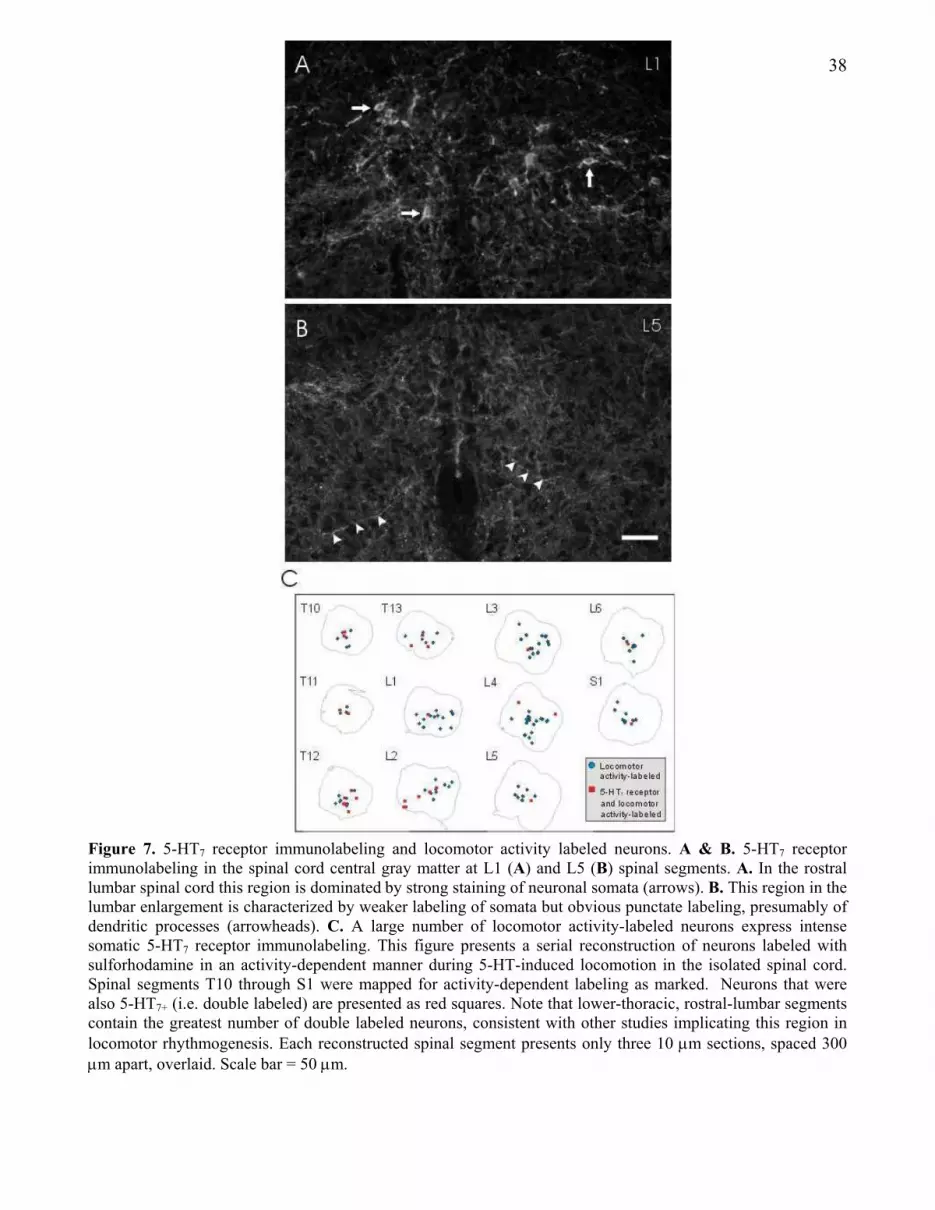
38
Figure 7. 5-HT7 receptor immunolabeling and locomotor activity labeled neurons. A & B. 5-HT7 receptor
immunolabeling in the spinal cord central gray matter at L1 (A) and L5 (B) spinal segments. A. In the rostral
lumbar spinal cord this region is dominated by strong staining of neuronal somata (arrows). B. This region in the
lumbar enlargement is characterized by weaker labeling of somata but obvious punctate labeling, presumably of
dendritic processes (arrowheads). C. A large number of locomotor activity-labeled neurons express intense
somatic 5-HT7 receptor immunolabeling. This figure presents a serial reconstruction of neurons labeled with
sulforhodamine in an activity-dependent manner during 5-HT-induced locomotion in the isolated spinal cord.
Spinal segments T10 through S1 were mapped for activity-dependent labeling as marked. Neurons that were
also 5-HT7+ (i.e. double labeled) are presented as red squares. Note that lower-thoracic, rostral-lumbar segments
contain the greatest number of double labeled neurons, consistent with other studies implicating this region in
locomotor rhythmogenesis. Each reconstructed spinal segment presents only three 10 µm sections, spaced 300 µm apart, overlaid. Scale bar = 50 µm.

39
Figure 8. Comparing the actions of monoamine transmitters in individual neurons. A. NA, 5-HT and
DA reversibly depress primary afferent-evoked EPSPs while ACh facilitated evoked responses. These
actions were common in many of the neurons recorded from the deep dorsal horn. B. The monoamines
convert neuronal firing properties from phasic to repetitive in response to current injection. Current
steps were delivered at 20 pA increments and voltage responses are presented superimposed. Figures
are presented as raw waveforms overlaid. Scale bars are 20 mV, 200 ms.


