Embed Size (px)
Citation preview

Musculoskeletal Pathology
�7�1 Integrin Does Not Alleviate Disease in a MouseModel of Limb Girdle Muscular Dystrophy Type 2F
Derek J. Milner and Stephen J. KaufmanFrom the Department of Cell and Developmental Biology,
University of Illinois, Urbana, Illinois
Transgenic expression of the �7�1 integrin in thedystrophic mdx/utr�/� mouse decreases develop-ment of muscular dystrophy and enhances longevity.To explore the possibility that elevating �7�1 integrinexpression could also ameliorate different forms ofmuscular dystrophy, we used transgenic technologyto enhance integrin expression in mice lacking �-sar-coglycan (� sgc), a mouse model for human limbgirdle muscular dystrophy type 2F. Unlike �7 trans-genic mdx/utr�/� mice, enhanced �7�1 integrin ex-pression in the � sgc-null mouse did not alleviatemuscular dystrophy in these animals. Expression ofthe transgene in the � sgc-null did not alleviate dys-trophic histopathology, nor did it decrease cardiomy-opathy or restore exercise tolerance. One hallmark ofintegrin-mediated alleviation of muscular dystrophyin the mdx/utr�/� background is the restoration ofmyotendinous junction integrity. As assessed byatomic force microscopy, myotendinous junctionsfrom normal and � sgc-null mice were indistinguish-able, thus suggesting the important influence of myo-tendinous junction integrity on the severity of mus-cular dystrophy and providing a possible explanationfor the inability of enhanced integrin expression toalleviate dystrophy in the � sgc-null mouse. Theseresults suggest that distinct mechanisms underlie thedevelopment of the diseases that arise from deficien-cies in dystrophin and sarcoglycan. (Am J Pathol 2007,170:609–619; DOI: 10.2353/ajpath.2007.060686)
The dystrophin-glycoprotein complex (DGC) of mem-brane-associated proteins is critical for the integrity ofskeletal muscle fibers.1 This complex consists of dystro-phin, the dystroglycans (� and �), the sarcoglycans (�, �,�, �, �, �), sarcospan, �-dystrobrevin, and the syntrophins(�1, �1, �2). Dystrophin binds to cytoskeletal actin and tothe transmembrane protein �-dystroglycan. The extracel-lular domain of �-dystroglycan binds to the peripheralmembrane protein �-dystroglycan, which binds laminin-2
in the basal lamina. �-Dystroglycan can also bind to theheparane sulfate proteoglycan agrin and perlecan in thebasement membrane of skeletal muscle. Thus, the DGCserves as a link between the extracellular matrix and thesubsarcolemmal cytoskeleton, sustaining proper myofi-ber architecture, facilitating myofiber alignment forproper force transmission, and protecting myofibers fromcontraction-induced damage. Mutations in genes encod-ing the different proteins of the DGC underlie differentforms of muscular dystrophy.1,2
The most common of these dystrophies is Duchennemuscular dystrophy (DMD), which arises from mutationsin the dystrophin gene. Mutations in genes encodingother members of the DGC can lead to additional mus-cular dystrophies. The limb girdle muscular dystrophies(LGMDs) are a heterogeneous group of diseases thathave highly variable onsets, progressions, and patternsof inheritance.1,2 These dystrophies are less prevalentthan DMD, each affecting �1 of every 20,000 persons,but they can be devastating disorders. Mutations in �, �,�, and � sarcoglycans are responsible for human LGMDtypes 2D, 2E, 2F, and 2C, respectively.3–7 Knockouts ofcorresponding sarcoglycan genes in mice show dystro-phic phenotypes as well.8–12
Mice that lack both dystrophin and utrophin (mdx:utr�/�) display a more severe phenotype than the dys-trophin-null mdx mouse, and the resulting disease moreclosely resembles that seen in DMD patients.13,14 Duringnormal embryonic development, utrophin exhibits a sar-colemmal distribution similar to dystrophin. At birth, dys-trophin replaces utrophin at the sarcolemma, and utro-phin becomes restricted to the neuromuscular andmyotendinous junctions.15 Utrophin is also found in the
Supported by the National Institutes of Health (to S.J.K.); the MuscularDystrophy Association (development award to D.J.M.); and the UnitedStates Department of Energy (grant DEFG02-91-ER45439 to the Centerfor Microanalysis of Materials, University of Illinois, for the atomic forcemicroscopy experiments).
Accepted for publication October 13, 2006.
Address reprint requests to Stephen J. Kaufman, Ph.D., Department ofCell and Developmental Biology, University of Illinois, B107 Chemical andLife Sciences Laboratory, 601 South Goodwin Ave., Urbana, IL 61801.E-mail: [email protected].
The American Journal of Pathology, Vol. 170, No. 2, February 2007
Copyright © American Society for Investigative Pathology
DOI: 10.2353/ajpath.2007.060686
609
sarcolemma of regenerated adult fibers, and it is in-creased in both mdx mice and DMD patients.14,16,17
The �7�1 integrin also binds laminin in the basementmembrane of skeletal muscle, and it provides an addi-tional linkage between the cytoskeleton and the extracel-lular matrix. The �7�1 integrin is abundant in adult skel-etal muscle, and it displays developmentally regulatedexpression of multiple isoforms comprised of differentcytoplasmic and extracellular domains.18 Experiments onmuscle biopsies from DMD patients and mdx mousemuscle demonstrated that �7 integrin transcript and pro-tein levels were elevated, suggesting that an increase inthe �7�1 integrin linkage system may compensate for theloss of the DGC-mediated linkage system resulting fromthe absence of dystrophin.19
Based on these observations, a hypothesis was devel-oped that increasing �7�1 integrin levels in mdx:utr�/�
mice might compensate for the absence of the DGC inthese animals and reduce the development of severemuscle pathology. Transgenic technology was used toproduce mdx:utr�/� mice with enhanced expression ofthe �7BX2 integrin isoform. As predicted, enhanced ex-pression of the �7 integrin significantly ameliorated thedystrophic phenotype in these animals.20 Transgenic an-imals showed a threefold increase in longevity, improvedmobility, reduced kyphosis, and maintenance of bodyweight when compared with nontransgenic mdx:utr�/�
mice. Transgenic expression of the �7�1 integrin chainalso reduced the degree of inflammatory cell infiltrationand reduced the expression of fetal myosin heavychain.20 Further study of these animals demonstratedthat enhanced expression of �7�1 integrin maintains thenormal architecture of the neuromuscular and myotendi-nous junctions and expands the regenerative capacity ofskeletal muscle.21
These results suggested that modulation of �7�1 inte-grin expression could potentially alleviate the develop-ment of dystrophic pathology in other forms of musculardystrophy. The � sarcoglycan (� sgc) gene is mutated inpatients with LGMD 2F, and � sgc-null mice display se-vere muscular dystrophy and cardiomyopathy.7,11,22 Todetermine whether enhanced expression of �7�1 integrincan alleviate other forms of muscular dystrophy, we pro-duced � sgc-null mice that express increased levels ofthe �7BX2 integrin isoform. Unlike mdx:utr�/� mice withenhanced expression of the �7�1 integrin, elevated ex-pression of the integrin in the � sgc-null background doesnot alleviate the dystrophic pathology associated with thelack of � sarcoglycan. These results suggest that dis-tinct mechanisms underlie the development of the dis-eases that arise from deficiencies in dystrophin andsarcoglycan.
Materials and Methods
Transgenic Mice
� sgc-null mice were derived as described11 and gener-ously provided by Dr. Kevin Campbell (University of Iowa,Iowa City, IA). Generation of transgenic � sgc-null mice
expressing the rat �7BX2 protein under control of theMCK promoter was performed by breeding �7BX2 trans-genic wild-type male mice with � sgc-null female mice toproduce �7BX2 transgenic mice heterozygous at the �sgc locus. �7 Transgene-positive � sgc heterozygotes(tg� �/�) were crossed with transgene-negative � sgcheterozygotes (tg� �/�) to produce transgene-positive �sgc-null (tg� �ko) and transgene-negative � sgc-null (tg�
�ko) animals. The production of transgenic mice express-ing the rat �7 integrin was as previously described,20 withone modification: a synthetic intron was inserted into thetransgene construct to further enhance transgene ex-pression.23 These transgenic mice yielded enhanced �7integrin expression levels sixfold greater than wild-typeanimals, and threefold greater than tg� �ko mice. Geno-typing of the � sgc locus and detection of the rat �7BX2transgene were performed by polymerase chain reaction(PCR) screening as described.11,20
Reverse Transcriptase (RT)-PCR
Mouse heart and hindlimb muscle were pulverized inliquid nitrogen and homogenized using a polytron. RNAwas extracted using Trizol (Invitrogen, Carlsbad, CA).RNA was treated with RNase-free DNase I (Invitrogen) for25 minutes at room temperature to remove potential con-taminating genomic DNA. RT-PCR reactions were per-formed using the Superscript one-step RT-PCR kit (In-vitrogen). For detection of the rat �7 transcript, theprimers used were: 5�-TTCATGTTGAAATAAGGCAG-GTT-3� (Rat�7 forward) and 5�-CACAGGAAAGACTTAG-GAGGG-3� (Rat�7 reverse). To ensure the quality of RNApreparations used for RT-PCR detection of rat integrintranscript, RT-PCR was performed to detect mouseGAPDH. For detection of mouse GAPDH, the primersused were: 5�-GAAGCTGTTGCAGCCTAGTC-3� (GAPDHforward) and 5�-CCATGGAGAAGGCCGGGG-3�(GAPDH reverse). Reactions were performed using 200ng of DNase I-treated RNA and performed for 30 cyclesof amplification. For each reaction, a control reactionlacking reverse transcriptase was done to ensure thatPCR products were not produced from genomic DNA.
Antibodies
The monoclonal antibody O26 was used to detect rat �7protein by immunofluorescence.24 Polyclonal anti-�7antibody CDB2 was used for Western blotting.25 Poly-clonal antibodies against �-sarcoglycan, �-sarcoglycanand sarcospan were generated as previously de-scribed8,26,27 and were kindly provided by Dr. KevinCampbell. Monoclonal antibodies against �-dystroglycan(NCL-b-DG) and utrophin (NCL-DRP2) were purchasedfrom Novocastra Laboratories, Newcastle Upon Tyne,UK. Monoclonal antibody against dystrophin (MAN-DRA-1) was purchased from Sigma, St. Louis, MO. AChRclusters were detected using rhodamine-labeled bunga-rotoxin purchased from Molecular Probes, Eugene, OR.20
Fluorescein isothiocyanate-labeled donkey anti-mouse
610 Milner and KaufmanAJP February 2007, Vol. 170, No. 2

and anti-rabbit antibodies were purchased from JacksonImmunoresearch Laboratories, West Grove, PA.
Western Blotting
Muscle tissue was pulverized in liquid nitrogen and ex-tracted twice in 200 mmol/L octyl-D-glucopyranoside, 50mmol/L Tris-HCl, pH 7.4, 2 mmol/L phenylmethyl sulfonylfluoride, 1:200 dilution of Protease Cocktail Set III (Cal-biochem, La Jolla, CA), 1 mmol/L CaCl2, and 1 mmol/LMgCl2 at 4°C for 30 minutes. Supernatants were com-bined and protein concentrations were determined byBradford assays. Equal amounts of protein were loadedon 8% sodium dodecyl sulfate-polyacrylamide gels andseparated under nonreducing conditions. Separated pro-teins were transferred to nitrocellulose and blocked over-night at 4°C using 5% milk in Tris-buffered saline-Tweenbuffer. For detection of integrin �7B, blocked filters wereincubated in a 1:1000 dilution of polyclonal anti-�7 anti-body CDB2, which recognizes the B-cytoplasmic domainof �7 integrin.25 Horseradish peroxidase-linked anti-rab-bit secondary antibody (Jackson Immunoresearch Lab-oratories), was used to detect primary antibodies. Immu-noreactive protein bands were detected using an ECLPlus kit (Amersham, Arlington Heights, IL), and blots werescanned with a Storm scanner (Molecular Dynamics,Sunnyvale, CA). Band intensities were determined usingImageQuant software (Molecular Dynamics).
Immunofluorescence
Muscle tissue from wild-type, nontransgenic � sgc-null(tg� �ko) and transgenic � sgc-null (tg� �ko) was snap-frozen in liquid nitrogen-cooled isopentane. Ten-�msections were fixed in �20°C acetone for 3 minutes,rehydrated in phosphate-buffered saline (PBS) for 10minutes, and blocked in PBS containing 5% bovine se-rum albumin for 30 minutes. For immunostaining of sec-tions with mouse monoclonal antibodies, goat anti-mouseFab fragments (Jackson Immunoresearch Laboratories)were included in the blocking solution at a concentrationof 70 �g/ml. Primary antibodies were detected using a1:100 dilution of fluorescein isothiocyanate-labeled don-key anti-mouse or anti-rabbit antibody in 1% bovine se-rum albumin in PBS. Rhodamine-labeled bungarotoxin(Molecular Probes, Eugene, OR) was used at 1:1000dilution to detect neuromuscular junctions. Coverslipswere mounted using Vectashield mounting medium (Vec-tor Laboratories, Burlingame, CA). Stained sections wereobserved with a Leica DMXRA2 microscope, and imageswere acquired using an AxioCam HRm digital camera(Zeiss, Thornwood, NY) and OpenLab software (Improvi-sion, Lexington, MA).
Histology and Evans Blue Dye Uptake
Ten-�m cryostat sections were fixed in 100% acetone at�20°C for 10 minutes, rinsed in tap water for 10 minutes,and stained with hematoxylin and eosin (H&E). Measure-ments of fiber cross-sectional area and central nuclei
were obtained using OpenLab software (Improvision).For each genotype, 200 to 300 fibers were measured insections of the soleus and gastrocnemius from each offour animals. For assessment of muscle membrane dam-age, mice were injected intraperitoneally with 50 �l/10 gbody weight of a filter-sterilized solution of Evans bluedye at a concentration of 10 mg/ml. Muscle tissue washarvested 24 to 48 hours later and snap-frozen in liquidnitrogen-cooled isopentane. Frozen sections were cut at10 �m, fixed in 100% acetone at �20°C for 10 minutes,washed with PBS for 10 minutes, and mounted withVectashield.
Treadmill Exercise
Six-week-old mice were injected with 50 �l of 10 mg/mlEvans blue dye per 10 g body weight 8 hours beforeexercise. Mice were allowed to warm up on a treadmillmoving at 10 m/minute for 10 minutes before being run at25 m/minute for 50 minutes. Duration of exercise at 25m/minute was recorded for each mouse. Mice that be-came exhausted and refused to continue running wereremoved, and the duration of exercise was recorded. Theanimals were sacrificed 36 to 48 hours after exercise, andhearts and skeletal muscle were harvested and observedfor Evans Blue dye uptake. Exercise was performed infour separate trials using one to two animals from eachgenotype per trial.
Atomic Force Microscopy
Muscle fibers were isolated from the flexor digitorumbrevis muscle of wild-type and � sgc-null mice by diges-tion with 0.2% collagenase (Worthington Biochemical,Lakewood, NJ) in Dulbecco’s modified Eagle’s medium,and fibers were washed and cultured in Tyrode’s solutioncontaining 5% horse serum. Fibers were cultured for 12hours and then adhered to coverslips coated with mouselaminin (Invitrogen) at 30 �g/cm2. Twelve to 24 hoursafter plating, adherent fibers were fixed with 2% parafor-maldehyde in Tyrode’s solution for 10 minutes, rinsedextensively with Tyrode’s solution, and then air-dried for 1hour. Dried fibers were then briefly washed twice withdeionized water and dried overnight. Atomic force mi-croscopy scans of isolated muscle fibers were generatedwith an MFP3D atomic force microscope (Asylum Re-search, Santa Barbara, CA) using oxide-sharpened sili-con nitride tapping tips (Budget Sensors, Sophia, Bul-garia). Imaging was done in AC mode at a set point of�700 mV and amplitude of 25 MHz.
Statistical Analysis
All averaged data are presented as the mean � SEM.Comparisons between groups were performed by anal-ysis of variance using Statview (SAS Institute Inc., SanFrancisco, CA). Differences were considered significantat P � 0.05.
�7 Integrin in a Model for LGMD 2F 611AJP February 2007, Vol. 170, No. 2

Results
Expression of the Rat �7BX2 Transgene inMouse Muscle
Cardiac and skeletal muscle from transgenic (tg� �ko)and nontransgenic (tg� �ko) � sgc-null mice were ana-lyzed for �7 transgene expression. RT-PCR analysis (Fig-ure 1A) detected the rat �7 transcript in tg� �ko skeletalmuscle, but not in tg� �ko skeletal muscle. Immunostain-ing of gastrocnemius muscle (Figure 1C) using a mono-clonal antibody that recognizes rat �7 demonstrated pos-itive staining in tg� �ko muscle, whereas immunostainingwas absent in tg� �ko muscle. Rat �7 transcript was notdetected in transgenic cardiac muscle RNA (Figure 1A)nor was rat protein detected in transgenic cardiac frozensections by immunofluorescence (not shown). The ab-sence of rat protein expression in the heart is not unex-pected because the activity of the MCK promoter regu-lating the rat �7 transgene is �100-fold lower in cardiaccompared with skeletal muscle, and transgenic mdx:utr�/� animals also show no expression of rat protein incardiac tissue.21,28
Western blotting (Figure 1B) of skeletal muscle ex-tracts using a polyclonal antibody that recognizes both
rat and mouse �7 integrin B cytoplasmic domain dem-onstrated increases in the amount of �7B integrin in bothtg� �ko and tg� �ko skeletal muscle. The tg� �ko hind-limb muscle has a twofold increase in endogenous �7Bintegrin protein when compared with wild-type muscle.This increase in mouse integrin is similar to the increasein �7 integrin levels seen in dystrophin-deficient mdxmice and �-sarcoglycan-null mice.9,19,20 tg� �ko musclehas a sixfold increase in integrin �7B levels when com-pared with normal mouse muscle. Differences betweenall groups were statistically significant (P � 0.009). Thus,the presence of the transgene results in expression of therat �7B integrin, and this results in a significant increasein total �7B integrin levels in tg� �ko skeletal musclewhen compared with both tg� �ko and wild-type skeletalmuscle.
No Improvement of Muscle Histopathology inTransgenic Animals
� sgc-null animals display muscle pathology that is typi-cally seen in dystrophic mice engineered to lack mem-bers of the DGC.11 By 3 weeks of age, central nucleatedfibers, necrotic fibers, inflammatory infiltration, and fiber
Figure 1. Transgene expression in �sgc-null muscle. A: RNA from heart (H) and hindlimb muscle (Sk) from nontransgenic (tg� �ko) and transgenic (tg� �ko)� sgc-null mice carrying the rat transgene under control of the MCK promoter was used for RT-PCR to detect expression of the transgene RNA. Rat �7 integrinexpression was only detected in transgenic skeletal muscle. Cardiac RNA from both tg� �ko and tg� �ko mice, as well as skeletal muscle RNA from tg� �ko mice,were negative for rat �7 transgene expression. Control reactions without reverse transcriptase ensured amplification of bands was not attributable to contaminatinggenomic DNA (middle). Detection of GAPDH RNA ensured that RNA was competent for use in RT-PCR reactions and gel loading was equivalent. B: Western blotsof extracted hindlimb skeletal muscle proteins from wild-type (lanes 1 and 2), tg� �ko (lanes 3 and 4), and tg� �ko (lanes 5 and 6) mice were probed withpolyclonal antibody against the �7B cytoplasmic domain to analyze the total amount of �7B integrin present. Arrows indicate the �120-kd full-length �7B (toparrow) and an �70-kd (bottom arrow) cleavage product quantified to determine the amount of total �7B. Analysis of scanned blots using ImageQuant imageanalysis software indicates tg� �ko muscle has a twofold increase in �7B integrin compared with wild type. Muscle from tg� �ko shows a sixfold increase in �7Bintegrin compared with wild type. Three separate gels consisting of protein samples from six different individuals of each genotype were analyzed. Differencesbetween all groups were statistically significant (P � 0.009). C: Immunostaining of mouse gastrocnemius muscle with monoclonal antibody O26 that selectivelyrecognizes rat �7 integrin under these conditions, shows sarcolemmal localization in tg� �ko sections, whereas tg� �ko sections are negative. Control sections(con) were incubated in the absence of primary antibody and demonstrate no staining. Scale bar � 50 �m.
612 Milner and KaufmanAJP February 2007, Vol. 170, No. 2

size variation can be observed in � sgc-null muscle. Asthe mice age, fibrosis, fatty infiltration, and fiber calcifi-cation can also be observed. These hallmarks of dystro-phy are seen in a variety of muscle groups in theseanimals.11
Analysis of H&E-stained sections from diaphragmand gastrocnemius of both tg� �ko and tg� �ko muscle(Figure 2A) demonstrated that enhanced expression of�7BX2 integrin had no impact on the dystrophic histo-pathology exhibited by � sgc-null animals. Pathologywas observed in tg� �ko and tg� �ko muscle in bothyoung (5 to 10 weeks) and older (6 to 8 months; notshown) animals, indicating that transgene expressiondid not reduce histopathology as the animals aged. In
addition to diaphragm and gastrocnemius muscle, pa-thology was also observed in several different musclegroups in both tg� �ko and tg� �ko mice, including thesoleus, biceps, quadriceps, and intercostal muscle(not shown). Membrane damage, a hallmark or dis-eased muscle, can be visualized by the uptake ofEvans blue dye into muscle fibers after systemic injec-tion. Muscles from both transgenic and nontransgenic� sgc-null mice demonstrate uptake of Evans blue dyein muscle fibers, and there was no apparent differencein the extent of dye uptake in tg� �ko and tg� �ko mice(not shown).
The characteristic increase in variation of muscle fibersize seen in dystrophic muscle was not changed in miceoverexpressing the integrin. Analysis of cross-sectionalareas (Figure 2B) of muscle fibers in the soleus andgastrocnemius muscles of transgenic and nontransgenic� sgc animals demonstrates the increase in variation offiber sizes in � sgc-null muscle is not appreciablychanged in the transgenic mice. This increase in fibersize variation is attributable to an increase in the popu-lation of small, regenerating fibers and large, hypertro-phic fibers in both tg� �ko and tg� �ko mice. There is noincrease in the number of hypertrophic fibers in tg� �komice. The number of fibers above the wild-type maximumcross-sectional areas are approximately equivalent in thegastrocnemius and soleus muscle of both tg� �ko andtg� �ko mice. Last, the extent of centrally localized nucleiis the same in both the transgenic and nontransgenicanimals. Approximately 56% of fibers contain central nu-clei (Figure 2C). Thus, enhanced expression of the�7BX2 integrin does not improve or prevent developmentof the dystrophic histopathology observed in � sgc-nullskeletal muscle.
No Restoration of Exercise Capacity inTransgenic Animals
A characteristic of the � sgc-null mouse is the devel-opment of cardiomyopathy with advancing age. Evansblue dye-positive cardiac lesions and fibrotic lesionsobserved by H&E-stained cardiac sections can be de-tected by 3 months of age, and cardiac lesions can beinduced in younger animals by subjecting them totreadmill exercise.11 Because the enhancement of�7BX2 integrin expression in skeletal muscle can alle-viate the cardiomyopathy seen in mdx:utr�/� miceeven without transgene expression in the heart,21,29 itwas of interest to determine whether the increasedexpression of �7BX2 integrin can also alleviate thecardiac pathology seen in � sgc-null mice. Mice 6weeks of age were injected with Evans blue dye andexercised on a treadmill operating at 20 m/minute forup to 50 minutes. Exercise tolerance (Figure 3A) wasdiminished in tg� �ko animals. The tg� �ko animalswithstood treadmill exercise for only half the averageduration of time tolerated by wild-type animals. Like-wise, tg� �ko animals showed reduced exercise toler-ance compared with wild-type animals, and there was
Figure 2. Histopathology in tg� �ko and tg� �ko skeletal muscle. A: Sectionsfrom the gastrocnemius of 10-week tg� �ko (a, c) and 10-week tg� �ko (b,d) male mice exhibit identical pathology. Central nuclei, necrotic fibers,infiltrating cells, calcification (c and d, arrowheads), and regenerating fibersare approximately equivalent regardless of the absence or presence of the �7transgene. B: Expression of the transgene does not alter the distribution offiber size in � sarcoglycan-null muscle. Histograms representing the distri-bution of fiber cross-sectional areas in wild-type (top), tg� �ko (middle), andtg� �ko (bottom) soleus and gastrocnemius muscle. In both muscle types,the distribution of fiber sizes is more variable in the tg� �ko and tg� �komuscle compared with wild type. C: The percentage of fibers with centralnuclei in gastrocnemius and soleus from tg� �ko and tg� �ko muscle areequivalent. n � 4 animals per genotype per muscle.
�7 Integrin in a Model for LGMD 2F 613AJP February 2007, Vol. 170, No. 2

no improvement in exercise tolerance compared withnontransgenic � sgc-null animals.
As expected, uptake of Evans blue dye was evident inthe myocardium of exercised � sgc-null mice (Figure 3B)but absent in the myocardium of exercised wild-typeanimals. Cardiac uptake of the dye was approximatelythe same in tg� �ko and tg� �ko hearts, indicating thatmyocardial damage can still occur in animals that ex-press enhanced levels of �7BX2 integrin in skeletal mus-cle. Dye uptake was absent in the myocardium fromnonexercised tg� �ko and tg� �ko animals (not shown).Evans blue dye was also observed in hindlimb muscle(Figure 3C) from both tg� �ko and tg� �ko exercisedmice, with no apparent difference in uptake between thegenotypes. Evans blue dye was also observed in nonex-ercised hind limbs from mice of both genotypes at 6weeks of age (not shown).
Maintenance of Dystrophin and �-DystroglycanProtein Expression and Localization
In several murine muscular dystrophies, loss of expres-sion of one component of the DGC leads to decreasesin protein levels and loss of sarcolemmal localization ofother members of the complex.1,30 Staining for othermembers of the sarcoglycan subcomplex of the DGCin � sgc-null animals shows, as expected, that theseproteins are mostly absent from the sarcolemma in �
sgc-null mice. Sarcospan (Figure 4A), �-sarcoglycan,and �-sarcoglycan (not shown) were not detected inthe sarcolemma of skeletal muscle in � sgc-null mice.The expression and localization of these proteins wasnot restored by enhanced expression of �7BX2 integrin(Figure 4A).
Figure 3. Exercise tolerance in 6-week tg� �ko and tg� �koanimals. A: Exercise endurance is significantly decreased in bothtg� �ko and tg� �ko animals (P � 0.003). No significant differ-ences in exercise endurance were found between tg� �ko andtg� �ko mice. n � 5. B: Evans blue dye uptake was approxi-mately the same in the myocardium from 6-week-exercised tg�
�ko and tg� �ko mice. Dye uptake was absent in wild-typeanimals. C: Hindlimbs from 6-week-old tg� �ko and tg� �koexercised mice exhibited similar levels of Evans blue dye uptake,whereas dye uptake in wild-type muscle was absent.
614 Milner and KaufmanAJP February 2007, Vol. 170, No. 2
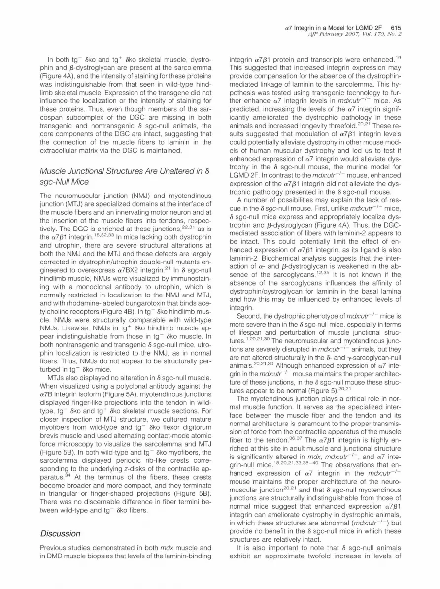
In both tg� �ko and tg� �ko skeletal muscle, dystro-phin and �-dystroglycan are present at the sarcolemma(Figure 4A), and the intensity of staining for these proteinswas indistinguishable from that seen in wild-type hind-limb skeletal muscle. Expression of the transgene did notinfluence the localization or the intensity of staining forthese proteins. Thus, even though members of the sar-cospan subcomplex of the DGC are missing in bothtransgenic and nontransgenic � sgc-null animals, thecore components of the DGC are intact, suggesting thatthe connection of the muscle fibers to laminin in theextracellular matrix via the DGC is maintained.
Muscle Junctional Structures Are Unaltered in �
sgc-Null Mice
The neuromuscular junction (NMJ) and myotendinousjunction (MTJ) are specialized domains at the interface ofthe muscle fibers and an innervating motor neuron and atthe insertion of the muscle fibers into tendons, respec-tively. The DGC is enriched at these junctions,22,31 as isthe �7�1 integrin.18,32,33 In mice lacking both dystrophinand utrophin, there are severe structural alterations atboth the NMJ and the MTJ and these defects are largelycorrected in dystrophin/utrophin double-null mutants en-gineered to overexpress �7BX2 integrin.21 In � sgc-nullhindlimb muscle, NMJs were visualized by immunostain-ing with a monoclonal antibody to utrophin, which isnormally restricted in localization to the NMJ and MTJ,and with rhodamine-labeled bungarotoxin that binds ace-tylcholine receptors (Figure 4B). In tg� �ko hindlimb mus-cle, NMJs were structurally comparable with wild-typeNMJs. Likewise, NMJs in tg� �ko hindlimb muscle ap-pear indistinguishable from those in tg� �ko muscle. Inboth nontransgenic and transgenic � sgc-null mice, utro-phin localization is restricted to the NMJ, as in normalfibers. Thus, NMJs do not appear to be structurally per-turbed in tg� �ko mice.
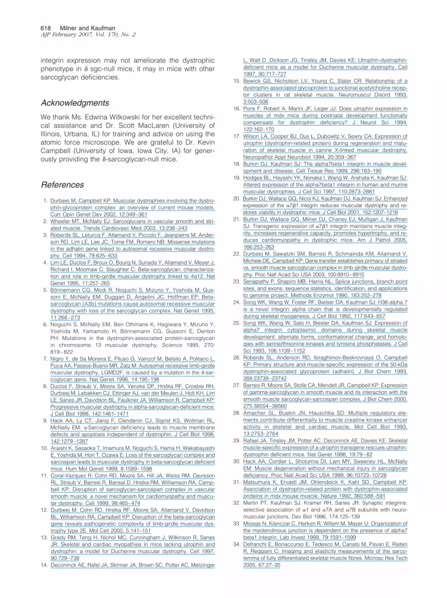
MTJs also displayed no alteration in � sgc-null muscle.When visualized using a polyclonal antibody against the�7B integrin isoform (Figure 5A), myotendinous junctionsdisplayed finger-like projections into the tendon in wild-type, tg� �ko and tg� �ko skeletal muscle sections. Forcloser inspection of MTJ structure, we cultured maturemyofibers from wild-type and tg� �ko flexor digitorumbrevis muscle and used alternating contact-mode atomicforce microscopy to visualize the sarcolemma and MTJ(Figure 5B). In both wild-type and tg� �ko myofibers, thesarcolemma displayed periodic rib-like crests corre-sponding to the underlying z-disks of the contractile ap-paratus.34 At the terminus of the fibers, these crestsbecome broader and more compact, and they terminatein triangular or finger-shaped projections (Figure 5B).There was no discernable difference in fiber termini be-tween wild-type and tg� �ko fibers.
Discussion
Previous studies demonstrated in both mdx muscle andin DMD muscle biopsies that levels of the laminin-binding
integrin �7�1 protein and transcripts were enhanced.19
This suggested that increased integrin expression mayprovide compensation for the absence of the dystrophin-mediated linkage of laminin to the sarcolemma. This hy-pothesis was tested using transgenic technology to fur-ther enhance �7 integrin levels in mdx:utr�/� mice. Aspredicted, increasing the levels of the �7 integrin signif-icantly ameliorated the dystrophic pathology in theseanimals and increased longevity threefold.20,21 These re-sults suggested that modulation of �7�1 integrin levelscould potentially alleviate dystrophy in other mouse mod-els of human muscular dystrophy and led us to test ifenhanced expression of �7 integrin would alleviate dys-trophy in the � sgc-null mouse, the murine model forLGMD 2F. In contrast to the mdx:utr�/� mouse, enhancedexpression of the �7�1 integrin did not alleviate the dys-trophic pathology presented in the � sgc-null mouse.
A number of possibilities may explain the lack of res-cue in the � sgc-null mouse. First, unlike mdx:utr�/� mice,� sgc-null mice express and appropriately localize dys-trophin and �-dystroglycan (Figure 4A). Thus, the DGC-mediated association of fibers with laminin-2 appears tobe intact. This could potentially limit the effect of en-hanced expression of �7�1 integrin, as its ligand is alsolaminin-2. Biochemical analysis suggests that the inter-action of �- and �-dystroglycan is weakened in the ab-sence of the sarcoglycans.12,35 It is not known if theabsence of the sarcoglycans influences the affinity ofdystrophin/dystroglycan for laminin in the basal laminaand how this may be influenced by enhanced levels ofintegrin.
Second, the dystrophic phenotype of mdx:utr�/� mice ismore severe than in the � sgc-null mice, especially in termsof lifespan and perturbation of muscle junctional struc-tures.1,20,21,30 The neuromuscular and myotendinous junc-tions are severely disrupted in mdx:utr�/� animals, but theyare not altered structurally in the �- and �-sarcoglycan-nullanimals.20,21,30 Although enhanced expression of �7 inte-grin in the mdx:utr�/� mouse maintains the proper architec-ture of these junctions, in the � sgc-null mouse these struc-tures appear to be normal (Figure 5).20,21
The myotendinous junction plays a critical role in nor-mal muscle function. It serves as the specialized inter-face between the muscle fiber and the tendon and itsnormal architecture is paramount to the proper transmis-sion of force from the contractile apparatus of the musclefiber to the tendon.36,37 The �7�1 integrin is highly en-riched at this site in adult muscle and junctional structureis significantly altered in mdx, mdx:utr�/�, and �7 inte-grin-null mice.18,20,21,33,38–40 The observations that en-hanced expression of �7 integrin in the mdx:utr�/�
mouse maintains the proper architecture of the neuro-muscular junction20,21 and that � sgc-null myotendinousjunctions are structurally indistinguishable from those ofnormal mice suggest that enhanced expression �7�1integrin can ameliorate dystrophy in dystrophic animals,in which these structures are abnormal (mdx:utr�/�) butprovide no benefit in the � sgc-null mice in which thesestructures are relatively intact.
It is also important to note that � sgc-null animalsexhibit an approximate twofold increase in levels of
�7 Integrin in a Model for LGMD 2F 615AJP February 2007, Vol. 170, No. 2

616 Milner and KaufmanAJP February 2007, Vol. 170, No. 2

endogenous �7 integrin, similar to the increase in en-dogenous �7 in mdx, mdx:utr�/�, and � sgc-nullmice.19–21,41 Although further transgenic enhancementof integrin expression provides partial alleviation ofdystrophy in the mdx:utr�/� mouse,20,21 the naturalincrease in endogenous integrin in the � sgc-null ani-mals may provide sufficient compensation to lessenthe severity of the dystrophy and maintain junctionalarchitecture, lifespan, and the ability to breed. Thatincreased levels of endogenous �7�1 integrin providecompensation for sarcoglycan loss is supported by thedevelopment of severe pathology in null mice lackingboth �7 integrin and �-sarcoglycan.41 These double-mutant mice exhibit a phenotype much more extremethan either the � sgc-null or the �7-null mice, and the
double-mutant mice exhibit rapid muscle degenerationand death by 1 month of age.41 Likewise, the pathologythat develops in mdx/�7�/� mice is much more severethan mice with either single mutation.42,43
Although primary deficiency of one of the sarcogly-cans typically leads to a secondary decrease in the othersarcoglycans, the phenotypes of the sarcoglycan-nullanimals are not identical.1 Mice with null mutations in thedifferent sarcoglycans exhibit differences in the severityof dystrophic pathology, cardiac pathology, and expres-sion of other members of the DGC.1,30 For example,muscle from � sgc-null mice exhibit normal resistance tomechanical strain and no contraction-induced injury afterexercise, in marked contrast to muscle from �-, �-, and�-sarcoglycan-null muscle.1,30 Thus, although enhanced
Figure 5. Myotendinous junctions of tg� �ko and tg� �ko mice. A: Mouse MTJs are visualized by immunostaining with a polyclonal antibody against �7B integrin.Wild-type, tg� �ko, and tg� �ko MTJ all show intense immunostaining for �7B integrin. No obvious alterations are observed in tg� �ko and tg� �ko MTJ comparedwith wild-type junctions. Images were not taken at identical exposure times but were optimized for visualization of junction structure. B: Three-dimensionaltopographic height images generated from atomic force microscopy scans of the sarcolemma (a, b) and MTJ (c, d) of isolated flexor digitorum brevis myofibersfrom wild-type and tg� �ko mice. Transverse rib-like crests corresponding to the underlying z-disks of the contractile apparatus are observed in both wild-typeand tg� �ko sarcolemmal sections (a and b, arrows). Finger-like projections (c and d, arrowheads) are observed at the termini of both wild-type and tg� �kofibers. N, nucleus. Scale bars: 10 �m (A); 1 �m (B).
Figure 4. Staining of DGC components and neuromuscular junctions in tg� �ko and tg� �ko mice. A: Immunostaining of dystrophin, �-dystroglycan (�-DG), andsarcospan (sspn) in gastrocnemius muscle from 10-week mice: dystrophin and �-dystroglycan immunostaining are present and approximately equivalent in tg�
�ko and tg� �ko mice. Immunostaining of sarcospan, a component of the sarcoglycan subcomplex, is absent in the sarcolemma of tg� �ko mice, and localizationof this protein is not retained in tg� �ko mice. B: Rhodamine bungarotoxin (btx) staining of mouse hindlimb muscle localizes at NMJs in wild-type, tg� �ko, andtg� �ko mice. Neuromuscular junctions of tg� �ko and tg� �ko mice appear unaltered compared with junctions from wild type. Immunolocalization of utrophin(utrn) demonstrates positive immunostaining that is restricted to the NMJ. Merged images indicate the localization of utrophin is restricted to the NMJ in wild-type,tg� �ko, and tg� �ko muscle. Scale bars � 50 �m.
�7 Integrin in a Model for LGMD 2F 617AJP February 2007, Vol. 170, No. 2
integrin expression may not ameliorate the dystrophicphenotype in � sgc-null mice, it may in mice with othersarcoglycan deficiencies.
Acknowledgments
We thank Ms. Edwina Witkowski for her excellent techni-cal assistance and Dr. Scott MacLaren (University ofIllinois, Urbana, IL) for training and advice on using theatomic force microscope. We are grateful to Dr. KevinCampbell (University of Iowa, Iowa City, IA) for gener-ously providing the �-sarcoglycan-null mice.
References
1. Durbeej M, Campbell KP: Muscular dystrophies involving the dystro-phin-glycoprotein complex: an overview of current mouse models.Curr Opin Genet Dev 2002, 12:349–361
2. Wheeler MT, McNally EJ: Sarcoglycans in vascular smooth and stri-ated muscle. Trends Cardiovasc Med 2003, 13:238–243
3. Roberds SL, Leturcq F, Allamand V, Piccolo F, Jeanpierre M, Ander-son RD, Lim LE, Lee JC, Tome FM, Romero NB: Missense mutationsin the adhalin gene linked to autosomal recessive muscular dystro-phy. Cell 1994, 78:625–633
4. Lim LE, Duclos F, Broux O, Bourg N, Sunada Y, Allamand V, Meyer J,Richard I, Moomaw C, Slaughter C: Beta-sarcoglycan: characteriza-tion and role in limb-girdle muscular dystrophy linked to 4q12. NatGenet 1995, 11:257–265
5. Bonnemann CG, Modi R, Noguchi S, Mizuno Y, Yoshida M, Gus-soni E, McNally EM, Duggan D, Angelini JC, Hoffman EP: Beta-sarcoglycan (A3b) mutations cause autosomal recessive musculardystrophy with loss of the sarcoglycan complex. Nat Genet 1995,11:266–273
6. Noguchi S, McNally EM, Ben Othmane K, Hagiwara Y, Mizuno Y,Yoshida M, Yamamoto H, Bonnemann CG, Gussoni E, DentonPH: Mutations in the dystrophin-associated protein-sarcoglycanin chromosome 13 muscular dystrophy. Science 1995, 270:819–822
7. Nigro Y, de Sa Moreira E, Piluso G, Vainzof M, Belsito A, Politano L,Puca AA, Passos-Bueno MR, Zatz M: Autosomal recessive limb-girdlemuscular dystrophy, LGMD2F, is caused by a mutation in the �-sar-coglycan gene. Nat Genet 1996, 14:195–198
8. Duclos F, Straub V, Moore SA, Venzke DP, Hrstka RF, Crosbie RH,Durbeej M, Lebakken CJ, Ettinger AJ, van der Meulen J, Holt KH, LimLE, Sanes JR, Davidson BL, Faulkner JA, Williamson R, Campbell KP:Progressive muscular dystrophy in alpha-sarcoglycan-deficient mice.J Cell Biol 1998, 142:1461–1471
9. Hack AA, Ly CT, Jiang F, Clendenin CJ, Sigrist KS, Wollman RL,McNally EM: �-Sarcoglycan deficiency leads to muscle membranedefects and apoptosis independent of dystrophin. J Cell Biol 1998,142:1279–1287
10. Araishi K, Sasaoka T, Imamura M, Noguchi S, Hama H, WakabayashiE, Yoshida M, Hori T, Ozawa E: Loss of the sarcoglycan complex andsarcospan leads to muscular dystrophy in beta-sarcoglycan deficientmice. Hum Mol Genet 1999, 8:1589–1598
11. Coral-Vazquez R, Cohn RD, Moore SA, Hill JA, Weiss RM, DavissonRL, Straub V, Barresi R, Bansal D, Hrstka RM, Williamson RA, Camp-bell KP: Disruption of sarcoglycan-sarcospan complex in vascularsmooth muscle: a novel mechanism for cardiomyopathy and muscu-lar dystrophy. Cell 1999, 98:465–474
12. Durbeej M, Cohn RD, Hrstka RF, Moore SA, Allamand V, DavidsonBL, Williamson RA, Campbell KP: Disruption of the beta-sarcoglycangene reveals pathogenetic complexity of limb-girdle muscular dys-trophy type 2E. Mol Cell 2000, 5:141–151
13. Grady RM, Teng H, Nichol MC, Cunningham J, Wilkinson R, SanesJR: Skeletal and cardiac myopathies in mice lacking utrophin anddystrophin: a model for Duchenne muscular dystrophy. Cell 1997,90:729–738
14. Deconinck AE, Rafel JA, Skinner JA, Brown SC, Potter AC, Metzinger
L, Watt D, Dickson JG, Tinsley JM, Davies KE: Utrophin-dystrophin-deficient mice as a model for Duchenne muscular dystrophy. Cell1997, 90:717–727
15. Bewick GS, Nicholson LV, Young C, Slater CR: Relationship of adystrophin-associated glycoprotein to junctional acetylcholine recep-tor clusters in rat skeletal muscle. Neuromuscul Disord 1993,3:503–506
16. Pons F, Robert A, Marini JF, Leger JJ: Does utrophin expression inmuscles of mdx mice during postnatal development functionallycompensate for dystrophin deficiency? J Neurol Sci 1994,122:162–170
17. Wilson LA, Cooper BJ, Dux L, Dubowitz V, Sewry CA: Expression ofutrophin (dystrophin-related protein) during regeneration and matu-ration of skeletal muscle in canine X-linked muscular dystrophy.Neuropathol Appl Neurobiol 1994, 20:359–367
18. Burkin DJ, Kaufman SJ: The alpha7beta1 integrin in muscle devel-opment and disease. Cell Tissue Res 1999, 296:183–190
19. Hodges BL, Hayashi YK, Nonaka I, Wang W, Arahata K, Kaufman SJ:Altered expression of the alpha7beta1 integrin in human and murinemuscular dystrophies. J Cell Sci 1997, 110:2873–2881
20. Burkin DJ, Wallace GQ, Nicol KJ, Kaufman DJ, Kaufman SJ: Enhancedexpression of the �7�1 integrin reduces muscular dystrophy and re-stores viability in dystrophic mice. J Cell Biol 2001, 152:1207–1218
21. Burkin DJ, Wallace GQ, Milner DJ, Chaney EJ, Mulligan J, KaufmanSJ: Transgenic expression of �7�1 integrin maintains muscle integ-rity, increases regenerative capacity, promotes hypertrophy, and re-duces cardiomyopathy in dystrophic mice. Am J Pathol 2005,166:253–263
22. Durbeej M, Sawatzki SM, Barresi R, Schmainda KM, Allamand V,Michele DE, Campbell KP: Gene transfer establishes primacy of striatedvs. smooth muscle sarcoglycan complex in limb girdle muscular dystro-phy. Proc Natl Acad Sci USA 2003, 100:8910–8915
23. Senapathy P, Shapiro MB, Harris NL: Splice junctions, branch pointsites, and exons: sequence statistics, identification, and applicationsto genome project. Methods Enzymol 1990, 183:252–278
24. Song WK, Wang W, Foster RF, Bielser DA, Kaufman SJ: H36-alpha 7is a novel integrin alpha chain that is developmentally regulatedduring skeletal myogenesis. J Cell Biol 1992, 117:643–657
25. Song WK, Wang W, Sato H, Biesler DA, Kaufman SJ: Expression ofalpha7 integrin cytoplasmic domains during skeletal muscledevelopment: alternate forms, conformational change, and homolo-gies with serine/threonine kinases and tyrosine phosphatases. J CellSci 1993, 106:1139–1152
26. Roberds SL, Anderson RD, Ibraghimov-Beskrovnaya O, CampbellKP: Primary structure and muscle-specific expression of the 50-kDadystrophin-associated glycoprotein (adhalin). J Biol Chem 1993,268:23739–23742
27. Barresi R, Moore SA, Stolle CA, Mendell JR, Campbell KP: Expressionof gamma-sarcoglycan in smooth muscle and its interaction with thesmooth muscle sarcoglycan-sarcospan complex. J Biol Chem 2000,275:38554–38560
28. Amacher SL, Buskin JN, Hauschka SD: Multiple regulatory ele-ments contribute differentially to muscle creatine kinase enhanceractivity in skeletal and cardiac muscle. Mol Cell Biol 1993,13:2753–2764
29. Rafael JA, Tinsley JM, Potter AC, Deconinck AE, Davies KE: Skeletalmuscle-specific expression of a utrophin transgene rescues utrophin-dystrophin deficient mice. Nat Genet 1998, 19:79–82
30. Hack AA, Cordier L, Shoturma DI, Lam MY, Sweeney HL, McNallyEM: Muscle degeneration without mechanical injury in sarcoglycandeficiency. Proc Natl Acad Sci USA 1999, 96:10723–10728
31. Matsumura K, Ervasti JM, Ohlendeick K, Kahl SD, Campbell KP:Association of dystrophin-related protein with dystrophin-associatedproteins in mdx mouse muscle. Nature 1992, 360:588–591
32. Martin PT, Kaufman SJ, Kramer RH, Sanes JR: Synaptic integrins:selective association of �1 and �7A and �7B subunits with neuro-muscular junctions. Dev Biol 1996, 174:125–139
33. Miosge N, Klenczar C, Herken R, Willem M, Mayer U: Organization ofthe myotendinous junction is dependent on the presence of alpha7beta1 integrin. Lab Invest 1999, 79:1591–1599
34. Defranchi E, Bonaccurso E, Tedesco M, Canato M, Pavan E, RaiteriR, Reggiani C: Imaging and elasticity measurements of the sarco-lemma of fully differentiated skeletal muscle fibres. Microsc Res Tech2005, 67:27–35
618 Milner and KaufmanAJP February 2007, Vol. 170, No. 2

35. Straub V, Duclos F, Venzke DP, Lee JC, Cutshall S, Leveille CJ,Campbell KP: Molecular pathogenesis of muscle degenerationin the delta-sarcoglycan-deficient hamster. Am J Pathol 1998,153:1623–1630
36. Tidball JG, Daniel TL: Myotendinous junctions of tonic muscle cells:structure and loading. Cell Tissue Res 1986, 245:315–322
37. Trotter JA: Functional morphology of force transmission in skeletalmuscle. A brief review. Acta Anat (Basel) 1993, 146:205–222
38. Law DJ, Tidball JG: Dystrophin deficiency is associated with myoten-dinous junction defects in prenecrotic and fully regenerated skeletalmuscle. Am J Pathol 1993, 142:1513–1523
39. MayerU, SaherG, Fassler R,BornemannA, Echtermeyer F, vonderMarkH,Miosge N, Poschl E, von der Mark K: Absence of integrin alpha 7 causesa novel form of muscular dystrophy. Nat Genet 1997, 17:318–323
40. Nawrotzki R, Willem M, Miosge N, Brinkmeier H, Mayer U: Defectiveintegrin switch and matrix composition at alpha 7-deficient myoten-dinous junctions precede the onset of muscular dystrophy in mice.Hum Mol Genet 2003, 12:483–495
41. Allikian MJ, Hack AA, Mewborn S, Mayer U, McNally EM: Geneticcompensation for sarcoglycan loss by integrin �7�1 in muscle. J CellSci 2004, 117:3821–3830
42. Rooney JE, Welser JV, Dechert MA, Flintoff-Dye NL, Kaufman SJ,Burkin DJ: Severe muscular dystrophy in mice that lack dystrophinand �7 integrin. J Cell Sci 2006, 119:2185–2195
43. Guo C, Willem M, Werner A, Raivich G, Emerson M, Neyses L, MayerU: Absence of alpha7 integrin in dystrophin-deficient mice causes amyopathy similar to Duchenne muscular dystrophy. Hum Mol Genet2006, 15:989–998
�7 Integrin in a Model for LGMD 2F 619AJP February 2007, Vol. 170, No. 2


