Embed Size (px)
Citation preview

Malays. Appl. Biol. (2022) 51(1): 1–20https://doi.org/10.55230/mabjournal.v51i1.2056
POTENTIAL MEDICINAL HERB FOR CARDIOVASCULAR HEALTH: A COMPREHENSIVE REVIEW ON
Salviae miltiorrhizae
TING WEI NG1, PAULINA PEI SUU TAN1, HOOI MIN LIM2, DEVARAJ NAVIN KUMAR3, SHAMSUL MOHD ZAIN4, TECK YEW LOW5, and YUH-FEN PUNG1*
1Division of Biomedical Science, Faculty of Science and Engineering, University of Nottingham Malaysia, 43500 Selangor, Malaysia
2 Department of Primary Care Medicine, Faculty of Medicine, University Malaya 50603 Kuala Lumpur, Malaysia3Department of Family Medicine, Faculty of Medicine and Health Sciences, Universiti Putra Malaysia, 43400
Serdang, Selangor, Malaysia4Department of Pharmacology, Faculty of Medicine, University Malaya, 50603 Kuala Lumpur, Malaysia
5UKM Medical Molecular Biology Institute (UMBI), Universiti Kebangsaan Malaysia, 56000 Kuala Lumpur, Malaysia
*E-mail: [email protected]
Accepted 7 March 2022, Published online 31 March 2022
ABSTRACT
Cardiovascular disease (CVD) and its associated risk factors have been ranked the number 1 cause of mortality in non-communicable diseases worldwide and Malaysia. The high statistic in CVD mortality indicates gaps and limitations in current treatment strategies using long-term drug prescription therapies. Hence, an immediate quest for alternative and effective treatments is needed. Medicinal herbs, which are ethnopharmacologically used to treat a wide range of conditions, have been used as an alternative or supplementary treatment for CVDs and their associated risk factors. The roots of Salviae miltiorrhizae have been traditionally used for centuries to treat various diseases including neurological disorders, cancer, and even coronary heart disease. Increasing evidence demonstrated a pharmacological basis for the action of S. miltiorrhizae and its active compounds, suggesting its potential in treating CVD. The objectives of this review were first to summarize published literature and synthesize the new body of knowledge on the use of S. miltiorrhizae as the potential medicinal herb that will positively impact the cardiovascular system, and secondly to elucidate the underlying mechanisms involved in promoting cardiovascular health. It is hoped that identification of key regulatory pathways by lipophilic and hydrophilic active compounds from S. miltiorrhizae will aid further investigation of its safety and efficacy to promote the use of evidence-based traditional medicinal herbs in alleviating symptoms and improve the prognosis of CVDs and their associated risk factors.
Key words: Atherosclerosis, diabetes, hypercholesterolemia, hyperlipidemia, hypertension
INTRODUCTION
Non-communicable diseases (NCDs) are a major health problem worldwide. Cardiovascular diseases (CVDs) and their associated risk factors were particularly found to contribute significantly to the number of NCD cases and are the leading cause of global mortality (Laslett et al., 2012). According to World Health Organization (WHO) in 2017, there was an alarming number of 17.7 million deaths caused by CVDs per year. This is attributed to 31% of overall deaths globally (Sazlina et al., 2020). Even in Malaysia, the National Health and Morbidity Survey (NHMS) reported that in 2015, coronary artery disease was the main cause of CVD
death, followed by stroke with a prevalence of 13.2%, and 6.9%, respectively (Sazlina et al., 2020). The prevalence of CVDs is predicted to increase up to more than 23.6 million by 2030 (Greenfield & Snowden, 2018). Consequently, deaths by CVDs have dominated the mortality rate worldwide.
CVDs include heart failure, myocardial infarction, atherosclerosis, coronary heart disease (CHD), stroke, cardiomyopathy, and peripheral artery disorders. Risk factors associated with CVDs are primarily caused by vascular dysfunction, often a result of thrombosis, atherosclerosis, obesity, diabetes, dyslipidemia, and hypertension, leading to subsequent damage of organs (Shaito et al., 2020). Many epidemiological studies have also indicated positive correlations between the incidence of CVDs and chronic alcohol consumption, smoking, * To whom correspondence should be addressed.
MEDICINAL HERB FOR CARDIOVASCULAR HEALTH2
psychosocial factors, physical inactivity, as well as insufficient consumption of vegetables and fruits (Rosiek & Leksowski, 2016; Sazlina et al., 2020). CVDs and their risk factors are commonly treated with continuous, long-term administration of aspirin, statins, beta-blockers, and angiotensin-converting enzyme (ACE) inhibitors (Rossello et al., 2015). Unfortunately, patient adherence to these classes of drugs has been proven poor due to direct and indirect costs of treatment, convenience, and lack of evidence for the efficacy of long-term medication intake and its effects after withdrawal (Nieuwlaat et al., 2013; McClellan et al., 2019). Inadequately treated or untreated patients due to poor medication adherence contribute to the plateau in CVD mortality trends that remain the leading cause of death worldwide (McClellan et al., 2019). Hence, this opens an avenue for alternative treatments other than western medicine. Besides utilizing western medicines as treatment for CVDs and their associated risk factors, medicinal herbs have been widely used as a potential alternative to treat CVDs (Pang et al., 2016).
To date, about 10,000 various phytochemicals have been discovered from medicinal herbs. However, there remains a wide range of unknown phytochemicals that have yet to be identified (Memariani et al., 2019). Since medicinal herbs possess beneficial phytochemicals that promote greater health, they are widely used in daily diet to improve the outcome of different diseases. Medicinal herbs can be prepared and consumed in various forms and ways, including ingesting whole herbs, syrup, teas, ointments, essential oils, and tablets or capsules that contain ground or powdered forms of raw herbs or even their dried extracts (Wachtel-Galor & Benzie, 2011). Over the decades, many studies have demonstrated the potential therapeutic benefits of medicinal herbs in treating CVDs and their associated risk factors. Therefore, there are attempts to enhance the research on medicinal herbs to maximize their efficacy in the treatments of CVDs.
Salviae miltiorrhizae Bunge, also known as Danshen (丹參) in Mandarin, is a popular traditional Chinese medicine used for treating various diseases including coronary heart disease, neurological disorders, and cancer (Su et al., 2015). In this review, we discussed the use of S. miltiorrhizae to treat CVDs and its associated risk factors in cell cultures, animal models, and clinical trials. It was hoped that this review would provide an overview of the beneficial effects of S. miltiorrhizae and promote more clinical trials on
the uses of S. miltiorrhizae to treat CVDs and its associated risk factors.
Salviae miltiorrhizaeSalviae miltiorrhizae, is a perennial plant
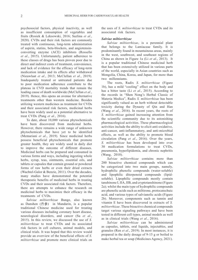
that belongs to the Lamiaceae family. It is predominantly found in mountainous areas, mainly in the west, southwest, and southeast regions of China as shown in Figure 1a (Li et al., 2013). It is a popular traditional Chinese medicinal herb that has been extensively utilized in various parts of the world, especially in Asian countries such as Mongolia, China, Korea, and Japan, for more than two millenniums.
The roots, Radix S. miltiorrhizae (Figure 1b), has a mild “cooling” effect on the body and has a bitter taste (Li et al., 2015). According to the records in “Shen Nong’s Herbal Classic of Materia Medica”, Radix S. miltiorrhizae has been significantly valued as an herb without detectable toxicity during the Dynasty of Qin and Han (Wang et al., 2016). In recent years, the roots of S. miltiorrhizae gained increasing attention from the scientific community due to its astonishing pharmacological activities. These pharmacological activities include the ability to elicit anti-oxidative, anti-cancer, anti-inflammatory, and anti-microbial effects, as well as the ability to promote blood circulation (Pang et al., 2016). Over the years, S. miltiorrhizae has been developed into over 30 medication formulations to treat CVDs, pneumonia, hyperlipidemia, and chronic nephritis (Wang, 2010).
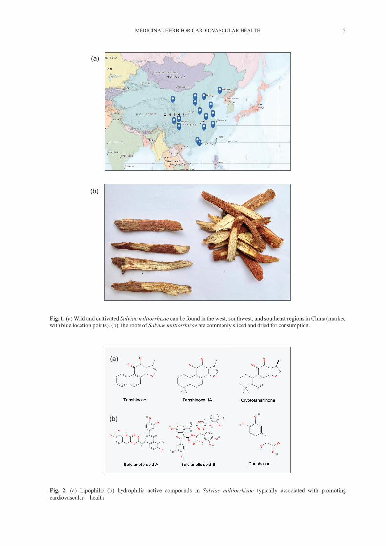
Salviae miltiorrhizae contains more than 200 bioactive chemical compounds which can be categorized into two main groups, namely, hydrophilic phenolic compounds (water-soluble) and lipophilic diterpenoid compounds (lipid-soluble). Lipophilic compounds mostly contain tanshinone I, IIA, IIB, and cryptotanshinone (Figure 2a); whilst the main type of hydrophilic compounds are phenolic acids such as miltirone, protocatechuic acid, and various types of salvianolic acids (Figure 2b). Moreover, components such as tannin and vitamin E have been discovered in extracts of S. miltiorrhizae. These bioactive chemical compounds target various signaling pathways and have been tested in different cell types, animal models as well as in clinical trials (Wang et al., 2016).
Salviae miltiorrhizae can be administered as capsules, tablets, oral liquids, injectables, and granules (Ren et al., 2019). In most instances, it is prepared in the daily dosage of 9-15 g or boiled to make herbal tea or soup (Medicines Agency, 2021).
MEDICINAL HERB FOR CARDIOVASCULAR HEALTH 3
Fig. 1. (a) Wild and cultivated Salviae miltiorrhizae can be found in the west, southwest, and southeast regions in China (marked with blue location points). (b) The roots of Salviae miltiorrhizae are commonly sliced and dried for consumption.
Fig. 2. (a) Lipophilic (b) hydrophilic active compounds in Salviae miltiorrhizae typically associated with promoting cardiovascular health
(a)
(b)
(a)
(b)

MEDICINAL HERB FOR CARDIOVASCULAR HEALTH4
Biological properties of Salviae miltiorrhizae and its underlying mechanisms
Salviae miltiorrhizae and its active compounds, mainly Danshensu, tanshinone IIA, salvianolic acid B, tanshinone VI, and cryptotanshinone, were found to elicit protective effects on endothelial cells, smooth muscle cells, and cardiac myocytes. Over the last few decades, researchers thoroughly examined the
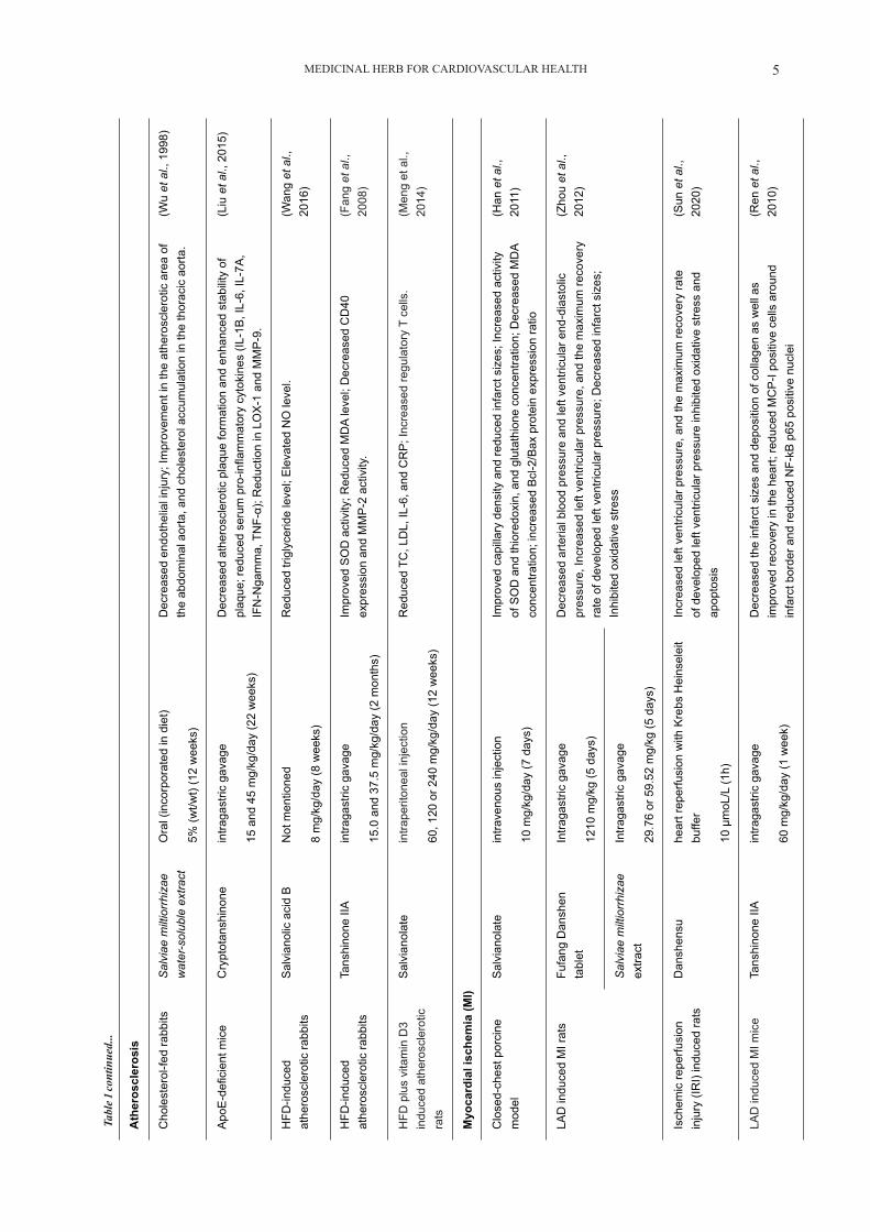
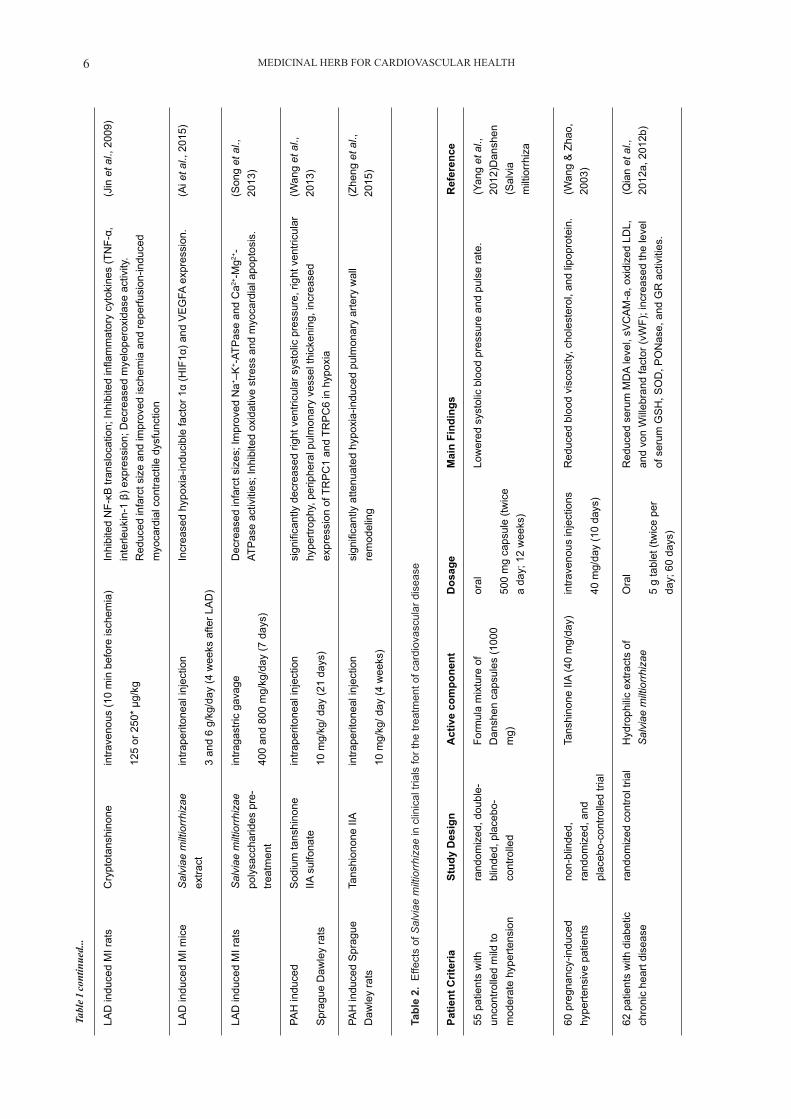
preclinical pharmacological activity of S. miltiorrhizae related to the treatment of CVD and its associated risk factors using different animal models. The following sections reviewed the effects of S. miltiorrizhae in different components of the cardiovascular system and summarise its beneficial outcomes in preclinical and clinical trials as shown in Table 1 and Table 2 respectively.
Tabl
e 1.
Effe
cts
of S
alvi
ae m
iltio
rrhi
zae
on h
yper
tens
ion,
ath
eros
cler
osis
, and
myo
card
ial i
sche
mia
in v
ivo
mod
els
In v
ivo
Mod
els
Act
ive
com
pone
ntR
oute
of a
dmin
istr
atio
n/
Dos
age/
Dur
atio
nM
ain
findi
ngs
Ref
eren
ces
Hyp
erte
nsio
n
Spo
ntan
eous
ly
hype
rtens
ive
rats
Dan
shen
suin
trape
riton
eal i
njec
tion
10 m
g/kg
/day
(6 w
eeks
)
Dec
reas
ed s
ysto
lic a
nd d
iast
olic
blo
od p
ress
ure;
Incr
ease
d in
duci
ble
NO
syn
thas
e ac
tivity
and
ser
um N
O c
onte
nt;
Low
ered
the
inci
denc
e of
ven
tricu
lar f
ibril
latio
n an
d ve
ntric
ular
ta
chyc
ardi
a; In
crea
sed
curr
ent d
ensi
ty o
f K+ a
nd c
urre
nt
dens
ity o
f Ca2+
-act
ivat
ed K
+ (K
Ca)
chan
nel i
n m
esen
teric
va
scul
ar s
moo
th m
uscl
e.
(Tan
g et
al.,
20
11a)
Isop
rote
reno
l in
duce
d m
yoca
rdia
l hy
pertr
ophy
rats
Dan
shen
suin
trape
riton
eal i
njec
tion
3 an
d 10
mg/
kg (d
ay 4
-7)
Red
uced
hea
rt w
eigh
t to
body
wei
ght i
ndex
and
arr
hyth
mia
sc
ores
; Im
prov
ed s
ysto
lic a
nd d
iast
olic
pre
ssur
es o
f the
left
vent
ricle
; Inc
reas
ed S
OD
and
Cx-
43 e
xpre
ssio
n.
(Tan
g et
al.,
20
11b)
Spo
ntan
eous
ly
hype
rtens
ive
rats
S
alvi
ae
milt
iorr
hiza
e in
trape
riton
eal i
njec
tion
1 g/
kg/d
ay (8
4 da
ys)
Red
uced
LV
mas
s in
dex,
siz
es o
f car
diom
yocy
tes,
and
co
llage
n vo
lum
e; c
ircum
fere
ntia
l are
a of
per
ivas
cula
r spa
ce
and
the
expr
essi
on o
f TN
F-α.
(Sun
&
Zhen
g, 2
007)
Phe
nyle
phrin
e in
duce
d m
ale
SD
ra
ts
Mag
nesi
um
tans
hino
ate
B
intra
veno
us in
ject
ion
0.7-
175
mg/
kg (2
0 m
ins
rest
ing
perio
d af
ter e
ach
dose
or
equi
libra
tion
15 m
in)
Red
uced
blo
od p
ress
ure
(mor
e th
an a
queo
us e
xtra
ct S
ME
)(L
eung
et a
l.,
2010
)
ET-
1 in
duce
d po
rtal
hype
rtens
ion
mic
eS
alvi
anol
ic
acid
B
intra
veno
us in
ject
ion
0.5
mg/
mou
se (3
day
s pr
e-tre
atm
ent)
Low
ered
the
aver
age
velo
city
of b
lood
flow
in th
e liv
er.
Tian
et a
l.,
2009
.
2-ki
dney
-2-
clip
indu
ced
reno
vasc
ular
hy
perte
nsio
n ra
t
Tans
hino
ne
IIA
intra
gast
ric g
avag
e
35 a
nd 7
0 m
g/kg
/day
(6 w
eeks
)
Atte
nuat
ed th
e re
sulti
ng in
ters
titia
l fib
rosi
s; D
ecre
ased
the
mR
NA
expr
essi
ons
of T
IMP
-1, T
IMP
-2, a
nd M
MP
-9; i
mpr
oved
ca
rdia
c fib
rosi
s an
d im
prov
ed c
ardi
ac fu
nctio
n
(Fan
g et
al.,
20
10)
MEDICINAL HERB FOR CARDIOVASCULAR HEALTH 5
Ath
eros
cler
osis
Cho
lest
erol
-fed
rabb
itsS
alvi
ae m
iltio
rrhi
zae
wat
er-s
olub
le e
xtra
ctO
ral (
inco
rpor
ated
in d
iet)
5% (w
t/wt)
(12
wee
ks)
Dec
reas
ed e
ndot
helia
l inj
ury;
Impr
ovem
ent i
n th
e at
hero
scle
rotic
are
a of
th
e ab
dom
inal
aor
ta, a
nd c
hole
ster
ol a
ccum
ulat
ion
in th
e th
orac
ic a
orta
.(W
u et
al.,
199
8)
Apo
E-d
efic
ient
mic
eC
rypt
otan
shin
one
intra
gast
ric g
avag
e
15 a
nd 4
5 m
g/kg
/day
(22
wee
ks)
Dec
reas
ed a
ther
oscl
erot
ic p
laqu
e fo
rmat
ion
and
enha
nced
sta
bilit
y of
pl
aque
; red
uced
ser
um p
ro-in
flam
mat
ory
cyto
kine
s (IL
-1B
, IL-
6, IL
-7A
, IF
N-N
gam
ma,
TN
F-α)
; Red
uctio
n in
LO
X-1
and
MM
P-9
.
(Liu
et a
l., 2
015)
HFD
-indu
ced
athe
rosc
lero
tic ra
bbits
Sal
vian
olic
aci
d B
Not
men
tione
d
8 m
g/kg
/day
(8 w
eeks
)
Red
uced
trig
lyce
ride
leve
l; E
leva
ted
NO
leve
l.(W
ang
et a
l.,
2016
)
HFD
-indu
ced
athe
rosc
lero
tic ra
bbits
Tans
hino
ne II
Ain
traga
stric
gav
age
15.0
and
37.
5 m
g/kg
/day
(2 m
onth
s)
Impr
oved
SO
D a
ctiv
ity; R
educ
ed M
DA
leve
l; D
ecre
ased
CD
40
expr
essi
on a
nd M
MP
-2 a
ctiv
ity.
(Fan
g et
al.,
20
08)
HFD
plu
s vi
tam
in D
3 in
duce
d at
hero
scle
rotic
ra
ts
Sal
vian
olat
e in
trape
riton
eal i
njec
tion
60, 1
20 o
r 240
mg/
kg/d
ay (1
2 w
eeks
)
Red
uced
TC
, LD
L, IL
-6, a
nd C
RP
; Inc
reas
ed re
gula
tory
T c
ells
.(M
eng
et a
l.,
2014
)
Myo
card
ial i
sche
mia
(MI)
Clo
sed-
ches
t por
cine
m
odel
Sal
vian
olat
ein
trave
nous
inje
ctio
n
10 m
g/kg
/day
(7 d
ays)
Impr
oved
cap
illar
y de
nsity
and
redu
ced
infa
rct s
izes
; Inc
reas
ed a
ctiv
ity
of S
OD
and
thio
redo
xin,
and
glu
tath
ione
con
cent
ratio
n; D
ecre
ased
MD
A
conc
entra
tion;
incr
ease
d B
cl-2
/Bax
pro
tein
exp
ress
ion
ratio
(Han
et a
l.,
2011
)
LAD
indu
ced
MI r
ats
Fufa
ng D
ansh
en
tabl
etIn
traga
stric
gav
age
1210
mg/
kg (5
day
s)
Dec
reas
ed a
rteria
l blo
od p
ress
ure
and
left
vent
ricul
ar e
nd-d
iast
olic
pr
essu
re, I
ncre
ased
left
vent
ricul
ar p
ress
ure,
and
the
max
imum
reco
very
ra
te o
f dev
elop
ed le
ft ve
ntric
ular
pre
ssur
e; D
ecre
ased
infa
rct s
izes
; In
hibi
ted
oxid
ativ
e st
ress
(Zho
u et
al.,
20
12)
Sal
viae
milt
iorr
hiza
e ex
tract
Intra
gast
ric g
avag
e
29.7
6 or
59.
52 m
g/kg
(5 d
ays)
Isch
emic
repe
rfusi
on
inju
ry (I
RI)
indu
ced
rats
Dan
shen
su
hear
t rep
erfu
sion
with
Kre
bs H
eins
elei
t bu
ffer
10 µ
moL
/L (1
h)
Incr
ease
d le
ft ve
ntric
ular
pre
ssur
e, a
nd th
e m
axim
um re
cove
ry ra
te
of d
evel
oped
left
vent
ricul
ar p
ress
ure
inhi
bite
d ox
idat
ive
stre
ss a
nd
apop
tosi
s
(Sun
et a
l.,
2020
)
LAD
indu
ced
MI m
ice
Tans
hino
ne II
Ain
traga
stric
gav
age
60 m
g/kg
/day
(1 w
eek)
Dec
reas
ed th
e in
farc
t siz
es a
nd d
epos
ition
of c
olla
gen
as w
ell a
s im
prov
ed re
cove
ry in
the
hear
t; re
duce
d M
CP
-I po
sitiv
e ce
lls a
roun
d in
farc
t bor
der a
nd re
duce
d N
F-kB
p65
pos
itive
nuc
lei
(Ren
et a
l.,
2010
)
Tabl
e 1 co
ntin
ued.
..
MEDICINAL HERB FOR CARDIOVASCULAR HEALTH6
LAD
indu
ced
MI r
ats
Cry
ptot
ansh
inon
ein
trave
nous
(10
min
bef
ore
isch
emia
)
125
or 2
50*
µg/k
g
Inhi
bite
d N
F-κB
tran
sloc
atio
n; In
hibi
ted
infla
mm
ator
y cy
toki
nes
(TN
F-α,
in
terle
ukin
-1 β
) exp
ress
ion;
Dec
reas
ed m
yelo
pero
xida
se a
ctiv
ity.
Red
uced
infa
rct s
ize
and
impr
oved
isch
emia
and
repe
rfusi
on-in
duce
d m
yoca
rdia
l con
tract
ile d
ysfu
nctio
n
(Jin
et a
l., 2
009)
LAD
indu
ced
MI m
ice
Sal
viae
milt
iorr
hiza
e ex
tract
intra
perit
onea
l inj
ectio
n
3 an
d 6
g/kg
/day
(4 w
eeks
afte
r LA
D)
Incr
ease
d hy
poxi
a-in
duci
ble
fact
or 1
α (H
IF1α
) and
VE
GFA
exp
ress
ion.
(Ai e
t al.,
201
5)
LAD
indu
ced
MI r
ats
Sal
viae
milt
iorr
hiza
e po
lysa
ccha
rides
pre
-tre
atm
ent
intra
gast
ric g
avag
e
400
and
800
mg/
kg/d
ay (7
day
s)
Dec
reas
ed in
farc
t siz
es; I
mpr
oved
Na+ –
K+ -
ATP
ase
and
Ca2+
-Mg2+
-AT
Pas
e ac
tiviti
es; I
nhib
ited
oxid
ativ
e st
ress
and
myo
card
ial a
popt
osis
.(S
ong
et a
l.,
2013
)
PAH
indu
ced
Spr
ague
Daw
ley
rats
Sod
ium
tans
hino
ne
IIA s
ulfo
nate
intra
perit
onea
l inj
ectio
n
10 m
g/kg
/ day
(21
days
)
sign
ifica
ntly
dec
reas
ed ri
ght v
entri
cula
r sys
tolic
pre
ssur
e, ri
ght v
entri
cula
r hy
pertr
ophy
, per
iphe
ral p
ulm
onar
y ve
ssel
thic
keni
ng, i
ncre
ased
ex
pres
sion
of T
RP
C1
and
TRP
C6
in h
ypox
ia
(Wan
g et
al.,
20
13)
PAH
indu
ced
Spr
ague
D
awle
y ra
tsTa
nshi
onon
e IIA
intra
perit
onea
l inj
ectio
n
10 m
g/kg
/ day
(4 w
eeks
)
sign
ifica
ntly
atte
nuat
ed h
ypox
ia-in
duce
d pu
lmon
ary
arte
ry w
all
rem
odel
ing
(Zhe
ng e
t al.,
20
15)
Tabl
e 2.
Effe
cts
of S
alvi
ae m
iltio
rrhi
zae
in c
linic
al tr
ials
for t
he tr
eatm
ent o
f car
diov
ascu
lar d
isea
se
Patie
nt C
riter
iaSt
udy
Des
ign
Act
ive
com
pone
ntD
osag
eM
ain
Find
ings
Ref
eren
ce
55 p
atie
nts
with
un
cont
rolle
d m
ild to
m
oder
ate
hype
rtens
ion
rand
omiz
ed, d
oubl
e-bl
inde
d, p
lace
bo-
cont
rolle
d
Form
ula
mix
ture
of
Dan
shen
cap
sule
s (1
000
mg)
oral
500
mg
caps
ule
(twic
e a
day;
12
wee
ks)
Low
ered
sys
tolic
blo
od p
ress
ure
and
puls
e ra
te.
(Yan
g et
al.,
20
12)D
ansh
en
(Sal
via
milt
iorr
hiza
60 p
regn
ancy
-indu
ced
hype
rtens
ive
patie
nts
non-
blin
ded,
ra
ndom
ized
, and
pl
aceb
o-co
ntro
lled
trial
Tans
hino
ne II
A (4
0 m
g/da
y)in
trave
nous
inje
ctio
ns
40 m
g/da
y (1
0 da
ys)
Red
uced
blo
od v
isco
sity
, cho
lest
erol
, and
lipo
prot
ein.
(Wan
g &
Zha
o,
2003
)
62 p
atie
nts
with
dia
betic
ch
roni
c he
art d
isea
sera
ndom
ized
con
trol t
rial
Hyd
roph
ilic
extra
cts
of
Sal
viae
milt
iorr
hiza
e O
ral
5 g
tabl
et (t
wic
e pe
r da
y; 6
0 da
ys)
Red
uced
ser
um M
DA
leve
l, sV
CA
M-a
, oxi
dize
d LD
L,
and
von
Will
ebra
nd fa
ctor
(vW
F); i
ncre
ased
the
leve
l of
ser
um G
SH
, SO
D, P
ON
ase,
and
GR
act
iviti
es.
(Qia
n et
al.,
20
12a,
201
2b)
Tabl
e 1 co
ntin
ued.
..
MEDICINAL HERB FOR CARDIOVASCULAR HEALTH 7
Effects of Salviae miltiorrhizae in preclinical trialsAnti-hypertensive
Hypertension is a common medical disorder where blood pressure in the arteries appear to be consistently higher than normal. The causes of high blood pressure might be associated with high cardiac output, accelerated heart rate, increased peripheral resistance, the decline in the density or number of capillaries, elevated vasoconstriction of active arteriolar, and/or reduced peripheral venous compliance. Long-term, uncontrolled hypertension can lead to stroke, heart failure, peripheral vascular disease, coronary artery disease, and even kidney failure (Lackland & Weber, 2015).
The hypotensive effect of S. miltiorrhizzae was tested in animal models. Tang et al. (2011a) discovered that 6-weeks intraperitoneal injection of Danshensu (10 mg/kg/day), a water-soluble isolate from S. miltiorrhizae, decreased blood pressure of spontaneously hypertensive rats (SHR) (145±3/ 103±10 mmHg to 116±7/ 87±2 mmHg) and significantly increased serum nitric oxide (NO), and activity of NO synthase (NOS). An increase in vasodilation from NO is important in attenuating hypertension and atherosclerosis as it protects the heart by regulating blood pressure and vascular tone, suppressing platelet aggregation and adhesion of leukocytes, as well as restricting the proliferation of smooth muscle cells (Zhao et al., 2015). Furthermore, incidences of ventricular fibrillation and tachycardia of the SHR rats reduced from 100% to 50%, and 30%, respectively (Tang et al., 2011a). Additionally, Danshensu increased the current density of Ca2+-activated K+ (KCa) channels and K+ (KV) channels of mesenteric vascular smooth muscles that are vital in modulating vascular tone and blood pressure (Tang et al., 2011a; Toth et al., 2013). These findings showed that Danshensu protected the cardiovascular system via modulation of vascular tone in the development of hypertension.
When the isoproterenol-induced myocardial hypertrophy rats were treated with Danshensu (3 & 10 mg/kg/day, intraperitoneal injection) on the 4th to 7th day, it was found that the heart to body weight index and arrhythmia scores were reduced. The systolic and diastolic pressures of the left ventricle along with electrocardiogram parameters were improved upon the administration of Danshensu. These results were shown to be due to the regulation of antioxidant enzymes, promoting connexin 43 (Cx-43) expression in the left ventricle (Tang et al., 2011b).
Hypertension also has a relatively close relationship with the elevated expression of pro-inflammatory myocardial TNF-α (Bergman et al., 1999). Salviae miltiorrhizae treatment (1 g/kg for 84 days, intraperitoneal injection) was given to the SHR and the results demonstrated that S. miltiorrhizae had significantly reduced the mass index of the left
ventricle, sizes of cardiomyocytes, collagen volume fraction, circumferential area of perivascular space respectively via decreased expression of TNF-α and hence, improve the hypertensive condition (Sun & Zheng, 2007). However, there was contradicting study indicating that the cardioprotective effect possessed by this herb was not associated with blood pressure (Sun & Zheng, 2007). Magnesium tanshinoate B (0.7-175 mg/kg) injected intravenously via the jugular vein into the high blood pressure male Sprague Dawley (SD) rats induced by phenylephrine was shown to have a hypotensive effect (Leung et al., 2010). Tian et al. (2009) revealed that S. miltiorrhizae (0.125 g per mouse) and salvianolic acid B (0.5 mg per mouse) reduced the average blood flow velocity in the liver of ET-1 induced portal hypertension mice. However, the mechanisms underlying its efficacy required further investigation.
Matrix metalloproteinases (MMPs) cause degradation of extracellular matrix components ultimately resulting in smooth muscle migration and proliferation and is tightly regulated by tissue inhibitors of metalloproteinases (TIMPs) which inhibit MMP activity (Galis & Khatri, 2002; Johnson, 2017). Disruption of the balance between MMPs and TIMPs results in pathophysiological conditions associated with vascular remodelling, wound healing, and inflammation such as vascular remodelling during early stages of hypertension, hypertrophy, or rupture of atherosclerotic plaques (Johnson, 2017). Proteases have been implicated in the development and progression of atherosclerosis, due to their ability to provoke focal destruction of the vascular extracellular matrix. Using renovascular hypertension rat model induced by 2-kidney-2-clip with the treatment of tanshinone IIA (70 mg/kg/day, intragastric gavage) for 6 weeks, Fang et al. (2010) concluded that the treatment could prevent cardiac fibrosis and hypertrophy in the left ventricle along with cardiac relaxation. Beneficial effects of tanshinone IIA were observed with the increase in the mRNA ratio of TIMP-2 to MMP-2, suggesting that tanshinone IIA exerts its effects by regulating MMPs and TIMPs at transcript levels (Fang et al., 2010).
Anti-atherosclerosis, anti-oxidation, and anti-inflammation
Atherosclerosis is a disorder in which the arteries become narrowed and lose elasticity due to abnormal accumulation of adhesive plaques (Aluganti Narasimhulu et al., 2016). Oxidative stress from an overproduction of reactive oxygen species (ROS) and inflammation is a critical contributor to atherosclerosis progression by causing several complications such as the aggravation of endothelial cells, disruption of vascular wall microenvironment, migration and adhesion of leucocytes, and increase in MMP production and degradation in collagen
MEDICINAL HERB FOR CARDIOVASCULAR HEALTH8
(Kattoor et al., 2017). Therefore, selectively targeting pro-oxidant components such as NADPH oxidase and lectin-type oxidized LDL receptor 1 (LOX-1), while enhancing the activity of antioxidants such as superoxide dismutase (SOD) is vital in atherosclerosis therapy to restore balance in the oxidant-antioxidant system and to regulate inflammation (Kattoor et al., 2017).
Several in vivo studies showed that S. miltiorrhizae was able to reduce triglyceride and total cholesterol levels, and exhibited potent anti-oxidative and anti-inflammatory properties, thus improving atherosclerosis (Wu et al., 1998; Fang et al., 2008; Liu et al., 2015; Wang et al., 2016). A water- soluble polyphenolic antioxidant isolated from the roots of this plant, was found to scavenge 1,1-diphenyl-2-picrylhydrazyl radicals and inhibit LDL oxidation more effectively than probucol. In order to evaluate the antiatherogenic potential, New Zealand White rabbits were fed for 12 weeks a normal diet, a high cholesterol diet, a high cholesterol diet containing 1% probucol, or a high cholesterol diet containing a 5% water-soluble extract of S. miltiorrhizae (SM). New Zealand white rabbits fed with a high cholesterol diet comprising of a 5% water-soluble extract of S. miltiorrhizae for 12 weeks were found to be more resistant against copper ion (Cu2+)-induced oxidative stress (Wu et al., 1998). Endothelium disruption has been reduced by 53% (p<0.05) as measured at the 6th week. The atherosclerotic lesion at the abdominal aorta was reduced by 56% (p<0.05) and cholesterol accumulation in the thoracic aorta was reduced by 50% (p<0.05) with the treatment of S. miltiorrhizae. This study concluded that the reduction in atherosclerosis by S. miltiorrhizae did not solely depend on the cholesterol-lowering efficacy but also on the anti-oxidant ability in preventing endothelial damage as well as inhibiting the formation of oxidized LDL in hypercholesterolemia subjects (Wu et al., 1998).
Liu et al. (2015) demonstrated that when cryptotanshinone (15 & 45 mg/kg/day, intragastric gavage) was given to high-fat diet apolipoprotein E deficient mice for 22 weeks, the generation of ROS from NADPH oxidase 4 and the NF-κB activation was inhibited. Subsequently, LOX-1 and MMP-9 expressions were also reduced. In another study, salvianolic acid B (8 mg/kg/day) was administered to the high-fat diet-induced atherosclerotic rabbits for 8 weeks. The study revealed that the release of NO was elevated while the triglyceride level was reduced and thus, the atherogenesis was subsequently inhibited (Wang et al., 2016). Fang et al. (2008) similarly demonstrated that tanshinone IIA possessed anti-atherosclerotic properties by regulating components of the cardiovascular anti-oxidant system. When administered for 2 months to high fatty diet rabbits in different dosages (15 & 37.5 mg/kg, intragastric gavage), tanshinone IIA substantially increased
superoxide dismutase (SOD) activity and caused a great reduction in pro-inflammatory malondialdehyde (MDA) levels (Fang et al., 2008). Moreover, the expression of pro-inflammatory molecule CD40 as well as the MMP-2 activity were both downregulated which suggested that tanshinone IIA attenuated MMP-2 via blocking CD40 inflammatory pathways (Fang et al., 2008). In summary, tanshinone IIA did not only possess the ability of anti-oxidation, but also anti-inflammation in alleviating atherosclerotic lesions.
Ability to reduce total cholesterol levels and reduce inflammation was also observed in hydrophilic S. miltiorrhizae extracts, such as salvianolate where salvianolic acid B was its main active component (85%) (Han et al., 2011; Meng et al., 2014). Salvianolate was given to a high-fat diet + vitamin D3-induced atherosclerotic rats for 12 weeks at a dosage of 60, 120, and 240 mg/kg (intraperitoneal injection) (Meng et al., 2014). Results demonstrated improvement of atherosclerosis development with declined levels of proinflammatory cytokines including IL-6 and C reactive protein (CRP) as well as an increase in regulatory T cells (Tregs) which possess the ability in preventing the progression of atherosclerosis through the reduction of IL-6 expression and CRP production. However, although salvianolate treatment reduced total cholesterol (TC) and LDL levels, it barely had any impact on the triglyceride and HDL levels (Poledne et al., 2009; He et al., 2010).
Prevention of myocardial ischemiaMyocardial ischemia (MI), also known as angina,
is a heart disease associated with irreversible damage to the cardiac muscle as a result of the deficiency of blood supply to the heart induced by partial blockage of the coronary arteries (Buja and Vender, 2016). MI develops several threats to the myocardium, including generation of ROS, overloading of intracellular calcium, dysfunctional endothelium, apoptosis of myocardium, increased adhesion of leukocytes, and degranulation of mast cells (Mozaffari et al., 2013).
Studies showed that salvianolate administration (10 mg/kg/day, intravenous injection) for 7 days consecutively enhanced myocardial microvessels reflow and elevated the density of capillary as well as reduced the infarct size in a porcine model (Han et al., 2011). The therapeutic effect of salvianolate was potentially associated with its capability in reducing oxidative stress and cell death. The decrease in oxidative stress was mediated by the increase of SOD and thioredoxin activities and glutathione concentration, as well as a decrease in the concentration of MDA. Subsequently, this would lead to the reduction in terminal deoxynucleotide transferase-mediated dUTP nick end labeling-positive cells and increased the ratio of B-cell lymphoma-2 (Bcl-2) to Bax expression, indicating suppression of cell apoptosis signaling pathways by salvianolate (Han et al., 2011).
MEDICINAL HERB FOR CARDIOVASCULAR HEALTH 9
Similarly, treatment of MI rats and ischemic reperfusion injured (IRI) rats with Fufang danshen tablet (1210 mg/kg, intragastric gavage) or aqueous S. miltiorrhizae extract (29.76 or 59.52 mg/kg, and intragastric gavage) for 5 days, and Danshensu (10 µmol/L, heart reperfusion) for 1 h, respectively, improved recovery of cardiac function as evidenced by improved hemodynamic parameters (i.e increased in left ventricular systolic pressure, decreased in left ventricular end-diastolic pressure) and decreased infarct size (Zhou et al., 2012; Sun et al., 2020). These improvements were also observed with a reduction in serum levels of oxidative markers including lactate dehydrogenase, glutamic oxalacetic transaminase, creatine kinase, and MDA, and an increase in anti-oxidant systems (SOD and glutathione peroxidase) (Zhou et al., 2012; Sun et al., 2020). Collectively, these observations indicated that hydrophilic active components in S. miltiorrhizae protected against MI through inhibiting oxidative stress and apoptosis
Tanshinone IIA treatment (60 mg/kg/day, intragastric gavage) was given to the permanent left anterior descending coronary artery (LAD) ligation induced myocardial ischemia (MI) rats for 7 days. Tanshionone IIA-treated rats demonstrated a reduction in the deposition of collagen and infarct sizes, consequently resulting in an improved recovery in the heart (Ren et al., 2010). Immunohistochemistry staining of myocardial tissue isolated from treatment groups revealed that tanshinone IIA reduced the number of the pro-inflammatory molecule, MCP-1, containing cells around the infarct border with reduced NF-kB p65 translocation to the nucleus, suggesting that tanshinone IIA elicits anti-inflammatory effects by preventing transcription of pro-inflammatory molecules as a cardioprotective response to MI (Ren et al., 2010).
Jin et al. (2009) investigated the cryptotanshinone treatment on the modifications of hemodynamic parameters including left ventricular dp/dtmax end-diastolic pressure, as well as the infarct size in the LAD-induced MI rat model. The research indicated that 250 µg/kg of cryptotanshinone prevented the alterations induced by MI via modulation of the inflammatory pathway by suppressing NF-κB translocation and lowering pro-inflammatory cytokine production, as well as restricting activation of myeloperoxidase and neutrophil infiltration through the regulation of p38 MAPK/ JNK/ ERK pathways in ischemic myocardial tissues.
Cardiac angiogenesis activation is an effective therapeutic approach for achieving the increased oxygen and nutritional demands of the stressed heart (Mozid et al., 2014). Vascular endothelial growth factor A (VEGFA) and Hypoxia-inducible factor 1 (HIF1α) are both important in the activation of cardiac angiogenesis (Giatromanolaki et al., 2008). Salviae miltiorrhizae extract (3 & 6 g/kg/day, intraperitoneal injection) administered for 4 weeks was shown to elevate the expression of VEGF-A and HIF1α in LAD-
induced MI mice, contributing to improved cardiac function and cardiac angiogenesis (Ai et al., 2015). Another study indicated that giving pre-treatment of 400 and 800 mg/kg by oral gavage of S. miltiorrhizae polysaccharides to LAD-induced MI rats respectively for 1 week improved the infarct sizes as well as enhanced the myocardial Na+-K+-ATPase and Ca2+-Mg2+-ATPase activities, which were believed to be correlated to myocardial injury (Ke et al., 2004; Song et al., 2013). Though S. miltiorrhizae polysaccharides were found to be beneficial against MI, more investigations on the usage of polysaccharides in the treatment of CVD are needed due to the shortage of appropriate methods in accessing its contributions.
Effects of Salviae miltiorrhizae in clinical trialsA randomized, double-blinded, placebo-
controlled, single-center clinical trial had proven that administration of S. miltiorrhizae could improve the hypertensive condition. A total of 55 patients having uncontrolled mild to moderate hypertension signed up for a conventional anti-hypertensive treatment were randomized into two groups receiving the Fufang Danshen extract capsules containing a mixture of 225 mg pure S. miltiorrhizae extract, 20 mg Rhodiola rosea extract, 100 mg Chrysanthemum extract, and 100 mg Pueraria extract (500 mg, twice a day) and placebo capsules respectively for 12 weeks (Yang et al., 2012). The results demonstrated that the systolic blood pressure and pulse rate of the S. miltiorrhizae-treated group had a significant reduction compared to the placebo group at week 12. The systolic blood pressure of the S. miltiorrhizae-treated group was reduced by 13.8 mmHg whilst the placebo group only decreased by 4.2 mmHg. There were no observable adverse effects between both groups. Hence, it was concluded that extract of S. miltiorrhizae had a high safety profile and was effective in reducing the systolic blood pressure and pulse rate of hypertensive patients (Yang et al., 2012). A non-blinded, randomized, and placebo-controlled clinical trial was performed to examine the efficacy of S. miltiorrhizae on 60 patients with pregnancy-induced hypertension (Wang & Zhao, 2003). Salviae miltiorrhizae injection containing tanshinone IIA (40 mg/day) was administered for 10 days. Results demonstrated that S. miltiorrhizae reduced blood viscosity, and both the levels of cholesterol and lipoprotein.
Another randomized controlled study was proven to soothe the symptoms of diabetic chronic heart disease. A total of 62 patients were treated with 5 g tablets containing hydrophilic extracts of S. miltiorrhizae for 60 days. The results demonstrated that there was a significant reduction in the serum MDA level, sVCAM-a, oxidized LDL, and von Willebrand factor (vWF) while the level of serum glutathione (GSH), SOD, paraoxonase (PONase), and glutathione reductase (GR) activities were increased (Wang et al., 2016).
MEDICINAL HERB FOR CARDIOVASCULAR HEALTH10
Elucidating molecular mechanisms of Salviae miltiorrhizae
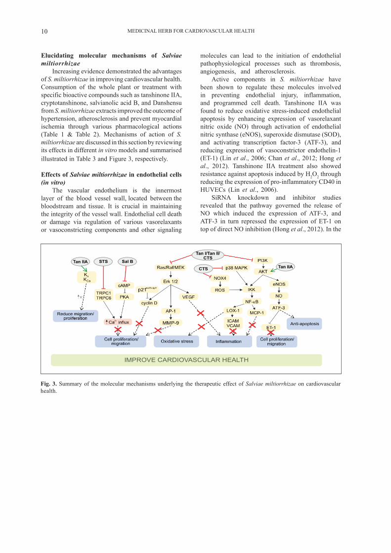
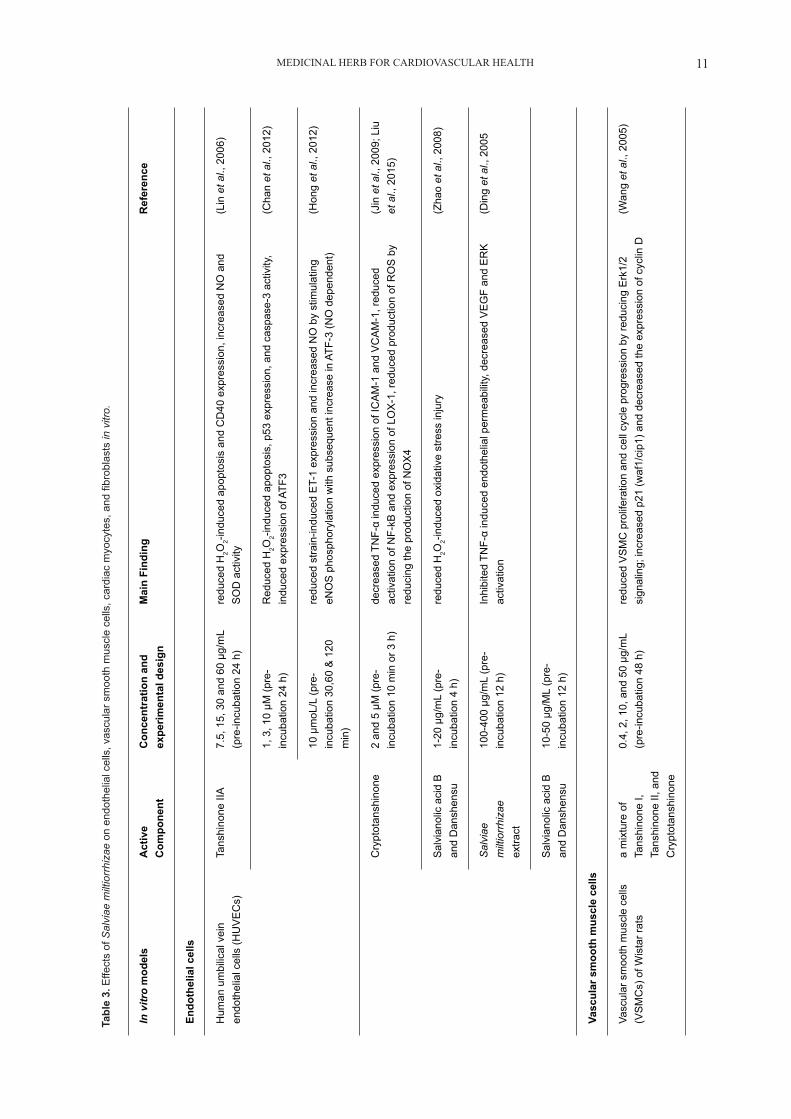
Increasing evidence demonstrated the advantages of S. miltiorrhizae in improving cardiovascular health. Consumption of the whole plant or treatment with specific bioactive compounds such as tanshinone IIA, cryptotanshinone, salvianolic acid B, and Danshensu from S. miltiorrhizae extracts improved the outcome of hypertension, atherosclerosis and prevent myocardial ischemia through various pharmacological actions (Table 1 & Table 2). Mechanisms of action of S. miltiorrhizae are discussed in this section by reviewing its effects in different in vitro models and summarised illustrated in Table 3 and Figure 3, respectively.
Effects of Salviae miltiorrhizae in endothelial cells (in vitro)
The vascular endothelium is the innermost layer of the blood vessel wall, located between the bloodstream and tissue. It is crucial in maintaining the integrity of the vessel wall. Endothelial cell death or damage via regulation of various vasorelaxants or vasoconstricting components and other signaling
molecules can lead to the initiation of endothelial pathophysiological processes such as thrombosis, angiogenesis, and atherosclerosis.
Active components in S. miltiorrhizae have been shown to regulate these molecules involved in preventing endothelial injury, inflammation, and programmed cell death. Tanshinone IIA was found to reduce oxidative stress-induced endothelial apoptosis by enhancing expression of vasorelaxant nitric oxide (NO) through activation of endothelial nitric synthase (eNOS), superoxide dismutase (SOD), and activating transcription factor-3 (ATF-3), and reducing expression of vasoconstrictor endothelin-1 (ET-1) (Lin et al., 2006; Chan et al., 2012; Hong et al., 2012). Tanshinone IIA treatment also showed resistance against apoptosis induced by H2O2 through reducing the expression of pro-inflammatory CD40 in HUVECs (Lin et al., 2006).
SiRNA knockdown and inhibitor studies revealed that the pathway governed the release of NO which induced the expression of ATF-3, and ATF-3 in turn repressed the expression of ET-1 on top of direct NO inhibition (Hong et al., 2012). In the
Fig. 3. Summary of the molecular mechanisms underlying the therapeutic effect of Salviae miltiorrhizae on cardiovascular health.
MEDICINAL HERB FOR CARDIOVASCULAR HEALTH 11
Tabl
e 3.
Effe
cts
of S
alvi
ae m
iltio
rrhi
zae
on e
ndot
helia
l cel
ls, v
ascu
lar s
moo
th m
uscl
e ce
lls, c
ardi
ac m
yocy
tes,
and
fibr
obla
sts
in v
itro.
In v
itro
mod
els
Act
ive
Com
pone
ntC
once
ntra
tion
and
expe
rimen
tal d
esig
nM
ain
Find
ing
Ref
eren
ce
Endo
thel
ial c
ells
Hum
an u
mbi
lical
vei
n en
doth
elia
l cel
ls (H
UV
EC
s)Ta
nshi
none
IIA
7.
5, 1
5, 3
0 an
d 60
μg/
mL
(pre
-incu
batio
n 24
h)
redu
ced
H2O
2-ind
uced
apo
ptos
is a
nd C
D40
exp
ress
ion,
incr
ease
d N
O a
nd
SO
D a
ctiv
ity(L
in e
t al.,
200
6)
1, 3
, 10
μM (p
re-
incu
batio
n 24
h)
Red
uced
H2O
2-ind
uced
apo
ptos
is, p
53 e
xpre
ssio
n, a
nd c
aspa
se-3
act
ivity
, in
duce
d ex
pres
sion
of A
TF3
(Cha
n et
al.,
201
2)
10 μ
moL
/L (p
re-
incu
batio
n 30
,60
& 1
20
min
)
redu
ced
stra
in-in
duce
d E
T-1
expr
essi
on a
nd in
crea
sed
NO
by
stim
ulat
ing
eNO
S p
hosp
hory
latio
n w
ith s
ubse
quen
t inc
reas
e in
ATF
-3 (N
O d
epen
dent
)(H
ong
et a
l., 2
012)
Cry
ptot
ansh
inon
e 2
and
5 μM
(pre
-in
cuba
tion
10 m
in o
r 3 h
)de
crea
sed
TNF-
α in
duce
d ex
pres
sion
of I
CA
M-1
and
VC
AM
-1, r
educ
ed
activ
atio
n of
NF-
kB a
nd e
xpre
ssio
n of
LO
X-1
, red
uced
pro
duct
ion
of R
OS
by
redu
cing
the
prod
uctio
n of
NO
X4
(Jin
et a
l., 2
009;
Liu
et
al.,
201
5)
Sal
vian
olic
aci
d B
an
d D
ansh
ensu
1-20
μg/
mL
(pre
-in
cuba
tion
4 h)
redu
ced
H2O
2-ind
uced
oxi
dativ
e st
ress
inju
ry(Z
hao
et a
l., 2
008)
Sal
viae
m
iltio
rrhi
zae
extra
ct
100-
400
μg/m
L (p
re-
incu
batio
n 12
h)
Inhi
bite
d TN
F-α
indu
ced
endo
thel
ial p
erm
eabi
lity,
dec
reas
ed V
EG
F an
d E
RK
ac
tivat
ion
(Din
g et
al.,
200
5
Sal
vian
olic
aci
d B
an
d D
ansh
ensu
10-5
0 μg
/ML
(pre
-in
cuba
tion
12 h
)
Vasc
ular
sm
ooth
mus
cle
cells
Vasc
ular
sm
ooth
mus
cle
cells
(V
SM
Cs)
of W
ista
r rat
s a
mix
ture
of
Tans
hino
ne I,
Ta
nshi
none
II, a
nd
Cry
ptot
ansh
inon
e
0.4,
2, 1
0, a
nd 5
0 μg
/mL
(pre
-incu
batio
n 48
h)
redu
ced
VS
MC
pro
lifer
atio
n an
d ce
ll cy
cle
prog
ress
ion
by re
duci
ng E
rk1/
2 si
gnal
ing;
incr
ease
d p2
1 (w
af1/
cip1
) and
dec
reas
ed th
e ex
pres
sion
of c
yclin
D(W
ang
et a
l., 2
005)
MEDICINAL HERB FOR CARDIOVASCULAR HEALTH12
Hum
an a
ortic
sm
ooth
mus
cle
cells
(HA
SM
Cs)
Tans
hino
ne II
A
15-1
00 μ
M (p
re-
incu
batio
n 1-
2 h)
Sup
pres
sed
TNF-
indu
ced
Akt
, ErK
and
c-ju
n ph
osph
oryl
atio
n, in
hibi
ted
IB p
hosp
hory
latio
n an
d p6
5 nu
clea
r tra
nslo
catio
n; d
ownr
egul
ate
MM
P-9
ex
pres
sion
(Jin
et a
l., 2
008)
Pul
mon
ary
arte
ry s
moo
th
mus
cle
cells
(PA
SM
Cs)
of
norm
oxic
rats
Sod
ium
ta
nshi
none
IIA
su
lfona
te
0 –
25 μ
M (p
re-in
cuba
tion
60 h
)bl
ocke
d hy
poxi
a-in
duce
d in
crea
se o
f TR
PC
1 an
d TR
PC
6 ex
pres
sion
, de
crea
sed
VS
MC
pro
lifer
atio
n. a
ttenu
ated
incr
ease
of S
OC
E, a
nd h
inde
red
the
elev
atin
g co
ncen
tratio
n of
bas
al in
trace
llula
r Ca2+
,
(Wan
g et
al.,
201
3)
Tans
hino
ne II
A
25 μ
g/m
L (c
ells
isol
ated
fro
m ra
ts tr
eate
d in
trape
riton
eally
for 4
w
eeks
)
Rec
over
ed th
e hy
poxi
a-do
wnr
egul
ated
I KV c
urre
nts
by d
ownr
egul
atin
g th
e ex
pres
sion
of K
v2.1
and
Kv1
.5(Z
heng
et a
l., 2
015)
Car
diom
yocy
tes
and
card
iofib
robl
asts
Car
diac
myo
cyte
s an
d fib
robl
asts
of n
eona
tal r
ats
Tans
hino
ne V
I10
μM
(pre
-incu
batio
n 24
h)
Inhi
bite
d ph
enyl
ephr
ine,
ET-
1, a
nd IG
F-1
indu
ced
prot
ein
synt
hesi
s; R
educ
ed
hype
rtrop
hy in
car
diac
fibr
obla
sts.
(Mak
i et a
l., 2
002)
Car
diac
myo
cyte
s an
d fib
robl
asts
of n
eona
tal r
ats
Tans
hino
ne II
A
(2-8
μM
)2-
8 μM
(pre
-incu
batio
n 1
h)re
duce
d TN
F-α-
indu
ced
expr
essi
on o
f MC
P-1
, TG
F-B
, and
CD
68 in
car
diac
fib
robl
asts
but
not
in c
ardi
omyo
cyte
s(R
en e
t al.,
201
0)
H9C
2 ce
ll lin
es o
f em
bryo
nic
rat h
eart
vent
ricle
Sal
vian
olic
aci
d B
0.00
1, 0
.01
and
0.1
mg/
mL
(pre
-incu
batio
n 2
h)R
educ
ed p
rodu
ctio
n of
ano
xia-
indu
ced
cAM
P an
d P
KA
follo
wed
by
Ca2+
influ
x, le
adin
g to
pro
tect
ive
effe
cts
on M
I (L
u et
al.,
201
2)
H9C
2 ce
ll lin
es o
f em
bryo
nic
rat h
eart
vent
ricle
Dan
shen
su10
µm
oL/L
(3 h
in
cuba
tion
afte
r hyp
oxia
)re
duce
d ap
opto
sis
by b
lock
ing
hypo
xia-
indu
ced
decr
ease
of B
cl-1
and
in
crea
se o
f Bax
(Sun
et a
l., 2
020)
Tabl
e 3 co
ntin
ued.
..
MEDICINAL HERB FOR CARDIOVASCULAR HEALTH 13
endothelium, ATF-3 typically induces the expression of genes that result in inflammation, apoptotic responses, and oxidative stress (Aung et al., 2016). Specifically, in regulating apoptosis cascades, ATF-3 was found to inhibit transcription of p53 by binding to the AP-1 element in the p53 gene promoter, resulting in suppression of TNF-α induced apoptotic pathways through caspase-3 (Kawauchi et al., 2002). Tanshinone IIA-treated HUVECs also demonstrated a reduction in p53 expression and caspase-3 activation further confirming its role in promoting endothelial survival during oxidative damage (Chan et al., 2012). On the other hand, ET-1 is a vasoconstricting peptide mainly formed in the endothelium and is involved in modulating vascular tone and has been shown to mediate cell proliferation in vascular smooth muscle cells, cardiomyocytes, and cardio fibroblasts through the Ras/Raf/ERK pathway (Cheng et al., 2003; Marasciulo et al., 2006). The present study was performed to examine the role of endogenous ET-1 in ET-1 - stimulated fibroblast proliferation and to investigate the regulatory mechanism of ET-1 - induced ET-1 gene expression in cardiac fibroblasts. Both ETA receptor antagonist [(hexahydro-1 H-azepinyl. Repression of ET-1 production in the endothelium by tanshinone IIA therefore reduced subsequent migration and proliferation pathways associated with cardiac and vascular remodeling.
Cryptotanshinone, a main bioactive diterpenoid found in S. miltiorrhizae, also possesses anti-oxidative and anti-inflammatory properties by regulating lectin-like oxidized LDL receptor-1 (LOX-1) expression. LOX-1 is a major receptor for the ROS-producing ox-LDL in endothelial cells and plays an important role in the formation of foam cells from macrophages, endothelial dysfunction observed in hypertension, atherogenesis, and plaque instability (Kattoor et al., 2017). In HUVECs treated with H2O2, oxidized low-density lipoprotein (oxLDL) and tumor necrosis factor-α (TNF-α), cryptotanshinone treatment inhibited the corresponding LOX-1 expression and nuclear transcription factor-kappa B (NF-κB) activation (Jin et al., 2009; Liu et al., 2015). Furthermore, cryptotanshinone disrupted LOX-1-mediated adhesion of THP-1 monocytes to HUVECs by lowering the expressions of vascular cell adhesion molecule-1 (VCAM-1), intracellular adhesion molecule-1 (ICAM-1), and E-selectin in HUVECs via inhibition of NOX4/ROS/ NF-κB signaling pathway and preventing translocation of NF-κB to the nucleus to induce gene transcription ( Jin et al., 2009; Liu et al., 2015. However, it is not known whether CTS can prevent experimental atherosclerosis. The present study was designed to investigate the protective effects of CTS on atherosclerosis and its molecular mechanisms of action. Higher expression and activation of VCAM-1 and ICAM-1, as well as E-selectin, are known to contribute to the binding
of circulating immune cells to endothelial cells surface, subsequently initiating the progression of atherosclerosis (Ling et al., 2012). Further inhibition of LOX-1 mediated downregulation of eNOS, upregulation of apoptotic proteins caspase 3 and caspase 9, and production of MMP-9 remains to be validated in endothelial cells, but are likely pathways of cryptotanshinone in attenuating atherosclerosis plaque formation, and improving plaque stability as observed in vivo models (Liu et al., 2015; Kattoor et al., 2017).
Endothelial hyperpermeability refers to dysfunction of the endothelial barrier, allowing increased cellular leakage of various pro-inflammatory, pro-oxidants molecules which results in exacerbated inflammation, ischemic reperfusion injury, or atherosclerosis (Kumar et al., 2010). Tyrosine kinase receptors such as vascular endothelial growth factor (VEGF) have been shown to mediate endothelial hyperpermeability (Kumar et al., 2010). Salviae miltiorrhizae extracts along with its hydrophilic active compounds including Danshensu and salvianolic acid B demonstrated the ability to suppress VEGF expression and attenuate TNF-α-induced hyperpermeability in HUVECs (Ding et al., 2005). To understand its mechanism of pharmacological action, its effects on endothelial monolayer permeability were studied. The present study demonstrated that decrease in VEGF expression by Danshensu and salvianolic acid B appeared to be dependent on abolishing ERK activation and reduction of ROS levels (Ding et al., 2005). Additionally, salvianolic acid B and Danshensu treatment (1-20 μg/mL) prevented H2O2 -induced injury in HUVECs attributed to its anti-oxidant properties as evidenced by high scavenging activities against various free radicals (Zhao et al., 2008).
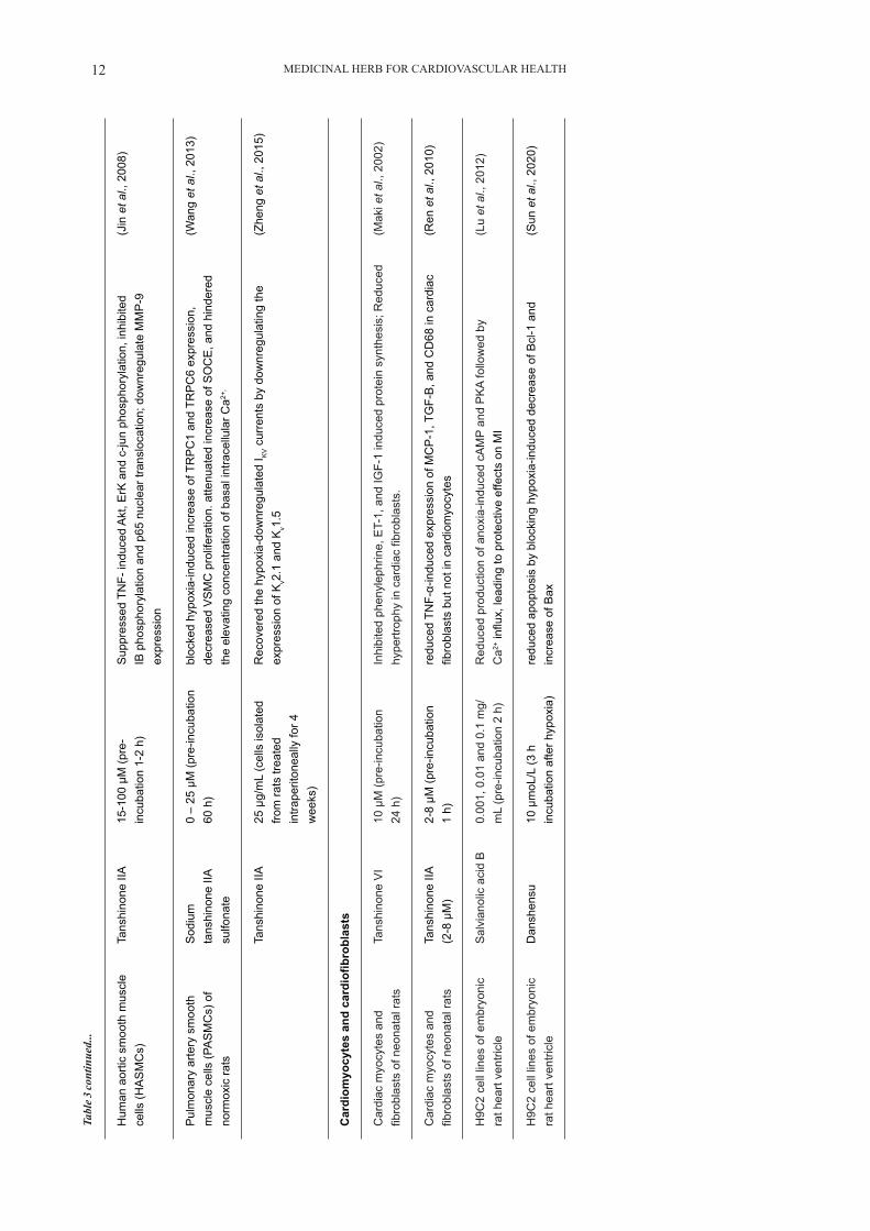
Effects of Salviae miltiorrhizae in vascular smooth muscle cells (in vitro)
VSMCs are a major cell type localized in the tunica media of the vasculature and function to mediate contraction and ECM production. VSMCs can migrate, proliferate, and differentiate into a more synthetic, less contractile phenotype in response to stress or pathological conditions, leading to VSMC dysfunction and remodeling associated with thickening of vessel walls and progression of cardiovascular disease (Guarner-Lans et al., 2020).
According to Wang et al. (2005), a combination treatment using lipohilic components tanshinone I, tanshinone II, and cryptotanshinone at the concentrations of 0.4, 2, 10, and 50 μg/mL, hindered the proliferation of the VSMCs in the Wistar rats. The reduced proliferation of VSMCs was mediated through the inhibition of extracellular signal-regulated protein kinase 1/2 (ERK 1/2) signaling and
MEDICINAL HERB FOR CARDIOVASCULAR HEALTH14
consequently decreasing the expression of cyclin D by increasing the expression of p21waf1/cip1. p21waf1/cip1 is implicated to inhibit cell cycle progression from G0/G1 to S phase thus inhibiting proliferation of VSMCs (Wang et al., 2005). In addition, tanshinone IIA suppressed the human aortic smooth muscle cells (hASMCs) migration via downregulating MMP-9 expression through inhibition of MAPK/ERK and PI3K/Akt pathways as evidenced by Akt, ErK, and c-jun phosphorylation. Interestingly, tanshinone IIA did not inhibit activation of other MAPKs (i.e. JNK & p38) in the VSMCs. In the PI3K/Akt pathway tanshinone IIA inhibited IκBα phosphorylation and p65 nuclear translocation, subsequently preventing transcription factor NF-κB to bind to the MMP-9 gene for further transcription, indicating that tanshinone IIA regulated MMP-9 expression at transcript levels.
VSMC proliferation and contraction are also regulated by intracellular Ca2+ concentration and cellular membrane potential. The influx of Ca2+ is mediated by transient receptor potential canonical channels (TRPC) which are a group of store-operated Ca2+ channels (SOCE) (Cheng et al., 2013; Wang et al., 2015). Sodium tanshinone IIA sulfonate treatment (12.5 μM) altered the proliferation and migration of hypoxic and normoxic pulmonary artery smooth muscle cells (PASMCs) by decreasing SOCE and hindering the elevating concentration of basal intracellular Ca2+ through disruption of TRPC1 and TRPC6 expression in distal PASMCs (Wang et al., 2013). During chronic hypoxia, severe exposure to hypoxic conditions suppressed the activity of KV channels in VSMCs and induced VSMC contraction and proliferation that results in pulmonary vascular remodeling typically observed in pulmonary hypertension (Wang et al., 2005). Danshensu therapy increased the expression of the K+ channel stimulated by K+ and Ca2+ in primary mesenteric vascular smooth muscle cells from spontaneously hypertensive rats (SHRs) when the tested potential was set at +60 mV. In another study, tanshinone IIA (25 μg/mL) therapy partially recovered the downregulated IKV currents induced by severe hypoxia in PASMCs (Zheng et al., 2015. These results indicate that active components in S. miltiorrhizae can be used in therapy for pathological conditions involving hypoxia such as pulmonary hypertension.
Effects of Salviae miltiorrhizae in cardiac myocytes and cardiac fibroblasts (in vitro)
Hypertrophy or remodeling of cardiac myocytes in response to stress and injury alters the contractility of the myocardium and eventually leads to left ventricular hypertrophy commonly observed in MI (Voulgari et al., 2010). Monocyte chemotactic protein (MCP)-1, which stimulates invasion, activation, and cytokine production of inflammatory cells, was highly expressed in the MI animal models (Kobusiak-Prokopowicz et al., 2007; Kohno et al.,
2008). In contrast, MCP-1 deficient mice showed lower macrophages recruitment in the ischemic heart, prolonged phagocytosis of damaged cardiac myocytes, reduced fibroblasts infiltration, and diminished left ventricular remodeling. Moreover, cyclic adenosine monophosphate (cAMP) stimulates the activation of protein kinase A (PKA) leading to the phosphorylation of L-type Ca2+ channels during myocardial ischemia, resulting in subsequent Ca2+ influx, and muscle contraction. Therefore, targeting MCP-1 and L-type Ca2+ channels would be beneficial in treating MI and its related cardiovascular diseases (Xia & Frangogiannis, 2007).
Tanshinone VI (10 μM) inhibited ET-1, insulin-like growth factor-1 (IGF-1), and phenylephrine-induced protein synthesis in neonatal rat myocardiocytes (Maki et al., 2002). It also reduced hypertrophy in cardiac fibroblasts induced by 5% of fetal bovine serum and 0.01 μM of IGF-1, through the reduced synthesis of collagen (Maki et al., 2002). Cardioprotective effects including a decrease in infarct size, decreasing collagen deposition, and improving heart recovery in tanshinone IIA-treated rats in vivo were attributed to a reduction in MCP-1, and TGF-β1 in cardiac fibroblasts and infiltration of macrophages (Ren et al., 2010). Moreover, the expression of NF-κB and its p65 subunit in the nuclei of infarcted cardiac cells decreased with the concentration of proinflammatory stimuli TNF- α (Ren et al., 2010). On the basis that TNF-α can induce MCP-1 activity and intensify the pro-inflammatory reactions via the p38 MAPK signaling pathways, it is conceivable that tanshinone IIA mechanistically inhibits expression of MCP-1 by blocking the MAPK/p38/ NF-κB pathway (Lam et al., 2008; Ho et al., 2008; Takahashi et al., 2008). Interestingly, inhibition of MCP-1 was not observed in cardiac myocytes indicating that tanshinone IIA may have some tissue selectivity. Studies showed that salvianolic acid B treatment (0.001, 0.01, & 0.1 mg/mL) for 2 hours reduced production of cAMP, inhibited PKA and Ca2+ influx, in H9C2 cells originating from embryonic rat heart ventricles leading to protective effects against MI (Lu et al., 2012). These results demonstrate that both lipophilic and hydrophilic components in S. miltiorrhizae are potential therapeutics in treating MI.
Danshensu, on the other hand, significantly attenuated cardiac function by decreasing oxidant markers in vivo and reducing ROS-dependent apoptosis in myocardial tissues from ischemia-reperfusion injury models (Sun et al., 2020). In vitro treatment of H9C2 cell lines of embryonic rat heart ventricles demonstrated that Danshensu (10 µmoL/L) reduced apoptosis by suppressing hypoxia-induced decrease of anti-apoptotic Bcl-1 and increase of apoptotic Bax (Sun et al., 2020). However, specific pathways in which danshensu acts mechanistically have yet to be elucidated.
MEDICINAL HERB FOR CARDIOVASCULAR HEALTH 15
CONCLUSION
The prevalence of CVDs and their associated risk factors continues to expand, resulting in high mortality and morbidity rates. As discussed in this review, there is increasing evidence indicating that extracts from S. miltiorrhizae appear to reduce the risk of CVDs and its associated risk factors in cell cultures, animal models, and clinical trials through anti-hypertensive, anti-oxidant, and anti-inflammatory properties. Both lipophilic and hydrophilic compounds from S. miltiorrhizae modulated signaling pathways including Ras/Raf/MEK, PI3K/Akt/, p38 MAPK/ NF-κB, and NOX4/ROS/NF-κB were associated with processes vital in the progression of CVDs, such as apoptosis, oxidative stress, inflammation, and cell proliferation and migration by regulating the expression of key molecules such as cyclin D, MMP-9, LOX-1, MCP-1, and ET-1. Overall mechanisms of action elucidated from in vitro models demonstrated that there was a pharmacological basis for S. miltiorrhizae in treating CVDs and its associated risk factors as observed in vivo models and clinical trials. Identification of pathways and key molecules regulated by individual S. miltiorrhizae active compounds will aid in improving formulation and dosage and strengthen the ability in predicting possible side effects or herb-drug interactions. While certain active compounds of S. miltiorrhizae appear to have cell-specific actions, tissue or cell selectivity of S. miltiorrhizae remains elusive. It is imperative for clinical trials involving greater sample sizes to test the safety and efficacy of S. miltiorrhizae formulated based on specific active compounds or taken as a whole, and its interactions with commonly prescribed drug-based therapies to further validate its use as supplements or nature-based remedies for the treatment or even prevention of CVDs.
ACKNOWLEDGMENTS
The authors are grateful to Shweta Manoharan for the help in the organization of the bibliography and references. This work was funded by the Malaysia Ministry of Higher Education Grant FRGS/1/2019/SKK08/UNIM/02/2 awarded to Yuh-Fen Pung.
CONFLICT OF INTEREST
The authors declare no conflict of interest.
REFERENCES
Ai, F., Chen, M., Li, W., Yang, Y., Xu, G., Gui, F., Liu, Z., Bai, X. & Chen, Z. 2015. Danshen improves damaged cardiac angiogenesis and cardiac function induced by myocardial infarction by modulating HIF1α/VEGFA signaling pathway. International Journal of Clinical and
Experimental Medicine, 8(10): 18311-18318.Aung, H.H., Altman, R., Nyunt, T., Kim, J.,
Nuthikattu, S., Budamagunta, M., Voss, J.C., Wilson, D., Rutledge, J.C., & Villablanca, A.C. 2016. Lipotoxic brain microvascular injury is mediated by activating transcription factor 3-dependent inflammatory and oxidative stress pathways. Journal of Lipid Research, 57(6): 955-968. https://doi.org/10.1194/jlr.M061853
Bergman, M.R., Kao, R.H., McCune, S.A. & Holycross, B.J. 1999. Myocardial tumor necrosis factor-α secretion in hypertensive and heart failure-prone rats. The American Journal of Physiology, 277(2): H543-H550. https://doi.org/10.1152/ajpheart.1999.277.2.H543
Buja, L.M. & Heide, R.S.V. 2016. Pathobiology of ischemic heart disease: Past, present and future. Cardiovascular Pathology, 25(3): 214-220. https://doi.org/10.1016/j.carpath.2016.01.007
Chan, P., Chen, Y.C., Lin, L.J., Cheng, T.H., Anzai, K., Chen, Y.H., Liu, Z.M., Lin, J.G. & Hong, H.J. 2012. Tanshinone IIA attenuates H2O2-induced injury in human umbilical vein endothelial cells. American Journal of Chinese Medicine, 40(6): 1307-1319. https://doi.org/10.1142/S0192415X12500966
Chen, H.H. & Wang, D.L. 2004. Nitric oxide inhibits matrix metalloproteinase-2 expression via the induction of activating transcription factor 3 in endothelial cells. Molecular Pharmacology, 65(5): 1130-1140. https://doi.org/10.1124/mol.65.5.1130
Cheng, K.T., Ong, H.L., Liu. X & Ambudkar, I.S. 2013. Contribution and regulation of TRPC channels in store-operated Ca2+ entry. Current Topics in Membranes, 71: 149-179. https://doi.org/10.1016/B978-0-12-407870-3.00007-X
Ding, M., Ye, T.X., Zhao, G.R., Yuan, Y.J. & Guo, Z.X. 2005. Aqueous extract of Salvia miltiorrhiza attenuates increased endothelial permeability induced by tumor necrosis factor-α. International Immunopharmacology, 5(11): 1641-1651. https://doi.org/10.1016/j.intimp.2005.05.005
Fang, J., Xu, S.W., Wang, P., Tang, F.T., Zhou, S.G., Gao, J., Chen, J.W., Huang, H.Q. & Liu, P.Q. 2010. Tanshinone II-A attenuates cardiac fibrosis and modulates collagen metabolism in rats with renovascular hypertension. Phytomedicine, 18(1): 58-64. https://doi.org/10.1016/j.phymed.2010.06.002
Fang, Z.Y., Lin, R., Yuan, B.X., Yang, G.D., Liu, Y. & Zhang, H. 2008. Tanshinone IIA downregulates the CD40 expression and decreases MMP-2 activity on atherosclerosis induced by high fatty diet in rabbit. Journal of Ethnopharmacology, 115(2): 217-222. https://doi.org/10.1016/j.jep.2007.09.025
Gerdes, N., Seijkens, T., Lievens, D., Kuijpers, M.J.,
MEDICINAL HERB FOR CARDIOVASCULAR HEALTH16
Winkels, H., Projahn, D., Hartwig, H., Beckers, L., Megens, R.T., Boon, L., Noelle, R.J., Soehnlein, O., Heemskerk, J.W., Weber, C. & Lutgens, E. 2016. Platelet CD40 exacerbates atherosclerosis by transcellular activation of endothelial cells and leukocytes. Arteriosclerosis, Thrombosis, and Vascular Biology, 36(3): 482-490. https://doi.org/10.1161/ATVBAHA.115.307074
Giatromanolaki, A., Koukourakis, M.I., Pezzella, F., Sivridis, E., Turley, H., Harris, A.L. & Gatter, K.C. 2008. Phosphorylated VEGFR2/KDR receptors are widely expressed in B-cell non-Hodgkin’s lymphomas and correlate with hypoxia inducible factor activation. Hematological Oncology, 26(4): 219–224. https://doi.org/10.1002/hon.861
Greenfield, D.M. and Snowden, J.A. 2019. Cardiovascular diseases and metabolic syndrome. In: The EBMT Handbook: Hematopoietic Stem Cell Transplantation and Cellular Therapies. 7th Ed. E. Carreras, C. Dufour, M. Mohty and N. Kröger (Eds.). Springer, Cham, Switzerland. pp. 415-420. https://doi.org/10.1007/978-3-030-02278-5_55
Guarner-Lans, V., Ramírez-Higueras, A., Rubio-Ruiz, M.E., Castrejon-Tellez, V., Soto, M.E. & Perez-Torres, T. 2020. Early programming of adult systemic essential hypertension. International Journal of Molecular Sciences, 21(4): 1-28. https://doi.org/10.3390/ijms21041203
Han, B., Zhang, X., Zhang, Q., Zhao, G., Wei, J., Ma, S., Zhu, W. & Wei, M. 2011. Protective effects of salvianolate on microvascular flow in a porcine model of myocardial ischaemia and reperfusion. Archives of Cardiovascular Diseases, 104(5): 313-324. https://doi.org/10.1016/j.acvd.2011.02.004
He, S., Li, M., Ma, X., Lin, J. & Li, D. 2010. CD4+CD25+Foxp3+ regulatory T cells protect the proinflammatory activation of human umbilical vein endothelial cells. Arteriosclerosis, Thrombosis, and Vascular Biology, 30(12): 2621-2630. https://doi.org/10.1161/ATVBAHA.110.210492
Ho, A.W.Y., Wong, C.K. & Lam, C.W.K. 2008. Tumor necrosis factor-α up-regulates the expression of CCL2 and adhesion molecules of human proximal tubular epithelial cells through MAPK signaling pathways. Immunobiology, 213(7): 533-544. https://doi.org/10.1016/j.imbio.2008.01.003
Hong, H.J., Hsu, F.L., Tsai, S.C., Lin, C.H., Liu, J.C., Chen, J.J., Cheng, T.H. & Chan, P. 2012. Tanshinone IIA attenuates cyclic strain-induced endothelin-1 expression in human umbilical vein endothelial cells. Clinical and Experimental Pharmacology and Physiology, 39(1): 63–68. https://doi.org/10.1111/j.1440-1681.2011.05637.x
Jin, U.H., Suh, S.J., Chang, H.W., Son, J.K., Lee, S.H., Son, K.H., Chang, Y.C. & Kim, C.H.
2008. Tanshinone IIA from Salvia miltiorrhiza BUNGE inhibits human aortic smooth muscle cell migration and MMP-9 activity through AKT signaling pathway. Journal of Cellular Biochemistry, 104(1): 15-26. https://doi.org/10.1002/jcb.21599
Jin, Y.C., Kim, C.W., Kim, Y.M., Nizamutdinova, I.T., Ha, Y.M., Kim, H.J., Seo, H.G., Son, K.H., Jeon, S.J., Kang, S.S., Kim, Y.S., Kam, S.C., Lee, J.H. & Chang, K.C. 2009. Cryptotanshinone, a lipophilic compound of Salvia miltiorrriza root, inhibits TNF-α-induced expression of adhesion molecules in HUVEC and attenuates rat myocardial ischemia/reperfusion injury in vivo. European Journal of Pharmacology, 614(1-3): 91-97. https://doi.org/10.1016/j.ejphar.2009.04.038
Johnson, J.L. 2017. Metalloproteinases in atherosclerosis. European Journal of Pharmacology, 816: 93-106. https://doi.o rg /10 .1016 / j . e jpha r.2017 .09 .007
Kattoor, A.J., Pothineni, N.V.K., Palagiri, D. & Mehta, J.L. 2017. Oxidative stress in atherosclerosis. Current Atherosclerosis Reports, 19(11): 42. https://doi.org/10.1007/s11883-017-0678-6
Kawauchi, J., Zhang, C., Nobori, K., Hashimoto, Y., Adachi, M.T., Noda, A., Sunamori, N., Kitajima, S. 2002. Transcriptional repressor activating transcription factor 3 protects human umbilical vein endothelial cells from tumor necrosis factor-α-induced apoptosis through down-regulation of p53 transcription. Journal of Biological Chemistry, 277(41): 39025-39034. https://doi.org/10.1074/jbc.M202974200
Ke, Y.S., Wang, D.G., Wang, H.G. & Yang, S.Y. 2004. Endoxin antagonist lessens myocardial ischemia reperfusion injury. Cardiovascular Drugs and Therapy, 18(4): 289-293. https://doi.org/10.1023/B:CARD.0000041248.20065.47
Kobusiak-Prokopowicz, M., Orzeszko, J., Mazur, G., Mysiak, A., Orda, A., Poreba, R. & Mazurek, W. 2007. Chemokines and left ventricular function in patients with acute myocardial infarction. European Journal of Internal Medicine, 18(4): 288–294. https://doi.org/10.1016/j.ejim.2007.02.001
Kohno, T., Anzai, T., Naito, K., Sugano, Y., Maekawa, Y., Takahashi, T., Yoshikawa, T. & Ogawa, S. 2008. Angiotensin-receptor blockade reduces border zone myocardial monocyte chemoattractant protein-1 expression and macrophage infiltration in post-infarction ventricular remodeling. Circulation Journal, 72(10): 1685-1692. https://doi.org/10.1253/circj.CJ-08-0115
Kumar, P., Shen, Q., Pivetti, C.D., Lee, E.S., Wu, M.H. & Yuan, S.Y. 2010 Molecular mechanisms of endothelial hyperpermeability: implications in Inflammation. Expert Review of Molecular
MEDICINAL HERB FOR CARDIOVASCULAR HEALTH 17
Medicine, 11: e19. https://doi.org/10.1017/S1462399409001112
Lackland, D.T. & Weber, M.A. 2015. Global burden of cardiovascular disease and stroke: Hypertension at the core. Canadian Journal of Cardiology, 31(5): 569-571. https://doi.org/10.1016/j.cjca.2015.01.009
Lam, S.Y., Tipoe, G.L., Liong, E.C. & Fung, M.L. 2008. Chronic hypoxia upregulates the expression and function of proinflammatory cytokines in the rat carotid body. Histochemistry and Cell Biology, 130(3): 549-559. https://doi.org/10.1007/s00418-008-0437-4
Laslett, L.J., Alagona, P. Jr., Clark, B.A. 3rd., Drozda, J.P. Jr., Saldivar, F., Wilson, S.R., Poe, C. & Hart, M. 2012. The worldwide environment of cardiovascular disease: Prevalence, diagnosis, therapy, and policy issues: A report from the American College of Cardiology. Journal of the American College of Cardiology, 60(25, Suppl.): S1-S49. https://doi.org/10.1016/j.jacc.2012.11.002
Leung, S.W.S., Zhu, D.Y. & Man, R.Y.K. 2010. Effects of the aqueous extract of Salvia Miltiorrhiza (Danshen) and its magnesium tanshinoate B-enriched form on blood pressure. Phytotherapy Research, 24(5): 769-774. https://doi.org/10.1002/ptr.3047
Li, M., Li, Q., Zhang, C., Zhang, N., Cui, Z., Huang, L. & Xiao, P. 2013. An ethnopharmacological investigation of medicinal Salvia plants (Lamiaceae) in China. Acta Pharmaceutica Sinica B, 3(4): 273-280. https://doi.org/10.1016/j.apsb.2013.06.001
Li, Y.H., Wang, F.Y., Feng, C.Q. & Yang, X.F. 2015. Studies on the active constituents in radix Salviae miltiorrhizae and their protective effects on cerebral ischemia reperfusion injury and its mechanism. Pharmacognosy Magazine, 11(41): 69-73. https://doi.org/10.4103/0973-1296.149706
Lin, R., Wang, W.R., Liu, J.T., Yang, G.D. & Han, C.J. 2006. Protective effect of tanshinone IIA on human umbilical vein endothelial cell injured by hydrogen peroxide and its mechanism. Journal of Ethnopharmacology, 108(2): 217-222. https://doi.org/10.1016/j.jep.2006.05.004
Ling, S., Nheu, L. & Komesaroff, P.A. 2012. Cell adhesion molecules as pharmaceutical target in atherosclerosis. Mini-Reviews in Medicinal Chemistry, 12(2): 175-183. https://doi.org/10.2174/138955712798995057
Liu, Z., Xu, S., Huang, X., Wang, J., Gao, S., Li, H., Zhou, C., Ye, J., Chen, S., Jin, Z.G. & Liu, P. 2015. Cryptotanshinone, an orally bioactive herbal compound from Danshen, attenuates atherosclerosis in apolipoprotein E-deficient mice: Role of lectin-like oxidized LDL receptor-1
(LOX-1). British Journal of Pharmacology, 172(23): 5661-5675. https://doi.org/10.1111/bph.13068
Lu, Y., Zheng, Y., Liu, X., Liang, X., Ngau, S., Li, T. & Zhang, W. 2012. Metabolomic profiles of myocardial ischemia under treatment with salvianolic acid B. Chinese Medicine, 7(1): 6. https://doi.org/10.1186/1749-8546-7-6
Maki, T., Kawahara, Y., Tanonaka, K., Yagi, A. & Takeo, S. 2002. Effects of tanshinone VI on the hypertrophy of cardiac myocytes and fibrosis of cardiac fibroblasts of neonatal rats. Planta Medica, 68(12): 1103-1107. https://doi.org/10.1055/s-2002-36337
Marasciulo, F., Montagnani, M. & Potenza, M. 2006 Endothelin-1: The yin and yang on vascular function. Current Medicinal Chemistry, 13(14): 1655-1665. https://doi.org/10.2174/092986706777441968
McClellan, M., Brown, N., Califf, R.M. & Warner, J.J. 2019. Call to action: Urgent challenges in cardiovascular disease: A presidential advisory from the American Heart Association. Circulation, 139(9): E44-E54. https://doi.org/10.1161/CIR.0000000000000652
Memariani, Z., Farzaei, M.H., Ali, A. and Momtaz, S. 2020. Nutritional and bioactive characterization of unexplored food rich in phytonutrients. In: Phytonutrients in Food: From Traditional to Rational Usage. S.M. Nabavi, I. Suntar, D. Barreca and H. Khan (Eds.). Woodhead Publishing, Cambridge, England. pp. 157–175. https://doi.org/10.1016/B978-0-12-815354-3.00001-0
Meng, C., Zhuo, X.Q., Xu, G.H. & Liu, J.L. 2014. Protection of salvianolate against atherosclerosis via regulating the inflammation in rats. Journal of Huazhong University of Science and Technology – Medical Science, 34(5): 646-651. https://doi.org/10.1007/s11596-014-1331-z
Mozaffari, M.S., Liu, J.Y., Abebe, W. & Baban, B. 2013. Mechanisms of load dependency of myocardial ischemia reperfusion injury. American Journal of Cardiovascular Disease, 3(4): 180-196.
Mozid, A.M., Holstensson, M., Choudhury, T., Haim, S.B., Allie, R., Martin, J., Sinusas, A.J., Hutton, B.F. & Mathur, A. 2014. Clinical feasibility study to detect angiogenesis following bone marrow stem cell transplantation in chronic ischaemic heart failure. Nuclear Medicine Communications, 35(8): 839-848. https://doi.org/10.1097/MNM.0000000000000130
Narasimhulu, C.A., Fernandez-Ruiz, I., Selvarajan, K., Jiang, X. Sengupta, B., Riad, A. & Parthasarathy, S. 2016. Atherosclerosis – Do we know enough already to prevent it? Current Opinion in Pharmacology, 27: 92-102. https://
MEDICINAL HERB FOR CARDIOVASCULAR HEALTH18
doi.org/10.1016/j.coph.2016.02.006Newby, A.C. 2005. Dual role of matrix
metalloproteinases (matrixins) in intimal thickening and atherosclerotic plaque rupture. Physiological Reviews, 85(1): 1-31. https://doi.org/10.1152/physrev.00048.2003
Nieuwlaat, R., Schwalm, J.D., Khatib, R. & Yusuf, S. 2013. Why are we failing to implement effective therapies in cardiovascular disease? European Heart Journal, 34(17): 1262-1269. https://doi.org/10.1093/eurheartj/ehs481
Pang, H., Wu, L., Tang, Y., Zhou, G., Qu, C. & Duan, J.A. 2016. Chemical analysis of the herbal medicine Salviae Miltiorrhizae Radix et Rhizoma (Danshen). Molecules, 21(1): 51. https://doi.org/10.3390/molecules21010051
Poledne, R., Lorenzová, A., Stávek, P., Valenta, Z., Hubáček, J., Suchánek, P. & Pitha, J. 2009. Proinflammatory status, genetics and atherosclerosis. Physiological Research, 58(Suppl. 2): S111-S118. https://doi.org/10.33549/10.33549/physiolres.931915
Qian, Q., Qian, S., Fan, P., Huo, D. & Wang, S. 2012a. Effect of Salvia miltiorrhiza hydrophilic extract on antioxidant enzymes in diabetic patients with chronic heart disease: A randomized controlled trial. Phytotherapy Research, 26 (1): 60–66. https://doi.org/10.1002/ptr.3513
Qian, S., Wang, S., Fan, P., Huo, D., Dai, L. & Qian, Q. 2012b. Effect of Salvia miltiorrhiza hydrophilic extract on the endothelial biomarkers in diabetic patients with chronic artery disease. Phytotherapy Research, 26 (10): 1575-1578. https://doi.org/10.1002/ptr.4611
Ren, J., Fu, L., Nile, S.H., Zhang, J. & Kai, G. 2019. Salvia miltiorrhiza in treating cardiovascular diseases: A review on its pharmacological and clinical applications. Frontiers in Pharmacology, 10: 753. https://doi.org/10.3389/fphar.2019.00753
Ren, Z.H., Tong, Y.H., Xu, W., Ma, J. & Chen, Y. 2010. Tanshinone II A attenuates inflammatory responses of rats with myocardial infarction by reducing MCP-1 expression. Phytomedicine, 17(3-4): 212-218. https://doi.org/10.1016/j.phymed.2009.08.010
Roffe, S., Hagai, Y., Pines, M. & Halevy, O. 2010. Halofuginone inhibits Smad3 phosphorylation via the PI3K/Akt and MAPK/ERK pathways in muscle cells: Effect on myotube fusion. Experimental Cell Research, 316(6): 1061-1069. https://doi.org/10.1016/j.yexcr.2010.01.003
Rosiek, A. & Leksowski, K. 2016. The risk factors and prevention of cardiovascular disease: The importance of electrocardiogram in the diagnosis and treatment of acute coronary syndrome. Therapeutics and Clinical Risk Management, 12: 1223-1229. https://doi.org/10.2147/TCRM.S107849
Rossello, X., Pocock, S.J. & Julian, D.G. 2015. Long-term use of cardiovascular drugs challenges for research and for patient care. Journal of the American College of Cardiology, 66 (11): 1273-1285. https://doi.org/10.1016/j.jacc.2015.07.018
Sazlina, S.G. Sooryanarayana, R., Ho, B.K., Omar, M.A., Krishnapillai, A.D., Tohit, N.M., Abidin, S.I.Z., Ariaratnam, S. & Ahmad, N.A. 2020. Cardiovascular disease risk factors among older people: Data from the National Health and Morbidity Survey 2015. PLOS ONE, 15(10): e0240826. https://doi.org/10.1371/journal.pone.0240826
Shaito, A., Thuan, D.T.B., Phu, H.T., Nguyen, T.H.D., Hasan, H., Halabi, S., Abdelhady, S., Nasrallah, G.K., Eid, A.H. & Pintus, G. 2020. Herbal medicine for cardiovascular diseases: efficacy, mechanisms, and safety. Frontiers in Pharmacology, 11: 422. https://doi.org/10.3389/fphar.2020.00422
Song, M., Huang, L., Zhao, G. & Song, Y. 2013. Beneficial effects of a polysaccharide from Salvia miltiorrhiza on myocardial ischemia-reperfusion injury in rats. Carbohydrate Polymers, 98(2):1631-1636. https://doi.org/10.1016/j.carbpol.2013.08.020
Su, C.Y., Ming, Q.L., Rahman, K., Han, T., Qin, L.P. 2015. Salvia miltiorrhiza: Traditional medicinal uses, chemistry, and pharmacology. Chinese Journal of Natural Medicines, 13(3): 163-182. https://doi.org/10.1016/S1875-5364(15)30002-9
Sun, D. Wei, Gao, Q. & Qi, X. 2020. Danshensu ameliorates cardiac ischaemia reperfusion injury through activating Sirt1/FoxO1/Rab7 signal pathway. Chinese Journal of Integrative Medicine, 26(4): 283-291. https://doi.org/10.1007/s11655-019-3165-9
Sun, L. & Zheng, Z. 2007. Effect of Salvia miltiorrhiza Bge on left ventricular hypertrophy and the expression of tumor necrosis factor-α in spontaneously hypertensive rats. Journal of Huazhong University of Science and Technology – Medical Science, 27(3): 245-247. https://doi.org/10.1007/s11596-007-0307-7
Takahashi, M., Suzuki, E., Takeda, R., Oba, S., Nishimatsu, H., Kimura, K., Nagano, T., Nagai, R. & Hirata, Y. 2008. Angiotensin II and tumor necrosis factor-α synergistically promote monocyte chemoattractant protein-1 expression: Roles of NF-κB, p38, and reactive oxygen species. American Journal of Physiology – Heart and Circulatory Physiology, 294(6): H2879-H2888. https://doi.org/10.1152/ajpheart.91406.2007
Tang, Y., Wang, M., Chen, C., Le, X., Sun, S. & Yin, Y. 2011b. Cardiovascular protection with Danshensu in spontaneously hypertensive rats. Biological and Pharmaceutical Bulletin, 34(10): 1596–1601. https://doi.org/10.1248/bpb.34.1596
MEDICINAL HERB FOR CARDIOVASCULAR HEALTH 19
Tang, Y., Wang, M., Le, X., Meng, J., Huang, L., Yu, P., Chen, J. & Wu, P. 2011a. Antioxidant and cardioprotective effects of Danshensu (3-(3, 4-dihydroxyphenyl)-2-hydroxy-propanoic acid from Salvia miltiorrhiza) on isoproterenol-induced myocardial hypertrophy in rats. Phytomedicine, 18(12): 1024-1030. https://doi.org/10.1016/j.phymed.2011.05.007
Tian, T. & Xu, L.M. 2009. Effects of Salviae miltiorrhizae and salvianolic acid B on microcirculation of liver in mice with portal hypertension. Journal of Chinese Integrative Medicine, 7(2): 151-156. https://doi.org/10.3736/jcim20090211
Toth, P., Csiszar, A., Tucsek, Z., Sosnowska, D., Gautam, T., Koller, A., Schwartzman, M.L., Sonntag, W.E. & Ungvari, Z. 2013. Role of 20-HETE, TRPC channels, and BKCa in dysregulation of pressure-induced Ca2+ signaling and myogenic constriction of cerebral arteries in aged hypertensive mice. American Journal of Physiology – Heart and Circulatory Physiology, 305(12): H1698-H1708. https://doi.org/10.1152/ajpheart.00377.2013
Voulgari, C., Papadogiannis, D. & Tentolouris, N. 2010. Diabetic cardiomyopathy: From the pathophysiology of the cardiac myocytes to current diagnosis and management strategies. Vascular Health and Risk Management, 6: 883-903. https://doi.org/10.2147/VHRM.S11681
Wachtel-Galor, S. and Benzie, I.F.F. 2011. Herbal medicine: An introduction to its history, usage, regulation, current trends, and research needs. In: Herbal Medicine: Biomolecular and Clinical Aspects. 2nd Ed. S. Wachtel-Galor and I.F.F. Benzie (Eds.). CRC Press/Taylor & Francis, Boca Raton, Florida. pp. 1–10. https://doi.org/10.1201/b10787-2
Wang, B.Q. 2010. Salvia miltiorrhiza: Chemical and pharmacological review of a medicinal plant. Journal of Medicinal Plants Research, 4(25): 2813-2820.
Wang, J., Fu, X., Yang, K., Jiang, Q., Chen, Y., Jia, J., Duan, X., Wang, E.W., He, J., Ran, P., Zhong, N., Semenza, G.L. & Lu, W. 2015. Hypoxia inducible factor-1-dependent up-regulation of BMP4 mediates hypoxia-induced increase of TRPC expression in PASMCs. Cardiovascular Research, 107(1): 108-118. https://doi.org/10.1093/cvr/cvv122
Wang, J., Jiang, Q., Wan, L., Yang, K., Zhang, Y., Chen, Y., Wang, E., Lai, N., Zhao, L., Jiang, H., Sun, Y., Zhong, N., Ran, P. & Lu, W. 2013. Sodium tanshinone IIA sulfonate inhibits canonical transient receptor potential expression in pulmonary arterial smooth muscle from pulmonary hypertensive rats. American Journal of Respiratory Cell and Molecular Biology, 48(1):
125-134. https://doi.org/10.1165/rcmb.2012-0071OC
Wang, J., Weigand, L., Wang, W., Sylvester, J.T. & Shimoda, L.A. 2005. Chronic hypoxia inhibits Kv channel gene expression in rat distal pulmonary artery. American Journal of Physiology – Lung Cellular and Molecular Physiology, 288(6): L1049-L1058. https://doi.org/10.1152/ajplung.00379.2004
Wang, L., Ma, R., Liu, C., Liu, H., Zhu, R., Guo, S., Tang, M., Li, Y., Niu, J., Fu, M., Gao, S. & Zhang, D. 2016. Salvia miltiorrhiza: A potential red light to the development of cardiovascular diseases. Current Pharmaceutical Design, 23(7): 1077–1097. https://doi.org/10.2174/1381612822666161010105242
Wang, X.F. and Zhao, M.Q. 2003. Ligustrazine and Salvia miltiorrhiza injection solution in complementary therapy of pregnancy-induced hypertension: clinical analysis of 60 cases. Di Yi Jun Yi Da Xue Xue Bao, 23 (9): 969-971.
Wu, Y.J., Hong, C.Y., Lin, S.J., Wu, P. & Shiao, M.S. 1998. Increase of vitamin E content in LDL and reduction of atherosclerosis in cholesterol-fed rabbits by a water-soluble antioxidant-rich fraction of Salvia miltiorrhiza. Arteriosclerosis, Thrombosis, and Vascular Biology, 18(3): 481-486. https://doi.org/10.1161/01.ATV.18.3.481
Xia, Y. & Frangogiannis, N.G. 2007. MCP-1/CCL2 as a therapeutic target in myocardial infarction and ischemic cardiomyopathy. Inflammation and Allergy – Drug Targets, 6(2): 101–107. https://doi.org/10.2174/187152807780832265
Yang, T.Y., Wei, J.C.C., Lee, M.Y., Chen, C.M.B. & Ueng, K.C. 2012. A randomized, double-blind, placebo-controlled study to evaluate the efficacy and tolerability of Fufang Danshen (Salvia miltiorrhiza) as add-on antihypertensive therapy in Taiwanese patients with uncontrolled hypertension. Phytotherapy Research, 26(2): 291-298. https://doi.org/10.1002/ptr.3548
Zhao, G.R., Zhang, H.M., Ye, T.X., Xiang, Z.J., Yuan, Y.J., Guo, Z.X. & Zhao, L.B. 2008. Characterization of the radical scavenging and antioxidant activities of danshensu and salvianolic acid B. Food and Chemical Toxicology, 46(1): 73-81. https://doi.org/10.1016/j.fct.2007.06.034
Zhao, W., Wu, C. & Chen, X. 2016. Cryptotanshinone inhibits oxidized LDL-induced adhesion molecule expression via ROS dependent NF-κB pathways. Cell Adhesion and Migration, 10(3): 248-258. https://doi.org/10.1080/19336918.2015.1119361
Zhao, Y., Vanhoutte, P.M. & Leung, S.W.S. 2015. Vascular nitric oxide: Beyond eNOS. Journal of Pharmacological Sciences, 129(2): 83-94. https://doi.org/10.1016/j.jphs.2015.09.002
Zheng, L., Liu, M., Wei, M., Liu, Y., Dong, M.,
MEDICINAL HERB FOR CARDIOVASCULAR HEALTH20
Luo, Y., Zhao, P., Dong, H., Niu, W., Yan, Z. & Li, Z. 2015. Tanshinone IIA attenuates hypoxic pulmonary hypertension via modulating KV currents. Respiratory Physiology & Neurobiology, 205: 120-128. https://doi.org/10.1016/j.resp.2014.09.025
Zhou, J., Du, T., Li, B., Rong, Y., Verkhratsky, A. & Peng, L. 2015. Crosstalk between MAPK/ERK and PI3K/AKT signal pathways during brain
ischemia/reperfusion. ASN Neuro, 7(5). https://doi.org/10.1177/1759091415602463
Zhou, R., He, L.F., Li, Y.F., Shen, Y., Chao, R.B. & Du, J.R. 2012. Cardioprotective effect of water and ethanol extract of Salvia miltiorrhiza in an experimental model of myocardial infarction. Journal of Ethnopharmacology, 139(2): 440-446. https://doi.org/10.1016/j.jep.2011.11.030


