Embed Size (px)
Citation preview

HAL Id: hal-02734393https://hal.archives-ouvertes.fr/hal-02734393
Submitted on 9 Jun 2020
HAL is a multi-disciplinary open accessarchive for the deposit and dissemination of sci-entific research documents, whether they are pub-lished or not. The documents may come fromteaching and research institutions in France orabroad, or from public or private research centers.
L’archive ouverte pluridisciplinaire HAL, estdestinée au dépôt et à la diffusion de documentsscientifiques de niveau recherche, publiés ou non,émanant des établissements d’enseignement et derecherche français ou étrangers, des laboratoirespublics ou privés.
A cysteine selenosulfide redox switch for proteinchemical synthesis
Vincent Diemer, Nathalie Ollivier, Bérénice Leclercq, Hervé Drobecq, JérômeVicogne, Vangelis Agouridas, Oleg Melnyk
To cite this version:Vincent Diemer, Nathalie Ollivier, Bérénice Leclercq, Hervé Drobecq, Jérôme Vicogne, et al.. Acysteine selenosulfide redox switch for protein chemical synthesis. Nature Communications, NaturePublishing Group, 2020, 11, pp.2558. �10.1038/s41467-020-16359-6�. �hal-02734393�
doi.org/10.26434/chemrxiv.11110133.v2
A Cysteine Selenosulfide Redox Switch for Protein Chemical SynthesisVincent Diemer, Nathalie Ollivier, Bérénice Leclercq, Hervé Drobecq, Jérôme Vicogne, Vangelis Agouridas,Oleg Melnyk
Submitted date: 26/02/2020 • Posted date: 26/02/2020Licence: CC BY-NC-ND 4.0Citation information: Diemer, Vincent; Ollivier, Nathalie; Leclercq, Bérénice; Drobecq, Hervé; Vicogne,Jérôme; Agouridas, Vangelis; et al. (2019): A Cysteine Selenosulfide Redox Switch for Protein ChemicalSynthesis. ChemRxiv. Preprint. https://doi.org/10.26434/chemrxiv.11110133.v2
The control of cysteine reactivity is of paramount importance for the synthesis of proteins using the nativechemical ligation (NCL) reaction. We discovered that this goal can be achieved in a traceless manner duringligation by appending a simple N-selenoethyl group to cysteine. While in synthetic organic chemistry thecleavage of carbon-nitrogen bonds is notoriously difficult, we found that N-selenoethyl cysteine (SetCys) losesits selenoethyl arm in water under mild conditions upon reduction of its selenosulfide bond. Detailedmechanistic investigations uncover a novel mode of reactivity for Cys. Its implementation in a processenabling the modular and straightforward assembly of linear or backbone cyclized polypeptides is illustratedby the synthesis of biologically active cyclic hepatocyte growth factor variants.
File list (2)
download fileview on ChemRxivArticle_R1_3.pdf (2.12 MiB)
download fileview on ChemRxivSupportingInformation_R1_3.pdf (7.07 MiB)

1
A cysteine selenosulfide redox switch for protein chemical synthesis
Vincent Diemer, Nathalie Ollivier, Bérénice Leclercq, Hervé Drobecq, Jérôme Vicogne,
Vangelis Agouridas*, Oleg Melnyk*
University of Lille, CNRS, Institut Pasteur de Lille, UMR CNRS 8204, INSERM U1019, Centre
d’Immunité et d’Infection de Lille, F-59000 Lille, France
Abstract
The control of cysteine reactivity is of paramount importance for the synthesis of proteins using the
native chemical ligation (NCL) reaction. We discovered that this goal can be achieved in a traceless
manner during ligation by appending a simple N-selenoethyl group to cysteine. While in synthetic
organic chemistry the cleavage of carbon-nitrogen bonds is notoriously difficult, we found that N-
selenoethyl cysteine (SetCys) loses its selenoethyl arm in water under mild conditions upon reduction
of its selenosulfide bond. Detailed mechanistic investigations uncover a novel mode of reactivity for
Cys. Its implementation in a process enabling the modular and straightforward assembly of linear or
backbone cyclized polypeptides is illustrated by the synthesis of biologically active cyclic hepatocyte
growth factor variants.
Introduction
In recent years, the study of protein function has made tremendous advances thanks to the
development of chemical synthetic tools and strategies for producing peptides and proteins. The vast
majority of proteins obtained this way are assembled using native chemical ligation (NCL1, Figure 1a)
or derived methods .2, 3, 4, 5 NCL involves the reaction of a peptide thioester with a Cys peptide to produce
a native peptide bond to Cys. The synthesis of complex protein scaffolds requires the control at some
point of the reactivity of Cys for orienting the order by which the peptide bonds connecting the various
peptide segments are produced (Figure 1a).6 Therefore, designing new strategies for modulating Cys

2
reactivity is a contemporary concern and stimulates the creativity of protein and organic chemists
worldwide.7, 8, 9, 10, 11
One hallmark of the Cys residue is its involvement in the formation of disulfide or selenosulfide
bonds (Figure 1b),12 which often play a critical role in protein folding. Nature also exploits the redox
properties of Cys thiols to control the activity of some enzymes featuring a Cys residue at their catalytic
site.13 Indeed, the conversion of a catalytic Cys thiol into a disulfide is a powerful means for shutting
down enzymatic activity because disulfides are poor nucleophiles compared to thiolates. Thioredoxin-
reductase or glutathione reductase are typical examples where the enzymes become active upon
reduction of a disulfide bond.13 In synthetic organic chemistry, the redox properties of the thiol group
also offer a simple means for controlling its reactivity.14 Unfortunately, acyclic dichalcogenide
derivatives of Cys are labile or in fast exchange under the reducing conditions used for performing NCL.
Consequently, such a bioinspired control of NCL by using Cys thiol as a redox switch has not so far
proved achievable. In practice, Cys reactivity is instead masked during protein assembly by introducing
classical alkyl- or acyl-based protecting groups on the -amino group, on the side-chain thiol or both
(for a recent review see reference 2).
To circumvent the high lability of Cys acyclic disulfides during NCL and to use Cys thiol as a redox
switch for controlling protein assembly, we sought to embed the Cys thiol in a cyclic dichalcogenide as
such species are known to be significantly more oxidizing than their linear counterparts.15 In this work,
we explored the properties of SetCys, the cyclic selenosulfide obtained by introducing a selenoethyl
appendage on the α-amino group of Cys (Figure 1c). We discovered that the products of NCL with
SetCys peptides vary with the strength of the reducing agent. Importantly, SetCys spontaneously loses
its selenoethyl arm in water at neutral pH in the presence of popular disulfide bond reductants such as
dithiothreitol (DTT) or tris(2-carboxyethyl)phosphine (TCEP). This chemical behavior contrasts with
the known difficulty in breaking carbon-nitrogen bonds, a process that usually requires harsch
conditions,16, 17 metal catalysis18 or radical reactions.19, 20 In contrast, the detailed mechanistic
investigations reported here point toward an anionic mechanism that depends on the ionization state of
SetCys in its ring-opened and reduced form. In this respect, SetCys uncovers a novel mode of reactivity
for Cys and provides a useful means for accessing complex protein scaffolds as illustrated by the total

3
one-pot synthesis of biologically active linear or backbone cyclized variants of the hepatocyte growth
factor (HGF) kringle 1 (K1) domain.
Results
SetCys peptides display an array of reactivities depending on the reducing environment
The NCL reaction is classically performed in the presence of aryl thiol catalyts,21 of which 4-
mercaptophenylacetic acid (MPAA) is considered as the gold standard.22 In addition to its catalytic
abilities, the latter also contributes to the maintenance of the reactants in a weakly reducing environment.
MPAA can possibly be complemented by DTT and TCEP, two powerful reductants that are popular
additives for NCL. Thus, MPAA and MPAA/DTT or MPAA/TCEP additive cocktails define two
extremes in reductive power applied to ligation mixtures.
We first examined the behavior of the SetCys residue in the presence of MPAA alone, i.e., weakly
reductive conditions, in the search for conditions where it could be silent. Exposure of a model SetCys
peptide to a large excess of MPAA at neutral pH led to no apparent change (Figure 2a, property 1). In a
second experiment, incubation of the SetCys peptide with a peptide thioester in the presence of MPAA
furnished a ligated peptide featuring an internal SetCys residue (Figure 2a, property 2). Although we
could not detect any reduced SetCys in the presence of MPAA alone (Figure 2a, property 1), perhaps
due to its oxidation by molecular oxygen during workup and analysis, the formation of the SetCys amide
product in this experiment shows that this species is likely present under these conditions. However, the
rate of ligation was more than 10 fold lower than the rate observed for NCL with a Cys peptide. This
observation prompted us to run a competitive reaction in which a peptide thioester and an equimolar
mixture of SetCys and Cys peptides were reacted in the presence of MPAA (Figure 2a, property 3).
Interestingly, this experiment resulted in the exclusive formation of the ligation product with the Cys
peptide. We further showed that the SetCys peptide does not interfere with NCL even when the thioester
component features a sterically demanding amino acid at its C-terminus, typically a valine residue (see
Supporting Information). We also verified that internal Cys residues are unable to activate SetCys
residue, which is therefore useful for the production of Cys-rich peptides (see Supporting Information).

4
Thus, the background NCL observed for a SetCys peptide in the presence of MPAA is unable to perturb
a regular NCL involving a Cys peptide.
The most striking property of SetCys was observed when the SetCys peptide was subjected to the strong
reducing conditions imposed by DTT or TCEP (Figure 2a, reaction 4). In this case, the reaction cleanly
furnished the Cys peptide. We further documented that the reaction of a SetCys peptide with a peptide
thioester in the presence of TCEP furnished a ligation product featuring a native Cys residue at the
ligation junction (Figure 2a, reaction 5). In contrast, the loss of the N-alkyl substituent was not observed
when the sulfur analog of SetCys, featuring a 2-mercaptoethyl group on the -nitrogen, was treated
similarly, even after extended reaction times (Figure 2b).23, 24 The reactivity observed for SetCys
depends specifically on the presence of selenium in its structure and, in that respect, SetCys is a novel
illustration of the high difference in reactivity than can exist between thiol and selenol compounds.25
Insights into the conversion of SetCys to a Cys residue
From a mechanistic point of view, the loss of the selenoethyl group from the SetCys residue
seems unlikely to involve radical intermediates since the reaction proceeds well in the presence of a
large excess of sodium ascorbate and MPAA,26, 27 two reagents known to be powerful quenchers of
alkylselenyl or alkylthiyl radicals. Omitting ascorbate during the treatment of SetCys peptide 1 by TCEP
yields the deselenized peptide Et-CALKEPVHGV-NH2 as the major product, whose formation
competes against the loss of the selenoethyl arm (see Supporting Information). Furthermore, the loss of
the selenoethyl limb is also observed when dithiothreitol is used as a reducing agent, definitely ruling
out the possibility that the reaction might involve a classical TCEP-induced dechalcogenation process.28,
29 Further insights into the species involved in the reaction come from the data shown in Figure 3b,
which presents the effect of pH on the rate of selenoethyl limb removal from a model SetCys peptide 2.
The pH-rate profile of the conversion of SetCys peptide 2 into cysteinyl peptide 3 shows a maximum at
pH 6.0 0.04 and two inflexion points at pH 4.8 and 7.3, which likely correspond to the pKas of the
SetCys selenol and ammonium groups respectively. These values are in agreement with the pKa values
reported for simple 2-selanylethylamines30 and Cys derivatives31 or estimated by calculation (Figure 3c).
The fact that the pH-rate profile of the reaction corresponds to the predominance zone for the

5
selenoate/ammonium zwitterionic intermediate 2+- led us to propose that the decomposition of SetCys
proceeds through the intramolecular substitution of the ammonium group by the selenide ion.
This mechanism results in the formation of an episelenide, which is known to be extremely unstable at
room temperature and spontaneously decomposes into ethylene and selenium (Figure 3a).17 While
selenium can be captured by TCEP in the form of the corresponding selenophosphine, whose formation
was indeed observed in these reactions, detection of ethylene gas was made difficult by the small scale
of synthesis.
The proposed mechanism is reminiscent of the cleavage of alkylamines by phenylselenol, albeit such
reactions usually require elevated temperatures and/or assistance by metals.16, 17, 32 Intrigued by the ease
of SetCys to Cys conversion, we sought to determine if the SetCys thiol participates to the departure of
the 2-selanylethyl limb. To this end, a N-(2-selanylethyl)-alanyl (SetAla) peptide analogue was prepared
and treated with MPAA/TCEP/ascorbate at the optimal pH for the SetCys to Cys conversion, i.e., pH
6.0 (Figure 3d). LC-MS analysis of the mixture showed the conversion of the SetAla residue into Ala,
but at a rate considerably lower (~ 8.5 fold) than those measured for the SetCys to Cys conversion. This
experiment shows that the departure of the 2-selanylethyl limb is greatly facilitated by the nearby SetCys
thiol, perhaps by allowing an intramolecular proton transfer as depicted in Figure 3a.
Insights into the mechanism of SetCys-mediated ligation
Having scrutinized the mechanism of SetCys conversion to a Cys residue under strong reductive
conditions, we next examined the species involved during ligation with a peptide alkyl thioester under
the same redox conditions. The monitoring of the reaction between SetCys peptide 1 and peptide
thioester 4 indicated that a first ligation product 5, containing an internal SetCys residue, accumulated
within the first minutes and then slowly disappeared over two days in favor of peptide 6 featuring a
native peptide bond to Cys (Figure 4a,b).
Regarding the mechanism of SetCys-mediated ligation under strong reducing conditions, we
hypothesized that the early formation of intermediate 5 is due to the interception of the reduced SetCys
unit 2 by the thioester component. The latter is likely to be present in the form of the aryl thioester 7,
produced in situ from peptide alkyl thioester 4 by thiol-thioester exchange with the MPAA catalyst

6
(Figure 4c). Of the two nucleophilic sites present in reduced SetCys unit, the selenol moiety is probably
the more reactive due to its lower pKa and higher nucleophilicity. The formation of tertiary amides of
type 5 is known to be reversible in the conditions used for the ligation through their capacity to undergo
an intramolecular nitrogen to selenium or sulfur acyl group migration.23, 24, 33 Therefore, SetCys peptide
2 is constantly present in solution and escapes the SetCys/SetCys amide equilibrium by irreversibly
losing its N-selenoethyl limb as discussed above. The Cys peptide 3 produced this way is expected to
undergo a classical NCL reaction with aryl thioester 7 to yield ligated Cys peptide 6. Although the
proposed mechanism arises from the properties of the SetCys unit described in Figure 2, we sought to
confront it to kinetic data for validation. In addition, the model also tests the possibility of a direct
conversion of SetCys amide 5 into final product 6, being fully aware that the cleavage of the selenoethyl
appendage from compound 5 through an ionic mechanism is unlikely due to the poor leaving group
ability of imido nitrogens.
We first determined the rate constants associated with the thiol-thioester exchange process involving
peptide thioester 4 and MPAA (k+2, k-2), with the conversion of SetCys peptide 2 into Cys peptide 3 (k1)
and with the reaction of peptide aryl thioester 7 with Cys peptide 3 (k4), from model reactions run
separately (see Supplementary Information). These rate constants were used to determine the remaining
kinetic parameters k+3, k-3 and k5 by fitting the experimental data of the SetCys-mediated ligation (circles
and triangles in Figure 4b) to the mechanistic model. The quality of the fit (dashed lines in Figure 4b, p
= 0.003) strongly suggests that the conversion of intermediate 5 to peptide 6 exclusively occurs through
SetCys peptide 2 since the kinetic constant for the direct process 5 6 is at least 100 fold below the
value measured for the decomposition of the SetCys into the Cys residue, i.e. 2 3. The second piece
of information provided by the model is that the SetCys peptide in its reduced form reacts almost as fast
with the peptide thioester component as does the Cys peptide. Finally, the model tells us that the loss of
the N-selenoethyl sidechain from SetCys peptide 2 is the rate limiting step of SetCys-mediated ligation.
SetCys redox-switch enables the straightforward synthesis of cyclic proteins in one-pot
Having characterized the differential reactivity of the SetCys unit under mild and strong
reducing conditions, we further sought to develop a simple process enabling the synthesis of cyclic

7
proteins using SetCys as a redox switch (Figure 5). The motivation for this particular application comes
from the observation that although a few studies pointed out the potential of protein cyclization for
improving protein thermal stability,34, 35 resistance to proteolytic degradation35 and potency,36 this
modification has not so far been widely used for the design of protein therapeutics. This situation
contrasts with the success of small cyclic protein scaffolds such as cyclotides used as platforms for drug
design,37 and the frequent use of macrocyclization in the development of small peptidic drugs.38 The fact
that protein cyclization is seldom used for protein optimization cannot be ascribed to inappropriate N-C
distances, because half of the protein domains found in the protein data bank (PDB) have their C and N
extremities joinable by linkers made of a few amino acids.39 Rather, this situation reflects the paucity of
tools for building cyclic proteins in a modular approach that facilitates the optimization of the linker
joining the N- and C-termini.40
Classical methods leading to cyclic proteins involve the macrocyclization of a bifunctional and linear
precursor,41 primarily by using the native chemical ligation reaction (NCL1) between a C-terminal
thioester group and an N-terminal cysteine (Cys) residue (Figure 5a).2, 42 Following this strategy, the
optimization of the linker requires the production of a library of extended precursors of varying length
and composition, an approach that inevitably makes the production of cyclic analogs cumbersome.
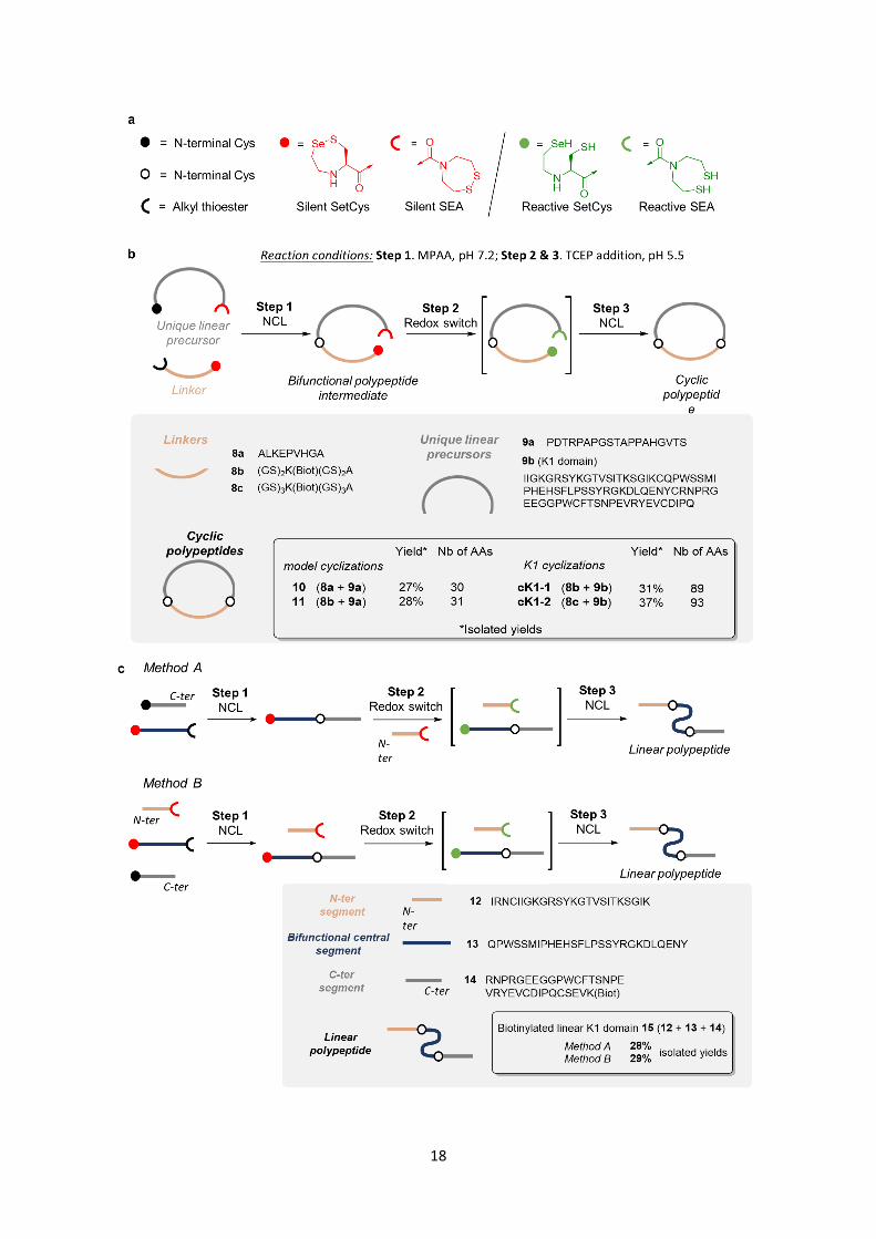
In this work, we sought to develop a modular one-pot method enabling the grafting of the linker to a
unique linear protein precursor (Figure 5b). This can be achieved by exploiting the silent properties of
the SetCys residue under mild reducing conditions for performing the first NCL (see property 3, Figure
2a), and then by using it as a redox switch for triggering the second cyclative NCL (see property 5,
Figure 2a). Regarding the acyl donors, the process utilizes the good reactivity of alkyl thioesters in the
presence of MPAA for the first ligation step. The second acyl donor is introduced as the bis(2-
sulfenylethyl)amido (SEA)43 latent thioester surrogate, which nicely complements the SetCys unit since
it can also be activated in the presence of TCEP.44, 45 The Cys / SEA couple of functional groups is
located on the linear protein precursor, while the SetCys / alkyl thioester functionalities are on the linker
peptide. In the presence of MPAA alone, the Cys-mediated NCL with the peptide alkyl thioester (Step
1, Figure 5b) exclusively yields a bifunctional polypeptide intermediate, which is activated at both ends
by the addition of TCEP in one-pot (Step 2) to produce the backbone-cyclized polypeptide (Step 3). The

8
process is highly tolerant of polypeptide length as similar isolated yields were obtained for the
production of medium to large cyclic peptides (30-93 AAs, ~27-37% overall yields). The examples
include the synthesis of two cyclic and biotinylated variants of hepatocyte growth factor / scatter factor
(HGF/SF) kringle 1 domain (K1), i.e., cK1-1 and cK1-2, from a unique 78 AA linear precursor. These
cyclic polypeptides differ by the length of the linker joining N and C-termini of the K1 protein (10 and
14 residues respectively).
SetCys chemistry proved equally useful for the C-to-N one-pot assembly of linear polypeptides from
three peptide segments (Figure 5a,c). The peptide segments could be added sequentially (Figure 5c,
Method A) or mixed altogether from the beginning of the assembly process (Figure 5c, Method B) with
equal selectivity and efficiency.
Folding and biological activity of biotinylated K1 cyclic analogs
The signaling of the HGF/SF/MET system plays a crucial role in the regeneration of several tissues such
as the liver or the skin, while its deregulation is often observed in cancer. The HGF/SF K1 domain
contains the main HGF/SF binding site for the MET tyrosine kinase receptor and thus constitutes an
interesting platform for designing future drugs based on this couple of proteins.46 In this study, we sought
to produce cyclic analogues of the K1 domain to investigate the tolerance of the K1/MET signaling
system to this modification. The X-ray crystal structure of the K1 protein shows that its tertiary structure
is made up of a series of loops stabilized by three disulfide bonds (Figure 6a).47 The N- and C-terminal
cysteine residues are on the opposite side of the MET binding loop and are linked by a disulfide bond.
The N- and C termini are thus close in space and can be joined by a peptide linker made of a few amino
acid residues which include a biotinylated lysine residue. The latter is used to multimerize the ligand
using streptavidin (S) due to the observation that multivalent presentation of the K1 domain is important
for achieving high binding and agonistic activities.46
The successful synthesis of the cyclic K1 polypeptides cK1-1 and cK1-2 set the stage for the
folding step. cK1-1 and cK1-2 were folded into cK1-1f and cK1-2f respectively using the
glutathione/glutathione disulfide redox system (Figure 6b,c).45 The folding mixtures converged to a
major form after 24 h and were purified by dialysis (Figure 6c, see also Supplementary Information).

9
Extensive proteomic analysis of the folded proteins cK1-1f and cK1-2f showed the exclusive formation
of the native pattern of disulfide bonds between Cys128-206, Cys149-189 and Cys177-201 as shown in
Figure 6b (see Supplementary Information). Thus the cyclization does not perturb the correct pairing of
the Cys residues.
cK1-1f and cK1-2f proteins were first analyzed for their capacity to bind to the recombinant
MET extracellular domain. The competitive AlphaScreen assay with recombinant NK1 protein
showed that the backbone cyclized proteins cK1-1f and cK1-2f were ~10 fold less potent in binding the
MET receptor than the biotinylated analog K1B (Figure 6d). This result was unexpected because the
cyclization site is diametrically opposite the MET binding site. The capacity of the cyclic K1 proteins
to activate the MET receptor was further examined using human HeLa cells (Figure 6e). MET
phosphorylation induced by the cyclic K1 proteins was found to be less than that observed with the
reference K1B analog. However, the tested K1 analogs triggered downstream signaling pathways, i.e.,
phosphorylation of AKT and ERK, with almost equal potency. Because previous studies showed marked
differences between MET phosphorylation levels and the strength of MET specific phenotypes induced
by HGF or HGF mimics,48 we further analyzed the capacity of the different K1 proteins to trigger the
scattering of human cells in vitro (Figure 6f). In this assay using human Capan 1 cells, the cyclic proteins
behaved similarly to the reference protein K1B in the concentration range tested (10 pM-100 nM) by
their capacity to induce a mesenchymal-like phenotype and cell scattering. To summarize, cyclization
in this case results in a significant loss of affinity, although the backbone cyclization site is opposite to
the receptor binding site. However, this loss of affinity does not translate into the cell scattering activity.
This work highlights the need for simple synthetic methods toward cyclic proteins to rapidly investigate
the interest of backbone cyclization for improving protein properties.
In conclusion, the cyclic selenosulfide derivative of cysteine, i.e. SetCys, shows an array of
reactivities in water depending on the reducing power of the solution. A striking property of SetCys is
its conversion to a native Cys residue by cleavage of a nitrogen-carbon bond under extremely mild
conditions if a strong reducing agent is present in the solution. This transformation also occurs in the
presence of a peptide thioester component and thus leads to the production of a native peptide bond to
Cys by native chemical ligation. In contrast, SetCys remains silent during NCL if it is conducted under
10
mild reducing conditions. The redox-switch properties of SetCys are particularly adapted for flexible
synthetic designs that involve nothing more than common additive that are routinely used in NCL
reaction.
Acknowledgements
We thank ANR for financial support (CyProt, ANR-19CE07-0020).
Author information
Affiliations
University of Lille, CNRS, Institut Pasteur de Lille, UMR CNRS 8204, INSERM U1019
Vincent Diemer, Nathalie Ollivier, Bérénice Leclercq, Hervé Drobecq, Jérôme Vicogne,
Vangelis Agouridas, Oleg Melnyk
Author contributions
VD performed the experiments and wrote the manuscript. NO prepared the linear K1 precursor.
HD performed the proteomic experiments. BL and JV performed the AlphaScreen and the cell-based
assay. VA performed the modelization study and wrote the manuscript. OM conceived the study and
wrote the manuscript.
Corresponding authors
Correspondence to Oleg Melnyk or Vangelis Agouridas.
Ethics declarations
Competing interests
The authors declare no competing interests.
Supplementary information

11
Experimental details, materials and methods, kinetic model for SetCys-mediated ligation, LC-MS data
and NMR spectra.
Data Availability
The source data underlying Figs 6d-f are provided as a Source Data file.

12
Figure 1. a) Principle of the native chemical ligation (NCL) between a C-terminal peptide thioester and
a cysteinyl (Cys) peptide. The control of the site of ligation requires masking any N-terminal Cys residue
present in the mixture. b) The reversible formation of disulfide or diselenide bonds is a hallmark of Cys
thiol chemistry in proteins and is used by nature as a redox switch to control Cys thiol reactivity. c) The
N-(2-selanylethyl) group of SetCys shuts down the nucleophilic properties of Cys thiol by formation of
a cyclic selenosulfide bond. It is removed spontaneously in water at neutral pH under reductive
conditions.

13
Figure 2. a) The reactivity of N-(2-selanylethyl)cysteine (SetCys) peptides is controlled by the reducing
strength of the mixture. The numbers on the arrows indicate different experimental conditions.
Condition 1-3: weakly reducing conditions, typically in the presence of an excess of 4-
mercaptophenylacetic acid (MPAA) at neutral pH. Conditions 4 and 5: strong reducing conditions,
typically in the presence of dithiothreitol (DTT) or tris(2-carboxyethyl)phosphine (TCEP) at neutral pH.
b) Ligation of N-(2-sulfanylethyl)cysteine (SutCys) peptides with peptide alkyl thioester under weak or
strong reducing conditions. Peptide sequences: peptide 1 = RLKEPVHGA-, peptide 2 = ALKEPVHGV-
NH2, peptide 3 = ILKEPVHGV-NH2.

14
Figure 3. a) Proposed mechanism for the loss of the N-selenoethyl appendage of N-terminal SetCys
peptides; b) Experimental pH-rate profile of the conversion of N-terminal SetCys peptides to the

15
corresponding N-terminal cysteinyl peptide (red diamonds). The data were fitted to a Gaussian (green
curve, p < 0.001) to determine the pH values for the inflexion points (pH 4.8 and 7.3). c) Different
ionization states for the SetCys residue in open form. The pKa values in bold correspond to the inflexion
points determined in b. The values in parentheses were calculated using ACDLabs software. d) Rate
of 2-selanylethyl limb cleavage in SetCys (, k = 2.53 x 10⁻³ min-1) and SetAla (, k = 2.96 x 10⁻⁴ min-
1) model peptides at pH 6.

16

17
Figure 4. a) Ligation of N-(2-selanylethyl)cysteine peptides with peptide alkyl thioester in NCL
standard conditions yields a product featuring a native peptide bond to cysteine; b) RP-HPLC monitoring
of the conversion of peptides 5 (black triangles) and 6 (brown circles) throughout the course of the
reaction. Fitting curves for each compound are represented by dashed lines; c) Proposed mechanistic
model for the ligation of N-(2-selanylethyl)cysteine peptides under strong reducing conditions. The rate
constants were obtained by software-assisted numerical integration of rate equations (Kintek
explorerTM).
18

19
Figure 5. a) Legend; b) One-pot grafting of a peptide linker to a unique linear precursor yields cyclic
polypeptides. Application to the total synthesis of cyclic and biotinylated analogs of HGF/SF K1 domain
(cK1-1, cK1-2); c) One-pot synthesis of linear K1 domain.

20

21
Figure 6. a) Primary and tertiary structure of HGF/SF K1 domain (pdb entry 1BHT). b) Biotinylated
K1 analogs tested for their capacity to bind MET receptor and induce MET-specific phenotypes. The
pattern of disulfide bonds determined experimentally corresponds to the native pattern found in K1
domain X-ray crystal structures. c) LC-MS monitoring of the folding of cK1-1 peptide into cK1-1f. d)
Competitive AlphaScreen assay with recombinant NK1 protein. K1B or cK1-1f or cK1-2f were mixed
with increasing concentrations of NK1 and with extracellular MET domain fused with human IgG1-Fc
(MET-Fc) and incubated with streptavidin AlphaScreen donor beads and Protein A acceptor beads. e)
HeLa cells were treated for 10 min with 300 pM mature HGF/SF (HGF), or with 10 nM / 100 nM K1/S,
cK1-1f/S and cK1-2f/S. Cell lysates were then analyzed by specific total MET and ERK or phospho-
MET, phospho-Akt and phospho-ERK Western blot; f) Cell scattering assay. Human Capan 1 isolated
cell islets were incubated for 18 h in culture media with 300 pM mature HGF/SF (HGF), or 100, 10, 1
nM and 100 and 10 pM K1B, cK1-1f and cK1-2f.
References
1. Dawson P. E., Muir T. W., Clark-Lewis I. & Kent S. B. H. Synthesis of proteins by native chemical ligation. Science 266, 776-779 (1994).
2. Agouridas V., El Mahdi O., Diemer V., Cargoet M., Monbaliu J.-C. M. & Melnyk O. Native chemical ligation and extended methods. Mechanisms, catalysis, scope and limitations. Chem. Rev. 12, 7328-7443 (2019).
3. Kulkarni S. S., Sayers J., Premdjee B. & Payne R. J. Rapid and efficient protein synthesis through expansion of the native chemical ligation concept. Nat. Rev. Chem. 2, 0122 (2018).
4. Conibear A. C., Watson E. E., Payne R. J. & Becker C. F. W. Native chemical ligation in protein synthesis and semi-synthesis. Chem. Soc. Rev. 47, 9046-9068 (2018).
5. Agouridas V., El Mahdi O., Cargoët M. & Melnyk O. A statistical view of protein chemical synthesis using NCL and extended methodologies. Bioorg. Med. Chem. 25, 4938-4945 (2017).
6. Raibaut L., Ollivier N. & Melnyk O. Sequential native peptide ligation strategies for total chemical protein synthesis. Chem. Soc. Rev. 41, 7001-7015 (2012).
7. Jbara M., Laps S., Morgan M., Kamnesky G., Mann G., Wolberger C. et al. Palladium prompted on-demand cysteine chemistry for the synthesis of challenging and uniquely modified proteins. Nat. Commun. 9, 3154 (2018).
8. Maity S. K., Mann G., Jbara M., Laps S., Kamnesky G. & Brik A. Palladium-assisted removal of a solubilizing tag from a Cys side chain to facilitate peptide and protein synthesis. Org. Lett. 18, 3026-3029 (2016).
9. Maity S. K., Jbara M., Laps S. & Brik A. Efficient palladium-assisted one-pot deprotection of (acetamidomethyl)cysteine following native chemical ligation and/or desulfurization to expedite chemical protein synthesis. Angew. Chem. Int. Ed. 55, 8108-8112 (2016).
10. Jbara M., Maity S. K., Seenaiah M. & Brik A. Palladium mediated rapid deprotection of N-terminal cysteine under native chemical ligation conditions for the efficient preparation of synthetically challenging proteins. J. Am. Chem. Soc. 138, 5069-5075 (2016).
11. Reddy P. S., Dery S. & Metanis N. Chemical synthesis of proteins with non-strategically placed cysteines using selenazolidine and selective deselenization. Angew. Chem. Int. Ed. 55, 992-995 (2015).
12. Poole L. B. The basics of thiols and cysteines in redox biology and chemistry. Free Radic. Biol. Med. 80, 148-157 (2015).
13. Klomsiri C., Karplus P. A. & Poole L. B. Cysteine-based redox switches in enzymes. Antioxid. Redox Signal. 14, 1065-1077 (2011).

22
14. Greene T. W. in Protection for the Thiol Group (eds Wuts P. G. M.) 837-894 (Wiley, 2014). 15. Lees W. J. & Whitesides G. M. Equilibrium constants for thiol-disulfide interchange reactions: a coherent,
corrected set. J. Org. Chem. 58, 642-647 (1993). 16. Reich H. J. & Cohen M. L. Organoselenium chemistry. Dealkylation of amines with benzeneselenol. J.
Org. Chem. 44, 3148-3151 (1979). 17. Krief A. & Hevesi L. Organoselenium Chemistry I. Functional Group Transformations (Springer-Verlag,
1988). 18. Ouyang K., Hao W., Zhang W.-X. & Xi Z. Transition-Metal-Catalyzed Cleavage of C–N Single Bonds. Chem.
Rev. 115, 12045-12090 (2015). 19. Loibl S. F., Harpaz Z. & Seitz O. A type of auxiliary for native chemical peptide ligation beyond cysteine
and glycine junctions. Angew. Chem. Int. Ed. 54, 15055-15059 (2015). 20. Yin H., Lu D., Wang S. & Wang P. Development of Powerful Auxiliary-Mediated Ligation To Facilitate
Rapid Protein Assembly. Org. Lett. 21, 5138-5142 (2019). 21. Dawson P. E., Churchill M. J., Ghadiri M. R. & Kent S. B. H. Modulation of reactivity in native chemical
ligation through the use of thiol additives. J. Am. Chem. Soc. 119, 4325-4329 (1997). 22. Johnson E. C. & Kent S. B. H. Insights into the mechanism and catalysis of the native chemical ligation
reaction. J. Am. Chem. Soc. 128, 6640-6646 (2006). 23. Melnyk O. & Agouridas V. From protein total synthesis to peptide transamidation and metathesis:
playing with the reversibility of N,S-acyl or N,Se-acyl migration reactions. Curr. Opin. Chem. Biol. 22, 137-145 (2014).
24. Ruff Y., Garavini V. & Giuseppone N. Reversible native chemical ligation: a facile access to dynamic covalent peptides. J. Am. Chem. Soc. 136, 6333-6339 (2014).
25. Reich H. J. & Hondal R. J. Why Nature Chose Selenium. ACS Chem. Biol. 11, 821-841 (2016). 26. Rohde H., Schmalisch J., Harpaz Z., Diezmann F. & Seitz O. Ascorbate as an alternative to thiol additives
in native chemical ligation. ChemBioChem 12, 1396-1400 (2011). 27. Dery S., Reddy P. S., Dery L., Mousa R., Dardashti R. N. & Metanis N. Insights into the deselenization of
selenocysteine into alanine and serine. Chem. Sci. 6, 6207-6212 (2015). 28. Metanis N., Keinan E. & Dawson P. E. Traceless ligation of cysteine peptides using selective
deselenization. Angew. Chem. Int. Ed. 49, 7049-7053 (2010). 29. Wan Q. & Danishefsky S. J. Free-radical-based, specific desulfurization of cysteine: a powerful advance
in the synthesis of polypeptides and glycopolypeptides. Angew. Chem. Int. Ed. 46, 9248-9252 (2007). 30. Yokoyama A., Sakurai H. & Tanaka H. Syntheses of related compounds of selenocysteamine and their
complex formation with metal ions. Chem. Pharm. Bull. 19, 1089-1094 (1971). 31. Jencks W. P. & Regenstein J. in Ionization constants of acids and bases (eds Lundblad R. L. & MacDonald
F. M.) 595–635 (CRC, 2010). 32. Tanini D. & Capperucci A. Ring opening reactions of heterocycles with selenium and tellurium
nucleophiles. New J. Chem. 43, 11451-11468 (2019). 33. Ollivier N., Blanpain A., Boll E., Raibaut L., Drobecq H. & Melnyk O. Selenopeptide transamidation and
metathesis. Org. Lett. 16, 4032-4035 (2014). 34. Camarero J. A., Fushman D., Sato S., Giriat I., Cowburn D., Raleigh D. P. et al. Rescuing a destabilized
protein fold through backbone cyclization. J. Mol. Biol. 308, 1045-1062 (2001). 35. Iwai H. & Pluckthun A. Circular beta-lactamase: stability enhancement by cyclizing the backbone. FEBS
Lett. 459, 166-172 (1999). 36. Camarero J. A. & Muir T. W. Biosynthesis of a head-to-tail cyclized protein with improved biological
activity. J. Am. Chem. Soc. 121, 5597-5598 (1999). 37. Craik D. J. Chemistry. Seamless proteins tie up their loose ends. Science 311, 1563-1564 (2006). 38. Craik D. J., Fairlie D. P., Liras S. & Price D. The future of peptide-based drugs. Chem. Biol. Drug Des. 81,
136-147 (2013). 39. Krishna M. M. & Englander S. W. The N-terminal to C-terminal motif in protein folding and function.
Proc. Natl. Acad. Sci. U. S. A. 102, 1053-1058 (2005). 40. Mulvenna J. P., Wang C. & Craik D. J. CyBase: a database of cyclic protein sequence and structure. Nucleic
Acids Res. 34, D192-D194 (2006). 41. White C. J. & Yudin A. K. Contemporary strategies for peptide macrocyclization. Nat. Chem. 3, 509-524
(2011). 42. Camarero J. A. & Muir T. W. Chemoselective backbone cyclization of unprotected peptides. Chem.
Commun., 1369-1370 (1997).
23
43. Ollivier N., Dheur J., Mhidia R., Blanpain A. & Melnyk O. Bis(2-sulfanylethyl)amino native peptide ligation. Org. Lett. 12, 5238-5241 (2010).
44. Boll E., Drobecq H., Ollivier N., Blanpain A., Raibaut L., Desmet R. et al. One-pot chemical synthesis of small ubiquitin-like modifier (SUMO) protein-peptide conjugates using bis(2-sulfanylethyl)amido peptide latent thioester surrogates Nat. Protoc. 10, 269-292 (2015).
45. Ollivier N., Vicogne J., Vallin A., Drobecq H., Desmet R., El-Mahdi O. et al. A one-pot three-segment ligation strategy for protein chemical synthesis. Angew. Chem. Int. Ed. 51, 209-213 (2012).
46. Simonneau C., Berenice L., Mougel A., Adriaenssens E., Paquet C., Raibaut L. et al. Semi-synthesis of a HGF/SF kringle one (K1) domain scaffold generates a potent in vivo MET receptor agonist. Chem. Sci. 6, 2110-2121 (2015).
47. Ultsch M., Lokker N. A., Godowski P. J. & de Vos A. M. Crystal structure of the NK1 fragment of human hepatocyte growth factor at 2.0 A resolution. Structure 6, 1383-1393 (1998).
48. Mekki M. S., Mougel A., Vinchent A., Paquet C., Copin M. C., Leroy C. et al. Hypoxia leads to decreased autophosphorylation of the MET receptor but promotes its resistance to tyrosine kinase inhibitors. Oncotarget 9, 27039-27058 (2018).

download fileview on ChemRxivArticle_R1_3.pdf (2.12 MiB)

S 1
Supporting Information for
A cysteine selenosulfide redox switch for protein chemical synthesis
Vincent Diemer, Nathalie Ollivier, Bérénice Leclercq, Hervé Drobecq, Jérôme Vicogne,
Vangelis Agouridas*, Oleg Melnyk*
University of Lille, CNRS, Institut Pasteur de Lille, UMR CNRS 8204, INSERM U1019, Centre
d’Immunité et d’Infection de Lille, F-59000 Lille, France
Corresponding author:
Dr Oleg Melnyk, E-mail : [email protected]
Dr Vangelis Agouridas, E-mail: [email protected]
Website: http://olegmelnyk.cnrs.fr
CBF group
Institut de Biologie de Lille
1 rue du Pr Calmette, CS 50447, 59021 Lille cedex, France

S 2
1 Table of contents
1 Table of contents ......................................................................................................... 2
2 Synthesis of Fmoc-protected SetCys amino acid ........................................................ 5
3 Peptide synthesis ........................................................................................................ 19
3.1 General procedures .................................................................................................... 19
3.2 Synthesis of SetCys peptides ..................................................................................... 22
SetCys peptide used for exploring SetCys reactivity (Figure 2 & Figure 3) .................... 22
SetCys peptides for cyclization studies (Figure 5) ............................................................ 23
Synthesis of peptide 8a .................................................................................................. 23
Synthesis of peptide 8b .................................................................................................. 24
Synthesis of peptide 8c .................................................................................................. 26
3.3 Preparation of SetAla peptide (Figure 3d) ................................................................. 27
3.4 Synthesis of SEAoff peptide segments for cyclization studies (Figure 5) .................. 32
Synthesis of peptide 9a...................................................................................................... 32
Synthesis of peptide 9b (linear K1 precursor)................................................................... 34
Synthesis of the peptide segments for the assembly of linear K1 peptide 9b ............... 35
Assembly of linear K1 peptide 9b ................................................................................. 36
4 Stability and reactivity of SetCys unit as a function of the reducing power of the
reaction mixture (Figure 2a) ..................................................................................................... 42
4.1 Stability of SetCys under low (property 1) or strong (property 4) reducing conditions
42
4.2 Stability of SetCys under strong (property 4) reducing conditions. Importance of
ascorbate ............................................................................................................................... 47
4.3 NCL under low reducing conditions (properties 2) ................................................... 52
4.4 SetCys is silent during NCL under low reducing conditions (property 3) ................ 57
4.4.1 Competitive ligation experiment involving a Cys and SetCys peptide .............. 57
4.4.2 Compatibility of SetCys with Cys-rich peptides ................................................ 61
4.5 NCL under strong reducing conditions (peptide alkyl thioester, TCEP/ascorbate,
MPAA, property 5) ............................................................................................................... 68
4.6 NCL under strong reducing conditions (peptide aryl thioester, TCEP/ascorbate, no
added MPAA, property 5) .................................................................................................... 73

S 3
4.7 NCL under strong reducing conditions (peptide alkyl thioester, DTT, MPAA,
property 5) ............................................................................................................................ 77
5 Stability and reactivity of N-(2-sulfanylethyl)Cys (SutCys) unit as a function of the
reducing power of the reaction mixture (Figure 2b) ................................................................ 79
5.1 Stability under strong reducing conditions ................................................................ 79
5.2 NCL under low reducing conditions ......................................................................... 80
5.3 NCL under strong reducing conditions ..................................................................... 84
6 Mechanistic studies (Figure 3) .................................................................................. 86
6.1 Effect of pH (Figure 3b) ............................................................................................ 86
6.2 Effect of Cys thiol: control experiment with a SetAla peptide (Figure 3d) .............. 87
6.2.1 Decomposition of SetAla peptide into Ala peptide under strong reducing
conditions .......................................................................................................................... 87
6.2.2 Ligation of SetAla peptide under strong reducing conditions ............................ 90
7 Kinetic model of SetCys-mediated NCL under strong reducing conditions (Figure 4)
92
7.1 Rate constants for a classical NCL (k+2, k-2 and k4, see Figure 4c) ........................... 93
7.2 Rate constant for the loss of the N-selenoethyl appendage (k1, Figure 4c) ............... 95
7.3 Kinetic model for SetCys-mediated NCL (Figure 4b,c) ............................................ 95
8 Synthesis of cyclic peptides (Figure 5a,b) ................................................................. 97
8.1 Synthesis of cyclic peptide 10 ................................................................................... 97
8.2 Synthesis of cyclic peptide 11 ................................................................................. 100
8.3 Synthesis of cK1-1 ................................................................................................... 103
8.3.1 Characterization of the cyclic backbone structure of cK1-1 ............................ 105
8.4 Synthesis of cK1-2 ................................................................................................... 108
8.4.1 Characterization of the cyclic backbone structure of cK1-2 ............................ 111
9 One-pot synthesis of linear K1 domain (Figure 5a,c) ............................................. 113
9.1 Preparation of the starting peptide segments ........................................................... 113
9.2 Method A ................................................................................................................. 114
9.3 Method B ................................................................................................................. 116
9.4 Characterization of linear K1 polypeptide ............................................................... 119
10 Folding, characterization and biological activity of cK1-1 and cK1-2 (Figure 6) .. 123
10.1 Folding (Figure 6b,c) ............................................................................................... 123

S 4
10.1.1 Characterization of folded cK1-1f and cK1-2f ................................................ 125
10.1.2 Disulfide bridge pattern of cK1-1f and cK1-2f ................................................ 127
10.2 Alphascreen Assay (Figure 6d) ............................................................................... 130
10.3 Biological assays (Figure 6e,f) ................................................................................ 130
10.3.1 Cell-based assay for MET signaling induced by cK1-1f and cK1-2f .............. 130
10.3.2 Scattering Assay ............................................................................................... 131
11 References ............................................................................................................... 132

S 5
2 Synthesis of Fmoc-protected SetCys amino acid
Fmoc-protected SetCys amino acid SI8 was prepared as described below in Figure S 1.
Figure S 1. Synthesis of the Fmoc-protected SetCys amino acid.
(2,2-Diethoxyethyl)(4-methoxybenzyl)selenide was prepared according to Abbas et al.1
2.1 Synthesis of Synthesis of H-Cys(Trt)-OMe
To a solution of cysteine methyl ester SI1 (856 mg, 5 mmol) in TFA (5 mL) was added
triphenylmethanol (1.43 g, 5.5 mmol) and the mixture was stirred at RT for 5 h. After
evaporation of the TFA, the residue was dissolved in MeOH and the mixture was stirred at RT
until the yellow color disappeared. The methanol was then evaporated under reduced pressure
and the residue was suspended in 0.25 M K2CO3 (30 mL). The obtained aqueous layer was
extracted with Et2O (2 × 20 mL) and the combined organic layers were dried over MgSO4.
After evaporation of the solvent, purification of the crude by column chromatography
(DCM/MeOH 98:2) provided the S-trityl protected amino acid SI2 (1.73 g) as a viscous oil with
91% yield.
NMR data are in agreement with the literature.2
1H NMR (300 MHz, CDCl3) δ 7.41-7.44 (m, 6H), 7.18-7.31 (m, 9H), 3.65 (s, 3H), 3.20 (dd, J
= 4.8 and 7.8 Hz, 1H), 2.59 (dd, J = 4.8 and 12.4 Hz, 1H), 2.46 (d, J = 7.8 and 12.4 Hz, 1H)

S 6
ppm. 13C NMR (75 MHz, CDCl3) δ 174.3 (C), 144.6 (3 × CH), 129.7 (6 × CH), 128.1 (6 ×
CH), 126.9 (3 × CH), 66.9 (C), 53.9 (CH), 52.3 (CH3), 37.2 (CH2) ppm.
Figure S 2. 1H NMR (300 MHz) spectrum of H-Cys(Trt)-OMe SI2 (CDCl3, 293 K).
Figure S 3. 13C JMOD NMR (75 MHz) spectrum of H-Cys(Trt)-OMe SI2 (CDCl3, 293 K).

S 7
2.2 Synthesis of N-[PMBSe-(CH2)2]-Cys(Trt)-OMe
(2,2-Diethoxyethyl)(4-methoxybenzyl)selenide SI3 (1.18 g, 3.72 mmol) was suspended in 1 M
formic acid (15 mL) and the mixture was heated overnight at 50 °C. Water (50 mL) was added
and the aqueous layer was extracted with Et2O (2 × 50 mL). The combined organic extracts
were washed with water (50 mL) and dried over MgSO4. After evaporation of the solvent under
reduced pressure, aldehyde SI4 was dissolved in anhydrous 1,2-dichloroethane (50 mL) under
argon. Activated powdered 3 Å molecular sieves (4 g), sodium triacetoxyborohydride (1.10 g,
5.21 mmol) and a solution of H-Cys(Trt)-OMe SI2 (1.41 g, 3.72 mmol) in DCE (35 mL) were
successively added to the solution of aldehyde. After 18 h stirring at RT, the reaction mixture
was filtered on a Büchner funnel and the solid was washed with additional portions of DCM.
The filtrate was evaporated under reduced pressure. Then 0.5 M K2CO3 (50 mL) was added to
the residue and the aqueous layer was extracted with DCM (3 × 50 mL). The combined organic
layers were dried over MgSO4 and the solvent was evaporated under reduced pressure.
Purification of the crude mixture by column chromatography (cyclohexane/ethyl acetate 80:20)
afforded the N-alkylated amino acid SI5 as a clear yellow oil (1.94 g, 86%).
1H NMR (300 MHz, CDCl3) δ 7.41 (d, J = 7.4 Hz, 6H), 7.19-7.32 (m, 9H), 7.17 (d, J = 8.6 Hz,
2H), 6.79 (d, J = 8.6 Hz, 2H), 3.77 (s, 3H), 3.71 (s, 2H), 3.66 (s, 3H), 2.99 (t, J = 6.5 Hz, 1H),
2.37-2.74 (m, 6H), 1.44-1.91 (m, 1H) ppm. 13C NMR (75 MHz, CDCl3) δ 173.5 (C), 158.4 (C),
144.6 (3 × C), 131.1 (C), 129.9 (2 × CH), 129.6 (6 × CH), 127.9 (6 × CH), 126.7 (3 × CH),
113.9 (2 × CH), 66.8 (C), 60.2 (CH), 55.2 (CH3), 51.9 (CH3), 47.3 (CH2), 34.6 (CH2), 26.3
(CH2), 23.9 (CH2) ppm. IR (ATR, cm-1) 3029, 2949, 1736, 1698, 1243, 1172, 738. HRMS
(ES+): Calcd. for C33H35NO3SNaSe: 628.1401, found: 628.1378. [α]D20 -19 ° (c 1.0,
chloroform).

S 8
Figure S 4. 1H NMR (300 MHz) spectrum of compound SI5 (CDCl3, 293 K).
Figure S 5. 13C JMOD NMR (75 MHz) spectrum of compound SI5 (CDCl3, 293 K).
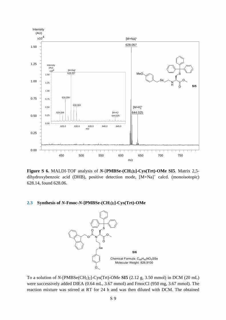
S 9
Figure S 6. MALDI-TOF analysis of N-[PMBSe-(CH2)2]-Cys(Trt)-OMe SI5. Matrix 2,5-
dihydroxybenzoic acid (DHB), positive detection mode, [M+Na]+ calcd. (monoisotopic)
628.14, found 628.06.
2.3 Synthesis of N-Fmoc-N-[PMBSe-(CH2)2]-Cys(Trt)-OMe
To a solution of N-[PMBSe(CH2)2]-Cys(Trt)-OMe SI5 (2.12 g, 3.50 mmol) in DCM (20 mL)
were successively added DIEA (0.64 mL, 3.67 mmol) and FmocCl (950 mg, 3.67 mmol). The
reaction mixture was stirred at RT for 24 h and was then diluted with DCM. The obtained
628.057
644.025
0.00
0.25
0.50
0.75
1.00
1.25
1.50
4x10
450 500 550 600 650 700 750
m/z
[M+Na]+
[M+K]+
Intensity
(AU)
628.057
626.056
630.063
624.056
644.025
0.00
0.25
0.50
0.75
1.00
1.25
1.50
4x10
625.0 630.0 635.0 640.0 645.0m/z
[M+Na]+
[M+K]+
Intensity
(AU)

S 10
organic layer was washed with water and was dried over MgSO4. After evaporation of the
solvent under reduced pressure, purification of the crude by column chromatography
(cyclohexane/EtOAc 7:3) provided the expected compound SI6 (1.87 g) as a white solid with
99% yield.
The titled compound was characterized by 1H and 13C NMR as a mixture of two conformers.
A/B = 37:63. The ratio A/B was calculated from the CH signals of the Fmoc protecting group.
1H NMR (300 MHz, CDCl3) δ 7.80 (d, J = 9.0 Hz, 0.64H, 2HA), 7.74 (d, J = 7.6 Hz, 2HB),
7.49-7.59 (m, 2HA + 2HB), 7.12-7.49 (m, 21HA + 19HB + residual CDCl3), 7.08 (d, J = 8.3 Hz,
2HB), 6.89 (d, J = 8.3 Hz, 2HA), 6.71 (d, J = 8.4 Hz, 2HB), 4.34-4.60 (m, 2HA + 2HB), 4.20 (t,
J = 5.9 Hz, HB), 4.13 (t, J = 5.2 Hz, HA), 3.75 (s, 3HA), 3.73 (s, 2HA), 3.68 (s, 3HB) 3.40-3.63
(m, HA + 5HB), 3.02-3.40 (m, 4HA + 2HB), 2.45-3.02 (m, 3HA + 3HB), 2.17-2.45 (m, 2HA +
2HB) ppm. 13C NMR (75 MHz, CDCl3) δ 170.0 (C, B), 169.7 (C, A), 158.4 (C, A +B), 155.2
(C, A + B), 144.5 (C, A + B), 143.7 (C, A + B), 141.4 (C, A), 141.3 (C, B), 131.3 (C, A), 130.9
(C, B), 129.8 (CH, A + B), 129.6 (CH, A + B), 128.0 (CH, A + B), 127.7 (CH, A + B), 127.1
(CH, A + B), 126.8 (CH, A + B), 124.3-125.3 (m, CH, A + B), 120.0 (CH, A + B), 113.9 (A +
B), 67.2 (CH2, A), 67.0 (CH2, B), 60.6 (CH, B), 59.8 (CH, A), 55.1 (CH3, A+ B), 52.3 (CH3,
B), 52.2 (CH3, A), 49.6 (CH2, A), 49.2 (CH2, B), 47.2 (CH, B), 47.1 (CH, A), 31.5 (CH2, A),
31.2 (CH2, B), 26.5 (CH2, A + B), 21.3 (CH2, B), 20.8 (CH2, A) ppm. IR (ATR, cm-1) 3054,
2949, 1742, 1699, 1509, 1445, 1283, 1245, 741, 701. HRMS (ES+): Calcd. for
C48H45NO5SNaSe: 850.2081, found: 850.2089. [α]D20 (c 1.0, CHCl3): -38 °.
Figure S 7. 1H NMR (300 MHz) spectrum of N-Fmoc-N-[PMBSe-(CH2)2]-Cys(Trt)-OMe
SI6 (CDCl3, 293 K).

S 11
Figure S 8. 13C JMOD NMR (75 MHz) spectrum of N-Fmoc-N-[PMBSe-(CH2)2]-Cys(Trt)-
OMe SI6 (CDCl3, 293 K).
Figure S 9. 1H-13C HSQC spectrum of N-Fmoc-N-[PMBSe-(CH2)2]-Cys(Trt)-OMe SI6
(CDCl3, 293 K).

S 12
Figure S 10. MALDI-TOF analysis of N-Fmoc-N-[PMBSe-(CH2)2]-Cys(Trt)-OMe SI6.
Matrix 2,5-dihydroxybenzoic acid (DHB), positive detection mode, [M+Na]+ calcd.
(monoisotopic) 850.22, found 849.9.
2.4 Synthesis of Fmoc-SetCys-OMe
To a solution of N-Fmoc-N-[PMBSe-(CH2)2]-Cys(Trt)-OMe SI6 (1.67 g, 2.02) in DCM (50
mL) was added NaHCO3 (508 mg, 6.06 mmol). The mixture was cooled to 0 °C and I2 (1.54 g,
6.06 mmol) dissolved in DCM (50 mL) was added dropwise. The reaction mixture was stirred
at RT for 30 min and was washed 1 M Na2S2O3 (50 mL) to remove the excess of I2. The organic
layer was separated and the aqueous layer was extracted with DCM (2 × 50 mL). Note that the
separation between the aqueous and organic layers was improved by addition of brine (50 mL).
All the organic layers were then combined, washed with brine (50 mL) and dried over MgSO4.
After evaporation of the solvent, purification of the crude by column chromatography
(cyclohexane/EtOAc 8:2) provided the expected compound SI7 (780 mg) as a glass with 83%
yield.
849.9
0
1000
2000
3000
4000
600 650 700 750 800 850 900 950
m/z
[M+Na]+
Intensity
(AU)
849.9
0
1000
2000
3000
4000
835 840 845 850 855 860 865
m/z
[M+Na]+Intensity
(AU)

S 13
The titled compound was characterized by 1H and 13C NMR as a mixture of two main
conformers. A/B = 45:55. The ratio A/B was calculated from the aromatic CH signals of the
Fmoc protecting group. Due to overlapping signals, no clear description of the 1H NMR
spectrum can be provided.
13C NMR (75 MHz, CDCl3) δ 170.0 (C), 169.7 (C), 155.6 (C), 155.2 (C), 143.90 (C), 143.88
(C), 143.84 (C), 143.6 (C), 141.6 (C), 141.53 (C), 141.51 (C), 127.91 (CH), 127.88 (CH),
127.86 (CH), 127.7 (CH), 127.34 (CH), 127.30 (CH), 127.25 (CH), 127.1 (CH), 124.8 (CH),
124.4 (CH), 124.3 (CH), 67.1 (CH2), 66.8 (CH2), 62.7 (CH), 52.7 (CH3), 52.4 (CH3), 49.8
(CH2), 47.4 (CH), 47.2 (CH), 38.6 (CH2), 29.3 (CH2), 29.1 (CH2) ppm. IR (ATR, cm-1) 2948,
1740, 1697, 1474, 1449, 1417, 1278, 738. HRMS (ES+) Calcd. for C21H21NO4NaSSe:
486.0254, found: 486.0246. [α]D20 (c 1.0, CHCl3): -29 °.
Figure S 11. 1H NMR (300 MHz) spectrum of Fmoc-SetCys-OMe SI7 (CDCl3, 293 K).

S 14
Figure S 12.13C JMOD NMR (75 MHz) spectrum of Fmoc-SetCys-OMe SI7 (CDCl3, 293 K).
Figure S 13. 1H-13C HSQC spectrum of Fmoc-SetCys-OMe SI7 (CDCl3, 293 K).

S 15
Figure S 14. LC-MS analysis of Fmoc-SetCys-OMe SI7. LC trace: eluent C 0.10% formic
acid in water, eluent D 0.10% formic acid in CH3CN/water: 4/1 by vol. C18 column, gradient
0-100% D in 4 min, 50 °C, 0.5 mL/min, UV detection. MS trace: [M+H]+ m/z calcd.
(monoisotopic mass) 464.04, found 464.0; [M+Na]+ m/z calcd. (monoisotopic mass) 486.04,
found 486.0.
Figure S 15. MALDI-TOF analysis of Fmoc-SetCys-OMe SI7. Matrix 2,5-dihydroxybenzoic
acid (DHB), positive detection mode, [M+Na]+ calcd. (monoisotopic) 486.04, found 486.2.
Time (min)
0.50 1.00 1.50 2.00 2.50 3.00 3.50 4.00 4.50 5.00 5.50 6.00 6.50 7.00 7.50 8.00
-2.0e-1
-1.0e-1
0.0
1.0e-1
2.0e-1
3.0e-1
4.0e-1
5.0e-1
6.0e-1
7.0e-1
8.0e-1
9.0e-1
1.0
1.1
1.2
5.37
m/z435 445 455 465 475 485 495 505 515 525
486.0
464.0
501.9
100
0
Intensity
(AU)[M+Na]+
[M+K]+
[M+H]+
Intensity
(AU)
486.2
0
250
500
750
1000
1250
200 300 400 500 600 700 800 900
m/z
[M+Na]+
Intensity
(AU)
486.2
0
250
500
750
1000
1250
480 482 484 486 488 490 492 494
m/z
[M+Na]+
Intensity
(AU)

S 16
2.5 Synthesis of Fmoc-SetCys-OH
To a solution of Fmoc-SetCys-OMe SI7 (411 mg) in dioxane (19 mL) was added 5 M HCl (9
mL) and the mixture was refluxed for 6 h. The reaction mixture was cooled to RT and 5%
K2CO3 (200 mL) was added to raise the pH above 9. The aqueous layer was then washed with
E2O (2 × 100 mL), acidified with concentrated HCl until pH ≈ 2-3 and extracted with DCM (3
× 100 mL). The organic layers (DCM) were combined and dried over MgSO4. Evaporation of
the solvent under reduced provided the expected amino acid SI8 (345 mg, 86%) as a white solid
which was used in the next step without further purification.
The titled compound was characterized by 1H and 13C NMR as a mixture of two main
conformers. Due to overlapping signals, no clear description of the 1H NMR spectrum can be
provided. IR (ATR, cm-1) 3000 (broad signal), 1699 (broad), 1476, 1450, 1419, 1286, 1190,
740. HRMS (ES+): Calcd. for C20H19NO4NaSSe: 472.0098, found: 472.0116. [α]D20 (c 1.0,
CHCl3): -32 °.
Figure S 16. 1H NMR (300 MHz) spectrum of Fmoc-SetCys-OH SI8 (CDCl3, 293 K).

S 17
Figure S 17.13C NMR (75 MHz) spectrum of Fmoc-SetCys-OH SI8 (CDCl3, 293 K).
Figure S 18. LC-MS analysis of Fmoc-SetCys-OH SI8. LC trace: eluent C 0.10% formic acid
in water, eluent D 0.10% formic acid in CH3CN/water: 4/1 by vol. C18 column, gradient 0-
100% D in 4 min, 50 °C, 0.5 mL/min, UV detection. MS trace: [M+H]+ m/z calcd.
(monoisotopic mass) 450.02, found 450.1; [M+Na]+ m/z calcd. (monoisotopic mass) 472.02,
found 472.0.
Time (min)
0.00 0.50 1.00 1.50 2.00 2.50 3.00 3.50 4.00 4.50 5.00 5.50 6.00 6.50 7.00 7.50 8.00
0.0
2.0e-1
4.0e-1
6.0e-1
8.0e-1
1.0
1.2
1.4
1.6
4.92
Intensity
(UV, AU)
m/z410 420 430 440 450 460 470 480 490 500 510 520 530
0
100 450.1
472.0
488.0
[M+H]+
[M+Na]+
[M+K]+
Intensity
(AU)

S 18
Figure S 19. MALDI-TOF analysis of Fmoc-SetCys-OH SI8. Matrix 2,5-dihydroxybenzoic
acid (DHB), positive detection mode, [M+Na]+ calcd. (monoisotopic) 472.01, found 471.95.
471.948
487.923
0.0
0.5
1.0
1.5
4x10
400 450 500 550 600 650 700 750m/z
471.948
469.949
473.956
467.950
0.00
0.25
0.50
0.75
1.00
1.25
1.50
4x10
462 464 466 468 470 472 474 476 478 480
m/z
Intensity (AU)
[M+Na]+
[M+Na]+
[M+K]+
Intensity (AU)

S 19
3 Peptide synthesis
CILKEPVHGV-NH2 has already been synthesized and characterized elsewhere.Erreur ! Signet non d
éfini.
3.1 General procedures
Peptide amides were synthesized on a NovaSyn TGR resin (0.25 mmol/g) using standard Fmoc
chemistry and an automated peptide synthesizer (Figure S 20). Note that the Fmoc-SetCys-OH
amino acid was manually introduced in the peptide sequence.
Figure S 20. General approach for the preparation of peptide amides.
SEAoff peptides were synthesized on SEA PS resin (0.16 mmol/g) using standard Fmoc
chemistry and an automated peptide synthesizer (Figure S 21). The SEAon peptide recovered
after acidic cleavage was converted into the corresponding SEAoff peptide by oxidation with
iodine. The same intermediate provided the MPA thioester via a SEA-MPA exchange reaction.
Detailed procedures to prepare these peptides are available in previous papers.3, 4, 5 Note that
first amino acids coupled to the resin, biotinylated lysine residues and the Fmoc-SetCys-OH
amino acids were introduced manually in the peptide sequence.

S 20
Figure S 21. Synthesis of SEAoff peptides and MPA thioesters.
Coupling of the first amino acid on SEA PS resin
The first amino acid (10 eq) was coupled to SEA PS resin (1 eq) using HATU (10 eq)/DIEA
(20 eq) activation in the minimal volume of DMF. The amino acid was preactivated for 2 min
and was then added to the resin swelled in the minimal volume of DMF. The beads were
agitated at RT for 1.5 h and the resin was then washed with DMF (3 × 2 min) and drained. The
absence of unreacted secondary amino groups was checked using chloranyl colorimetric assay.
A capping step was then performed using Ac2O/DIEA/DMF 10:5:85 (2 × 5 min) and the resin
was washed with DMF (3 × 2 min).
Automated peptide elongation
Peptide elongation was performed using standard Fmoc chemistry on an automated peptide
synthesizer without microwaves. Couplings were performed using 4-fold molar excess of each
Fmoc L-amino acid, 3.6-fold molar excess of HBTU, and 8-fold molar excess of DIEA. A
capping step was performed with Ac2O/DIEA/DMF 10:5:85 before Fmoc deprotection
(piperidine/DMF 80:20).
Coupling of the biotinylated lysine
To a solution of HATU (190 mg, 0.5 mmol, 5 eq) in DMF (3 mL) were successively added
FmocLys(Biot)OH (300 mg, 0.64 mmol, 6.4 eq) and DIEA (174 μL, 1.0 mmol, 10 eq) and the
mixture was stirred at RT for 2 min. The preactivated amino acid was then added to the resin
(0.1 mmol, 1 eq) swelled in a minimal volume of DMF. The beads were agitated at RT for 1.5
h and were then washed with DMF (3 × 2 min). A capping step was then performed using
Ac2O/DIEA/DMF 10:5:85 (2 × 5 min). Finally, the resin was washed again with DMF (3 × 2
min).

S 21
Coupling of the Fmoc-SetCys-OH amino acid
To a solution of Fmoc-SetCys-OH (44.8 mg, 0.10 mmol, 2 eq) in DMF (2.5 mL) were
successively added HOBt (15.3 mg, 0.10 mmol, 2 eq) and DIC (15.6 μL, 0.10 mmol, 2 eq) and
the mixture was stirred at RT for 2 min. The preactivated amino acid was then added to the
resin (0.1 mmol, 1 eq) swelled in a minimal volume of DMF. The beads were agitated at RT
for 3 h and washed with DMF (3 × 2 min). The resin was then treated with a 80:20 mixture of
DMF and piperidine (2 × 10 min) to remove the Fmoc protecting group of the Aze(Se,S) amino
acid. Finally the solid support was washed with DMF (3 × 2 min).
Final peptide deprotection and cleavage
At the end of the synthesis, the resin was washed with CH2Cl2 (3 × 2 min) and diethyl ether (3
× 2 min) and dried in vacuo. The crude peptide was cleaved from the solid support by reaction
TFA cocktails, precipitated by addition of cold diethyl ether/heptane 1:1 (20 mL per mL of TFA
cocktail) and recovered by centrifugation.
Oxidation of the SEA group (SEAon → SEAoff)
The SEAon peptide recovered after precipitation was solubilized in deionised water, lyophilized
and dissolved in AcOH/water 1:4. A solution of I2 in DMSO (≈ 100 mg/mL) was added
dropwise until complete oxidation of the SEA group (persistence of the yellow color of iodine
in the reaction mixture). After 30 s stirring, a solution of DTT in AcOH/water: 1/4 (≈ 100
mg/mL) was added to consume the excess of I2. Then the peptide was immediately purified by
HPLC.
Transthioesterification (SEAon → MPA)
The SEAon peptide recovered after precipitation was converted into the corresponding MPA
thioester by reaction with MPA (5% in volume) at pH 4.0. This reaction was performed in
absence of TCEP when the SetCys amino acid is included in the peptide sequence (TCEP
induces the conversion of the SetCys amino acid into cysteine). A detailed procedure is given
below for the synthesis of MPA thioesters.

S 22
3.2 Synthesis of SetCys peptides
SetCys peptide used for exploring SetCys reactivity (Figure 2 & Figure 3)
SetCys-ALKEPVHGV-NH2 peptide 1 was synthesized on 0.1 mmol scale as described in the
general procedure and was cleaved from the solid support using TFA/TIS/H2O/EDT
92.5:2.5:2.5:2.5 (12 mL). The titled peptide which was recovered by precipitation from
Et2O/heptane was solubilized in water and lyophilized and was used in the next step without
further purification (48 mg, 32%).
Characterization of SetCys-ALKEPVHGV-NH2 peptide 1:
Figure S 22. LC-MS analysis of SetCys-ALKEPVHGV-NH2 peptide 1. LC trace: eluent A
0.10% TFA in water, eluent B 0.10% TFA in CH3CN/water: 4/1 by vol. C18 column, gradient
0-100% B in 30 min, 30 °C, 1 mL/min, light scattering detection. MS trace: m/z calcd. for
[M+H]+ (monoisotopic mass): 1157.50, found: 1157.6.
Time (min)
0.00 5.00 10.00 15.00 20.00 25.00 30.00
0.000
1.000
2.000
3.000
4.000
5.000
6.000
7.000
8.00010.35
Intensity
(light scattering, AU)
m/z400 600 800 1000 1200 1400 1600 1800
0
100579.2
386.6
1157.6
[M+2H]2+
[M+H]+
[M+3H]3+
Intensity
(AU)

S 23
Figure S 23. MALDI-TOF analysis of SetCys-ALKEPVHGV-NH2 peptide 1. Matrix α-
cyano-4-hydroxycinnamic acid, positive detection mode, calcd. for [M+H]+ (monoisotopic):
1157.50, found: 1157.8.
SetCys peptides for cyclization studies (Figure 5)
Synthesis of peptide 8a
SetCys-ALKEPVHGA-MPA peptide 8a was synthesized on 0.08 mmol scale as described in
the general procedure. TFA/H2O/TIS/thiophenol 92.5:2.5:2.5:2.5 (8 mL) was used as cleavage
cocktail. The SEAon peptide which was recovered by precipitation from Et2O/heptane was
immediately converted into the corresponding MPA thioester using the following procedure.
MPA (1 mL) was dissolved in 19 mL of water and the pH of the solution was adjusted to 4.0
by addition of 6 M NaOH. The SEAon peptide recovered after acidic cleavage was directly
dissolved in this solution of MPA and the reaction mixture was stirred under inert atmosphere
(glovebox) at 37 °C for 16 h. The reaction mixture was then acidified with 10% AcOH in water
(10 mL) and extracted with Et2O (5 ×) to remove the MPA. Purification of the crude by semi-
preparative HPLC (eluent A 0.10% TFA in water, eluent B 0.10% TFA in CH3CN/water: 4/1
by vol, 6 mL/min, 0-35% eluent B in 25 min, C18 XBridge 5 μm (10 × 250 mm) column,
detection at 215 nm, 30 °C) provided SetCys-ALKEPVHGA-MPA peptide 8a as a white solid
after lyophilisation (25.3 mg, 20%).
1157.8
0
2
4
6
8
800 1000 1200 1400 1600 1800 2000
m/z
x104
Intensity
(AU)[M+H]+
1157.8
1179.8
1195.8
0
2
4
6
8
x104
1150 1160 1170 1180 1190 1200 1210
m/z
Intensity
(AU) [M+H]+
[M+Na]+
[M+K]+
S 24
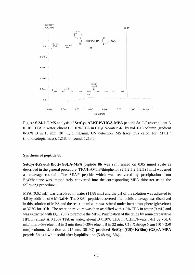
Figure S 24. LC-MS analysis of SetCys-ALKEPVHGA-MPA peptide 8a. LC trace: eluent A
0.10% TFA in water, eluent B 0.10% TFA in CH3CN/water: 4/1 by vol. C18 column, gradient
0-50% B in 15 min, 30 °C, 1 mL/min, UV detection. MS trace: m/z calcd. for [M+H]+
(monoisotopic mass): 1218.45, found: 1218.5.
Synthesis of peptide 8b
SetCys-(GS)2-K(Biot)-(GS)2A-MPA peptide 8b was synthesized on 0.05 mmol scale as
described in the general procedure. TFA/H2O/TIS/thiophenol 92.5:2.5:2.5:2.5 (5 mL) was used
as cleavage cocktail. The SEAon peptide which was recovered by precipitation from
Et2O/heptane was immediately converted into the corresponding MPA thioester using the
following procedure.
MPA (0.62 mL) was dissolved in water (11.88 mL) and the pH of the solution was adjusted to
4.0 by addition of 6 M NaOH. The SEAon peptide recovered after acidic cleavage was dissolved
in this solution of MPA and the reaction mixture was stirred under inert atmosphere (glovebox)
at 37 °C for 16 h. The reaction mixture was then acidified with 1.5% TFA in water (9 mL) and
was extracted with Et2O (5 ×) to remove the MPA. Purification of the crude by semi-preparative
HPLC (eluent A 0.10% TFA in water, eluent B 0.10% TFA in CH3CN/water: 4/1 by vol, 6
mL/min, 0-5% eluent B in 3 min then 5-30% eluent B in 32 min, C18 XBridge 5 μm (10 × 250
mm) column, detection at 215 nm, 30 °C) provided SetCys-(GS)2-K(Biot)-(GS)2A-MPA
peptide 8b as a white solid after lyophilisation (5.48 mg, 8%).
Time (min)
0.00 2.00 4.00 6.00 8.00 10.00 12.00 14.00
0.0
2.0e-1
4.0e-1
6.0e-1
8.0e-1
1.0
11.27
Intensity
(UV, AU)
m/z
300 500 700 900 1100 1300 1500 1700 19000
100609.7
1218.5
[M+2H]2+
[M+H]+
Intensity
(AU)

S 25
Characterization of SetCys-(GS)2-K(Biot)-(GS)2A-MPA peptide 8b:
Figure S 25. LC-MS analysis of SetCys-(GS)2-K(Biot)-(GS)2A-MPA peptide 8b. LC trace:
eluent A 0.10% TFA in water, eluent B 0.10% TFA in CH3CN/water: 4/1 by vol. C18 column,
gradient 0-50% B in 15 min, 30 °C, 1 mL/min, light scattering detection. MS trace: m/z calcd.
for [M+H]+ (monoisotopic mass): 1317.37, found: 1317.5.
Figure S 26. MALDI-TOF analysis of SetCys-(GS)2-K(Biot)-(GS)2A-MPA peptide 8b.
Matrix α-cyano-4-hydroxycinnamic acid, positive detection mode, calcd. for [M+Na]+
(monoisotopic): 1339.37, found: 1339.2.
Time (min)
0.00 1.00 2.00 3.00 4.00 5.00 6.00 7.00 8.00 9.00 10.00 11.00 12.00 13.00 14.00 15.00
0.000
0.200
0.400
0.600
0.800
1.000
1.200
1.400
1.600
10.05
Intensity
(light scattering, AU)
m/z
1300 1320 1340 1360 1380 1400 14200
100 1317.5
1316.51315.4
1313.4
1311.2
1319.6
1320.8
1321.4
Intensity
(AU)
1339.2
0
1000
2000
3000
4000
1000 1200 1400 1600 1800 2000
m/z
[M+Na]+
Intensity
(AU)
1339.2
1355.2
1317.2
0
1000
2000
3000
4000
1300 1310 1320 1330 1340 1350 1360 1370
m/z
[M+H]+
[M+Na]+
[M+K]+
Intensity
(AU)

S 26
Synthesis of peptide 8c
SetCys-(GS)3-K(Biot)-(GS)3A-MPA peptide 8c was synthesized on 0.05 mmol scale as
described in the general procedure. TFA/H2O/TIS/thiophenol 92.5:2.5:2.5:2.5 (5 mL) was used
as cleavage cocktail. The SEAon peptide which was recovered by precipitation from
Et2O/heptane was immediately converted into the corresponding MPA thioester using the
following procedure.
MPA (1.25 mL) was dissolved in water (23.75 mL) and the pH of the solution was adjusted to
4.0 by addition of 6 M NaOH. The SEAon peptide recovered after acidic cleavage was dissolved
in this solution of MPA and the reaction mixture was stirred at 37 °C for 5.5 h. The reaction
was monitored by HPLC and was stopped before completion to limit the formation of the side
product which results from the slow cyclisation of SetCys-(GS)3-K(Biot)-(GS)3A-MPA
peptide 8c. The reaction mixture was then acidified with 10% TFA in water (until pH 3.2) and
was extracted with Et2O (5 ×) to remove the MPA. Purification of the crude by semi-preparative
HPLC (eluent A 0.10% TFA in water, eluent B 0.10% TFA in CH3CN/water: 4/1 by vol, 6
mL/min, 0-5% eluent B in 3 min then 5-35% eluent B in 32 min, C18 XBridge 5 μm (10 × 250
mm) column, detection at 215 nm, 30 °C) provided SetCys-(GS)3-K(Biot)-(GS)3A-MPA
peptide 8c as a white solid after lyophilisation (16.6 mg, 19%).
Characterization of SetCys-(GS)3-K(Biot)-(GS)3A-MPA peptide 8c:
Figure S 27. LC-MS analysis of SetCys-(GS)3-K(Biot)-(GS)3A-MPA peptide 8c. LC trace:
eluent A 0.10% TFA in water, eluent B 0.10% TFA in CH3CN/water: 4/1 by vol. C18 column,
gradient 0-50% B in 15 min, 30 °C, 1 mL/min, light scattering detection. MS trace: m/z calcd.
for [M+H]+ (monoisotopic mass): 1605.48, found: 1605.6.
Time (min)
0.00 1.00 2.00 3.00 4.00 5.00 6.00 7.00 8.00 9.00 10.00 11.00 12.00 13.00 14.00 15.00
0.000
0.400
0.800
1.200
1.600
2.000
2.400
9.76
Intensity
(light scattering, AU)
m/z
300 500 700 900 1100 1300 1500 1700 1900
0
100803.3
1605.6
[M+2H]2+
[M+H]+
Intensity
(AU)

S 27
Figure S 28. MALDI-TOF analysis of SetCys-(GS)3-K(Biot)-(GS)3A-MPA peptide 8c.
Matrix α-cyano-4-hydroxycinnamic acid, positive detection mode, calcd. for [M+Na]+
(monoisotopic): 1627.46, found: 1627.5.
3.3 Preparation of SetAla peptide (Figure 3d)
Synthesis of Fmoc-SetAla-OH SI12
Figure S 29. Synthesis of the Fmoc-protected SetAla amino acid SI12.
1627.5
0
500
1000
1500
2000
2500
3000
800 1000 1200 1400 1600 1800 2000
m/z
[M+Na]+
[M+H]+
[M+K]+
Intensity
(AU)
1627.5
1643.5
1605.5
0
500
1000
1500
2000
2500
3000
1580 1600 1620 1640 1660 1680
m/z
[M+Na]+
[M+H]+
[M+K]+
Intensity
(AU)

S 28
(2,2-Diethoxyethyl)(4-methoxybenzyl)selenide SI3 (230 mg, 0.73 mmol) was suspended in 1
M formic acid (6 mL) and the mixture was heated overnight at 50 °C. Water (50 mL) was added
and the aqueous layer was extracted with Et2O (2 × 50 mL). The combined organic extracts
were washed with water (50 mL) and dried over MgSO4. After evaporation of the solvent under
reduced pressure, aldehyde SI4 was dissolved in anhydrous 1,2-dichloroethane (15 mL) under
argon. H-Ala-OMe.HCl SI9 (106 mg, 0.76 mmol), DIEA (146 µL, 0.84 mmol, activated
powdered 3 Å molecular sieves (760 mg) and sodium triacetoxyborohydride (208 mg,
0.98 mmol) were successively added to the solution of aldehyde. After 20 h stirring at RT, the
reaction mixture was filtered on a Büchner funnel and the solid was washed with additional
portions of DCM. The filtrate was evaporated under reduced pressure. Then 0.5 M K2CO3 (30
mL) and saturated NaCl (30 mL) were added to the residue and the aqueous layer was extracted
with DCM (3 × 50 mL). The combined organic layers were dried over MgSO4 and the solvent
was evaporated under reduced pressure.
The N-alkylated derivative SI10 contained in the residue (272 mg) was dissolved in DCM (4
mL). DIEA (0.14 mL, 0.82 mmol) and FmocCl (212 mg, 0.82 mmol) were added to the solution
of SI10. After 24 h stirring at RT, the reaction mixture was diluted with an additional portion
of DCM. The organic layer was washed with water and was dried over MgSO4. After
evaporation of the solvent under reduced pressure, purification of the crude by column
chromatography (cyclohexane/EtOAc 7:3) provided the Fmoc-protected derivative SI11, in
mixture with an impurity that resulted from FmocCl decomposition (310 mg).
To the compound SI11 isolated by column chromatography (140 mg) were added dioxane (6
mL) and 5 M HCl (3 mL) and the mixture was refluxed for 6 h. The reaction mixture was then
diluted with 1 M HCl (40 mL) and the aqueous layer was extracted with DCM (3 × 40 mL).
The combined organic layers were dried over MgSO4 and the solvent was evaporated under
reduced pressure. Purification of the crude by column chromatography (DCM/MeOH 99:1 →
95:5) provided the expected amino acid SI12 as a glass (55 mg, 31% from SI3).
The titled compound was characterized by 1H and 13C NMR as a mixture of conformers. Due
to overlapping signals, no clear description of the 1H NMR spectrum can be provided.
IR (ATR, cm-1) 3065, 2995, 2932, 1739, 1696, 1509, 1284, 1244, 1172, 1101, 726. [α]D20 (c
1.0, CHCl3): -9 °.
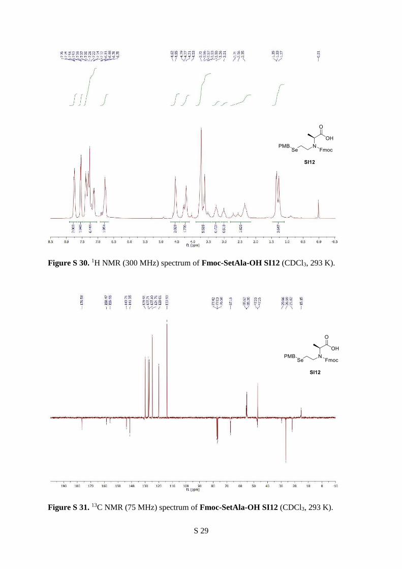
S 29
Figure S 30. 1H NMR (300 MHz) spectrum of Fmoc-SetAla-OH SI12 (CDCl3, 293 K).
Figure S 31. 13C NMR (75 MHz) spectrum of Fmoc-SetAla-OH SI12 (CDCl3, 293 K).

S 30
Figure S 32. LC-MS analysis of Fmoc-SetAla-OH SI12. LC trace: eluent C 0.10% formic acid
in water, eluent D 0.10% formic acid in CH3CN, gradient 40-100% D in 10 min, 0.4 mL/min,
Atlantis T3 3 µm 2.1 × 50 mm column; 50 °C; UV detection. MS trace: [M+Na]+ m/z calcd.
(monoisotopic mass) 562.11, found 562.33.
Elongation of the SetAla peptide
SetAla peptide was elongated according to general procedure presented in section 3.1. Note that
a protocol similar to the one described for SetCys was used to couple the SetAla amino acid at
the N-terminus of the elongated peptide.
Cleavage of the SetAla peptide
PMB-SeCH2CH2-Ala-ALKEPVHGV-NH2 peptide was synthesized on 0.05 mmol scale.
TFA/H2O/TIS 95:2.5:2.5 (5 mL) was used as cleavage cocktail.
Removal of PMB protecting group
Figure S 33. Removal of PMB protecting group.
The PMB-protected peptide which was recovered by precipitation from Et2O/heptane (43.0 mg)
was converted into the final diselenide by oxidation with iodine using the following procedure.
The crude recovered after precipitation (21.0 mg) was solubilized in AcOH/water 1:4 (13.2 mL)
and a solution of I2 in DMSO (109 µL, 61.4 mg/mL) was added dropwise. After 30 min stirring
0 1 2 3 4 5 6 7 8 9 10 11Time (min)
0
500000
1000000
1500000
2000000 5.87
Intensity
(UV, µAU
510 540 570 600 630 660 690
m/z
0
20
40
60
80
100660.00
562.33
[M+Na]+[M+K+2ACN]+
Relative
abundance

S 31
at RT, a solution of sodium ascorbate in water (9.9 mg/mL) was added dropwise until the
complete disappearance of I2 color. Purification of the crude by preparative HPLC (eluent A
0.10% TFA in water, eluent B 0.10% TFA in CH3CN, 20 mL/min, 0-10% eluent B in 4 min
then 10-30% eluent B in 29 min, C18 XBridge 5 μm (19 × 150 mm) column, detection at 215
nm, RT) provided the expected diselenide as a white solid after lyophilisation (8.7 mg, 24%
overall starting from solid support).
Characterization of the diselenide (SetAla-ALKEPVHGV-NH2)2:
Figure S 34. MALDI-TOF analysis of the diselenide (SetAla-ALKEPVHGV-NH2)2. Matrix
α-cyano-4-hydroxycinnamic acid, positive detection mode. The diselenide as well as the selenol
are detected by MALDI-TOF analysis. Calcd. for [M+H]+ (selenol): 1127.55, found: 1127.53;
calcd for [M+H]+ (diselenide): 2252.07, found: 2252.05.
1127.535
2252.049
0
1
2
3
4
5
6x10⁴
1000 1200 1400 1600 1800 2000 2200 2400
m/z
[M+H]+
[M+H]+
Intensity
(AU)
1127.535
0
2
4
6x10⁴
1122 1126 1130m/z
2252.049
0
1
2
3
x104
2244 2248 2252 2256m/z
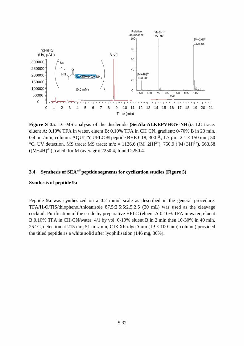
S 32
Figure S 35. LC-MS analysis of the diselenide (SetAla-ALKEPVHGV-NH2)2. LC trace:
eluent A: 0.10% TFA in water, eluent B: 0.10% TFA in CH3CN, gradient: 0-70% B in 20 min,
0.4 mL/min; column: AQUITY UPLC ® peptide BHE C18, 300 Å, 1.7 µm, 2.1 × 150 mm; 50
°C, UV detection. MS trace: MS trace: m/z = 1126.6 ([M+2H]2+), 750.9 ([M+3H]3+), 563.58
([M+4H]4+); calcd. for M (average): 2250.4, found 2250.4.
3.4 Synthesis of SEAoff peptide segments for cyclization studies (Figure 5)
Synthesis of peptide 9a
Peptide 9a was synthesized on a 0.2 mmol scale as described in the general procedure.
TFA/H2O/TIS/thiophenol/thioanisole 87.5:2.5:5:2.5:2.5 (20 mL) was used as the cleavage
cocktail. Purification of the crude by preparative HPLC (eluent A 0.10% TFA in water, eluent
B 0.10% TFA in CH3CN/water: 4/1 by vol, 0-10% eluent B in 2 min then 10-30% in 40 min,
25 °C, detection at 215 nm, 51 mL/min, C18 Xbridge 5 μm (19 × 100 mm) column) provided
the titled peptide as a white solid after lyophilisation (146 mg, 30%).
0 1 2 3 4 5 6 7 8 9 10 11 12 13 14 15 16 17 18 19 20 21
Time (min)
0
50000
100000
150000
200000
250000
300000
8.64Intensity
(UV, µAU)
550 650 750 850 950 1050 1150m/z
0
20
40
60
80
100750.92
1126.58
563.58
Relative
abundance[M+3H]3+
[M+2H]2+
[M+4H]4+

S 33
Characterization of peptide 9a:
Figure S 36. LC-MS analysis of peptide 9a. LC trace: eluent A 0.10% TFA in water, eluent B
0.10% TFA in CH3CN/water: 4/1 by vol. C18 column, gradient 0-50% B in 15 min, 30 °C, 1
mL/min, light scattering detection. MS trace: m/z = 1062.5 ([M+2H]2+), 708.7 ([M+3H]3+).
Calcd. for M (average): 2124.5, found: 2123.5.
Figure S 37. MALDI-TOF analysis of peptide 9a. Matrix α-cyano-4-hydroxycinnamic acid,
positive detection mode, calcd. for [M+H]+ (monoisotopic): 2123.94, found: 2124.1.
Time (min)
0.00 2.00 4.00 6.00 8.00 10.00 12.00 14.00
0.000
0.200
0.400
0.600
0.800
1.000
12.64
1.200
Intensity
(light scattering, AU)
m/z400 600 800 1000 1200 1400 1600 1800
0
100
found: 2123.5
[M+3H]3+
708.7
1062.6
[M+2H]2+
Intensity
(AU)
2124.1
0
2
4
6
8
1000 1500 2000 2500 3000 3500
m/z
x104
Intensity
(AU)[M+H]+
2124.1
2146.1
0
2
4
6
8
x104
2120 2125 2130 2135 2140 2145 2150
m/z
Intensity
(AU)[M+H]+
[M+Na]+

S 34
Synthesis of peptide 9b (linear K1 precursor)
The linear K1 precursor 9b used for the synthesis of K1 cyclic derivatives was prepared as
described in Figure S 38. Its sequence corresponds to HGF 128-205 using Uniprot numbering
of amino acid residues (P14210).
Figure S 38. Assembly of linear K1 precursor 9b.

S 35
Synthesis of the peptide segments for the assembly of linear K1 peptide 9b
AcA-K1[128-148]-SEAoff 6 and K1[149-176]-SEAoff Erreur ! Signet non défini. have already b
een synthesized and characterized elsewhere.
K1[177-205]-SEAoff (C(StBu)RNPRGEEGGPWC(StBu)FTSNPEVRYEVC(StBu)DIPQ-
SEAoff) was synthesized on a 1.80 mmol scale as described in the general procedure.
TFA/H2O/TIS/thioanisole/EDT 90:1.67:5:1.67:1.67 was used as cleavage cocktail. Purification
of the crude by preparative HPLC (eluent A 0.10% TFA in water, eluent B 0.10% TFA in
CH3CN/water: 4/1 by vol, 25 °C, detection at 215 nm, 51 mL/min, 0-28% eluent B in 5 min
then 28-42% in 25 min, C18 Xbridge 5 μm (19 × 100 mm) column) provided the titled peptide
as a white solid after lyophilisation (404 mg, 5%).
Characterization of K1[177-205]-SEAoff:
Figure S 39. LC-MS analysis of K1[177-205]-SEAoff. LC trace: eluent A 0.10% TFA in water,
eluent B 0.10% TFA in CH3CN/water: 4/1 by vol. C18 column, gradient 0-100% B in 30 min,
30 °C, 1 mL/min, light scattering detection. MS trace: m/z = 1860.8 ([M+2H]2+), 1241.8
([M+3H]3+); calcd. for M: 3721.4, found: 3721.0.
Time (min)
0.00 2.00 4.00 6.00 8.00 10.00 12.00 14.00 16.00 18.00 20.00 22.00 24.00 26.00 28.00 30.00
0.000
0.040
0.080
0.120
0.160
0.200
0.240
0.280
0.320
18.81
m/z1200 1300 1400 1500 1600 1700 1800
0
100 1241.8
1860.8
Intensity
(AU) [M+3H]3+
[M+2H]2+

S 36
Figure S 40. MALDI-TOF analysis of K1[177-205]-SEAoff. Matrix α-cyano-4-
hydroxycinnamic acid, positive detection mode, calcd. for [M+H]+ (monoisotopic) 3719.58,
found 3720.0.
Assembly of linear K1 peptide 9b
Step 1. Synthesis of peptide thioester AcA-K1[128-148]-MPA
TCEP·HCl (286 mg) was dissolved in a mixture of MPA (0.5 mL) and H2O (9.5 mL) and the
pH was adjusted to 4.0 by addition of 6 M NaOH. AcA-K1[128-148]-SEAoff (23.1 mg, 7.79
μmol) was added to the solution (7.78 mL) and the mixture was stirred at 37 °C for 18 h. Note
that a white solid precipitated during the reaction. The addition of Gn·HCl (7.35 g, final
concentration: 6 M) and MPA (0.255 mL, final concentration: 5% v/v) did not allow the
dissolution of the precipitate. The reaction mixture was acidified with AcOH (1.2 mL) and
extracted with Et2O (3 ×) to remove the MPA. The crude was purified by HPLC (eluent A =
water containing 0.1% TFA, eluent B=acetonitrile in water 4/1 containing 0.1% TFA, 30°C,
detection at 215 nm, 6 mL/min, 0-15% eluent B in 5 min, then 15-35% eluent B in 60 min,
C18XBridge column) to give AcA-K1[128-148]-MPA as a with solid (15.1 mg, 66%).
3720.0
0
2000
4000
6000
8000
2600 2800 3000 3200 3400 3600 3800 4000 4200 4400
m/z
Intensity(AU)
[M+H]+
3720.0
0
2000
4000
6000
8000
3700 3705 3710 3715 3720 3725 3730 3735 3740
m/z
Intensity(AU)
[M+H]+

S 37
Characterization of AcA-K1[128-148]-MPA:
Figure S 41. LC-MS analysis of AcA-K1[128-148]-MPA. LC trace: eluent A 0.10% TFA in
water, eluent B 0.10% TFA in CH3CN/water: 4/1 by vol. C18 Xbridge BEH 300 Å 5 μm (4.6
× 250 mm) column, 30 °C, gradient 0-100% B in 30 min, 1 mL/min, light scattering detection.
MS trace: m/z = 1185.0 ([M+2H]2+), 790.2 ([M+3H]3+), 593.1 ([M+4H]4+); calcd. for M
(average): 2368.8, found: 2368.2.
Figure S 42. MALDI-TOF analysis of AcA-K1[128-148]-MPA. Matrix 2,5-dihydroxybenzoic
acid (DHB), positive detection mode, calcd. for [M+H]+ (monoisotopic) 2368.27, found 2368.6.
Time (min)
0.00 5.00 10.00 15.00 20.00 25.00 30.00
0.000
0.500
1.000
1.500
2.000
2.500
3.000
3.500
12.68
Intensity
(light scattering, AU)
m/z
200 400 600 800 1000 1200 1400 1600 18000
100
found: 2368.25
790.2
[M+4H]4+
593.1
1185.0
[M+3H]3+
[M+2H]2+
Intensity
(AU)
2368.6
0.0
0.2
0.4
0.6
0.8
1.0
1000 1500 2000 2500 3000 3500
m/z
x104
[M+H]+Intensity
(AU)
2368.6
2390.6
0.0
0.2
0.4
0.6
0.8
1.0
x104
2340 2350 2360 2370 2380 2390 2400 2410
m/z
[M+H]+
[M+Na]+
Intensity
(AU)

S 38
Step 2-3. Synthesis of peptide thioester AcA-K1[128-176]-MPA
To a solution of Gn·HCl (574 mg) in 0.1 M, pH 7.4 phosphate buffer (600 μL) was added
MPAA (33.4 mg, 200 mM) and the pH of the mixture was adjusted to 7.44 by addition of 6 M
NaOH (80 μL). K1[149-176]- SEAoff (21.5 mg, 5.22 μmol, 1.05 eq) and AcA-K1[128-148]-
MPA (14.6 mg, 4.97 μmol, 1 eq) were then successively dissolved in the MPAA solution (710
μL) and the mixture was stirred at 37 °C. The progress of the reaction leading to peptide AcA-
K1[128-176]-SEAoff was followed by HPLC.
After completion of the NCL ligation (24h), SEA peptide AcA-K1[128-176]-SEAoff was
directly converted into the corresponding MPA thioester by addition MPA (5% v/v) and TCEP
(100 mM). To perform this reaction, TCEP (222 mg, 111 mM) and MPA (388 μL, 5.55 % v/v)
were added to the solution of Gn·HCl (4.01 g) in 0.1 M, pH 7.4 phosphate buffer (3.81 mL)
and the pH of the mixture was adjusted to 4.00 by addition of 6 M NaOH (600 μL). The previous
ligation mixture containing AcA-K1[128-176]-SEAoff was then diluted with the MPA/TCEP
solution (6.38 mL) and the pH was readjusted to 4.00 by addition of 6 N HCl (25 μL). In these
conditions ([TCEP] = 100 mM, 5% MPA v/v), the reaction mixture was stirred at 37 °C for 18
h. After completion of the reaction, the mixture was acidified with AcOH (0.7 mL) and
extracted with Et2O (5 ×) to remove the MPA and the MPAA. The crude was purified by HPLC
(eluent A = water containing 0.1% TFA, eluent B=acetonitrile in water 4/1 containing 0.1%
TFA, 50°C, detection at 215 nm, 6 mL/min, 0-20% eluent B in 5 min, then 20-40% eluent B in
60 min, C18XBridge column) to give AcA-K1[128-176]-MPA as a with solid after
lyophilisation (20.8 mg, 62%).
Characterization of AcA-K1[128-176]-MPA:
Figure S 43. MALDI-TOF analysis of AcA-K1[128-176]-MPA. Matrix 2,5-dihydroxybenzoic
acid (DHB), positive detection mode, calcd. for [M+H]+ (average) 5689.52, found 5689.5.
5689.5
0
1000
2000
3000
4000
3000 4000 5000 6000 7000 8000 9000
m/z
[M+H]+
DHB
adduct
Intensity
(AU)

S 39
Figure S 44. LC-MS analysis of AcA-K1[128-176]-MPA. LC trace: eluent A 0.10% TFA in
water, eluent B 0.10% TFA in CH3CN/water: 4/1 by vol. C18 Xbridge BEH 300 Å 5 μm (4.6
× 250 mm) column, 50 °C, gradient 0-100% B in 30 min, 1 mL/min, UV detection. MS trace:
m/z = 1422.8 ([M+4H]4+), 1138.4 ([M+5H]5+), 948.7 ([M+6H]6+), 813.4 ([M+7H]7+), 711.9
([M+8H]8+); calcd. for M: 5688.5, found: 5687.0.
Step 4-5. Synthesis of peptide K1[128-205]-SEAoff (9b)
To a solution of Gn·HCl (574 mg) in 0.1 M, pH 7.4 phosphate buffer (600 μL) was added
MPAA (33.7 mg) and the pH of the mixture was adjusted to 7.39 by addition of 6 M NaOH (75
μL). K1[177-205]-SEAoff (13.5 mg, 3.25 μmol, 1.05 eq) and AcA-K1[128-176]-MPA (20.8
mg, 3.10 μmol, 1 eq) were then successively dissolved in the MPAA solution (442 μL) and the
mixture was stirred at 37 °C. The progress of the reaction leading to peptide AcA-K1[128-205]-
SEAoff was followed by HPLC.
After completion of the NCL ligation (24h), the AcA protecting group of AcA-K1[128-205]-
SEAoff was removed by reaction with NH2OH. To perform this deprotection, the reaction
mixture was acidified with AcOH (110 μL) and treated with a 0.71 M solution of NH2OH·HCl
in H2O (27.3 μL, 19.4, μmol, 6.25 eq). After 2 h stirring at 37 °C, the reaction mixture was
diluted with H2O (570 μL) and extracted with Et2O (5 ×) to remove the MPAA. The crude was
further diluted with H2O (1.14 mL, [AcOH] ≈ 5% v/v) and purified by HPLC (eluent A = water
containing 0.1% TFA, eluent B=acetonitrile in water 4/1 containing 0.1% TFA, 50°C, detection
at 215 nm, 6 mL/min, 0-25% eluent B in 5 min, then 25-75% eluent B in 75 min, C18XBridge
column) to give K1[128-205]-SEAoff (9b) as a with solid after lyophilisation(10.9 mg, 34%).
Time (min)
5.00 10.00 15.00 20.00 25.00 30.00
0.0
4.0e-1
8.0e-1
1.2
1.6
2.0
2.4
14.00
1.95
Intensity
(UV, AU)
2.8
m/z
200 400 600 800 1000 1200 1400 1600 18000
100
found: 5687.0813.4
711.9
[M+6H]+6
948.7
1422.81138.4
[M+7H]+7
[M+8H]+8
[M+5H]+5 [M+4H]+4
Intensity
(AU)

S 40
Characterization of peptide 9b:
a)
b)
Figure S 45. LC-MS analysis of peptide 9b. a) LC trace. Eluent A 0.10% TFA in water, eluent
B 0.10% TFA in CH3CN, ACQUITY UPLC Peptide BEH 300Å 1.7 μm (2.1 × 150 mm)
column, 50 °C, gradient 0-70% B in 20 min, 0.4 mL/min, UV detection. b) MS trace: m/z =
1493.42 ([M+6H]6+), 1280.25 ([M+7H]7+), 1120.42 ([M+8H]8+), 996.08 ([M+9H]9+), 896.75
([M+10H]10+), 815.25 ([M+11H]11+), 747.33 ([M+12H]12+), 689.92 ([M+13H]13+); calcd. for M
(average): 8955.26, found: 8955.81.
0 2 4 6 8 10 12 14 16 18 20
Time (min)
0
500000
1000000
1500000
11.31
Intensity
(UV, µAu)
300 500 1000 1500 2000m/z
0
20
40
60
80
1001280.25
1493.42
1120.42
996.08
896.75
815.25
747.33
689.92
Relative abundance
(%)

S 41
Figure S 46. MALDI-TOF analysis of peptide 9b. Matrix 2,5-dihydroxybenzoic acid (DHB),
positive detection mode, calcd. for [M+H]+ (average) 8956.22, found: 8957.4.
8957.4
5000 6000 7000 8000 9000 10000 11000 12000 13000
m/z
Intensity
(AU)
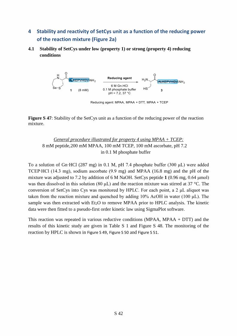
S 42
4 Stability and reactivity of SetCys unit as a function of the reducing power
of the reaction mixture (Figure 2a)
4.1 Stability of SetCys under low (property 1) or strong (property 4) reducing
conditions
Figure S 47: Stability of the SetCys unit as a function of the reducing power of the reaction
mixture.
General procedure illustrated for property 4 using MPAA + TCEP:
8 mM peptide, 200 mM MPAA, 100 mM TCEP, 100 mM ascorbate, pH 7.2
in 0.1 M phosphate buffer
To a solution of Gn·HCl (287 mg) in 0.1 M, pH 7.4 phosphate buffer (300 μL) were added
TCEP∙HCl (14.3 mg), sodium ascorbate (9.9 mg) and MPAA (16.8 mg) and the pH of the
mixture was adjusted to 7.2 by addition of 6 M NaOH. SetCys peptide 1 (0.96 mg, 0.64 μmol)
was then dissolved in this solution (80 μL) and the reaction mixture was stirred at 37 °C. The
conversion of SetCys into Cys was monitored by HPLC. For each point, a 2 μL aliquot was
taken from the reaction mixture and quenched by adding 10% AcOH in water (100 μL). The
sample was then extracted with Et2O to remove MPAA prior to HPLC analysis. The kinetic
data were then fitted to a pseudo-first order kinetic law using SigmaPlot software.
This reaction was repeated in various reductive conditions (MPAA, MPAA + DTT) and the
results of this kinetic study are given in Table S 1 and Figure S 48. The monitoring of the
reaction by HPLC is shown in Figure S 49, Figure S 50 and Figure S 51.
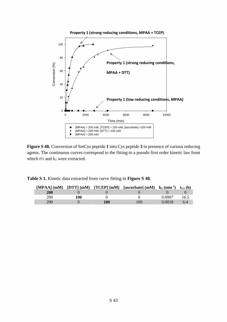
S 43
Time (min)
0 2000 4000 6000 8000 10000
Co
nve
rsio
n (
%)
0
20
40
60
80
100
[MPAA] = 200 mM, [TCEP] = 100 mM, [ascorbate] =100 mM
[MPAA] = 200 mM, [DTT] = 100 mM
[MPAA] = 200 mM
Figure S 48. Conversion of SetCys peptide 1 into Cys peptide 3 in presence of various reducing
agents. The continuous curves correspond to the fitting to a pseudo first order kinetic law from
which t½ and k1 were extracted.
Table S 1. Kinetic data extracted from curve fitting in Figure S 48.
[MPAA] (mM) [DTT] (mM) [TCEP] (mM) [ascorbate] (mM) k1 (min-1) t1/2 (h)
200 0 0 0 0 0
200 100 0 0 0.0007 16.5
200 0 100 100 0.0018 6.4
Property 1 (low reducing conditions, MPAA)
Property 1 (strong reducing conditions, MPAA + TCEP)
Property 1 (strong reducing conditions,
MPAA + DTT)

S 44
Figure S 49. Monitoring of the conversion of SetCys peptide 1 into cysteinyl peptide 3 using
MPAA + TCEP. LC trace: eluent A 0.10% TFA in water, eluent B 0.10% TFA in CH3CN/water:
4/1 by vol., C18 column, gradient 0-50% B in 15 min, 30 °C, 1 mL/min, UV detection at 215
nm.
0
100
200
300
400
Intensity
(UV, mAU)
0.0 2.0 4.0 6.0 8.0 10.0 12.0 15.0
0
100
200
300
400
Time (min)
0
100
200
300
400
Se=TECP

S 45
Figure S 50. Monitoring of the conversion of SetCys peptide 1 into cysteinyl peptide 3 using
DTT. LC trace: eluent A 0.10% TFA in water, eluent B 0.10% TFA in CH3CN/water: 4/1 by
vol., C18 column, gradient 0-50% B in 15 min, 30 °C, 1 mL/min, UV detection at 215 nm.
Intensity
(UV, mAU)
0
100
200
300
400
0
100
200
300
400
Time (min)
0.0 2.0 4.0 6.0 8.0 10.0 12.0 15.0
0
100
200
300
400
b) t = 25 h

S 46
Figure S 51. Stability of the SetCys peptide 1 in the presence of MPAA. LC trace: eluent A
0.10% TFA in water, eluent B 0.10% TFA in CH3CN/water: 4/1 by vol., C18 column, gradient
0-50% B in 15 min, 30 °C, 1 mL/min, UV detection at 215 nm.
Time (min)
0.0 2.0 4.0 6.0 8.0 10.0 12.0 15.0
0
100
200
300
400
500
Intensity
(UV, mAU)
0.0 2.0 4.0 6.0 8.0 10.0 12.0 15.0
0
100
200
300
400a) t = 20 s

S 47
4.2 Stability of SetCys under strong (property 4) reducing conditions. Importance of
ascorbate
Figure S 52. Treatment of SetCys by TCEP in absence of ascorbate.
To a solution of Gn·HCl (287 mg) in pH 7.4 0.1 M phosphate buffer (300 μL) were added
TCEP.HCl (14.3 mg) and MPAA (16.8 mg) and the pH of the mixture was adjusted to 6.0 by
addition of 6 M NaOH. SetCys peptide 1 (0.48 mg, 0.32 μmol) was then dissolved in the
buffered solution (320 μL) and the reaction mixture was stirred at 37 °C. The progress of the
reaction was monitored by HPLC. For each point, an 8 μL aliquot was taken from the reaction
mixture and quenched by adding 10% AcOH in water (100 μL). The sample was then extracted
with Et2O to remove MPAA prior to HPLC analysis.
At pH 6.0 and at the beginning of the reaction, the deselenization of SetCys peptide 1 into the
corresponding N-ethyl cysteinyl derivative (Et-CALKEPVHGV-NH2) dominates in the
mixture (Figure S 53a). At longer reaction times, the SetCys peptide 1 also yields the Cys peptide
by losing its selenoethyl arm (Figure S 53b). An increase of the reaction mixture pH (from 6.00
to 7.20) favors the deselenization process and leads nearly exclusively to the formation of the
N-ethylcysteinyl peptide product (Figure S 54).

S 48
Figure S 53. HPLC monitoring of the treatment of SetCys peptide 1 by TCEP in the absence
of ascorbate performed at pH 6.0. Eluent A: 0.10% TFA in water, eluent B: 0.10% TFA in
CH3CN, gradient: 0-50% B in 15 min, 30 °C, 1 mL/min, C18 column, UV detection (215 nm).
0.0 2.0 4.0 6.0 8.0 10.0 12.0 15.0
-100
0
100
200
300
400
500
Intensity
(UV, mAU)
Time (min)
Intensity
(UV, mAU)
Time (min)
0.0 2.0 4.0 6.0 8.0 10.0 12.0 15.0
-100
0
100
200
300
400
500
Se=TCEP

S 49
Figure S 54. HPLC monitoring of the treatment of SetCys peptide 1 by TCEP in the absence
of ascorbate performed at pH 7.20. Eluent A: 0.10% TFA in water, eluent B: 0.10% TFA in
CH3CN, gradient: 0-50% B in 15 min, 30 °C, 1 mL/min, C18 column, UV detection (215 nm).
The Et-CALKEPVHGV-NH2 peptide produced by deselenization of SetCys peptide 1 under
the above conditions was isolated by HPLC (Figure S 55) and its sequence was confirmed by
MALDI MS-MS analysis (Figure S 56, Figure S 57).
Intensity
(UV, mAU)
Time (min)
0.0 2.0 4.0 6.0 8.0 10.0 12.0 15.0
-100
0
100
200
300
400
500
Intensity
(UV, mAU)
Time (min)
0.0 2.0 4.0 6.0 8.0 10.0 12.0 15.0
-100
0
100
200
300
400
500
Se=TCEP

S 50
Figure S 55. UPLC-MS analysis of Et-CALKEPVHGV-NH2. a) LC trace. Eluent A: 0.10%
TFA in water, eluent B: 0.10% TFA in CH3CN, ACQUITY UPLC Peptide BEH 300 Å 1.7 μm
(2.1 × 150 mm) column, 50 °C, gradient 0-70% B in 20 min, 0.4 mL/min, UV detection (215
nm). b) MS trace. Calcd. for [M+H]+ (monoisotopic): 1079.60, found: 1079.42.
0 2 4 6 8 10 12 14 16 18 20
Time (min)
-60000
-30000
0
30000
60000
90000
7.77
Intensity
(UV, µAU)
400 600 800 1000 1200 1400 1600 1800 2000
m/z
0
20
40
60
80
100
[M+H]+
1079.42
[M+Na]+
1102.33
[2M+H]+
540.33
Relative
abundance

S 51
Figure S 56. MALDI-TOF analysis of Et-CALKEPVHGV-NH2 peptide. Matrix α-cyano-4-
hydroxycinnamic acid, positive detection mode, calcd. for [M+H]+ (monoisotopic): 1079.60,
found: 1079.64.
Figure S 57. MALDI-TOF-TOF sequencing of Et-CALKEPVHGV-NH2 peptide. Matrix α-
cyano-4-hydroxycinnamic acid, positive detection mode. The MS-MS analysis confirms that
the SetCys residue is converted into a N-ethyl cysteine residue (J in the figure) in the peptide
sequence.
[M+H]+
1079.636
[M+K]+
1117.589
[M+Na]+
1101.616
0.00
0.25
0.50
0.75
1.00
1.25
x104
975 1000 1025 1050 1075 1100 1125 1150 1175 1200
m/z
Intensity
(AU)
m/z100 200 300 400 500 600 700 800 900 1000 1100 1200
0
2
4
6
8
10
12
14
16
18
20
22
Abs. Int. * 1000
a J A H G V
b L K E P V H Gy V P E K L A J
104.08
a 1175.14
a 2
203.14
b 2
316.22
b 3
311.24
y 3
444.31
b 4
410.29
y 4
545.34
a 5
573.34
b 5
507.34
y 5
670.39
b 6
636.36
y 6
741.46
a 7
769.43
b 7
764.45
y 7
878.52
a 8
906.50
b 8
878.52
y 8
935.51
a 9
963.52
b 9
948.57
y 9
1034.48
a 10
1079.68
y 10

S 52
4.3 NCL under low reducing conditions (properties 2)
When SetCys peptide 1 and peptide thioester 4 were reacted under mild reducing conditions
(MPAA), the resulting peptide featuring a SetCys unit in the form of a cyclic selenosulfide at
the ligation junction was recovered as the only product of the ligation with 41% yield (Figure
S 58). The insertion of the SetCys amino acid into the peptide sequence was confirmed by MS-
MS analysis (Figure S 62).
Figure S 58. Ligation reaction of SetCys peptide 1 with peptidyl MPA-thioester 4.
Protocol
To a solution of Gn·HCl (1.03 g) in 0.1 M, pH 7.4 phosphate buffer (1.08 mL) was added
MPAA (60.6 mg) and the pH of the mixture was adjusted to 7.20 by addition of 6 M NaOH.
SetCys-ALKEPVHGV-NH2 1 (23.2 mg, 15.5 μmol, 1.2 eq) and RLKEPVHGA-MPA 4 (20.0
mg, 12.9 μmol, 1 eq) were then successively dissolved in this solution (1.61 mL) and the
reaction mixture was stirred at 37 °C for 24 h. The mixture was then acidified with 10% AcOH
in water (22.5 mL) and extracted with Et2O (5 ×) to remove the MPAA. Purification of the
crude by HPLC (eluent A = water containing 0.1% TFA, eluent B=acetonitrile in water 4/1
containing 0.1% TFA, 50°C, detection at 215 nm, 3 mL/min, 0-10% eluent B in 5 min, then 10-
35% eluent B in 35 min, then 35-50% eluent B in 15 min, C18XBridge column) provided
RLKEPVHGA-SetCys-ALKEPVHGV-NH2 as a white solid after lyophilisation (15.06 mg,
41%).

S 53
Figure S 59. Monitoring of the ligation between SetCys peptide 1 and peptidyl MPA thioester
RLKEPVHGA-MPA. LC trace: eluent A 0.10% TFA in water, eluent B 0.10% TFA in
CH3CN/water: 4/1 by vol., C18 column, gradient 0-50% B in 15 min, 30 °C, 1 mL/min, UV
detection at 215 nm.
Intensity
(UV, mAU)
0
100
200
300
400
500
600
0
100
200
300
400
500
600
Time (min)
0.0 2.0 4.0 6.0 8.0 10.0 12.0 15.0
0
100
200
300
400
500
600
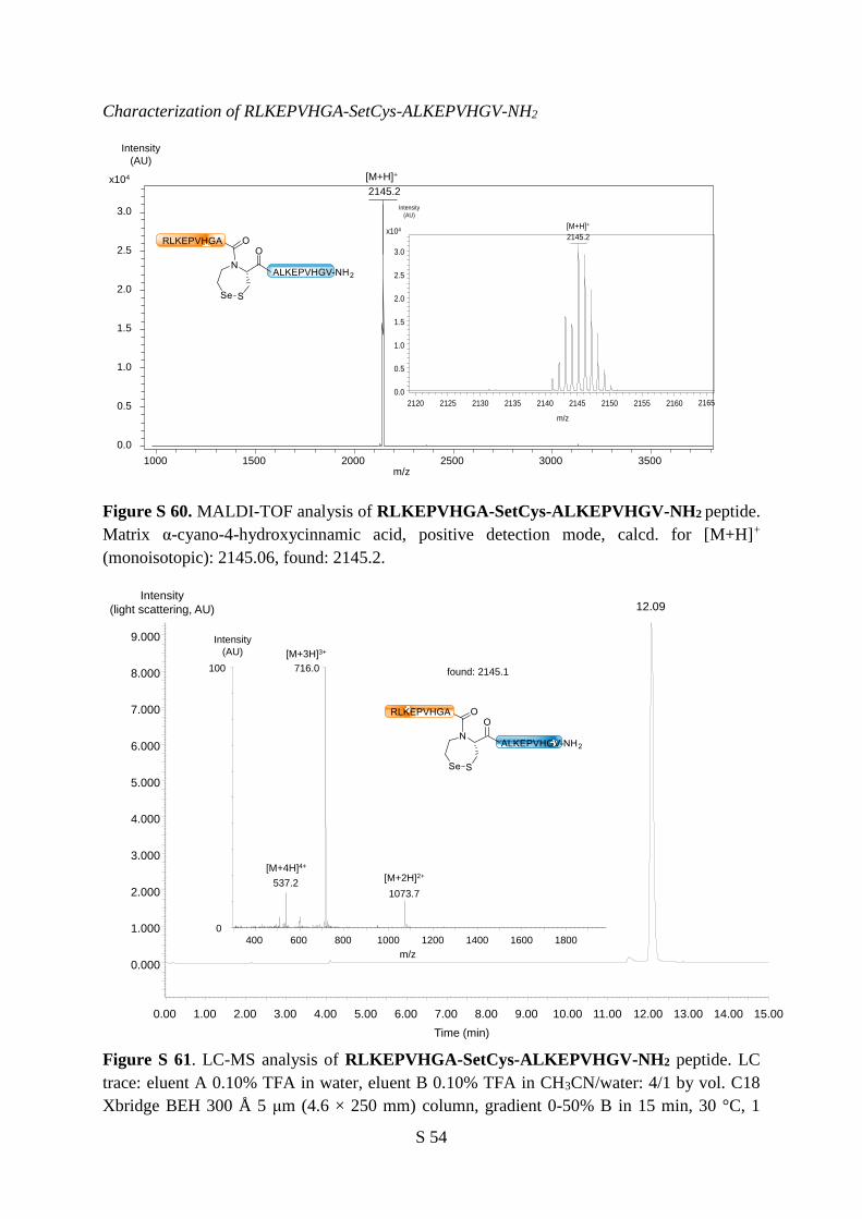
S 54
Characterization of RLKEPVHGA-SetCys-ALKEPVHGV-NH2
Figure S 60. MALDI-TOF analysis of RLKEPVHGA-SetCys-ALKEPVHGV-NH2 peptide.
Matrix α-cyano-4-hydroxycinnamic acid, positive detection mode, calcd. for [M+H]+
(monoisotopic): 2145.06, found: 2145.2.
Figure S 61. LC-MS analysis of RLKEPVHGA-SetCys-ALKEPVHGV-NH2 peptide. LC
trace: eluent A 0.10% TFA in water, eluent B 0.10% TFA in CH3CN/water: 4/1 by vol. C18
Xbridge BEH 300 Å 5 μm (4.6 × 250 mm) column, gradient 0-50% B in 15 min, 30 °C, 1
2145.2
0.0
0.5
1.0
1.5
2.0
2.5
3.0
x104
1000 1500 2000 2500 3000 3500m/z
[M+H]+
Intensity
(AU)
2145.2
0.0
0.5
1.0
1.5
2.0
2.5
3.0
x104
2120 2125 2130 2135 2140 2145 2150 2155 2160 2165
m/z
[M+H]+
Intensity
(AU)
Time (min)
0.00 1.00 2.00 3.00 4.00 5.00 6.00 7.00 8.00 9.00 10.00 11.00 12.00 13.00 14.00 15.00
0.000
1.000
2.000
3.000
4.000
5.000
6.000
7.000
8.000
9.000
12.09Intensity
(light scattering, AU)
m/z
400 600 800 1000 1200 1400 1600 18000
100 found: 2145.1716.0
537.21073.7
Intensity
(AU) [M+3H]3+
[M+2H]2+[M+4H]4+

S 55
mL/min, light scattering detection. MS trace: 1073.7 ([M+2H]2+), 716.0 ([M+3H]3+), 537.2
([M+4H]4+); calcd. for M (average): 2144.4, found: 2145.1.
Figure S 62. MALDI-TOF-TOF sequencing of RLKEPVHGA-SetCys-ALKEPVHGV-NH2
peptide. Matrix α-cyano-4-hydroxycinnamic acid, positive detection mode. The MS-MS
analysis confirms the presence of SetCys (J on the figure) residue in the sequence of the peptide.
Kinetic study
The progress of the reaction was monitored by HPLC. For each point, a 2 μL aliquot was taken
from the reaction mixture and quenched by adding 10% AcOH in water (100 μL). The sample
was then extracted with Et2O to remove MPAA prior to HPLC analysis. The monitoring of the
ligation points out that the MPAA peptide thioester produced from RLKEPVHGA-MPA 4 by
transthioesterification with MPAA quickly accumulates in the reaction mixture. This
intermediate is then slowly consumed with concomitant formation of the ligated product.
m/z2 0 0 4 0 0 6 0 0 8 0 0 1 0 0 0 1 2 0 0 1 4 0 0 1 6 0 0 1 8 0 0 2 0 0 0 2 2 0 0 2 4 0 0
0
2
4
6
8
1 0
1 2
1 4
1 6
Ab s. In t. * 1 0 0 0
a R L H G A Hb R L K E P V H G A J A L K E G Vy V P E K L A J H E K L R
1 2 9 .0 9
a 1
1 5 7 .0 3
b 1
2 4 2 .2 8
a 2
2 7 0 .2 8
b 2
3 9 8 .4 0
b 3
3 1 1 .2 9
y 3
4 9 9 .4 8
a 4
5 2 7 .4 7
b 4
4 0 9 .4 1
y 4
6 2 4 .5 6
b 5
5 0 7 .4 4
y 5
6 9 5 .6 6
a 6
7 2 3 .6 3
b 6
6 3 6 .5 1
y 6
8 3 2 .7 6
a 7
8 6 0 .7 2
b 7
7 6 4 .6 4
y 7
8 8 9 .7 9
a 8
9 1 7 .7 7
b 8
8 7 7 .7 3
y 8
9 6 0 .8 6
a 9
9 8 8 .8 0
b 99 4 8 .7 9
y 9
1 1 9 7 .8 1
b 1 0
1 1 5 6 .7 2
y 1 0
1 2 6 8 .8 8
b 1 1
1 3 5 3 .9 4
a 1 2
1 3 8 1 .9 0
b 1 2
1 2 8 5 .8 9
y 1 2
1 5 0 9 .9 7
b 1 3
1 4 2 2 .8 5
y 1 3 1 6 0 9 .9 8
a 1 4
1 6 3 8 .8 8
b 1 4
1 6 1 8 .8 6
y 1 5
1 8 0 6 .8 8
a 1 6
1 7 4 7 .9 3
y 1 61 9 4 3 .8 9
a 1 7
1 9 7 1 .8 2
b 1 7
1 8 7 6 .8 3
y 1 7
2 0 2 8 .5 3
b 1 8
1 9 8 9 .8 2
y 1 8
2 0 9 9 .9 0
a 1 9
2 1 2 7 .3 1
b 1 9
2 1 4 5 .2 2
y 1 9
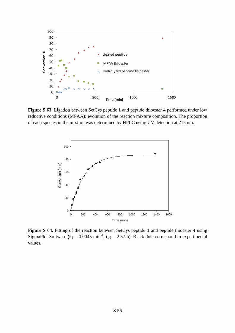
S 56
Figure S 63. Ligation between SetCys peptide 1 and peptide thioester 4 performed under low
reductive conditions (MPAA): evolution of the reaction mixture composition. The proportion
of each species in the mixture was determined by HPLC using UV detection at 215 nm.
Time (min)
0 200 400 600 800 1000 1200 1400 1600
Co
nve
rsio
n (
min
)
0
20
40
60
80
100
Figure S 64. Fitting of the reaction between SetCys peptide 1 and peptide thioester 4 using
SigmaPlot Software (k1 = 0.0045 min-1; t1/2 = 2.57 h). Black dots correspond to experimental
values.
0
10
20
30
40
50
60
70
80
90
100
0 500 1000 1500
Co
nve
rsio
n %
Time (min)
RLKEPVHGA-Aze(Se,S)-ALKEPVHGV-NH2
RLKEPVHGA-MPAA
RLKEPVHGA-OHHydrolyzed peptide thioester
MPAA thioester
Ligated peptide

S 57
4.4 SetCys is silent during NCL under low reducing conditions (property 3)
4.4.1 Competitive ligation experiment involving a Cys and SetCys peptide
Figure S 65. SetCys is silent during NCL under low reducing conditions.
General procedure illustrated with RLKEPVHGA-MPA 4 as peptide thioester:
To a solution of Gn·HCl (287 mg) in 0.1 M, pH 7.4 phosphate buffer (300 μL) was added
MPAA (16.8 mg) and the pH of the mixture was adjusted to 7.22 by addition of 6 M NaOH.
SetCys-ALKEPVHGV-NH2 (1.32 mg, 0.88 μmol, 1.2 eq), CILKEPVHGV-NH2 (1.27 mg,
0.88 μmol, 1.2 eq) and RLKEPVHGA-MPA (1.14 mg, 0.735 μmol, 1 eq) were then
successively dissolved in this solution (92 μL) and the reaction mixture was stirred at 37 °C.
The progress of the kinetically favored ligation (RLKEPVHGA-MPA + CILKEPVHGV-
NH2) was monitored by HPLC. For each point, a 2 μL aliquot was taken from the reaction
mixture and quenched by adding 10% AcOH in water (100 μL). The sample was then extracted
with Et2O to remove MPAA prior to HPLC analysis. The kinetic data were then fitted to a
pseudo-first order kinetic law and the results are given in Table S 2.
The reaction was repeated using ILKEPVHGV-MPA instead RLKEPVHGA-MPA as
peptidyl thioester. In both cases, SetCys-ALKEPVHGV-NH2 peptide was recovered
unreacted at the end of the ligation (Figure S 67, Figure S 68).

S 58
Figure S 66. Ligations using respectively RLKEPVHGA-MPA and ILKEPVHGV-MPA as
acyl donor. The continuous curves correspond to the fitting to a pseudo first order kinetic law
from which t½ and k1 were extracted.
Table S 2. Rate constants and half-reaction times determined from data in Figure S 66.
Product of ligation k1 (min-1) t1/2 (h)
RLKEPVHGA-CILKEPVHGV-NH2 0.0512 0.22
ILKEPVHGV-CILKEPVHGV-NH2 0.0044 2.62

S 59
Figure S 67. Monitoring of the competitive reaction between SetCys peptide 1 and cysteinyl
peptide CILKEPVHGV-NH2 with peptidyl MPA-thioester 4. LC trace: eluent A 0.10% TFA in
water, eluent B 0.10% TFA in CH3CN/water: 4/1 by vol., C18 column, gradient 0-50% B in 15
min, 30 °C, 1 mL/min, UV detection at 215 nm.
100
200
300
400
Intensity
(UV, mAU)
0
100
200
300
400
500
0
0.0 2.0 4.0 6.0 8.0 10.0 12.0 15.0
100
200
300
400
500
Time (min)
0

S 60
Figure S 68. Monitoring of the competitive reaction between SetCys peptide 1 and cysteinyl
peptide CILKEPVHGV-NH2 with peptidyl MPA-thioester ILKEPVHGV-MPA. LC trace:
eluent A 0.10% TFA in water, eluent B 0.10% TFA in CH3CN/water: 4/1 by vol., C18 column,
gradient 0-50% B in 15 min, 30 °C, 1 mL/min, UV detection at 215 nm.
0
100
200
300
400
500
Intensity
(UV, mAU)
0
100
200
300
400
500
0.0 2.0 4.0 6.0 8.0 10.0 12.0 15.0
0
100
200
300
400
500
Time (min)
b) t = 23 h
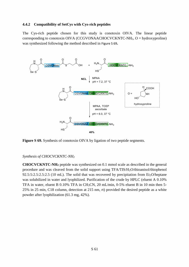
S 61
4.4.2 Compatibility of SetCys with Cys-rich peptides
The Cys-rich peptide chosen for this study is conotoxin OIVA. The linear peptide
corresponding to conotoxin OIVA (CCGVONAACHOCVCKNTC-NH2, O = hydroxyproline)
was synthesized following the method described in Figure S 69.
Figure S 69. Synthesis of conotoxin OIVA by ligation of two peptide segments.
Synthesis of CHOCVCKNTC-NH2
CHOCVCKNTC-NH2 peptide was synthesized on 0.1 mmol scale as described in the general
procedure and was cleaved from the solid support using TFA/TIS/H2O/thioanisol/thiophenol
92.5:5:2.5:2.5:2.5 (10 mL). The solid that was recovered by precipitation from Et2O/heptane
was solubilized in water and lyophilized. Purification of the crude by HPLC (eluent A 0.10%
TFA in water, eluent B 0.10% TFA in CH3CN, 20 mL/min, 0-5% eluent B in 10 min then 5-
25% in 25 min, C18 column, detection at 215 nm, rt) provided the desired peptide as a white
powder after lyophilization (61.3 mg, 42%).

S 62
Characterization of CHOCVCKNTC-NH2
Figure S 70. UPLC-MS analysis of CHOCVCKNTC-NH2. a) LC trace. Eluent A 0.10% TFA
in water, eluent B 0.10% TFA in CH3CN, ACQUITY UPLC Peptide BEH 300 Å 1.7 μm (2.1
× 150 mm) column, 50 °C, gradient 0-70% B in 20 min, 0.4 mL/min, UV detection. b) MS
trace: Calcd. for [M+H]+ (monoisotopic): 1122.42, found: 1122.42.
0 4 8 12 16 20
Time (min)
0
250000
500000
750000
1000000
1200000
5.94
Intensity
(UV, µAU)
300 600 900 1200 1500 1800 2000
m/z
0
20
40
60
80
100
[M+H]+
1122.42
[M+2H]2+
561.67
[M+Na]+
1144.25
Relative
abundance
a)
b)

S 63
Synthesis of peptide thioester SetCysCGVONAA-MPA
SetCysCGVONAA-MPA peptide was synthesized on 0.05 mmol scale as described in the
general procedure. TFA/H2O/TIS/thiophenol/thioanisol 87.5:2.5:5:2.5:2.5 (8 mL) was used as
the cleavage cocktail. The SEAon peptide which was recovered by precipitation from
Et2O/heptane was immediately converted into the corresponding MPA thioester using the
following procedure.
MPA (0.625 mL) was dissolved in 11.88 mL of water and the pH of the solution was adjusted
to 4.0 by addition of 6 M NaOH. The SEAon peptide recovered after acidic cleavage was added
to this solution of MPA. Due to the low solubility of the peptide, solid Gn∙HCl (1.99 g) was
added to the reaction mixture. Once the pH was readjusted to 4.0 by addition of 6 M NaOH, the
reaction was stirred under inert atmosphere at 37 °C for 7 h. The reaction mixture was then
acidified with AcOH (1 mL) and extracted with Et2O (5 ×) to remove the MPA. Purification of
the crude by semi-preparative HPLC (eluent A 0.10% TFA in water, eluent B 0.10% TFA in
CH3CN, 20 mL/min, 0-10% eluent B in 5 min then 10-25% in 30 min, C18 column, detection
at 215 nm, 30 °C) provided SetCysCGVONAA-MPA peptide as a white solid after
lyophilisation (7.22 mg, 14% overall yield).

S 64
Characterization of SetCysCGVONAA-MPA
Figure S 71 . UPLC-MS analysis of SetCysCGVONAA-MPA. a) LC trace. Eluent A 0.10%
TFA in water, eluent B 0.10% TFA in CH3CN, ACQUITY UPLC Peptide BEH 300 Å 1.7 μm
(2.1 × 150 mm) column, 50 °C, gradient 0-70% B in 20 min, 0.4 mL/min, UV detection. b) MS
trace: Calcd. for [M+H]+ (monoisotopic): 944.21, found: 944.08.
0 4 8 12 16 20
Time (min)
0
40000
80000
120000
160000
8.10
Intensity
(UV, µAU)
400 800 1200 1600 2000
m/z
0
20
40
60
80
100
[M+H]+
944.08
[M+Na]+
966.25
Relative
abundance
a)
b)

S 65
Assembly of conotoxin OIVA sequence
To a solution of Gn·HCl (287 mg) in 0.1 M, pH 7.4 phosphate buffer (300 μL) was added
MPAA (16.8 mg) and the pH of the mixture was adjusted to 7.25 by addition of 6 M NaOH.
Peptide CHOCVCKNTC-NH2 (2.50 mg, 1.70 μmol, 1 equiv) and peptide
SetCysCGVONAA-MPA (1.80 mg, 1.70 μmol, 1 equiv) were successively dissolved in the
MPAA solution (0.472 mL) and the mixture was stirred at 37 °C. The progress of the ligation
leading to the elongated SeCys peptide intermediate was followed by HPLC (Figure S 72).
After completion of the NCL ligation (2 h), the second step of the process leading to the
conversion of the SetCys residue into Cys residue was induced by addition of TCEP. To a
solution of Gn·HCl (287 mg) in 0.1 M, pH 7.4 phosphate buffer (300 μL) were added
TCEP∙HCl (28.6 mg), sodium ascorbate (9.9 mg) and MPAA (16.8 mg) and the pH of the
mixture was adjusted to 5.85 by addition of 6 M NaOH. The previous ligation mixture
containing the elongated SetCys peptide intermediate was then diluted with the solution of
TCEP (0.472 mL) and the pH of the new mixture was readjusted to 5.98 by addition of 6 M
HCl. The reaction mixture was then stirred at 37 °C for 24 h. After completion of the reaction
(Figure S 72), the mixture was acidified with 7.5 % AcOH in water (8 mL) and extracted with
Et2O (5 ×) to remove the MPAA. The crude was purified by HPLC (eluent A: H2O containing
0.1% TFA, eluent B=acetonitrile containing 0.1% TFA, 0-5% eluent B in 5 min then 5-20%
eluent B in 30 min, 6 mL/min, C18 column, RT, detection at 215 nm) to give the linear sequence
of conotoxin OVIA as a white solid after lyophilization (1.48 mg, 40%).
Figure S 72. Conotoxin OIVA assembly monitored by UPLC-MS. a) Analysis of the reaction
mixture 2 h after mixing the two partners of the ligation reaction. The elongated SetCys peptide
is detected in mixture with its reduced form, due to the addition of TCEP in the analyzed sample
(to reduce MPAA adducts). b) Analysis of the reaction mixture 24 h after the addition of TCEP
and ascorbate. LC trace: eluent A 0.10% TFA in water, eluent B 0.10% TFA in CH3CN,
RT: 0.00 - 21.00
0 1 2 3 4 5 6 7 8 9 10 11 12 13 14 15 16 17 18 19 20 21
Time (min)
0
100000
200000
300000
400000
500000
600000
700000
800000
uA
U
0
100000
200000
300000
400000
500000
600000
700000
800000
uA
U
7.82
1.25
7.32
NL:
1.63E6
UV_VIS_1
UV 2
vd166-170-
d3
NL:
1.67E6
UV_VIS_1
UV 2
vd166-170-
d8
400 800 1200 1600 2000
m/z
0
20
40
60
80
100
982.08
[M+2H]2+
1962.08
[M+H]+654.83
[M+3H]3+
Relative
abundance
400 800 1200 1600 2000
m/z
0
20
40
60
80
100
927.75
[M+2H]2+
1854.25
[M+H]+
618.75
[M+3H]3+
Relative
abundance

S 66
ACQUITY UPLC Peptide BEH 300 Å 1.7 μm (2.1 × 150 mm) column, 50 °C, gradient 0-70%
B in 20 min, 0.4 mL/min, UV detection.
Characterization of linear conotoxin OVIA peptide
0 4 8 12 16 20
Time (min)
0
250000
500000
750000
1000000
1250000
7.26
Intensity
(UV, µAU)
400 800 1200 1600 2000
m/z
0
20
40
60
80
100
[M+2H]2+
927.58
[M+H]+
1853.42
[M+3H]3+
618.83
Relative
abundance

S 67
c)
d)
Figure S 73. Analysis of linear conotoxin OIVA. a) LC trace. Eluent A 0.10% TFA in water,
eluent B 0.10% TFA in CH3CN, ACQUITY UPLC Peptide BEH 300 Å 1.7 μm (2.1 × 150 mm)
column, 50 °C, gradient 0-70% B in 20 min, 0.4 mL/min, UV detection. b) MS trace: Calcd.
for [M+H]+ (monoisotopic): 1853.70, found: 1853.42. c) MALDI-TOF analysis of conotoxin
OIVA linear peptide. Matrix α-cyano-4-hydroxycinnamic acid, positive detection mode, calcd.
0
1
2
3
x105
1000 1200 1400 1600 1800 2000 2200 2400
m/z
[M+H]+
1853.654
Intensity
(AU)
[M+Na]+
1875.655
[M+K]+
1891.621
m/z200 400 600 800 1000 1200 1400 1600 1800 2000
0
1
2
3
4
5
6
7
8
9
10
11
Abs. Int. * 1000
a C
b V H V C K
y K C V H C A A N O V G C C
75.93
a 1
264.15
b 3
335.24
y 3
335.24
a 4
363.24
b 4
464.40
y 4
567.49
y 5
666.07
y 6
661.49
b 7
882.57
y 8
835.52
b 9
1019.65
y 9
944.60
a 10
972.56
b 10
1122.64
y 10
1193.66
y 11
1188.61
b 12
1264.67
y 12
1287.60
b 13
1378.64
y 13
1390.60
b 14
1491.69
y 14
1518.65
b 15
1590.68
y 15
1647.74
y 16
1733.77
b 17
1750.91
y 17
1853.74
y 18

S 68
for [M+H]+ (monoisotopic): 1853.70, found: 1853.65. d) MALDI-TOF-TOF sequencing of
conotoxin OIVA linear peptide. Matrix α-cyano-4-hydroxycinnamic acid, positive detection
mode. The MS-MS analysis confirms that the Ala-Cys junction formation by NCL as well as
the TCEP-induced conversion of the N-terminal SetCys residue into Cys.
4.5 NCL under strong reducing conditions (peptide alkyl thioester, TCEP/ascorbate,
MPAA, property 5)
When SetCys peptide 1 and peptide thioester 4 were reacted in the presence of the TCEP, native
peptide 6 was recovered as the only ligation product with 64% yield (Figure S 74).
Figure S 74. Model ligation of SetCys peptide 1 and peptidyl MPA-thioester 4 in the
presence of TCEP
Protocol
To a solution of Gn·HCl (574 mg) in 0.1 M, pH 7.4 phosphate buffer (600 μL) were added
TCEP.HCl (28.7 mg), sodium ascorbate (19.8 mg) and MPAA (33.6 mg) and the pH of the
mixture was adjusted to 7.17 by addition of 6 M NaOH. RLKEPVHGA-MPA 4 (6.04 mg,
3.89 μmol, 1 eq) and SetCys-ALKEPVHGV-NH2 1 (7.00 mg, 4.67 μmol, 1.2 eq) were then
successively dissolved in this solution (212 μL) and the reaction mixture was stirred at 37 °C
for 4 days. The mixture was then acidified with 5% AcOH in water (10 mL) and extracted with
Et2O (5 ×) to remove the MPAA. Purification of the crude by HPLC (eluent A = water
containing 0.1% TFA, eluent B=acetonitrile in water 4/1 containing 0.1% TFA, 50°C, detection
at 215 nm, 3 mL/min, 0-10% eluent B in 5 min, then 10-25% eluent B in 30 min, C18XBridge
column) provided RLKEPVHGACALKEPVHGV-NH2 6 as a white solid after
lyophilisation(6.81 mg, 64%).

S 69
Characterization of RLKEPVHGA-CALKEPVHGV-NH2 6
Figure S 75. MALDI-TOF analysis of RLKEPVHGA-CALKEPVHGV-NH2 peptide 6.
Matrix α-cyano-4-hydroxycinnamic acid, positive detection mode, calcd. for [M+H]+
(monoisotopic): 2039.13, found: 2039.2.
Figure S 76. LC-MS analysis of RLKEPVHGA-CALKEPVHGV-NH2 peptide 6. LC trace:
eluent A 0.10% TFA in water, eluent B 0.10% TFA in CH3CN/water: 4/1 by vol. C18 Xbridge
BEH 300 Å 5 μm (4.6 × 250 mm) column, gradient 0-50% B in 15 min, 30 °C, 1 mL/min, UV
detection. MS trace: m/z = 1020.4 ([M+2H]2+), 680.7 ([M+3H]3+), 510.7 ([M+4H]4+); calcd.
for M (average): 2039.4, found 2039.2.
2039.2
0
2000
4000
6000
8000
1000 1500 2000 2500 3000 3500
m/z
Intensity
(AU)
2039.2
0
2000
4000
6000
8000
2034 2036 2038 2040 2042 2044 2046 2048 2050 2052 2054
m/z
Intensity(AU)
Time (min)
0.00 2.00 4.00 6.00 8.00 10.00 12.00 14.00
0.0
2.0e-1
4.0e-1
6.0e-1
8.0e-1
1.0
1.2
10.65
Intensity
(UV, AU)
m/z
400 600 800 1000 1200 1400 1600 18000
100
found: 2039.2
680.7
510.7
1020.4
[M+3H]3+
[M+2H]2+
[M+4H]4+
Intensity
(AU)

S 70
The presence of the newly formed cysteine residue into the peptide sequence was confirmed by
MS-MS analysis (Figure S 77).
Figure S 77. MALDI-TOF-TOF sequencing of RLKEPVHGA-CALKEPVHGV-NH2
peptide 6. Matrix α-cyano-4-hydroxycinnamic acid, positive detection mode. The MS-MS
analysis confirms the presence of Cys residue in the sequence of the peptide (in place of the
SetCys amino acid).
The determination of the optical purity for the Ala residue involved in the newly formed
junction was performed by chiral GC-MS analysis after acid hydrolysis in deuterated acid. This
analysis performed by the compagny C.A.T. GmbH & Co Chromatographie und
Analysentechnik KG indicated that the Ala residue did not epimerize to a significant extent
during the NCL: 1.69% D-Ala content was measured in the isolated peptide 6.
Besides peptide 6, RLKEPVHGA-OH (0.97 mg, Figure S 78) and CALKEPVHGV-NH2 (1.12
mg, Figure S 79) were isolated as side products from the reaction mixture. Peptide
RLKEPVHGA-OH results from the partial hydrolysis of the peptidyl MPA-thioester 4
whereas the cysteinyl peptide CALKEPVHGV-NH2 results from the decomposition of the
unreacted SetCys peptide 1.
m/z2 0 0 4 0 0 6 0 0 8 0 0 1 0 0 0 1 2 0 0 1 4 0 0 1 6 0 0 1 8 0 0 2 0 0 0
0
5
1 0
1 5
2 0
2 5
3 0
Ab s. In t. * 1 0 0 0
a R L K E H G A C A L Hb R L K E P V H G A C A L K E P V H Gy E K L H E K L R
1 2 9 .2 0
a 1
1 5 7 .2 5
b 1
2 4 2 .3 9
a 2
2 7 0 .4 0
b 2
3 7 0 .4 4
a 3
3 9 8 .5 2
b 3
3 1 1 .4 0
y 3
4 9 9 .6 0
a 4
5 2 7 .5 9
b 4
6 2 4 .6 5
b 5
5 0 7 .5 6
y 5
6 9 5 .7 7
a 6
7 2 3 .7 5
b 6
6 3 6 .6 1
y 6
8 3 2 .8 7
a 7
8 6 0 .8 4
b 7
7 6 4 .7 7
y 7
8 8 9 .8 7
a 8
9 1 7 .8 9
b 8
8 7 7 .8 5
y 8
9 6 0 .9 7
a 9
9 8 8 .9 2
b 9
1 0 6 3 .9 9
a 1 0
1 0 9 1 .9 4
b 1 0
1 1 3 5 .0 3
a 1 1
1 1 6 2 .9 8
b 1 1
1 2 4 8 .1 1
a 1 2
1 2 7 6 .0 8
b 1 2
1 1 8 0 .0 2
y 1 2
1 4 0 4 .0 7
b 1 3
1 3 1 7 .0 3
y 1 3
1 5 0 5 .0 7
a 1 4
1 5 3 3 .0 3
b 1 4
1 6 3 0 .0 0
b 1 5
1 5 1 3 .0 2
y 1 5
1 7 0 1 .0 2
a 1 6
1 7 2 9 .0 0
b 1 6
1 6 4 1 .9 0
y 1 6
1 8 3 8 .1 1
a 1 7
1 8 6 5 .9 9
b 1 7
1 7 6 9 .9 1
y 1 7
1 9 2 3 .2 2
b 1 8
1 8 8 2 .9 6
y 1 8
1 9 9 3 .6 1
a 1 9
2 0 3 9 .2 7
y 1 9

S 71
Characterization of peptide RLKEPVHGA-OH:
Figure S 78. LC-MS analysis of RLKEPVHGA-OH. a) LC trace. Eluent A 0.10% TFA in
water, eluent B 0.10% TFA in CH3CN, ACQUITY UPLC Peptide BEH 300Å 1.7 μm (2.1 ×
150 mm) column, 50 °C, gradient 0-70% B in 20 min, 0.4 mL/min, UV detection at 215 nm. b)
MS trace: calcd. for [M+H]+ (monoisotopic): 1006.57, found: 1006.50.
0 3 6 9 12 15 18 21
Time (min)
0
200000
400000
600000
6.58
Intensity
(UV, μAU)
400 800 1200 1600 2000
m/z
0
50
100
1006.50
[M+H]+
503.83
[M+2H]2+
Relative
abundance

S 72
Characterization of peptide CALKEPVHGV-NH2:
Figure S 79. UPLC-MS analysis of CALKEPVHGV-NH2. The peptide was isolated as a
mixture of monomer and oxidazed dimer. a) LC trace. Eluent A 0.10% TFA in water, eluent B
0.10% TFA in CH3CN, ACQiITY UPLC Peptide BEH 300 Å 1.7 μm (2.1 × 150 mm) column,
50 °C, gradient 0-70% B in 20 min, 0.4 mL/min, UV detection at 215 nm. b) MS trace. For the
monomer, calcd. for [M+H]+ (monoisotopic): 1051.56, found: 1051.42. For the dimer, m/z =
1050.83 ([M+2H]2+), 700.92 ([M+3H]3+), 526.00 ([M+4H]4+); calcd. for M (average): 2100.54,
found: 2099.81.
0 3 6 9 12 15 18 21
Time (min)
0
100000
200000
300000
3800008.19
7.56
Intensity
(UV, μAU)
400 800 1200 1600 2000
m/z
0
50
100
1051.42
[M+H]+
526.25
[M+2H]2+
1073.42
[M+Na]+
Relative
abundance
(%)
400 800 1200 1600 2000
m/z
0
50
100
526.00
[M+4H]4+
Relative
abundance
(%)
700.92
[M+3H]3+
1050.83
[M+2H]2+

S 73
4.6 NCL under strong reducing conditions (peptide aryl thioester, TCEP/ascorbate, no
added MPAA, property 5)
When SetCys peptide 1 and peptidyl MPAA-thioester ILKEPWHGA-MPAA were reacted in
the presence of the TCEP, peptide ILKEPWHGA-CALKEPVHGV-NH2 was recovered as
the only ligation product with 40% isolated yield (Figure S 80).
Figure S 80. MPAA-free model ligation of SetCys peptide 1 and peptidyl MPAA-thioester
ILKEPWHGA-MPAA in the presence of TCEP.
Synthesis and characterization of ILKEPWHGA-MPAA peptide
ILKEPWHGA-MPAA peptide was synthesized from the corresponding peptide hydrazide
using Liu’s method.7
0 4 8 12 16 20
Time (min)
0
500000
1000000
1500000
10.21
Intensity
(UV, µAU)a)

S 74
Figure S 81. UPLC-MS analysis of ILKEPWHGA-MPAA. a) LC trace. Eluent A 0.10% TFA
in water, eluent B 0.10% TFA in CH3CN, ACQUITY UPLC Peptide BEH 300 Å 1.7 μm (2.1
× 150 mm) column, 50 °C, gradient 0-70% B in 20 min, 0.4 mL/min, UV detection. b) MS
trace:. Calcd. for [M+H]+ (monoisotopic): 1200.58, found: 1200.67.
Protocol
To a solution of Gn·HCl (287 mg) in 0.1 M, pH 7.4 phosphate buffer (300 μL) were added
sodium ascorbate (9.9 mg) and TCEP∙HCl (14.3 mg) and the pH of the mixture was adjusted to
7.27 by addition of 6 M NaOH. SetCys peptide 1 (2.00 mg, 1.33 μmol, 1.2 eq) and peptidyl
MPAA-thioester ILKEPWHGA-MPAA (1.71 mg, 1.10 μmol, 1 eq) were then successively
dissolved in this solution (139 μL) and the reaction mixture was stirred at 37 °C. The mixture
was then acidified with 7.5% AcOH in water (8 mL) and extracted with Et2O (5 ×). Purification
of the crude by HPLC (eluent A = water containing 0.1% TFA, eluent B = acetonitrile
containing 0.1% TFA, gradient: 0-10% eluent B in 5 min then 10-25% in 25 min, 6 mL/min,
C18 column, RT, detection at 215 nm,) provided ILKEPWHGA-CALKEPVHGV-NH2
peptide as a white solid after lyophilisation (1.17 mg, 40%).
300 700 1100 1500 1900
m/z
0
20
40
60
80
100
[M+H]+
1200.67
Relative
abundance
[M+2H]2+
601.00
b)

S 75
Characterization of ILKEPWHGACALKEPVHGV-NH2 peptide
Figure S 82. UPLC-MS analysis of ILKEPWHGACALKEPVHGV-NH2. a) LC trace. Eluent
A 0.10% TFA in water, eluent B 0.10% TFA in CH3CN, ACQUITY UPLC Peptide BEH 300
Å 1.7 μm (2.1 × 150 mm) column, 50 °C, gradient 0-70% B in 20 min, 0.4 mL/min, UV
detection. b) MS trace: m/z = 1853.42 ([M+2H]2+), 927.58 ([M+3H]3+), 618.83 ([M+4H]4+).
Calcd. for M (average): 2083.49, found: 2082.99.
0 1 2 3 4 5 6 7 8 9 10 11 12 13 14 15 16 17 18 19 20 21
Time (min)
0
100000
200000
300000
3800009.44
Intensity
(UV, µAU)
400 800 1200 1600 2000
m/z
0
20
40
60
80
100
[M+2H]2+
1042.33
[M+3H]3+
695.33
[M+4H]4+
521.83
Relative
abundance
a)
b)
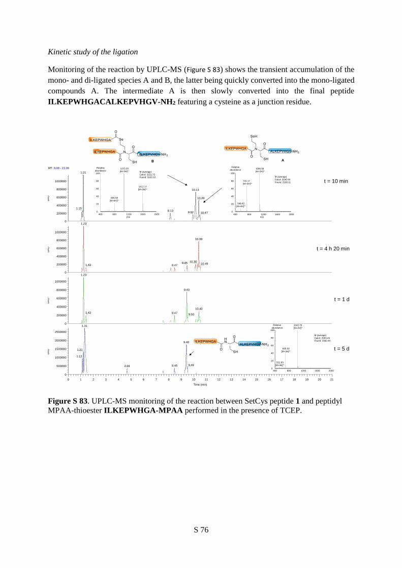
S 76
Kinetic study of the ligation
Monitoring of the reaction by UPLC-MS (Figure S 83) shows the transient accumulation of the
mono- and di-ligated species A and B, the latter being quickly converted into the mono-ligated
compounds A. The intermediate A is then slowly converted into the final peptide
ILKEPWHGACALKEPVHGV-NH2 featuring a cysteine as a junction residue.
Figure S 83. UPLC-MS monitoring of the reaction between SetCys peptide 1 and peptidyl
MPAA-thioester ILKEPWHGA-MPAA performed in the presence of TCEP.
RT: 0.00 - 21.00
0 1 2 3 4 5 6 7 8 9 10 11 12 13 14 15 16 17 18 19 20 21
Time (min)
0
500000
1000000
1500000
2000000
2500000
uA
U
0
200000
400000
600000
800000
1000000
uA
U
0
200000
400000
600000
800000
1000000
uA
U
0
200000
400000
600000
800000
1000000
uA
U
1.21
10.13
10.39
1.158.13
9.92 10.47
1.23
10.39
10.309.45 10.491.42 8.47
1.23
9.43
10.40
1.42 8.479.50
1.31
9.40
1.21
1.12
9.498.464.68
NL:
1.15E6
UV_VIS_1
UV 2
VD166-180-
C3
NL:
1.14E6
UV_VIS_1
UV 2
vd166-178-
c8
NL:
1.09E6
UV_VIS_1
UV 2
vd166-180-
c11
NL:
2.69E6
UV_VIS_1
UV 2
vd166-180-
c15
400 800 1200 1600 2000
m/z
0
20
40
60
80
100
1075.00
[M+3H]3+
1612.17
[M+2H]2+
806.58
[M+4H]4+
Relative
abundanceM (average)
Calcd: 3222.72
Found: 3222.22
400 800 1200 1600 2000m/z
0
20
40
60
80
100
1096.08
[M+2H]2+
731.17
[M+3H]3+
548.42
[M+4H]4+
M (average)
Calcd: 2190.50
Found: 2190.11
Relative
abundance
M (average)
Calcd: 2083.49
Found: 2083.44
400 800 1200 1600 20000
20
40
60
80
100
1042.75
[M+2H]2+
695.50
[M+3H]3+
521.83
[M+4H]4+
Relative
abundance
t = 10 min
t = 4 h 20 min
t = 1 d
t = 5 d

S 77
4.7 NCL under strong reducing conditions (peptide alkyl thioester, DTT, MPAA,
property 5)
When SetCys peptide 1 and peptide thioester 4 were reacted in the presence of the DTT, native
peptide 6 was produced with 55% yield (Figure S 84).
Figure S 84. Ligation of SetCys peptide 1 and peptide thioester 4 in the presence of DTT.
Protocol
To a solution of Gn·HCl (574 mg) in pH 7.4 0.1 M phosphate buffer (600 μL) were added DTT
(15.4 mg) and MPAA (33.6 mg) and the pH of the mixture was adjusted to 7.24 by addition of
6 M NaOH. Peptide thioester 4 (6.04 mg, 3.89 μmol, 1 eq) and SetCys peptide 1 (7.00 mg, 4.67
μmol, 1.2 eq) were then successively dissolved in this solution (487 μL) and the reaction
mixture was stirred at 37 °C. The reaction was monitored by HPLC (Figure S 85). After
completion of the reaction (4 days), the mixture was then acidified with 10% AcOH in water
(8 mL) and extracted with Et2O (5 ×) to remove the MPAA. Purification of the crude by HPLC
(eluent A: water containing 0.1% TFA, eluent B: acetonitrile containing 0.1% TFA, gradient:
0-10% eluent B in 5 min then 10-30% eluent B in 30 min, 6 mL/min, C18 column, rt, detection
at 215 nm) provided peptide 6 as a white solid after lyophilisation (5.83 mg, 55%).

S 78
Figure S 85. HPLC-MS monitoring of the reaction between SetCys peptide 1 and peptide
thioester 4 performed in the presence of DTT. LC trace: eluent A 0.10% TFA in water, eluent
B 0.10% TFA in CH3CN/water: 4/1 by vol. C18 Xbridge BEH 300 Å 5 μm (4.6 × 250 mm)
column, gradient 0-50% B in 15 min, 30 °C, 1 mL/min, UV detection (215 nm).
-50
125
250
375
500
625
800
0.0 2.0 4.0 6.0 8.0 10.0 12.0 15.0
-50
500
1 000
1 500
2 200
Time (min)
Intensity(UV, mAU)
a) t = 10 min

S 79
5 Stability and reactivity of N-(2-sulfanylethyl)Cys (SutCys) unit as a function
of the reducing power of the reaction mixture (Figure 2b)
The SutCys-ALKEPVHGV-NH2 peptide used as model compound in this study was
synthesized according to Ruff et al.8
5.1 Stability under strong reducing conditions
Figure S 86: Stability of the SutCys-ALKEPVHGV-NH2 peptide under strong reductive
conditions.
To a solution of Gn·HCl (287 mg) in 0.1 M phosphate buffer (300 μL) were added TCEP.HCl
(14.3 mg), sodium ascorbate (9.9 mg) and MPAA (16.8 mg) and the pH of the mixture was
adjusted to 6.95 by addition of 6 M NaOH. SutCys-ALKEPVHGV-NH2 peptide (0.23 mg,
0.16 μmol) was then dissolved in this solution (160 μL) and the reaction mixture was stirred at
37 °C.
The reaction was monitored by HPLC (Figure S 87). For each point, an 8 μL aliquot was taken
from the reaction mixture and quenched by adding 10% AcOH in water (100 μL). The sample
was then extracted with Et2O to remove MPAA prior to HPLC analysis.
The HPLC monitoring showed that the SutCys residue didn’t lose its sulfanylethyl arm, even
after several days of incubation in presence of TCEP. Neither increasing (from 6.95 to 7.92)
nor decreasing (from 6.95 to 6.06) the pH of the reaction mixture changed the outcome of the
reaction.

S 80
Figure S 87. Stability of the SutCys peptide in the presence of MPAA + TCEP (pH = 6.06).
LC trace: eluent A 0.10% TFA in water, eluent B 0.10% TFA in CH3CN/water: 4/1 by vol.,
C18 column, gradient 0-50% B in 15 min, 30 °C, 1 mL/min, UV detection at 215 nm.
5.2 NCL under low reducing conditions
When SutCys-ALKEPVHGV-NH2 peptide and peptidyl thioester 4 were reacted under mild
reducing conditions (MPAA), the resulting peptide featuring the SutCys unit in the form of a
cyclic disulfide at the ligation junction was recovered as the only product of the ligation with
53% yield (Figure S 88).
Time (min)
0.0 2.0 4.0 6.0 8.0 10.0 12.0 15.0
0
100
200
300
400
500
600
Intensity
(UV, mAU)
0
100
200
300
400
500
600

S 81
Figure S 88. Ligation reaction of SutCys-ALKEPVHGV-NH2 peptide with peptidyl MPA-
thioester 4 under low reducing conditions.
Protocol
To a solution of Gn·HCl (1.03 g) in 0.1 M, pH 7.4 phosphate buffer (1.08 mL) was added
MPAA (60.6 mg) and the pH of the mixture was adjusted to 7.21 by addition of 6 M NaOH.
SutCys-ALKEPVHGV-NH2 peptide (22.4 mg, 15.4 μmol, 1.2 eq) and RLKEPVHGA-MPA
peptide 4 (20.0 mg, 12.9 μmol, 1 eq) were then successively dissolved in this solution (1.61
mL) and the reaction mixture was stirred at 37 °C for 24 h. The mixture was then acidified with
10% AcOH in water (23 mL) and extracted with Et2O (5 ×) to remove the MPAA. Purification
of the crude by HPLC (eluent A: water containing 0.1% TFA, eluent B: acetonitrile in water
4/1 containing 0.1% TFA, 50°C, detection at 215 nm, 6 mL/min, 0-10% eluent B in 5 min, then
10-35% eluent B in 35 min, C18XBridge column) provided RLKEPVHGA-SutCys-
ALKEPVHGV-NH2 peptide as a white solid after lyophilisation (19.2 mg, 53%).

S 82
Figure S 89. Monitoring of the ligation between SutCys peptide SutCysALKEPVHGV-NH2
and peptidyl MPA thioester RLKEPVHGA-MPA. LC trace: eluent A 0.10% TFA in water,
eluent B 0.10% TFA in CH3CN/water: 4/1 by vol., C18 column, gradient 0-50% B in 15 min,
30 °C, 1 mL/min, UV detection at 215 nm.
0
100
200
300
400
500
600
Time (min)
0.0 2.0 4.0 6.0 8.0 10.0 12.0 15.0
0
100
200
300
400
500
600
Intensity
(UV, mAU)
0
100
200
300
400
500
a) t = 20 s
b) t = 5.3 h
b) t = 22 h
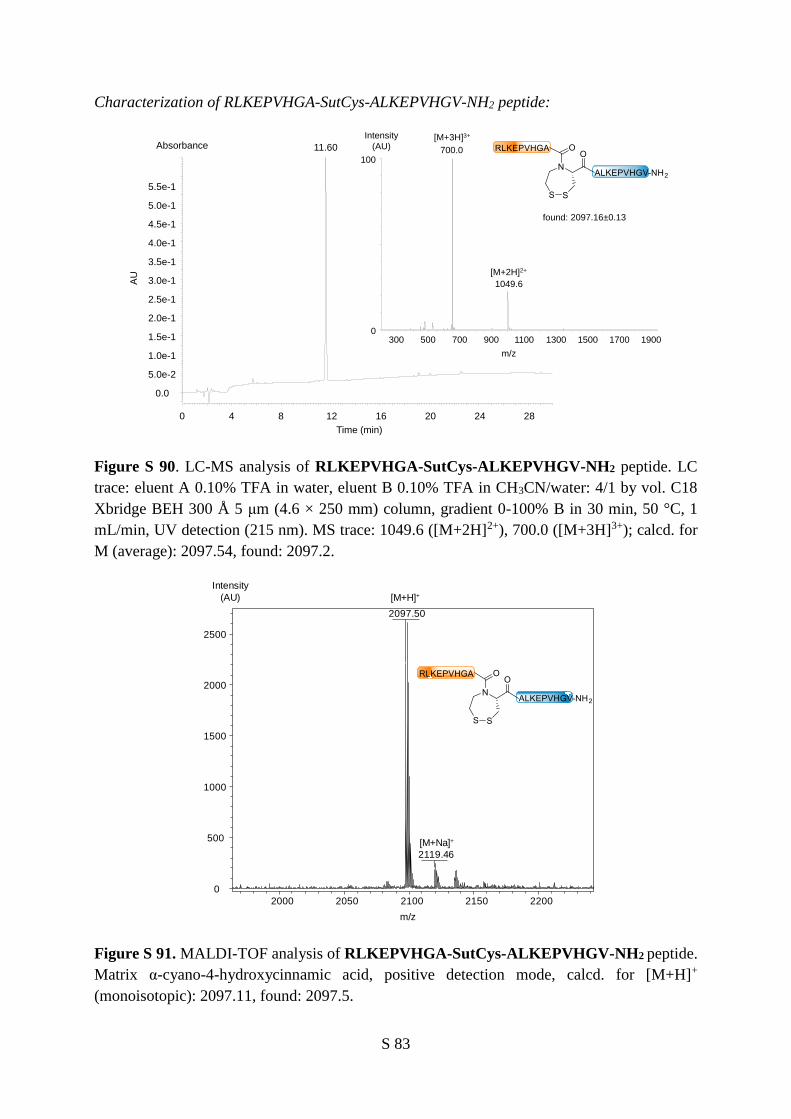
S 83
Characterization of RLKEPVHGA-SutCys-ALKEPVHGV-NH2 peptide:
Figure S 90. LC-MS analysis of RLKEPVHGA-SutCys-ALKEPVHGV-NH2 peptide. LC
trace: eluent A 0.10% TFA in water, eluent B 0.10% TFA in CH3CN/water: 4/1 by vol. C18
Xbridge BEH 300 Å 5 μm (4.6 × 250 mm) column, gradient 0-100% B in 30 min, 50 °C, 1
mL/min, UV detection (215 nm). MS trace: 1049.6 ([M+2H]2+), 700.0 ([M+3H]3+); calcd. for
M (average): 2097.54, found: 2097.2.
Figure S 91. MALDI-TOF analysis of RLKEPVHGA-SutCys-ALKEPVHGV-NH2 peptide.
Matrix α-cyano-4-hydroxycinnamic acid, positive detection mode, calcd. for [M+H]+
(monoisotopic): 2097.11, found: 2097.5.
Time (min)
0 4 8 12 16 20 24 28
AU
0.0
5.0e-2
1.0e-1
1.5e-1
2.0e-1
2.5e-1
3.0e-1
3.5e-1
4.0e-1
4.5e-1
5.0e-1
5.5e-1
11.60Absorbance
m/z
300 500 700 900 1100 1300 1500 1700 19000
100
found: 2097.16±0.13
700.0
1049.6
[M+3H]3+
[M+2H]2+
Intensity
(AU)
2097.50
2119.46
0
500
1000
1500
2000
2500
2000 2050 2100 2150 2200
m/z
Intensity
(AU) [M+H]+
[M+Na]+

S 84
5.3 NCL under strong reducing conditions
Figure S 92: Ligation reaction of SutCys-ALKEPVHGV-NH2 peptide with peptidyl MPA-
thioester 4 under strong reducing conditions.
Protocol
To a solution of Gn·HCl (573 mg) in 0.1 M phosphate buffer (0.6 mL) were successively added
TCEP (53.7 mg) and MPAA (33.6 mg) and the pH of the mixture was adjusted to 7.2 by addition
of 6 M NaOH. SutCys-ALKEPVHGV-NH2 (8.00 mg, 5.51 µmol, 1.25 eq) and peptidyl
thioester 4 (6.83 mg, 4.40 μmol, 1 eq) were then successively dissolved in this solution (0.55
mL) and the reaction mixture was maintained at 37 °C.
After 48 h reaction, a 2 μL aliquot was taken from the reaction mixture and quenched by adding
10% AcOH in water (100 μL). The sample was then extracted with Et2O to remove MPAA
prior to LC-MS analysis (Figure S 93). The ligation product (tR = 11.48 min) featuring the
SutCys unit in its reduced, ring-opened form was detected in the crude, in mixture with the
unreacted SutCys-ALKEPVHGV-NH2 peptide (tR = 10.10 min) and with the peptide resulting
from the hydrolysis of the starting peptide thioester (tR = 8.37 min).
The ligation product was isolated in the form of the cyclic disulfide after oxidation with iodine.
Experimentally, the ligation mixture was acidified with 10% AcOH in water (3.5 mL) and
extracted with Et2O (5 ×) to remove the MPAA. A solution of iodine (158 mg) in DMSO (2
mL) was then added dropwise until persistency of the yellow color of iodine. The excess of
iodine was then quenched with a solution of DTT (40 mg) in 20% AcOH (4 mL) and the mixture
was diluted with water (final volume: 10 mL). Purification of the crude by HPLC (eluent A:
water containing 0.1% TFA, eluent B: acetonitrile in water 4/1 containing 0.1% TFA, 50°C,
detection at 215 nm, 6 mL/min, 0-10% eluent B in 5 min, then 10-30% eluent B in 30 min,
C18XBridge column) provided RLKEPVHGA-SutCys-ALKEPVHGV-NH2 as a white
solide after lyophilisation (3.70 mg, 30%).

S 85
Figure S 93. LC-MS analysis of the ligation mixture. LC trace: eluent A 0.10% TFA in water,
eluent B 0.10% TFA in CH3CN/water: 4/1 by vol. C18 Xbridge BEH 300 Å 5 μm (4.6 × 250
mm) column, gradient 0-100% B in 30 min, 50 °C, 1 mL/min, UV detection (215 nm). MS
trace for SutCys ligated peptide (tR = 11.48 min): 1050.8 ([M+2H]2+), 700.8 ([M+3H]3+);
calcd. for M (average): 2099.55, found: 2099.6.
Time (min)
4 8 12 16 20
-4.0e-2
0.0
4.0e-2
8.0e-2
1.2e-1
1.6e-1
2.0e-1
2.4e-1
2.8e-1
11.48
10.10
8.37
m/z
300 500 700 900 1100
0
100
found: 2099.59±0.07
[M+3H]3+
700.8
1050.8
[M+2H]2+
Intensity
(AU)
Absorbance
0

S 86
6 Mechanistic studies (Figure 3)
6.1 Effect of pH (Figure 3b)
General procedure illustrated with the following conditions:
1 mM peptide, 200 mM MPAA, 100 mM TCEP, 100 mM ascorbate, pH 6.9
in 0.1 M phosphate buffer
To a solution of Gn·HCl (287 mg) in 0.1 M, pH 7.4 phosphate buffer (300 μL) were added
TCEP.HCl (14.3 mg), sodium ascorbate (9.9 mg) and MPAA (16.8 mg) and the pH of the
mixture was adjusted to 6.90 by addition of 6 M NaOH. SetCys peptide 1 (0.48 mg, 0.32 μmol)
was then dissolved in the buffered solution (320 μL) and the reaction mixture was stirred at 37
°C. The progress of the reaction was monitored by HPLC. For each point, an 8 μL aliquot was
taken from the reaction mixture and quenched by adding 10% AcOH in water (100 μL). The
sample was then extracted with Et2O to remove MPAA prior to HPLC analysis. The kinetic
data were then fitted to a pseudo-first order kinetic law using KinteK Explorer SoftwareTM
(Figure S 94).
Figure S 94. Fitting of the reaction leading to a loss of the N-selenoethyl appendage on SetCys
peptides at pH 6.90 using KinteK Explorer SoftwareTM (Version 7.2.180216., Kintek
Corporation - https://kintekcorp.com/software/). Red diamonds correspond to experimental
values / Gray line corresponds to fitted data.
0
20
40
60
80
100
120
0 500 1000 1500 2000 2500 3000
Co
nve
rsio
n (
%)
Time (min)
Cys peptide (fit)
Cys peptide (exp)

S 87
The reaction leading to a loss of the N-selenoethyl appendage on SetCys peptides was repeated
at various pH (4.99 < pH < 8.49). Corresponding rate constants are given in Table S 3.
Table S 3. Effect of the pH on the conversion of SetCys-ALKEPVHGV-NH2 1 into
CALKEPVHGV-NH2 3. Rate constants k1 were extracted using KinteK Explorer SoftwareTM.
pH k1 (min-1) standard error
4.99 0.00137 0.00083
5.25 0.00191 0.00074
5.54 0.00203 0.00061
5.98 0.00253 0.00043
6.52 0.00202 0.00126
6.90 0.00174 0.00024
7.22 0.00119 0.00080
7.55 0.00089 0.00003
8.03 0.00047 0.00005
8.49 0.00014 0.00003
6.2 Effect of Cys thiol: control experiment with a SetAla peptide (Figure 3d)
6.2.1 Decomposition of SetAla peptide into Ala peptide under strong reducing
conditions
Figure S 95. Decomposition of SetAla peptide into an Ala peptide.
To a solution of Gn·HCl (287 mg) in 0.1 M, pH 7.4 phosphate buffer (300 μL) were added
TCEP.HCl (14.3 mg), sodium ascorbate (9.9 mg) and MPAA (16.8 mg) and the pH of the
mixture was adjusted to 5.97 by addition of 6 M NaOH. SetAla peptide (0.47 mg, 0.16 μmol)
was then dissolved in the buffered solution (320 μL) and the reaction mixture was stirred at 37
°C. The progress of the reaction was monitored by HPLC (Figure S 96). For each point, an 8 μL
aliquot was taken from the reaction mixture and quenched by adding 10% AcOH in water (100
μL). The sample was then extracted with Et2O to remove MPAA prior to HPLC analysis. The

S 88
kinetic data were then fitted to a pseudo-first order kinetic law using KinteK Explorer
SoftwareTM (Figure S 97).
Figure S 96. Monitoring of the conversion of SetAla peptide 1 into Ala peptide using MPAA
+ TCEP. LC trace: eluent A 0.10% TFA in water, eluent B 0.10% TFA in CH3CN, C18 column,
gradient 0-50% B in 15 min, 30 °C, 1 mL/min, UV detection at 215 nm.
Time (min)
0.0 2.0 4.0 6.0 8.0 10.0 12.0 15.0
0
125
250
375
500
625
0
100
200
300
400
Intensity
(UV, mAU)
0.0 2.0 4.0 6.0 8.0 10.0 12.0 15.0
0
100
200
300
400a) t = 1.5 h
b) t = 22 h
b) t = 142 h
Se=TECP

S 89
Figure S 97. Fitting of the reaction leading to a loss of the N-selenoethyl appendage on SetAla
peptides at pH 5.97 using KinteK Explorer SoftwareTM (Version 7.2.180216., Kintek
Corporation - https://kintekcorp.com/software/). k = 0.000296 ± 0.000091. Red diamonds
correspond to experimental values / Black line corresponds to fitted data.
The AALKEPVHGV-NH2 peptide produced by decomposition of SetAla peptide during the
reaction was purified by HPLC and its sequence was confirmed by MS-MS analysis (Figure S
98 and Figure S 99).
Figure S 98. MALDI-TOF analysis of AALKEPVHGV-NH2 peptide. Matrix α-cyano-4-
hydroxycinnamic acid, positive detection mode, calcd. for [M+H]+ (monoisotopic): 1019.60,
found: 1019.53.
0
10
20
30
40
50
60
70
80
90
100
0 2000 4000 6000 8000 10000
Co
nve
rsio
n (
%)
Time (min)
AALKEPVHGV-NH2 (exp)
AALKEPVHGV-NH2 (fit)
1019.532
1057.462
1041.485
1005.799
0
2
4
6
8
4x10
960 980 1000 1020 1040 1060 1080 1100 1120 1140
m/z
[M+H]+
[M+Na]+
[M+K]+
Intensity
(AU)
AALKEPVHGV-NH2

S 90
Figure S 99. MALDI-TOF-TOF sequencing of AALKEPVHGV-NH2 peptide. Matrix α-
cyano-4-hydroxycinnamic acid, positive detection mode. The MS-MS analysis confirms that
the SetAla residue is converted into the Ala amino acid in the peptide.
6.2.2 Ligation of SetAla peptide under strong reducing conditions
Figure S 100. Ligation reaction of SetAla peptide with peptidyl MPA-thioester 4.

S 91
Protocol
To 500 µL of a 6M solution of Gn·HCl in 0.1 M pH 7.4 phosphate buffer were successively
added MPAA (16.8 mg), TCEP (14.3 mg) and sodium ascorbate (9.9 mg). The pH of the
mixture was adjusted to 7.28 by addition of 6 M NaOH (61,5 µL). In 140 µL of the previous
solution were dissolved SetAla-ALKEPVHGV-NH2 peptide (1.97 mg, 1.2 eq) and
RLKEPVHGA-MPA thioester (1.73 mg, 1 eq). A pH of 6.90 was measured after addition of
the peptide segments. The reaction mixture was stirred at 37 °C and monitored by HPLC (Figure
S 101). For each point, a 2 μL aliquot was taken from the reaction mixture and quenched by
adding 10% AcOH in water (100 μL). The sample was then extracted with Et2O to remove
MPAA prior to HPLC analysis.
Figure S 101. Monitoring of the reaction between a SetAla peptide and a peptidyl MPA-
thioester in the presence of MPAA, TCEP and sodium ascorbate. LC trace: eluent A 0.10%
TFA in water, eluent B 0.10% TFA in CH3CN, C18 column, gradient 0-50% B in 15 min, 30 °C,
1 mL/min, UV detection at 215 nm.
5.0 6.3 7.5 8.8 10.0 11.3 12.5 13.8 15.0min
t0
t2h40
t7days

S 92
7 Kinetic model of SetCys-mediated NCL under strong reducing conditions (Figure 4)
SetCys peptide 1 and peptidyl thioester 4 can be assembled in the presence of TCEP into ligated
product 6 having a cysteine as a junction residue. Such a reaction implies that the N-selenoethyl
appendage of the SetCys unit is lost during the process.
A kinetic study of the ligation shows also the transient accumulation of species 5 at the
beginning of the reaction (Figure S 102).
Figure S 102. HPLC monitoring of the reaction between SetCys peptide 1 and peptide thioester
4 performed in the presence of TCEP. For each point, a 2 μL aliquot was taken from the reaction
mixture and quenched by adding 10% AcOH in water (100 μL). The sample was then extracted
with Et2O to remove MPAA prior to HPLC analysis. The proportion of each species in the
mixture was determined by HPLC using UV detection at 215 nm.
On the basis of the preceding observations, we have proposed the following mechanistic model
(Figure S 103).
0
10
20
30
40
50
60
70
80
90
100
0 500 1000 1500 2000 2500 3000 3500 4000 4500
con
vers
ion
(%)
Time (min)
Rdt RLKEPVHGA-NH(Se,S)AzeALKEPVHGV-NH2
Rdt RLKEPVHGA-CALKEPVHGV-NH2
Fraction RLKEPVHGA-MPA
Fraction RLKEPVHGA-OH
Peptide intermediate 5
Ligated peptide 6
Peptide thioester 4
Hydrolyzed peptide thioester

S 93
Figure S 103. Mechanistic model for the NCL of SetCys peptides
The purpose of this model is to clarify the following points:
- Determination of rate constants and identification of the rate-limiting step.
- Are the kinetic parameters extracted from the model in accord with experimental data?
- Based on the fact that SetCys peptide 1 evolves into Cys peptide 3, is such a reaction
also possible from intermediate 5 to generate ligated products at Cys of type 6?
Methodological approach
In order to determine the rate constants involved in the different steps of this complex process,
the system was decomposed into simpler reactions, wherefrom kinetic parameters were
extracted using KinteK Explorer SoftwareTM (Version 7.2.180216, Kintek Corporation -
https://kintekcorp.com/software/).
7.1 Rate constants for a classical NCL (k+2, k-2 and k4, see Figure 4c)
The kinetic profile of an NCL reaction was first studied in order to determine the rate constants
of the thiol-thioester exchange step (k+2 and k-2) and of the capture of the transient arylthioester
by a cysteinyl peptide (k4) (Figure S 104). Importantly, a model peptidyl thioester possessing a
C-terminal alanyl residue was used in this study.

S 94
Figure S 104. Kinetic study of the NCL reaction at submillimolar peptide concentration
At the peptide millimolar concentration range, NCL reactions are under the kinetic control of
thiol-thioester exchanges. Such conditions do not allow to observe the accumulation of the
transient aryl thioester 7 and make the determination of k4 rate constant impossible.
In order to extract second-order rate constants, the thioester and the Cys peptide were reacted
at a respective concentration of 0.1 mM and 0.15 mM (in phosphate buffer 0.1 M, 6 M Gn.HCl,
200 mM MPAA, 100 mM TCEP, pH 7.2, 37°C). 50 µL of the reaction mixture were collected
every ~20 minutes and added to 50 µL of a 50% acetic acid aqueous solution. The sample was
extracted three times by Et2O and the aqueous layer was analyzed by RP-HPLC (eluent A 0.1%
TFA in water, eluent B 0.1% TFA in CH3CN/water: 4/1 by vol. C18 Xbridge BEH 300 Å 5 μm
(4.6 × 250 mm) column, gradient 0-50% B in 15 min, 1 mL/min, 30° C, detection at 215 nm)
The chromatograms were processed on the basis of the absorbance signal at 215 nm. All
datasets were fitted with an exponential analytical function to obtain an estimate of the standard
deviation. A subsequent global fit allowed determining rate constants k+2, k-2, k4 (Table S 4 and
Figure S 105).
Table S 4. Second-order rate constants for the model NCL reaction
value (M-1.min-1) Std error
k+2 0.257 0.006
k-2 129 17
k4 243 3

S 95
Figure S 105. Fitting of the NCL reaction at submillimolar concentration of peptides using
KinteK Explorer SoftwareTM (Version 7.2.180216., Kintek Corporation -
https://kintekcorp.com/software/). Red squares / Blue diamonds correspond to experimental
values.
7.2 Rate constant for the loss of the N-selenoethyl appendage (k1, Figure 4c)
The experimental approach for this reaction is documented in full in section 4.1 of the SI. Note
that the reduction of SetCys peptide 1 by TCEP was neglected since this reaction proceeds
almost instantly at pH 7.2.
As previously determined, k1 = 0.00119 ± 0.00080 min-1 at pH 7.2, 37 °C.
Figure S 106. Mechanistic model for the loss of the N-selenoethyl appendage on SetCys
peptides.
7.3 Kinetic model for SetCys-mediated NCL (Figure 4b,c)
The rate constants determined in the model NCL and arm loss reactions (i.e. k+2, k-2, k1, k4)
were used to fit kinetic data collected for the NCL of SetCys peptides (Figure S 107) with the
model proposed in Figure S 103 and to determine k+3, k-3 and k5 (Table S 5).
0
10
20
30
40
50
60
70
80
90
100
0 200 400 600 800 1000 1200
Co
nve
rsio
n (
%)
Time (min)
MPAA thioester (fit)
Ligation product (fit)
MPAA thioester (exp)
Ligation product (exp)

S 96
Figure S 107. Ligation of SetCys peptide 1 with MPA-thioester 4 in NCL standard conditions.
RP-HPLC monitoring of the formation of peptides 5 (black triangles) and 6 (brown circles)
throughout the course of the reaction. The continuous lines correspond to the fitting of
experimental data using KinteK Explorer SoftwareTM (Version 7.2.180216., Kintek Corporation
- https://kintekcorp.com/software/).
Table S 5. Rate constants for the model NCL reaction of SetCys peptides.
Value Std err
k1 0.0012 min-1 0.0008
k+2 0.257 M-1.min-1 0.006
k-2 129 M-1. min-1 17
k+3 182 M-1. min-1 49
k-3 0.0632 M-1.min-1 0.0314
k4 243 M-1.min-1 3
k5 0 min-1 nd
Note that according to the proposed model, best fit is obtained for a value of k5 being null which
excludes the loss of the N-selenoethyl appendage from the ligated product.
0
10
20
30
40
50
60
70
80
90
100
0 1000 2000 3000 4000 5000
Co
nve
rsio
n (%
)
Time (min)

S 97
8 Synthesis of cyclic peptides (Figure 5a,b)
Figure S 108. One-pot synthesis of cyclic peptides 10, 11, cK1-1 and cK1-2.
8.1 Synthesis of cyclic peptide 10
To a solution of Gn·HCl (287 mg) in 0.1 M, pH 7.4 phosphate buffer (300 μL) was added
MPAA (16.8 mg) and the pH of the mixture was adjusted to 7.20 by addition of 6 M NaOH.
Peptide 9a (4.50 mg, 1.82 μmol, 1 eq) and peptide 8a (2.84 mg, 1.82 μmol, 1 eq) were then
successively dissolved in the MPAA solution (228 μL) and the mixture was stirred at 37 °C.
The progress of the reaction leading to the bifunctional peptide intermediate was followed by
HPLC (Figure S 109).
After completion of the NCL ligation (3 h), the second step of the process leading to the
cyclisation of the peptide was induced by addition of TCEP. To a solution of Gn·HCl (1.32 g)
in 0.1 M, pH 7.4 phosphate buffer (1.38 mL) were added TCEP∙HCl (87.7 mg), sodium
ascorbate (60.6 mg) and MPAA (77.4 mg) and the pH of the mixture was adjusted to 5.51 by
addition of 6 M NaOH. The previous ligation mixture containing the bifunctional peptide

S 98
intermediate was then diluted with the solution of TCEP (2.05 mL) and the mixture was stirred
at 37 °C. After completion of the reaction (40 h), the mixture was acidified with 10 % AcOH
in water (5 mL) and diluted with water (2.5 mL). The mixture was then extracted with Et2O (5
×) to remove the MPAA and purified by HPLC (eluent A = water containing 0.1% TFA, eluent
B=acetonitrile in water 4/1 containing 0.1% TFA, 50°C, detection at 215 nm, 6 mL/min, 0-10%
eluent B in 5 min, then 10-35% eluent B in 30 min, C18XBridge column) to give the cyclic
peptide 10 as a with solid after lyophilization (1.65 mg, 27%).
Figure S 109. Assembly of the cyclic peptide 10 monitored by LC-MS (a and b) and HPLC (c).
a) Analysis of the reaction mixture 190 min after mixing the two partners of the ligation
reaction. b) Analysis of the reaction mixture 16 h after the addition of TCEP. c) Analysis of the
reaction mixture 22 h after the addition of TCEP. LC trace: eluent A 0.10% TFA in water,
eluent B 0.10% TFA in CH3CN/water: 4/1 by vol., C18 column, gradient 0-50% B in 15 min,
50 °C (for a and b) or 30 °C (for c), 1 mL/min, UV detection at 215 nm.
0.00 3.00 6.00 9.00 12.00 15.00
0.0
2.0e-1
4.0e-1
6.0e-1
11.47
Intensity
(UV, AU)
m/z
400 600 800 1000 1200 1400 1600 1800
0
100
50M (average)
Calcd: 3147.5
Found: 3147.8
Relative
abundance
1575.1
[M+2H]2+
1050.3
[M+3H]3+
788.0
[M+4H]4+
Time (min)
0.00 3.00 6.00 9.00 12.00 15.00
0.0
2.0e-1
4.0e-1 10.89
Intensity
(UV, AU)
m/z
400 600 800 1000 1200 1400 1600 18000
100599.6
M (average)
Calcd: 2907.3
Found: 2907.2
Relative
abundance970.1
[M+3H]3+
1454.5
[M+2H]2+
727.9
[M+4H]4+
50
0.0 2.0 4.0 6.0 8.0 10.0 12.0 15.0
-50
125
250
375
500
625
800
Time (min)
Intensity
(UV, mAu)
b)
c)

S 99
Figure S 110. LC-MS analysis of purified cyclic peptide 10. LC trace: eluent A 0.10% TFA in
water, eluent B 0.10% TFA in CH3CN/water: 4/1 by vol. C18 column, gradient 0-100% B in
30 min, 50 °C, 1 mL/min, light scattering detection. MS trace: m/z 1455.3 ([M+2H]2+), 970.0
([M+3H]3+), 727.8 ([M+4H]+); calcd. for M (average): 2907.3, found: 2907.1.
Time (min)
0.00 5.00 10.00 15.00 20.00 25.00 30.00
0.000
0.100
0.200
0.300
0.400
0.500
0.600
0.700
0.800
10.88
Intensity
(light scattering, AU)
m/z
200 400 600 800 1000 1200 1400 1600 1800
0
100
found: 2907.1
970.0
727.8
1455.3
Intensity
(UA)
[M+4H]4+
[M+3H]3+
[M+2H]2+

S 100
Figure S 111. MALDI-TOF analysis of cyclic peptide 10. Matrix α-cyano-4-hydroxycinnamic
acid, positive detection mode, calcd. for [M+H]+ (monoisotopic): 2906.40, found: 2906.1.
8.2 Synthesis of cyclic peptide 11
To a solution of Gn·HCl (287 mg) in 0.1 M, pH 7.4 phosphate buffer (300 μL) was added
MPAA (16.8 mg) and the pH of the mixture was adjusted to 7.20 by addition of 6 M NaOH.
peptide 9a (4.50 mg, 1.82 μmol, 1 eq) and peptide 8b (2.61 mg, 1.82 μmol, 1 eq) were then
successively dissolved in the MPAA solution (228 μL) and the mixture was stirred at 37 °C.
The reaction yielding the bifunctional peptide intermediate was monitored by HPLC.
After completion of the NCL ligation (3.5 h), the second step of the process leading to the
cyclisation of the peptide was induced by addition of TCEP. To a solution of Gn·HCl (1.32 g)
in 0.1 M, pH 7.4 phosphate buffer (1.38 mL) were added TCEP∙HCl (87.7 mg), sodium
ascorbate (60.6 mg) and MPAA (77.4 mg) and the pH of the mixture was adjusted to 5.50 by
addition of 6 M NaOH. The previous ligation mixture containing the bifunctional peptide
intermediate was then diluted with the solution of TCEP (2.05 mL) and the mixture was stirred
at 37 °C. After completion of the reaction (4 days), the mixture was acidified with 10 % AcOH
in water (7.5 mL) and diluted with water (10 mL). The crude was then extracted with Et2O (5
×) to remove the MPAA and purified by HPLC (eluent A = water containing 0.1% TFA, eluent
B=acetonitrile in water 4/1 containing 0.1% TFA, 50°C, detection at 215 nm, 6 mL/min, 0-5%
eluent B in 3 min, then 5-35% eluent B in 32 min, C18XBridge column) to give the cyclic
peptide 11 as a with solid after lyophilization (1.65 mg, 28%).
2906.1
0
1
2
3
4
5
4x10
750 1000 1250 1500 1750 2000 2250 2500 2750 3000 3250
m/z
[M+H]+
Intensity
(AU)
2906.1
2928.1 2944.0
0
1
2
3
4
5
x104
2860 2880 2900 2920 2940 2960
m/z
[M+H]+Intensity
(AU)
[M+Na]+ [M+K]+

S 101
Figure S 112. Assembly of the cyclic peptide 11 monitored by LC-MS. a) Analysis of the
reaction mixture 190 min after mixing the two partners of the ligation reaction. b) Analysis of
the reaction mixture 4 d after the dilution with TCEP solution. LC trace: eluent A 0.10% TFA
in water, eluent B 0.10% TFA in CH3CN/water: 4/1 by vol. C18 column, gradient 0-50% B in
15 min, 50 °C, 1 mL/min, UV detection at 215 nm.
Time (min)
0.00 3.00 6.00 9.00 12.00 15.00
-1.0e-1
0.0
3.0e-1
6.0e-1
9.0e-1
10.39
0.0
2.0e-1
4.0e-1
6.0e-1
8.0e-1
11.45
Intenisty
(UV, AU)
m/z
400 600 800 1000 1200 1400 1600 18000
100
1625.0
[M+2H]2+
M (average)
Calcd: 3246.5
Found: 3245.9
1083.3
[M+3H]3+
50
Relative
abundance
m/z
400 600 800 1000 1200 1400 1600 18000
100
1503.9
[M+2H]2+
M (average)
Calcd: 3006.3
Found: 3006.0
Relative
abundance1002.9
[M+3H]3+
a)
b)

S 102
Figure S 113. LC-MS analysis of purified cyclic peptide 11. LC trace: eluent A 0.10% TFA in
water, eluent B 0.10% TFA in CH3CN/water: 4/1 by vol. C18 Xbridge BEH 300 Å 5 μm (4.6
× 250 mm) column, 30 °C, gradient 0-50% B in 15 min, 1 mL/min, light scattering detection.
MS trace: m/z = 1503.7 ([M+2H]2+), 1002.9 ([M+3H]3+); calcd for M: 3006.3, found: 3005.9.
Time (min)
0.00 2.00 4.00 6.00 8.00 10.00 12.00 14.00
0.000
0.200
0.400
0.600
0.800
1.000
10.32
Intensity
(light scattering, AU)
m/z
400 600 800 1000 1200 1400 1600 18000
100
found 3005.9
1002.9
1503.7
Intensity
(AU) [M+3H]3+
[M+2H]2+
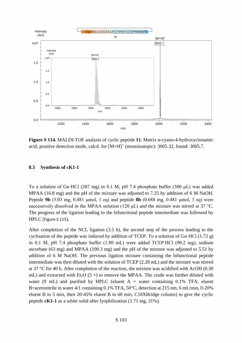
S 103
Figure S 114. MALDI-TOF analysis of cyclic peptide 11. Matrix α-cyano-4-hydroxycinnamic
acid, positive detection mode, calcd. for [M+H]+ (monoisotopic): 3005.32, found: 3005.7.
8.3 Synthesis of cK1-1
To a solution of Gn·HCl (287 mg) in 0.1 M, pH 7.4 phosphate buffer (300 μL) was added
MPAA (16.8 mg) and the pH of the mixture was adjusted to 7.25 by addition of 6 M NaOH.
Peptide 9b (5.03 mg, 0.481 μmol, 1 eq) and peptide 8b (0.688 mg, 0.481 μmol, 1 eq) were
successively dissolved in the MPAA solution (120 μL) and the mixture was stirred at 37 °C.
The progress of the ligation leading to the bifunctional peptide intermediate was followed by
HPLC (Figure S 115).
After completion of the NCL ligation (3.5 h), the second step of the process leading to the
cyclisation of the peptide was induced by addition of TCEP. To a solution of Gn·HCl (1.72 g)
in 0.1 M, pH 7.4 phosphate buffer (1.80 mL) were added TCEP∙HCl (90.2 mg), sodium
ascorbate (63 mg) and MPAA (100.3 mg) and the pH of the mixture was adjusted to 5.51 by
addition of 6 M NaOH. The previous ligation mixture containing the bifunctional peptide
intermediate was then diluted with the solution of TCEP (2.28 mL) and the mixture was stirred
at 37 °C for 40 h. After completion of the reaction, the mixture was acidified with AcOH (0.30
mL) and extracted with Et2O (5 ×) to remove the MPAA. The crude was further diluted with
water (9 mL) and purified by HPLC (eluent A = water containing 0.1% TFA, eluent
B=acetonitrile in water 4/1 containing 0.1% TFA, 50°C, detection at 215 nm, 6 mL/min, 0-20%
eluent B in 5 min, then 20-45% eluent B in 60 min, C18XBridge column) to give the cyclic
peptide cK1-1 as a white solid after lyophilization (1.71 mg, 31%).
3005.7
0.0
0.5
1.0
1.5
x104
2200 2400 2600 2800 3000 3200 3400
m/z
Intensity
(AU)
3005.7
0.0
0.5
1.0
1.5
x104
2960 2980 3000 3020 3040 3060
m/z
[M+H]+
Intensity
(AU)
[M+H]+
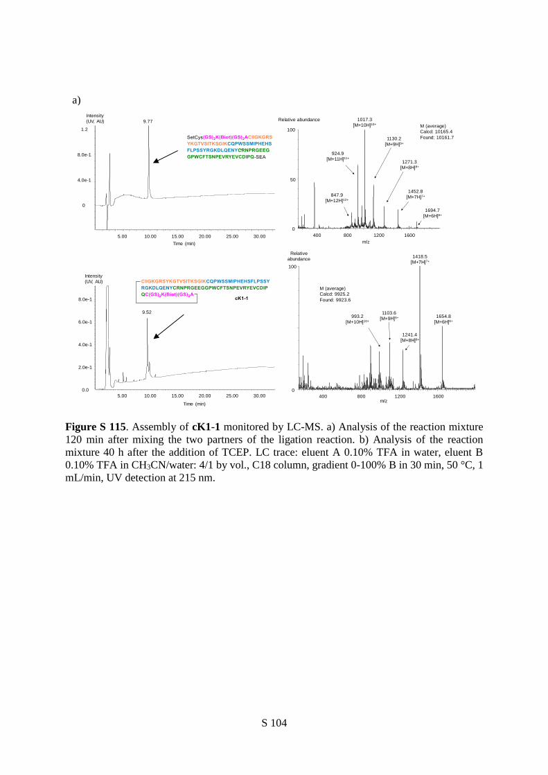
S 104
Figure S 115. Assembly of cK1-1 monitored by LC-MS. a) Analysis of the reaction mixture
120 min after mixing the two partners of the ligation reaction. b) Analysis of the reaction
mixture 40 h after the addition of TCEP. LC trace: eluent A 0.10% TFA in water, eluent B
0.10% TFA in CH3CN/water: 4/1 by vol., C18 column, gradient 0-100% B in 30 min, 50 °C, 1
mL/min, UV detection at 215 nm.
Time (min)
5.00 10.00 15.00 20.00 25.00 30.00
0
4.0e-1
8.0e-1
1.2
9.77
Intensity
(UV, AU)
Time (min)
5.00 10.00 15.00 20.00 25.00 30.00
0.0
2.0e-1
4.0e-1
6.0e-1
8.0e-1
9.52
Intensity
(UV, AU)
m/z
400 800 1200 1600
0
100
50
1017.3
[M+10H]10+
1130.2
[M+9H]9+
1452.8
[M+7H]7+
1694.7
[M+6H]6+
M (average)
Calcd: 10165.4
Found: 10161.7
Relative abundance
1271.3
[M+8H]8+
847.9
[M+12H]12+
924.9
[M+11H]11+
400 800 1200 1600
0
100
993.2
[M+10H]10+
1103.6
[M+9H]9+
1418.5
[M+7H]7+
1654.8
[M+6H]6+
M (average)
Calcd: 9925.2
Found: 9923.6
Relative
abundance
1241.4
[M+8H]8+
m/z
a)

S 105
a)
b)
Figure S 116. LC-MS analysis of cK1-1. a) LC trace. Eluent A 0.10% TFA in water, eluent B
0.10% TFA in CH3CN, ACQUITY UPLC Peptide BEH 300Å 1.7 μm (2.1 × 150 mm) column,
50 °C, gradient 0-70% B in 20 min, 0.4 mL/min, UV detection. b) MS trace: m/z = 1655.25
([M+6H]6+), 1419.00 ([M+7H]7+), 1241.83 ([M+8H]8+), 1104.00 ([M+10H]10+); calcd. for M
(average): 9925.19, found: 9926.28.
8.3.1 Characterization of the cyclic backbone structure of cK1-1
Reduction-alkylation
cK1-1 (50 µg) was treated with a solution of iodoacetamide in 0.025 M ammonium bicarbonate
(10 mg/mL, 50 µL) for 15 min. The alkylation step was monitored by MALDI-TOF mass
spectrometry.
0 2 4 6 8 10 12 14 16 18 20
Time (min)
0
100000
200000
300000
400000
500000
10.78
:
300 500 700 900 1100 1300 1500 1700 1900m/z
0
20
40
60
80
1001419.00
1241.83
1655.25
1104.00
Relative abundance
(%)

S 106
Figure S 117. Monitoring of the cK1-1 alkylation step by MALDI-TOF mass spectrometry
(matrix: 2,5-dihydroxybenzoic acid).
Trypsin cleavage
Trypsin (0.1 mg/mL, 0.5 µL) was added to the mixture to cleave the alkylated cyclic peptide.
The fragments resulting from the enzymatic cleavage were identified by MALDI-TOF MS
(Figure S 118a) and the MS-MS sequencing of the ion at m/z 2756.36 confirmed the Gln-Cys
and Ala-Cys junctions formed during the cyclisation process (Figure S 118b).

S 107
a)
b)
Figure S 118. MALDI-TOF TOF sequencing of cyclic peptide cK1-1 after alkylation and
trypsin cleavage using 2,5-dihydroxybenzoic acid as matrix (positive reflector mode). a) MS
analysis of the enzymatic lysate (after 90 min). b) MS-MS sequencing of the ion at m/z 2756.36.
m/z5 0 0 1 0 0 0 1 5 0 0 2 0 0 0 2 5 0 0 3 0 0 0
0
1 0
2 0
3 0
4 0
5 0
6 0
7 0
8 0
9 0
1 0 0
Ab s. In t. * 1 0 0 0
b I I
y I A S G S G K* S G S G C Q P I D C V
2 9 2 .8 7
b 2
2 0 3 .9 4
y 2
3 1 6 .9 4
y 3
6 6 6 .7 7
b 5
5 8 9 .9 0
y 5
7 5 1 .8 0
a 6
7 7 9 .8 1
b 6
6 6 0 .9 1
y 6
7 4 8 .9 1
y 7
8 0 4 .8 7
y 8
8 9 1 .8 8
y 9
9 4 8 .8 8
y 1 0
1 3 0 2 .9 6
y 1 1
1 3 8 9 .9 7
y 1 2
1 4 4 7 .0 1
y 1 3
1 8 0 7 .0 9
b 1 4
1 5 3 4 .0 6
y 1 4
1 5 9 1 .0 8
y 1 5
1 7 5 1 .1 8
y 1 6
1 8 7 9 .3 4
y 1 7
1 9 7 6 .4 5
y 1 8
2 0 8 9 .6 1
y 1 9
2 2 0 4 .7 6
y 2 0 2 4 3 9 .2 0
b 2 1
2 3 6 4 .9 7
y 2 1
2 5 5 3 .0 0
b 2 2
2 4 6 4 .6 2
y 2 22 7 1 0 .9 2
a 2 4
2 7 5 6 .4 4
y 2 4

S 108
8.4 Synthesis of cK1-2
To a solution of Gn·HCl (287 mg) in 0.1 M, pH 7.4 phosphate buffer (300 μL) was added
MPAA (16.8 mg) and the pH of the mixture was adjusted to 7.26 by addition of 6 M NaOH (31
μL). Peptide 9b (5.00 mg, 0.479 μmol, 1 eq) and peptide 8c (0.822 mg, 0.479 μmol, 1 eq) were
successively dissolved in the MPAA solution (120 μL) and the mixture was stirred at 37 °C.
The progress of the ligation leading to the bifunctional peptide intermediate was followed by
HPLC (Figure S 119).
After completion of the NCL ligation (3.5 h), the second step of the process leading to the
cyclisation of the peptide was induced by addition of TCEP in the reaction mixture. To a
solution of Gn·HCl (1.72 g) in 0.1 M, pH 7.4 phosphate buffer (1.80 mL) were added
TCEP.HCl (90.1 mg), sodium ascorbate (62.4 mg) and MPAA (100.8 mg) and the pH of the
mixture was adjusted to 5.52 by addition of 6 M NaOH (260 μL). The previous ligation mixture
containing the bifunctional peptide intermediate was then diluted with this solution of TCEP
(2.28 mL) and the reaction mixture was stirred at 37 °C for 40 h. After completion of the
reaction, the mixture was acidified with AcOH (0.30 mL) and extracted with Et2O (5 ×) to
remove the MPAA. The crude was further diluted with water (9 mL) and purified by HPLC
(eluent A = water containing 0.1% TFA, eluent B=acetonitrile in water 4/1 containing 0.1%
TFA, 50°C, detection at 215 nm, 6 mL/min, 0-20% eluent B in 5 min, then 20-45% eluent B in
60 min, C18XBridge column) to give the cyclic peptide cK1-2 as a white solid after
lyophilization (2.04 mg, 37%).
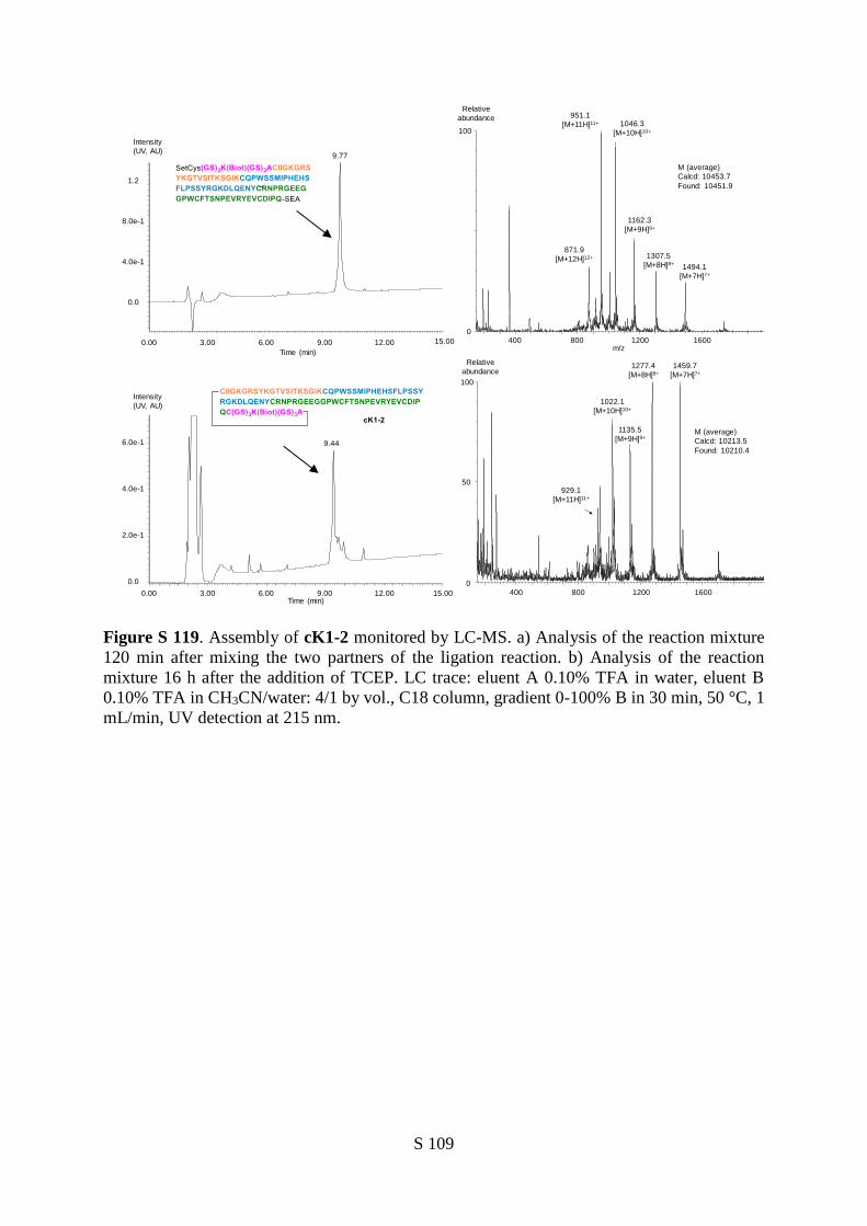
S 109
Figure S 119. Assembly of cK1-2 monitored by LC-MS. a) Analysis of the reaction mixture
120 min after mixing the two partners of the ligation reaction. b) Analysis of the reaction
mixture 16 h after the addition of TCEP. LC trace: eluent A 0.10% TFA in water, eluent B
0.10% TFA in CH3CN/water: 4/1 by vol., C18 column, gradient 0-100% B in 30 min, 50 °C, 1
mL/min, UV detection at 215 nm.
m/z400 800 1200 1600
0
1001046.3
[M+10H]10+
1162.3
[M+9H]9+
1494.1
[M+7H]7+
M (average)
Calcd: 10453.7
Found: 10451.9
1307.5
[M+8H]8+
871.9
[M+12H]12+
951.1
[M+11H]11+
Relative
abundance
400 800 1200 1600
0
100
1277.4
[M+8H]8+
1135.5
[M+9H]9+
1459.7
[M+7H]7+
M (average)
Calcd: 10213.5
Found: 10210.4
1022.1
[M+10H]10+
929.1
[M+11H]11+
Relative
abundance
50
Time (min)0.00 3.00 6.00 9.00 12.00 15.00
0.0
2.0e-1
4.0e-1
6.0e-1 9.44
Intensity
(UV, AU)
Time (min)
0.00 3.00 6.00 9.00 12.00 15.00
0.0
4.0e-1
8.0e-1
1.2
9.77
Intensity
(UV, AU)

S 110
a)
b)
Figure S 120. LC-MS analysis of cK1-2. a) LC trace. Eluent A 0.10% TFA in water, eluent B
0.10% TFA in CH3CN, ACQUITY UPLC Peptide BEH 300Å 1.7 μm (2.1 × 150 mm) column,
50 °C, gradient 0-70% B in 20 min, 0.4 mL/min, UV detection. b) MS trace: m/z = 1702.83
([M+6H]6+), 1459.92 ([M+7H]7+), 1277.67 ([M+8H]8+), 1135.83 ([M+9H]9+), 1022.33
([M+10H]10+); calcd. for M: 10213.45, found: 10212.71.
0 2 4 6 8 10 12 14 16 18 20
Time (min)
0
500000
1000000
1500000
2000000
2500000
10.69
300 500 700 900 1100 1300 1500 1700 1900
m/z
0
20
40
60
80
1001459.92
1277.67
1135.831702.83
1022.33
Relative abundance
(%)

S 111
8.4.1 Characterization of the cyclic backbone structure of cK1-2
Reduction-alkylation
cK1-2 (20 µg) was dissolved in a solution of dithiothreitol in 0.025 M ammonium bicarbonate
(1 mg/mL, 20 µL) and the mixture was treated with iodoacetamide (10 mg/mL in 0.025 M
ammonium bicarbonate, 20 µL) for 15 min. The alkylation step was monitored by MALDI-
TOF mass spectrometry.
Figure S 121. Monitoring of the cK1-2 alkylation step by MALDI-TOF mass spectrometry
(matrix: 2,5-dihydroxybenzoic acid).

S 112
Trypsin cleavage
Trypsin (0.1 mg/mL, 1 µL) was added to the mixture to cleave the alkylated cyclic peptide.
The fragments resulting from the enzymatic cleavage were identified by MALDI-TOF MS
(Figure S 122a) and the MS-MS sequencing of the ion at m/z 3044.05 confirmed the Gln-Cys
and Ala-Cys junctions formed during the cyclisation process (Figure S 122b).
a)
b)
Figure S 122. MALDI-TOF TOF sequencing of cyclic peptide cK1-2 after alkylation and
trypsin cleavage using 2,5-dihydroxybenzoic acid as matrix (positive reflector mode). a) MS-
analysis of the enzymatic lysate. b) MS-MS sequencing of the ion at m/z 3044.05.
m/z5 0 0 1 0 0 0 1 5 0 0 2 0 0 0 2 5 0 0 3 0 0 0 3 5 0 0
0
2
4
6
8
1 0
1 2
1 4
1 6
1 8
Ab s. In t. * 1 0 0 0
b Iy I I C A S G S G S G K* S G S G S G C Q P I D C V E
2 9 3 .1 5
b 2
2 0 4 .2 1
y 2
3 1 7 .2 3
y 3
4 3 0 .3 1
y 4
6 6 7 .2 2
b 5
5 9 0 .2 9
y 5
7 5 2 .3 4
a 6
7 8 0 .3 1
b 6
6 6 1 .3 5
y 6
7 4 8 .3 6
y 7
8 0 5 .3 8
y 8
8 9 2 .4 3
y 9
9 4 9 .4 2
y 1 0
1 0 3 6 .4 9
y 1 1
1 0 9 3 .5 1
y 1 2
1 4 4 7 .7 0
y 1 3
1 5 3 4 .7 4
y 1 4
1 5 9 1 .7 9
y 1 5
1 9 5 2 .0 3
b 1 6
1 6 7 8 .7 8
y 1 6
1 7 3 5 .8 8
y 1 7
1 8 2 2 .7 6
y 1 8
1 8 7 9 .9 7
y 1 9
2 0 4 0 .0 5
y 2 0
2 1 6 8 .2 6
y 2 1
2 2 6 5 .2 8
y 2 2
2 3 7 8 .3 8
y 2 3
2 4 9 3 .3 0
y 2 4
2 6 5 3 .5 2
y 2 5
2 8 4 1 .7 5
b 2 6
2 7 5 2 .6 6
y 2 6
2 8 8 1 .6 3
y 2 7
2 9 9 8 .2 9
a 2 8

S 113
9 One-pot synthesis of linear K1 domain (Figure 5a,c)
9.1 Preparation of the starting peptide segments
The sequence of the target K1 polypeptide corresponds to HGF 125-209 using Uniprot
numbering of amino acid residues (P14210), with an additional biotinylated lysine residue at
the C-terminus.
The SEA peptide segment [125-148]K1-SEAoff was synthesized as described elsewhere.9
The biotinylated cysteinyl peptide segment [177-209]K1-NH2 was synthesized as described
elsewhere.10
Synthesis of the bifunctional peptide SetCys-[150-176]K1-MPA
SetCys-[150-176]K1-SEA peptide was synthesized on 0.05 mmol scale as described in the
general procedure. TFA/H2O/TIS/thiophenol/thioanisol 87.5:2.5:5:2.5:2.5 (8 mL) was used as
cleavage cocktail. The SEAon peptide which was recovered by precipitation from Et2O/heptane
was immediately converted into the corresponding MPA thioester using the following
procedure.
MPA (0.625 mL) was dissolved in water (11.88 mL) and the pH of the solution was adjusted
to 4.0 by addition of 6 M NaOH. The SEAon peptide recovered after acidic cleavage was added
to this solution of MPA. Gn∙HCl (8.58 g) was added until complete dissolution of the peptide.
Once the pH readjusted to 4.0 by addition of 6 M NaOH, the reaction was stirred under inert
atmosphere (glovebox) at 37 °C for 6 h. The reaction mixture was then acidified with AcOH
(1.5 mL) and extracted with Et2O (5 ×) to remove the MPA. Purification of the crude by semi-
preparative HPLC (eluent A: 0.10% TFA in water, eluent B: 0.10% TFA in CH3CN, 20
mL/min, 0-10% eluent B in 5 min then 10-35% in 30 min, C18 column, detection at 215 nm,
30 °C) provided SetCys-[150-176]K1-MPA peptide as a white solid after lyophilisation (34.7
mg, 17%).

S 114
Figure S 123. UPLC-MS analysis of SetCys-[150-176]K1-MPA peptide. a) LC trace. Eluent
A: 0.10% TFA in water, eluent B: 0.10% TFA in CH3CN, ACQUITY UPLC Peptide BEH 300
Å 1.7 μm (2.1 × 150 mm) column, 50 °C, gradient 0-70% B in 20 min, 0.4 mL/min, UV
detection (215 nm). b) MS trace. m/z = 1766.25 ([M+2H]2+), 1177.75 ([M+3H]3+), 883.50
([M+4H]4+); calcd. for M (average): 3530.25, found: 3530.82.
9.2 Method A
This first one-pot method of synthesis (method A) relies on the sequential addition of the
peptide segments (Figure S 124). Elongation of the SetCys peptide segment is first performed by
NCL between segments 13 and 14 under the mild reductive conditions imposed by the presence
of the MPAA catalyst. After completion of the reaction, the SEA peptide segment 12 is added
in the reaction mixture and the second ligation leading to the target K1 polypeptide is triggered
by the concomitant activation of SetCys and the SEA groups with TCEP.
0 2 4 6 8 10 12 14 16 18 20
Time (min)
0
200000
400000
600000
11.24
Intensity
(UV, µAU)
400 700 1000 1300 1600 1900
m/z
0
20
40
60
80
100
[M+3H]3+
1177.75
[M+2H]2+
1766.25
[M+4H]4+
883.50
Relative
abundance
a)
b)

S 115
Figure S 124. One-pot three peptide segment assembly of linear K1 polypeptide (method A).
Synthesis of linear K1 using sequential addition of peptides (method A):
To a solution of Gn·HCl (287 mg) in 0.1 M, pH 7.4 phosphate buffer (300 μL) was added
MPAA (16.8 mg) and the pH of the mixture was adjusted to 7.22 by addition of 6 M NaOH.
Peptide [177-209]K1-NH2 14 (2.78 mg, 0.61 μmol, 1 eq) and peptide SetCys-[150-176]K1-
MPA 13 (2.50 mg, 0.61 μmol, 1 eq) were successively dissolved in the MPAA solution (152
μL) and the mixture was stirred at 37 °C. The progress of the ligation leading to the elongated
SeCys peptide was followed by UPLC-MS (Figure S 125).
After completion of the NCL ligation (2.5 h), the second step of the process that finalizes the
assembly of the linear peptide K1 was performed by adding the SEA peptide [125-148]K1-
SEA 12 and TCEP in the reaction mixture. To a solution of Gn·HCl (287 mg) in 0.1 M, pH 7.4
phosphate buffer (300 μL) were added TCEP∙HCl (28.6 mg), sodium ascorbate (19.8 mg) and
MPAA (16.8 mg) and the pH of the mixture was adjusted to 5.23 by addition of 6 M NaOH.
The SEA peptide [125-148]K1-SEA was dissolved in the previous ligation mixture containing
the elongated SetCys peptide and the resulting mixture was then diluted with the solution of
TCEP (152 µL). The pH was readjusted to 5.52 by addition of 6 M HCl and the mixture was
stirred at 37 °C for 40 h. After completion of the reaction (Figure S 125), the mixture was diluted
with 7.5% AcOH in water (8 mL) and extracted with Et2O (5 ×) to remove the MPAA.
Purification of the crude by HPLC (eluent A: 0.1% TFA in water, eluent B: 0.1% TFA in
acetonitrile, 6 mL/min, 0-10% eluent B in 5 min then 10-35% eluent B in 30 min, C18 column,
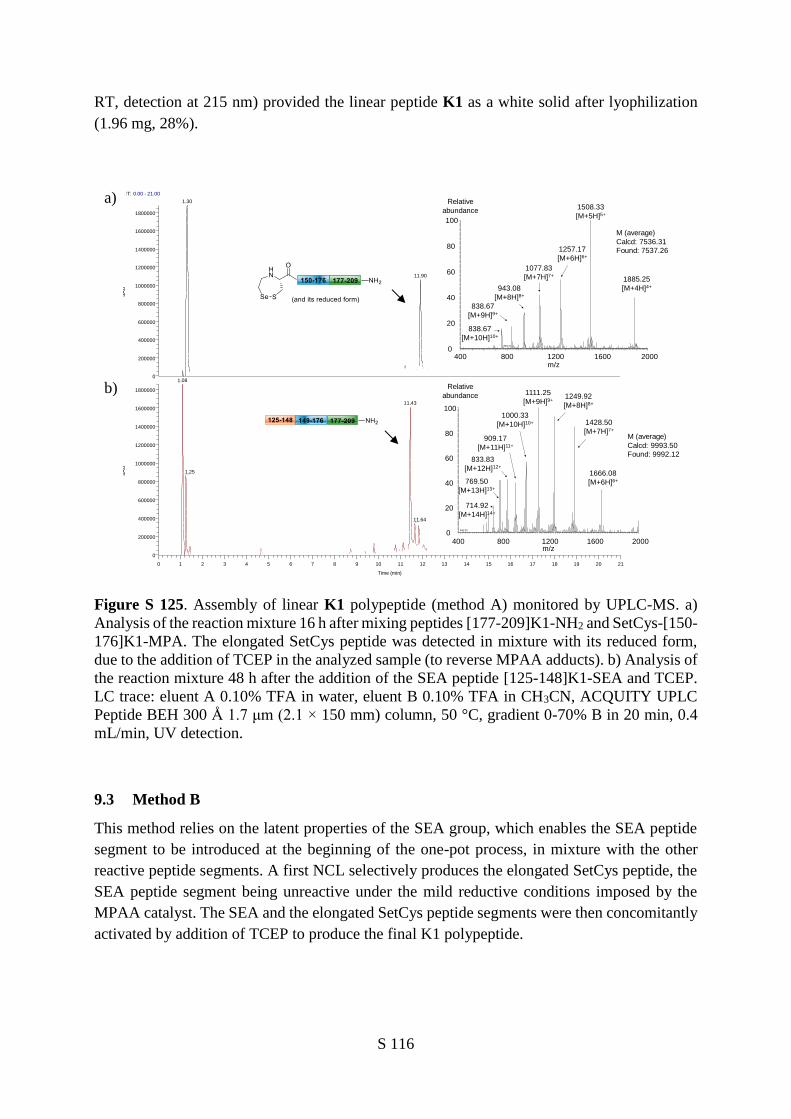
S 116
RT, detection at 215 nm) provided the linear peptide K1 as a white solid after lyophilization
(1.96 mg, 28%).
Figure S 125. Assembly of linear K1 polypeptide (method A) monitored by UPLC-MS. a)
Analysis of the reaction mixture 16 h after mixing peptides [177-209]K1-NH2 and SetCys-[150-
176]K1-MPA. The elongated SetCys peptide was detected in mixture with its reduced form,
due to the addition of TCEP in the analyzed sample (to reverse MPAA adducts). b) Analysis of
the reaction mixture 48 h after the addition of the SEA peptide [125-148]K1-SEA and TCEP.
LC trace: eluent A 0.10% TFA in water, eluent B 0.10% TFA in CH3CN, ACQUITY UPLC
Peptide BEH 300 Å 1.7 μm (2.1 × 150 mm) column, 50 °C, gradient 0-70% B in 20 min, 0.4
mL/min, UV detection.
9.3 Method B
This method relies on the latent properties of the SEA group, which enables the SEA peptide
segment to be introduced at the beginning of the one-pot process, in mixture with the other
reactive peptide segments. A first NCL selectively produces the elongated SetCys peptide, the
SEA peptide segment being unreactive under the mild reductive conditions imposed by the
MPAA catalyst. The SEA and the elongated SetCys peptide segments were then concomitantly
activated by addition of TCEP to produce the final K1 polypeptide.
RT: 0.00 - 21.00
0 1 2 3 4 5 6 7 8 9 10 11 12 13 14 15 16 17 18 19 20 21
Time (min)
0
200000
400000
600000
800000
1000000
1200000
1400000
1600000
1800000
uA
U
0
200000
400000
600000
800000
1000000
1200000
1400000
1600000
1800000
uA
U
1.30
11.90
1.08
11.43
1.25
11.64
NL:
1.88E6
UV_VIS_1
UV 2
VD166-
172-C5
NL:
1.85E6
UV_VIS_1
UV 2
vd166-172-
c9
1257.17
[M+6H]6+
1508.33
[M+5H]5+
1077.83
[M+7H]7+
943.08
[M+8H]8+
838.67
[M+9H]9+
1885.25
[M+4H]4+
M (average)
Calcd: 7536.31
Found: 7537.26
Relative
abundance
400 800 1200 1600 2000m/z
0
20
40
60
80
100
803.17
838.67
[M+10H]10+
400 800 1200 1600 2000m/z
0
20
40
60
80
100
446.33
1428.50
[M+7H]7+
1249.92
[M+8H]8+
1111.25
[M+9H]9+
1000.33
[M+10H]10+
769.50
[M+13H]13+
909.17
[M+11H]11+
833.83
[M+12H]12+
1666.08
[M+6H]6+
714.92
[M+14H]14+
M (average)
Calcd: 9993.50
Found: 9992.12
Relative
abundance
a)
b)

S 117
Figure S 126. One-pot three peptide segment assembly of linear K1 polypeptide (method B).
To a solution of Gn·HCl (287 mg) in 0.1 M, pH 7.4 phosphate buffer (300 μL) was added
MPAA (16.8 mg) and the pH of the mixture was adjusted to 7.28 by addition of 6 M NaOH.
Peptide [125-148]K1-SEA (2.18 mg, 0.61 μmol, 1 eq), [177-209]K1-NH2 (2.78 mg, 0.61 μmol,
1 eq) and peptide SetCys-[150-176]K1-MPA (2.50 mg, 0.61 μmol, 1 eq) were successively
dissolved in the MPAA solution (152 μL) and the mixture was stirred at 37 °C. The progress of
the ligation leading to the elongated SeCys peptide was followed by UPLC-MS (Figure S 127a),
the SEA peptide being unreactive in these mild reductive conditions.
After completion of the NCL ligation (2.5 h), the second step of the process that ligates the
elongated SetCys peptide with the SEA segment was induced by addition of TCEP. To a
solution of Gn·HCl (287 mg) in 0.1 M, pH 7.4 phosphate buffer (300 μL) were added
TCEP∙HCl (28.6 mg), sodium ascorbate (19.8 mg) and MPAA (16.8 mg) and the pH of the
mixture was adjusted to 5.40 by addition of 6 M NaOH. The previous ligation mixture
containing the elongated SetCys peptide and SEA segment was then diluted with the solution
of TCEP (152 µL). The pH was readjusted to 5.45 by addition of 6 M HCl and the mixture was
stirred at 37 °C for 40 h. After completion of the reaction (Figure S 127b), the mixture was diluted
with 7.5% AcOH in water (8 mL) and extracted with Et2O (5 ×) to remove the MPAA.
Purification of the crude by HPLC (eluent A: 0.1% TFA in water, eluent B: 0.1% TFA in
acetonitrile, 6 mL/min, 0-10% eluent B in 5 min then 10-35% eluent B in 30 min, C18 column,
RT, detection at 215 nm) provided the linear peptide K1 as a white solid after lyophilization
(2.05 mg, 29%).

S 118
Figure S 127. Assembly of linear K1 polypeptide (method B) monitored by UPLC-MS. a)
Analysis of the reaction mixture 2.5 h after mixing peptides [125-148]K1-SEA, [177-209]K1-
NH2 and SetCys-[150-176]K1-MPA. The elongated SetCys peptide and the remaining SEA
peptide segments are detected in mixture with their reduced form, due to the addition of TCEP
in the analyzed sample (to reverse MPAA and StBu adducts). b) Analysis of the reaction
mixture 40 h after the addition of TCEP. LC trace: eluent A 0.10% TFA in water, eluent B
0.10% TFA in CH3CN, ACQUITY UPLC Peptide BEH 300 Å 1.7 μm (2.1 × 150 mm) column,
50 °C, gradient 0-70% B in 20 min, 0.4 mL/min, UV detection.
a
b

S 119
9.4 Characterization of linear K1 polypeptide
Figure S 128. LC-MS analysis of linear K1 polypeptide. a) LC trace. Eluent A 0.10% TFA in
water, eluent B 0.10% TFA in CH3CN, ACQUITY UPLC Peptide BEH 300 Å 1.7 μm (2.1 ×
150 mm) column, 50 °C, gradient 0-70% B in 20 min, 0.4 mL/min, UV detection. b) MS trace:
m/z = 1666.17 ([M+6H]6+), 1428.42 ([M+7H]7+), 1250.08 ([M+8H]8+), 1111.25 ([M+9H]9+),
1000.25 ([M+10H]10+), 909.42 ([M+11H]11+), 833.75 ([M+12H]12+), 769.58 ([M+13H]13+),
714.67 ([M+14H]14+), 667.17 ([M+15H]15+). Calcd. for M (average): 9993.40, found: 9992.14.
0 4 8 12 16 20
Time (min)
0
400000
800000
1200000
1600000
2000000
11.40
Intensity
(uV, µAU)
400 800 1200 1600 2000
m/z
0
20
40
60
80
100
[M+8H]8+
1250.08[M+9H]9+
1111.25[M+7H]7+
1428.42
[M+10H]10+
1000.25
[M+11H]11+
909.42
[M+12H]12+
833.75
[M+13H]13+
769.58
[M+14H]14+
714.67
[M+15H]15+
667.17
[M+6H]6+
1666.17
Relative
abundanceb)
a)

S 120
Figure S 129. MALDI-TOF analysis of linear K1 polypeptide. Matrix: 2,5-dihydroxybenzoic
acid, positive detection mode, calcd. for [M+H]+ (average): 9994.4, found: 9993.1.
Proteomic analysis of K1 polypeptide
Alkylation
Synthetic K1 polypeptide (20 µg) was treated with a solution of iodoacetamide in 0.025 M
ammonium bicarbonate (1 mg/mL, 20 µL) for 15 min. The alkylation step was monitored by
MALDI-TOF mass spectrometry.
[M+H]+
9993.063
5012.423
0.0
0.5
1.0
1.5
2.0
2.5
4x10
4000 8000 12000 16000 20000
m/z
Intensity
(AU)
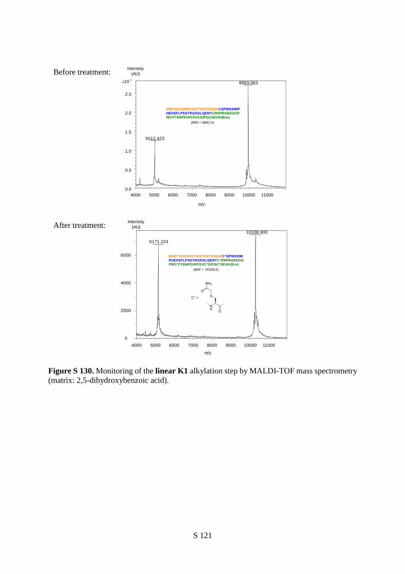
S 121
Figure S 130. Monitoring of the linear K1 alkylation step by MALDI-TOF mass spectrometry
(matrix: 2,5-dihydroxybenzoic acid).
9993.063
5012.423
0.0
0.5
1.0
1.5
2.0
2.5
4x10
4000 5000 6000 7000 8000 9000 10000 11000
m/z
Intensity
(AU)
Intensity
(AU)
10338.900
5171.224
0
2000
4000
6000
4000 5000 6000 7000 8000 9000 10000 11000
m/z
Before treatment:
After treatment:

S 122
Trypsin cleavage
Trypsin (0.5 mg/mL, 2 µL) was added to the mixture to cleave the alkylated linear polypeptide.
The fragments resulting from the enzymatic cleavage were identified by MALDI-TOF MS.
Figure S 131. MALDI-TOF analysis of the enzymatic lysate. Matrix: 2,5-dihydroxybenzoic
acid, positive detection mode. Analysis of the fragments produced by enzymatic cleavage of
alkylated K1 confirms the Tyr-Cys junction formed by NCL and the conversion of the SetCys
residue into Cys residue during the second ligation step.

S 123
10 Folding, characterization and biological activity of cK1-1 and cK1-2 (Figure
6)
10.1 Folding (Figure 6b,c)
cK1-1 and cK1-2 were simultaneously folded using a mixture of oxidized and reduced
gluthatione.
- K1 cyclized with the short biotinylated linker (n = 2)
- K1 cyclized with the long biotinylated linker (n = 3)
General procedure:
The cyclic polypeptide cK1-1 or cK1-2 was dissolved in 10 mM PBS, 138 mM NaCl, 2.7 mM
KCl buffer pH 7.4 containing 10 % by vol of glycerol, 1 mM reduced glutathione, 0.2 mM
oxidized glutathione (33 mL/μmol of peptide). The reaction mixture was gently stirred at 4 °C
for 36 h.
The folding of cK1-1 and cK1-2 was followed by LC-MS. Results are given in Figure S 132-
Figure S 135.
Figure S 132. Kinetics of cK1-1 folding studied by LC-MS. Elution conditions: eluent A 0.10%
TFA in water, eluent B 0.10% TFA in CH3CN/water: 4/1 by vol., XBridge Peptide BHE C18
3.5 µm 4.6×150 mm column, gradient 0-50% B in 15 min, 50 °C, 1 mL/min, light scattering
detection.

S 124
Figure S 133. Kinetics of cK1-2 folding studied by LC-MS. Elution conditions: eluent A 0.10%
TFA in water, eluent B 0.10% TFA in CH3CN/water: 4/1 by vol., XBridge Peptide BHE C18
3.5 µm 4.6×150 mm column, gradient 0-50% B in 15 min, 50 °C, 1 mL/min, light scattering
detection.
The mixtures were then centrifugated (15300 × g) at 4 °C during 20 min to eliminate the
insoluble aggregates. The mixtures were transferred in an ultrafiltration column (Vivaspin, 2
mL) with a cut-off of 3000 MWCO and centrifugated (12000 × g) at 4 °C.
Once concentrated, the proteins were dialysed twice (respectively 1 h and 18 h) at 4 °C against
10 mM PBS, 138 mM NaCl, 2.7 mM KCl buffer pH 7.4 containing 10 % in vol of glycerol (2
× 0.3 L). Finally, the concentration of the folded proteins was determined using BCA assay.
Table S 6. Folding results
peptide Initial mass of peptide
(in mg)
Volume after
dialys (in μL)
Concentration of peptide
determined by BCA (in μmol)
Yield
(in %)
cK1-1 1.056 312 30 12
cK1-2 1.084 230 26 7

S 125
10.1.1 Characterization of folded cK1-1f and cK1-2f
a)
b)
Figure S 134. LC-MS analysis of folded cK1-1f. a) LC trace. Eluent A 0.10% TFA in water,
eluent B 0.10% TFA in CH3CN, ACQUITY UPLC Peptide BEH 300Å 1.7 μm (2.1 × 150 mm)
column, 50 °C, gradient 0-70% B in 20 min, 0.4 mL/min, UV detection. b) MS trace. m/z =
1654.17 ([M+6H]6+), 1418.17 ([M+7H]7+), 1241.08 ([M+8H]8+), calcd for M (average):
9919.13, found: 9919.95.
0 2 4 6 8 10 12 14 16 18 20
Time (min)
0
200000
400000
600000
10.33
Intensity
(UV, µAu)
300 500 700 900 1100 1300 1500 1700 1900
m/z
0
20
40
60
80
1001418.17
1241.08
1654.17
Relative abundance
(%)

S 126
a)
b)
Figure S 135. LC-MS analysis of folded cK1-2f. a) LC trace. Eluent A 0.10% TFA in water,
eluent B 0.10% TFA in CH3CN, ACQUITY UPLC Peptide BEH 300Å 1.7 μm (2.1 × 150 mm)
column, 50 °C, gradient 0-70% B in 20 min, 0.4 mL/min, UV detection. b) MS trace: m/z =
1702.33 ([M+6H]6+), 1459.25 ([M+7H]7+), 1277.08 ([M+8H]8+), 1135.33 ([M+9H]9+); calcd
for M (average): 10207.39, found 10208.34.
0 2 4 6 8 10 12 14 16 18 20
Time (min)
-100000
0
100000
200000
300000
400000
10.29
300 500 700 900 1100 1300 1500 1700 1900m/z
0
20
40
60
80
1001459.25
1277.08
1702.331135.33
Relative abundance
(%)
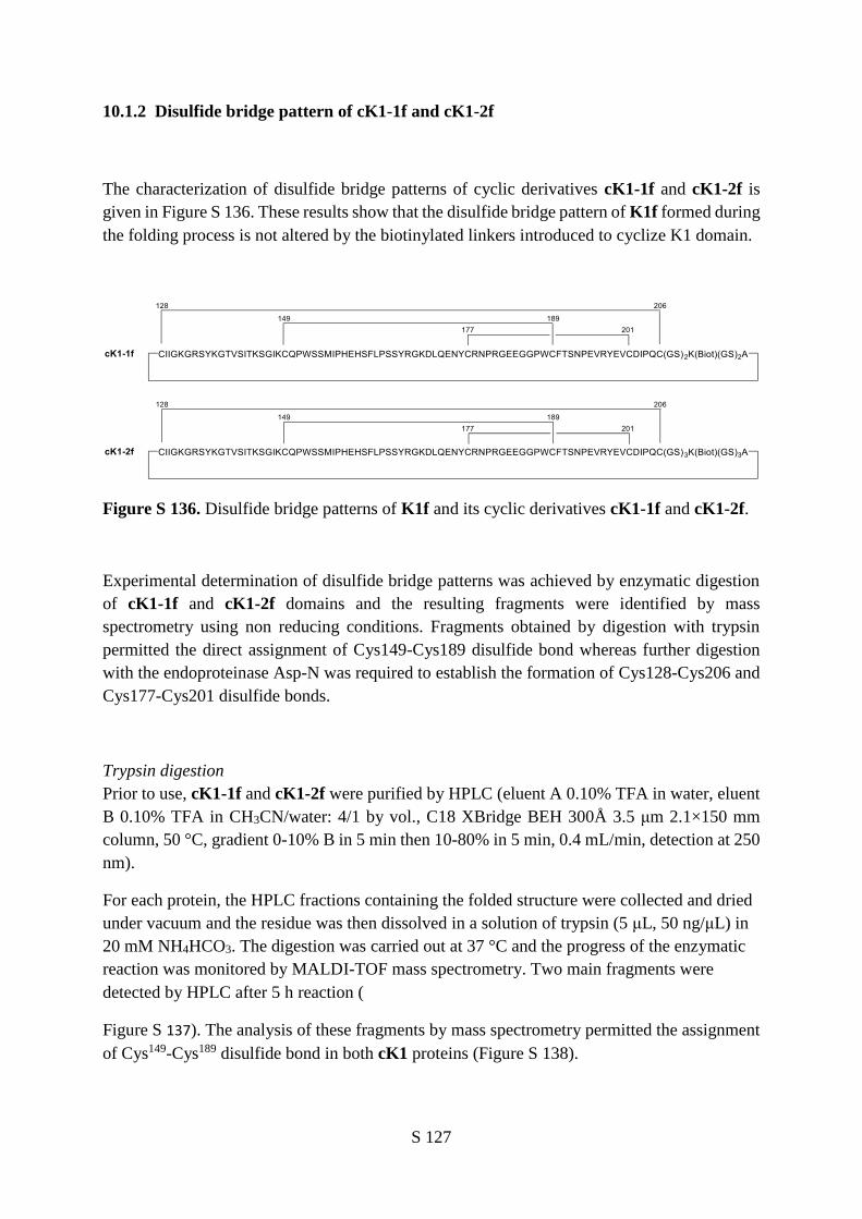
S 127
10.1.2 Disulfide bridge pattern of cK1-1f and cK1-2f
The characterization of disulfide bridge patterns of cyclic derivatives cK1-1f and cK1-2f is
given in Figure S 136. These results show that the disulfide bridge pattern of K1f formed during
the folding process is not altered by the biotinylated linkers introduced to cyclize K1 domain.
Figure S 136. Disulfide bridge patterns of K1f and its cyclic derivatives cK1-1f and cK1-2f.
Experimental determination of disulfide bridge patterns was achieved by enzymatic digestion
of cK1-1f and cK1-2f domains and the resulting fragments were identified by mass
spectrometry using non reducing conditions. Fragments obtained by digestion with trypsin
permitted the direct assignment of Cys149-Cys189 disulfide bond whereas further digestion
with the endoproteinase Asp-N was required to establish the formation of Cys128-Cys206 and
Cys177-Cys201 disulfide bonds.
Trypsin digestion
Prior to use, cK1-1f and cK1-2f were purified by HPLC (eluent A 0.10% TFA in water, eluent
B 0.10% TFA in CH3CN/water: 4/1 by vol., C18 XBridge BEH 300Å 3.5 μm 2.1×150 mm
column, 50 °C, gradient 0-10% B in 5 min then 10-80% in 5 min, 0.4 mL/min, detection at 250
nm).
For each protein, the HPLC fractions containing the folded structure were collected and dried
under vacuum and the residue was then dissolved in a solution of trypsin (5 μL, 50 ng/μL) in
20 mM NH4HCO3. The digestion was carried out at 37 °C and the progress of the enzymatic
reaction was monitored by MALDI-TOF mass spectrometry. Two main fragments were
detected by HPLC after 5 h reaction (
Figure S 137). The analysis of these fragments by mass spectrometry permitted the assignment
of Cys149-Cys189 disulfide bond in both cK1 proteins (Figure S 138).
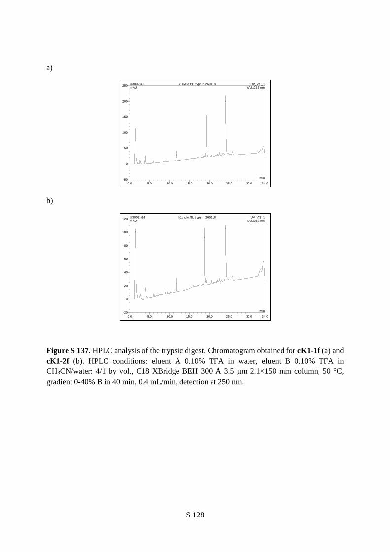
S 128
a)
b)
Figure S 137. HPLC analysis of the trypsic digest. Chromatogram obtained for cK1-1f (a) and
cK1-2f (b). HPLC conditions: eluent A 0.10% TFA in water, eluent B 0.10% TFA in
CH3CN/water: 4/1 by vol., C18 XBridge BEH 300 Å 3.5 μm 2.1×150 mm column, 50 °C,
gradient 0-40% B in 40 min, 0.4 mL/min, detection at 250 nm.
0.0 5.0 10.0 15.0 20.0 25.0 30.0 34.0
-50
0
50
100
150
200
250U3002 #90 k1cyclo PL trypsin 260118 UV_VIS_1mAU
min
WVL:215 nm
0.0 5.0 10.0 15.0 20.0 25.0 30.0 34.0
-20
0
20
40
60
80
100
120U3002 #91 k1cyclo GL trypsin 260118 UV_VIS_1mAU
min
WVL:215 nm

S 129
Figure S 138. MALDI-TOF analysis of the trypsic digest: results obtained with cK1-1f (A) and
cK1-2f (B). Matrix 2,5-dihydroxybenzoic acid (DHB), positive detection mode.
Asp-N digestion
Each trypsic digest was diluted with 0.1% TFA in water and the resulting mixture was purified
by HPLC in order to isolate the fragment containing in its structure cysteines 128, 177, 201 and
206. The HPLC fractions in which this fragment was detected were concentrated and dried
under vacuum and the obtained residue was then dissolved in a solution of endoproteinase N-
Asp (3 μL, 0.01 mg/mL) in 20 mM NH4HCO3. The digestion was carried out at 37 °C and was
run for 2 min. The analysis of the new fragments by mass spectrometry permitted the
assignment of Cys126-Cys206 and Cys177-Cys201 disulfide bonds for both cK1 proteins (Figure S
139).
Figure S 139. MALDI-TOF analysis of the endoproteinase N-Asp digest: results obtained with
cK1-1f (A) and cK1-2f (B). Matrix 2,5-dihydroxybenzoic acid (DHB), positive detection mode.
A)
B)
A)
B)

S 130
10.2 Alphascreen Assay (Figure 6d)
Competition assays for binding of K1B, cK1-1f or cK1-2f to recombinant MET-Fc protein in
competition with increasing concentrations of NK1 were performed in 384-well microtiter
plates (OptiPlate™-384, PerkinElmer©, CA, USA, 40 μL of final reaction volume). Final
concentrations were 20 nM for K1B, cK1-1f or cK1-2f, 2 nM for MET-Fc, 0–300 nM for NK1,
10 μg mL–1 for streptavidin coated donor beads and protein A-conjugated acceptor beads. The
buffer used for preparing all protein solutions and the bead suspensions was: PBS, 5 mM
HEPES pH 7.4, 0.1% BSA.
K1B, cK1-1f or cK1-2f (5 μL, 20 nM) was mixed with a solution of hMET-Fc (5 μL, 2 nM)
and with solutions of NK1 (10 μL, 0-300 nM). The mixture was incubated for at 23 °C 60 min
(final volume 20 μL). Protein A-conjugated acceptor beads (10 μL, 50 μg mL–1) were then
added to the vials. The plate was incubated at 23 °C for 30 min in a dark box. Finally,
streptavidin coated donor beads (10 μL, 50 μg mL–1) were added and the plate was further
incubated at 23 °C for 30 min in a dark box. The emitted signal intensity was measured using
standard Alpha settings on an EnSpire® Multimode Plate Reader (PerkinElmer).
10.3 Biological assays (Figure 6e,f)
K1B was produced as described in Simonneau et al.Erreur ! Signet non défini.
10.3.1 Cell-based assay for MET signaling induced by cK1-1f and cK1-2f
The assay was performed according to Simonneau et al.Erreur ! Signet non défini. HeLa cells were t
reated for 10 min with 300 pM mature HGF/SF (HGF), or with 10 nM / 100 nM K1/S, cK1-
1f/S and cK1-2f/S.
Cell lysates were then analyzed by specific total MET and ERK or phospho-MET, phospho-
Akt and phospho-ERK Western blot. Cells were collected by scraping and then lysed on ice
with a lysis buffer (20 mM HEPES pH 7.4, 142 mM KCl, 5 mM MgCl2, 1 mM EDTA, 5%
glycerol, 1% NP40 and 0.1% SDS) supplemented with freshly added protease and phosphatase
inhibitors (#P8340 and #P5726, respectively, Sigma). Lysates were clarified by centrifugation
(20,000 × g, 15 min) and protein concentration was determined (BCA protein assay Kit,
Pierce®, Thermo scientific, IL, USA). The same protein amount of cell extracts was separated
by either classical SDS-PAGE or NuPAGE (4-12% or 10% Bis-Tris precast gels) (Life
technologies) and electrotransferred to polyvinylidene difluoride (PVDF) membranes (Merck
Millipore). Membranes were probed with indicated primary antibodies, followed by incubation
with appropriate HRP conjugated secondary antibodies. Protein-antibody complexes were
visualized by chemiluminescence with the SuperSignal® West Dura Extended Duration
Substrate (Thermo scientific), using a LAS-3000 imaging system (Fujifilm, Tokyo, Japan) or
X-ray films (CL-Xposure Film, Thermo scientific).
S 131
10.3.2 Scattering Assay
The assay was performed according to Simonneau et al.10
Capan cells were seeded at low density (2,000 cells/well on a 12-well plate) to form compact
colonies. After treatment, when colony dispersion was observed, the cells were fixed and
colored by Hemacolor® stain (Merck, Darmstadt, Germany) according to the manufacturer’s
instructions. Representative images were captured using a phase contrast microscope with 40x
magnification (Nikon Eclipse TS100, Tokyo, Japan).
S 132
11 References
1. Abbas M., Bethke J. & Wessjohann L. A. One pot synthesis of selenocysteine
containing peptoid libraries by Ugi multicomponent reactions in water. Chem.
Commun., 541-543 (2006).
2. Miura C., Kiyama M., Iwano S., Ito K., Obata R., Hirano T. et al. Synthesis and
luminescence properties of biphenyl-type firefly luciferin analogs with a new, near-
infrared light-emitting bioluminophore. Tetrahedron 69, 9726-9734 (2013).
3. Boll E., Drobecq H., Ollivier N., Blanpain A., Raibaut L., Desmet R. et al. One-pot
chemical synthesis of small ubiquitin-like modifier (SUMO) protein-peptide
conjugates using bis(2-sulfanylethyl)amido peptide latent thioester surrogates Nat.
Protoc. 10, 269-292 (2015).
4. Ollivier N., Raibaut L., Blanpain A., Desmet R., Dheur J., Mhidia R. et al. Tidbits for
the synthesis of bis(2-sulfanylethyl)amido (SEA) polystyrene resin, SEA peptides and
peptide thioesters. J. Pept. Sci. 20, 92–97 (2014).
5. Ollivier N., Dheur J., Mhidia R., Blanpain A. & Melnyk O. Bis(2-sulfanylethyl)amino
native peptide ligation. Org. Lett. 12, 5238-5241 (2010).
6. Ollivier N., Desmet R., Drobecq H., Blanpain A., Boll E., Leclercq B. et al. A simple
and traceless solid phase method simplifies the assembly of large peptides and the
access to challenging proteins. Chem. Sci. 8, 5362-5370 (2017).
7. Fang G.-M., Li Y.-M., Shen F., Huang Y.-C., Li J.-B., Lin Y. et al. Protein chemical
synthesis by ligation of peptide hydrazides. Angew. Chem. Int. Ed. 50, 7645-7649
(2011).
8. Ruff Y., Garavini V. & Giuseppone N. Reversible native chemical ligation: a facile
access to dynamic covalent peptides. J. Am. Chem. Soc. 136, 6333-6339 (2014).
9. Ollivier N., Vicogne J., Vallin A., Drobecq H., Desmet R., El-Mahdi O. et al. A one-
pot three-segment ligation strategy for protein chemical synthesis. Angew. Chem. Int.
Ed. 51, 209-213 (2012).
10. Simonneau C., Berenice L., Mougel A., Adriaenssens E., Paquet C., Raibaut L. et al.
Semi-synthesis of a HGF/SF kringle one (K1) domain scaffold generates a potent in
vivo MET receptor agonist. Chem. Sci. 6, 2110-2121 (2015).

download fileview on ChemRxivSupportingInformation_R1_3.pdf (7.07 MiB)


