Embed Size (px)
Citation preview

183
Gunilla SWEDJEMARK1 and Jan STENLID2
"The Forestry Research Institute of Sweden, Uppsala Science Park, S-751 83 Uppsala, Sweden.#Department of Forest Mycology and Pathology, Swedish University of Agricultural Sciences, Box 7026, S-750 07 Uppsala, Sweden.
Received 10 February 2000 ; accepted 16 June 2000.
The wood of one Picea abies stump, including its roots (1±3 m in length), was sliced into 2 cm thick discs. The stump originated from
a thinning conducted 7 yr prior to the investigation in a 30 yr old spruce stand planted on previous farmland that became
heavily infected by Heterobasidion annosum. After incubation of the wood discs, interaction zones were observed on the surfaces and
H. annosum was isolated from the areas between the zones and from the zone lines, resulting in 296 isolates. The isolates were
tested with somatic incompatibility to detect 35 different genets. Of the 27 genets colonising the upper part of the stump (excluding
the roots), 12 had proceeded into the roots. Seven genets found in the roots were not found in the upper part of the stump and one
genet isolated from an interaction zone on the top of the stump was not found elsewhere in the stump or in the roots. Two of the
genets had grown into root contact with other trees.
INTRODUCTION
Heterobasidion annosum is a serious pathogen on conifers
throughout the boreal and temperate zones of the Northern
Hemisphere. Trunks of infected Norway spruce trees can be
decayed up to 12 m height (Stenlid & Wa$ sterlund 1986). Since
the pathogen is a poor interspecific competitor (Rishbeth
1950, Capretti & Mugnai 1989, Holmer & Stenlid 1994)
spores can only successfully germinate on uncolonised wood
such as freshly cut stumps. The fungus colonises the stump
and is then able to spread vegetatively to adjacent trees via
root contacts. Stumps left after thinning or clear-cuttings may
act as substrate for the fungus for at least 30 years (Greig &
Pratt 1976, Stenlid 1987). The lateral spread from the primary
infection centre varies between 0±1 and 2 m per year (Fowler
1962, Hodges 1969, Swedjemark & Stenlid 1993). The growth
rate of H. annosum is higher in dead than in live wood (Bendz-
Hellgren et al. 1999).
In Scandinavia, H. annosum consists of two intersterility
groups (IS groups). The S group is mainly confined to Norway
spruce while the P group is most often found on Scots pine,
but also on other coniferous and broadleaf trees (Korhonen
1978, Capretti, Goggioli & Mugnai 1994, Swedjemark &
Stenlid 1995). Two groups are identified by their ability to
heterokaryotize homokaryotic tester strains of known IS
group (Korhonen 1978). The intersterility is not complete and
some mycelia are able to mate with both IS groups (Korhonen,
Stenlid & Capretti 1998).
A basidiomycete population is composed of genetically
different individuals which are able to recognise self and non-
self through a system called somatic incompatibility (Todd &
Rayner 1980, Stenlid 1985, Rayner & Boddy 1988, Hansen,
Stenlid & Johansson 1993a, b). In H. annosum this is a
polygenic system where differences at one or more loci result
in an incompatibility reaction (Hansen et al. 1993a, b). Brasier
& Rayner (1987) proposed that the term ‘genet ’ should
describe all parts of a vegetative mycelium that have the same
set of somatic incompatibility genes.
In zones of somatic incompatibility between two hetero-
karyotic strains, new combinations of the four nuclei involved
can occur (Hansen et al. 1993a). Such new combinations are
normally short-lived and confined to small areas between the
interacting mycelia. Hypothetically, they could escape away
from the progenitors when growing in wood by entering into
new wood cells not colonised by other mycelia.
The silvicultural treatment, age and history of a stand are
important factors for the size and number of fungal genets in
a forest area (Dahlberg & Stenlid 1990, Smith, Bruhn &
Anderson 1992, Swedjemark & Stenlid 1993). The population
density of H. annosum varied between 25 and 4800 genets per
hectare (Stenlid 1985, 1987, Piri, Korhonen & Sairanen 1990,
Swedjemark & Stenlid 1993, Garbelotto, Cobb & Bruns 1994,
Morrison & Pellow 1994, Mokritsky 1994), where the dense
populations with relatively small genets were the result of
strong disturbance through forestry and the sparse populations
with large genets were found at sites with a long forest
history of relatively extensive forestry. Swedjemark & Stenlid
(1993) found that 31% of the thinning-stumps in the
Mycol. Res. 105 (2) : 183–189 (February 2001). Printed in the United Kingdom.
A highly diverse population of Heterobasidion annosum in asingle stump of Picea abies

A diverse Heterobasidion annosum population 184
investigated stand contained more than one genet of H.
annosum. The maximum number of genets obtained from the
top of one stump was 13, while from trees nearby only one
genet was obtained. The analysed stump in the present
investigation originated from this stand.
The purpose of this study was to analyse the distribution
and development of H. annosum genets in a population
colonising a single stump 7 years after infection.
MATERIAL AND METHODS
Stand description and history
The stand was situated at Siaro$ , an island in the archipelago
on the Swedish east coast, 50 km north of Stockholm
(Swedjemark & Stenlid 1993). Norway spruce was planted at
a 1±5 m spacing on 7±5 ha abandoned farmland during
1961}62. No silvicultural operations were carried out until
1982. A thinning was then made during July and August.
Sixty percent of the stems were left, which corresponds to a
basal area of 35±5 m# ha−". No signs of decay were found in
the stumps at the time of thinning, and since no heavy
machinery was allowed in the stand the remaining trees were
free from logging and extraction damage.
Disease pattern in the stand
By 1968}87 some trees had died and resin flow was present
on several stems. In 1989, parts of the stand were surveyed for
Fig. 1. Norway spruce stump created at a thinning containing several different genets of H. annosum. Each genet occupies a distinct
sector.
disease and mortality (Swedjemark & Stenlid 1993). Decay
was then detected in 63% of the analysed stems and in 70%
of the stumps (Fig. 1) left after the thinning (thinning stump).
Between 1982 and 1990, 4.5% of the trees had died, mainly
caused by H. annosum infection. Infected thinning-stumps
contained between one (36% of the infected stumps) and 13
fungal genets at the level 0–5 cm from the stump top, while
the trees contained only one genet each. Ninety-five percent
of the collected isolates were assigned to the S group, 2% to
the P group, and 3% could not be classified. S and P isolates
coexisted on stumps but only S isolates were found in the
trees. The smallest genets were confined to one stump while
the largest genet encompassed one stump and two trees and
measured 5 m laterally from the stump to the most distant tree
(Swedjemark & Stenlid 1993). After clear cutting 1993, all
stumps were screened for visible signs of decay. About 85%
of the stumps showed decay.
Sample collection
A stump analysed in the study in 1989, that in the top 5 cm
contained 10 different genets of both the S and P IS groups,
was chosen for the detailed stump study. The stump, including
about 1±3 m of all its major roots, was dug up in spring
1991 and transported to the laboratory. The surrounding trees
and stumps were carefully mapped (ca 5 m from the stump)
and contacts between roots from the stump and other tree or
stump roots were marked.
The complete stump was consecutively cut into 2 cm thick

G. Swedjemark and J. Stenlid 185
Fig. 2. Norway spruce stump, including its roots, colonised by H.
annosum. The stump was cut into 2 cm thick discs, H. annosum was
collected from each sector and tested for somatic incompatibility to
detect single genets.
discs (Fig. 2). All discs were numbered and incubated under
humid conditions in 20 °C for 7 d. Colonies of H. annosum
were recognised from their conidial stage. Any distinct
interaction zone visible on the discs, was identified and
numbered. Corresponding areas on the following discs were
numbered according to previous discs even if the interaction
zones were less distinct. For all discs originating from roots,
the distance to the stump was noted. Distinct areas bordered
by interaction zones from the upper surface from all 4 discs
Fig. 3. Stump discs after sampling. Drawings of the distinct areas bordered by interaction zones from the upper surface of all 4 discs
(I–IV) from the investigated stump, excluding the roots. Any distinct area between interaction zones visible on the discs, was identified
and lettered. In disc I and II, corresponding areas were lettered according to the previous disc. On disc III and IV each isolate has a new
identity due to difficulties to interpret the interaction zones. Dark areas represent wood that did not show signs of decay. (Isolates
sampled from the bottom surface of the discs are not documented in this figure.)
from the stump (excluding the roots) were documented
(Fig. 3).
Isolations were made under a dissecting microscope. A
sterilised needle, slightly touching the conidiophores, was used
to transfer conidia to an Hagem agar (HA) plate (Swedjemark
& Stenlid 1993). Isolations were made from both sides on all
discs from all distinct areas and areas which possibly could
contain a fungal genet. Isolations from all contact points
between the stump roots and other roots were also performed.
The interaction zones were studied under the dissecting
microscope and when conidiophores were observed on the
zones, isolates from those were made separately.
Somatic incompatibility tests
Isolates were paired by placing 3¬3 mm inocula 2–3 cm
apart on HA plates. The morphology of the interaction was
studied 4–5 weeks later after incubation in 20 °C. A compatible
reaction shows a continuous mycelial matt when isolates of
the same genetical origin were paired. When genetically
different isolates were paired a zone with sparse mycelial
growth is observed between the two isolates (incompatible
reaction). Isolates of different intersterility groups create a
morphologically different pigmented zone between them
when paired (Stenlid 1985). Somatic incompatibility was
studied between isolates as follows :

A diverse Heterobasidion annosum population 186
(A) Among all isolates in the upper part of the stump (discs
1–4, including the tap-root).
(B) Among all isolates within the same root.
(C) Between isolates originating from the stump and
isolates originating from the roots.
(D) Between isolates from other roots in contact with the
investigated stump and isolates from the contacted root
originating from the stump.
(E) Between isolates from interaction zones and all isolates
from the stump, roots and root contacts.
(F) Between the isolates sampled in 1989 and all isolates
from the stump, the roots, the contacts and the reaction zones
(3 isolates sampled in 1989 were lost due to contamination
during storage, leaving 7 isolates for somatic incompatibility
tests).
Approximately 6500 pairings were made.
Intersterility group studies
Isolates were assigned to IS group according to their ability to
heterokaryotize homokaryotic known test strains (Korhonen
1978, Stenlid & Karlsson 1991).
RESULTS
The stump was 22 cm diam and about 25 cm high with ten
roots, diameters ranging between 2 cm and 11 cm at the root
base. The tap-root was heavily decayed, which explains why
only the top 5 cm could be recovered. The longest root
obtained was 125 cm. Sampled root contacts were between
25 cm and 125 cm away from the stump.
None of the isolates obtained in 1989 from this stump was
found in any tree in the stand. From decayed trees within 5 m
from this stump only H. annosum genets with known
origin from other stumps were isolated
Table 1. Details of the distribution of isolates of Heterobasidion annosum and genets, determined from somatic incompatibility, at various levels in the
examined stump. The (1 P) indicates that one of the genets in this category is of the P intersterility group. Three of the isolates sampled in 1989 were
lost due to contamination during storage, leaving 7 isolates for somatic incompatibility tests.
Origin of
isolate
Total
No. of
isolates
No. of genets
after
confrontation
within the
unit
No. of genets
that were not
found in the
wood of the
upper part of
the stump in
1990
No. of genets
found in the
upper part of
the stump in
1990 and
elsewhere
Stump}root
diam at
the root base
and at the
root end (cm)
Root
length
(cm)
Upper part of
the stump
62 24 (1 P) — 12 22 —
Root 1 65 5 3 2 10–2 56
Root 2 17 1 1 — 4–0±5 40
Root 3 75 3 0 3 7–0±8 54
Root 4 24 2 0 2 11–2 20
Root 5 15 3 3 — 3–1±5 20
Root 6 1 1 0 1 4–3 20
Root 7 2 1 0 1 2–1 35
Root 8 3 1 0 1 3–1 20
Root contacts 14 2 0 2 — 125
Reaction zones 8 3 1 2 — —
Genets isolated
in 1989
10(7) 10(7) 3 (1 P) 7 — —
Total 296 56 11 — — —
On the top and the second disc of the stump 14 distinct
interaction zones were observed similar to those described by
Swedjemark & Stenlid (1993). On the 3rd disc 13 and on the
4th disc, 16 distinctly separated areas were noted (Fig. 3). On
most root discs, a similar pattern of interaction zone-bordered
sections were noted. Two roots, 4 and 2±5 cm diam, were free
from conidiophores. No root grafts were observed at points
of contact between the roots. Of 23 analysed root contacts,
14 developed conidiophores of H. annosum following
incubation.
From the complete stump, a total of 296 successful isolates
were made. Of those, 62 isolates were from the upper part of
the stump (disc 1–4 including about 5 cm of the tap-root), 217
from the roots, 14 from root contacts and 8 isolates were
collected from interaction zones (one of those originated from
a root). From the investigation in 1989, 7 genets were
included (Table 1).
In a few cases it was difficult to interpret somatic
incompatibility tests, but it eventually proved possible to
classify a majority of these after repeated confrontation. Only
two isolates in root 1 did not form the typical self or non-self
morphology when confronted with other mycelia. Absence of
clamp connections suggested that these isolates were
homokaryotic.
After confrontations within each unit (stump or root) a total
of 49 genets were noted. In the upper part of the stump 27
genets were identified and in the roots and interaction zones
19 and 3 genets were identified, respectively (Table 1). Of the
27 genets colonising the upper part of the stump, 12 had
proceeded into the roots. On one root two different genets
were present on the same disc. Of the genets from roots and
interaction zones, seven and one, respectively, were distinct
from the ones in the upper part of the stump. From the isolates
sampled in 1989, three genets were not identical with stump-
isolates from 1991 (Table 1). Following confrontations among
G. Swedjemark and J. Stenlid 187
Table 2. Distribution of genets within the stump at different levels after
somatic incompatibility tests. Isolates sampled from the bottom surfaces of
the discs are not included in this Table (3 separate genets from bottom
surfaces on discs III and IV, respectively).
Disc I Disc II Disc III Disc IV
AI¯A
IIA
II¯A
Iab¯ ED
I1, 14¯GJ
II
BI¯B
IIB
II¯B
Ic¯ F
I2¯ 2
CI¯C
IIC
II¯C
Idefhkm¯ JKLN
I3¯ 3
EDI¯ ED
IIED
II¯ ED
Igi¯M
I4-, 5¯B
I
FI¯ F
IIFII
¯ FI
j¯BI
6¯ 6
GI¯H
IIGJ
II¯ 1, 14
IVl¯ 1 7, 8¯A
I
HI¯H
IH
II¯G
I9, 12, 15¯ L
II
II¯ I
IIIII
¯ II
11¯ 11
JKLNI¯K
IIK
II¯ JKLN
I13¯ JKLN
I
MI¯ gi
IIILII
¯ 12IV
16¯ ED
MII
¯MII
17¯ 17
NII
¯NII
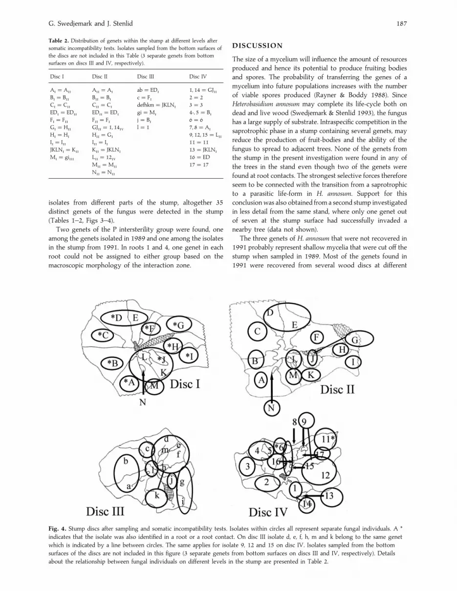
isolates from different parts of the stump, altogether 35
distinct genets of the fungus were detected in the stump
(Tables 1–2, Figs 3–4).
Two genets of the P intersterility group were found, one
among the genets isolated in 1989 and one among the isolates
in the stump from 1991. In roots 1 and 4, one genet in each
root could not be assigned to either group based on the
macroscopic morphology of the interaction zone.
Fig. 4. Stump discs after sampling and somatic incompatibility tests. Isolates within circles all represent separate fungal individuals. A *
indicates that the isolate was also identified in a root or a root contact. On disc III isolate d, e, f, h, m and k belong to the same genet
which is indicated by a line between circles. The same applies for isolate 9, 12 and 15 on disc IV. Isolates sampled from the bottom
surfaces of the discs are not included in this figure (3 separate genets from bottom surfaces on discs III and IV, respectively). Details
about the relationship between fungal individuals on different levels in the stump are presented in Table 2.
DISCUSSION
The size of a mycelium will influence the amount of resources
produced and hence its potential to produce fruiting bodies
and spores. The probability of transferring the genes of a
mycelium into future populations increases with the number
of viable spores produced (Rayner & Boddy 1988). Since
Heterobasidium annosum may complete its life-cycle both on
dead and live wood (Swedjemark & Stenlid 1993), the fungus
has a large supply of substrate. Intraspecific competition in the
saprotrophic phase in a stump containing several genets, may
reduce the production of fruit-bodies and the ability of the
fungus to spread to adjacent trees. None of the genets from
the stump in the present investigation were found in any of
the trees in the stand even though two of the genets were
found at root contacts. The strongest selective forces therefore
seem to be connected with the transition from a saprotrophic
to a parasitic life-form in H. annosum. Support for this
conclusionwas also obtained from a second stump investigated
in less detail from the same stand, where only one genet out
of seven at the stump surface had successfully invaded a
nearby tree (data not shown).
The three genets of H. annosum that were not recovered in
1991 probably represent shallow mycelia that were cut off the
stump when sampled in 1989. Most of the genets found in
1991 were recovered from several wood discs at different

A diverse Heterobasidion annosum population 188
depths in the stump. Interaction zones at this stump age are
not fully reflecting the distribution of fungal genets. For
example, sectors D and E at the level of disc I (Fig. 4) were
both inhabited by the same genet, seven years after felling.
One possible explanation for this could be that one genet had
invaded and taken over the domain of a second one that
subsequently became excluded from the stump. Support for
this interpretation also comes from a previous investigation
in the same geographical area, indicating that the number of
genets at the stump surfaces is reduced with time (Swedjemark
& Stenlid 1993).
It is interesting to speculate on the origin of the genets that
were only found in the roots and interaction zones away from
the surface of the stumps. Several possibilities exist. One
possible scenario is that all 35 genets established more or less
simultaneously from 70 individual spores with distinct nuclei
at the time of thinning in 1982. Based on dissimilar growth
rates they would subsequently have spread out at different
depths in the stump, possibly also dying back in the older
parts of the mycelium, and by radial expansion covered large
parts of each section. However, theoretically, only 9 different
spores with dissimilar nuclei would be needed to form 35
distinct dikaryotic mycelia. Since mycelia sharing one of their
mating types have previously been reported from the same
stump (Chase & Ullrich 1983), it seems likely that not all
genets have established from two spores, different from all
others. Furthermore, the positioning of some of the genets
away from direct contact with any airborne inocula in the
interaction zone between two other genets is hard to explain
with the 70 spores scenario. The spatial positioning of the 35
genets in the stump and its roots, which are sometimes non-
matching, is also hard to explain from a simultaneous
establishment at the stump surface.
Another possibility is that some of the genets established
from spores had been washed down through the soil and onto
the surface of the roots. Following the dying off of the stump
roots, the spores may have germinated and mycelia established
in the roots. This is a distinct possibility and some authors
have also described viable spores washed through sand (Molin
1957), and successful inoculations of H. annosum spores onto
weakened roots (Kuhlman 1969). However, the root infections
would not explain the higher diversity of genets in the upper
part of the stump or the presence of small distinct genets in
interaction zones.
To get a full understanding of the origin of genets we need
to consider the possibility of multiple mating among
established mycelia and reassortment of nuclei in interaction
zones between somatically incompatible heterokaryons. One
homokaryotic mycelium can, for example, mate with several
other mycelia that establish in its vicinity, thereby creating
different genets that share one of their nuclei. The other
possibility of reassorting nuclei between already established
heterokaryons was proposed by Hansen et al. (1993a) and has
some support from our finding that distinct genets were
detected inside the stump in the interaction zone between two
other genets. Both these possibilities would extend the
distribution of one nucleotype outside the extension of one
genet, and open up novel prospects when defining selection
pressures on a population.
ACKNOWLEDGEMENT
We would like to thank Karin Grip for technical assistance and the Swedish
Council of Forestry and Agricultural Research for financial support.
REFERENCES
Bendz-Hellgren, M., Brandtberg, P-O., Johansson, M., Swedjemark, G. &
Stenlid, J. (1999) Growth rate of Heterobasidion annosum in Picea abies
established on forest land and arable land. Scandinavian Journal of Forest
Research 14 : 402–407.
Brasier, C. M. & Rayner, A. D. M. (1987) Whither terminology below the
species level in the fungi ? In Evolutionary Biology of the Fungi (A. D. M
Rayner, C. M. Brasier & D. Moore, eds) : 379–388. Cambridge University
Press, Cambridge, UK.
Capretti, P. & Mugnai, L. (1989) Biological control of Heterobasidion annosum
in Silver fir (Abies alba) stands. In Proceedings of the 7th International
Conference on Root and Butt Rots, Vernon & Victoria, Canada 1988 (D. J.
Morrison ed.) : 277–287. Minister of Supply and Services, Ottawa.
Capretti, P., Goggioli, V. & Mugnai, L. (1994) Intersterility groups of
Heterobasidion annosum in Italy : Distribution, hosts and pathogenicity tests.
In Proceedings of the 8th International Conference on Root and Butt Rots, Wik,
Sweden and Haikko, Finland 1993 (M. Johansson & J. Stenlid, eds) : 218–226.
INFO}REPRO, Uppsala.
Chase, T. & Ullrich, R. C. (1983) Sexuality, distribution and dispersal of
Heterobasidion annosum in pine plantations in Vermont. Mycologia 75 :
825–831.
Dahlberg, A. & Stenlid, J. (1990) Population structure and dynamics in Suillus
bovinus as reflected by spatial distribution of fungal clones. New Phytologist
115 : 487–495.
Fowler, M. E. (1962) Fomes annosus in north eastern United States. In
Conference and study tour on Fomes annosus in Scotland (1960) : 19–20.
IUFRO, Florence.
Garbelotto, M., Cobb, F., Bruns, T., Otrosina, W., Slaughter, G. & Popenuck,
T. (1994) Preliminary results on the genetic structure of Heterobasidion
annosum occuring in Italy. In Proceedings of the 8th International Conference on
Root and Butt Rots, Wik, Sweden and Haikko, Finland 1993 (M. Johansson &
J. Stenlid, eds) : 227–232. INFO}REPRO, Uppsala.
Greig, B. J. W. & Pratt, J. E. (1976) Some observations on the longevity of
Fomes annosus in conifer stumps. European Journal of Forest Pathology 6 :
250–253.
Hansen, E. M., Stenlid, J. & Johansson, M. (1993a) Somatic incompatibility
and nuclear reassortment in Heterobasidion annosum. Mycological Research
97 : 1223–1228.
Hansen, E. M., Stenlid, J. & Johansson, M. (1993b) Genetic control of somatic
incompatibility in the root rotting basidiomycete Heterobasidion annosum.
Mycological Research 97 : 1229–1233.
Hodges, C. S. (1969) Modes of infection and spread of Fomes annosus. Annual
Review of Phytopathology 7 : 247–266.
Holmer, L. & Stenlid, J. (1994) Biological control of Heterobasidion annosum by
cord-forming basidiomycetes. In Proceedings of the 8th International Conference
on Root and Butt Rots, Wik, Sweden and Haikko, Finland 1993 (M. Johansson
& J. Stenlid, eds) : 686–695. INFO}REPRO, Uppsala.
Korhonen, K. (1978) Intersterility groups of Heterobasidion annosum.
Communicationes Instituti Forestalis Fennise. 94 (6) : 1–25.
Korhonen, K. Stenlid, J., Capretti, P. & Karjalainen, R. (1998) Distribution of
Heterobasidion annosum intersterility groups in Europe. In Heterobasidion
annosum: biology, ecology, impact and control (S. Woodward, J. Stenlid, R.
Karjalainen & A. Hu$ ttermann, eds) : 93–104. CAB International,
Wallingford.
Kuhlmann, E. G. (1969) Inoculation of loblolly pine seedlings with Fomes
annosus in the greenhouse. Canadian Journal of Botany 47 : 2079–2082.
Mokritsky, V. A. (1994) Population structure of Heterobasidion annosum in
pure pine stands in Ukraina. In Proceedings of the 8th International Conference
on Root and Butt Rots, Wik, Sweden and Haikko, Finland 1993 (M. Johansson
& J. Stenlid, eds) : 340–348.
Molin, N. (1957) The infection biology of Fomes annosus. Meddelanden frac nStatens Skogsforskningsinstitut 47 : 1–36.

G. Swedjemark and J. Stenlid 189
Morrison, D. & Pellow, K. (1994) Development of Armillaria root disease in
a 25-year-old Douglas-fir plantation. In Proceedings of the 8th International
Conference on Root and Butt Rots, Wik, Sweden and Haikko, Finland 1993 (M.
Johansson & J. Stenlid, eds) : 560–571. INFO REPRO, Uppsala.
Piri, T., Korhonen, K. & Sairanen, A. (1990) Occurrence of Heterobasidion
annosum in pure and mixed spruce stands in southern Finland. Scandinavian
Journal of Forest Research 5 : 113–125.
Rayner, A. D. M. & Boddy, L. (1988) Fungal Decomposition of Wood. John
Wiley & Sons, Chichester.
Rishbeth, J. (1950) Observations on the biology of Fomes annosus with
particular reference to East Anglian pine plantations. I. The outbreaks of
disease and ecological status of the fungus. Annals of Botany 14 : 365–383.
Smith, M. L., Bruhn, J N. & Anderson, J. B. (1992) The fungus Armillaria
bulbosa is among the largest and oldest living organisms. Nature 356 :
428–431.
Stenlid, J. (1985) Population structure of Heterobasidion annosum as determined
by somatic incompatibility, sexual incompatibility and isoenzyme patterns.
Canadian Journal of Botany 63 : 2268–2273.
Stenlid, J. (1987) Controlling and predicting the spread of Heterobasidion
annosum from infected stumps and trees of Picea abies. Scandinavian Journal
of Forest Research 2 : 187–198.
Stenlid, J., Karlsson, J. O. (1991) Partial intersterility in Heterobasidion annosum.
Mycological Research 95 : 1153–1159.
Stenlid, J., Wa$ sterlund, I. (1986) Estimating the frequency of stem rot in Picea
abies using an increment borer. Scandinavian Journal of Forest Research 1 :
303–308.
Swedjemark, G. & Stenlid, J. (1993) Population dynamics of the root rot
fungus Heterobasidion annosum following thinning of Picea abies. Oikos 66 :
247–254.
Swedjemark, G. & Stenlid, J. (1995) Susceptibility of conifer and broadleaf
seedlings to Swedish S- and P-strains of Heterobasidion annosum under
greenhouse conditions. Plant Pathology 44 : 73–79.
Todd, N. K. & Rayner, A. D. M. (1980) Fungal individualism. Science Progress
66 : 331–354.
Corresponding Editor : D. J. Bond


