Embed Size (px)
Citation preview
ARTICLE
A Homozygous Mutation in Human PRICKLE1Causes an Autosomal-RecessiveProgressive Myoclonus Epilepsy-Ataxia Syndrome
Alexander G. Bassuk,1,2,3 Robyn H. Wallace,7 Aimee Buhr,2 Andrew R. Buller,1 Zaid Afawi,8
Masahito Shimojo,9 Shingo Miyata,10 Shan Chen,1 Pedro Gonzalez-Alegre,4 Hilary L. Griesbach,5
Shu Wu,1 Marcus Nashelsky,6 Eszter K. Vladar,11,12 Dragana Antic,11,12 Polly J. Ferguson,1
Sebahattin Cirak,16 Thomas Voit,17 Matthew P. Scott,12,13,14,15 Jeffrey D. Axelrod,11
Christina Gurnett,18 Azhar S. Daoud,19 Sara Kivity,20 Miriam Y. Neufeld,8 Aziz Mazarib,22
Rachel Straussberg,21 Simri Walid,23 Amos D. Korczyn,24 Diane C. Slusarski,5 Samuel F. Berkovic,25,*and Hatem I. El-Shanti1,2,26
Progressive myoclonus epilepsy (PME) is a syndrome characterized by myoclonic seizures (lightning-like jerks), generalized convulsive
seizures, and varying degrees of neurological decline, especially ataxia and dementia. Previously, we characterized three pedigrees of
individuals with PME and ataxia, where either clinical features or linkage mapping excluded known PME loci. This report identifies
a mutation in PRICKLE1 (also known as RILP for REST/NRSF interacting LIM domain protein) in all three of these pedigrees. The
identified PRICKLE1 mutation blocks the PRICKLE1 and REST interaction in vitro and disrupts the normal function of PRICKLE1 in
an in vivo zebrafish overexpression system. PRICKLE1 is expressed in brain regions implicated in epilepsy and ataxia in mice and
humans, and, to our knowledge, is the first molecule in the noncanonical WNT signaling pathway to be directly implicated in human
epilepsy.
Introduction
More than a dozen clinico-molecular forms of progressive
myoclonus epilepsy (PME) are known, including Unver-
richt-Lundborg disease (MIM 254800 resulting from
CSTB mutations [MIM 601145]), Lafora disease (MIM
254780 resulting from EPM2A [MIM 607566] or NHLRC1
[MIM 608072] mutations), the family of neuronal ceroid
lipofuscinoses (with a variety of molecular defects includ-
ing PPT1 [MIM 256730], CLN4 [MIM 204300], and CLN5
[MIM 256731] mutations), and myoclonic epilepsy with
ragged red fibers (MERFF [MIM 545000] with mitochon-
drial t-RNA mutations). Previously, we characterized three
families with individuals affected with PME and ataxia but
normal brain imaging, where either clinical features or
linkage mapping excluded known PME loci.1–3
This report identifies a mutation in PRICKLE1 (MIM
608500) in all three of these pedigrees. PRICKLE1 is part
of the noncanonical or planar cell polarity (WNT/PCP)
572 The American Journal of Human Genetics 83, 572–581, Novem
pathway, in which some WNT family members activate a
b-CATENIN (CTNNB1 [MIM 116806])-independent path-
way.4 In Drosophila and vertebrates, the WNT/PCP path-
way likely regulates cell polarization.5 Depleting Prickle
genes in the zebrafish embryo alters the convergent-exten-
sion movements essential for gastrulation and disrupts
normal calcium signaling.6–8 PRICKLE1 is part of a gene
family encoding proteins containing a highly conserved
PET domain, which mediates Prickle1-protein-binding
interactions.6,9–11 Prickle1 was discovered independently
based on its ability to bind and functionally interact with
the RE1-SILENCING TRANSCRIPTION FACTOR (REST
[MIM 600571], which was thus separately named Rilp,
for REST/NRSF interacting LIM domain protein), an essen-
tial regulator of neural genes.12,13 The PRICKLE1 mutation
identified in this study is located in the PET domain and
disrupts the PRICKLE1 and REST interaction in vitro and
alters the normal function of PRICKLE1 in an in vivo
zebrafish overexpression system.
1Department of Pediatrics, 2Graduate Program in Genetics, 3Graduate Program in Neuroscience, 4Department of Neurology, 5Department of Biology,6Department of Pathology, University of Iowa, Iowa City, IA 52242, USA; 7Queensland Brain Institute, The University of Queensland, Brisbane 4072, Aus-
tralia; 8Department of Neurology, Tel Aviv Sourasky Medical Center and Sackler Faculty of Medicine, Tel Aviv University, Tel Aviv 64239, Israel; 9Depart-
ment of Molecular and Cellular Biochemistry, University of Kentucky, Louisville, KY 40536, USA; 10Department of Anatomy & Neuroscience, Graduate
School of Medicine, Osaka University, Osaka 565-0871, Japan; 11Department of Pathology, 12Department of Developmental Biology, 13Department of
Genetics, 14Department of Bioengineering, 15Howard Hughes Medical Institute, Stanford University School of Medicine, Stanford, CA 94305, USA;16Universitatskinderklinik Essen, Abteilung fur Allgemeine Padiatrie mit Schwerpunkt Neuropadiatrie, 45122 Essen, Germany; 17Institut de Myologie
Groupe Hospitalier Pitie-Salpetriere, 75013 Paris, France; 18Department of Neurology, Division Pediatric Neurology, Washington University School of
Medicine, St. Louis, MO 63110, USA; 19Department of Pediatrics, Jordan University of Science and Technology, Irbid 22110, Jordan; 20Epilepsy Unit,21Department of Child Neurology, Schneider Children’s Medical Center of Israel, Petach Tikvah 49202, Israel; 22Kupat Holim Clalit, Nazareth 16000, Israel;23Department of Neurology, Western Galilee Hospital, Nahariya 22100, Israel; 24Sieratzki Chair of Neurology, Tel Aviv University, Ramat Aviv 69978, Israel;25Epilepsy Research Centre and Department of Medicine, University of Melbourne (Austin Health), Heidelberg West, Victoria 3161, Australia; 26Shafallah
Medical Genetics Center, Doha, Qatar
*Correspondence: [email protected]
DOI 10.1016/j.ajhg.2008.10.003. ª2008 by The American Society of Human Genetics. All rights reserved.
ber 7, 2008
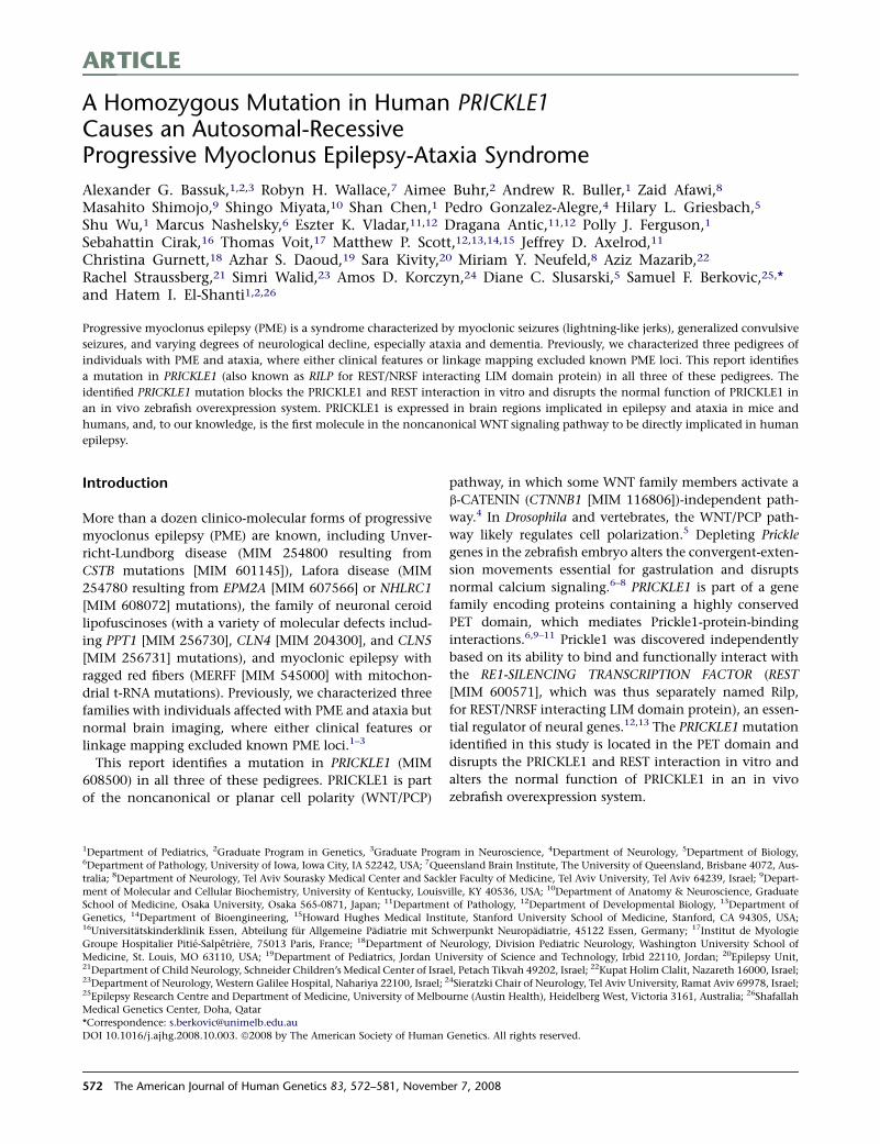
Figure 1. Pedigrees of the Affected Families, Representative Sequences, and Evolutionary Comparison of the Altered PRICKLE1Amino AcidNine nuclear families from three pedigrees including 23 subjects with progressive myoclonus epilepsy and ataxia (pink symbol). Boxes onpedigrees indicate individuals previously reported by Berkovic et al.1 (A), El-Shanti et al.2 (B), and Straussberg et al.3 (C). Dotted linesindicate individuals believed to be related, but the exact relationship was unknown. Subjects who probably had the familial syndromebut were not personally examined are shown in orange. Shared chromosome 12 haplotypes of affected subjects are shown on the top right.Haplotypes were remarkably stable within nuclear families and extended pedigrees. Individuals with epilepsy or ataxia, clinically distinctfrom the familial syndrome (green and purple symbols), did not share the haplotypes or have the PRICKLE1 mutation. Representative DNAsequence chromograms from normal, carrier, and affected (mutant) individuals are in the bottom left panel with the red asterisks denotingthe position of the abnormal nucleotide. Amino acid sequence alignment surrounding the altered amino acid for PRICKLE proteins inmultiple species. pk1, Prickle1 protein; pk2, Prickle2 protein; esn, espinas protein; zfish, zebrafish. Accession numbers for the proteinsequences are: human-pk1, NP_694571; human-pk2, NP_942559; mouse-pk1, NP_001028389; mouse-pk2, NP_001074615; platypus-pk1, XP_001505284; platypus-pk2, XP_001508261; chicken-pk1, XP_416036; chicken-pk2, XP_001234704; frog-pk1, NP_001016939;frog-pk-2, NP_001103517; zfish-pk1, NP_899185; zfish-pk2, NP_899186; fruit fly-pk1, NP_724534; fruit fly-esn, CAB64381; worm-pk1,NP_741435. The amino acid altered in the families and the corresponding amino acid in Prickle proteins from other species are boxed in red.
Material and Methods
SubjectsClinical details of the three pedigrees were previously described;1–3
pedigree B was subsequently expanded with eight more affecteds
identified in three nuclear families. Clinical studies were approved
by the Institutional Review Boards of the Tel Aviv Sourasky Medi-
cal Center and the Jordan University of Science and Technology.
Informed consent was obtained from participating subjects and
their legal guardians. The control brain specimens were obtained
from a 60-year-old male with cirrhosis who died suddenly of
The America
atherosclerotic heart disease, after exemption by the Institutional
Review Board of the University of Iowa and within guidelines
established by Iowa statute.
Fine Mapping and HaplotypingMicrosatellite markers within the chromosome 12 pericentro-
meric linkage region were selected from the Marshfield human
linkage map. Genotyping of 47 individuals from 3 families (Fig-
ure 1) was performed by the Australian Genome Research Facility.
Marker order is based on the current human sequence map (NCBI
Build 36.3).
n Journal of Human Genetics 83, 572–581, November 7, 2008 573
ResequencingPRICKLE1 amplicons (Table S2 available online) were sequenced
with an automated ABI sequencer with dye terminator chemistry.
After DNA amplification, unincorporated PCR primers and dNTPs
in the sample were removed prior to sequencing by isolation of the
desired band in a 2% agarose gel, followed by column purification.
Sequences were analyzed with the computer program PHRED,
which calls the bases, and PHRAP that assembled the sequence
on a PC.
Control GenotypingThe 1054 individuals from the HGD-CEPH panel and the 300
Middle Eastern individuals were genotyped with the Taqman (ABI)
assay on an ABI 7900 HT Fast Real Time PCR machine with the fol-
lowing primers according to manufacturer’s instructions: PR1ex4-
ex4F, GAAAAAAGAGTTGCAGGTGTTCAGT; PR1ex4-ex4R, TTAAT
TGTTCCTCTTCCCAGTGCTT; PR1ex4-ex4V1, VIC, CTCAGCGGA
AGAAA; PR1ex4-ex4M1, FAM, CTCAGCAGAAGAAA.
The entire PRICKLE1 gene was also directly resequenced in an
additional 288 individuals, including 96 individuals of Jorda-
nian-Palestinian ancestry.
Immunohistochemistry and Immunostaining
ProtocolsFor mouse sections and HeLa cells, the solution used was PBS 13
Dulbecco’s (pH 7.4) and the blocking solution for rabbit antibodies
used was serum-free blocking media (Dako, X0909). For mouse
combined with rabbit antibodies, we used M.O.M. Mouse Ig Block-
ing Reagent (Vector, BMK-2202); add 2 drops of stock solution to
2.5 ml of PBS. For human sections, the solution used was PBS 1X
Dulbecco’s (pH 7.4) and the blocking solution was serum-free
blocking media (Dako, X0909).
Antibodies
Rabbit polyclonal antibody to mouse Prickle1 was produced with
amino acids 808–822 by A.G.B. and with amino acids 339–514 by
D.A. Specificity for Prickle1 and not Prickle2 was confirmed by im-
munostaining of myc-tagged cDNAs encoding Prickle1 or Prickle2
proteins into HeLa cells (data not shown). NeuN (Chemicon),
GFAP (Dako), myc 9E10 (Sigma), and GFAP (Santa Cruz) anti-
bodies were used at a 1:400–1:500 dilutions for immunohisto-
chemistry and immunoblotting. Mouse and rabbit antibodies
were diluted in M.O.M. Diluent (600 ml of protein concentrate
stock solution added to 7.5 ml of PBS [Vector, BMK-2202]). Se-
quential rabbit antibodies were diluted in PBS 1X. Secondary
antibodies were diluted 1/500 in blocking for mouseþrabbit
antibodies or in PBS for rabbit antibody. We used the goat anti-rab-
bit IgG (Fab0)2-Alexa Fluor 568 conjugated antibody (Invitrogen,
A21069) for the anti-GFAP antibody and the goat anti-mouse
IgG (Fab0)2-Alexa Fluor 568 conjugated Ab (Invitrogen, A11019)
for anti-NeuN antibody. We used the goat anti-rabbit IgG (Fab0)2-
Alexa Fluor 488 conjugated Ab (Invitrogen, A11070) for the
Prickle1 antibodies. The nuclear counterstain To-Pro3 (Invitrogen,
T3605) was diluted 1/2000 in PBS 1X or DAPI. Slides were
mounted with Vector Labs, H-1000.
Staining Protocol, Mouse Sections
Procedure. Procedures were performed at room temperature or as
indicated. Sections were deparaffinized with autostainer program
#3 and rehydrated with ddH2O. To perform antigen retrieval by
microwave, citrate buffer (pH 6.0) was prewarmed for ~30 s in a Tef-
lon Coplin jar, and slides were added under the following condi-
tions: restriction temperature, þ95�C; wattage, #6 (601 W); pulse
574 The American Journal of Human Genetics 83, 572–581, Novemb
5 min, hold 5 min (no microwave applied), pulse 5 min, then slides
were left in the dish to cool to room temperature for about 20 min.
Next, slides were washed in PBS 1X (pH 7.4), 3x3 min. Sections were
permeabilized with 0.1% Triton X-100 in PBS for 10 min, washed in
PBS 1X (pH 7.4), 3x3 min. Blocking was performed for rabbit and
mouse primary antibodies together by applying the working solu-
tion of M.O.M. mouse Ig blocking reagent X1 hr, then PBS 3x3
min, then working solution M.O.M. diluent for 5 min.
Primary Antibodies. Serum-free blocking media was added for
1 hr, then primary antibodies were added at the following dilu-
tions: rabbit anti-prickle 1 antibodies 1/250-1/375 were added
with mouse anti-NeuN (Chemicon, MA13377) 1/500 or rabbit
anti-prickle 1 affinity-purified antibody 1/250-1/375 was added
followed by rabbit anti-GFAP Ab (Dako, Z0344) 1/1000 and stored
in a slide folder at þ4�C.
Sequential Stainings. The following variation was used. Sections
were blocked (serum-free protein block) for 30 min, then primary
antibody (anti-GFAP antibody) was added for 1 hr, then washed
with PBS 1X (pH 7.4), 6 times for 3 min, and then the secondary
antibody-conjugated fluorophore was added (in the dark) for
30 min. The slide was then washed with PBS 1X (pH 7.4), 6x for
3 min. Nuclear counterstain (in the dark) was then added for
5 min. Slides were then rinsed in PBS 1X and mounted in Vecta-
Shield and stored in slide-folder at þ4�C. Nuclei were viewed in
the Far red channel (647 nm Ex). Anti-Prickle1 antibodies were
viewed in the green channel (488 nm Ex), and anti-GFAP or
anti-NeuN antibodies were viewed in red channel (568 nm Ex).
Staining Protocol, Human Sections
Procedure. Procedures were performed at room temperature or as
indicated. Sections were deparaffinized (with an autostainer)
and rehydrated with ddH2O. Antigen retrieval was performed
by microwave staining. Citrate buffer (pH 6.0) was prewarmed
in the microwave for ~30 s in a Teflon Coplin jar. Slides were
then added with temperature restriction: þ95�C, wattage: #6
(601 W), pulse 5 min, hold 5 min (no microwave applied), pulse
5 min. Slides were then cooled to room temperature for about
20 min, washed in PBS 1X (pH 7.4), 3x for 3 min, and permeabi-
lized with 0.1% Triton X-100 in PBS for 10 min, washed in PBS
1X (pH 7.4), 3x3 min blocked with serum-free blocking media
for 1 hr. Primary antibodies were added for 1 hr, then washed
with PBS, 6x for 3 min, secondary antibodies were added in the
dark for 1 hr, then washed with PBS, 6x for 3 min. Slides were
mounted with VectaShield and sections were stored in a slide
folder at þ4�C.
Primary Antibodies. Primary antibodies were diluted in PBS 1X.
Rabbit anti-prickle 1 antibody 1/250-1/375 and mouse anti-
NeuN (Chemicon, MA13377) 1/500 were used together, and rabbit
anti-prickle 1 antibody 1/250-1/375 and rabbit anti-GFAP Ab
(Dako, Z0344) 1/1000 were used sequentially. Goat anti-rabbit
IgG (Fab0)2-Alexa Fluor 488 conjugated antibody (Invitrogen,
A11070) secondary antibody was diluted 1/500 in PBS. To-Pro3
(Invitrogen, T3605) was diluted 1/2000 in PBS 1X and was used
as the nuclear counterstain. Slides were mounted with VectaShield
(Vector Labs, H-1000).
Sequential Stainings. After the first primary and secondary anti-
bodies were applied, slides were blocked (serum-free protein
block) for 30 min, and then the second primary antibody was ap-
plied for 1 hr, then the slides were washed in PBS 1X (pH 7.4), 6x
for 3 min, and then the second secondary antibody-fluorophore
conjugated was added in the dark for 30 min, and then washed
in PBS 1X (pH 7.4), 6x for 3 min. Nuclear counterstain was then
added (in the dark) for 5 min, and slides were rinsed with PBS
er 7, 2008
1X, and mounted in VectaShieldand and stored in a slide-folder at
þ4�C. Nuclei are viewed in the far red channel (647 nm Ex), anti-
Prickle1 antibodies are viewed in the green channel (488 nm Ex),
and anti-NeuN or GFAP antibodies are viewed in red channel (568
nm Ex).
HeLa Cell Staining
Mouse monoclonal anti-Myc 9E10 (Sigma) 1/1000 was added
(EGFP viewed directly in green channel), then PBS was added 6
times for 3 min followed by secondary antibodies in the dark for
1 hr, then PBS 6 times for 3 min, and slides were then mounted
with VectaShield and double labeled sections were stored with
Ms þ Rb Abs in slide folder at þ4�C.
Confocal MicroscopyAll confocal microscopy was performed on a Zeiss 510 Confocal
Microscope at the University of Iowa or on a Leica confocal micro-
scope at Stanford University. The thickness of the confocal images
and all details of exposure time are embedded within the digital
photographs used in this manuscript and available upon request.
PlasmidsThe full-length human PRICKLE1 cDNA in the PCR-bluntII-TOPO
(Open Biosystems) was cloned into the EcoRI (50) and XhoI (30)
sites of the pCS2þ vector, with the EGFP epitope added in-frame
to the 30 end of the gene by PCR site-directed mutagensis/chimer-
agnesis. The R104Q encoding mutation was introduced by site-
directed mutagenesis, and the entire cDNA was resequenced after
introduction of the mutation to insure that no additional muta-
tions were introduced. Myc-REST was the same construct used pre-
viously to define the Prickle-REST interaction.12,13
TransfectionsTransfections into HeLa cells were performed as previously de-
scribed.12,13
Nuclear REST Measurement. To measure the impact of the
PRICKLE1 mutation on the localization of REST, cells transfected
with wild-type and myc-REST or mutant PRICKLE1 and myc-
REST and quantified with a variation of the method previously re-
ported.14,15 All GFPþ/mycþ cells were counted and a score of 4
was assigned for myc nuclear fluorescence much greater than
myc cytoplasmic fluorescence, 3 for myc nuclear fluorescence
greater than myc cytoplasmic fluorescence, 2 for myc nuclear fluo-
rescence equal to myc cytoplasmic fluorescence, 1 for myc nuclear
fluorescence less than myc cytoplasmic fluorescence, and 0 for
myc nuclear fluorescence much less than myc cytoplasmic fluores-
cence. The mean REST nuclear score was calculated for 100 GFPþ/
mycþ cells in each condition.
CoimmunoprecipitationsCoimmunoprecipitations with slight variations as previously de-
scribed.12,13 In brief, myc-mouse REST and GFP-human Prickle1
plasmids were cotransfected into HeLa cells according to the Lip-
ofectamine 2000 protocol. After 48 hr incubation, cells were lysed
with 300 ml cold NET-N þ Roche Protease Inhibitor. Lysates were
sonicated 3 times for 4 s. 60 ml of the crude lysates were removed
and mixed with 110 ml of denaturing loading buffer. The remain-
ing lysates were precleared by adding 5 ml of anti-rabbit beads and
rotating at room temperature for 20 min. The beads were removed
by centrifuging samples briefly at 10,000 rpm and removing the
supernatant. Precleared lysates were then mixed with 10 ml rabbit
anti-GFP conjugated beads (Santa Cruz) and rotated for 2 hr at
The America
4�C, followed by 30 min rotation at room temperature. The super-
natant was removed and the beads rinsed 3 times with 500 mL of
cold NET-N lysis buffer. After rinsing, 80 ml of denaturing loading
buffer was added to the immunoprecipitated samples. 15 ml of
IP lysates or 10 ml of crude lysates were loaded onto a 10% SDS-
PAGE gel, ran for 75 min at 200V, and electroblotted overnight
at 140 mA. Membrane was incubated 1:2000 anti-GFP (Roche)
or anti-myc (Sigma) for 4 hr at 4�C.
PCR Conditions for SequencingPrimers used are listed in Table S2. For exons 2–8, the mix used for
PCR was: 2 ml DNA at 20 ng/ml, 2 ml 103 NH4 reaction buffer, 2 ml
dNTP (5 mM), 0.75 ml MgCl2 (50 mM), 0.2 ml Biolase Taq (5 mm/ml),
0.5 ml forward primer, 0.5 ml reverse primer (20 mM per primer), add
DDH2O to 20 ml. For exon 1, Invitrogen Platinum Pfx DNA poly-
merase was used. PCR cycling began after 10 min at 94�C as fol-
lows: (94�C, 45 s; annealing temperature, 30 s, 72�C, 45 s) 335 cy-
cles, followed by 72�C for 8 min, then 4�C until removed from
thermocycler.
Zebrafish Constructs and AnalysisZebrafish prickle cDNAs and wild-type and mutant (R / Q) PCR
products were cloned into the Gateway vector system (Invitro-
gen). Both myc-tagged and untagged forms were created. Syn-
thetic RNA was made with the mMessage machine kit (Ambion)
and injected at the 1–8 cell stage. Alterations to gastrulation move-
ments were monitored by live image capture at 20 hr postfertiliza-
tion and older. Additionally, embryos were fixed at the 8–10
somite stage for whole-mount in situ hybridization with the
molecular marker MyoD, which labels the midline as well as the
developing somites. Equivalent protein expression was verified by
myc-immunostaining and western blotting.
Statistical AnalysisThe results of the zebrafish injections were subjected to the Fisher’s
exact test with the following contingency numbers: wild-type Zfsh
pk1: 6 normal embryos, 36 defective embryos; mutant Zfish pk1:
20 normal embryos, 30 defective embryos; total: 26 normal em-
bryos, 66 defective embryos.
Results
We analyzed disease features and genetics in three consan-
guineous Middle-Eastern pedigrees with autosomal-reces-
sive progressive myoclonic epilepsy syndromes with
ataxia1–3 that most closely resembled Unverricht-Lund-
borg disease, but without mutations in CSTB. Pedigree A
(Figure 1) from Northern Israel was originally reported as
progressive myoclonus epilepsy (a new form of Unver-
richt-Lundborg disease; EPM1B) beginning between 5
and 10 years; individuals were seen in adolescence or adult
life and difficulty walking prior to onset of myoclonus was
reported but not directly observed.1 Part of pedigree B was
seen in Jordan when subjects were young and the impres-
sive feature was an early-onset ataxia with later myoclonus
and seizures.2 Re-evaluation subsequently showed features
of progressive myoclonus epilepsy that was also present in
newly identified members of this pedigree residing in Jor-
dan and Northern Israel. Similarly, pedigree C was initially
n Journal of Human Genetics 83, 572–581, November 7, 2008 575
regarded as a predominant ataxic syndrome when children
were examined in the first decade, but later re-examination
showed florid progressive myoclonic epilepsy.3 The three
affected children had impaired upgaze that was also ob-
served in some affected members of pedigrees A and B. Fea-
tures of a mild sensory neuropathy seen in pedigree C were
not seen in the other families.
In all three pedigrees, comprising nine nuclear families,
ataxia began at 4 to 5 years and evolved into an unequivo-
cal PME phenotype with ataxia. In many forms of PME,
cognitive decline is severe and generally occurs early; how-
ever, in this disorder intellect is usually preserved. Further-
more, in all affected individuals tested, brain magnetic
resonance imaging gave unremarkable results. Table 1
summarizes the clinical and molecular features compared
to Unverricht-Lundborg disease.
Previous linkage analysis mapped the disease locus in
pedigree A and part of pedigree B (Figure 1) to the pericen-
tromeric region of chromosome 12.1,2 Pedigree B was next
expanded considerably, and in all three pedigrees, haplo-
type analysis of microsatellite markers showed an identical
haplotype over 12 Mb in pedigrees A and B, whereas pedi-
gree C shared only the distal 250,000 base pairs with ped-
igrees A and B (Figure 1). Pedigrees A and B shared the same
family name and were thought to be distantly related but
the relationship could not be established with genealogical
information for 5–6 generations. Pedigree C resided in a dif-
ferent village, had a different family name, and elders de-
nied a connection to the family name of pedigrees A and
B and to their villages.
Resequencing the entire coding region and intron-exon
boundries of 47 genes in the original linkage region (Tables
S1 and S2) revealed a single, shared, missense-nucleotide
change in the coding region of PRICKLE1 in all three fam-
ilies (c.311G / A [R104Q]; Figure 1, bottom left). The al-
tered amino acid in PRICKLE1 lies within the highly con-
served PET domain and is invariant in evolution from
humans to worms and in the related PRICKLE2 and
Drosophila Espinas proteins (Figure 1, bottom right). This
nucleotide change segregated with the clinical phenotype
in all three pedigrees and was neither present in any of
1054 individuals from the CEPH-HGD control DNA sam-
ples,16 nor found in 300 samples from unrelated Middle-
Eastern individuals without epilepsy. Resequencing the en-
tire PRICKLE1 coding region showed no other variants in
another 288 control individuals without PME (including
96 Middle-Eastern individuals).
Taken together, the high degree of conservation of the
residue affected by the missense change, the absence of
the identified nucleotide change in more than 1300 con-
trols (2600 chromosomes), and the lack of other PRICKLE1
variants in nearly 300 additional controls strongly support
that the PRICKLE1 gene mutation causes this autosomal-
recessive, progressive myoclonus epilepsy-ataxia syndrome.
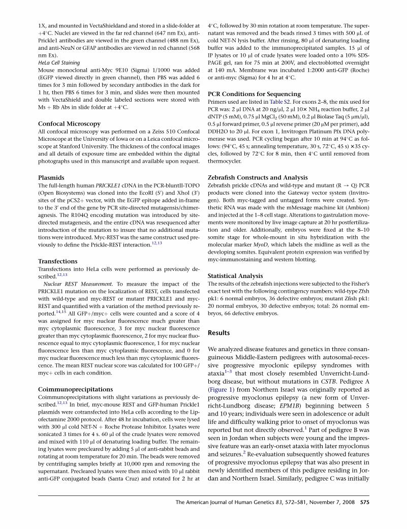
Prickle1 is expressed in multiple brain regions through-
out mouse embryonic development, including regions
implicated in epilepsy such as the hippocampus, cerebral
576 The American Journal of Human Genetics 83, 572–581, Novem
cortex, and thalamus, as well as the primitive cerebel-
lum17–20 (and data not shown). We performed immuno-
histochemical analysis with two different Prickle1-specific
antibodies, each directed against a distinct epitope. In this
way we also detected Prickle1 expression in the postnatal
murine thalamus, hippocampus, cerebral cortex, and cere-
bellum (Figure 2). Costaining these tissues with the neu-
ron-specific marker NeuN and the glia-specific marker
GFAP demonstrated that Prickle1 is specifically expressed
in neurons, but not in glia (Figures 2C, 2D, 2G, 2H, 2K,
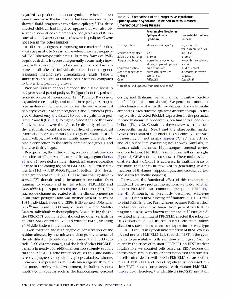
and 2L; cerebellum costaining not shown). Similarly, in
human adult thalamus, hippocampus, cerebral cortex,
and cerebellum, PRICKLE1 is in neurons rather than glia
(Figure 3; GFAP staining not shown). These findings dem-
onstrate that PRICKLE1 is expressed in multiple areas of
the brain thought to be involved in generating seizures
(neurons of thalamus, hippocampus, and cerebral cortex)
and ataxia (cerebellar neurons).
To evaluate the functional effect of this mutation on
PRICKLE1-partner protein interactions, we tested whether
mutant PRICKLE1 can coimmunoprecipitate REST (Fig-
ure 4). Although, as previously described, wild-type
PRICKLE1 binds REST directly,12,13 mutant PRICKLE1 fails
to bind REST in vitro. Furthermore, because REST nuclear
localization is altered in brains from patients with Hun-
tington’s disease with known mutations in Huntingtin,21
we tested whether mutant PRICKLE1 affected the subcellu-
lar localization of REST. Indeed, in HeLa cells, immunoloc-
alization shows that whereas overexpression of wild-type
PRICKLE1 results in cytoplasmic retention of REST, overex-
pressed mutant PRICKLE1 fails to retain REST in the cyto-
plasm (representative cells are shown in Figure 5A). To
quantify the effect of mutant PRICKLE1 on REST nuclear
localization, we counted cells based on REST expression
in the cytoplasm, nucleus, or both cytoplasm and nucleus,
in cells cotransfected with RESTþPRICKLE1 versus REST þmutant PRICKLE1 and found significantly increased nu-
clear REST in cells cotransfected with mutant PRICKLE1
(Figure 5B). Therefore, the identified PRICKLE1 mutation
Table 1. Comparison of the Progressive MyoclonusEpilepsy-Ataxia Syndrome Described Here to ClassicalUnverricht-Lundborg Disease
Progresssive MyoclonusEpilepsy-AtaxiaSyndrome
Unverricht-LundborgDiseasea
First symptom ataxia around age 4 yr myoclonic or
tonic-clonic seizures
Seizure onset: mean 7 yr 10–11 yr
Seizure onset: range 5–10 yr 6–16 yr
Progressive features worsening myoclonus,
ataxia, impaired up-gaze
worsening myoclonus,
ataxia
Cognitive decline mild or absent mild or absent
Mode of inheritance autosomal recessive autosomal recessive
Linkage 12p11–q13 21q22.3
Gene PRICKLE1 Cystatin B
a Modified and updated from Berkovic et al.1
ber 7, 2008
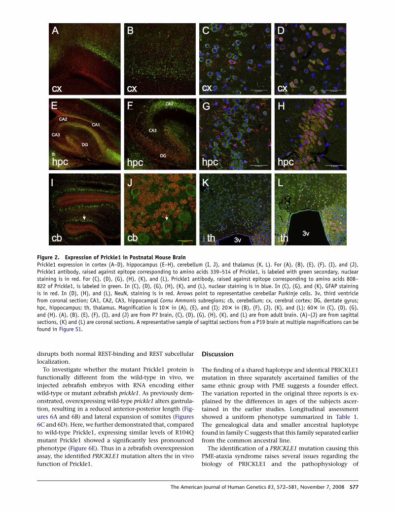
Figure 2. Expression of Prickle1 in Postnatal Mouse BrainPrickle1 expression in cortex (A–D), hippocampus (E–H), cerebellum (I, J), and thalamus (K, L). For (A), (B), (E), (F), (I), and (J),Prickle1 antibody, raised against epitope corresponding to amino acids 339–514 of Prickle1, is labeled with green secondary, nuclearstaining is in red. For (C), (D), (G), (H), (K), and (L), Prickle1 antibody, raised against epitope corresponding to amino acids 808–822 of Prickle1, is labeled in green. In (C), (D), (G), (H), (K), and (L), nuclear staining is in blue. In (C), (G), and (K), GFAP stainingis in red. In (D), (H), and (L), NeuN, staining is in red. Arrows point to representative cerebellar Purkinje cells. 3v, third ventriclefrom coronal section; CA1, CA2, CA3, hippocampal Cornu Ammonis subregions; cb, cerebellum; cx, cerebral cortex; DG, dentate gyrus;hpc, hippocampus; th, thalamus. Magnification is 103 in (A), (E), and (I); 203 in (B), (F), (J), (K), and (L); 603 in (C), (D), (G),and (H). (A), (B), (E), (F), (I), and (J) are from P7 brain, (C), (D), (G), (H), (K), and (L) are from adult brain. (A)–(J) are from sagittalsections, (K) and (L) are coronal sections. A representative sample of sagittal sections from a P19 brain at multiple magnifications can befound in Figure S1.
disrupts both normal REST-binding and REST subcellular
localization.
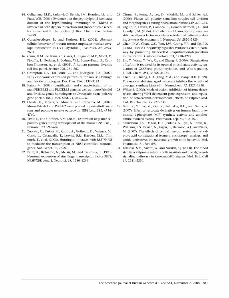
To investigate whether the mutant Prickle1 protein is
functionally different from the wild-type in vivo, we
injected zebrafish embryos with RNA encoding either
wild-type or mutant zebrafish prickle1. As previously dem-
onstrated, overexpressing wild-type prickle1 alters gastrula-
tion, resulting in a reduced anterior-posterior length (Fig-
ures 6A and 6B) and lateral expansion of somites (Figures
6C and 6D). Here, we further demonstrated that, compared
to wild-type Prickle1, expressing similar levels of R104Q
mutant Prickle1 showed a significantly less pronounced
phenotype (Figure 6E). Thus in a zebrafish overexpression
assay, the identified PRICKLE1 mutation alters the in vivo
function of Prickle1.
The America
Discussion
The finding of a shared haplotype and identical PRICKLE1
mutation in three separately ascertained families of the
same ethnic group with PME suggests a founder effect.
The variation reported in the original three reports is ex-
plained by the differences in ages of the subjects ascer-
tained in the earlier studies. Longitudinal assessment
showed a uniform phenotype summarized in Table 1.
The genealogical data and smaller ancestral haplotype
found in family C suggests that this family separated earlier
from the common ancestral line.
The identification of a PRICKLE1 mutation causing this
PME-ataxia syndrome raises several issues regarding the
biology of PRICKLE1 and the pathophysiology of
n Journal of Human Genetics 83, 572–581, November 7, 2008 577
Figure 3. Expression of PRICKLE1 in Adult Human BrainLow-power (A–D) and high-power (E–H) confocal images from immunostaining of adult human thalamus (A, E), hippocampus (B, F),cerebral cortex (C, G), and cerebellum (D, H). PRICKLE1 staining is in green, NeuN is in red, and nuclear staining is in blue. Scale markersare represented on the bottom right corner of each image.
neurological disease in these affected individuals.
PRICKLE1 is part of the noncanonical or planar cell polar-
ity (WNT/PCP) signaling pathway. In vivo studies in de-
veloping flies, frogs, and fish clearly demonstrated that
Prickle1 is important for planar polarity signaling. Re-
cently, mice lacking Prickle1 were shown to die early in
gestation, confirming an essential role for Prickle1 in
development (H. Tao et al., 2008, Jap. Soc. Dev. Biol., ab-
stract). At least some PRICKLE1 functions seem to be pres-
ent in protein with the R104Q mutation because the mu-
tation does not affect PRICKLE1 protein expression nor
subcellular localization (Figure 5) and it still retains some
wild-type function in our zebrafish overexpression system
(Figure 6).
Our in vitro studies suggest that PRICKLE1 normally
binds and translocates REST to the cytoplasm, thereby
preventing REST from silencing target genes. The
R104Q PRICKLE1 mutation lies within a known protein
binding domain and thus disrupts REST binding (Fig-
ure 4), blocking the normal transport of REST out of
the nucleus (Figure 5). These results suggest that tissues
expressing mutant PRICKLE1 contain constitutively ac-
tive REST which inappropriately downregulates REST tar-
get genes. This is significant because in addition to silenc-
ing neuronal genes in nonneuronal cells and neuronal
precursors, REST also regulates target genes in mature
neurons.22 REST targets include ion channels and neuro-
transmitters, and the PME-ataxia syndrome may occur
when brain regions expressing mutant PRICKLE1 misex-
press these target genes. Although Prickle function was
implicated in the control of cell division and morphogen-
esis during zebrafish neurulation23 and REST activity was
578 The American Journal of Human Genetics 83, 572–581, Novemb
recently described in fish and frogs,24 a role for the
PRICKLE/REST interaction during neurogenesis has not
yet been studied.
Figure 4. R104Q Mutant PRICKLE1 Has Impaired NRSF/RESTBindingCoimmunoprecipitation of REST with wild-type (WT) or R104Qmutant (MUT) encoding PRICKLE1 demonstrates decreased RESTbinding for MUT PRICKLE1. MYC-REST and either GFP-tagged WTor R104Q MUT PRICKLE1 plasmids were transfected into HeLa cells.Cell lysates were prepared in RIPA buffer and immunoprecipitatedwith agarose-conjugated anti-GFP antibody (A) or anti-MYC anti-body (B). Immunoprecipitates (IP) were subjected to SDS-PAGEfollowed by western blotting (WB) with MYC or GFP antibodies.WB antibody noted to left of gels. The input (1/5) of immunopre-cipitation is shown in the ‘‘Lys’’ lanes. Arrows to the right of thegels note the position of MYC-REST and GFP-PRICKLE1.
er 7, 2008
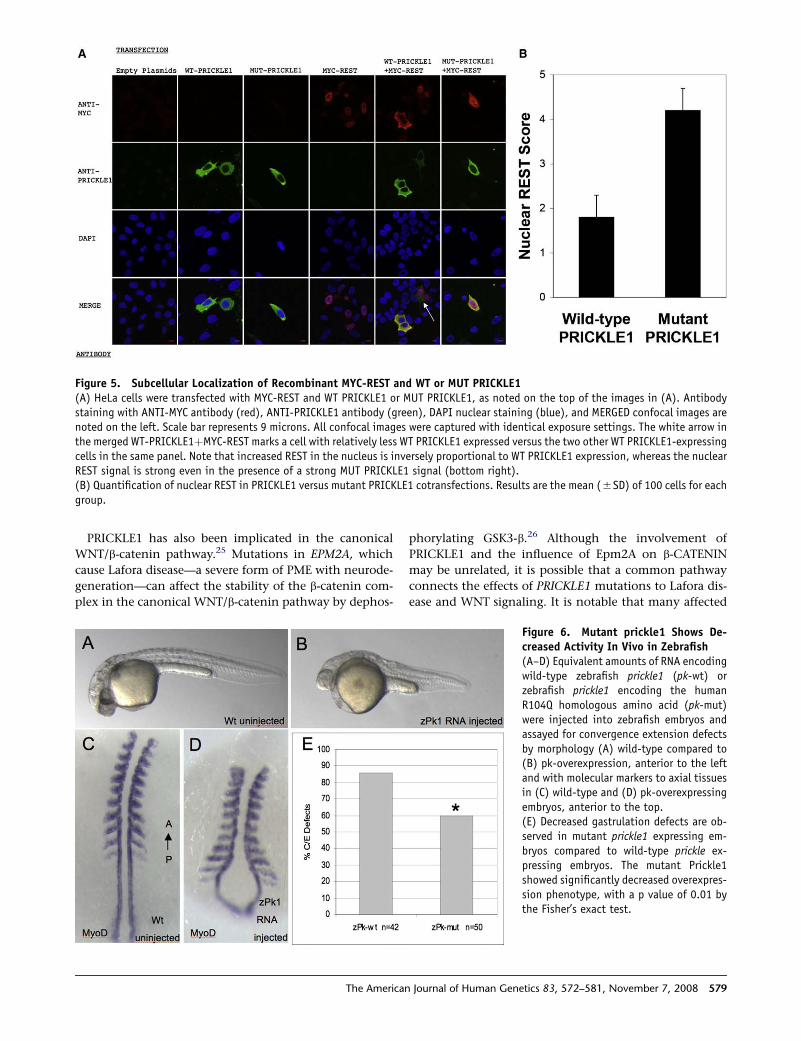
Figure 5. Subcellular Localization of Recombinant MYC-REST and WT or MUT PRICKLE1(A) HeLa cells were transfected with MYC-REST and WT PRICKLE1 or MUT PRICKLE1, as noted on the top of the images in (A). Antibodystaining with ANTI-MYC antibody (red), ANTI-PRICKLE1 antibody (green), DAPI nuclear staining (blue), and MERGED confocal images arenoted on the left. Scale bar represents 9 microns. All confocal images were captured with identical exposure settings. The white arrow inthe merged WT-PRICKLE1þMYC-REST marks a cell with relatively less WT PRICKLE1 expressed versus the two other WT PRICKLE1-expressingcells in the same panel. Note that increased REST in the nucleus is inversely proportional to WT PRICKLE1 expression, whereas the nuclearREST signal is strong even in the presence of a strong MUT PRICKLE1 signal (bottom right).(B) Quantification of nuclear REST in PRICKLE1 versus mutant PRICKLE1 cotransfections. Results are the mean (5SD) of 100 cells for eachgroup.
PRICKLE1 has also been implicated in the canonical
WNT/b-catenin pathway.25 Mutations in EPM2A, which
cause Lafora disease—a severe form of PME with neurode-
generation—can affect the stability of the b-catenin com-
plex in the canonical WNT/b-catenin pathway by dephos-
The America
phorylating GSK3-b.26 Although the involvement of
PRICKLE1 and the influence of Epm2A on b-CATENIN
may be unrelated, it is possible that a common pathway
connects the effects of PRICKLE1 mutations to Lafora dis-
ease and WNT signaling. It is notable that many affected
Figure 6. Mutant prickle1 Shows De-creased Activity In Vivo in Zebrafish(A–D) Equivalent amounts of RNA encodingwild-type zebrafish prickle1 (pk-wt) orzebrafish prickle1 encoding the humanR104Q homologous amino acid (pk-mut)were injected into zebrafish embryos andassayed for convergence extension defectsby morphology (A) wild-type compared to(B) pk-overexpression, anterior to the leftand with molecular markers to axial tissuesin (C) wild-type and (D) pk-overexpressingembryos, anterior to the top.(E) Decreased gastrulation defects are ob-served in mutant prickle1 expressing em-bryos compared to wild-type prickle ex-pressing embryos. The mutant Prickle1showed significantly decreased overexpres-sion phenotype, with a p value of 0.01 bythe Fisher’s exact test.
n Journal of Human Genetics 83, 572–581, November 7, 2008 579
individuals with PME respond to medications such as val-
proic acid, which also affects the WNT/b-catenin signaling
pathway through inhibition of GSK3-b.27,28 On the other
hand, valproic acid was also shown to alter inositol levels,
which would interfere with calcium regulation.29–31
It is not yet clear how a mutation in human PRICKLE1
leads to the PME syndrome, which is pathophysiologically
characterized by increased cortical hyperexcitability with
involvement of the cerebellum and probably other deep
gray matter nuclei as well.
Future analysis of the effects of specific point mutations
of Prickle1 in animal models will further elucidate the mo-
lecular mechanisms underlying this PME-ataxia syndrome.
Understanding the molecular and cellular basis of the dis-
ease may lead to improved diagnostic and therapeutic
approaches for afflicted individuals.
Supplemental Data
Supplemental Data include one figure and two tables and can be
found with this article online at http://www.ajhg.org/.
Acknowledgments
We thank the members of the families for their participation. We
thank Chantal Allamargot and Kathy Walters for their assistance
with immunostaining and confocal microscopy at the University
of Iowa, and Kaye Suyama for her work with the antibodies at Stan-
ford. A.G.B. is supported by NIH/NINDS grant K08NS48174. M.P.S.
is an investigator of the Howard Hughes Medical Institute. D.C.S.
and H.L.G. were supported by NIH CA112369. M.S. was supported
by NIH/NCRR P20RR020171 and NIH MH067123. A.B. was sup-
ported by a grant from the Epilepsy Foundation. We thank Jeffrey
Murray for access to the CEPH-HGD panel and for commentary
on the manuscript. The authors have no disclosures and no con-
flicts of interest. A.G.B. wrote the manuscript, performed all immu-
nohistochemical studies in HeLa cells and half of the tissue immu-
nostaining, and oversaw all aspects of the resequencing, control
genotyping, and coimmunoprecipitations. H.E. and A.D. clinically
evaluated part of pedigree B. S.B. clinically evaluated all the pedi-
grees and integrated the clinical data, together with Z.A., S.K.,
A.K., M.N., S.W., A.Z., and R.S. H.E. and S.B. designed the mapping
strategies. R.W., A.B., and S.C. performed the fine mapping, rese-
quencing, and genotyping studies. A.B. compiled Table S1. S.C.
compiled Table S2. A.R.B. performed the coimmunoprecipitations.
P.F. and J.M. assisted in the design and analysis of he genotyping as-
says. M.N. ascertained and prepared the specimens for human tis-
sue staining. S.W. prepared the Prickle1 antibody at the University
of Iowa. P.G. assisted in the development of the cell-culture exper-
iments. D.C.S. oversaw all zebrafish injections and analyses per-
formed by H.L.G. M.S. provided the myc-REST construct and guid-
ance in cell transfections. S.M. provided a mouse prickle1-GFP
expression vector that was used in pilot studies in preparation for
the use of a human Prickle1-EGFP construct. J.A., M.P.S., D.A.,
and E.K.V. produced the Prickle1 antibodies and performed the
immunostaining at Stanford University.
Received: August 19, 2008
Revised: September 28, 2008
Accepted: October 3, 2008
Published online: October 30, 2008
580 The American Journal of Human Genetics 83, 572–581, Novemb
Web Resources
The URLs for data presented herein are as follows:
Online Mendelian Inheritance in Man (OMIM), http://www.ncbi.
nlm.nih.gov/Omim/
PHRAP, http://www.phroap.org
References
1. Berkovic, S.F., Mazarib, A., Walid, S., Neufeld, M.Y., Manelis, J.,
Nevo, Y., Korczyn, A.D., Yin, J., Xiong, L., Pandolfo, M., et al.
(2005). A new clinical and molecular form of Unverricht-
Lundborg disease localized by homozygosity mapping. Brain
128, 652–658.
2. El-Shanti, H., Daoud, A., Sadoon, A.A., Leal, S.M., Chen, S.,
Lee, K., and Spiegel, R. (2006). A distinct autosomal recessive
ataxia maps to chromosome 12 in an inbred family from Jor-
dan. Brain Dev. 28, 353–357.
3. Straussberg, R., Basel-Vanagaite, L., Kivity, S., Dabby, R., Cirak,
S., Nurnberg, P., Voit, T., Mahajnah, M., Inbar, D., Saifi, G.M.,
et al. (2005). An autosomal recessive cerebellar ataxia syn-
drome with upward gaze palsy, neuropathy, and seizures. Neu-
rology 64, 142–144.
4. Ciani, L., and Salinas, P.C. (2005). WNTs in the vertebrate ner-
vous system: from patterning to neuronal connectivity. Nat.
Rev. Neurosci. 6, 351–362.
5. Veeman, M.T., Axelrod, J.D., and Moon, R.T. (2003). A second
canon. Functions and mechanisms of beta-catenin-indepen-
dent Wnt signaling. Dev. Cell 5, 367–377.
6. Carreira-Barbosa, F., Concha, M.L., Takeuchi, M., Ueno, N.,
Wilson, S.W., and Tada, M. (2003). Prickle 1 regulates cell
movements during gastrulation and neuronal migration in
zebrafish. Development 130, 4037–4046.
7. Veeman, M.T., Slusarski, D.C., Kaykas, A., Louie, S.H., and
Moon, R.T. (2003). Zebrafish prickle, a modulator of nonca-
nonical Wnt/Fz signaling, regulates gastrulation movements.
Curr. Biol. 13, 680–685.
8. Takeuchi, M., Nakabayashi, J., Sakaguchi, T., Yamamoto, T.S.,
Takahashi, H., Takeda, H., and Ueno, N. (2003). The prickle-
related gene in vertebrates is essential for gastrulation cell
movements. Curr. Biol. 13, 674–679.
9. Bastock, R., Strutt, H., and Strutt, D. (2003). Strabismus is
asymmetrically localised and binds to Prickle and Dishevelled
during Drosophila planar polarity patterning. Development
130, 3007–3014.
10. Bellaiche, Y., Beaudoin-Massiani, O., Stuttem, I., and Schweis-
guth, F. (2004). The planar cell polarity protein Strabismus
promotes Pins anterior localization during asymmetric divi-
sion of sensory organ precursor cells in Drosophila. Develop-
ment 131, 469–478.
11. Tree, D.R., Shulman, J.M., Rousset, R., Scott, M.P., Gubb, D.,
and Axelrod, J.D. (2002). Prickle mediates feedback amplifica-
tion to generate asymmetric planar cell polarity signaling. Cell
109, 371–381.
12. Shimojo, M., and Hersh, L.B. (2003). REST/NRSF-interacting
LIM domain protein, a putative nuclear translocation recep-
tor. Mol. Cell. Biol. 23, 9025–9031.
13. Shimojo, M., and Hersh, L.B. (2006). Characterization of the
REST/NRSF-interacting LIM domain protein (RILP): localiza-
tion and interaction with REST/NRSF. J. Neurochem. 96,
1130–1138.
er 7, 2008
14. Galigniana, M.D., Radanyi, C., Renoir, J.M., Housley, P.R., and
Pratt, W.B. (2001). Evidence that the peptidylprolyl isomerase
domain of the hsp90-binding immunophilin FKBP52 is
involved in both dynein interaction and glucocorticoid recep-
tor movement to the nucleus. J. Biol. Chem. 276, 14884–
14889.
15. Gonzalez-Alegre, P., and Paulson, H.L. (2004). Aberrant
cellular behavior of mutant torsinA implicates nuclear enve-
lope dysfunction in DYT1 dystonia. J. Neurosci. 24, 2593–
2601.
16. Cann, H.M., de Toma, C., Cazes, L., Legrand, M.F., Morel, V.,
Piouffre, L., Bodmer, J., Bodmer, W.F., Bonne-Tamir, B., Cam-
bon-Thomsen, A., et al. (2002). A human genome diversity
cell line panel. Science 296, 261–262.
17. Crompton, L.A., Du Roure, C., and Rodriguez, T.A. (2007).
Early embryonic expression patterns of the mouse Flamingo
and Prickle orthologues. Dev. Dyn. 236, 3137–3143.
18. Katoh, M. (2003). Identification and characterization of hu-
man PRICKLE1 and PRICKLE2 genes as well as mouse Prickle1
and Prickle2 genes homologous to Drosophila tissue polarity
gene prickle. Int. J. Mol. Med. 11, 249–256.
19. Okuda, H., Miyata, S., Mori, Y., and Tohyama, M. (2007).
Mouse Prickle1 and Prickle2 are expressed in postmitotic neu-
rons and promote neurite outgrowth. FEBS Lett. 581, 4754–
4760.
20. Tissir, F., and Goffinet, A.M. (2006). Expression of planar cell
polarity genes during development of the mouse CNS. Eur. J.
Neurosci. 23, 597–607.
21. Zuccato, C., Tartari, M., Crotti, A., Goffredo, D., Valenza, M.,
Conti, L., Cataudella, T., Leavitt, B.R., Hayden, M.R., Tim-
musk, T., et al. (2003). Huntingtin interacts with REST/NRSF
to modulate the transcription of NRSE-controlled neuronal
genes. Nat. Genet. 35, 76–83.
22. Palm, K., Belluardo, N., Metsis, M., and Timmusk, T. (1998).
Neuronal expression of zinc finger transcription factor REST/
NRSF/XBR gene. J. Neurosci. 18, 1280–1296.
The America
23. Ciruna, B., Jenny, A., Lee, D., Mlodzik, M., and Schier, A.F.
(2006). Planar cell polarity signalling couples cell division
and morphogenesis during neurulation. Nature 439, 220–224.
24. Olguin, P., Oteiza, P., Gamboa, E., Gomez-Skarmeta, J.L., and
Kukuljan, M. (2006). RE-1 silencer of transcription/neural re-
strictive silencer factor modulates ectodermal patterning dur-
ing Xenopus development. J. Neurosci. 26, 2820–2829.
25. Chan, D.W., Chan, C.Y., Yam, J.W., Ching, Y.P., and Ng, I.O.
(2006). Prickle-1 negatively regulates Wnt/beta-catenin path-
way by promoting Dishevelled ubiquitination/degradation
in liver cancer. Gastroenterology 131, 1218–1227.
26. Liu, Y., Wang, Y., Wu, C., and Zheng, P. (2006). Dimerization
of Laforin is required for its optimal phosphatase activity, reg-
ulation of GSK3beta phosphorylation, and Wnt signaling.
J. Biol. Chem. 281, 34768–34774.
27. Chen, G., Huang, L.D., Jiang, Y.M., and Manji, H.K. (1999).
The mood-stabilizing agent valproate inhibits the activity of
glycogen synthase kinase-3. J. Neurochem. 72, 1327–1330.
28. Wiltse, J. (2005). Mode of action: inhibition of histone deace-
tylase, altering WNT-dependent gene expression, and regula-
tion of beta-catenin–developmental effects of valproic acid.
Crit. Rev. Toxicol. 35, 727–738.
29. Galit, S., Shirley, M., Ora, K., Belmaker, R.H., and Galila, A.
(2007). Effect of valproate derivatives on human brain myo-
inositol-1-phosphate (MIP) synthase activity and amphet-
amine-induced rearing. Pharmacol. Rep. 59, 402–407.
30. Shimshoni, J.A., Dalton, E.C., Jenkins, A., Eyal, S., Ewan, K.,
Williams, R.S., Pessah, N., Yagen, B., Harwood, A.J., and Bialer,
M. (2007). The effects of central nervous system-active val-
proic acid constitutional isomers, cyclopropyl analogs, and
amide derivatives on neuronal growth cone behavior. Mol.
Pharmacol. 71, 884–892.
31. Tokuoka, S.M., Saiardi, A., and Nurrish, S.J. (2008). The mood
stabilizer valproate inhibits both inositol- and diacylglycerol-
signaling pathways in Caenorhabditis elegans. Mol. Biol. Cell
19, 2241–2250.
n Journal of Human Genetics 83, 572–581, November 7, 2008 581