Embed Size (px)
Citation preview
A microinjection technique for targeting regions of embryonic andneonatal mouse brain in vivo
Steve Davidson1,2,3, Hai Truong2, Yasushi Nakagawa1,2, and Glenn J Giesler Jr1,21Graduate Program in Neuroscience, University of Minnesota, Minneapolis, MN. 554552Department of Neuroscience, University of Minnesota, Minneapolis, MN. 55455
AbstractA simple pressure injection technique was developed to deliver substances into specific regions ofthe embryonic and neonatal mouse brain in vivo. The retrograde tracers Fluorogold and cholera toxinB subunit were used to test the validity of the technique. Injected animals survived the duration oftransport (24–48 hrs) and then were sacrificed and perfused with fixative. Small injections (≤ 50 nL)were contained within targeted structures of the perinatal brain and labeled distant cells of origin inseveral model neural pathways. Traced neural pathways in the perinatal mouse were further examinedwith immunohistochemical methods to test the feasibility of double labeling experiments duringdevelopment. Several experimental situations in which this technique would be useful are discussed,for example, to label projection neurons in slice or culture preparations of mouse embryos andneonates. The administration of pharmacological or genetic vectors directly into specific neuraltargets during development should also be feasible. An examination of the form of neural pathwaysduring early stages of life may lead to insights regarding the functional changes that occur duringcritical periods of development and provide an anatomic basis for some neurodevelopmentaldisorders.
Keywordsfetus; embryo; retrograde; mouse; development; tracer
1. INTRODUCTIONMany contributions have been made to improve our understanding of the nervous system bytargeting specific regions of the brain. As information about the localization of function hasincreased, the ability to precisely inject neural tracers, pharmacological agents or gene vectorshas become increasingly important. Tools to manipulate the genome of mice have made thisspecies the principal model for the study of mammalian neural development. However,technical challenges such as small size and anesthetic requirements have preventedinvestigators from targeting specific neural structures in the embryonic and neonatal mouse
© 2009 Elsevier B.V. All rights reserved.Corresponding Author: Glenn J Giesler Jr., Department of Neuroscience, University of Minnesota, 6-145 Jackson Hall, Church St. SE,Minneapolis, MN. 55455, Phone: (612) 624-3984, Fax: (612) 626-5009, [email protected] address: Washington University Pain Center and Department of Anesthesiology, Washington University School of Medicine,St. Louis, MO. 63110Publisher's Disclaimer: This is a PDF file of an unedited manuscript that has been accepted for publication. As a service to our customerswe are providing this early version of the manuscript. The manuscript will undergo copyediting, typesetting, and review of the resultingproof before it is published in its final citable form. Please note that during the production process errors may be discovered which couldaffect the content, and all legal disclaimers that apply to the journal pertain.
NIH Public AccessAuthor ManuscriptBrain Res. Author manuscript; available in PMC 2011 January 11.
Published in final edited form as:Brain Res. 2010 January 11; 1307: 43–52. doi:10.1016/j.brainres.2009.10.024.
NIH
-PA Author Manuscript
NIH
-PA Author Manuscript
NIH
-PA Author Manuscript
brain. Recently, injection of genetic material for electroporation into the ventricles ofembryonic mice has been described (Saito 2006; Shimogori and Ogawa, 2008). However, asimple method for the injection of neural tracers, pharmacological agents or gene vectors intotargeted regions of the grey matter has not been communicated. Here we used neural tracersto demonstrate the targeting of specific regions of the perinatal mouse brain. The technique isfast and simple and requires minimal equipment.
In vivo microinjection provides opportunities to examine neuroanatomy during developmentand also to conduct subsequent experiments with identified projection neuron populations. Forexample, in vivo microinjection of retrograde tracers permits the examination of functionalcharacteristics of identified projection neurons in slice or culture preparations. Such studieshave been previously performed, but only in older rats which prevent the option to exploittransgenic strategies that are available in the mouse (Dahlhaus et al., 2005; Sooksawate et al.,2005; Evans et al., 2003). Targeting of specific brain regions with tracers during developmentalperiods can be combined with immunohistochemical methods and the administration ofpharmacological treatments. This type of study could lead to a more complete understandingof the cellular and molecular mechanisms that regulate perinatal pharmacology. For example,little is known about the neural mechanisms that regulate the often altered effects ofmedications and other drugs when administered during the perinatal period, including duringpregnancy (Thompson et al., 2009; Fitzgerald and Walker 2009).
Despite technical difficulties, some progress has been made in microinjecting substances intoperinatal organisms. Stereotaxic placement of electrodes or pipettes has been described forneonatal rats (Hoorneman, 1985; Joosten and van Eden, 1989), and a stereotaxic apparatus forthe neonatal rat or adult mouse is now commercially available. The direct injection of a neuraltracer into the cortex of embryonic rats has been demonstrated in vivo (Sbriccolli et al., 1999).However, mice are now the model organism of choice for many fields including developmentalneurobiology because of the availability of strains with targeted genetic alterations. Despitethe rapid growth in use of the mouse, knowledge of mouse neuroanatomy during earlydevelopment is lacking and therefore complete descriptions of the neurons that expressidentified genes are missing. This report introduces a basic in vivo technique for microinjectionof small volumes of biologically interesting substances into targeted brain regions of perinatalmice. Neuroanatomical tract tracing combined with immunohistochemistry is used todemonstrate the targeting of regions within the deep forebrain and cerebellum with the widelyadopted retrograde tracers Fluorogold and cholera toxin B subunit (CTB).
2. RESULTSNeonatal Injections
Under hypothermia-induced anesthesia, neonatal mice (postnatal day 0 – 5) were positionedupon a stage in either a forebrain or hindbrain configuration and injected through the skullusing a pulled glass pipette containing the retrograde tracer Fluorogold or CTB. All animalsrecovered quickly after the injection and their behaviors, including nursing, wereindistinguishable from non-injected littermates. Two days after the injection neonates wereperfused with 4% paraformaldehyde and dissected. Whole brains injected with the tracerFluorogold were inspected under a handheld UV light to determine whether the injection wassuccessful (Figure 1A). In some cases the dye failed to flow from a clogged pipette tip andthese brains were discarded before sectioning. All CTB injected brains were sectioned. Aftera couple of litters for learning trials, the success rate for hitting selected neural targets inneonates reached a plateau of about 70%.
An example of a mouse cerebellum injected at P0 with 50 nL of 4% Fluorogold is shown inFigure 1. A coronal section through the cerebellum shows the injection site and extent of tracer
Davidson et al. Page 2
Brain Res. Author manuscript; available in PMC 2011 January 11.
NIH
-PA Author Manuscript
NIH
-PA Author Manuscript
NIH
-PA Author Manuscript
spread (Fig. 1B). Retrogradely labeled neurons were found in the inferior olive of thecontralateral medulla (Fig. 1C) and in Clarke’s column of the ipsilateral thoracic spinal cord(Fig. 1D). To show that alternatives to Fluorogold can be injected using this technique, CTBconjugated with Alexa Fluor 594 was injected into P0 neonates. CTB can work as a retrogradetracer and has the advantage of not inducing necroses at the injection site. The results of asuccessful injection of 50 nL of CTB show the tracer contained completely within the thalamus(Fig 2A). Another injection of CTB is shown into a well circumscribed region within thestriatum at the level of the anterior commissure (Fig 2B).
Embryonic InjectionInjections into 4–7 embryos typically took 30 minutes. Twenty-four hours after tracerinjections the dam was re-anesthetized and embryos were removed from the uterus forperfusion. In successful experiments, over 90% of embryos within a litter survived. Sometimes(~20%) an entire litter was lost due to early parturition, or less frequently the entire litter didnot survive but remained within the uterus until the second laparotomy. Surviving wholeembryos were placed under a UV light to determine whether Fluorogold was injectedsuccessfully and embryos containing tracer were perfused. All CTB injected embryos wereperfused and sectioned. Of the injected embryos approximately 25% contained dye in thetargeted region that did not spread appreciably into adjacent grey matter. Therefore a singlepregnant dam would typically yield 1–2 successfully injected embryos. A typical example ofa successful injection into the thalamus of an E17 mouse is shown in Figure 3A. Labeled cellsof origin were observed in the spinal cord (Fig 3B) and the morphology of individual cellsincluding their processes could be observed (Fig 3C). Another embryo received an injectioninto the basal ganglia at the level of the anterior commissure (Fig 4A). Labeled cells of originwere observed within the substantia nigra pars compacta (SNC) and the ventral tegmental area(VTA; Fig 4B).
Combined retrograde labeling and immunohistochemistryRetrograde labeling was combined with immunohistochemistry to determine the effectivenessa double labeling strategy in perinatal mice. Fluorogold (50 nL) was injected into the striatumof a P0 neonatal mouse and the retrograde labeling was observed in the ipsilateral SNC andVTA (Fig 5A). Morphological features of individual projection neurons are shown at highermagnification in Figure 5B. An antibody against tyrosine hydroxylase (TH) was used todetermine whether retrogradely labeled neurons could be characterized as dopaminergic.Tissue processed for immunoreactivity to TH displayed bilateral labeling for cells within boththe VTA and SNC (Fig 5C). Numerous double labeled cells located only on the side ipsilateralto the injection site are shown in Figure 5D, indicating that dopaminergic neurons project tothe striatum by P2 (arrows). Additionally, some retrogradely labeled neurons were observedwithout immunoreactivity for TH, suggesting a population of neurons projecting to the striatumat P2 that do not produce dopamine (arrowheads).
3. DISCUSSIONThe direct injection of substances into the grey matter of adult organisms has lead to moreprecise localization of function and a better understanding of anatomic connectivity. However,the difficulty of targeting substances into specific brain areas of living embryos and neonateshas retarded the analyses of neural development. The focus of this report was to demonstratethat precise microinjections of biologically active substances can be made into regions of theembryonic and neonatal mouse brain. We used the neural tracers Fluorogold and CTB to showthat neural structures can be specifically targeted with small, well circumscribed injections inthe perinatal mouse. Retrogradely labeled cells of origin were identified in several neuralpathways, demonstrating that tracing studies using direct microinjection techniques is a
Davidson et al. Page 3
Brain Res. Author manuscript; available in PMC 2011 January 11.
NIH
-PA Author Manuscript
NIH
-PA Author Manuscript
NIH
-PA Author Manuscript
feasible strategy for the study of perinatal mouse neuroanatomy. Additionally, we showed thatimmunohistochemistry can be used in combination with tracing studies.
Comparison of the pattern of retrogradely labeled cells from injections into the striatum,thalamus and cerebellum were consistent with previously described patterns of labeling in ratsand adult mice. Each of the projections systems that were targeted: the spinothalamic tract, theclimbing fibers of the inferior olive, and the substantia nigra-ventral tegmental areadopaminergic pathway, appeared homologous to previously reported patterns in adult rats andmice (Sotelo et al., 1984; Burstein et al., 1990; Anderson et al., 2001; Hu et al. 2004). Thissuggests that many long neural pathways are formed early in mouse development and areamenable to further study. Immunohistochemistry methods and Fluorogold were demonstratedhere to be combined successfully in perinatal mice. In prior studies, in situ for RNA detectionand immunohistochemistry have been successfully combined with Fluorogold labeling in adultrodents (Burgunder and Young, 1988; Fried et al., 1989; Ju et al., 1989) suggesting possiblefuture strategies for experiments with perinatal mice.
The study of perinatal organisms has shown that sensory and motor function depends on criticalperiods of plasticity that occur during development in which proper growth and refinement ofneural circuitry is established (Hensch, 2004; Innocenti, 2007; Trachtenberg and Stryker,2001). Aberrant development of neural pathways from environmental stress or geneticmutation is thought to contribute to the establishment of neurodevelopmental disorders(Fumagalli et al., 2007). An understanding of the molecular mechanisms involved in plasticityduring critical periods has advanced quickly with the use of mutant mouse models (Taha andStryker, 2005). However, neuroanatomical observations have been missing from studies ofneurons at the origin of specific pathways in wild type and transgenic mouse models. Inaddition, transcription factors and other molecular markers that are expressed transiently in thedeveloping embryo and neonate are used now as a principal means of identification ofpopulations of neurons in the mouse spinal cord (Lee and Jessell, 1999; Helms and Johnson,2003), cortex (Molyneaux et al., 2007; Leone et al., 2008), cerebellum (Wang and Zoghbi,2001) and in the thalamus (Vue et al., 2007). However, these studies have lacked the capabilityto determine whether molecularly identified neural populations are interneurons or part of aprojection system. The microinjection of neural tracers into embryonic and neonatal mousebrain provides a means to identify and quantify projection neurons that are also molecularlyidentified. Furthermore, transgenic mice may be examined for altered pathway formation,which may explain certain developmental phenotypes and catalyze the determination of genefunction.
Several methods have been previously described to circumvent direct injection into embryosand neonates in vivo. For example, crystallized lipophilic dyes (e.g. DiI) or horseradishperoxidase (HRP) have been placed directly into fixed neural tissue and allowed to passivelydiffuse along axonal pathways (Godement et al., 1987; van der Togt and Feirabend, 1990; Huet al., 2004). Unfortunately, these dyes travel relatively short distances, can require longtransport times and may produce spurious or unwanted transcellular labeling (Godement et al.,1987; Kageyama and Robertson, 1993). Other studies have taken advantage of explanted tissuepreparations to trace longer pathways using dextran-amines or HRP (Smalheiser et al., 1981;Glover et al., 1986). The value of these methods is limited by the dyes available for transportin fixed tissue or the selection of model organisms and tissues for viable explants.
More importantly, the use of fixed or ex vivo tissue prevents the subsequent examination ofphysiological characteristics. For example, retrogradely labeled neurons have been used inconjunction with in slice electrophysiology or primary cell culture preparations; but suchstudies have previously used only older rats (Dahlhaus et al., 2005; Sooksawate et al., 2005;Evans et al., 2003) which are, unlike mice, impractical for the examination of the functional
Davidson et al. Page 4
Brain Res. Author manuscript; available in PMC 2011 January 11.
NIH
-PA Author Manuscript
NIH
-PA Author Manuscript
NIH
-PA Author Manuscript
roles of specific genes. Combining electrophysiology or calcium imaging with identifiedprojection neurons in transgenic and wild type mice may yield important and novelcontributions to our understanding of the functional significance of gene expression duringdevelopment.
The flexibility of the injection apparatus permits the introduction of other fluid or suspendedsubstances. Vectors for genetic materials such as adeno- and lentiviruses and pharmacologicalagents can be injected into targeted embryonic and neonatal brain sites. For example, Welchet al. (2007) injected into the striatum of young null mutant mice a lentivirus containing a copyof the deleted gene. The reintroduction of the gene into a targeted neural region produced arelatively quick and inexpensive tissue specific knock-in model which allowed the study ofthe expression of the gene on behavior during adulthood. The reintroduction of a deleted geneor over-expression of a gene into a targeted neural structure can provide information on therole of that gene in specific regions of the brain.
The retrograde tracers Fluorogold and CTB were used in these experiments because eachprovides a means to clearly identify the site of the injection. Fluorogold is thought to passthrough the lipid membrane of axons and terminals and is then driven into vesicles by a pHgradient and carried to the soma (Dado et al., 1990; Wessendorf 1991). Although easy to use,Fluorogold produces necrosis at the injection site prohibiting its use in behavioral experiments.CTB on the other hand is non-toxic and can be conjugated to various fluorescent probes tolabel multiple pathways simultaneously (Conte et al., 2009). The technique introduced herewas designed to allow classical microinjection techniques to be applied to embryonic andneonatal mice. The advantage of a pressure injection system is that pipettes can be loaded withthe substance of the investigators choice. The retrograde labeling provides evidence that neuralpathways in embryonic and neonatal mouse are extant and appropriate for further study. Theincorporation of microinjection into experiments on the developing mouse should help toreconcile classical neuroanatomy with studies in perinatal mice and may be an important toolfor increasing our understanding of the neural mechanisms that produce developmentaldisorders and for determining the success of therapeutic interventions during early life.
4. EXPERIMENTAL PROCEDUREAnimals
Timed pregnant female mice (CD1, Charles River) and neonatal mice, born on postnatal day0 (P0), were used in this study with the approval of the University of Minnesota InstitutionalCare Facility. Neonates were housed with their mother before and after the injection.
Stereotaxic ApparatusMost manipulators from stereotaxic systems can be adapted for these experiments. We usedan electrode manipulator stand with a 3-axis manipulator and an attached microinjection unit(Model 1449 Electrode Manipulator Stand, Model 1460-61 Electrode Manipulator, and Model5000 Microinjection Unit; David Kopf Instruments, Tejunga, CA). A 5 µL manual syringe(Model 95, Hamilton, Reno, NV) was fitted to the Microinjection Unit. Pipette tips were pulledfrom thin walled borosilicate glass capillary tubes (with filament) with an outer diameter of1.0 mm and inner diameter of 0.76 mm. The pipette was attached to the needle of the syringewith cyanoacrylate adhesive. Pipettes were backfilled with light mineral oil and then forwardfilled with 4% Fluorogold (Fluorochrome, LLC, Denver, CO) or CTB conjugated to AlexaFluor 594 (Invitrogen, Carlsbad, CA) dissolved at 5.0mg/ml in PBS. For injections intoembryos, a short length of PE-10 plastic tubing was glued surrounding the pipette shaft leavingonly a portion of the tip exposed. The tubing worked as a stopper so that the depth of theinjection would not exceed that which was desired. All injections were done with the aid of a
Davidson et al. Page 5
Brain Res. Author manuscript; available in PMC 2011 January 11.
NIH
-PA Author Manuscript
NIH
-PA Author Manuscript
NIH
-PA Author Manuscript
dissecting microscope. Anatomical borders in the perinatal mouse brain were determined withthe assistance of the Atlas of the Developing Mouse Brain (Paxinos et al., 2007).
Neonatal InjectionsFor injections into neonatal mice, a stage was cut from a small block of expanded polystyrenefoam and fixed to the center of the base plate with adhesive. This stage was used for pups fromP0 to P5 to position the body and help stabilize the head (Figure 6).
A neonate was placed in a container of crushed ice for about two minutes until the onset ofhypothermia induced anesthesia. In albino mice the transverse and superior sagittal sinuses canbe easily seen through the skin and skull. In pigmented mice the sinuses become difficult todiscern after about P3. The sinuses were used as landmarks to identify the desired pipetteinsertion point. A dot was drawn on the skin surface at the insertion point with a fine felt-tippedmarker (Figure 7A). For injections into the thalamus of P0–P5 neonates, the pipette insertionpoint was 0.7 mm lateral of the superior sagittal sinus and 1.2 mm rostral of the transversesinus. The pipette was inserted to a depth of 2.2 mm from the surface of the skin. For injectionsinto the caudate/putamen the insertion point was 1.3 mm lateral of the superior sagittal sinus,2.2 mm rostral of the transverse sinus and the depth from the skin was 2.0 mm. The cerebellumcould be directly observed through the bone and skin and a dot was placed on the skin at theinjection site. The depth for injections into the cerebellum was 1.8 mm from the surface of theskin. For neonates at P5 (and older), the parietal bone is thick enough to prevent easypenetration by the pipette. In these animals a 30 gauge needle was first used to make a smallpuncture at the pipette insertion point.
Once anesthetized, the neonate was removed from the ice and placed on the stage in theappropriate orientation for forebrain (Figure 6B) or hindbrain/cerebellar (Figure 6C) injections.The head was held in position by the finger tips of a second investigator. Importantly, the skinaround the pipette insertion point was gently stretched taut with the fingertips while the headwas held in place (Figure 7B). This greatly improved the ability of the pipette to penetrate theskin and bone at the designated location by preventing the skin from slipping over the bone.The pipette was lowered to the skin surface under a dissecting microscope, and then loweredto the appropriate depth. Thirty to 50 nL of 4% Fluorogold or CTB was delivered over 15seconds and the pipette was removed after 60 seconds. The neonate was immediately placedunder a warming lamp and on a warming pad. After approximately one minute color,movement, and reaction to touch appeared normal and the neonate was returned to the homecage. No animal failed to recover after this procedure. Neonates could be injected one afteranother as long as care was taken to ensure the tip of the pipette did not clog with tissue orcrystallized dye. A typical litter of 10 pups could be injected in about an hour.
Embryonic surgery and injectionsAll instruments were sterilized and sterile saline was used to keep the exposed organs moistduring the surgery. Timed pregnant females carrying embryonic day 17 (E17) embryos wereanesthetized with sodium pentobarbital (60 mg/kg ip) and placed on a heating pad that wasplaced over the base plate of the stereotaxic apparatus. The hair covering the abdomen wasclipped and the skin swabbed with an antiseptic. A 2.5 cm longitudinal incision was made withscissors along the midline of the abdomen to gain entry to the abdominal cavity. A portion ofthe uterus containing 2 or 3 embryos was gently extracted at one time. Ritodrine hydrochloride(100 µL at 14 mg/ml in saline) was applied directly to the surface of the exposed uterus to relaxthe smooth muscle. An individual embryo was manipulated in utero under a dissectingmicroscope using hand-held ring forceps (Figure 8). Care was taken to avoid contact of theforceps with the placenta. A second forceps with flat, blunted edges was used to assist inpositioning the embryo within the uterine sac. The head of the embryo was gently positioned
Davidson et al. Page 6
Brain Res. Author manuscript; available in PMC 2011 January 11.
NIH
-PA Author Manuscript
NIH
-PA Author Manuscript
NIH
-PA Author Manuscript
so as to be upright with the crown facing the pipette tip. For injections into the striatum thehead was rotated slightly along the longitudinal axis so that the pipette would penetrate morelaterally. The head of the embryo was held in direct contact with the uterine wall. The transverseand superior sagittal sinuses can be seen through the uterine wall and through the skin and boneof the embryo and were used as landmarks. Once the embryo was held stationary a secondinvestigator lowered the pipette through the uterine wall, to the depth of the stopper (Figure 8,inset) and 30 nL of Fluorogold was ejected. The pipette remained in place for 10 seconds aftercompletion of the injection and then was withdrawn. A few easily accessible embryos on eachside were injected and then placed back into the body cavity. The mother was sutured andrecovered on a warming pad for 1 hour and then moved back to her home cage. The motherwas re-anesthetized 24 hours later with pentobarbital and the abdominal incision reopened toaccess the embryos for hypothermia induced anesthesia and fixation by perfusion.
Fixation and HistologyFluorogold transports quickly after an injection and animals sacrificed after two hours fortransport displayed bright labeling of fiber bundles up to 2 mm away from the injection site(unpublished observations). For robust filling of cells we allowed a transport time of 24 hoursfor embryonic injections and 48 hours for neonatal injections. After this time the embryo orneonate was put on ice for hypothermia induced anesthesia. The mouse was then pinned ventralside up to a polystyrene block. Under a dissection microscope the ribcage was cut out with finescissors to provide access to the heart. The right atrium was lanced and a blunted 27 gaugeneedle inserted into the apex of the heart. Five milliliters of 0.9% saline and then 5 mL of 4%paraformaldehyde was perfused to fix the tissue. Solutions were delivered by pump at 1 mL/minute. Animals were then dissected so that only the skull and vertebral column remainedintact, i.e. organs, limbs, skin were all removed. The combined skull and vertebral columncontaining the brain and spinal cord were placed in a container of 4% PFA at 4 degreesovernight. The next day the nervous system was carefully removed from skull and vertebraeunder a dissecting scope. The brain and spinal cord were separated at the level of the tectumand embedded in cutting medium. Both were then sectioned in 50 micron sections on a cryostat,mounted onto a gel coated slide, dried and coverslips applied.
For immunohistochemistry after tracer injection, brains were fixed by intracardiac perfusionof 4% paraformaldehyde/0.1M phosphate buffer. Brains were submerged overnight in the samefixative. After washing with phosphate buffered saline (PBS) and cryoprotection in 30%sucrose, brains were frozen in OCT compound (Sakura). Twenty micron thick sections werecollected on Superfrost Plus slides (Fisher, Pittsburgh, PA) and dried on a slide warmerovernight. Sections were then rinsed in PBS and fixed 4% paraformaldehyde/0.1M phosphatebuffer. After three washes in PBS, the slides were immersed in boiled 10 mM citrate buffer(pH 6) for 5 minutes prior to blocking with 3% donkey serum / 0.3% Triton X-100/PBS for 1hour. Anti-tyrosine hydroxylase mouse monoclonal antibody (Millipore, MAB318) was usedat 1:200 dilution in the blocking solution and incubation was performed overnight at 4°C. Onthe second day, after washing in PBS, sections were incubated for 1 hour with Cy3-conjugatedanti-mouse IgG antibody (Jackson ImmunoResearch, West Grove, PA). After dehydration inascending concentrations of ethanol and clearing in xylene, slides were mounted in DPXmounting medium (EM Sciences, Hatfield, PA).
Fluorescence was visualized with an Olympus BX50 microscope under custom filter sets (forAlexa Fluor 594 and Cy3: exciter 541–551; emitter 572–608; for Fluorogold: exciter 351–371;emitter 420, longpass). Micrographs were captured digitally with a Scion 1.4 megapixelgrayscale scope-mounted camera and Scion Capture v2.0 software. Brightness and contrast ofwhole images was uniformly adjusted using Adobe Photoshop CS3.
Davidson et al. Page 7
Brain Res. Author manuscript; available in PMC 2011 January 11.
NIH
-PA Author Manuscript
NIH
-PA Author Manuscript
NIH
-PA Author Manuscript
AcknowledgmentsThis work was supported by National Institute of Neurological Disorders and Stroke, Grants NS-047399 andNS-059199 and by the Graduate School of the University of Minnesota.
REFERENCESAnderson DW, Neavin T, Smith JA, Schneider JS. Neuroprotective effects of pramipexole in young and
aged MPTP-treated mice. Brain Res 2001;905:44–53. [PubMed: 11423078]Burgunder JM, Young WS 3rd. The distribution of thalamic projection neurons containing
cholecystokinin messenger RNA, using in situ hybridization histochemistry and retrograde labeling.Brain Res 1988;464:179–189. [PubMed: 3208109]
Burstein R, Dado RJ, Giesler GJ Jr. The cells of origin of the spinothalamic tract of the rat: a quantitativereexamination. Brain Res 1990;511:329–337. [PubMed: 2334851]
Conte WL, Kamishina H, Reep RL. Multiple neuroanatomical tract-tracing using fluorescent Alexa Fluorconjugates of cholera toxin subunit B in rats. Nat Protoc 2009;4:1157–1166. [PubMed: 19617887]
Dado RJ, Burstein R, Cliffer KD, Giesler GJ Jr. Evidence that Fluoro-Gold can be transported avidlythrough fibers of passage. Brain Res 1990;533:329–333. [PubMed: 1705157]
Dahlhaus A, Ruscheweyh R, Sandkühler J. Synaptic input of rat spinal lamina I projection andunidentified neurones in vitro. J Physiol 2005;566(Pt 2):355–368. [PubMed: 15878938]
Evans C, Baxi S, Neff R, Venkatesan P, Mendelowitz D. Synaptic activation of cardiac vagal neuronsby capsaicin sensitive and insensitive sensory neurons. Brain Res 2003;979(1–2):210–215. [PubMed:12850588]
Fumagalli F, Molteni R, Racagni G, Riva MA. Stress during development: Impact on neuroplasticity andrelevance to psychopathology. Prog Neurobiol 2007;81:197–217. [PubMed: 17350153]
Fried K, Arvidsson J, Robertson B, Brodin E, Theodorsson E. Combined retrograde tracing and enzyme/immunohistochemistry of trigeminal ganglion cell bodies innervating tooth pulps in the rat.Neuroscience 1989;33:101–109. [PubMed: 2481244]
Fitzgerald M, Walker SM. Infant pain management: a developmental neurobiological approach. Nat ClinPract Neurol 2009;5:35–50. [PubMed: 19129789]
Glover JC, Petursdottir, Jansen JKS. Fluorescent dextran-amines used as axonal tracers in the nervoussystem of the chicken embryo. J Neurosci Methods 1986;18:243–254. [PubMed: 2432362]
Godement P, Vanselow J, Thanos S, Bonhoeffer F. A study in developing visual systems with a newmethod of staining neurones and their processes in fixed tissue. Development 1987;101:697–713.[PubMed: 2460302]
Helms AW, Johnson JE. Specificatin of dorsal spinal cord interneurons. Curr Opin Neurobiol2003;13:42–49. [PubMed: 12593981]
Hensch TK. Critical period regulation. Annu Rev Neurosci 2004;27:549–579. [PubMed: 15217343]Hoorneman EM. Stereotaxic operation in the neonatal rat; a novel and simple procedure. J Neurosci
Methods 1985;14:109–116. [PubMed: 2993760]Hu Z, Cooper M, Crockett DP, Zhou R. Differentiation of the midbrain dopaminergic pathways during
mouse development. J Comp Neurol 2004;476:301–311. [PubMed: 15269972]Innocenti GM. Subcortical regulation of cortical development: some effects of early, selective
deprivations. Prog Brain Res 2007;164:23–37. [PubMed: 17920424]Joosten EA, van Eden CG. An anterograde tracer study on the development of corticospinal projections
from the medial prefrontal cortex in the rat. Brain Res Dev Brain Res 1989;45:313–319.Ju G, Han ZS, Fan LZ. Fluorogold as a retrograde tracer used in combination with immunohistochemistry.
J Neurosci Methods 1989;29:69–72. [PubMed: 2668650]Kageyama GH, Robertson RT. Transcellular retrograde labeling of radial glial cells with WGA-HRP and
DiI in neonatal rat and hamster. Glia 1993;9:70–81. [PubMed: 7503953]Lee KJ, Jessell TM. The specification of dorsal cell fates in the vertebrate central nervous system. Annu
Rev Neurosci 1999;22:261–294. [PubMed: 10202540]Leone DP, Srinivasan K, Chen B, Alcamo E, McConnell SK. The determination of projection neuron
identity in the developing cerebral cortex. Curr Opin Neurobiol 2008;18:28–35. [PubMed: 18508260]
Davidson et al. Page 8
Brain Res. Author manuscript; available in PMC 2011 January 11.
NIH
-PA Author Manuscript
NIH
-PA Author Manuscript
NIH
-PA Author Manuscript
McIlhinney RA, Bacon SJ, Smith AD. A simple and rapid method for the production of cholera B-chaincoupled to horseradish peroxidase for neuronal tracing. J Neurosci Methods 1988;22:189–194.[PubMed: 3283465]
Molyneaux BJ, Arlotta P, Menezes JR, Macklis JD. Neuronal subtype specification in the cerebral cortex.Nat Rev Neurosci 2007;8:427–437. [PubMed: 17514196]
Paxinos, G.; Halliday, G.; Watson, C.; Koutcherov, Y.; Wang, HQ. Atlas of the Developing Mouse Brain.Netherlands: Elsevier, Amsterdam; 2007.
Saito T. In vivo electroporation in the embryonic mouse central nervous system. Nat Protoc 2006;1:1552–1558. [PubMed: 17406448]
Sbriccoli A, Carretta D, Santarelli M, Pinto F, Granato A, Minciacchi D. A simple pressure microinjectionsystem for delivery of small substance volumes to the brain: applications to the developmental studyof thalamo-cortical projections in foetal and neonatal rats. Brain Res Protocol 1999;3:257–263.
Shimogori T, Ogawa M. Gene application with in utero electroporation in mouse embryonic brain.Develop Growth Differ 2008;50:499–506.
Smalheiser NR, Crain SM, Bornstein MB. Development of ganglion cells and their axons in organizedcultures of fetal mouse retinal explants. Brain Res 1981;204:159–178. [PubMed: 6166351]
Sooksawate T, Saito Y, Isa T. Electrophysiological and morphological properties of identified crossedtecto-reticular neurons in the rat superior colliculus. Neurosci Res 2005;52:174–184. [PubMed:15893578]
Sotelo C, Bourrat F, Triller A. Postnatal development of the inferior olivary complex in the rat. II.Topographic organization of the immature olivocerebellar projection. J Comp Neurol 1984;222:177–199. [PubMed: 6321565]
Taha SA, Stryker MP. Molecular substrates of plasticity in the developing visual cortex. Prog Brain Res2005;147:103–114. [PubMed: 15581700]
Thompson BL, Levitt P, Stanwood GD. Prenatal exposure to drugs: effects on brain development andimplications for policy and education. Nat Rev Neurosci 2009;10:303–312. [PubMed: 19277053]
Trachtenberg JT, Stryker MP. Rapid anatomical plasticity of horizontal connections in the developingvisual cortex. J Neurosci 2001;21:3476–3482. [PubMed: 11331376]
van der Togt C, Feirabend HKP. Post-fixation horseradish peroxidase tracing in rat fetus and pups. JNeurosci Methods 1990;31:65–73. [PubMed: 2308383]
Vue TY, Aaker J, Taniguchi A, Kazemzadeh C, Skidmore JM, Martin DM, Martin JF, Treier M,Nakagawa Y. Characterization of progenitor domains in the developing mouse thalamus. J CompNeurol 2007;505:73–91. [PubMed: 17729296]
Wang VY, Zoghbi HY. Genetic regulation of cerebellar development. Nat Rev Neurosci 2001;2:484–491. [PubMed: 11433373]
Welch JM, Lu J, Rodriguiz RM, Trotta NC, Peca J, Ding JD, Feliciano C, Chen M, Adams JP, Luo J,Dudek SM, Weinberg RJ, Calakos N, Wetsel WC, Feng G. Cortico-striatal synaptic defects andOCD-like behaviours in Sapap3-mutant mice. Nature 2007;448:894–900. [PubMed: 17713528]
Wessendorf MW. Fluoro-Gold: composition, and mechanism of uptake. Brain Res 1991;553:135–148.[PubMed: 1933270]
Davidson et al. Page 9
Brain Res. Author manuscript; available in PMC 2011 January 11.
NIH
-PA Author Manuscript
NIH
-PA Author Manuscript
NIH
-PA Author Manuscript
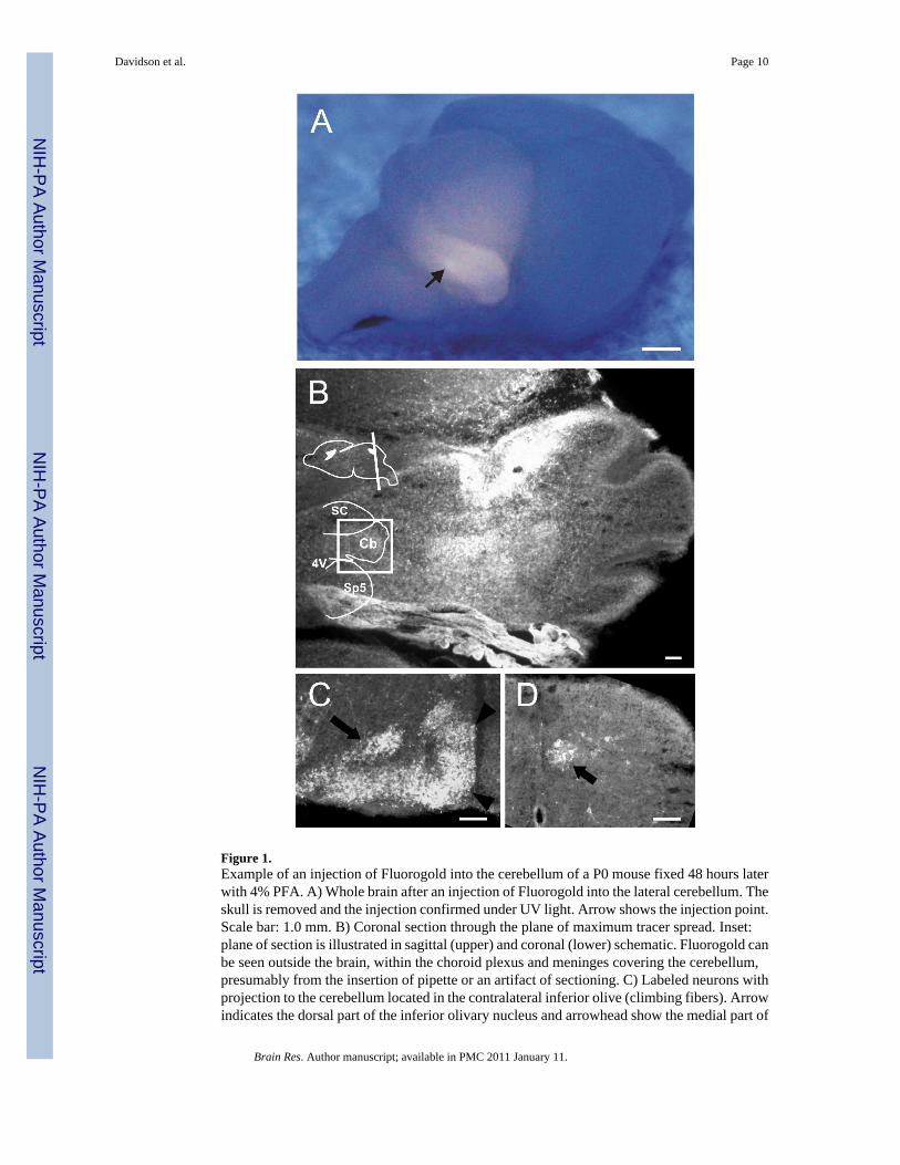
Figure 1.Example of an injection of Fluorogold into the cerebellum of a P0 mouse fixed 48 hours laterwith 4% PFA. A) Whole brain after an injection of Fluorogold into the lateral cerebellum. Theskull is removed and the injection confirmed under UV light. Arrow shows the injection point.Scale bar: 1.0 mm. B) Coronal section through the plane of maximum tracer spread. Inset:plane of section is illustrated in sagittal (upper) and coronal (lower) schematic. Fluorogold canbe seen outside the brain, within the choroid plexus and meninges covering the cerebellum,presumably from the insertion of pipette or an artifact of sectioning. C) Labeled neurons withprojection to the cerebellum located in the contralateral inferior olive (climbing fibers). Arrowindicates the dorsal part of the inferior olivary nucleus and arrowhead show the medial part of
Davidson et al. Page 10
Brain Res. Author manuscript; available in PMC 2011 January 11.
NIH
-PA Author Manuscript
NIH
-PA Author Manuscript
NIH
-PA Author Manuscript

the inferior olivary nucleus. D) Clarke’s column (arrow) of the ipsilateral thoracic spinal cord.Abbreviations: 4V, fourth ventricle; Cb, cerebellum; SC, superior colliculus; Sp5, spinaltrigeminal nucleus.
Davidson et al. Page 11
Brain Res. Author manuscript; available in PMC 2011 January 11.
NIH
-PA Author Manuscript
NIH
-PA Author Manuscript
NIH
-PA Author Manuscript
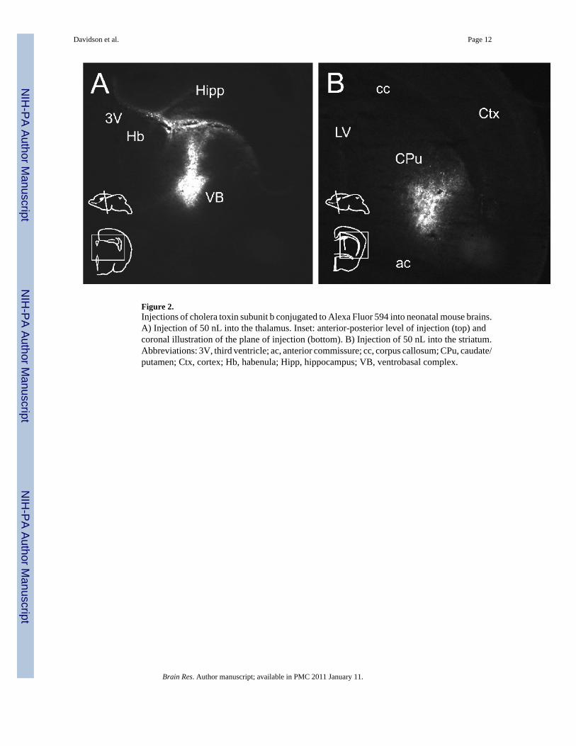
Figure 2.Injections of cholera toxin subunit b conjugated to Alexa Fluor 594 into neonatal mouse brains.A) Injection of 50 nL into the thalamus. Inset: anterior-posterior level of injection (top) andcoronal illustration of the plane of injection (bottom). B) Injection of 50 nL into the striatum.Abbreviations: 3V, third ventricle; ac, anterior commissure; cc, corpus callosum; CPu, caudate/putamen; Ctx, cortex; Hb, habenula; Hipp, hippocampus; VB, ventrobasal complex.
Davidson et al. Page 12
Brain Res. Author manuscript; available in PMC 2011 January 11.
NIH
-PA Author Manuscript
NIH
-PA Author Manuscript
NIH
-PA Author Manuscript
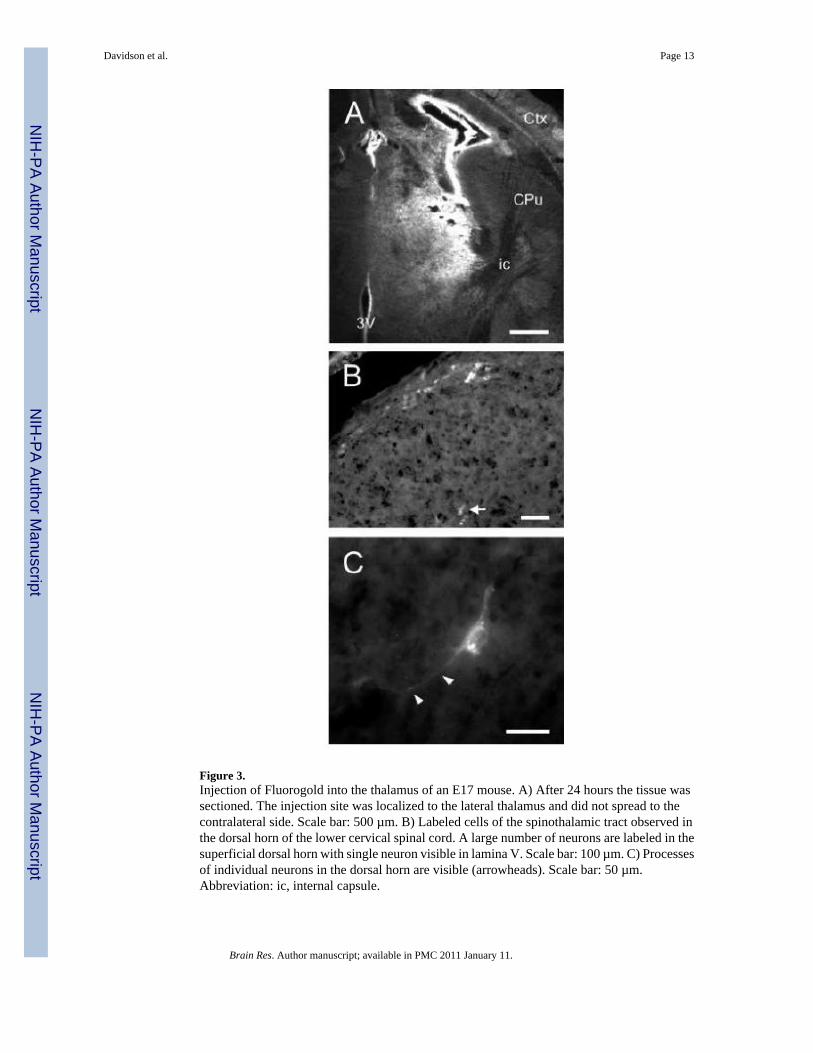
Figure 3.Injection of Fluorogold into the thalamus of an E17 mouse. A) After 24 hours the tissue wassectioned. The injection site was localized to the lateral thalamus and did not spread to thecontralateral side. Scale bar: 500 µm. B) Labeled cells of the spinothalamic tract observed inthe dorsal horn of the lower cervical spinal cord. A large number of neurons are labeled in thesuperficial dorsal horn with single neuron visible in lamina V. Scale bar: 100 µm. C) Processesof individual neurons in the dorsal horn are visible (arrowheads). Scale bar: 50 µm.Abbreviation: ic, internal capsule.
Davidson et al. Page 13
Brain Res. Author manuscript; available in PMC 2011 January 11.
NIH
-PA Author Manuscript
NIH
-PA Author Manuscript
NIH
-PA Author Manuscript
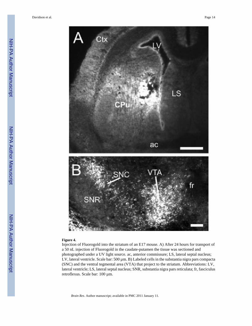
Figure 4.Injection of Fluorogold into the striatum of an E17 mouse. A) After 24 hours for transport ofa 50 nL injection of Fluorogold in the caudate-putamen the tissue was sectioned andphotographed under a UV light source. ac, anterior commissure; LS, lateral septal nucleus;LV, lateral ventricle. Scale bar: 500 µm. B) Labeled cells in the substantia nigra pars compacta(SNC) and the ventral tegmental area (VTA) that project to the striatum. Abbreviations: LV,lateral ventricle; LS, lateral septal nucleus; SNR, substantia nigra pars reticulata; fr, fasciculusretroflexus. Scale bar: 100 µm.
Davidson et al. Page 14
Brain Res. Author manuscript; available in PMC 2011 January 11.
NIH
-PA Author Manuscript
NIH
-PA Author Manuscript
NIH
-PA Author Manuscript
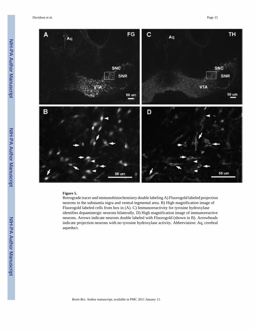
Figure 5.Retrograde tracer and immunohistochemistry double labeling A) Fluorogold labeled projectionneurons in the substantia nigra and ventral tegmental area. B) High magnification image ofFluorogold labeled cells from box in (A). C) Immunoreactivity for tyrosine hydroxylaseidentifies dopaminergic neurons bilaterally. D) High magnification image of immunoreactiveneurons. Arrows indicate neurons double labeled with Fluorogold (shown in B). Arrowheadsindicate projection neurons with no tyrosine hydroxylase activity. Abbreviation: Aq, cerebralaqueduct.
Davidson et al. Page 15
Brain Res. Author manuscript; available in PMC 2011 January 11.
NIH
-PA Author Manuscript
NIH
-PA Author Manuscript
NIH
-PA Author Manuscript
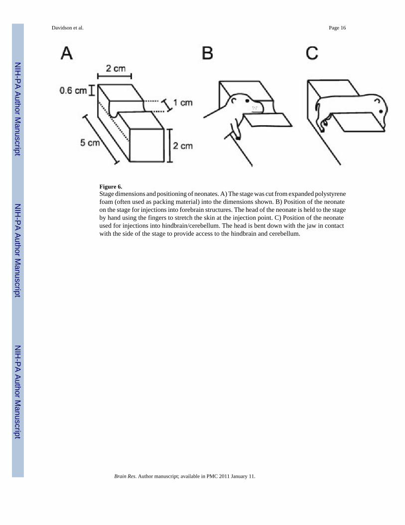
Figure 6.Stage dimensions and positioning of neonates. A) The stage was cut from expanded polystyrenefoam (often used as packing material) into the dimensions shown. B) Position of the neonateon the stage for injections into forebrain structures. The head of the neonate is held to the stageby hand using the fingers to stretch the skin at the injection point. C) Position of the neonateused for injections into hindbrain/cerebellum. The head is bent down with the jaw in contactwith the side of the stage to provide access to the hindbrain and cerebellum.
Davidson et al. Page 16
Brain Res. Author manuscript; available in PMC 2011 January 11.
NIH
-PA Author Manuscript
NIH
-PA Author Manuscript
NIH
-PA Author Manuscript
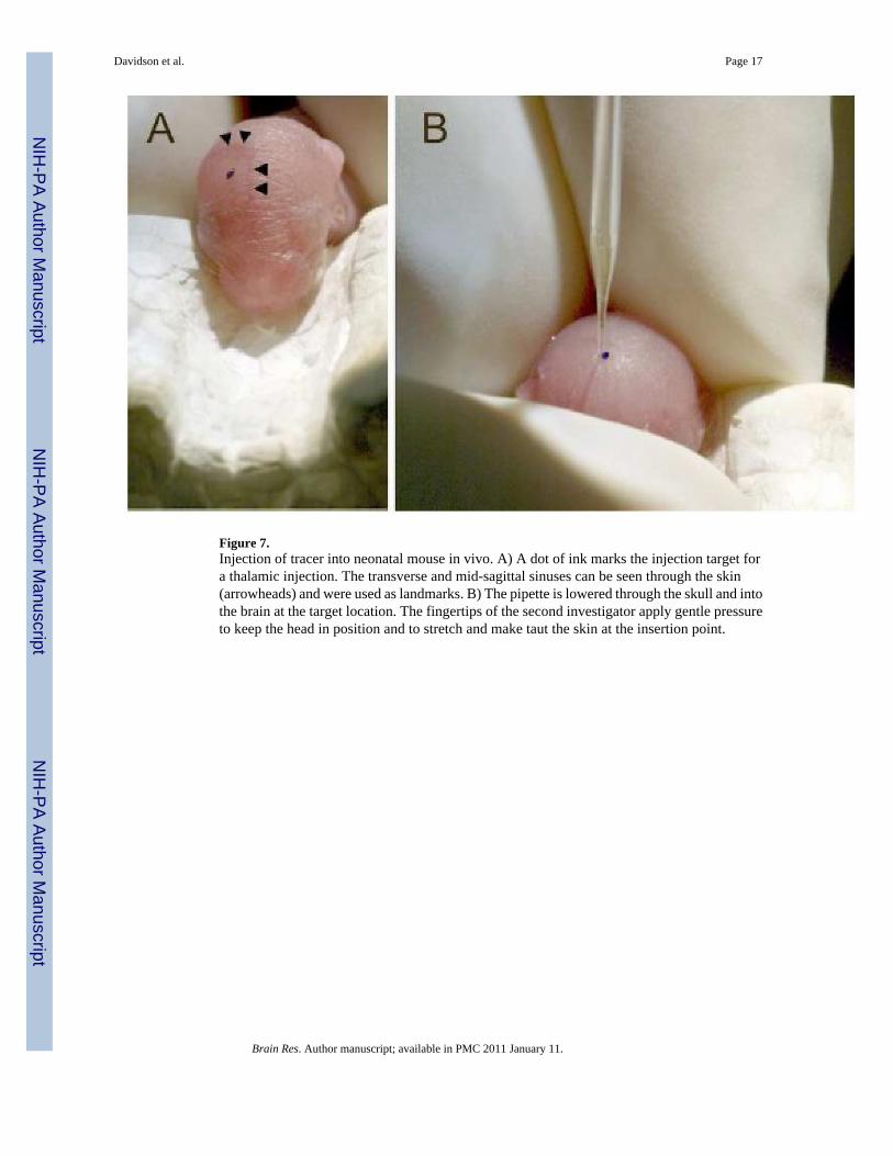
Figure 7.Injection of tracer into neonatal mouse in vivo. A) A dot of ink marks the injection target fora thalamic injection. The transverse and mid-sagittal sinuses can be seen through the skin(arrowheads) and were used as landmarks. B) The pipette is lowered through the skull and intothe brain at the target location. The fingertips of the second investigator apply gentle pressureto keep the head in position and to stretch and make taut the skin at the insertion point.
Davidson et al. Page 17
Brain Res. Author manuscript; available in PMC 2011 January 11.
NIH
-PA Author Manuscript
NIH
-PA Author Manuscript
NIH
-PA Author Manuscript
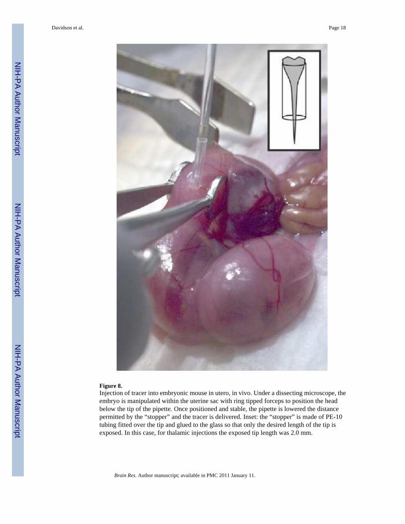
Figure 8.Injection of tracer into embryonic mouse in utero, in vivo. Under a dissecting microscope, theembryo is manipulated within the uterine sac with ring tipped forceps to position the headbelow the tip of the pipette. Once positioned and stable, the pipette is lowered the distancepermitted by the “stopper” and the tracer is delivered. Inset: the “stopper” is made of PE-10tubing fitted over the tip and glued to the glass so that only the desired length of the tip isexposed. In this case, for thalamic injections the exposed tip length was 2.0 mm.
Davidson et al. Page 18
Brain Res. Author manuscript; available in PMC 2011 January 11.
NIH
-PA Author Manuscript
NIH
-PA Author Manuscript
NIH
-PA Author Manuscript


![[P] Neonatal Hypoglycaemia - Healthpoint](https://img.pdfslide.net/doc/110x75/631a78a8c51d6b41aa04d74d/p-neonatal-hypoglycaemia-healthpoint.jpg)


